student manual - mrschamberlain.com biology/labs/bioradpcrpv92.pdf · to obtain dna for use in the...
TRANSCRIPT
Student Manual
Introduction to PCR�—The Polymerase Chain ReactionYou are about to perform a procedure known as PCR1�—the amplification of a specific
sequence of your own DNA in a test tube. This particular piece of DNA is present in thegenes of many but not all people. Analysis of the data generated in this laboratory willenable you to determine whether your genomic DNA carries this piece of DNA, or not.
The genome, composed of DNA, is our hereditary code. This is the so-called"hard-wiring", the blueprint that controls much of why we look like we do, why we actlike we do, and how we do the things we do. Molecular biology is the study of genes andthe molecular details that regulate the flow of genetic information from DNA, to RNAand proteins, from generation to generation. Biotechnology uses this knowledge to manip-ulate organisms' (microbes, plants or animals) DNA to help solve human problems.
Within the molecular framework of biology, DNA, RNA and proteins are closely tiedto each other. Because proteins and enzymes ultimately play such a critical role in thelife process, scientists have spent many lifetimes studying proteins in an attempt to under-stand how they work. With this understanding it was believed we could cure, prevent andovercome disease and physical handicaps as well as explain exactly how and why organ-isms exist, propagate and die. However, the complete answer to how and why does not liesolely in the knowledge of how enzymes function, we must learn how they are made. Ifeach enzyme is different, then what controls these differences and what is the blueprint forthis difference? That answer lies within our genome, or genetic code.
Thus, you may realize why researchers today, in an attempt to understand themechanisms behind the various biological processes, study nucleic acids as well as pro-teins in order to get a complete picture. In the last 20 years, many advances in the areasof nucleic acid techniques have allowed researchers the means to study the roles thatnucleic acids play in biology. It took the imagination and hard work of many scientists toreveal the answers to one of the most mysterious puzzles of life �– understanding themechanisms that control how DNA is translated into proteins within living cells.
Before Beginning this Lab, See If You Can Answer the FollowingQuestions
How is DNA faithfully passed on from generation to generation? What causes geneticdisease in some people but not others? How do scientists obtain DNA to study? Whatsecrets can DNA tell us about our origins? What human problems can an understanding ofDNA help us solve? Should we unlock the secrets held in this most basic building block oflife?
PCR Set the Stage for a Scientific RevolutionIn 1983, Kary Mullis2 at Cetus Corporation developed the molecular biology technique
that has since revolutionized genetic research. This technique, termed the polymerasechain reaction (PCR), transformed molecular biology into a multidisciplinary research fieldwithin 5 years of its invention. Before PCR, the molecular biology techniques used to studyDNA required such a high level of expertise that relatively few scientists could use them.
The objective of PCR is to produce a large amount of DNA in a test tube (in vitro),starting from only a trace amount. Technically speaking, this means the controlled enzy-matic amplification of a DNA sequence, or gene, of interest. The template strands can beany form of double-stranded DNA such as genomic DNA. A researcher can take trace
33

amounts of genomic DNA from a drop of blood, a single hair follicle or cheek cell (intheory, only a single template strand is needed to copy and generate millions of new identical DNA molecules) and make enough to study. Prior to PCR, this would have beenimpossible. It is the ability to amplify the precise sequence of DNA of interest that is thetrue power of PCR.
PCR has made an impact on four main areas of genetic research: gene mapping, cloning,DNA sequencing and gene detection. PCR is now used as a medical diagnostic tool to detectspecific mutations that may cause genetic disease3, is used in criminal investigations andcourts of law to identify suspects on the molecular level4, and has been a powerful tool in thesequencing of the human genome5. Prior to PCR the use of molecular biology techniques fortherapeutic, forensic, pharmaceutical, agricultural or medical diagnostic purposes was notpractical nor cost effective. The development of PCR technology transformed molecularbiology from a difficult science to one of the most accessible and widely used disciplines ofbiotechnology.
Now, let�’s extract some of your own DNA.
34

Lesson 1 Procedure for DNA Extraction and TemplatePreparation
To obtain DNA for use in the polymerase chain reaction you will extract the DNA fromyour own living cells. It is interesting to note that DNA can be also extracted from mummiesand fossilized dinosaur bones. In this lab activity, you will be isolating DNA from epithelialcells that line the inside of your cheek. This is accomplished by using a sterile pipet tip togently scrape the inside of both your cheeks about 10 times each to scoop up the cells liningthe surface. You will then boil the cells to rupture them and release the DNA they contain.To obtain pure DNA for PCR you will use the following procedure.
The cheek cells in the pipet tip are transferred into a micro test tube containing 200 µl ofInstaGene matrix. This particulate matrix is made up of negatively charged microscopicbeads that "chelate", or grab metal ions out of solution. It acts to trap metal ions, such asMg2+, which are required as catalysts or cofactors in enzymatic reactions. Your cheek cellswill then be lysed or ruptured by heating to release all of their cellular constituents, includingenzymes that were once contained in the cheek cell lysosomes. Lysosomes are sacs withinthe cells cytoplasm that contain powerful enzymes, such as DNases, which are used by cellsto digest the DNA of invading viruses. When you rupture the cells, these DNases can digestthe released DNA of interest. However, when the cells are lysed in the presence of thechelating beads, the cofactors are adsorbed and are not available to the enzymes. This virtu-ally blocks all enzyme degradation of the extracted DNA and results in a population of intactgenomic DNA molecules that will be used as the template in your PCR reaction.
Your isolated cheek cells are first suspended in the InstaGene matrix and incubated at56 °C for 10 minutes. This "preincubation" step helps to soften the plasma membranes andrelease clumps of cells from each other. The increased temperature also acts to inactivateenzymes such as DNases, which will degrade the DNA template. After this 10 minuteincubation period, the cells are then placed into a boiling (100°C) water bath for 6 minutes.The boiling ruptures the cells and releases the DNA from the cell nucleus. Your extractedgenomic DNA will then be used as the target template for PCR amplification.
35

Lesson 1 Lab�—DNA Extraction and Template Preparation
Workstation Checklist
Your workstation. Materials and supplies that should be present at your workstationprior to beginning this lab are listed below.
Student workstations Number (✔✔)Screwcap tubes with InstaGene�™ matrix 8 ❐
Styrofoam microtube rack 2 ❐
P-20 micropipet 1 ❐
P-200 micropipet 1 ❐
Pipet tips (filter type), 20�–200 µl 1 box ❐
Lab marker 1 ❐
Waste container 1 ❐
Copy of Quick Guide or protocol 1 ❐
Instructor's (common) workstation. A list of materials, supplies, and equipment thatshould be present at a common location to be accessed by your team is also listed below.
P-200 micropipet 1�–8 ❐
Pipet tips (filter type), 20�–200 µl 1 box ❐
Water baths (56 and 100°C) 2 ❐
Centrifuge 1 ❐
36

Lab Protocol for Lesson 11. Each member of your team should have two screwcap tubes, each containing 200 µl
of InstaGene matrix. Label the tube on the cap and on the side with your initials. In addition, label the tubes as "tube 1" and "tube 2". Each person should wash his or herhands before beginning step 2.
2. Using a sterile 20�–200 µl filter pipet tip, gently scrape the inside of both cheeks 10 timeseach with the tip. This is most easily done by pinching and extending the corner of yourmouth with one hand, and scraping the cheek with the tip in the other hand. Use firm,but gentle pressure. The goal is to remove the surface layer of epithelial cells from yourcheek lining. You should see a small volume of white cells in the pipet tip.
3. Place the tip that contains your cheek cells into the screwcap tube labeled as "tube 1".
4. Using a second sterile 20�–200 µl filter pipet tip, gently scrape the inside of both cheeks10 times each with the tip. Place the tip that contains your cheek cells into your screwcap tube labeled as "tube 2".
5. Place the tips on the end of a P-200 micropipet that is set on a 100 µl setting. Pipetup and down 5 times into the InstaGene matrix �– the action of pipetting up anddown mixes and transfers your cheek cells into the InstaGene matrix.
37

6. Screw the lid tightly on your tubes and place in the foam microtube holder. When allmembers of your team have collected their samples, float the rack and tubes in a 56°Cwater bath for 10 min. At the halfway point (5 minutes), remix the contents of the tubesby shaking or vortexing several times and place back in the water bath for the remaining5 minutes.
7. Remove the tubes from the water bath and remix by shaking the tubes several times.Now float the rack of tubes in a 100°C water bath for 6 minutes.
8. After 6 minutes, remove the tubes from the 100°C water bath and shake or vortex gentlyseveral times to resuspend the sample. Place the eight tubes in a balanced arrangementin the centrifuge. Pellet the matrix by spinning for 5 minutes at 6,000 x g (or 10 minutesat 2,000 x g) in the centrifuge.
9. Using a 200 µl pipet tip, remove 170 µl of the supernatant from one of your screwcaptubes and transfer the supernatant into the other. You will now be left with one screwcaptube that contains your isolated genomic DNA.
10. Store your screwcap tube in the refrigerator until the next laboratory period or proceedto Lesson 2 if you have enough time to set up and perform PCR reactions.
38
56°C, 10 minWater bath
100°C, 6 minWater bath
Centrifuge

Lesson 1 DNA Extraction and Template Preparation
Focus Questions
1. Why is it necessary to trap the metal ions in the cheek cell solution before theboiling/lysis step at 100 °C? What would happen if you did not put in the InstaGene�™
matrix?
2. What is needed from the cheek cells in order to conduct the polymerase chain reaction?
3. What structures must be broken in order to release the DNA from a cell?
4. Why do you think the extracted cheek cell DNA is stored cold in the InstaGene matrixafter boiling the samples?
39

Lesson 2 PCR AmplificationIt is estimated that there are 30,000�–50,000 individual genes in the human genome.
The true power of PCR is the ability to target and amplify a specific piece of DNA (orgene) out of a complete genome.
The recipe for a PCR amplification of DNA requires a simple mixture of ingredients.To replicate a given piece of DNA, the reaction mixture requires the following components.
1. Intact DNA template�—containing the sequence of DNA to be amplified.
2. Individual deoxynucleotide bases (A, T, G and C)�—raw material of DNA.
3. DNA polymerase�—an enzyme that assembles the nucleotides into a new DNA chain.
4. Magnesium ions�—a cofactor (catalyst) required by DNA polymerase to create theDNA chain.
5. Oligonucleotide primers�—pieces of DNA complementary to the template that tellDNA polymerase exactly where to start making copies.
6. Salt buffer�—provides the optimum ionic environment and pH for the PCR reaction.
The template DNA in this exercise is genomic DNA that was extracted from yourcheek cells. When all the other components are combined under the right conditions a copyof the original double stranded template DNA molecule is made�—doubling the number oftemplate strands. Each time this cycle is repeated, copies are made from copies and thenumber of template strands doubles�—from 2 to 4 to 8 to 16 and so on�—until after 20cycles there are 1,048,554 exact copies of the target sequence.
PCR makes use of the same basic processes that cells use to duplicate their DNA.
1. Complementary DNA strand hybridization
2. DNA strand synthesis via DNA polymerase
The two DNA primers provided in this kit are designed to flank, or bracket, a DNAsequence within your genome and thus provide the exact start signal for the DNA polymeraseto "zero-in on" and begin synthesizing (replicating) copies of that target DNA.Complementary strand hybridization takes place when the two different primers anneal, orhybridize, to each of their respective complementary base pair sequences on the templateDNA molecule.
The primers are two short single-stranded DNA molecules (23 bases long), one that iscomplementary to a portion of the 5'�–3' strand, and another that is complementary to theportion of the 3'�–5' strand of your DNA. These primers hybridize to the separated templatestrands and serve as starting points for DNA amplification.
The enzyme, called Taq DNA polymerase, extends the annealed primers' sequence by"reading" the strand and synthesizing the complementary sequence. In this way, Taq replicatesthe template DNA strand(s). This polymerase has been isolated from a heat stable bacterium(Thermus aquaticus) which in nature lives within the steam vents in the Yellowstone NationalPark.6 For this reason the enzymes within these bacteria have evolved to withstand hightemperatures (94°C) also used in each cycle of the PCR reaction.
40

PCR�—Step by StepPCR amplification includes three main steps, a denaturation step, an annealing step
and an extension step (these steps are summarized in Figure 9). During denaturation, thereaction mixture is heated to 94°C for 1 minute, which results in the melting out or separationof the double-stranded DNA template into two single stranded molecules. In PCR amplification, DNA templates must be separated before the polymerase can generate a newcopy. The harsh temperature required to melt the DNA strands normally would destroythe activity of most enzymes, but because Taq polymerase has been isolated from bacteriathat thrive in the high temperatures of hot springs, it remains active.
Fig. 9. A complete cycle of PCR.
During the annealing step, the oligonucleotide primers "anneal to" or find their complementarysequences on the two single-stranded template strands of DNA. In these annealed positions, theycan act as "primers" for Taq DNA polymerase. They are called primers because they "prime" thesingle stranded DNA molecules by providing a short sequence of double stranded DNA for Taqpolymerase to extend and build a new complementary strand. Binding of the primers to their template sequences is also highly dependent on temperature. In this lab exercise, a 60°C annealingtemperature is optimum for primer binding.
During the extension step, the job of Taq DNA polymerase is to add nucleotides (A, T, Gor C) one at a time to the primer to create a complementary copy of the DNA template. During
41
Denature Strands 95°C
Anneal Primers 60°C(Taq Polymerase recognizesdouble stranded substrate)
Extend 72°C(Synthesize New Strand)
Repeat Cycle 40 Times
5'
5'
5'
5'
5'
5'
5'
5'
5'
5'
3'
3'
3'
3'
3'
3'
3'
3'
3'
3'
5'
5'3'
3'

polymerization, the reaction temperature is increased to 72°C, the temperature that producesoptimal Taq polymerase activity. The three steps of denaturation, annealing, and extension,combine to form one "cycle" of PCR, and a complete PCR amplification undergoes 40 cycles.
The entire 40 cycle reaction is carried out in a test tube which is placed into the thermalcycler (also called a Gene Cycler). The Gene Cycler contains an aluminum block that holdsthe samples and can be rapidly heated and cooled across extreme temperature differences.The rapid heating and cooling of this thermal block is defined as "temperature cycling" or"thermal cycling".
Temperature Cycle=Denaturation Step (94°C)+Annealing Step (60°C)+Extension Step (72°C)
42

Lesson 2 Lab�–PCR Amplification
Workstation Checklist
Your workstation. Materials and supplies that should be present at your workstationprior to beginning this lab are listed below.
Student workstations Number (✔✔)PCR tubes 4 ❐
Microtubes, capless 4 ❐
Master mix 1 ❐
P-20 micropipet 1 ❐
Pipet tips (filter type), 2�–20 µl 8 tips ❐
Foam microtube rack 2 ❐
Lab marker 1 ❐
Ice bucket with ice 1 ❐
Waste container 1 ❐
Copy of Quick Guide or protocol 1 ❐
Instructor's (common) workstation
Gel trays 1/station ❐
Molten agarose (see advance prep) 40 ml/gel ❐
Lab tape for gel trays 1/station ❐
Centrifuge 1 ❐
Gene Cycler or MyCycler 1 ❐
43
Lab Protocol for Lesson 2
1. Obtain your screwcap tube that contains your genomic DNA template from therefrigerator and place on your lab bench. Centrifuge your tubes for 2 minutes at 6,000 x g or for 5 minutes at 2,000 x g in a centrifuge.
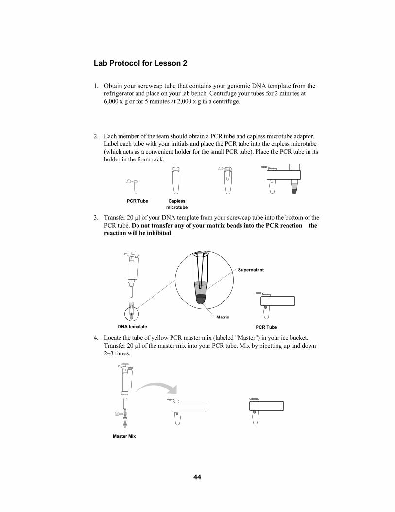
2. Each member of the team should obtain a PCR tube and capless microtube adaptor.Label each tube with your initials and place the PCR tube into the capless microtube(which acts as a convenient holder for the small PCR tube). Place the PCR tube in itsholder in the foam rack.
3. Transfer 20 µl of your DNA template from your screwcap tube into the bottom of thePCR tube. Do not transfer any of your matrix beads into the PCR reaction�—thereaction will be inhibited.
4. Locate the tube of yellow PCR master mix (labeled "Master") in your ice bucket.Transfer 20 µl of the master mix into your PCR tube. Mix by pipetting up and down2�–3 times.
44
PCR Tube Capless microtube
DNA template
Supernatant
Matrix
PCR Tube
Master Mix

5. Remove your PCR tube from its holder and place the tube in the Gene Cycler.
6. When all of the PCR samples are in the Gene Cycler, the teacher will begin the PCRreaction. The reaction will undergo 40 cycles of amplification, which will takeapproximately 3 hours.
7. If your teacher instructs you to do so, you will now pour your agarose gels (the gelsmay have been prepared ahead of time by the teacher).
45

Lesson 2 PCR Amplification
Focus Questions
1. Why is it necessary to have a primer on each side of the DNA segment to be amplified?
2. How did Taq polymerase acquire its name?
3. Why are there nucleotides (A, T, G, and C) in the master mix? What are the othercomponents of the master mix and what are their functions?
4. Describe the three main steps of each cycle of PCR amplification and what reactionsoccur at each temperature.
5. Explain why the precise length target DNA sequence doesn�’t get amplified until thethird cycle. You may need to use additional paper and a drawing to explain youranswer.
46
Lesson 3 Analyzing Your DNA Using Gel Electrophoresis
What Are You Looking At?
Before you analyze your PCR products, it is important to review the details of the tar-get sequence being explored.
What Can Genes and DNA Tell Us?
It is estimated that the 23 pairs, or 46 chromosomes, of the human genome(23 chromosomes come from the mother and the other 23 come from the father)contain approximately 30,000�–50,000 genes. Each chromosome contains a series ofspecific genes. The larger chromosomes contain more DNA, and therefore moregenes, compared to the smaller chromosomes. Each one of the homologous chromosomes (pairs) contains similar genes.
Each gene holds the code for a particular protein. Interestingly, the 30,000�–50,000genes only comprise 5% of the total chromosomal DNA. The other 95% is non-codingDNA. This non-coding DNA is interspersed in blocks between functional segments ofgenes and within genes, splitting them into segments. The exact function of the non-coding(intergenic) DNA is not yet known, although it is thought that non-coding DNA allowsfor the accumulation of mutations and variations within organisms.
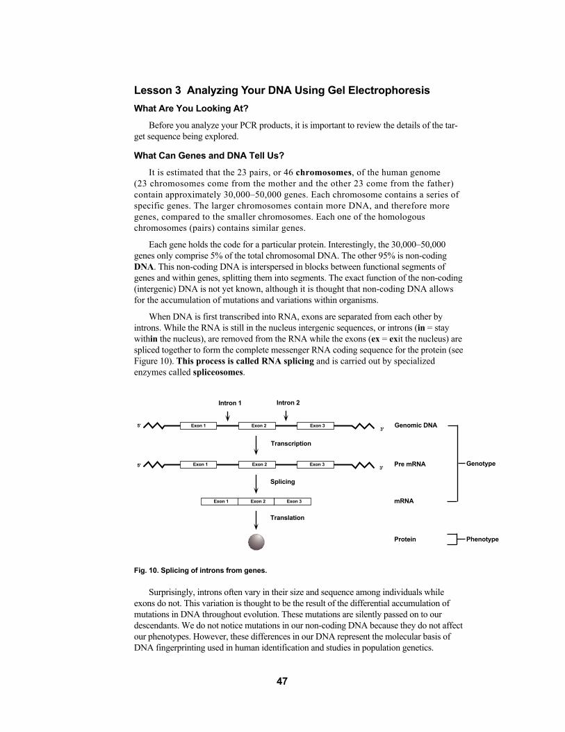
When DNA is first transcribed into RNA, exons are separated from each other byintrons. While the RNA is still in the nucleus intergenic sequences, or introns (in = staywithin the nucleus), are removed from the RNA while the exons (ex = exit the nucleus) arespliced together to form the complete messenger RNA coding sequence for the protein (seeFigure 10). This process is called RNA splicing and is carried out by specializedenzymes called spliceosomes.
Fig. 10. Splicing of introns from genes.
Surprisingly, introns often vary in their size and sequence among individuals whileexons do not. This variation is thought to be the result of the differential accumulation ofmutations in DNA throughout evolution. These mutations are silently passed on to ourdescendants. We do not notice mutations in our non-coding DNA because they do not affectour phenotypes. However, these differences in our DNA represent the molecular basis ofDNA fingerprinting used in human identification and studies in population genetics.
47
Intron 1
Exon 15'
5'3'
3'Exon 2 Exon 3 Genomic DNA
Genotype
Phenotype
Pre mRNA
mRNA
Protein
Exon 3Exon 2Exon 1
Exon 1 Exon 2 Exon 3
Intron 2
Transcription
Splicing
Translation

The Target Sequence�–Can You Say "Alu"?
The genome contains small repetitive DNA elements that have become randomly insertedinto the human genome over millions of years. One such repetitive element is called the"Alu sequence"7 (see Figure 11). This is a DNA sequence about 300 base pairs longthat is repeated, one copy at a time, almost 500,000 times throughout the humangenome8. The origin and function of these randomly repeated sequences is not yet known.
Fig. 11. Location of an Alu repetitive element within an intron.
Some of these Alu sequences have characteristics that make them very useful togeneticists. When present within introns of certain genes, they can either be associated witha disease or merely used to estimate relatedness among individuals. In this exercise, analy-sis of a single Alu repeat is used to estimate its frequency in the population and as asimple measure of molecular genetic variation�—with no reference to disease or related-ness among individuals.
In this lab you will be hunting for an Alu element in the PV92 region of chromosome16. This particular Alu element is dimorphic, meaning that the element is present insome individuals and not others. Some people have the insert in one copy of their 16th chromosomes (one allele), others may have the insert in both copies of chromosome 16 (twoalleles), while some may not have the insert on either copy of the chromosome (see Figure12). The presence or absence of this insert can be detected using the polymerase chainreaction followed by agarose gel electrophoresis.
Since you are amplifying a region of DNA contained within an intron, the region ofDNA is never really used in your body. So if you don�’t have it, don�’t worry.
The primers in this kit are designed to bracket the region within the PV92 region that is641 base pairs in length if the intron does not contain the Alu insertion or 941 base pairs inlength if Alu is present. This increase in size is due to the 300 base pair sequence contributedby the Alu insert. When your PCR products are electrophoresed on an agarose gel, thereare three distinct outcomes that can be visualized in the gel.
If both chromosomes contain Alu inserts, then each amplified PCR product will be 941base pairs long. On a gel they will migrate at the same speed so there will be one bandthat corresponds to 941 base pairs. If neither chromosome contains the insert then eachamplified PCR product will be 641 base pairs and they will migrate as one band that corresponds to 641 base pairs. If you have an Alu insert on one chromosome but not theother, then there will be one PCR product of 641 base pairs and one of 941 base pairs. Theresulting gel will reveal two bands.
48
5' 3'
Intron
ALU

Fig. 12. The presence or absence of the Alu insert within the PV92 region of chromosome 16.
Electrophoresis separates DNA fragments according to their relative size (molecularweight). DNA fragments are loaded into an agarose gel slab, which is placed into a chamberfilled with a conductive liquid buffer solution. A direct current is passed between wireelectrodes at each end of the chamber. DNA fragments are negatively charged, and whenplaced in an electric field will be drawn toward the positive pole and repelled by the negativepole. The matrix of the agarose gel acts as a molecular sieve through which smaller DNAfragments can move more easily than larger ones. Over a period of time smaller fragmentswill travel farther than larger ones. Fragments of the same size stay together and migrate inwhat appear as single "bands" of DNA in the gel. In the sample gel below (Fig. 13), PCRamplified bands of 941 bp and 641 bp are separated based upon their size
Fig. 13. Separation of DNA bands based on size. This gel depicts the electrophoretic separation ofthe EZ Load DNA molecular mass ruler, which contains 1,000 bp, 700 bp, 500 bp, 200 bp and 100 bp fragments (lane 1), two homozygous (+/+) 941 bp fragments (lanes 2 and 6), three homozygous (-/-)641 bp fragments (lanes 3, 5, and 8), and two heterozygous (+/-) 941/641 bp fragments (lanes 4 and 7).
49
ALU
PV92 Genotype DNA size of PCR Products
Homozygous (+/+) 941 base pairs
641 base pairs
941 + 641 base pairs
Homozygous (-/-)
Heterozygous (+/-)
ALU
ALU
1 2 3 4 5 6 7 8
(bp)
1000700500
200100

50
Lesson 3 Lab�–Analyzing Your DNA Using Gel Electrophoresis
Workstation Checklist
Your workstation. Materials and supplies that should be present at your workstationprior to beginning this lab are listed below.
Student workstations Number/Station (✔✔)Agarose gel 1 ❐
Student PCR samples 1/student ❐
MMR-DNA standard 1 ❐
LD loading dye 1 ❐
P-20 micropipet 1 ❐
Pipet tips (filter type), 2�–20 µl 11 tips ❐
Lab marker 1 ❐
Styrofoam microtube rack 1 ❐
Gel box and power supply 1 ❐
Gel staining tray 1 ❐
Waste container 1 ❐
Copy of Quick Guide or protocol 1 ❐
Instructor's workstation
1x TAE electrophoresis buffer 275 ml/gel box ❐
Bio-Safe DNA stain�—1x solution 500 ml ❐
MMR-DNA standard 1 ❐
Positive control samples (two each) 6 ❐
PV92 homozygous (+/+)
PV92 homozygous (-/-)
PV92 heterozygous (+/-)Centrifuge (optional equipment) 1 ❐
Shaking platform (optional equipment)* 1 ❐
* Strongly recommended.

Lab Protocol for Lesson 31. Remove your PCR samples from the Gene Cycler and place in the microtube rack. If a
centrifuge is available, place the PCR tubes in the adaptor and pulse spin the tubes(~3 seconds at 2,000 x g) to bring the condensation that formed on the lids to the bottom of the tubes. Add 10 µl of 5x loading dye to each of your PCR tubes.
2. Obtain a pre-poured agarose gel (either the one you poured or pre-poured by your teacher).Place the casting tray with the solidified gel in it, into the platform in the gel box. Thewells should be at the (-) cathode end of the box, where the black lead is connected.Very carefully remove the comb from the gel by pulling it straight up.
3. Pour ~275 ml of electrophoresis buffer into the electrophoresis chamber. Pour bufferinto the gel box until it just covers the wells.
4. Using a separate tip for each sample, load the samples into the first seven wells of thegel in the following order:
Lane Sample Volume Loaded
1 MMR-DNA Standard 10 µl
2 Homozygous (+/+) control 20 µl
3 Homozygous (-/-) control 20 µl
4 Heterozygous (+/-) control 20 µl
5 Student 1 20 µl
6 Student 2 20 µl
7 Student 3 20 µl
8 Student 4 20 µl
51
+-

5. Secure the lid on the gel box. The lid will attach to the base in only one orientation: redto red and black to black. Connect electrical leads to the power supply.
6. Turn on the power supply. Set it to 100 V and electrophorese the samples for 30 min-utes.
7. When the electrophoresis is complete, turn off the power and remove the lid from thegel box. Carefully remove the gel tray and the gel from the gel box. Be careful, the gelis very slippery. Nudge the gel off the gel tray with your thumb and carefully slide itinto your plastic staining tray.
8. Pour 60 ml of Bio-Safe DNA stain into your plastic staining tray, cover with plasticwrap, and let the gel sit in the stain overnight. If a shaking table is available, gentlyshake the gels in the stain overnight.
52
+
-

53
Lesson 3 Analyzing Your DNA Using Gel Electrophoresis
Focus Questions
1. Explain the difference between an intron and an exon.
2. Why do the two possible PCR products differ in size by 300 base pairs?
3. Explain how agarose electrophoresis separates DNA fragments of interest. Why does asmaller DNA fragment move faster than a larger one?
4. What kind of controls are run in this experiment? Why are they important? Could therebe others used?

Lesson 4 Lab�–Final Analysis and Interpretation of ResultsThe moment of truth has arrived. What is your genotype? Are you a homozygote or a
heterozygote? To find out you will have to destain your agarose gel. After 5�–10 minutes,the excess blue stain will begin to diffuse out of the agarose leaving the remaining dyebound to the DNA in the gel. Destaining increases the contrast and will allow you to visual-ize the DNA fragments you generated using PCR. To increase the contrast between theblue background of the gel and DNA bands, gels can be destained for an additional 1�–6hours.
To make detailed observations of your gel, lay the gel on a white background or youmay wish to view your gel with a light box (a light bulb under a piece of white acrylic).The light box will provide a brighter background to view your stained gel. You may alsotrace the DNA bands in your gel by placing a clear sheet of acetate over the gel. Trace theoutline of the gel, the sample wells and the DNA bands with a permanent marker.
Lesson 4 Analysis and Interpretation of Results
Workstation Checklist
Your workstation. Materials and supplies that should be present at your workstationprior to beginning this lab are listed below.
Student workstations Number/Station (✔✔)Water for destaining gels 60 ml ❐
Gel support membrane (optional) 1 ❐
Copy of Quick Guide or protocol 1 ❐
Acetate for tracing gels 1 ❐
Instructors workstation
None required
54

Lab Protocol for Lesson 4
Protocol
1. Pour off the Bio-Safe DNA stain into a bottle or another appropriate container anddestain the gel with 60 ml of water for ~5 minutes.
2. Pour the water out of the staining tray. Ask the instructor how to properly dispose ofthe stain.
3. Trim away any empty lanes of the gel with a knife or razor blade. Let the gel dry on gelsupport film or in your staining tray on your lab bench for 3�–5 days. When the gel isdry, tape it into your lab notebook for a permanent record.
4. With the help of your instructor, determine whether you are homozygous or heterozygous for the Alu insertion. First look at the control gel and note the migrationpattern of the homozygous (+/+), the homozygous (-/-), and the heterozygous (+/-)samples (also check the example on page 50). You may notice that in the heterozygoussample the smaller 641 base pair fragment is more intense than the larger 941 bpfragment. This difference is due to the fact that the smaller piece is amplified moreefficiently than the larger fragment. Copies of the shorter piece can be made at a fasterrate than the bigger piece so more fragments of the shorter piece are created per cycle.
55

Analysis
Compare your sample lanes with the control gel lanes, using the DNA size marker onboth gels as a reference. Mark the location and size of your fragment, or fragments. Bycomparing your DNA migration pattern to the controls, determine whether you arehomozygous (+/+), homozygous (-/-), or heterozygous (+/-). If your sample lane is blank,discuss with your classmates and teacher the possible reasons for lack of amplification.
Remember that the interpretation of the gel itself allows you to determine your geneticmakeup only at the site, or gene locus (location), being studied. There are three possiblegenotypes for the Alu insert at the location you have amplified. Use the informationgathered from your gel to determine the number of sub-populations in your class: homozygous (+/+), homozygous (-/-), or heterozygous (+/-). Tally the class results in thetable on page 58.
A major factor affecting the reliability of DNA fingerprinting evidence in forensics ispopulation genetics and genetic statistics. In humans there are hundreds of loci or DNAsegments, like Alu, that can be selected and used for fingerprinting analysis. Depending ondemographic factors such as ethnicity or geographic isolation, some populations showmuch less variation in particular DNA segments than others. The degree of variation willaffect the statistical odds of more than one individual having the same sequence. If 33% (1out of three individuals) of a given population has the same frequency in its DNA fingerprinting pattern for a certain DNA segment, then little information will be obtainedfrom using that segment alone to identify an individual.
When performing a DNA fingerprint for identifying a suspect in a criminal case orpaternity suit what you want is not a 1 out of 3 (1/3) chance of a match in a population.What you want is 1 in 10 million (1/107) chance of a match. The frequency of a particularDNA pattern turning up in a population becomes extremely low when a series of DNAsegments is selected and amplified, rather than just one segment alone. Amplifying multiple DNA segments from a single sample of genomic DNA can serve as apowerful tool to discriminate between individuals in a population. For DNA fingerprintingto be admissible as evidence in a court of law it is necessary for 30 to 40 different DNAsegments on multiple chromosomes to be selectively amplified and analyzed. Therefore inanalyzing how incriminating the DNA evidence is, one needs to ask the question:
Statistically, how many people in a population may have the same DNA pattern astaken from a crime scene: 1 in 1,000,000? 1 in 10,000? Or 1 in 10?
In actuality, the Alu insert such as the one you have "fingerprinted" in this lab has beenused to study the geographic migration patterns of different human populations over thecourse of human evolution8. The data from these studies have been published and yoursamples can be compared to the data collected from much larger populations.
56

Lesson 4 Analysis and Interpretation of Results
Focus Questions
Remember that the value of this Alu insert being in a non-coding region of the TPAgene is that it is not related to a particular disease, nor does it code for any proteinsequence. It�’s just non-coding DNA that can be used as a tool to study human genotypicfrequencies.
Because Alu repeats have become integrated into the general population at random, theAlu insert in the TPA gene is very useful in the study of the gene frequencies in localizedhuman populations. Theoretically, in some geographically isolated populations allindividuals may be homozygous (+/+), in others the individuals may all be homozygous (-/-) while in a ("melting-pot") population the three genotypes (polymorphisms) may existin equilibrium. The results you obtain in this lab provide a real life opportunity to use theHardy-Weinberg equation to examine and study genotypic and allelic frequencies of theAlu insert in your class.
The Hardy-Weinberg equation, p2 + 2pq + q2 = 1, describes the frequencies of allelesin the gene pool of an entire population. In this case the entire population is your class:
p2 = the frequency of an individual homozygous (+/+) for the Alu insert
q2 = the frequency of an individual homozygous (-/-) for the lack of an Alu insert
2pq = the frequency of an individual heterozygous (+/-) for the Alu insert
By determining frequencies of the Alu genotype within your class population, theallelic frequencies can also be calculated. Additionally, the genotypic frequencies of yourclass population can be compared to published results of larger population sizes9.
1. What is your genotype for the Alu insert in your PV92 region?
2. What are the observed (actual) genotypic frequencies of (+/+), (+/-), or (-/-) in yourclass population? Fill in the table below with your class data.
Table 1. Observed Class Genotypic Frequencies
Category Number of genotypes Frequency (# of genotypes/Total)
Homozygous (+/+) p2 =Heterozygous (+/-) pq =Homozygous (-/-) q2 =
Total = = 1
57

3. What is the frequency of each allele in your overall class sample? Fill in the tablebelow with your class data. Remember, a class of 32 students (N), will have a total of2(N) = 64 alleles.
Table 2. Calculated Allelic Frequencies for the Class
Category Number Class Allelic Frequency
total (+) alleles = p p/Total =total (-) alleles = q q/Total =
Total alleles = = 1.00
Hint: The expected allelic frequencies can be calculated from the Hardy-Weinberg equationusing your data from Table 1.
The number of p alleles is determined by adding the two(+) alleles of (p2) plus the one(+)allele from (2pq). Mathematically, this can be written as: p = 2(p2) + (2pq)
The number of q alleles is determined by adding the 2(-) alleles of (q2) plus the one(-) allelefrom (2pq). Mathematically, this can be written as: q = 2(q2) + (2pq)
The allelic frequency can be calculated by dividing the number of each allele by the totalnumber of alleles.
p = number of p alleles total number of alleles
q = number of q allelestotal number of alleles
4. The following table presents data from a USA-wide random population study.
Table 3. Sampling of USA Genotypic Frequencies for Alu
Category Number of each genotype Frequency
Homozygous (+/+) = p2 p2 = 2,422 0.2422Heterozygous (+/-) = 2pq pq = 5,528 0.5528Homozygous (-/-) = q2 q2 = 2,050 0.2050
Number of Samples Studied Total = 10,000 = 1.00
Now, using the data above, calculate the allelic frequencies for the USA data as youdid for your class population in Table 2.
Table 4. Calculated Allelic Frequencies for USA
Category Number Frequency
total (+) alleles = p p/Total =total (-) alleles = q q/Total =
Total alleles = = 1.00
5. How does your actual class data for allelic frequencies compare with that of the randomsampling of the USA population? Would you expect them to match? What reasons canyou think of to explain the differences or similarities?
58

Lesson 5 Analysis of Results using BioinformaticsBioinformatics is a discipline that integrates mathematical, statistical, and computer
tools to collect and process biological data. Bioinformatics has become an important tool inrecent years for analyzing the extraordinarily large amount of biological information that isbeing generated by researchers around the world. In Lesson 5, students will perform abioinformatics exercise to investigate the genotypic frequencies for the Alu polymorphismin their class population and compare them with the genotypic frequencies of other populations.
Following PCR amplification and electrophoresis of their samples, students will analyzetheir experimental data to determine their genotypes for the Alu insertion within the PV92locus on chromosome 16. The classroom genotype data can then be entered into Allele Serverof Cold Spring Harbor Laboratory's Dolan DNA Learning Center. Allele Server is a Web-based database that contains genotype data from a range of populations around theworld as well as other classrooms and teacher training workshops. It also provides a collection of statistical analysis tools to examine the Alu insertion polymorphism at the population level. Students can either analyze their classroom data as an individual populationor compare their population with other populations in the database.
Once students enter classroom data into Allele Server, they can perform a Chi-squareanalysis to compare the Alu genotype frequencies within the class population with those predicted by the Hardy-Weinberg equation. The genotypic frequencies of the class populationcan also be compared with the genotypic frequencies of another population in the database,using the Chi-square analysis.
Getting Started on Allele Server
Note: The Dolan DNA Learning Center web site is continually updated. Some of thefollowing information may change.
1. On your Web browser, go to vector.cshl.org
2. Click on Resources
3. Click on BioServers
4. Under Allele Server, click on Register. Registration is free and allows you to set up apersonal account. There is no need to register everytime you return to this site.
Using Allele Server
1. Log in to Allele Server using the username and password you registered
2. Once you have logged in, follow instructions provided in the pop-up window for usingAllele Server. You may also open a new window and go todnalc.org/help/sad/topic_3.html to get more detailed instructions. Follow the detailedinstructions on how to populate the workspace, analyze groups, compare group, andquery the database.
Remember that as a registered user, you may store any groups that you loaded in yourpersonal Allele Server database and analyze them at your convenience.
59

Appendix AReview of Molecular Biology
This section provides an overview of terms and concepts with which students shouldbe familiar in order to get the most out of this lab.
Genome�—The sequence of DNA molecules within the nucleus which code for all proteinsfor a given species. Each segment of DNA which encodes a given protein is called a gene.The information contained in the genome comprises the organisms hereditary code.
Molecular Biology�—The study of genes and the molecular details which regulate the flowof genetic information from DNA, to RNA and proteins, and from generation to generation.
Biotechnology�—The manipulation of organisms (microbes, plants or animals) DNA tohelp solve human problems.
Any living organism functions based on the complicated interactions between nucleicacids, proteins, lipids (fat) and carbohydrates. In nearly all cases, certain proteins termedenzymes control the almost infinite number of interactions and life processes in livingcreatures. Think of enzymes and proteins as all the different people on earth. Each personperforms a different role, function or job on this planet and although people are not theactual physical make-up of buildings, documents, food and roads it is the people that makethese buildings and roads and write the documents and plant and nurture the crops. In thesame way, enzymes and proteins do not comprise bones, lipids, sex hormones and sugars,but enzymes control these structures, their interactions and processes.
Because proteins and enzymes ultimately play such a critical role in the life process,scientists have spent many lifetimes studying proteins in an attempt to understand how theywork and how they can be controlled. With a complete understanding we could cure, preventand overcome many diseases and physical handicaps as well as explain exactly how andwhy organisms exist, propagate and die. However, the complete answers do not lie solelyin the knowledge of how enzymes function, we must learn how they are made. Before wecan control enzymes, we must understand where they come from and what is the basis ofthe molecular information that encodes proteins. That answer lies within our genetic code.
Each living organism has its own blueprint for life. This blueprint defines how anorganism will look and function (using enzymes as a means to form the look and controlthe functions). The blueprint codes for all the different enzymes. With amazing precisionthis blueprint gets passed on from generation to generation of each species for as long asthat species continues to propagate.
The transfer of this blueprint from generation to generation is called heredity. Theblueprint for any organism is called its genome. The hereditary code is encrypted withinthe sequence of the DNA molecules that make up the genome. The molecule that comprisesthe genome and thus the hereditary code is DNA (deoxyribonucleic acid).
The genome consists of very long DNA/protein complexes called chromosomes.Prokaryotes, organisms lacking a true nucleus, have only one chromosome. All otherspecies, eukaryotes, have a defined cell nucleus which contains multiple chromosomes.The nucleus is a defined, membrane-enclosed region of the cell which contains the chromosomes. The number of chromosomes varies with the organism �– from 2 or 3 insome yeasts and up to 100 or so in some fish. Humans have 46.
In most cases chromosomes come in nearly identical pairs (one member of the chromosomepair from each parent). In general, the members of a pair differ in small details from each other,since they come from different parents, but are otherwise identical or homologous. Cells with
60

homologous pairs of chromosomes are called diploid. Nearly all cells of an organism arediploid. Cells that have only one chromosome of each pair are called haploid. All sperm and ovaare haploid.
The process of forming sperm and ova is called meiosis. Meiosis starts with a diploidcell that divides into two haploid cells. When the sperm fertilizes the ovum the two nucleifuse and thus the new nucleus contains pairs of each chromosome, one partner from eachparent. The result is called a diploid zygote.
All cells of diploid organisms duplicate chromosomal pairs when they divide (exceptwhen sperm and ova are formed) so that all body cells (called somatic cells) of an organismare diploid. The process of cell division in which the chromosomes are duplicated and eachdaughter cell gets pairs of chromosomes is called mitosis. It is through the processes ofmitosis and meiosis that the hereditary code is passed from cell to cell and generation togeneration. Now that we know where the code is and how that code is passed on, we needto know how the code produces the enzymes that control life. The actual DNA code fora protein is contained within a segment of a chromosome called a gene. In nearly all cases,diploid organisms will have the same gene on a specific chromosome pair. The gene oneach chromosome of a specific chromosome pair is also called an allele.
To clarify, a gene encodes a particular protein that performs a particular function. Anallele refers to the same gene existing on each chromosome of a specific chromosome pair.Thus, there are genes for hair color and there is an allele for the hair color gene on eachchromosome pair. The gene or allele DNA code can also be called the genotype.
When the protein is made from this code and performs its function, the physical trait orresult that is seen is called the phenotype. In many cases the two alleles on the specificchromosome pair coding for a protein differ slightly in their respective DNA code (genotype).Any slight difference in code between the two alleles can result in two different proteinsthat although intended to perform basically the same function, actually carry out that function slightly differently, causing different results and thus different phenotypes.
Therefore, it is not only the various combinations of chromosomes a parent contributesto each offspring but also the various combinations of alleles and how each of the enzymescoded from the alleles work together that decide how we look and allow us to function. Thevarious combinations are nearly infinite and that is why we are all different. The study ofgenotypes and phenotypes is often referred to as Mendelian genetics (after Mendel, theindividual who pioneered the study of heredity and genetics).
DNA�—What is it?A DNA molecule is a long polymer consisting of four different components called
bases. The four bases are also called nucleotides. It is the various combinations of thesefour bases or nucleotides that create a unique DNA code or sequence (also genotype, gene,and allele). Nucleotides are comprised of three different components:
1. nitrogen base
2. ribose sugar
3. phosphate group
Each nucleotide contains the same ribose sugar and the phosphate group. What makeseach nucleotide unique is its nitrogen base. There are four nitrogen bases.
61

Adenine (A)
Thymine (T)
Guanine (G)
Cytosine (C)
A long DNA nucleotide "chain" or sequence is created by the connection of thephosphate group to the ribose sugar of the next nucleotide. A long strand of nucleotidescan be created by the connection of phosphates and ribose sugars. This connection orchain creates the "backbone" of the DNA molecule.
To designate the different ends of this single-stranded chain we must make use of sometypical biochemistry terminology. The carbons on any sugar are numbered. The ribose sugarof a nucleotide contains 5 carbons. The phosphate group (PO4) of a given nucleotide isconnected to the 5' carbon of its ribose sugar. This phosphate group is connected to the 3'carbon on the ribose sugar of the next nucleotide.
Thus, the end of a single-strand DNA molecule that has a free phosphate group (i.e., notattached to another nucleotide's ribose sugar) is called the 5' end and the end of the single-strandchain that has an open hydroxyl group (OH) on the 3' carbon of the ribose sugar (because nosubsequent phosphate of another nucleotide is attached) is called the 3' end. (See Figure 14.)
Fig. 14. Structure of deoxyribonucleic acid.
It has become standard that single-stranded DNA molecules are viewed with the 5' endon the left and the 3' end on the right. Therefore, a single-stranded DNA chain's sequence isaligned from left to right starting on the left with the 5' nucleotide and moving to the rightuntil the 3' nucleotide is last. Most DNA sequences are read 5' to 3'.
However, the long DNA molecules or chains that comprise the chromosomes are not single-stranded molecules. From X-ray crystallography patterns of DNA, and someimaginative molecular model building, Watson and Crick (along with some help fromMesselson and Stahl) deduced that DNA is in fact a double-stranded molecule with thetwo single-strands of DNA held together by hydrogen bonds between the nitrogen bases (A,T, G, and C). This double-stranded molecule is often called a duplex. There are severalimportant properties of double-stranded DNA molecules.
62
5'-phosphate
Ribose sugar
3'-hydroxyl
Thymine
Nitrogen Base
O O
O
O
O N
N H
OH
O
CH2
CH3
O P
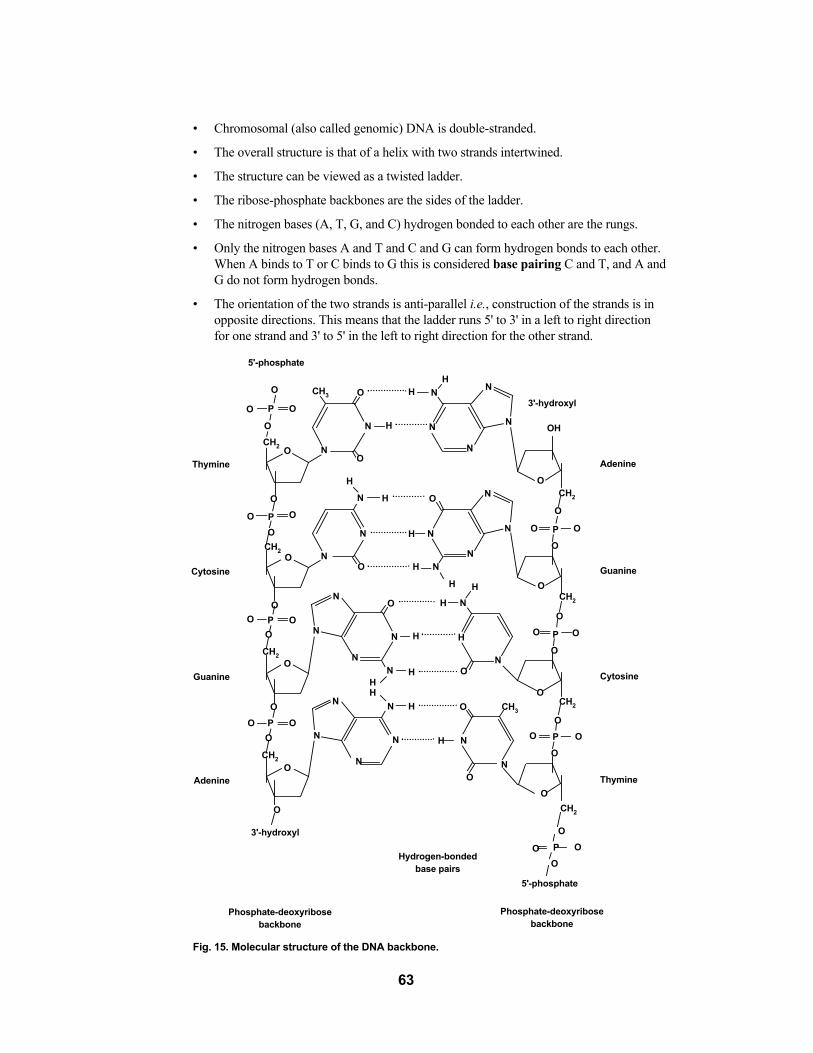
�• Chromosomal (also called genomic) DNA is double-stranded.
�• The overall structure is that of a helix with two strands intertwined.
�• The structure can be viewed as a twisted ladder.
�• The ribose-phosphate backbones are the sides of the ladder.
�• The nitrogen bases (A, T, G, and C) hydrogen bonded to each other are the rungs.
�• Only the nitrogen bases A and T and C and G can form hydrogen bonds to each other.When A binds to T or C binds to G this is considered base pairing C and T, and A andG do not form hydrogen bonds.
�• The orientation of the two strands is anti-parallel i.e., construction of the strands is inopposite directions. This means that the ladder runs 5' to 3' in a left to right directionfor one strand and 3' to 5' in the left to right direction for the other strand.
Fig. 15. Molecular structure of the DNA backbone.
63
5'-phosphate
3'-hydroxyl
O
O
O
CH2
CH2
CH2
CH2
CH2
CH2
CH2
CH2
CH3
CH3
P O
O
O H
H
H
H
H
H
H
H
H
HH
HHH
H
H
H
N
NN
N
N
N
N
N
N
N
N
N
N
N
N N
N
N
N
O
O
O
O
O
O
O
O
OO
O
O
O
O
O
O
O
OO
O
5'-phosphate
3'-hydroxyl
Adenine
Guanine
Cytosine
Thymine Adenine
Guanine
Cytosine
Thymine
Hydrogen-bondedbase pairs
Phosphate-deoxyribosebackbone
Phosphate-deoxyribosebackbone
O
O
O
O
P
P
P
P
O
O
O O
O
O
O O
O
O
O
O
O
O
O
O
O
O
P
P
O
P
N
N
N
N
N
N
N
N
N
N
OH

It follows From This Structure That
�• Because A only binds to T and G only binds to C, the two strands will have exactly theopposite, or complementary, sequence running in opposite directions (one strand 5' to3', the other 3' to 5').
�• One strand is called the sense strand and the other is antisense.
�• These two complementary strands anneal or hybridize to each other through hydrogenbonds between the bases.
�• A new single-strand can be synthesized using its complement as the template for newstrand synthesis.
�• Each strand, then, carries the potential to deliver and code for the same information.
The length of any double-stranded DNA molecule is given in terms of base pairs (bp).If a DNA strand contains over a thousand base pairs, the unit of measure is kilobases (kb).If there are over one million base pairs in a strand the unit of measure is megabases (Mb).
Fig. 16. DNA (deoxyribonucleic acid)�—A long chainlike molecule that stores genetic information. DNAis graphically represented in a number of different ways, depending on the amount of detail desired.
64

DNA Replication�—Strand Synthesis
New strands are synthesized by enzymes called DNA polymerases. New strands arealways synthesized in a 5' to 3' direction. For a new single strand of DNA to be synthesized,another single strand is necessary. The single strand of DNA that will be used to synthesizeits complementary strand is called the template strand.
However, in order for the DNA polymerase to start synthesizing a new complementarystrand, a section of double-stranded DNA must be present for the DNA polymerase toinitiate synthesis. A short strand (20�–50 base pairs) used to anneal to the template strand tocreate the double-stranded start site for the DNA polymerase is called an oligonucleotideprimer. This primer is almost always a short strand of nucleotides complementary to a partof the template where the researcher wants synthesis to begin. It must have a free 3'hydroxyl group (OH) for the DNA polymerase to attach the 5' phosphate of the nextnucleotide.
The DNA polymerase grabs free (single) nucleotides from the surrounding environmentand joins the 5' phosphate of the new nucleotide to the 3' hydroxyl group (OH) of the newcomplementary strand. This 5' to 3' joining process creates the backbone of the new DNAstrand.
The newly synthesized strand maintains its complementarity with the template strandbecause the DNA polymerase only joins two nucleotides during new strand synthesis if thenew nucleotide has its complement on the template strand. For example, the DNA polymerasewill only join a G to the 3' end of the newly synthesized strand if there is the C counterpart onthe template strand to form a hydrogen bond. Guanine will not be joined to the new strand ifadenine, thymine or guanine is the opposite nucleotide on the template strand.
DNA polymerase and strand synthesis allow DNA to replicate during mitosis.Interestingly, both new DNA strands are synthesized simultaneously from the two originalDNA template strands during mitotic DNA replication.
As you can see DNA, RNA and proteins are closely tied to each other. Thus you canrealize why researchers today, in an attempt to understand the mechanisms behind thevarious life processes, must study the nucleic acids as well as the proteins to get completeanswers about the flow of information carried in the genetic code. In the last 20 years,many gains in the areas of nucleic acid techniques have finally allowed researchers themeans to study the roles of nucleic acids in life processes.
Individual discoveries by many scientists have contributed the pieces that have begunto solve one of the most mysterious puzzles of life�—understanding the hereditary code. In1985, enough pieces of the puzzle were in place for a major breakthrough to occur. Thiselucidation of how the necessary molecular components interact to faithfully replicateDNA within living cells led to the development of a technique for creating DNA in a testtube. This technique is called the polymerase chain reaction, or PCR.
65

Appendix BGlossary of Terms
Aliquot The division of a quantity of material into smaller,equal parts.
Alu A small piece of repetitive DNA which contains theAlu I restriction enzyme site, from which the sequenceobtained its name.
Annealing Binding of oligonucleotide primers to complementarysequences on the template DNA strands.
Chelation To bind metal ions in solution. An example of acommon chelating agent is EDTA orEthyleneDiamine Tetraacetic Acid.
ChelexTM Microscopic beads which chelate divalent cations andcomprise the InstaGene matrix.
Cofactors Ions or small molecules needed by an enzyme tofunction properly. For example, Taq DNA polymeraseneeds Mg++ in order to function properly. Mg++
would therefore be considered a cofactor.
Denature The process of melting apart two complementaryDNA strands. In vivo denaturation is accomplished byenzymes; in the PCR reaction denaturation isaccomplished by heat.
DNases Digestive enzymes which degrade DNA.
dNTPs Commonly used abbreviation for all four deoxynucleotidetriphosphates (dATP, dTTP, dGTP, dCTP) used in synthesizing DNA.
Ethidium bromide A fluorescent dye molecule which intercalatesbetween DNA base pairs and fluoresces whenexposed to ultraviolet light.
Eukaryotes Organisms that are made up of cells containing amembrane-bound nucleus that contains the geneticmaterial (DNA).
Exon The region of a transcribed messenger RNA moleculethat gets spliced together and leaves the nucleus fortranslation into protein sequence.
Extension This refers to the process of Taq polymerase addingdNTPs (deoxynucleotide triphosphates-dATP, dTTP,dCTP, or dGTP) onto the ends of the oligonucleotideprimers. Extension follows the base pairing rule andproceeds in the 5'�–3' direction.
Genomic DNA The sum total of the DNA that is found within thenucleus of a cell.
Hardy-Weinberg Equilibrium The conditions that enable a population to maintain itsgenetic frequencies. These conditions are: large population, random mating, no immigration or emigration, no mutations, and no natural selection.
66

Homologous chromosomes A pair of complementary chromosomes that containthe same genetic sequences, or genes, with one chromosome inherited from the mother and onechromosome inherited from the father.
InstaGene�™ matrix Microscopic beads that bind divalent cations insolution. The binding or sequestering of divalentcations prevents their availability to enzymes that candegrade the DNA template.
Intron The region of a transcribed messenger RNA that isspliced out of the mRNA and is not translated intoprotein sequence.
Lysis The process of rupturing a cell to release its constituents.In this exercise, human cheek cells are lysed to releasegenomic DNA for PCR reactions.
Master mix The main solution of a PCR reaction which containsall of the necessary components (dNTPs, primer,buffer, salts, polymerase, magnesium) of the reactionexcept the template DNA.
Mendelian inheritance For each inherited characteristic, an organism containstwo genes, or alleles, one inherited from each parent.There can be two forms of alleles, dominant andrecessive, and dominant alleles mask the expressionof recessive alleles.
Messenger RNA A type of RNA that is synthesized from the geneticmaterial (DNA) and that attaches to ribosomes and istranslated into protein.
Nucleotides The fundamental unit of DNA or RNA. They consistof a sugar (deoxyribose or ribose), phosphate, andnitrogenous base (adenine, thymine, cytosine, orguanine and uracil in place of thymine in RNA).
PCR Polymerase chain reaction. The process of amplifyingor synthesizing DNA within a test tube.
Primers A small series of nucleotides (usually 16�–24 bases inlength) that recognize a particular sequence ofnucleotides on the target DNA sequence. Primers forthe polymerase chain reaction are usually synthesizedin a laboratory.
Reagents Materials needed to conduct an experiment. They areusually solutions or mixtures of various solutions.
Taq polymerase Heat stable DNA polymerase that was isolated fromthe heat stable bacteria Thermus aquaticus. This DNApolymerase is commonly used in PCR reactions.
Template The strand of DNA that contains the target sequencesof the oligonucleotide primers and that will be copiedinto its complementary strand.
67

Appendix CPCR Amplification and Sterile Technique
PCR is a powerful and sensitive technique which enables researchers to produce largequantities of DNA from very small amounts of starting material. Because of this sensitivity,contamination of PCR reactions with unwanted, extraneous DNA is always a possibility.Therefore, utmost care must be taken to prevent cross-contamination of samples. Steps tobe taken to prevent contamination and failed experiments include:
1. Filter-type pipet tips. The end of the barrel of micropipets can easilybecome contaminated with DNA molecules that are aerosolized. Pipet tips whichcontain a filter at the end can prevent aerosol contamination from micropipets. DNAmolecules which are found within the micropipet can not pass through the filter andcan not contaminate PCR reactions. Xcluda® aerosol barrier pipet tips (catalog number211-2006-EDU and 211-2016-EDU) are ideal pipet tips to use in PCR reactions.
2. Aliquot reagents. Sharing of reagents and multiple pipettings into the same reagent tubewill most likely introduce contaminants into your PCR reactions. When at all possible,aliquot reagents into small portions for each team, or if possible, for each student. If analiquotted reagent tube does become contaminated, then only a minimal number of PCRreactions will become contaminated and fail.
3. Change pipet tips. Always change pipet tips when entering a reagent for the first time.If a pipet tip is used repeatedly, contaminating DNA molecules on the outside of the tipwill be passed into other solutions, resulting in contaminated PCR reactions. If you areat all unsure if your pipet tip is clean, use the safe rule of thumb and discard the tip andget a new one. The price of a few extra tips is a lot smaller than the price of failed reactions.
4. Use good sterile technique. When opening, aliquotting, or pipetting reagents, leavethe tube open for as little time as possible. Tubes which are open and exposed to the aircan easily become contaminated by DNA molecules which are aerosolized or whichare present in your mouth/breath, etc. Go into reagent tubes efficiently, and close themwhen you are finished pipetting. Also, try not to pick tubes up by the rim or cap asyou can easily introduce contaminating DNA molecules from your fingertips.
68