studies on the chemical composition of muscle...
TRANSCRIPT

J. Exp. Biol. (1961), 38, 707-7*8 -707
Great Britain
STUDIES ON THE CHEMICAL COMPOSITIONOF MUSCLE TISSUE
II. THE ABDOMINAL FLEXOR MUSCLES OF THE LOBSTERNEPHROPS NORVEGICUS (L.)
BY JAMES D. ROBERTSON
Department of Zoology, University of Glasgow and the Marine Station, Millport
(Received 8 June 1961)
I. INTRODUCTION
This is the second of a series of papers on the composition of muscles of some lowervertebrates and invertebrates, particularly from the standpoints of the osmoticconcentration of various constituents and the accumulation or reduction of ions inmuscle relative to those of the blood plasma. A comprehensive analysis of the muscleand plasma of the decapod crustacean Nephrops norvegicus (L.) has been made for allinorganic and organic compounds believed to be of osmotic significance. An attempthas been made to assign concentrations to the intracellular phase of muscle, afterappropriate corrections for extracellular fluid, and to see how far the summation ofions and molecules compares with the experimentally determined osmotic concentra-tion of the muscle and plasma.
Male specimens of Nephrops were obtained by trawling, and thereafter kept3-4 days in the aquarium tanks at Millport. A chloride determination on the sea waterwas made each time an animal was used. Over the period of the analyses and experi-ments the values ranged between 1776 and 18-80 g. Cl/1., with a mean of 18-4.
Blood was collected by pipette through the arthrodial membranes at the bases ofthe legs; after the cells had aggregated the blood was centrifuged and the clear plasmaused for analysis. The muscles analysed were the paired longitudinal flexors of theabdomen. These were lightly blotted between sheets of filter paper and separateweighed samples were used for the determination of cations (sodium, potassium,calcium and magnesium), chloride, sulphate and water content. Methods used inthese inorganic analyses of muscle and plasma have been given previously (Robertson,19606, c). In addition, further samples of muscle and plasma were analysed for otherions and compounds as follows: bicarbonate of muscle, method of Conway & Fearon(1944); lactate, method of Hullin & Noble (1953) on 10% trichloracetic acid filtratesof muscle and plasma; reducing sugar on zinc hydroxide nitrates after Somogyi (1945);inorganic and organic phosphate, fractionation procedure after Umbreit, Burris &Stauffer (1949), determination of phosphorus according to Sumner (1944); a-amino Nof free amino acids on tungstic acid filtrates after Frame, Russell & Wilhelmi (1943)with the improvements of Russell (1944); arginine on tungstic acid filtrates by Mac-pherson's (1946) method, with the substitution of oxine for a-naphthol (Janus, 1956);proline according to Chinard (1952), after removing lysine with permutit (Troll &
J-ondsley, 1955); trimethylamine oxide and glycine betaine on 10% trichloracetic acid

708 JAMES D. ROBERTSON
filtrates after Kermack, Lees & Wood (1955), with the substitution of gravimetri^estimation of the mixed reineckates of these two compounds for colorimetry.
Estimates of total extracellular fluid in muscle, the blood plus interstitial fluid, wereobtained by injecting into the sternal sinus solutions of inulin, sucrose or sodiumthiosulphate, substances believed to enter cells only with difficulty, and comparingtheir distribution in samples of blood and muscle on a water-content basis, afterperiods of 1-10 hr. The volume or weight of water deemed to be extracellular couldthen be expressed as a percentage of total muscle water ('permeations' in Conway &Fitzgerald's (1942) terminology) and compared with the chloride and copper ' permea-tions' of the muscle, that is, the chloride and copper contents of muscle waterexpressed as percentages of those in the plasma water.
Before the injections 9ome blood was withdrawn equal in volume to that of thesolution to be injected. Inulin (Kerfut) was used in a 3% solution in sea water(2 ml. into animals of about 150 g.), sucrose and sodium thiosulphate in isosmoticsolution (usually 1 and 0-5 ml., respectively). Inulin was determined photometricallyin zinc hydroxide filtrates by the method of Roe, Epstein & Goldstein (1949), sucroseby the same method or by Somogyi's (1945) copper reagents after hydrolysis of thesucrose in the filtrates with an invertase preparation, and sodium thiosulphate in atungstic acid filtrate according to Gilman, Philips & Koelle (1946). Choride wasestimated in other aliquots of the same muscle nitrates by Sendroy's silver iodatemethod or the Volhard titration (Robertson, 1960c).
A comparison of the copper content of plasma and muscle enables an estimate to bemade of the volume of blood in the muscle, since haemocyanin of the plasma is thechief copper-containing compound in the tissues. This comparison gives a maximumvalue for the blood-space in the muscle, since traces of copper inside the cells wouldreduce the estimate. While copper can be determined in ashed blood plasma or intrichloracetic extracts by direct colorimetric measurement of the copper-diethyl-dithiocarbamate complex, this fails with the muscle of Nephrops (and cephalopods)because the traces of zinc in the tissue precipitate and cause cloudiness of the solution.The complex was therefore extracted with amyl alcohol according to the method ofEden & Green (1940), but trichloracetic acid filtrates of blood and muscle weresubstituted for the preliminary ashing procedure of these authors.
In all muscle analyses involving the production of filtrates, measured quantities ofreagents were added to weighed samples which were then ground thoroughly withquartz sand in a mortar, and aliquots of each filtrate were taken for analysis. Since thewater content of the muscle was also determined, the results of analyses could beexpressed in relation to water content.
Measurements of total osmotic concentration were made by a thermo-electricvapour-pressure method (Krogh, 1939, p. 211) on the plasma and muscle-juice ofspecimens, the juice being obtained with a small tissue-press (Krogh, 1938). Byworking rapidly the measurements on the juice could be completed within 20-60 min.of removing the muscle, thus minimizing changes due to breakdown of labilecompounds.
Some inorganic analyses were made on the muscle-juice by the methods used forblood and whole muscle, but the small amount of magnesium was determined in theash by means of titan yellow (Heagy, 1948).

Composition of muscle tissue. II 709
II. INORGANIC IONS
(1) Composition of whole muscle and plasma
Three comparisons of muscle, plasma and sea water show close agreement (Table 1).Total concentrations of inorganic ions in the plasma and sea water are practicallyidentical in two cases, within 1 -8 % of each other in the third. Ionic regulation in theplasma consists of increases in sodium and calcium over sea-water values, markeddecreases in magnesium and sulphate, slight decreases in potassium, and virtualidentity of chloride with that of the water; this is in agreement with a previous study(Robertson, 1949).
Table 1. Ionic composition of whole muscle and blood plasma
(1) MusclePlasmaSea water
(2) MusclePlasmaSea water
(3) MusclePlasmaSea water
MeansMusclePlasmaSea water
Na
84-4527456
83-2SO458
82-1512458
8 3 3517457
K
173-58-597
159-27-79-8
167-09-69-8
166-68-69-8
Ca
5-2413-9io-o
5-2617-1IO-I
5-1217-5IO-I
5-2i16-2IO-I
mg. ions/kg, water
Mg
2O-69-3
52-O
16-910-953-3
19-810-952-3
19110-452-3
Cl
120-753°533
9i-3528536117-8523536
IO99537535
SO,
4-316-827-4
2-117-527-6
3-o21-727-6
3-118-727-5
Total P
149-8
141-5
I43-O
144-8
Totalmg. ions
55811061088
50010941094
53810941094
53210981093
Water(g./kg. or
g-/l.)
749947986
752935986
768946986
756943986
The figures for P (trichloracetic acid-soluble P) are really mg. atoms. The mg. ions are slightly lower,since the adenosine triphosphate molecule has 3 atoms P.
The sum of inorganic ions and acid-soluble phosphorus in muscle comes to onlyone-half of the ionic concentration of the plasma in each case, therefore the other halfof the osmotic concentration (assuming identity between muscle and blood con-centrations) must be made up chiefly by organic compounds other than those ofphosphorus (see §111 below). The pattern of ions in the muscle relative to the plasmais consistent: higher concentrations of potassium and magnesium, and lower con-centrations of sodium, calcium, chloride and sulphate, with very high levels of acid-soluble phosphorus. Magnesium in the plasma is kept low partly by its selectiveexcretion in the secretion of the antennal glands (Robertson, 1949); the approximatedoubling of its concentration in the water of the muscle still leaves it well below thesea-water level.
(2) Apparent extracellular spaces in muscle
Tables 2 and 2 a give the results of experiments with injected solutions of inulin,sucrose and sodium thiosulphate, with simultaneous measurements of chloride andcopper. The samples of muscle taken for the analyses were quite large (ca. 8-10 g.)

710 JAMES D. ROBERTSON
in relation to the total abdominal flexor muscles, so that they should be fully repre-sentative and allow of replicate analyses. The chloride space is obtained by expressingthe chloride concentration of the muscle as a percentage of the chloride of the plasma(both on a water-content basis). If there were no chloride inside the cells, the spacewould represent the volume of muscle water which is extracellular, having the samechloride concentration as the plasma.
Table 2. Apparent extracellular spaces or 'permeations' in Nephrops muscle (concen-trations in muscle as percentages of those in plasma, on water-content basis)
Extracellular
Chloride
208619-871816182221-0622-0620-951981
——
Mean 20-12
3.B. ±049
spaces as % total
Tnnll'n
9-2611-02IO-63l6-2214-1612*41II-33IO-7214-38IO-7O
12-08
±0-69
muscle water
Copper
—4-853-02——
4-943-443-395-00—
4-n
±0-37
A/Tn^rli* rhlnririn
mg./g. water
3-843613-383-333-854-063-823-53——
3-68
±0-09
Time allowed1UI UUfUlUULlUll
(hr.)1
a2
336-5
1 0
1 0
2 *
3*
—
• Antennal gland openings closed with dental cement.
Table 2 a. Apparent extracellular spaces
Extracellular spaces as % total muscle waterTime allowed
Sodium Muscle chloride for distributionChloride Sucrose thiosulphate Copper mg./g. water (hr.)
1729 17-43 — 4-6o 3-07 215-50 17-29 — — 2-75 2
I5-95 14-94 — — 2-81 41863 — 18-63 — 3-<>9 1
In Nephrops the chloride space is usually the largest, but it is equalled by oneestimation of the sodium thiosulphate space, and exceeded by the sucrose space intwo out of three cases. The copper space is the smallest. It represents the plasmawater in the total muscle water, the copper being derived from the protein haemocyanin.
Inulin spaces are always lower than the corresponding chloride spaces, and two tothree times as large as the copper spaces. They range between 9 and 16%, with amean value of 12%. For the following reasons the inulin space has been taken asapproximating the extracellular space:
(1) Theoretically, the large size of the inulin molecule, (M.W. about 5000) shouldmake it difficult for this carbohydrate to enter cells.
(2) The inulin spaces are smaller than the sucrose and sodium thiosulphate spaces,and fairly consistent, after periods of 1-10 hr.
(3) The smaller molecule of sucrose (M.W. 342) and thiosulphate (ionic weight 112)give spaces practically equivalent to chloride spaces.

Composition of muscle tissue. II 711
The simplest assumption is that the distribution of inulin gives a measure of extra-cellular fluid, and that the smaller sucrose molecule and thiosulphate ion have beenable to diffuse to a small extent into the cells during the 1-4 hr. of the experiments.Sucrose is known to diffuse slowly into amphibian muscle cells (Krogh & Lindberg,1944), while inulin seems to measure extracellular space in mammalian muscle(Conway & Fitzgerald, 1942).
On this basis, there is a small amount of chloride inside the muscle cells of Nephrops(see below, Table 3). This agrees with work on isolated muscle fibres of Carcimu,another marine decapod crustacean, in which there are about 53 mg. ions/kg, intra-cellular water (Shaw, 1955).
Accepting an inulin space of 12 % as the extracellular volume in total muscle water,and an average copper space of 4% as the volume of plasma, the difference of 8%would be a measure of interstitial fluid volume.
(3) Intracellular ionic concentrations of muscle
By subtracting from the mean concentrations of ions per kilogram muscle water(Table 1) values representing 120 ml. extracellular fluid (based on the composition ofplasma), and then expressing the remaining intracellular concentrations in 880 ml. asconcentrations per kilogram water, the intracellular composition is found (Table 3).
Table 3. Intracellular composition of Nephrops muscle compared with blood plasma
mg. ions/kg, water
Na K Ca Mg Cl SO4 HCO, Lactate P Total
Muscle cells 24-5 188 3-72 20-3 53-1 i*oa I-9° 8-8 164-3 466Plasma 517 8-6 i6-a 10-4 527 18-7 4-13 0-36 o-8i 1103
muscle cell , „Ratio: — ; 0-047 21-9 o-a3 i'97 o-ioi 0-055 o-4° 33'8 203 0-42
plasmaWater content of cells—731 g./kg.
The chief differences between intracellular and whole muscle concentrations are thereduced sodium and chloride values of the former, because of the subtraction of thesodium- and chloride-rich extracellular fluid. This results in a lower total concentra-tion of analysed ions, 466 against 532 mg. ions. Potassium and magnesium are theonly cations to become concentrated in the cells, the former to about twenty times andthe latter to twice its level in the plasma. Sodium and sulphate fall to about a twentiethof their plasma values, calcium to a quarter, and chloride to a tenth.
If we assume that most of the cellular potassium is held electrostatically by non-diffusible organic anions, particularly organic phosphates, and that the low value forsodium is maintained by a process of active extrusion of sodium ions, and, furtherthat the cells are permeable to small ions (e.g. Ussing, 1949; Hodgkin, 1951), it ispossible that cellular potassium stands in Donnan equilibrium with the outsidepotassium, and
In Nephrops these ratios are respectively 21-9 and 9-9. For these to represent aJ)onnan equilibrium, 50% of the potassium would need to be in an unionized complex.
45 Exp. Biol. 38, 4
712 JAMES D. ROBERTSON
Evidence for the binding of about a quarter of the potassium is given in § IV, zb.this is accepted, the concentration of potassium above about 135 mg. ions or a 15-foldincrease over the plasma value must be maintained by an active metabolic process.
III. ORGANIC CONSTITUENTS
(1) The phosphorus compounds
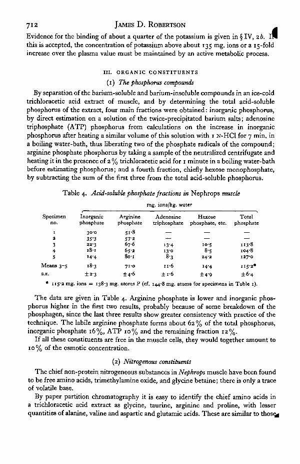
By separation of the barium-soluble and barium-insoluble compounds in an ice-coldtrichloracetic acid extract of muscle, and by determining the total acid-solublephosphorus of the extract, four main fractions were obtained: inorganic phosphorus,by direct estimation on a solution of the twice-precipitated barium salts; adenosinetriphosphate (ATP) phosphorus from calculations on the increase in inorganicphosphorus after heating a similar volume of this solution with 1 N-HC1 for 7 min. ina boiling water-bath, thus liberating two of the phosphate radicals of the compound;arginine phosphate phosphorus by taking a sample of the neutralized centrifugate andheating it in the presence of 2 % trichloracetic acid for 1 minute in a boiling water-bathbefore estimating phosphorus; and a fourth fraction, chiefly hexose monophosphate,by subtracting the sum of the first three from the total acid-soluble phosphorus.
Table 4. Acid-soluble phosphate fractions in Nephrops muscle
mg. ions/kg, water
Specimenno.
i
3
345
Means 3-5S.E.
Inorganicphosphate
30-035-322318-114-4
±2-3
Argininephosphate
5 1 *57-2676
8o-i
71-0
±4-6
Adenosinetriphosphate
13-413-08-3
n-6±i-6
Hexosephosphate, etc.
10-58-5
24-2
14-4
±4-9
Totalphosphati
H3-8104-8127-0
115-2*
±6-4
• 115-2 mg. ions = 138-3 mg. atoms P (cf. 144-8 mg. atoms for specimens in Table 1).
The data are given in Table 4. Arginine phosphate is lower and inorganic phos-phorus higher in the first two results, probably because of some breakdown of thephosphagen, since the last three results show greater consistency with practice of thetechnique. The labile arginine phosphate forms about 62 % of the total phosphorus,inorganic phosphate 16%, ATP 10% and the remaining fraction 12%.
If all these constituents are free in the muscle cells, they would together amount to10% of the osmotic concentration.
(2) Nitrogenous constituents
The chief non-protein nitrogeneous substances in Nephrops muscle have been foundto be free amino acids, trimethylamine oxide, and glycine betaine; there is only a traceof volatile base.
By paper partition chromatography it is easy to identify the chief amino acids ina trichloracetic acid extract as glycine, taurine, arginine and proline, with lesserquantities of alanine, valine and aspartic and glutamic acids. These are similar to those^

Composition of muscle tissue. II 713
found in an extensive study of the muscle of the lobster Homarus by Kermack et al.(1955), and by Camien, Sarlet, Duchateau & Florkin (1951) in the muscles of Homarusand the crabs Maia and Eriocheir.
From the standpoint of the present investigation it was deemed sufficient to esti-mate total a-amino N in tungstic acid filtrates of Nephrops muscle by Folin's naphtho-quinone sulphonate reagent as used by Russell (1944), although this photometricmethod may not be as accurate as manometric analysis, according to Chinard &Van Slyke (1947).
It was found that taurine N reacted with the naphthoquinone and was measuredwith the a-amino N of the other amino acids. Proline did not develop the full colourexpected on the basis of its amino N, and, as found previously by Chinard & VanSlyke, is subject to fading. On the basis of a glycine standard (used throughout) itgave 70 % of the theoretical colour 15-25 min. after final dilution. Some estimations ofproline were therefore made using the method of Troll & Lindsley (1955), so thatcorrected figures could be given for total amino acids.
Table 5. Nitrogenous constituents of Nephrops muscle
Specimenno.
1
2
34S6789
1 0
11
1 3
13141516
1718
19202 1
Amino acids(a-amino N)
481457482441
445
45533949953955i53 i494————————
mM./kg.
Trimethylamineoxide
————
49-263-039'i62-s50-836-153-927-439-6
115-659'687-472-928-460-953-4
IO2-O
water
Betaine
————
66-o58-594-063-061-470-268-650-865-348-676-44i-544-482-3—
94-6—
Chloride
79-286-79 9 2—
137-9130-3898
105-8105-198-8
104-884-087-2————————
Mean 476 59-0 65-7 100-7
S.E. 16-4 ±5-9 ±4-2 ±5-2
Volatile base (ammonium ions) of specimens 19, 20 and 21 was 3-0, 3-4 and 2-4 mg. ions/kg, water.
These figures include all the arginine. While it was possible by working rapidly toget a tungstic acid filtrate in which only about half of the labile arginine phosphatehad broken down (giving 56-61 mia. free arginine as against the mean of 71 mM.arginine phosphate (Table 4) and 93 mM. free arginine when splitting was complete),the analyses were usually delayed 12-24 nr-> when most or all of the arginine com-
had split into its components.43-3

714 JAMES D. ROBERTSON
From Table 5 it is seen that amino acids varied from 339 to 551 mM./kg. water,with a mean of 476 mM., thus constituting a very large proportion of the osmoticconcentration of whole muscle, some 40-50% (see §IV).
The nitrogenous bases trimethylamine oxide and betaine are present in aboutequimolar concentration, with means of 59 and 66 mM., respectively. The former wasmore variable than the latter. Kermack et al. found 75 and 67 mM. respectively perkilogram fresh muscle in Homarus, approximately 100 and 90 mM. per kilogram waterof the muscle. Shaw's (19586) figure for trimethylamine oxide in the muscle fibres ofCarcinus, 90 mM., is of a similar order. From the osmotic standpoint these two basesform about 13% of the total particle concentration in Nephrops (§IV).
Table 5 a. Some amino acids of Nephrops muscle
mM/kg. water
Totala-amino N
44553955i53i494
Mean 513
8.K. ±10-2
Proline
1189373
106120
1 0 2
±8-7
Arginine
931007i97
106
93
± 6 0
Glycine, taurine,etc (by difference)
234346407328268
3'7
±3°-3In three other specimens, glutamine averaged 28-4 mM./kg. water (27'3, 24-9, 33-o), using the method
of Kermack et aL (1955).
In Table 5 a are a few analyses of proline and arginine in specimens 5 and 10-13 °fTable 5. They show that the concentrations of these amino acids are very similar.
(3) Soluble carbohydrates
Carbohydrates contributing to the osmotic pressure of muscle and plasma aresugars and lactic acid. Boyland (1928) has given some data for the muscles of lobsters.Expressed in relation to water content and molality, lower carbohydrates (calculatedas glucose) were 3-4 (1-5-5-9) mM./kg. water, lactic acid 9-1 (4-4-13-3) mM. in Homarusvulgaris; for Palinurus vulgaris these were 40 (0-7-5-9) m M- anc^ J9"7 (11-8-26-7) mM.,respectively.
A few analyses of reducing sugar and lactate in Nephrops muscle and plasma aregiven in Table 6. The mean values for plasma, o-8 mM. and 0-3 mg. ions, are of thesame order as those in intermoult Carcinus (Robertson, 19606). Reducing sugar valuesin muscle are about ten times as high as in the plasma, being somewhat higher thanin Boyland's analyses. The significance of the values of reducing sugar obtained onfiltrates of muscle by femcyanide (Boyland) or copper reagents (the present analysis)is somewhat questionable, as glucose will form only part of the reducing substances,another fraction being the hexose phosphates, of which some at least have a reducingvalue when unhydrolysed.
True resting values for lactate are difficult to obtain. The lower values of speci-mens 5 and 6 were obtained by quickly cutting out the muscle and dropping it into

Composition of muscle tissue. II 715
weighed beakers containing ice-cold trichloracetic acid, weighing, and then grindingwith quartz sand and more acid in a cold mortar. Perhaps the mean of the threelowest figures, 8-4, or 7-8 mg. ions for two males, would be nearer the values in theliving animal at rest.
123456*
Table 6. Soluble carbohydrates in muscle and plasma
Reducing sugar Lactate
Plasma
mg./l.
134-6140-21683120-6
Mean —
S.E.
mM./kg.water
0790-830090-71
0-83
±0-06
Muscle
1-4941-0840-6411-167
mM./kg.water
II-O8-o4-78-7
Plasma
mg./l.
17-030-040-0
7-014-3
±1-3 —
$ specimen.
mg. ions/kg,water
0-200-360-480080-17
0-26
±0-07
Muscle
1-5*30-5642-3061-471O-4730-641
mg. ions/kg,water
22-88-5
35-922-O719-6
17-7
±4-5
Table 7. Osmotic concentrations of plasma and muscle-juice(Krogh-Baldes method)
cimer
1
2
3456789
ean
1 Sea water
3-1323-0343-0123-022
2-99730973-0113-on3-009
3-034
±0-0128
Plasma
3-1703-037306830193-0093-0303-0222-9822-976
3-035
±00193
Muscle-juice*
3.262 (1-5 hr.)3-037 (0-4 hr.)3-133 (0-3 hr.)3-131 (0-4 hr).2-994 (o-9 hr.)3-186 (0-9 hr.)2-989 (o-8 hr.)3-055 (0-5 hr.)3-079(1 -3 hr.)
3-096
± 0-0302
Plasma as %sea water
101-5IOO-I
101-9
999100-4IOI-I100-4
9 9 0
989
100-4
±0-344
Muscle-juice as% plasma
102-9ioo-oIO2-I103-7
99-5105-1989
102-41035102*0
±0-703
• Figures in brackets are times between removal of muscle and completion of estimation.Concentrations are expressed in relation to % NaCl solution (g./ioo g. water). On standard NaCl
solutions, s.D. of method was 0-75 % (N = 9).
IV. OSMOLALITY AND CATION-ANION BALANCE
(1) Direct measurements of osmolality
These are given in Table 7. The plasma of nine specimens was isosmotic with seawater within 1-9%, the mean value being 100-4% that °f ^ e medium. This is theusual finding in marine decapod crustaceans (Robertson, 1960 a, b). Compared withthe plasma the juice expressed from the abdominal flexor muscles of the samespecimens was more variable in concentration; in six cases it was slightly but definitelyhyperosmotic by 2-5%. Delay in measuring the vapour pressure of the juice or

716 JAMES D. ROBERTSON
delay in putting a sample of muscle in the press resulted in higher positive differencesfrom the plasma, plus 5-8%. These high values are apparently due to the breakdownof labile compounds such as arginine phosphate, with a consequent increase inosmotic concentration. The mean difference between plasma and muscle-juice was2'O % in the nine estimations done within 1 -5 hr. of removing the muscle, 1 -7 % in theseven done within an hour; the first difference is significant (P < 0-05), the secondnot, by the f-test.
It is reasonable to conclude that the mean difference of plus 2-0 % is due to partialbreakdown of labile compounds during the working of the press at room temperature(17-190 C), and that the muscle cells are in life isosmotic with the interstitial fluidand plasma.
Table 8. Osmotic concentration of plasma, whole muscle, and muscle cells
mg. ions or mM./kg. water
Constituent
SodiumPotassiumCalciumMagnesiumAmmoniumChlorideSulphateBicarbonateLactateInorganic phosphateArginine phosphateAdenosine triphosphateRemaining acid-solublephosphate
Amino acidsTrimethylamine oxideBetaineReducing sugar
Plasma
S'78-6
1 6 2io'40 2 8
52718-74-i30-260-63——0 1 8
3-77
—0-83
Whole muRde
83-21666
5-211 9 12-93
10993-12-167-8
i8-371-0n-614-4
4°5*5 9 °65-7
Muscle cells
24-5188
3-7220-3
3'353-i
1-02
I OO
8-820-780-713-216-4
46o»67-074-6
—
Total 1108 1045 1037
• Excluding arginine of arginine phosphate.
(2) Osmolality by summation of chemical constituents
From the data given in §11 and III, we can calculate the osmolality by summing themg. ions and millimoles non-electrolytes, neglecting for the moment the osmoticcoefficients of the salts and nitrogenous compounds, and the possibility that some ofthe 'ions' are bound. This is set out in Table 8, in which the figures for the musclecells have been calculated on the basis that plasma and interstitial water form anextracellular volume of 12% of total muscle water. The reducing sugar value of8-i mM. in whole muscle (Table 6) has been omitted in Table 8, since presumably it islargely included under 'remaining acid-soluble P', that is, hexose phosphates, etc.,of 14-4 mM. Similarly, to avoid including arginine twice, 71 mM. (the value forarginine phosphate) has been subtracted from the mean of 476 mM. amino acids inwhole muscle (Table 5).
The agreement between the total concentrations in cells and plasma is fairly good.However, the apparent balance is subject to several uncertainties, of which one is

Composition of muscle tissue. II 717
possible error involved in adding up constituents from different animals. Thus oneseries of specimens was used for inorganic ions, a second series for the phosphatecompounds, and two further series for the nitrogenous compounds. This error wasminimized by using animals in the spring and summer months, and in as healthy acondition as possible 3-4 days after being trawled.
Two further uncertainties are the possibilities that some of the ions are not freebut bound to protein molecules, and that the osmotic coefficients of the salts (or ions)and organic compounds in solution may depart from unity.
(a) Departure from ideal laws of solutions. If the term 'milliosmoles' (often takenas equivalent to mg. ions in an electrolyte) is used as suggested by Dick (1959) as aunit of osmotic pressure, then a solution of A 1-858° C. contains 1000 m-osmoles. Seawater of this freezing-point depression contains 1129 mg. ions per kilogram solventwater, and is isosmotic with 0-553 M-NaCl (Sverdrup, Johnson & Fleming, 1942;Robinson, 1954).
It is probably safe to assume that the predominately inorganic plasma ions haveproportionally the same osmotic concentration as the ions of sea water; thus the1108 mg. ions would equal 981 m-osmoles. But the muscle cell contains a mixture ofions and organic compounds. In the cell solution one might assume that the aminoacids and other nitrogenous compounds would behave approximately as ideal non-electrolytes, and that the inorganic ions and phosphates would behave like plasma ions.
At the molal concentration of 0-98, the osmotic concentrations of the amino acidsand nitrogenous bases would be in proportion to their partial molal concentration,taking into account the osmotic coefficients. The latter do not depart from unity inthe cases of several amino acids and betaine for which data are available. Thus at1 molal the coefficients are 0-928 for glycine (Smith & Smith, 1937), 1-046 for prolineand 1-115 f°r betaine (Smith & Smith, 1940), and 1-003 f°r alanine which behaveslike an almost ideal solute (Robinson, 1952).
Assuming about 200 mM. glycine and 120 mM. proline, the 320 mM. of these twoamino acids would equal 311 m-osmoles. The betaine concentration of 74-6 mM. equals83-2 m-osmoles. Thus the sum of these three constituents, 395 mM., equals 394m-osmoles, a rather fortuitous result. Lacking any information on the other aminoacids and on trimethylamine oxide, we may assume for present purposes ideal behaviour,like alanine: the 602 mM. nitrogenous constituents would equal 602 m-osmoles.
Accepting the same average osmotic coefficient for cellular ions as for sea water andplasma (o-886), the 436 mg. ions would equal 386 m-osmoles, giving a total cellularosmolality of 602 + 386 = 988 m-osmoles, a figure very close to the plasma value of981.
(b) The binding of ions. When Nephrops muscle is subjected to pressure in a tissue-press an almost clear fluid is obtained which has a water content rather lower thanthat of the plasma, 914-936 g./kg., but much higher than that of whole muscle,740-765 g./kg. In composition this muscle juice is quite different from the plasma,although isosmotic with it (Table 7), and differs in several respects from wholemuscle, as shown in Table 9. On a water-content basis its Chloride content is practic-ally identical with that of muscle, but sodium and potassium are reduced by about aquarter, magnesium by over a half, calcium by nine-tenths and acid-soluble phos-
by a fifth.

718 JAMES D. ROBERTSON
MuscleMuscle-juiceJuice as % muscleNo. of pairedestimations
Na
87666-576
2
Table 9.
K
165-3122-8
742
Composition of muscle-juice
mg.
Ca
7-91079
10
I
ions/kg, water
Mg
23-7946
4°1
Cl
94-8 (±2-9)94-6 (±2-4)
1006
P*
163-2
127-2
78I
Water
754 (±i-3)930 (±i-8)
7
• mg. atoms. (± ) = S.B.Four unpaired analyses for Mg were: muscle 11-5, 16-2, muscle-juice 4-04, 5-63 mg. ions/kg, water.
Table 10. Approximate amount of ion-binding {based on data of Table 9)
mg. ions/kg, solvent water
Muscle cellCellular portion ofmuscle-juice f
Free ionsBound ions
Na
2 9 1
5-J
18%82%
K
18661383
74%26%
Ca
6-80
0 %100%
Mg
25-59 3
36%64%
Cl
35-935-7
9 9 %1 %
P*
185-2144-4
78%2 2 %
• mg. atoms.•f I.e. excluding extracellular component of juice.
It seems impossible to escape the conclusion that these reductions are due to theholding back by proteins of some of the 'ions', those bound in complexes. Neithercontractile proteins nor the soluble proteins of the sarcoplasm appear to cross the cellwalls under pressure. Only the free ions, including all the chloride of the cell, passout in the expressed juice, together with the amino acids and nitrogenous bases.Collectively, the osmotic concentration of all these ions and nitrogenous compoundsis 980-1000 m-osmoles, making the muscle-juice isosmotic with the plasma (seeTable 7). It is these free constituents that make up the osmotically active portion ofthe muscle cell.
The proportion of extracellular fluid in the muscle-juice has not been measured,but is presumably the same as that in whole muscle, about 12% of the water content.Accepting this, and assuming that the average composition of the plasma (Table 8)represents that of extracellular fluid as a whole, appropriate calculations can be madefor the composition of the muscle cells and cellular portion of the muscle-juice inthese specimens. The calculated composition is given in Table 10.
All the calcium of the cell seems to be bound, as well as over 60 % of the magnesium,82 % of the sodium and 26 % of the potassium, while none of the chloride is bound andonly 22% of the phosphorus. Which phosphorus compounds are bound have notbeen determined, possibly ATP (Dubuisson, 1942), as this would reduce the numberof phosphorus atoms by 25 %.
If these corrections are applied to the ions of the muscle cells as given in Table 8,the 436 mg. ions become 337 mg. ions, which when multiplied by the average osmoticcoefficient of 0-886 are equivalent to 299 m-osmoles. A revised osmotic balance sheetcan now be made up (Table 11). The correction for ion-binding in the plasma is vem
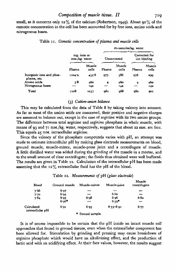
Composition of muscle tissue. II 719
small, as it concerns only 12% of the calcium (Robertson, 1949). About 92% of theosmotic concentration in the cell has been accounted for by free ions, amino acids andnitrogenous bases.
Table 11. Osmotic concentration of plasma and muscle cells
m-osmoles/kg. water
Inorganic ions and phos-phates, etc.
Amino acidsNitrogenous bases
Total
mg. ions ormM./kg. water
MusclePlasma cells
1104-3 435-6
3-8 460— 142
1108 1037
Unconnected
Plasma
977
4
981
Musclecells
386
46014a
988
Corrected forion binding
MusclePlasma cells
976 »99
4 460— 14a
980 901
(3) Cation-onion balance
This may be calculated from the data of Table 8 by taking valency into account.As far as most of the amino acids are concerned, their positive and negative chargesare assumed to balance out, except in the case of arginine with its two amino groups.The difference between total arginine and arginine phosphate in whole muscle, withmeans of 93 and 71 mM./kg. water, respectively, suggests that about 22 mM. are free.This equals 25 mM. intracellular arginine.
Since the valency of the phosphate compounds varies with pH, an attempt wasmade to estimate intracellular pH by making glass electrode measurements on blood,ground muscle, muscle-mince, muscle-press juice and a centrifugate of muscle.A little distilled water was added during the grinding of the muscle in a mortar, andto the small amount of clear centrifugate; the fluids thus obtained were well buffered.The results are given in Table 12. Calculation of the intracellular pH has been madeassuming that the 12% extracellular fluid has the pH of the blood.
Table 12. Measurements o/pH (glass electrode)
Blood
7667-7°7-84
Calculatedintracellular pH
Ground muscle
6-976-946-956-96*
6-91
Muscle-mince
6 9 8
693
• Second sample.
Muscle-juice
6-6o6966-59*
6-53-6-91
Musclecentrifugate
682
6-77
It is of course impossible to be certain that the pH inside an intact muscle cellapproaches that found in ground tissues, even when the extracellular component hasbeen allowed for. Stimulation by grinding and pressing may cause breakdown ofarginine phosphate which would have an alkalinizing effect, and the production oflactic acid with an acidifying effect. At their face values, however, the results suggest

720 JAMES D. ROBERTSON
an internal pH of 6-8-6-9 in the muscle cells. These values are similar to those of̂Caldwell (1958) on Carcinus muscle.
In Table 13 the equivalents of the phosphate compounds have been calculated forboth pH 6 and 7.
Table 13. Cation-onion balance
m.equiv./kg. water
Ion
SodiumPotassiumCalciumMagnesiumAmmoniumArginine (net +™ charge)
Total cation*
ChlorideSulphateBicarbonateLactateInorganic phosphateArginine phosphate "jAdenosine triphosphate >Hexose monophosphate, etc. J
Total anion3
Plasma
5178-6
32-42O-8
0-3—
579-1
5*737-4
4-i0-31-2
ca. 0-4
57O-4
Muscle cell
Free + bound
24-5188
7-440-6
3-325
288-8
53-i2-O
1 98-8
33-o»-23-i83-0-80-750-1-42-931-8-23-6
264-6-236-1
constituents
Free
4-4139
0
1463-3
25
186-3
53-i2 - 0
1-98-8
33-o#-23-i83-9-80-7
—31-8-23-6
214-5-193-2
• Assuming intracellular pH of 7-0; second figure for pH 6-0. Equivalents of phosphates calculatedfrom data of S8ren9en (1912): inorganic phosphate; Meyerhof & Lohmann (1928): arginine phosphate;Alberty, Smith & Bock (1951): adenosine triphosphate; Meyerhof & Lohmann (1927): hexosemonophosphate.
The small deficiency of anions in the plasma is probably made up by proteinate. Inthe muscle cells about three-quarters of the anions investigated are phosphates, andthe deficiency of 24-53 m-equiv. in the third column may be met by proteins. Thishas not been investigated in Nephrops. Dubuisson (1954) calculates that myosinateand myogenate in frog muscle cells at pH 6-8 may contribute 24 (22 + 2) m-equiv. tothe anions, but doubts whether they are free. Conway's (1957) estimates of the chargesof myosin and myogen of rat muscle are much greater, 61 m-equiv. at pH 7 and 30 atpH6.
However, it is certain that some of the other constituents are bound wholly or inpart in complexes, and in the last column of the table are given the estimates of freeions based for inorganic ions on the composition of the cellular portion of muscle-juice. Presumably the sarcolemma, myosin and actin are the chief binders of the ions,removing all the cellular calcium and most of the magnesium and sodium from freesolution, as well as a small proportion of the potassium. It has been assumed alsothat the adenosine triphosphate is bound to myosin and that the only proteins to beconsidered are those of the sarcoplasm, the myogen complex, whose net contributionto free anions may be quite small.
Cation-anion balance is fair, considering the uncertainties and difficulties of inter-pretation, and would be satisfactory if the intracellular pH were closer to 6 than to 7.

Composition of muscle tissue. II 721
V. ALTERATIONS IN THE BLOOD-MUSCLE STEADY STATE
The ionic steady state in the plasma of Nephrops and other decapod crustaceans, inwhich levels of ions are maintained different from those of the surrounding sea waterdespite some degree of ionic permeability of the gills, is a process controlled by theantennal glands and gills with the expenditure of energy (Robertson, 1949; 1960 a).Likewise, ionic and amino acid regulation in muscle, in which levels are maintainedoften vastly different from those of the plasma, requires energy. Death of a lobsterbrings about changes in the permeability of the cells, with a tendency towardsequalizing the concentrations of ions and nitrogenous compounds in plasma andmuscle.
Table 14. Changes in the plasma-muscle steady state after death
mg. ions or mM./kg. water
Plasma Muscle
Cl a-amino N Cl a-amino NNormal* 527 3-8 110 476Dead 12 hr. 516 2-3 105 475Dead 24 hr. 364 161-6 278 316
• Mean data from Tables 1, 5 and 8.The dead animals were kept at 14-160 C.
In Table 14 are given chloride and a-amino N analyses of plasma and muscle oftwo dead specimens, one 12 hr., the other 24 hr. after respiratory movements and theantennal reflexes had ceased. The first specimen showed no differences from normalanimals, but the second showed marked changes; chloride had fallen to about 70%in the plasma and increased to nearly three times its usual value in the muscle,whereas amino N had fallen in the muscle and increased in the plasma from the orderof 2-4 mM. to 162 mM. These changes imply leakage of amino acids from muscle cellsto extracellular fluid, and permeation of the muscle cells with chloride from the plasma,both processes tending to equalize the concentrations on both sides of the cellmembranes.
Since the gills or integument of most marine invertebrates including crustaceansare permeable to the ions of sea water (Krogh, 1939), an attempt was made to ascertainwhether changes brought about in the plasma by altering the levels of ions in the seawater would cause corresponding changes in the muscle.
Two Nephrops were kept for 3 days in sea water in which the potassium and calciumconcentrations had been increased to about three times normal by adding approxi-mately isosmotic solutions of the chlorides. Inspection of Table 15 shows thatpotassium of the plasma was much higher than in normal animals, having increasedin proportion to the difference in external level; muscle potassium was higher byonly 10 and 18%. Calcium of the plasma was up to twice, and in the muscle to threeand five times, the usual concentration. T-tests showed that the differences in thepotassium and calcium levels of the muscle were significant.
To be certain that the intracellular concentrations increased would require know-ledge of the extracellular phase in the muscle of these specimens. This can be assumed
be of the same order as found previously in normal sea water (§11, 2). On this

722 JAMES D. ROBERTSON
basis the intracellular concentrations in mg. ions/kg, water are: K 188 ± 4-62 (and 212-915-96 (K- and Ca-rich); Ca 3-71 ±0-19 and 17-4314-94; these differencesare significant in each case (P < 0-05). It is therefore probable that the increasedconcentrations in muscle are in both the extracellular and intracellular phases.
The experiment leads to the conclusion that the level of potassium and calcium ionsin plasma and muscle in Nephrops is broadly dependent on the concentrations of theseions in sea water.
Table 15. Changes in ions of plasma and muscle in sea water with raisedlevels of potassium and calcium
Na
K
Ca
Mg
Cl
so4
Total
Water(mg./ml.)or (mg./g.)
mg.
Sea water
457
9-8
IO-I
saa
535
a7"5
109a
986
>
ions/kg, water
Plasma
517
8-6
1 6 a
10-4
587
18-7
1098
943
Muscle
83-a
1666
5-a
1 9 1
n o
3 1
387
756
<
mg.
Sea water
4 0 9
37-1
a7-7
46-7
534
a4-7
1069
986
>
ions/kg, water
Plasma
4 4 4461
a 5 iai-83i-73 6 3
15-713-5
5345a4
«4'51 7 8
ioss1074
945933
Muscle
87-86 0 0
183-7196-8a5-3«3519-5n-8
14a1 0 1
—
—
458383
763760
Normal sea water, 3 specimens K- and Ca-rich sea water, 2 specimens
Data in normal sea water from Table 1.t-test on muscle:K i66-6±4-i3 (normal) and 190-3 ±6-55; Ca 5-31 ±0-04 and 19-4±5-9.Differences significant at P = 0-05.
VI. DISCUSSION
It has long been known that the ash content of marine invertebrate muscle is abouthalf that of the blood (Fredericq, 1904) and that the osmotic concentration must bemade up by organic substances. The pattern of inorganic ions in crustacean andmolluscan muscle has been studied by Bialaszewicz & Kupfer (1936) and Hayes &Pelluet (1947), and more recently by Potts (1958) and Shaw (1955): on a molar basispotassium usually exceeds sodium by a factor approaching two, calcium and magnesiumare present in relatively low concentration, and chloride usually exceeds sodium.Only in the last 10 years has the quantitative importance of free amino acids in musclebeen realized, by finding some 200-350 mM./kg. fresh tissue in lobsters (Homarus,Nephrops), crabs (Carcinus, Eriocheir) and mussels (Mytilus) (Camien et al. 1951;Kermack et al. 1955; Robertson, 1957; Shaw, 19586; Potts, 1958). Of lesser import-ance, at least in crustaceans, are the nitrogenous bases trimethylamine oxide andglycine betaine which are present in concentrations of about 70 mM./kg. in Homarusmuscle (Kermack et al.).
In the present paper analyses of whole muscle of Nephrops have been given for

Composition of muscle tissue. II 723
e constituents and others including the principal organic phosphorus compounds.On a water-content basis the sum of ions and molecules comes to 94% of that in theplasma, the plasma itself being isosmotic with the external sea water within 1 %. Thegreat importance of non-protein amino acids of muscle is evident from the fact thaton the average 405 mM. are present, 476 mM. if arginine phosphate is included.Quantitatively more important than arginine phosphate in the muscle are potassium,chloride and sodium ions, with trimethylamine oxide and betaine only slightly lessconcentrated than in Homarus.
Whole muscle includes an extracellular phase which seems to be measured by theinulin space or permeation, forming about 12% of the total muscle water. This inulinpermeation was consistently less than that calculated from injections of sucrose orsodium thiosulphate; these latter substances appear to enter the cells slightly. Fromcopper analyses a maximum blood space of 4% of the extracellular water has beenestimated, and the difference of 8 % has been attributed to the interstitial fluid roundthe individual cells of the muscle.
Zuckerkandl (i960) who has discussed various inconsistencies and anomalies in themeasurement of blood volume in crustaceans is prepared to believe in the existence ofa non-circulatory component of extracellular fluid from the fact that the dilution ofthe circulating blood at moult does not correspond with the volume of extracellularfluid (sea water) incorporated. It has been shown, however, that about a third of thewater absorbed at moult by Carcinus is taken up by the cells; it does not all remain inthe blood and interstitial fluid compartments (Robertson, 1960^). The finding of aspace in muscle into which inulin and other substances diffuse and from which bloodwith its haemocyanin is excluded gives experimental support for an interstitial phase,but the morphological separation of blood and interstitial fluid has still to be worked out.
Intracellular concentrations of ions and molecules in Nephrops muscle obtained byappropriate corrections for the 12% extracellular fluid do not differ in total by morethan 1 % from those of whole muscle. Concentrations of sodium and chloride changefrom 83 and n o mg. ions to 25 and 53 mg. ions, respectively, because of the highcontent of these ions in the extracellular fluid. Most of the other inorganic ions andall the organic compounds show a higher level inside the cells than in whole muscle,for example, potassium 188 compared to 167 mg. ions, and the amino acids 460 com-pared to 405 mM./kg. water.
The only data comparable in scope are those of Shaw (1955, 1958a, b) on musclefibres of the chela of Carcinus maenas prepared by a method involving washing witha solution of isosmotic glucose to remove the extracellular fluid. Despite the twodifferent methods of obtaining intracellular concentrations, the pattern of results onthe two animals is remarkably similar. In mg. ions Carcinus has rather lower figuresfor potassium (146) and arginine phosphate (59), a slightly higher sodium (54), andan identical chloride concentration (53).
Shaw (1955) was inclined to accept the concept of a Donnan equilibrium betweenexternal and internal concentrations of potassium and chloride for Carcinus in normalstrength sea water, finding on average

724 JAMES D. ROBERTSON
Later findings (Shaw, 1958a) of a higher mean potassium ratio of 12 (ii-2-i3'6)against this, unless some 20% of the intracellular potassium is bound in a proteincomplex. In Nephrops the corresponding ratios are quite unequal, 21-9 and 9-9.
Alterations in the external medium can cause alterations in the ions of musclethrough changes in the blood. Thus the potassium contents of Carcinus muscle can belowered by keeping crabs in an artificial sea water of reduced potassium content(Shaw, 1958 a). Similarly, the potassium and calcium concentrations of Nephropsmuscle can be raised by keeping this lobster in water of high potassium and calciumcontent.
Comparable analyses of other marine invertebrate muscles are few. Mytilusadductor muscles studied by Potts (1958) show similar intracellular concentrations ofpotassium and higher concentrations of sodium and chloride. The fast adductormuscle of Pecten is more like the crustacean muscles in these three ions. UnlikeNephrops and Carcinus, these bivalves do not reduce the magnesium content of theblood relative to that of sea water (Robertson, 1949), and this is correlated with higherintracellular concentrations of magnesium, at least in Mytilus where the fast adductorhas a value of 34 mM./kg. fibre water, almost double that of the crustaceans.
Direct measurements of osmolality on juice pressed from Nephrops muscle haveshown that the concentration of osmotically active substances increases with time, butif the measurements are made within 1 hr., the vapour pressure of the juice does notdiffer significantly from that of the plasma (mean difference +17%) .
Cryoscopic methods applied to frozen ground mammalian muscle mixed with salineshow a rapid increase in osmotic concentration in a few minutes (Conway & McCor-mack, 1953), due to the breakdown of ATP, hexose esters, creatine phosphate andglycogen (Conway, Geoghegan & McCormack, 1955), but extrapolation to zero timeshows that the muscle and plasma are normally in osmotic equilibrium. This con-clusion can probably be extended to other mammalian tissues, despite the contradic-tions in the literature (Robinson, i960).
Among invertebrates, Potts (1952) found the freezing point of Mytilus muscle fibresto be within 1*5% of that of the blood, confirming Krogh's (1939) vapour-pressuredeterminations of muscle juice from this bivalve. But Shaw (1958 b) found the musclefibres of Carcinus to be 7-8% hyperosmotic to the plasma, using the same micro-freezing point method used by Potts. Shaw stored the fibres in glass capillaries at— 8o° C for an unstated period, and he attributed the result to the breakdown ofarginine phosphate and ATP during storage. It is perhaps unfortunate that he did notuse the full potentiality of the method by making some measurements immediately,before serious breakdown of labile compounds had taken place.
Analysis of the juice pressed from muscle in Nephrops shows it to contain little ifany protein. In this respect it is unlike the juice obtained after grinding muscle insaline, from which the myogen fraction of muscle proteins is obtained. Apart fromits extracellular fluid component, it should be an ultra-filtrate of the cells. If theproteins of the cells and all the ions were completely free in solution one would expecta Donnan equilibrium ratio in such ions as Na+, K+ and Cl~, but the results (Table 10)were far from this expectation: Cl~ ions seemed to pass in equal concentration intothe juice, but all the other ions including phosphates were kept back by cell consti-tuents in variable degree, calcium completely.

Composition of muscle tissue. II 725
A working hypothesis to explain the findings is to assume that most of the proteinis not freely in solution, and thus does not impose a larger Donnan value for thechloride concentration of the juice. It is possible, alternatively, that a small fractionof the intracellular chloride is bound to proteins as Scatchard, Scheinberg & Arm-strong (1950) find in sodium chloride solutions with serum-albumin, thus causing theDonnan ratio ClJClj to exceed 1. But the large apparent diminution in cellular Na+,Ca++ and Mg++ to 18, o and 36%, respectively, in the juice, and the smaller diminu-tion of K+ and acid-soluble phosphate to 74 % and 78 % suggest that the variabledecreases reflect the binding by cellular proteins and the sarcolemma of significantamounts of these ' ions' in osmotically inactive complexes.
Support for the binding of a proportion of the alkali-metal ions is still small(Robinson, i960). Steinbach (1950) investigated a centrifuged homogenate of frogthigh muscles and found no potassium binding in the residue compared to the super-natant liquid, but a small amount of sodium binding (2-3 % of the total). On the otherhand, Stone & Shapiro (1948) found that only about 78 % of the potassium in a homo-genate of rat gastrocnemius muscle could diffuse across a collodion membrane. Theolder work of Quagliariello (1929) showed that 34% of the potassium, 44% of thecalcium and 57 % of the magnesium in the juice pressed from minced and ground dogmuscle did not appear in an ultrafiltrate, presumably on account of binding by proteinsin the juice.
Binding of much of the calcium and magnesium of muscle is less disputable. Apartfrom Quagliariello'8 results, Conway (1957) states from unpublished work that one-half of the calcium and magnesium of frog muscle fibres is probably bound to protein.Hasselbach (1957) has analysed different components of the structural proteins ofrabbit muscle and finds much of the calcium and some of the magnesium bound toactin and L-myosin.
In drawing up a final osmotic balance sheet for Nephrops muscle cells allowance hasbeen made for the fact that the 602 mM, of amino acids and nitrogenous bases areprobably behaving as ideal non-electrolytes, while the 436 mg. ions of the cell, bothinorganic ions and organic phosphates, have an over-all osmotic coefficient of about0-89, which is that of a sea water having the same osmotic concentration as the cell.The calculated total in milliosmoles is then within 1 % of that of the plasma, but ifallowance is made for the probable binding of some of the four principal cations andadenosine triphosphate the final figure is 92 % of the plasma value.
VII. SUMMARY
1. Comprehensive analyses have been made of the muscle and plasma for inorganicions, organic phosphates, soluble carbohydrates, amino acids and nitrogenous bases.
2. Compared with a total of 1108 mg. ions/kg, water in the plasma, the musclehad 1045, of which 515 were mg. ions (Na, K, Ca, Mg, Cl, SO4, lactate, HCO3,inorganic and arginine phosphates, adenosine triphosphate, hexose phosphates, etc.),and the remainder amino acids (405 mM.) and the nitrogenous compounds trimethyl-amine oxide (59 mM.) and betaine (66 mM.).
3. Measurements of the apparent extracellular spaces in the muscle following^ljections of inulin, sucrose and sodium thiosulphate have led to the conclusion that

726 JAMES D. ROBERTSON
the inulin space or permeation, 12-1 % ±0-69 (S.E.) of the total muscle water, is thflbest indication of total extracellular fluid in the muscle. The copper space, 4-11 ± 0-37,represents a maximum value for the blood space (that filled with haemocyanin),leaving 8-o% as the volume occupied by interstitial fluid.
4. Intracellular concentrations of potassium and chloride do not conform to valuesexpected on the basis of a Donnan equilibrium with plasma: [KJ/fKJ = 21-9,[cy/[cy = 9-9.
5. Mean values in mg. ions/kg, water for the whole muscle concentrations ofphosphate ions are inorganic phosphate 18-3, arginine phosphate 71-0, adenosinetriphosphate 11-6, remaining acid-soluble phosphate, chiefly hexose phosphates, 14*4,total 115-2.
6. Total non-protein amino acids of muscle from measurements of a-amino N are339-551 mM., mean 476. These are chiefly glycine, proline, arginine, taurine, alanineand glutamine. Most of the arginine is combined as arginine phosphate.
7. Direct measurements of osmolality of the plasma using Krogh-Baldes thermo-couples give a mean value of 100-4 ± °"34 (N = 9), taking sea water as 100. Measure-ments of the juice expressed from muscle with a tissue-press give a mean value com-pared with plasma (100) of 102-0 ± 0-70 if done within 1-5 hr. of removing the muscle,101-7 ±0-87 for seven estimations done within an hour; the latter result is not signifi-cantly different from the plasma values. Delay in measurement leads to higher values(plus 5-8 % on plasma), owing to the breakdown of labile compounds.
8. Compared with whole muscle on a water-content basis, the concentrations ofions in the muscle-juice are Na 76%, K.74%, Ca 10%, Mg40%, Cl 100%, totalacid-soluble P 78%. This is interpreted as showing binding by proteins of fractionsof all these ions except chloride. If the juice is corrected for its extracellular com-ponent, approximate percentages of ions bound inside the muscle cells are Na 82%,K 26%, Ca 100%, Mg 64% and P 22%; all the cellular chloride is free.
9. A final osmotic balance sheet for Nephrops muscle cells, in which considerationhas been given to such factors as binding of ions, osmotic coefficients of ions andprobable ideal behaviour of the nitrogenous compounds, shows that about 92 % ofthe total osmotic concentration (in milliosmoles) has been accounted for.
10. Increasing the concentration of potassium and calcium in sea water leads toincreases in the values of these ions in both plasma and muscle.
I am indebted to the Director and Staff of the Millport Marine Station for facilitiesand kindness during my visits. Some of the apparatus used was bought with a RoyalSociety grant.
REFERENCES
ALBERTY, R. A., SMITH, R. M. & BOCK, R. M. (1951). The apparent ionization constants of theadenosinephosphates and related compounds. J. Biol. Chem. 193, 435—34.
BIALASZEWICZ, K. & KUPFER, C. (1936). De la composition minerale des muscles des animaux marins.Arch. int. Physiol. 43, 398-404.
BOYIANB, E. (1928). Chemical changes in muscle. Part II. Invertebrate muscle. Part III. Vertebratecardiac muscle. Biochem. J. aa, 362-80.
CALDWHLL, P. C. (1958). Studies on the internal pH of large muscle and nerve fibres. J. Physiol. 143,22-62.
CAMIEN, M. N., SARLET, A., DUCHXTKAU, G. & FLORKIN, M. (1951). Non-protein amino acids in muscleand blood of marine and fresh water Crustacea. J. Biol. Chem. 193, 881-5.
CHINARD, F. P. (195a). Photometric estimation of proline and omithine. J. Biol. Chem. 199, 91—5-
Composition of muscle tissue. II 727"HINARD, F. P. & VAN SLYKB, D. D. (1947). Comparison of a modified Folia photometric procedure
and the ninhydrin manometric method for the determination of amino acid nitrogen in plasma.J. BioL Chan. 169, 571-81.
CONWAY, E. J. (1957). Nature and significance of concentration relations of potassium and sodium ionsin skeletal muscle. PhytioL Rev. 37, 84-133.
CONWAY, E. J. & FBARON, P. J. (1944). Acid-labile COt in mammalian muscle and the pH of themuscle fibre. J. Phytiol. 103, 274-89.
CONWAY, E. J. & FITZGERALD, O. (1943). Diffusion relations of urea, inulin and chloride in somemammalian tissues. J. Phytiol. 101, 86-105.
CONWAY, E. J., GEOOHEOAN, H. & MCCORMACK, J. I. (1955). Autolytic changes at zero centigradein ground mammalian tissues. J. Phytiol. 130, 427-37.
CONWAY, E. J. & MCCORMACK, J. I. (1953). The total intracellular concentration of mammaliantissues compared with that of the extracellular fluid. J. PhytioL 120, 1-14-
DICK, D. A. T. (1959). Osmotic properties of living cells. Int. Rev. CytoL 8, 387-448.DUBUISSON, M. (1943). Sur la repartition des ions dans le muscle stri6. Arch. int. Physiol. 5a, 439-63.DUBUISSON, M. (1954). Muscular Contraction. Thomas: Springfield, U . S AEDEN, A. & GREEN, H. H. (1940). Micro-determination of copper in biological material. Biochem. J.
34, 1202-8.FRAME, E. G., RUSSELL, J. A. & WILHELMI, A. E. (1943). The colorimetric estimation of amino nitrogen
in blood. J. Biol. Chem. 149, 255-70.FREDBRICQ, L. (1904). Sur la concentration moleculaire du sang et des tissus chez les animaux aquatiques.
Arch. Biol. ao, 709-30.GILMAN, A., PHILIPS, F. S. & KOBLLE, E. S. (1946). The renal clearance of thiosulphate with observa-
tions on its distribution. Amer. J. PhytioL 146, 348-57.HASSELBACH, W. (1957). Die Bindung von Adenosindiphosphat, von anorganischem Phosphat und von
Erdalkalien an die Strukturproteine des Muskels. Biochim. Biophyt. Acta, as, 563-74.HAYES, F. R. & PELLUET, D. (1047). The inorganic constitution of molluscan blood and muscle J. Mar.
Biol. Alt. U.K. a6, 580-9.HEAOY, F. C. (1948). The use of polyvinyl alcohol in the colorimetric determination of magnesium in
plasma or serum by means of titan yellow. Canad. J. Ret. E, a6, 295*8.HODOKLN, A. L. (1951). The ionic basis of electrical activity in nerve and muscle. Biol. Rev. a6,
339-409.HULLIN, R. P. & NOBLE, R. L. (1953). The determination of lactic acid in microgram quantities.
Biochem. J. 55, 289-91.JANUS, J. W. (1956). Determination of arginine. Nature, Land., 177, 539.KERMACK, W. O., LEES, H. & WOOD, J. D. (1955). Some non-protein constituents of the tissues of the
lobster. Biochem. J. 60, 424-8.KROOH, A. (1938). Extracellular and intracellular fluid. Acta med. tcand. Suppl. 90, 9-18.KROOH, A. (1939). Otmotic Regulation m Aquatic Animah. Cambridge University Press.KROOH, A. & LINDBERO, A.-L. (1944). The exchange of ions between cells and extracellular fluid.
III. The exchange of sodium with glucose in the frog's heart. Acta phytiol. tcand. 7, 338-43.MACPHKRSON, H. T. (1046). The basis amino-acid content of proteins. Biochem. J. 40, 470-81.MBYERHOF, O. & LOHMANN, K. (1927). Uber die enzymatische Milchsflurebildung im Muskelextrakt.
IV. Die Spaltung der Hexose monophosphorsauren. Bioch. Z. 185, 1I3-64.MBYERHOF, O. & LOHMANN, K. (1928). Uber die natilrlichen Guanidinophosphoreauren (Phos-
phagene) in der quergestreiften Muskulatur. II. Die physikalisch-chemischen Eigenschaften derGuanidinophosphoreauren. Biochem. Z. 196, 40-72.
POTTS, W. T. W. (1952). Measurements of osmotic pressure in single cells. Nature, Land., 169, 834.POTTS, W. T. W. (1958). The inorganic and amino acid composition of some lamellibranch muscles.
J. Exp. Biol. 35, 749-64.QUAOLIARIELLO, G. (1929). Na, K, Ca e Mg nel succo muscolare a nel suo ultrafiltrato. R.C. Accad.
lincei, Cl. Sci. Ser. vi, 9, 1029-30.ROBERTSON, J. D. (1949). Ionic regulation in some marine invertebrates. J. Exp. BioL a6, 182-300.ROBERTSON, J. D. (1957). Osmotic and ionic regulation in aquatic invertebrates. In Recent Advancet
in Invertebrate Phytiology (editor B. T. Scheer), pp. 329-46. University of Oregon Publications,Eugene.
ROBERTSON, J. D. (1960a). Ionic and osmotic regulation. In The Phytiology of Cruttacea (editorT. H. Waterman), vol. 1, pp. 317-39. New York: Academic Press.
ROBERTSON, J. D. (19606). Ionic regulation in the crab Carcinui maenat (L.) in relation to the moultingcycle. Comp. Biochem. Phytiol. 1, 183-212.
ROBERTSON, J. D. (1960c). Studies on the chemical composition of muscle tissue. I. The muscles of thehagfish Myxine glutinoia L. and the Roman eel Muraena helena L. J. Exp. Biol. 37, 879-88.
JOBINSON, J. R. (i960). Metabolism of intracellular water. Phytiol. Rev. 40, 112-49.pBlNSON, R. A. (1953). The vapor pressure of aqueous solutions of ajanine. J. BioL Chem. 199, 7J-3.
46 Exp. BioL 38 4

728 JAMES D. ROBERTSON
ROBINSON, R. A. (1954). The vapour pressure and osmotic equivalence of sea water. J. Mar. Biol.U.K. 33, 449-55-
ROE, J. H., EPSTEIN, J. H. & GOLDSTEIN, N. P. (1949). A photometric method for the determination ofimilin in plasma and urine. J. Biol. Chem. 178, 839-45.
RUSSELL, J. A. (1944). Note on the colorimetric determination of amino nitrogen. J. Biol. Chem. 156,467-8.
SCATCHAHD, G., SCHMNBERG, I. H. & ARMSTRONG, S. H. Jr. (1950). Physical chemistry of proteinsolutions. IV. The combination of human serum albumin with chloride ion. J. Amer. Chem. Soc.7». 535-4°-
SHAW, J. (1955). Ionic regulation in the muscle fibres of Carcinus maenas. I. The electrolyte composi-tion of single fibres. J. Exp. Biol. 33, 383-96.
SHAW, J. (1958a). Further studies on ionic regulation in the muscle fibres of Carcinus maenas. J. Exp.Biol. 35, 903-19.
SHAW, J. (19586). Oamoregulation in the muscle fibres of Carcinus maenas. J. Exp. Biol. 35, 930-9.SMITH, P. K. & SMITH, E. R. B. (1940). Thermodynamic properties of solutions of amino acids and
related sybstances. V. The activities of some hydroxy- and N-methylamino acids and proline inaqueous solution at twenty-five degrees. J. Biol. Chem. 133, 57-64.
SMITH, R. B. & SMITH, P. K. (1937). The activity of glycine in aqueous solution at twenty-five degrees.J. Biol. Chem. 117, 209-16.
SOMOGYI, M. (1945). A new reagent for the determination of sugars. J. BioL Chem. 160, 61-8.SORENSEN, S. P. L. (1912). Uber die Messung und Bedeutung der Wasserstoffionenkonzentration bei
biologiachen Processen. Ergtbn. Physiol. 13, 393—532.STEINBACH, H. B. (1950). Ion binding in muscle homogenates. J. Gen. PhysioL 163, 236-46.STONE, D. & SHAPIRO, S. (1948). Investigations of free and bound potassium in rat brain and muscle.
Amer. J. Physiol. 155, 141-6.SUMNER, J. B. (1944). A method for the colorimetric determination of phosphorus. Science, 100, 413-14.SVERDRUP, H. U., JOHNSON, M. W. & FLEMING, R. H. (1942). The Oceans. New York: Prentice Hall.TROLL, W. & LINDSLEY, J. (1955). A photometric method for the determination of proline. J. Biol.
Chem. 315, 655-60.UMBREIT, W. W., BURRIS, R. H. & STAUFFER, J. F. (1949). Manometric Techniques in Tissue Metabolism,
2nd ed. Minneapolis: Burgess.USSING, H. H. (1949). Transport of ion» across cellular membranes. Physiol. Rev. 39, 127-55.ZUCKERKANDL, E. (i960). Y a-t-il plus d'un 'espace extracellulaire' chez les Crustacea decapodes?
CaMers de biologie marine, I, 25-35.