substrate binding and formation of an occluded state in
TRANSCRIPT
Substrate Binding and Formation of an Occluded State in theLeucine Transporter
Leyla Celik,*y Birgit Schiøtt,y and Emad Tajkhorshid**Department of Biochemistry, Center for Biophysics and Computational Biology, and Beckman Institute, University of Illinois atUrbana-Champaign, Urbana, Illinois; and yiNANO and inSPIN Centers, Department of Chemistry, University of Aarhus, DK-8000Aarhus C, Denmark
ABSTRACT Translocation through the extracellular vestibule and binding of leucine in the leucine transporter (LeuT) havebeen studied with molecular dynamics simulations. More than 0.1 ms of all-atom molecular dynamics simulations have beenperformed on different combinations of LeuT, bound substrate, and bound structural Na1 ions to describe molecular eventsinvolved in substrate binding and in the formation of the occluded state and to investigate the dynamics of this state. Threestructural features are found to be directly involved in the initial steps of leucine transport: a Na1 ion directly coordinated toleucine (Na-1), two aromatic residues closing the binding site toward the extracellular vestibule (Tyr-108 and Phe-253), and asalt bridge in the extracellular vestibule (Arg-30 and Asp-404). These features account for observed differences betweensimulations of LeuT with and without bound substrate and for a possible pathway for leucine binding and thereby formation ofthe occluded LeuT binding site.
INTRODUCTION
Neurotransmitter transporters have been attracting increasing
attention because of their involvement in numerous physio-
logical processes. Of particular importance are the monoamine
(serotonin, dopamine, and norepinephrine) transporters, which
are members of the neurotransmitter sodium symporter (NSS)
family and responsible for reuptake of biogenic monoamines
after signaling in the brain. They are involved in psychological
disorders such as depression, obsessive-compulsive disorders,
Parkinson’s disease, and epilepsy (1–4). The transport of
monoamines by NSSs is driven by the ionic gradient across the
cellular membrane. All depend on symport of Na1, but some
also symport Cl�, and a few antiport K1 (5). Experimental
studies indicating different ion/substrate stoichiometries in the
NSSs (5) suggest that, despite a high degree of similarity, the
transport mechanism in NSSs is not entirely uniform. The
molecular details of the transport mechanism and substrate/
inhibitor selectivity are so far poorly understood, primarily
because of lack of detailed structural information. Efforts were
previously put into constructing models of the serotonin and
dopamine transporter binding sites from structures of lactose
permease and a Na1/H1 antiporter (6–9) even though these
proteins belong to different transporter families. Other ap-
proaches have included developing ligand-based descriptors
for the binding sites through, e.g., QSAR studies (10–14).
The first x-ray structure of the bacterial leucine transporter
from Aquifex aeolicus (LeuT) (15) was solved in 2005. An-
other five structures, including bound tricyclic antidepres-
sants (TCAs), have recently been published as well (16,17).
LeuT belongs to the same family of NSSs as the monoamine
transporters and, thus, presents a highly relevant model for
investigation of the mechanism of transport in the family.
Structural differences between LeuT and mammalian NSSs
are primarily located in the loop regions, generally longer in
the latter. The overall homology between human NSS pro-
teins and LeuT is ;20–25%, but up to ;50% similarity can
be detected in the transmembrane (TM) segments, clearly
indicating the relevance of the mechanism of transport in
LeuT to neurotransmitter transport in NSSs. Because of the
high similarity, the structure of LeuT has recently been em-
ployed to improve and refine the amino acid sequence align-
ment of NSSs to LeuT (18) and to build homology models
of the serotonin, dopamine, and norepinephrine transporters
to study substrate and antidepressant binding and selectivity
(19–24,72), providing new opportunities for the field of neu-
rotransmitter research.
The crystal structure of LeuT (15) revealed a dimer with
a crystallographic twofold axis. Each monomer has 12 TM
a-helices (H1–H12) showing an inverted repeat motif be-
tween helices 1–5 and 6–10, respectively. A substrate mol-
ecule, Leu, and two structural Na1 ions (denoted Na-1 and
Na-2) were bound in a closed binding site of each monomer.
Two partially unwound helices, H1 and H6 (the N-terminal
halves are denoted H1a and H6a, and the C-terminal halves
H1b and H6b, respectively), make up the binding site to-
gether with residues from H3 and H8, which are slightly
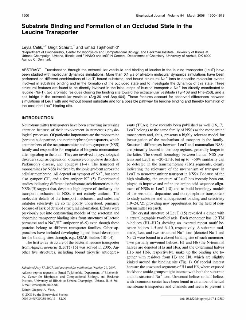
kinked around the binding site (Fig. 1). Of special interest
here are the unwound segments of H1 and H6, where exposed
backbone amide groups might interact with both the substrate
and the structural Na1 ions. Unwound helices or half-helices
with a common center have been found in a number of helical
membrane transporters and channels and seem to present a
doi: 10.1529/biophysj.107.117580
Submitted July 17, 2007, and accepted for publication October 29, 2007.
Address reprint requests to Emad Tajkhorshid, Department of Biochemis-
try, Center for Biophysics and Computational Biology, and Beckman
Institute, University of Illinois at Urbana-Champaign, Urbana, IL 61801.
E-mail: [email protected].
Editor: Gregory A. Voth.
� 2008 by the Biophysical Society
0006-3495/08/03/1600/13 $2.00
1600 Biophysical Journal Volume 94 March 2008 1600–1612

common structural motif of functional importance in this
family of proteins (15,25–29).
Aquifex aeolicus, the bacterium from which LeuT is iso-
lated, is a thermophilic organism, exhibiting a strong binding
affinity for Leu at room temperature (KD � 20 nM) with a
slow transport rate (kcat� 1.2 h�1) (16). The Leu binding site
is adapted to the amphiphilic structure of the substrate; highly
hydrophobic regions are complemented by hydrophilic ones
to optimally match the aliphatic side chain of the substrate
and its charged groups, respectively (Fig. 1). A number of
aromatic residues constitute an aromatic cage around the
binding site with residues Tyr-108 (in H3) and Phe-253 (in
H6) functioning as a ‘‘lid’’ covering the extracellular en-
trance of the binding pocket (Fig. 1). Above the aromatic lid,
residues Arg-30 (in H1) and Asp-404 (in H10), positioned
across the lumen from each other, form a water-mediated salt
bridge. In the initial crystal structure of LeuT (15), this salt
bridge and the above-mentioned aromatic lid close the
binding site to the extracellular lumen, whereas the path to
the intracellular side is closed by ;20 A of tightly packed
FIGURE 1 (A) The crystal structure of LeuT. The 12 TM a-helices in LeuT are bundled together in a unique spiral-like fold. (B) The two unwound helices,
H1 (magenta) and H6 (green), as well as helices H3 (yellow) and H8 (blue) are important for substrate binding and transport. H10 (light blue) and the
extracellular loop 4 (EL4) (light blue) provide residues close to the suspected pathway. The Arg-30–Asp-404 salt bridge is water (red balls) mediated. (C)
Substrate binding in LeuT. Snapshot of Leu (pink) and interacting residues (cyan) together with Na-1 (yellow). Note how residues interacting with the substrate
functional groups are located in the unwound parts of H1 (magenta) and H6 (green). Possible hydrogen bonds to Leu(N) are marked with heteroatom distance.
(D) The substrate, Leu (pink), and Na-1 (yellow sphere) in a surface representation of the LeuT binding site. The front of the figure is cut away to clearly show
the hydrophobic (white) and hydrophilic (magenta) duality of the binding site.
Initial Steps of Transport in LeuT 1601
Biophysical Journal 94(5) 1600–1612
protein residues. In the recently published structures of LeuT
with TCAs bound in the extracellular vestibule, Arg-30
forms a direct (not water-mediated) salt bridge with Asp-404
(16,17). Arg-30, Asp-404, and Asp-401 are the only charged
residues found in the extracellular vestibule of LeuT, sug-
gesting their importance and possible interactions with the
zwitterionic Leu substrate during transport. Indeed, muta-
tional studies on the g-aminobutyric acid and serotonin
transporters have shown that these three residues are crucial
for transport in NSSs (30,31).
The two structural Na1 ions, Na-1 and Na-2, are tightly
coordinated in LeuT; Na-1 is positioned in the substrate bind-
ing site, directly coordinated by the substrate, whereas Na-2
is positioned somewhat farther away. Na-1 is found in an
octahedral (six-liganded) coordination interacting with two
backbone carbonyls, two side-chain amide carbonyls, and
a hydroxyl group besides one of the oxygen atoms of the
carboxylate group of the substrate. Na-2, which has no
contact to the substrate, is bound in a trigonal bipyramidal
(five-liganded) site composed of three backbone carbonyls
and two hydroxyl groups. A similar picture of substrate
and sodium binding is found in GltPh, an aspartate transporter
from Pyrococcus horikishii, and a bacterial homolog of the
mammalian glutamate transporters (25,26). The family of glu-
tamate transporters is functionally related to the NSS family,
although instead of 12, they include only 8 TM a-helices per
monomer. The crystal structure of GltPh showed a trimer
rather than a dimer as found for LeuT. Despite this structural
difference, the substrate binding sites are organized in the
same way and include free backbone carbonyl groups from
unwound or half-helices and two Na1 binding sites in close
proximity of the substrate (15,25,26).
Membrane transporters are generally believed to operate
by a mechanism known as the alternate access model de-
scribed by Jardetzky in 1966 (32). In this model, the binding
of substrate triggers a structural transition between two states,
an inward open and an outward open, thereby inducing the
translocation of the substrate across the membrane. Kinetic
studies indicate the involvement of a third and possibly a
fourth state in the transport facilitated by NSSs (33,34). The
recent structures of LeuT (15–17) and GltPh (25,26) directly
show the involvement of a so-called ‘‘occluded’’ state as a
third structural state in the mechanism: a transporter con-
formation with a closed binding site but where the main parts
of the transporter lumen are still exposed to the extracellular
side.
Although the substrate/ion stoichiometry has been exten-
sively studied for different NSSs (5), the sequence of binding
and release of the ions and the substrate during transport in
NSSs are poorly understood. Another important question
concerns the conformational changes of the transporters
during the transport mechanism. Because of the static nature
of x-ray crystallography, the structure does not provide direct
information about possible conformational changes to other
states. For example, because of lack of a structural model for
the outward open state, it is unknown if major conformational
changes are involved in the formation of the occluded state
after the binding of substrate from the extracellular lumen, or if
primarily rearrangement of amino acid side-chain groups are
involved. However, for the serotonin transporter, it has been
suggested, based on cysteine-scanning experiments, that for-
mation of the occluded state does not involve major confor-
mational changes (35), as the accessibility of the tested
residues did not change much between an apotransporter and
a transporter with the substrate bound.
Complementary to static structures from x-ray crystallog-
raphy, molecular dynamics (MD) simulations have proven
very effective in providing dynamic information on biolog-
ical systems (36–40). Despite time-scale limitations, previ-
ous studies have shown the usefulness of equilibrium MD
simulations in describing physiologically relevant conforma-
tional states of proteins (41–45). Many processes, however,
cannot be described by equilibrium MD simulations. For ex-
ample, translocation of a substrate across the protein usually
follows a longer time scale than sampled in presently acces-
sible unbiased MD simulations. Biased methods, such as
steered MD (SMD (46–48)), have been developed in which
an external force or moving constraint is employed to accel-
erate certain molecular events. The application of the method
to membrane proteins has been demonstrated, e.g., for aqua-
porins (49,50), lactose permease (51), and the outer membrane
transporter BtuB (52). Although the resulting biased trajectories
do not sample perfectly equilibriated structures and pro-
cesses, numerous studies have revealed that careful appli-
cation of such methods can provide relevant and useful
information (46–51,53,54). In modeling of membrane trans-
porters, MD and SMD simulations have been employed to
directly study or deduce individual steps involved in the
transport mechanism (45,52,55).
In this article we present the results of more than 0.1 ms of
simulations of an all-atom membrane-embedded model of
the outward-occluded conformation of the LeuT dimer. MD
simulations have been conducted with different setups, sys-
tematically exploring the effect of the presence and absence
of substrate and sodium ions on LeuT dynamics and disen-
tangling the binding of Leu in LeuT. Furthermore, pulling
simulations, employing different force profiles and pulling
velocities to either Leu alone or Leu and Na-1 together, were
performed to study the unbinding of the substrate from the
binding site and to study the formation of the open state from
the occluded state, thereby revealing the differences between
an outward-open and the outward-occluded state as well as
proposing a likely pathway for Leu binding to LeuT. This is
the first such study of the NSS transport mechanism and can
thus be utilized in further studies of monoamine transporters.
Results show a very tight binding of Leu in the occluded
binding site, highly influenced by the presence of free po-
lar groups in the unwound backbones of H1 and H6. We
have, furthermore, identified a possible binding pathway for
Leu that has been well reproduced in pulling simulations
1602 Celik et al.
Biophysical Journal 94(5) 1600–1612

employing different pulling schemes. This pathway espe-
cially involves the aromatic residues closing the binding site
and a conserved salt bridge in the extracellular vestibule of
LeuT.
METHODS
Model building
The 1.65 A resolution x-ray structure of LeuT (15) was obtained from the
Protein Data Bank (56) (accession code 2A65). The structure includes the
protein residues 5–132 and 135–515 from one of the monomers in the dimer.
Furthermore, a substrate leucine (Leu), two sodium and one chloride ions,
five detergent molecules, and 210 water molecules were included in the PDB
file. The detergent molecules and the chloride ion were removed. Two
missing residues, Asn-133 and Ala-134 located in extracellular loop 2 (EL2),
were built as follows: first two glycine residues were introduced to complete
the backbone, the residues were then mutated to alanine, and finally, the
asparagine side chain was built for residue 133. Residues 132–135 were
minimized after each step of the modeling process. Coordinates for missing
atoms of side chains and hydrogen atoms were constructed with the psfgen
plugin in VMD (57) employing the CHARMM32 topology for proteins and
lipids (58–61). No information is available as to whether LeuT is functional
in a monomeric or a dimeric form, but it was crystallized as a dimer (15).
Other studies have shown that the biogenic amine transporters are functional
as dimers (62,63). The dimer was constructed by applying a crystallographic
twofold symmetry and space group using program O (64) to the monomer
found in the pdb-file. As a further incentive to model LeuT as a dimer, we
observed that the expected usual interactions between the lipid head groups
and charged and polar residues would induce a deep depression of the lipid
bilayer on one side of the transporter if it were modeled as a monomer. The
inclusion of the dimer in the model further allowed us to double the statistics,
as two trajectories, one from each monomer, are produced for every setup
employed. As described below, to further improve the statistics, some SMD
simulations were performed twice. Reproducibility of the observed molec-
ular events in repeated simulations will provide additional reliability for the
obtained results.
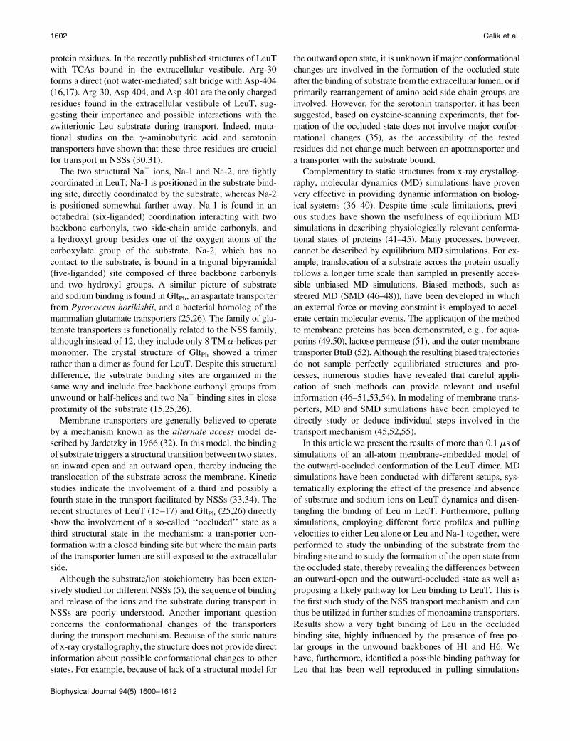
A preequilibrated and solvated 140 3 125 A2 membrane bilayer con-
sisting of palmitoyl-oleoyl phosphatidylethanolamine lipids was created
using the membrane plugin in VMD (57). The LeuT dimer was embedded in
the bilayer by aligning the lipid head groups with a ring of charged and
aromatic residues at the surface of the dimer. The lipid bilayer was large
enough to provide at least 15 A of lipid padding around the dimer along any
direction. Layers of water 20 A thick were added to the two sides of the
membrane to fully solvate the lipid-protein system. The system was then
neutralized with sodium and chloride ions to a physiological concentration
of 0.2 M. This resulted in a total atom count of 142,405, with dimensions of
130 3 113 3 90 A3 after equilibration (Fig. 2).
The Leu substrate is modeled as a zwitterion, charged on both the amino
and carboxylate groups. The two charged groups will be referred to as the
ammonium and the carboxylate groups, respectively.
Assignment of protonation states
The titration states of ionizable amino acids (aspartate, glutamate, lysine,
arginine, histidine, and tyrosine) were assigned based on a pKa calculation
using the web application H11 from Virginia Technical University (65).
Glutamate residues 112 (in H3), 287 (in H7), and 419 (in H10) exhibited high
pKa values of 10.2, 13.9, and 23.1, respectively, and were thus modeled as
neutral during the simulations. The three affected residues are all located
facing other acidic residues (either an aspartate or a glutamate), presenting an
opportunity for them to interact with each other. All other glutamate residues
were modeled as charged. Of the seven histidine residues present in LeuT,
His-74 and His-391 had high pKa values (13.2 and 7.3, respectively) and
were thus modeled in their protonated form. His-7, His-377, and His-480
were modeled as neutral with the proton on Ne, whereas His-510 was
modeled with a proton on Nd to maximize favorable interactions with the
local microenvironment. All aspartate, arginine, and lysine residues were
modeled as charged, and tyrosines as neutral.
Simulation details
All simulations were performed using the CHARMM32 force field for
proteins and lipids (58–60) including the CMAP corrections (61) with the
MD program NAMD 2.6 (66,67). After an initial Conjugate Gradient min-
imization for 500 iterations, lipid tails were melted in a 500-ps NVT simu-
lation at 310 K in which all atoms but the lipid tails were held fixed. The
system was then equilibrated for a 2-ns constraint-free simulation with a time
step of 1 fs. Equilibration was performed under periodic boundary conditions
in the NPT ensemble employing the particle mesh Ewald algorithm (68) for
calculation of long-range electrostatics. Constant temperature was achieved
by employing Langevin dynamics with a damping constant of 0.1 ps�1 for
melting of lipid tails and 5 ps�1 for the equilibration phase. The Langevin
Piston method (69) was employed to maintain a constant pressure of 1 atm
with a piston period of 100 fs and a piston decay of 50 fs. The van der Waals
interactions were accounted for to a cut-off distance of 12 A and gradually
dampened by use of a switching function from 10 A.
Production dynamics (MD and SMD) was performed in the NPT en-
semble with a Langevin damping constant of 0.5 ps�1 and imposition of a
constant area to the membrane patch. Coordinates for analysis were saved
every picosecond. Four equilibrium MD simulations were set up to study the
dynamics and stability of LeuT with or without substrate/sodium ions; setup
1 is the holo system represented by a dimer of LeuT; setup 2 is the apo
transporter devoid of both substrate and the sodium ions; setup 3 is the apo
transporter including sodium ions; and setup 4 included substrate but no
sodium ions. Together these four simulations were used to study the dy-
namics of the substrate in the binding pocket as well as to deduce information
about the sequence of sodium and substrate binding to the transporter.
In pulling simulations, a moving constraint is applied to the center of mass
(COM) of a group of atoms. The chosen group of atoms will thus be forced to
generally move along a specified direction while still being able to move
FIGURE 2 The simulated system from a side view. The two transporter
monomers are depicted in green and magenta cartoons, lipid tails in cyan
lines, lipid heads in CPK, water in transparent, and ions as van der Waals
spheres in yellow (Na1) and cyan (Cl�). The limited interaction between the
two monomers is through H11 and H12, which are not directly involved in
substrate binding or transport, suggesting that the two monomers function
independently.
Initial Steps of Transport in LeuT 1603
Biophysical Journal 94(5) 1600–1612

freely along all other degrees of freedom (46–48). Constant-velocity SMD
simulations were performed using the tclforces command in NAMD 2.6
(66,67). The force was applied to the COM of different groups of atoms
(Table 1). The force constant employed was 500 pN/A following the stiff
spring protocol according to Park et al. (70) while different pulling velocities
were systematically investigated (see Table 1). The system was oriented such
that the applied force was along the membrane normal, i.e., along the putative
transport pathway. To prevent net translation of the system that might arise as
a result of the pulling force acting on the substrate, lipid phosphorus atoms as
well as Ca atoms of residues Phe-51, Ile-204, and Phe-496 were constrained
with a harmonic potential during the pulling simulations. In total, 14 pulling
simulations were conducted to study substrate unbinding from LeuT. All
performed simulations are summarized in Table 1. In setups 5–10 Leu was
pulled out of the binding site at four different velocities, whereas in setups
11–18 both the substrate Leu and Na-1 were pulled simultaneously. As in-
dicated in Table 1 several of the SMD simulations were repeated to improve
the statistics at given velocities. In general, faster SMD simulations were
initially used to explore the process and to identify events that might be of
interest. This was then followed by increasingly slower simulations (as much
as computational resources allowed) to verify the reproducibility of major
events at slower pulling speeds.
Data analysis
Analysis of the computed trajectories was performed with VMD 1.8.6 (57)
and the included Tcl-scripting facility. All molecular images were rendered
by VMD. The root mean-square deviation (RMSD) of protein Ca atoms in
each simulation was calculated with respect to the initial minimized struc-
ture. Interaction energies were calculated via the NAMDEnergy plugin of
VMD (57).
RESULTS AND DISCUSSION
Setups 1–4 were first made to study equilibrium dynamics of
LeuT in the presence and absence of the Leu substrate and the
two bound sodium ions. In addition to providing dynamic
information about the structural stability of LeuT, these
simulations might also shed light on the question of the se-
quence of binding of substrate and sodium ions to the trans-
porter. From these four setups the specific binding of the
substrate and the two sodium ions is studied and electrostatic
interactions and the importance of the unwound helices are
investigated. The binding of Leu to LeuT, a key step in the
transport mechanism, is then studied by pulling simulations
in setups 5–18 of the reverse unbinding event. Different pull-
ing velocities are applied to either the COM of the substrate
(setups 5–10) or to the COM of Leu and Na-1 together (setups
11–18), inducing substrate unbinding toward the extracellular
lumen (see Table 1). Two different classes of setups, namely
equilibrium MD simulations in 1–4 and pulling simulations in
setups 5–18, are included to examine if unbinding of Leu is
dependent on a simultaneous transport of Na-1 or if the two are
independent of each other. Although the pulling speed in SMD
simulations does not allow for the principle of microscopic
reversibility to exactly apply, we expect to capture major steps
and molecular events involved in initial association and
binding of the substrate from the periplasm by simulating the
reverse process, i.e., unbinding of the substrate from the
binding pocket. In the following, specific atoms of individual
amino acids are referred to as, e.g., Ala-22(O), Gly-26(N), and
Ser-256(OH), respectively, for backbone amide O and N
atoms and a side-chain hydroxyl group.
Protein stability and dynamics of thebinding pocket
No major conformational changes of the protein backbone
were observed throughout the performed MD and SMD
simulations. RMSD values for LeuT Ca atoms were found to
be between 1.5 A and 2.2 A for all modeled systems, and the
overall structure of the transporter is conserved throughout
(see Supplementary Material for average RMSD values for
all simulations). Of particular interest are the unwound heli-
ces, which, despite their apparent structural flexibility,
maintain their initial conformation and exhibit thermal fluc-
tuations comparable to other parts of the protein and to the
experimentally found B-factors in the x-ray diffraction ex-
periment (15). Although no global protein conformational
change is evident, we do observe a partial opening of the
binding site in some of the MD simulations. This opening is
furnished merely by rotation of the side chain (specifically
x1 and x2 dihedral angles) of Tyr-108 and Phe-253, the two
residues that form the aromatic lid of the binding site (see
Figs. 3 and 4). Interestingly, there seems to be a correlation
between this opening and the occupancy of the binding
pocket; when Leu is present in the binding site (setup 1),
these dihedral angles do not exhibit large fluctuations. Re-
moval of either the substrate or the sodium ions, however,
results in an increased flexibility of these side chains and, as a
result, a partial opening of the binding site. In the SMD
simulations we furthermore observe that release of Leu from
TABLE 1 Simulation setups reported in this study
Setup v/A/ns* t/nsy Leu Na-1 Na-2 Pulled group
1 N/A 10.0 1 1 1 N/A
2 N/A 10.0 – – – N/A
3 N/A 10.0 – 1 1 N/A
4 N/A 5.0 1 – – N/A
5 1.0 10.0 1 1 1 Leu
6 2.5 5.0 1 1 1 Leu
7 4.0 4.0 1 1 1 Leu
8 4.0 3.7 1 1 1 Leu
9 10.0 2.0 1 1 1 Leu
10 10.0 2.0 1 1 1 Leu
11 1.0 13.4 1 1 1 Leu / Na-1
12 2.5 6.0 1 1 1 Leu / Na-1
13 4.0 4.0 1 1 1 Leu / Na-1
14 4.0 4.5 1 1 1 Leu / Na-1
15 6.0 3.0 1 1 1 Leu / Na-1
16 8.0 3.0 1 1 1 Leu / Na-1
17 10.0 5.0 1 1 1 Leu / Na-1
18 10.0 3.0 1 1 1 Leu / Na-1
In the analysis the nomenclature 1A will refer to monomer A in setup 1.
Note that some of the simulation setups are repeated under the same
conditions to improve sampling.
*Pulling velocity in SMD simulations.ySimulation time.
1604 Celik et al.
Biophysical Journal 94(5) 1600–1612

the binding site only requires the aromatic lid to open, sug-
gesting, along with the results of the MD simulations in
setups 1 and 2, that no major conformational changes of the
protein backbone are necessary for the initial binding of leu-
cine in LeuT and formation of the occluded state. Because only
side-chain movement is necessary for opening of the binding
site, a close similarity between the outward open and occluded
states is expected, a conclusion that is in line with what has
been also hypothesized for the serotonin transporter (35).
In setup 1 Leu is tightly bound to the LeuT binding site
with five possible hydrogen bonds (not all present at the same
time) to its ammonium group (Asn-21(O), Ala-22(O), Phe-
253(O), Thr-254(O), and Ser-256(OH)) and two to its car-
boxylate group (Gly-26(N) and Tyr-108(OH)) as well as a
salt bridge between its carboxylate group and Na-1 (Fig. 1).
We also observe interactions between the Leu carboxylate
group and backbone amide groups of residues Gly-24 and
Leu-25 in the unwound part of H1. These interactions do not
adopt a geometry that can be classified as hydrogen bonds,
the amide N-H to Leu O angles are far from linear, but they do
show highly favorable electrostatic interactions. The residues
involved in hydrogen bonds to Leu via polar backbone
groups are all found in the termini of the broken helices, H1
and H6, located near the center of the TM segment of LeuT,
suggesting significance of these in substrate binding. Tyr-108
and Ser-256, which interact with the substrate through their
polar side chains, are positioned in the middle of H3 and H6b,
respectively. Average distances for direct interactions be-
tween Leu and the protein calculated from the 10-ns simu-
lation of setup 1 are listed in Table 2. The observed binding of
Leu in LeuT is similar to that observed for dopamine in an
MD simulation of dopamine binding in a homology model of
the dopamine transporter based on the structure of LeuT (23).
Interaction energies between the substrate and various
parts of the protein and the bound sodium ions are presented
in Table 3. The interaction energy between the substrate
and LeuT is primarily electrostatic in nature with EElect ��100 kcal/mol, of which ;75% is from interactions to H1
and H6. Helix H3 also has a large contribution, whereas a
small unfavorable interaction energy is calculated between
H8 and Leu. An additional EElect � �40 kcal/mol can be
found for interactions between Leu and Na-1. The computed
high electrostatic energy between Leu and the protein is in
accord with the experimentally measured very low rate of
transport of leucine through LeuT (16). Furthermore, it can
be speculated that the electrostatic repulsion between H8 and
Leu can trigger major conformational changes needed for
transitioning between the outward-facing and inward-facing
states of LeuT. A similar reasoning was recently presented
for the serotonin and dopamine transporters, where H8 is
speculated to initiate the formation of the inward-facing form
through interaction with the dopamine and serotonin sub-
strates, respectively (71).
Unwound helices present an interesting structural feature
in LeuT. It can be expected that a bound substrate in a binding
site composed of unwound helices can be, at least partly,
stabilized by the helical macrodipoles. Furthermore, the
backbone peptide groups of the unwound segments can di-
rectly participate in hydrogen bonding to the substrate. To
investigate the contribution of these effects in stabilization of
the substrate in the binding pocket, we decomposed the com-
puted electrostatic interaction energies between Leu and LeuT
into contributions from individual residues. Very high elec-
trostatic interaction energies are detected, showing the sig-
nificance of direct interactions between the charged groups of
the substrate and the exposed polar backbone groups of the
TABLE 2 Direct interactions between Leu and the binding site
of LeuT
Interaction Monomer A Monomer B Crystal
Leu(OT1)–Na-1 2.25 6 0.11 2.24 6 0.10 2.5
Leu(OT1)–Gly-24(N) 3.37 6 0.23 3.82 6 0.21 3.8
Leu(OT1)–Leu-25(N) 4.49 6 0.22 4.55 6 0.21 4.6
Leu(OT1)–Gly-26(N) 3.55 6 0.20 3.60 6 0.19 3.3
Leu(OT2)–Gly-24(N) 3.93 6 0.21 3.96 6 0.22 3.8
Leu(OT2)–Leu-25(N) 3.07 6 0.16 3.08 6 0.15 3.1
Leu(OT2)–Gly-26(N) 2.85 6 0.12 2.83 6 0.12 2.7
Leu(OT2)–Tyr-108(OH) 2.70 6 0.12 2.69 6 0.12 2.7
Leu(N)–Asn-21(O) 3.58 6 0.24 3.44 6 0.21 3.6
Leu(N)–Ala-22(O) 3.21 6 0.25 3.29 6 0.26 2.9
Leu(N)–Phe-253(O) 2.81 6 0.13 2.80 6 0.12 2.9
Leu(N)–Thr-254(O) 2.95 6 0.16 2.96 6 0.17 3.1
Leu(N)–Ser-256(OH) 2.81 6 0.10 2.81 6 0.10 2.8
Average distances and standard deviations from the equilibrium simulation
of setup 1 are given in A. Distances observed in the crystal structure are
included for comparison.
TABLE 3 Average electrostatic interaction energies and
standard deviations between the substrate and various parts
of LeuT are calculated and compared for the 10 ns simulation
of setup 1
Leu to: Monomer A, kcal/mol Monomer B, kcal/mol
Full monomer �99.6 6 7.9 �98.4 6 8.0
H1 �55.8 6 5.9 �53.8 6 5.8
H3 �13.6 6 2.6 �14.1 6 2.7
H6 �42.6 6 4.3 �42.5 6 4.4
H8 5.4 6 1.6 5.1 6 1.4
Na-1 �36.9 6 5.8 �38.1 6 5.7
Na-2 �2.4 6 2.3 �1.8 6 2.3
Asn-21 (H1) �10.5 6 1.6 �10.6 6 1.7
Ala-22 (H1) 1.8 6 2.5 2.6 6 2.3
Val-23 (H1) �4.1 6 0.6 �4.0 6 0.6
Gly-24 (H1) �8.5 6 1.2 �8.3 6 1.2
Leu-25 (H1) �4.2 6 1.6 �3.8 6 1.4
Gly-26 (H1) �15.0 6 1.5 �15.1 6 1.5
Arg-30 (H1) �7.6 6 1.1 �7.2 6 1.0
Tyr-108 (H3) �13.2 6 2.6 �13.5 6 2.6
Phe-253 (H6) �18.1 6 2.0 �18.2 6 2.0
Thr-254 (H6) �4.7 6 2.5 �4.7 6 2.4
Leu-255(H6) 0.0 6 0.6 0.0 6 0.6
Ser-256 (H6) �14.1 6 1.9 �14.1 6 1.9
Ala-22 and Thr-254 are also coordinated to Na-1 and have smaller or
unfavorable contributions to the binding of Leu.
Initial Steps of Transport in LeuT 1605
Biophysical Journal 94(5) 1600–1612

unwound helices in the binding site. Further analysis of the
results, however, also shows that the total electrostatic inter-
action energy between each helix and Leu is slightly higher
than the sum of interaction energies of residues directly in-
teracting with Leu, indicating participation of other elements
of the unwound helices in stabilization of the substrate in the
occluded binding site (see Table 3). Therefore, we conclude
that, although the binding energy seems to be primarily caused
by the numerous direct hydrogen bonds to charged groups of
Leu, the macrodipole of the unwound helices may also play a
role in stabilizing the bound substrate.
Coordination of the two structural sodium ions during MD
simulations likewise presents a picture of tight binding. For
both ions average coordination distances are within 0.1 A of
those observed in the crystal structure (average distances and
standard deviations for sodium coordination in setups 1 and 3
are tabulated in the Supplementary Material). The presence
(setup 1) or absence (setup 3) of the substrate does not affect
the Na-1 coordination significantly; indeed, in setup 3 (in
which Leu is removed), the missing substrate coordination is
taken up by a water molecule entering from the bulk, thereby
maintaining the octahedral coordination. Before water pen-
etrates into the binding site, the five interactions to LeuT
remain in a square pyramidal coordination, similar to the one
found in setup 1, leaving the sixth coordination site unoc-
cupied. This observation, along with the strong binding of
the Na1 ion in the Na-1 binding site as found in our pulling
simulations, might suggest that the binding of Na-1 to the
transporter precedes substrate association. This Na1 ion most
likely enters an empty binding pocket (substrate-free) with a
few water molecules comprising its first hydration shell,
though some of these water molecules will be gradually re-
placed by protein side chains as the ion settles in its binding
site. A complete dehydration would occur when the substrate
binds in the binding pocket. Although our results can be in-
terpreted consistently with a model in which Na-1 binding
precedes that of the substrate, we note that the possibility of
simultaneous binding of the sodium ion and the substrate
should also be kept in mind.
No significant electrostatic interaction, either direct or long
range, can be identified between the substrate and Na-2. It
might be inferred that the binding of Leu is independent of
Na-2, at least during the initial association of the substrate to
the transporter, and that Na-2 might not be involved in the
transport mechanism but merely have a structural role. It
should be kept in mind, however, that subsequent steps in the
transport mechanism might involve major protein confor-
mational changes and substrate translocation that would
bring the substrate and Na-2 close to each other; such effects,
however, can only be speculated at this time.
After removal of the two sodium ions (setup 4), water from
the solvation box penetrates into the Na-1 binding site and
coordinates the Leu ammonium group. At the same time the
aromatic lid opens slightly with a rotation of the two aromatic
rings, allowing additional water molecules to interact with
the Leu carboxylate group. In the absence of sodium ions, the
substrate binding site shows larger thermal fluctuations than
when sodium ions are present. For example, in setup 4A,where these fluctuations are particularly large, hydrogen
bonds between the Leu ammonium group and Asn-21(O) and
Ser-256(OH) are broken, and the ammonium group is posi-
tioned closer to the water molecule located in the Na-1 site.
Meanwhile, a new interaction is established between Ser-
355(OH), located in H8, and the Leu carboxylate group.
Together, these events result in a rotation of the substrate side
chain from a configuration approximately parallel to the
membrane plane to an almost perpendicular one, with the
charged carboxylate group pointing toward the extracellular
vestibule and the side chain toward the cytoplasm. This new
conformation is observed for more than 2 ns and may indicate
a destabilization of substrate binding in the absence of
Na-1, providing further support for the key role of Na-1 in
Leu binding.
Pathway for substrate unbinding in LeuT
A total of 69 ns of pulling simulations in 14 setups (setups
5–18) were conducted to investigate the unbinding of sub-
strate from the binding pocket (see Table 1). That gives a total
of 28 unbinding trajectories because of the dimeric structure
of the system.
The majority, 18, of the trajectories resulted in a pathway
that appears physiologically relevant and is consistent with
data from unbiased simulations of setups 1–4. This pathway
is reproducible in simulations with pulling velocities ranging
from 1 to 10 A/ns, strongly suggesting that the observed
pathway and the mechanism represent highly relevant fea-
tures of the natural unbinding process. During the unbinding
process interactions to the binding site residues are broken
one by one as Leu passes the aromatic lid, but new interac-
tions are formed along the extracellular vestibule, leading to a
specific path where Leu is passed along from one set of
specific interactions to another. Throughout this identified
translocation mechanism Na-1 stays in its original binding
pocket. It should be noted that the large degree of repro-
ducibility is remarkable for the pulling simulations, thereby
indicating that the collected trajectories are of biological
relevance. Because the sampled pathway was observed to be
largely independent of the pulling velocity applied on either
Leu or Leu/Na-1, it is very likely that even slower pulling will
result in a similar trajectory.
In another five of the SMD trajectories, where either a
higher pulling velocity is employed or when both Leu and
Na-1 are pulled, Na-1 shows a different behavior, namely, it
moves along with the substrate. However, this does not affect
the sampled pathway or the translocation steps involved in
Leu unbinding; the only difference is that Na-1 travels tightly
coordinated to Leu along the same pathway. This gives a total
of 23 of 28 trajectories sampling the same unbinding path-
way for Leu. We note that in several trajectories, although
1606 Celik et al.
Biophysical Journal 94(5) 1600–1612
Na-1 was being pulled along with Leu, Na-1 did not leave its
binding site despite the applied force, indicating a strong
tendency of Na-1 to stay bound to the protein even after Leu
has left the binding site.
A rare event, which was observed only in the last five fast-
pulling trajectories, was translocation of Na-1 and Leu on
opposite sides of helices H1 and H6. This pathway exposes
Na-1 to a highly hydrophobic environment and is concluded
to be an artifact of the fast-pulling protocols employed in
these few simulations.
As in the equilibrium MD simulations, Na-2 is highly
stable in all SMD simulations, further demonstrating that,
most likely, there is no connection between the bound Na-2
and the initial translocation of Leu in the extracellular lumen
of LeuT (average distances and standard deviations for Na-2
coordination during the SMD simulations are tabulated in the
Supplementary Material). This observation is consistent with
the notion that the nature of Na-2 is mostly structural, and
Na-1 is the only sodium ion directly involved in initial asso-
ciation of Leu to LeuT.
Transfer pathway for substrate unbinding
Under the assumptions that binding and unbinding are reverse
processes and, as discussed above, that the nonequilibrium
pulling simulations very closely sample the equilibrium
pathway, a binding pathway for Leu to LeuT can be extracted
from the reverse unbinding pathway found in SMD simula-
tions. The unbinding pathway of Leu from LeuT, as observed
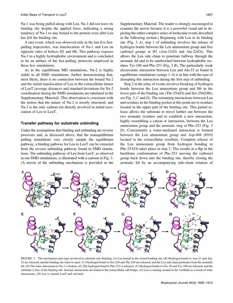
in our SMD simulations, is illustrated with a cartoon in Fig. 3.
(A movie of the unbinding mechanism is provided in the
Supplementary Material. The reader is strongly encouraged to
examine the movie because it is a powerful visual aid in de-
picting the rather complex series of molecular events described
in the following section.) Beginning with Leu in its binding
site (Fig. 3 A), step 1 of unbinding involves the release of
hydrogen bonds between the Leu ammonium group and free
carbonyl groups in H1 (Asn-21(O) and Ala-22(O)). This
allows the Leu side chain to penetrate halfway through the
aromatic lid and to be sandwiched between hydrophobic res-
idues Tyr-108 and Phe-253 (Fig. 3 B). The particularly weak
electrostatic interaction between Leu and Ala-22 as found in
equilibrium simulations (setups 1–4) is in line with the ease of
disrupting this interaction during the first step of unbinding.
Step 2 in the relay of events involves breaking of hydrogen
bonds between the Leu ammonium group and H6 in the
lower part of the binding site (Thr-254(O) and Ser-256(OH);
see Fig. 3, C and D). The remaining interactions between Leu
and residues in the binding pocket at this point are to residues
located in the upper part of the binding site. This partial re-
lease allows the substrate to travel farther out between the
two aromatic residues and to establish a new interaction,
highly resembling a cation–p interaction, between the Leu
ammonium group and the aromatic ring of Phe-253 (Fig. 3
D). Concurrently a water-mediated interaction is formed
between the Leu ammonium group and Asp-404 (H10)
located in the extracellular vestibule. Complete release of
the Leu ammonium group from hydrogen bonding to
Phe-253(O) takes place in step 3. This results in a flip in the
backbone conformation of Phe-253 moving the carbonyl
group back down into the binding site, thereby closing the
aromatic lid by an accompanying side-chain rotation of
FIGURE 3 The mechanism and steps involved in substrate (un-)binding. (A) Leu bound in the closed binding site. (B) Hydrogen bonds to Asn-21 and Ala-
22 are released, and the binding site starts to open. (C) Hydrogen bonds to Ser-256 and Thr-254 are released, and the Leu side chain protrudes from the aromatic
lid. (D) The ionic interaction to Na-1 is broken. (E) The hydrogen bond to Phe-253 is released. (F) Hydrogen bonds to Gly-26 and Tyr-108 are released, and the
substrate is free of the binding site. Instead, interactions are found to the extracellular salt bridge. (G) Leu is turning around in the vestibule as a result of ionic
interactions. (H) Leu is outside LeuT and solvated.
Initial Steps of Transport in LeuT 1607
Biophysical Journal 94(5) 1600–1612

Phe-235 (Fig. 3 E). Simultaneously a direct salt bridge is
formed between Leu(N) and Asp-404.
Na-1 is affected by the translocation of the substrate from
the binding pocket. During the initial phase of the induced
unbinding of the substrate, Na-1 is partially, but only tran-
siently, displaced from its binding site (Fig. 3 C). The dis-
placement seems to coincide with the formation of a strong
double interaction with both carboxylate oxygen atoms of the
substrate, which transiently forms along the dislocation of the
substrate in the binding pocket. However, because of an even
stronger interaction of Na-1 with its binding site, it soon falls
back into its original binding site (EElect � �160 kcal/mol to
LeuT, Fig. 3 D where the sodium ion is located behind the
unwound part of H1). In some of the simulations the inter-
action between Na-1 and the substrate is broken during step
2, in others at a later time, mostly just before or during step 3.
The disruption of this interaction coincides with a large drop
in the applied force; for setup 7 this appears after 1.5 ns, when
the COM of Leu has moved ,5 A (see figures in the Sup-
plementary Material), indicating that Na-1 contributes a
major interaction energy to the binding of the substrate. A
possible scenario could be that the binding of Na-1 precedes
that of Leu. In this case, a bound Na-1 will contribute to
electrostatic attraction of the substrate toward the binding
pocket during its translocation from the vestibule into the
binding pocket, and in particular during its final orientation in
the binding pocket.
After step 3 only one of the Leu carboxylate oxygen atoms
remains coordinated to the binding site through hydrogen
bonds to Gly-26(N) and Tyr-108(OH) in the aromatic lid (Fig.
3 E). These bonds are subsequently broken in step 4, allowing
Leu to slide between the two salt-bridge-forming residues in
the extracellular vestibule, Arg-30 and Asp-404 (Fig. 3 F).
During the initial stages of unbinding, the Leu side chain
moves ahead of the charged backbone. This is primarily
because of the strong electrostatic interaction between the
residues in the binding pocket and the Leu backbone, which
makes it the last part to leave the binding site. Later along the
permeation pathway, the Leu side chain is caught by aliphatic
and aromatic residues, Val-29, Leu-33, Ala-319, and Phe-
320, in the extracellular vestibule. Additional water-mediated
interactions to Asp-401 pull the substrate out of the extra-
cellular vestibule in an orientation where the charged back-
bone of Leu is first (Fig. 3, C–H). After the release of the side
chain (step 5), the substrate is almost fully solvated in the
entrance of the extracellular lumen of LeuT (Fig. 3 H).
The maximum buildup force during the SMD simulations
is ;1500 pN with a single peak developed during the first
three steps. The force decreases slightly after step 2 but does
not drop significantly until after the interaction between the
substrate and Na-1 is broken. A second force peak (of ;800
pN) is found around step 4, although no force is needed to
relinquish the interactions to the salt-bridge residues. Graphs
of a representative force profile are available in the Supple-
mentary Material.
After release of the substrate, up to three water molecules
enter the binding site to replace the interactions of the sub-
strate head groups with the protein and Na-1. This is similar
to what is observed in the equilibrium simulation of the apo
system (setup 3). This observation is a further indicative of
the fact that the SMD simulations are highly relevant, as they
converge toward molecular assemblies that are highly similar
to those seen in the equilibrium simulations.
Formation of the occluded state
The crystal structure of LeuT is suggested to represent the
occluded state of the transporter, in which the aromatic lid
separates the substrate from the extracellular vestibule lead-
ing to the extracellular lumen (15). During the simulation of
setup 1 this lid remains tightly closed (average distances
between closest carbon atoms of ;4.3 A in both monomers).
This is also found in one of the monomers in setup 2 (2B),
whereas monomer 2A and setups 3 and 4 show an average
distance of ;5–7 A, thus showing a partially opened lid. The
opening is facilitated by rotation of the two aromatic side
chains of the lid. In setup 1 the dihedral angles of these side
chains are stable at either ;90� (both residues, x2) or ;200�(both residues, x1). In setups 2, 3, and 4, a higher degree of
flexibility is observed, and either one of the residues changes
conformation into a different rotamer state to stabilize the lid
in its new, partly opened, conformation. The partly open
binding site is also observed in the SMD simulations after
release of the Leu(N)–Phe-253(O) interaction, resulting in a
partially open lid, similar to the one found in setup 3, also
with a binding site with both Na1 ions, but no Leu present.
Arg-30, Asp-401, and Asp-404 are the only charged resi-
dues found in the extracellular vestibule of LeuT and must
therefore be responsible for the electrostatic properties of the
presumed binding pathway. Arg-30 and Asp-404 have been
shown to be vital for transport in NSSs (15,30,31). The ob-
served water-mediated contact of these residues in the crystal
structure of LeuT is preserved during our equilibrium sim-
ulation (setup 1). These two residues were proposed to
constitute a second gate, which would be closed through
water-mediated interactions once the substrate is bound to the
binding site (15). The water-mediated interactions are also
found in the beginning of the SMD simulations, i.e., while the
substrate is still in the binding pocket, and even in setup 4,
where the sodium ions are excluded from the simulation.
Removal of the substrate from the binding site, both in setup
3 and during later stages of the SMD simulations (when the
substrate has left the binding site), induces the formation of a
direct, bifurcated, salt bridge between Arg-30 and Asp-404
similar to the one found in structures of LeuT with bound
TCA (16,17), the two situations are depicted in Fig. 4, A and
B. Apart from the directly bound and water-bridged con-
figurations, SMD simulations revealed a third mode of in-
teraction between Arg-30 and Asp-404 arising during the
substrate translocation, in which Leu is positioned
1608 Celik et al.
Biophysical Journal 94(5) 1600–1612

between the two residues while forming salt bridges to both
(Fig. 3 F).
Because of the strong interaction between Na-1 and the
substrate in its bound form, as observed in our equilibrium
and SMD simulations (above), Na-1 is most likely needed to
mediate the displacement of Leu from the salt bridge site
(between Arg-30 and Asp-404) into the binding site. Com-
bined with the results from MD simulations of setup 4, we
propose that both Leu and Na-1 need to be present in the
binding site for formation and stabilization of the occluded
state. Furthermore, in addition to playing a gating role, we
propose that Arg-30 and Asp-404 facilitate the substrate
binding by coordinating the substrate in the extracellular
vestibule of LeuT, then positioning and orienting it properly
for interactions to Na-1 and residues located in the binding
site. It has to be noted that, despite the computational effort of
this study, the limited time scale of MD and SMD simulations
in general does not allow one to exclude the possibility of
large protein conformational changes being involved in the
formation of the occluded state. However, the consistency
found between the results of our simulations and the con-
clusions reached in various studies of the serotonin trans-
porter (35) provides a strong support for the significance of
the observed phenomena in this study.
Implications for binding and transport of leucinein LeuT
Based on the crystal structures of LeuT and GltPh, Gouaux and
co-workers (15,25,26) suggested a transport mechanism in-
cluding a minimum of three states: outward open, outward
occluded, and inward open states. Our simulations of the apo
system with and without sodium ions (setups 2 and 3) indicate
that LeuT does not necessarily undergo large conformational
changes to open toward the extracellular cavity; instead, a few
water molecules might penetrate into the binding site to
occupy available coordination sites in the transporter while
the hydrophobic part of the binding pocket remains empty.
Residues Arg-30 and Asp-404 have been suggested to be part
of a gating mechanism in the NSS family (30,31). In the
crystal structure of the outward occluded conformation they
interact through two water molecules, whereas in the simu-
lation of setup 3 (sodium ions present) they become directly
connected, a configuration also found after substrate release in
our pulling simulations. Together with the observed interac-
tion with Asp-401, this suggests these three charged residues
play a role in attracting the substrate, via electrostatic inter-
actions, into the extracellular vestibule, where it is then guided
into the binding site. Asp-401 is located close to the surface of
the transporter. Once an interaction between Leu and Asp-401
is established, the substrate can readily form an additional
hydrogen bond to Asp-404. From this point, the substrate can
then establish an interaction to Arg-30.
After insertion between Arg-30 and Asp-404, the substrate
can be guided into the binding site by establishing stepwise
interactions with various parts of the binding pathway, as
observed in the reverse from the SMD simulations. Electro-
static attraction between Leu and Na-1 (�40 kcal/mol to�60
kcal/mol) are more favorable than those to Arg-30 (;�25
kcal/mol) and would initially draw the carboxylate group
toward the binding site, where interactions to Gly-26(N) and
Tyr-108(OH) could be formed on the way. As a result, the
interaction from Leu(N) to Asp-404 would be reduced and
formation of a hydrogen bond to Phe-253(O) be more favor-
able, ensuring a rotation of its aromatic ring and thus opening
of the ‘‘lid’’ to allow for full binding of the substrate in a
highly orchestrated mechanism.
FIGURE 4 Water-mediated (A) and
direct (B) interaction in the salt bridge
between Arg-30 and Asp-404 in the
extracellular vestibule. The aromatic lid
on the extracellular side of the binding
site can be either closed (C) or open
(D), depending on the presence/absence
of Leu. The open lid can also be seen
during SMD simulation (E).
Initial Steps of Transport in LeuT 1609
Biophysical Journal 94(5) 1600–1612

The initial stabilizing interactions between Leu and the
extracellular vestibule are also important in desolvation of the
highly charged substrate and likely in orienting the substrate
for an optimal insertion into the binding pocket. These two
factors may explain the experimentally observed require-
ments for the three charged residues for transport (15,30,31).
The binding of the substrate and the two sodium ions re-
veals a clear role for the unwound helices, that is, to provide
free carbonyl groups for binding of the transported entities.
An additional role can be found for H6. The large loop in the
middle of H6 interacts with the hydrophobic side chain of the
substrate. Furthermore, the interaction between Leu(N) and
Phe-253 is relevant for the opening and closing of the aro-
matic lid.
The proposed pathway for substrate binding is similar to
the recently suggested pathway for dopamine binding in the
dopamine transporter (23). Furthermore, the observation that
no major conformational changes take place during occlusion
is consistent with experimental observations made on the
serotonin transporter (35). Because of the rather scarce in-
formation on LeuT in the literature, it is difficult, at this time,
to provide additional experimental evidence supporting the
binding pathway suggested for Leu. One possible way to
validate our results would be solid-state NMR measurements
with labeled Leu and selectively labeled LeuT, which would
enable one to identify long-lived interaction sites during the
transport. Another possibility is to perform single-point
mutagenesis on LeuT and study binding and transport in the
mutants. Some mutagenesis studies have appeared in the
literature showing that residues corresponding to Arg-30 and
Asp-404 in other NSSs are essential for activity (30,31).
Other interesting residues to study would be the hydro-
phobic residues interacting with the Leu side chain during
the transport process, namely Leu-29, Val-33, Ala-319, and
Phe-320. The first two of these residues are located in the
C-terminal part of H1 and become close to Leu during the
unbinding; they might be the ones holding the side chain
during the rotation of the molecule. The two other residues
are located in the EL4 loop (the position of Phe-320 is de-
picted in Fig. 1 B) and pointing down into the extracellular
vestibule. As observed for the structure of LeuT with bound
TCA (16,17), the EL4 loop is flexible, and Ala-319 and
Phe-320 are in close contact with Leu during the end of the
unbinding pathway.
The next step in the transport mechanism will be to pro-
ceed from the outward-occluded state to an inward-facing
conformation. Based on the present structures of LeuT, this
will require considerable conformational changes of the pro-
tein, which present a much more challenging problem to sim-
ulation methodologies and beyond the scope of this study.
CONCLUSION
Reuptake of neurotransmitters is a physiologically important
process, but, until recently, the lack of structural knowledge
had hampered structural studies including atomic details of
the transport mechanism. In this study, using a structure of a
bacterial homolog, the LeuT, we report extensive simulations
in which several relevant aspects of the transport mechanism
have been investigated. By simulating the process of sub-
strate unbinding toward the extracellular vestibule, we pro-
vide a detailed view of a possible binding pathway along with
the steps involved in the process, thus proposing a mecha-
nism for initial substrate association to LeuT. The binding
process involves a salt bridge in the extracellular lumen of
LeuT (Arg-30 and Asp-404) to which an incoming zwitter-
ionic substrate initially binds. Sliding through the opened
space between these two residues, the substrate establishes
multiple interactions with other residues along the pathway,
thus penetrating deeper into the center of LeuT. Investigation
of the pH dependence of Leu association and the effect of
mutation of either of the salt bridge residues would provide
a strong test for examining the recruiting and/or gating role
of this residue pair. Access of the substrate to the binding
pocket is possible only through an opening of the aromatic
lid on top of the binding site. The substrate then moves into
the binding site and participates in several interactions, pri-
marily to one of the bound sodium ions (Na-1) and to residues
in the unwound parts of H1 and H6. Side-chain conforma-
tional changes, namely the opening and closing of the aro-
matic lid, seem to be one of the main steps involved in the
transition of LeuT between the outward open and occluded
states. This proposal could be tested by site-directed muta-
genesis introducing a tryptophan residue instead of Phe-253
in the aromatic lid. If there is no major functional effect of
LeuT on this mutation, FRET studies aimed at probing
movements in the aromatic lid should be able to provide
information in this regard. One of the structurally bound
sodium ions in the LeuT binding site (Na-1) is found to play a
major role in substrate binding, whereas the other sodium ion
(Na-2) interacts with the substrate in neither the outward
open nor the occluded state. Therefore, we propose that
during the transport cycle, the binding of Na-1 precedes that
of the substrate or accompanies substrate binding. This can
be measured experimentally by studying the association rates
of Leu as a function of Na1 concentration. A strong depen-
dence of the association rate of Leu is expected if binding
requires either initial or simultaneous sodium ion binding.
Experiments along these lines are in progress in our collab-
orators’ laboratory.
SUPPLEMENTARY MATERIAL
To view all of the supplemental files associated with this
article, visit www.biophysj.org.
We thank Anne-Marie Lund Winther and Prof. Poul Nissen, Department of
Molecular Biology, University of Aarhus, for providing the LeuT dimer and
Dr. Steffen Sinning for inspiring discussions.
This work was supported by grants from the National Institutes of Health,
R01-GM067887 and P41-RR05969, and from the Lundbeck Foundation,
1610 Celik et al.
Biophysical Journal 94(5) 1600–1612

the Danish Natural Science Research Council, and iNANO. The authors
acknowledge supercomputer time provided by a TeraGrid grant
(MCA06N060) at the National Center for Supercomputing Applications
and Indiana University as well as the computer time provided by the
University of Illinois at Urbana-Champaign Turing cluster and the Danish
Center for Scientific Computing.
REFERENCES
1. Torres, G. E., R. R. Gainetdinov, and M. G. Caron. 2003. Plasmamembrane monoamine transporters: structure, regulation and function.Nat. Rev. Neurosci. 4:13–25.
2. Hahn, M. K., and R. D. Blakely. 2002. Monoamine transporter genestructure and polymorphisms in relation to psychiatric and othercomplex disorders. Pharmacogenomics J. 2:217–235.
3. Masson, J., C. Sagne, M. Hamon, and S. E. Mestikawy. 1999. Neuro-transmitter transporters in the central nervous system. Pharmacol. Rev.51:439–464.
4. Owens, M. J., and C. B. Nemeroff. 1994. Role of serotonin in thepathophysiology of depression: focus on the serotonin transporter. Clin.Chem. 40:288–295.
5. Rudnick, G. 2002. Mechanisms of biogenic amine neurotransmittertransporters. In Neurotransmitter transporters: structure, function, andregulation. 2nd edition. M. E. A. Reith, editor. Humana Press, Totowa,NJ. 25–52.
6. Ravna, A. W., I. Sylte, and S. G. Dahl. 2003. Molecular model of theneural dopamine transporter. J. Comput. Aided Mol. Des. 17:367–382.
7. Ravna, A. W., I. Sylte, and S. G. Dahl. 2003. Molecular mechanism ofcitalopram and cocaine interactions with neurotransmitter transporters.J. Pharmacol. Exp. Ther. 307:34–41.
8. Dahl, S. G., I. Sylte, and A. W. Ravna. 2004. Structures and models oftransporter proteins. J. Pharmacol. Exp. Ther. 309:853–860.
9. Ravna, A. W., I. Sylte, K. Kristiansen, and S. G. Dahl. 2006. Putativedrug binding conformations of monoamine transporters. Bioorg. Med.Chem. 14:666–675.
10. Gundertofte, K., K. P. Bøgesø, and T. Liljefors. 1997. A stereoselectivepharmacophoric model of the serotonin reuptake site. In Computer-assisted lead finding and optimization. H. vad de Waterbeemd, B.Testa, and G. Folkers, editors. Verlag Helvetica Chimica Acta, Basel.
11. Pratuangdejkul, J., B. Schneidier, P. Jaudon, V. Rosilio, E. Baudoin, S.Loric, M. Conti, J. M. Launay, and P. Manivet. 2005. Definition of anuptake pharmacophore of the serotonin transporter through 3D-QSARanalysis. Curr. Med. Chem. 12:2393–2410.
12. Hoffman, B. T., T. Kopajtic, J. L. Katz, and A. H. Newman. 2000. 2DQSAR modeling and preliminary database searching for dopaminetransporter inhibitors using genetic algorithm variable selection ofmolconn Z descriptors. J. Med. Chem. 43:4151–4159.
13. Kulkarni, S. S., A. H. Newman, and W. J. Houlihan. 2002. Three-dimensional quantitative structure-activity relationships of mazindolanalogues at the dopamine transporter. J. Med. Chem. 45:4119–4127.
14. Christensen, H., S. Boye, J. Thinggaard, S. Sinning, O. Wiborg, B.Schiøtt, and M. Bols. 2007. QSAR studies and pharmacophore iden-tification for arylsubstituted cycloalkenecarboxylic acid methyl esterswith affinity for the human dopamine transporter. Bioorg. Med. Chem.15:5262–5274.
15. Yamashita, A., S. K. Singh, T. Kawate, Y. Jin, and E. Gouaux. 2005.Crystal structure of a bacterial homologue of Na1/Cl�-dependentneurotransmitter transporters. Nature. 437:215–223.
16. Singh, S. K., A. Yamashita, and E. Gouaux. 2007. Antidepressantbinding site in a bacterial homologue of neurotransmitter transporters.Nature. 448:952–956.
17. Zhou, Z., J. Zhen, N. K. Karpowich, R. M. Goetz, C. J. Law, M. E. A.Reith, and D.-N. Wang. 2007. LeuT-desipramine structure reveals howantidepressants block neurotransmitter reuptake. Science. 317:1390–1393.
18. Beuming, T., L. Shi, J. A. Javitch, and H. Weinstein. 2006. Acomprehensive structure-based alignment of prokaryotic and eukary-otic neurotransmitter/Na1 symporters (NSS) aids in the use of the LeuTstructure to probe NSS structure and function. Mol. Pharmacol. 70:1630–1642.
19. Ravna, A. W., M. Jaronczyk, and I. Sylte. 2006. A homology model ofSERT based on the LeuTAa template. Bioorg. Med. Chem. Lett. 16:5594–5597.
20. Jørgensen, A. M., L. Tagmose, A. M. M. Jørgensen, S. Topiol, M.Sabio, K. Gundertofte, K. P. Bøgesø, and G. H. Peters. 2007.Homology modeling of the serotonin transporter: Insights into theprimary escitalopram-binding site. ChemMedChem. 2:815–826.
21. Jørgensen, A. M., L. Tagmose, A. M. M. Jørgensen, K. P. Bøgesø, andG. H. Peters. 2007. Molecular dynamics simulations of Na1/Cl�-dependent neurotransmitter transporters in a membrane-aqueous sys-tem. ChemMedChem. 2:827–840.
22. Paczkowski, F. A., I. A. Sharpe, S. Dutertre, and R. J. Lewis. 2007.Chi-conopeptide and tricyclic antidepressant interactions at the norep-inephrine transporter define a new transporter model. J. Biol. Chem.282:17837–17844.
23. Huang, X., and C. Zhan. 2007. How dopamine transporter interactswith dopamine: insights from molecular modeling and simulation.Biophys. J. 93:3627–3639.
24. Indarte, M., J. D. Madura, and C. K. Surratt. 2007. Dopaminetransporter comparative molecular modeling and binding site predic-tion using the LeuTAa leucine transporter as a template. Proteins Struct.Funct. Bioinformatics. 70:1033–1046.
25. Yernool, D., O. Boudker, Y. Jin, and E. Gouaux. 2004. Structure of aglutamate transporter homologue from pyrococcus horikoshi. Nature.431:811–818.
26. Boudker, O., R. M. Ryan, D. Yernool, K. Shimamoto, and E. Gouaux.2007. Coupling substrate and ion binding to extracellular gate of asodium-dependent aspartate transporter. Nature. 445:387–393.
27. Doyle, D. A., J. M. Cabral, R. A. Pfuetzner, A. Kuo, J. M. Gulbis, S. L.Cohen, B. T. Chait, and R. MacKinnon. 1998. The structure of thepotassium channel: Molecular basis of K1 conduction and selectivity.Science. 280:69–77.
28. Hunte, C., E. Screpanti, M. Venturi, A. Rimon, E. Padan, and H.Michel. 2005. Structure of a Na1/H1 antiporter and insights intomechanism of action and regulation by pH. Nature. 435:1197–1202.
29. Sui, H., B. Han, J. K. Lee, P. Walian, and B. K. Jap. 2001. Structuralbasis of water-specific transport through the AQP1 water channel.Nature. 414:872–878.
30. Pantanowitz, S., A. Bendahan, and B. I. Kanner. 1993. Only one of thecharged amino acids located in the transmembrane a-helices of theg-aminobutyric acid transporter (subtype A) is essential for its activity.J. Biol. Chem. 268:3222–3225.
31. Cao, Y., M. Li, S. Mager, and H. A. Lester. 1998. Amino acid residuesthat control pH modulation of transport-associated current in mamma-lian serotonin transporters. J. Neurosci. 18:7739–7749.
32. Jardetzky, O. 1966. Simple allosteric model for membrane pumps.Nature. 211:969–970.
33. Jones, S. R., J. D. Joseph, L. S. Barak, M. G. Caron, and R. M.Wightman. 1999. Dopamine neuronal transport kinetics and effects ofamphetamine. J. Neurochem. 73:2406–2414.
34. Schenk, J. 2002. The functioning neuronal transporter for dopamine:kinetic mechanisms and effects of amphetamines, cocaine and meth-ylphenidate. Prog. Drug Res. 59:111–131.
35. Rudnick, G. 2006. Serotonin transporters—structure and function.J. Membr. Biol. 213:101–110.
36. Adcock, S. A., and J. A. McCammon. 2006. Molecular dynamics:Survey of methods for simulating the activity of proteins. Chem. Rev.105:1589–1615.
37. Karplus, M., and J. Kuriyan. 2005. Chemical theory and computationspecial feature: molecular dynamics and protein function. Proc. Natl.Acad. Sci. USA. 102:6679–6685.
Initial Steps of Transport in LeuT 1611
Biophysical Journal 94(5) 1600–1612
38. Tajkhorshid, E., J. Cohen, A. Aksimentiev, M. Sotomayor, and K.Schulten. 2005. Towards understanding membrane channels. In Bac-terial ion channels and their eukaryotic homologues. B. Martinac andA. Kubalski, editors. American Society of Microbiology Press,Washington, DC. 153–190.
39. Gumbart, J. C., Y. Wang, A. Aksimentiev, E. Tajkhorshid, and K.Schulten. 2005. Molecular dynamics simulations of proteins in lipidbilayers. Curr. Opin. Struct. Biol. 15:423–431.
40. Tajkhorshid, E., A. Aksimentiev, I. Balabin, M. Gao, B. Isralewitz,J. C. Phillips, F. Zhu, and K. Schulten. 2003. Large scale simulation ofprotein mechanics and function. Adv. Protein Chem. 66:195–247.
41. Celik, L., J. D. D. Lund, and B. Schiøtt. 2007. Conformational dynamicsof the estrogen receptor a: molecular dynamics simulations of the influenceof binding site structure on protein dynamics. Biochemistry. 46:1743–1758.
42. Lindorff-Larsen, K., R. B. Best, M. A. DePristo, C. M. Dobson, and M.Vendruscolo. 2005. Simultaneous determination of protein structureand dynamics. Nature. 433:128–132.
43. Hornak, V., A. Okur, R. C. Rizzo, and C. Simmerling. 2006. HIV-1protease flaps spontaneously open and reclose in molecular dynamicssimulations. Proc. Natl. Acad. Sci. USA. 103:915–920.
44. Hornak, V., A. Okur, R. C. Rizzo, and C. Simmerling. 2006. HIV-1protease flaps spontaneously close to the correct structure in simula-tions following manual placement of an inhibitor into the open state.J. Am. Chem. Soc. 128:2812–2813.
45. Yin, Y., M. Ø. Jensen, E. Tajkhorshid, and K. Schulten. 2006. Sugarbinding and protein conformational changes in lactose permease. Biophys.J. 91:3972–3985.
46. Israelewitz, B., M. Gao, and K. Schulten. 2001. Steered moleculardynamics and mechanical functions of proteins. Curr. Opin. Struct.Biol. 11:224–230.
47. Gao, M., M. Sotomayor, E. Villa, E. Lee, and K. Schulten. 2006.Molecular mechanisms of cellular mechanics. Phys. Chem. Chem. Phys. 8:3692–3706.
48. Sotomayor, M., and K. Schulten. 2007. Single-molecule experiments invitro and in silico. Science. 316:1144–1148.
49. Jensen, M. Ø., S. Park, E. Tajkhorshid, and K. Schulten. 2002.Energetics of glycerol conduction through aquaglyceroporin GlpF.Proc. Natl. Acad. Sci. USA. 99:6731–6736.
50. Wang, Y., K. Schulten, and E. Tajkhorshid. 2005. What makes anaquaporin a glycerol channel? A comparative study of AqpZ and GlpF.Structure. 13:1107–1118.
51. Jensen, M. Ø., Y. Yin, E. Tajkhorshid, and K. Schulten. 2007. Sugartransport across lactose permease probed by steered molecular dynam-ics. Biophys. J. 93:92–102.
52. Gumbart, J. C., M. C. Wiener, and E. Tajkhorshid. 2007. Action at adistance: mechanics of force propagation in TonB-dependent outermembrane transport. Biophys. J. 93:496–504.
53. Gao, M., D. Craig, O. Lequin, I. Campbell, V. Vogel, and K. Schulten.2003. Structure and functional significance of mechanically unfoldedfibronectin type III1 intermediates. Proc. Natl. Acad. Sci. USA. 100:14784–14789.
54. Marszalek, P. E., H. Lu, H. Li, M. Carrion-Vazquez, A. F. Oberhauser,K. Schulten, and J. M. Fernandez. 1999. Mechanical unfolding inter-mediates in titin modules. Nature. 402:100–103.
55. Arkin, I. T., H. Xu, M. Ø. Jensen, E. Arbely, E. R. Bennett, K. J.Bowers, E. Chow, R. O. Dror, M. P. Eastwood, R. Flitman-Tene, B. A.Gregersen, J. L. Klepeis, I. Kolossvary, Y. Shan, and D. E. Shaw.2007. Mechanism of Na1/H1 antiporting. Science. 317:799–803.
56. Berman, H. M., J. Westbrook, Z. Feng, G. Gilliland, T. N. Bhat, H.Weissig, I. N. Shindyalov, and P. E. Bourne. 2000. The protein databank. Nucleic Acids Res. 28:235–242.
57. Humphrey, W., A. Dalke, and K. Schulten. 1996. VMD: visualmolecular dynamics. J. Mol. Graph. 14:33–38.
58. MacKerell, A. D. J., D. Bashford, M. Bellott, R. L. Dunbrack, Jr., J. D.Evanseck, M. J. Field, S. Fischer, J. Gao, H. Guo, S. Ha, D. Joseph-McCarthy, L. Kuchnir, K. Kuczera, F. T. K. Lau, C. Mattos,S. Michnick, T. Ngo, D. T. Nguyen, B. Prodhorn, W. E. Reiher III,B. Roux, M. Schlenkrich, J. C. Smith, R. Stote, J. Straub, M. Watanabe,J. Wiorkiewicz-Kuczera, D. Yin, and M. Karplus. 1998. All-atomempirical potential for molecular modeling and dynamics studies ofproteins. J. Phys. Chem. B. 102:3586–3616.
59. MacKerell, A. D. J. 2001.Atomistic models and force fields. InComputational biochemistry and biophysics. O. M. Becker, A. D. J.MacKerell, B. Roux, and M. Watanabe, editors. Marcel Dekker, NewYork. 7–38.
60. MacKerell, A. D. J. 2004. Empirical force fields for biological macro-molecules: Overview and issues. J. Comput. Chem. 25:1584–1604.
61. MacKerell, A. D., M. Feig, and C. L. Brooks III. 2004. Extending thetreatment of backbone energetics in protein force fields: limitations ofgas-phase quantum mechanics in reproducing protein conformationaldistributions in molecular dynamics simulations. J. Comput. Chem.25:1400–1415.
62. Kocabas, A. M., G. Rudnick, and F. Kilic. 2003. Functional conse-quences of homo- but not hetero-oligomerization between transportersfor the biogenic amine neurotransmitters. J. Neurochem. 85:1513–1520.
63. Just, H., H. H. Sitte, J. A. Schmid, M. Freissmuth, and O. Kudlacek.2004. Identification of an additional interaction domain in transmem-brane domains 11 and 12 that supports oligomer formation in thehuman serotonin transporter. J. Biol. Chem. 279:6650–6657.
64. Jones, T. A., J.-Y. Zou, S. W. Cowan, and M. Kjeldgaard. 1991.Improved methods for building protein models in electron densitymaps and the location of errors in these models. Acta Crystallogr. A.47:110–119.
65. Onufriev, A., D. A. Case, and G. M. Ullmann. 2001. A novel view ofpH titration in biomolecules. Biochemistry. 40:3413–3419.
66. Kale, L., R. Skeel, M. Bhandakar, R. Brunner, A. Gursoy, N. Krawetz,J. Phillips, A. Shinozaki, K. Varadarajan, and K. Schulten. 1999.NAMD2: greater scalability for parallel molecular dynamics. J. Comput.Phys. 151:283–312.
67. Phillips, J. C., R. Braun, W. Wang, J. Gumbart, E. Tajkhorshid,E. Villa, C. Chipot, R. D. Skeel, L. Kale, and K. Schulten. 2005.Scalable molecular dynamics with NAMD. J. Comput. Chem. 26:1781–1802.
68. Darden, T., D. York, and L. Pedersen. 1993. Particle mesh Ewald: AnN*log(N) method for Ewald sums in large systems. J. Chem. Phys. 98:10089–10092.
69. Feller, S. E., Y. Zhang, R. W. Pastor, and B. R. Brooks. 1995. Constantpressure molecular dynamics simulation: The langevin piston method.J. Chem. Phys. 103:4613–4621.
70. Park, S., F. Khalili-Araghi, E. Tajkhorshid, and K. Schulten. 2003.Free energy calculation from steered molecular dynamics sim-ulations using Jarzyinski’s equality. J. Chem. Phys. 119:3559–3566.
71. Guptaroy, B., M. Zhang, F. Binda, E. Bowton, L. Johnson, A. Galli, J.Javitch, and M. Gnegy. 2006. An N-terminal mutation of the dopaminetransporter results in an efflux promoting conformation. Program No.35.13. 2006 Neuroscience meeting planner. Society for Neuroscience,Atlanta, GA. http://www.sfn.org/am2006/.
72. Celik, L., S. Sinning, K. Severinsen, C. G. Hansen, M. S. Møller, M.Bols, O. Wiborg, and B. Schiøtt. 2008. Binding of serotonin to thehuman serotonin transporter. Molecular modelling and experimentalvalidation. J. Am. Chem. Soc. In press.
1612 Celik et al.
Biophysical Journal 94(5) 1600–1612