summer 2013 issue - mississippi society of radiologic technologists
TRANSCRIPT
L e g i s l a t i v e U p d a t e :
L e t t e r f r o m t h e M S R T P r e s i d e n t :
T h e B E A M
To My MSRT Family,
I want to start off by saying congratulations to all of the recent graduates in our Radiologic
Technology profession. Whether you continue on to further your education or decide to
step immediately into the workforce, implement the skills that you have learned over these
past couple of years to set goals and light your pathway to success. The road ahead may
not be easy, but it is worth it and will pay off. This profession and society offers many
avenues to aid in becoming the best technologist that you can be.
Myself, along with six other MSRT members, represented Mississippi this year at R.T. in
D.C. I want to personally thank Jennifer, Shazowee, Diane, Robbie, and Lee for all of their
dedication and hard work in Washington, D.C. On the state and national level, the MSRT
is continuing to make a solid push in getting the CARE bill enacted. The ASRT decided to
try a different strategy by attaching the CARE bill to various other legislations in hopes of
creating a greater chance of the bill finally getting passed. Continue to check the MSRT
website at www.msrt.biz for updates on the CARE bill.
Conference time will be here before we know it and I look forward to seeing you all there
in Biloxi, MS, this October!! Now is a great time to be creatively thinking about what
exhibit you may want to enter to win the competition or brushing up on your knowledge to
help your program take home the bragging rights of prep bowl champions. Also, use this
opportunity to network and meet the technologists throughout the state as this could be
beneficial in future employment opportunities. Please continue to visit www.msrt.biz for
up-to-date information on the 72nd Annual MSRT Conference and other upcoming
events. Lastly, I would just like to thank all of my mentors and friends on the Board of
Directors of the MSRT. My success would be a failure without each one of you helping me
out along the way!
John Melvin, M.S., R.T. (R)
MSRT President
Inside this issue:
Letter from the MSRT President
1
Legislative Update 1
CARE Bill Update 2
ASRT Affiliate Delegate Report
4
Senior Tribute 10
MSRT Scholarship Recipients
21
Conference Info 24
Student Paper—
2nd Place Recipient
(Conference 2012)
31
Student Paper—
3rd Place Recipient
(Conference 2012)
38
Student Paper 44
Student Paper 46
Miscellaneous Info 48
Letter from the
Editor 52
2 0 1 3 S u m m e r
M i s s i s s i p p i S o c i e t y o f R a d i o l o g i c T e c h n o l o g i s t s A f f i l i a t e d w i t h t h e A m e r i c a n S o c i e t y o f R a d i o l o g i c T e c h n o l o g i s t s
I’m happy to report that the Registration of Medical Radiation Technologist legislation was passed in the House
and Senate then signed by Governor Phil Bryant March 21, 2013.
Our legislation was up for repealer July 2013. The state department of health had some minor word changes
they wanted, which did not change the intent of the bill.
For clarification purposes, we added after Fluoroscopic: “both stationary and mobile (c-arm)”.
Our bill is next up for repealer July 2016.
Respectfully submitted,
Diane Mayo, R.T.(R)(CT)
Legislative Chair
P a g e 1
P a g e 2
CARE Bill Update
R.T. in D.C.
The ASRT’s RT in DC this year was March 3-6. Mississippi was represented by six MSRT members: myself, along with John Melvin, Shaz Edgerton, Robbie Nettles, and our two student delegates, Jennifer Tucker and Lee Brown. RA Jeff Crowley joined us to work on the MARCA legislation as well as the CARE. We had a full day of education and prep work for our congressional visits on Monday and were all ready to storm the Hill Tuesday. Due to an unexpected approaching winter storm, two of our members left for home Tuesday afternoon; however, us remaining four decided to take our chances with the impending snow so we could make sure we were able to keep all of our appointments. We started our day on the Hill with an early Mississippi Breakfast sponsored by Senator Roger Wicker and we were able to meet with Senator Wicker briefly. Next we met with Senator Wicker’s Health LA(Legislative Assistant). Then, while in the same building, we met with Senator Cochran’s Health LA (appointments are set up in advance by us). Next we made our way across the US Capitol grounds to the House side and met with the Health LAs for Representatives Harper, Nunnelee, Thompson, and Palazzo. We had great meetings with everyone and, since all of our congressional members have been past co-sponsors of the CARE legislation, we strongly feel they all will be again this congress. Since we have been working on the passage of the CARE (Consistency, Accuracy, Responsibility and Excellence in Medical Imaging and Radiation Therapy) legislation for about 13 years, the ASRT decided to take a different approach to passage. The plan was, and still is, to have our legislation attached with any medical type bills. Our lobbyists are working behind the scenes to try and make this happen. This is the main message we presented to the LAs we met with. They all suggested we still needed to have bills introduced in Congress as in the past, along with having the bill attached to something else. Well….unsolicited by us, this is what has happened since our visits on March 5:
March 13 — Reps. Ed Whitfield and John Barrow introduced the CARE legislation in the House……H.R. 1146
March 21 — Senators Harkins and Enzi introduced the CARE legislation in the Senate…..S. 642 March 14 — Rep. David Reichert introduced H.R. 1148, MARCA, which is Medical Access to
Radiology Care Act of imaging services performed by qualified radiologist assistants under the direction of a supervising radiologist.
MARCA would save Medicare dollars because services performed by qualified radiology physician extenders would be reimbursed at 85% of the physician fee. This is where everyone’s help is needed. We need you, your co-workers, family, and friends to contact our state legislators and ask for their support of these bills as a co-sponsor:
S. 642 - Senator Wicker’s office has contacted me to say he will be signing on again but, as of yet, he has not done so. We all need to contact him asking him to do so.
P a g e 3
CARE Bill Update
R.T. in D.C.
H.R. 1146 - Rep. Harper has already signed on as a co-sponsor.
Please contact your representatives and ask they sign on again this congress. They have all been co-sponsors in the past congresses.
H.R. 1148- Reps. Thompson, Nunnelee, and Palazzo
Contact all four representatives and ask for their support. Last congress, Nunnelee was our only co-sponsor for this bill.
Just keep your email, fax, or phone call brief. Tell them who and what you are, and where you live (they like and want to hear from constituents). Just let them know you support CARE and/or MARCA, and make “the ask” for them to sign on as a co-sponsor. If you are an ASRT member and prefer to send a “form” type of letter, go to ASRT.org, look under legislative issues, and go to ASRT eAdvocacy for you. It will let you send an email quickly and easily. Thanks again for your dedication to the profession and for your ongoing support of the CARE bill and MARCA. If you have further questions, please don’t hesitate to contact me. BTW- the remaining RT in DC’ers made it home late March 5. Our next day flight out was canceled, but we were able to get out the night before so we didn’t get stranded in DC! Sincerely, Diane Mayo, R.T.(R)(CT) ASRT Committee on R.T. Advocacy Member [email protected]
P a g e 4
ASRT Affiliate Delegates’ Report to the Membership
2013 ASRT Annual Governance and House of Delegates Meeting
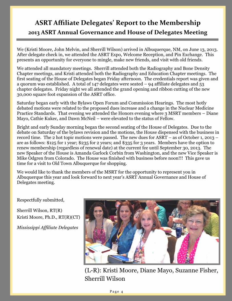
We (Kristi Moore, John Melvin, and Sherrill Wilson) arrived in Albuquerque, NM, on June 13, 2013. After delegate check in, we attended the ASRT Expo, Welcome Reception, and Pin Exchange. This presents an opportunity for everyone to mingle, make new friends, and visit with old friends.
We attended all mandatory meetings. Sherrill attended both the Radiography and Bone Density Chapter meetings, and Kristi attended both the Radiography and Education Chapter meetings. The first seating of the House of Delegates began Friday afternoon. The credentials report was given and a quorum was established. A total of 147 delegates were seated – 94 affiliate delegates and 53 chapter delegates. Friday night we all attended the grand opening and ribbon cutting of the new 30,000 square foot expansion of the ASRT office.
Saturday began early with the Bylaws Open Forum and Commission Hearings. The most hotly debated motions were related to the proposed dues increase and a change in the Nuclear Medicine Practice Standards. That evening we attended the Honors evening where 3 MSRT members – Diane Mayo, Cathie Kukec, and Dawn McNeil – were elevated to the status of Fellow.
Bright and early Sunday morning began the second seating of the House of Delegates. Due to the debate on Saturday of the bylaws revision and the motions, the House dispensed with the business in record time. The 2 hot topic motions were passed. The new dues for ASRT – as of October 1, 2013 – are as follows: $125 for 1 year; $235 for 2 years; and $335 for 3 years. Members have the option to renew membership (regardless of renewal date) at the current fee until September 30, 2013. The new Speaker of the House is Amanda Garlock Corbin from Washington, and the new Vice Speaker is Mike Odgren from Colorado. The House was finished with business before noon!!! This gave us time for a visit to Old Town Albuquerque for shopping.
We would like to thank the members of the MSRT for the opportunity to represent you in Albuquerque this year and look forward to next year’s ASRT Annual Governance and House of Delegates meeting.
Respectfully submitted,
Sherrill Wilson, RT(R)
Kristi Moore, Ph.D., RT(R)(CT) Mississippi Affiliate Delegates
(L-R): Kristi Moore, Diane Mayo, Suzanne Fisher,
Sherrill Wilson
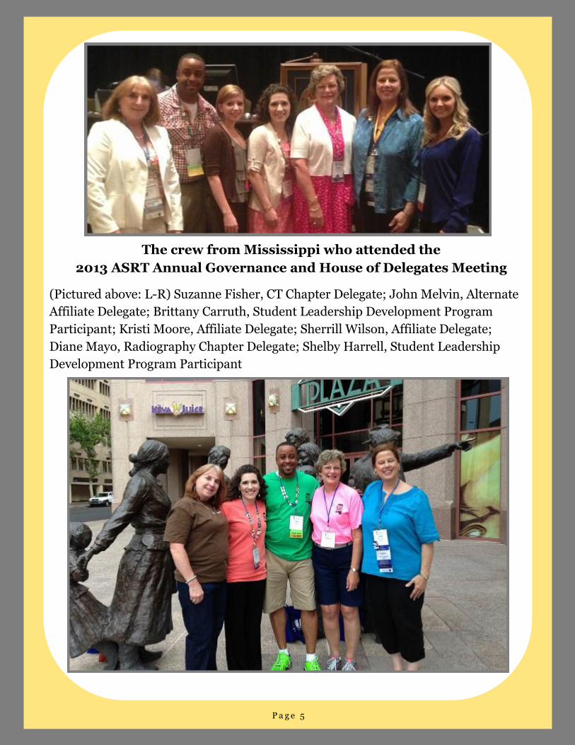
The crew from Mississippi who attended the
2013 ASRT Annual Governance and House of Delegates Meeting
(Pictured above: L-R) Suzanne Fisher, CT Chapter Delegate; John Melvin, Alternate
Affiliate Delegate; Brittany Carruth, Student Leadership Development Program
Participant; Kristi Moore, Affiliate Delegate; Sherrill Wilson, Affiliate Delegate;
Diane Mayo, Radiography Chapter Delegate; Shelby Harrell, Student Leadership
Development Program Participant
P a g e 5
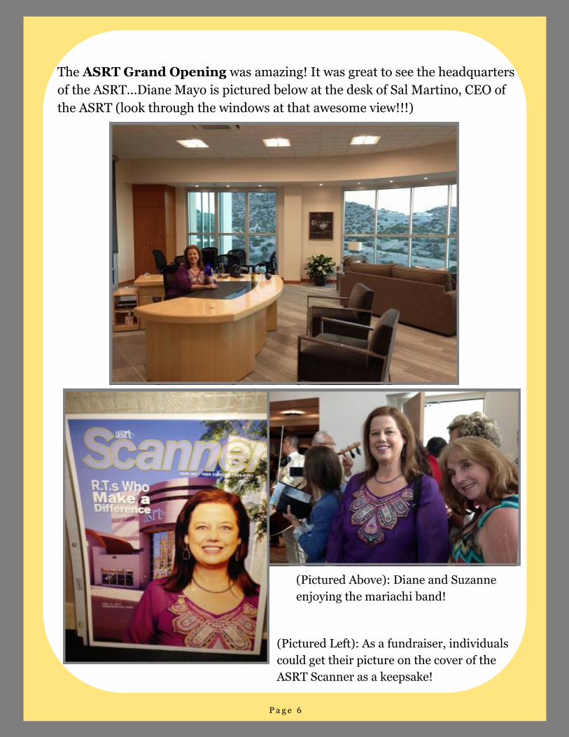
The ASRT Grand Opening was amazing! It was great to see the headquarters
of the ASRT...Diane Mayo is pictured below at the desk of Sal Martino, CEO of
the ASRT (look through the windows at that awesome view!!!)
(Pictured Left): As a fundraiser, individuals
could get their picture on the cover of the
ASRT Scanner as a keepsake!
(Pictured Above): Diane and Suzanne
enjoying the mariachi band!
P a g e 6

P a g e 7
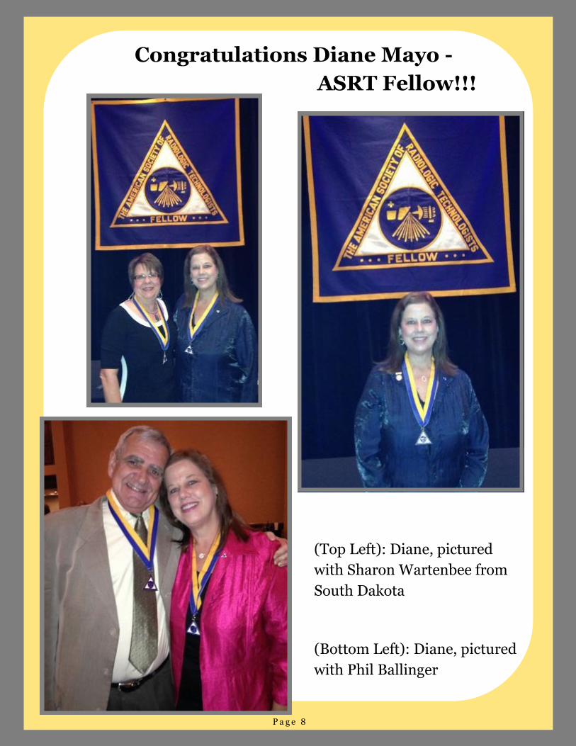
Congratulations Diane Mayo -
ASRT Fellow!!!
(Top Left): Diane, pictured
with Sharon Wartenbee from
South Dakota
(Bottom Left): Diane, pictured
with Phil Ballinger
P a g e 8
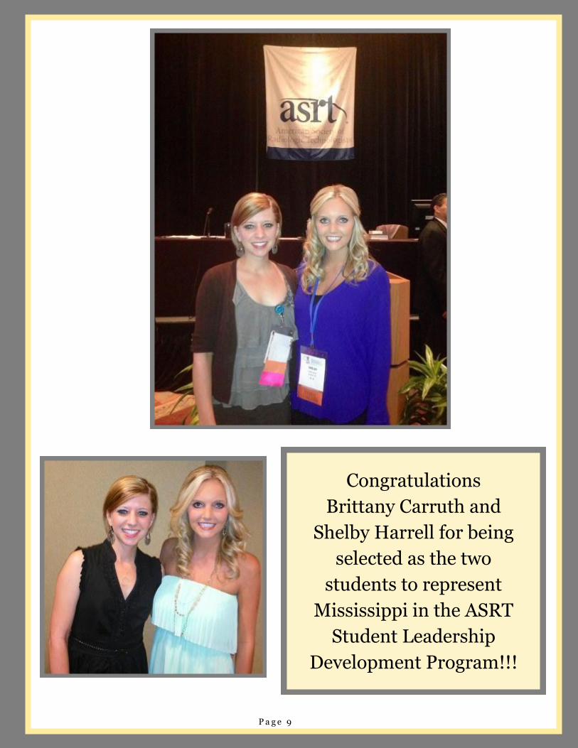
Congratulations
Brittany Carruth and
Shelby Harrell for being
selected as the two
students to represent
Mississippi in the ASRT
Student Leadership
Development Program!!!
P a g e 9

Congratulations
Graduates
Class of 2013 P a g e 1 0
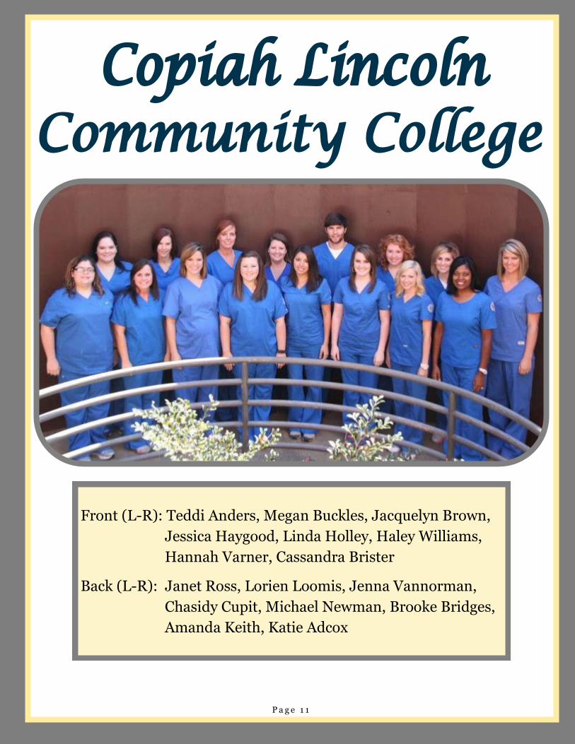
Front (L-R): Teddi Anders, Megan Buckles, Jacquelyn Brown,
Jessica Haygood, Linda Holley, Haley Williams,
Hannah Varner, Cassandra Brister
Back (L-R): Janet Ross, Lorien Loomis, Jenna Vannorman,
Chasidy Cupit, Michael Newman, Brooke Bridges,
Amanda Keith, Katie Adcox
P a g e 1 1
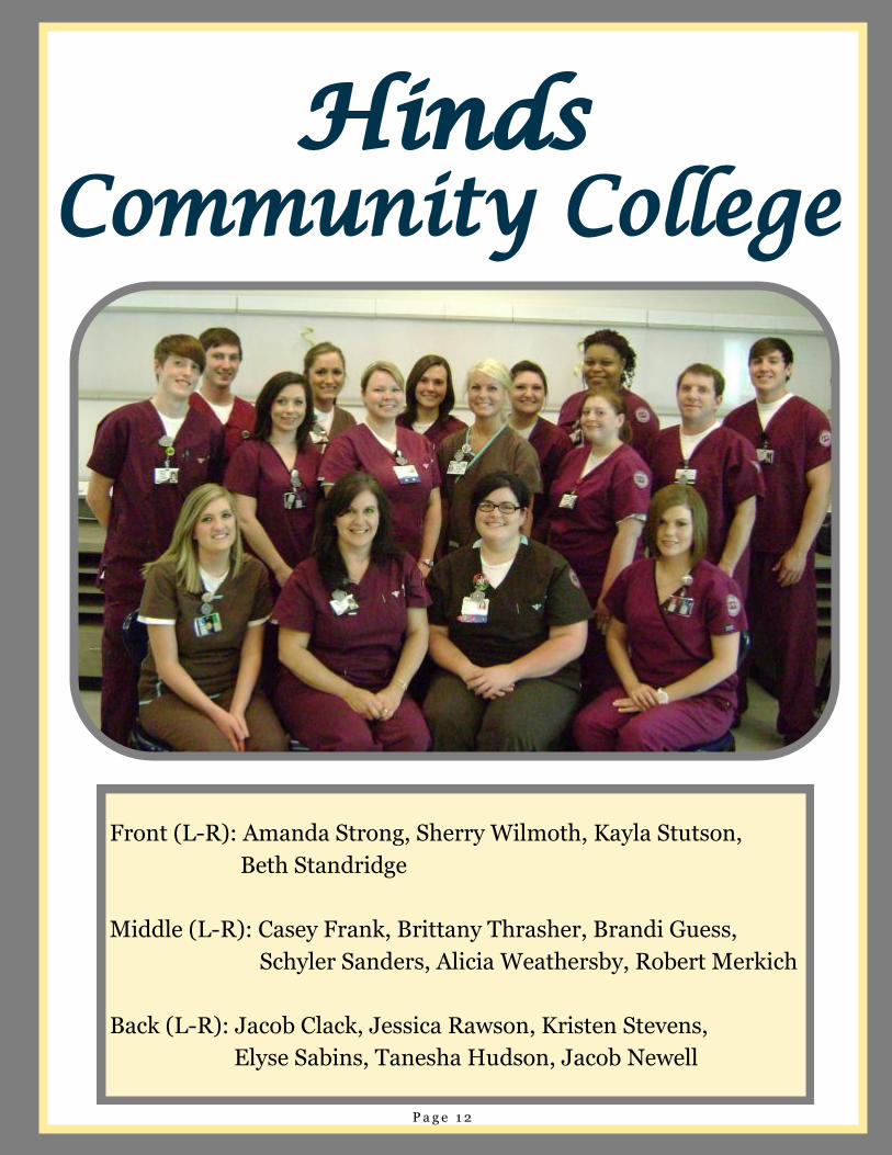
Front (L-R): Amanda Strong, Sherry Wilmoth, Kayla Stutson,
Beth Standridge
Middle (L-R): Casey Frank, Brittany Thrasher, Brandi Guess,
Schyler Sanders, Alicia Weathersby, Robert Merkich
Back (L-R): Jacob Clack, Jessica Rawson, Kristen Stevens,
Elyse Sabins, Tanesha Hudson, Jacob Newell
P a g e 1 2
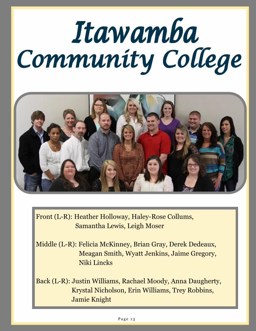
Front (L-R): Heather Holloway, Haley-Rose Collums,
Samantha Lewis, Leigh Moser
Middle (L-R): Felicia McKinney, Brian Gray, Derek Dedeaux,
Meagan Smith, Wyatt Jenkins, Jaime Gregory,
Niki Lincks
Back (L-R): Justin Williams, Rachael Moody, Anna Daugherty,
Krystal Nicholson, Erin Williams, Trey Robbins,
Jamie Knight
P a g e 1 3

Kneeling (L-R): Maggie Callicott, Chelsey Merritt, Susan Tisdale,
Kasey Bishop
Standing (L-R): Amy Firmin, Chase Powell, Jayln Nicholson,
Alley Mooney, Kristin Powell, Nguyen Phan,
Cody Eidson, Meagen Tucker
P a g e 1 4
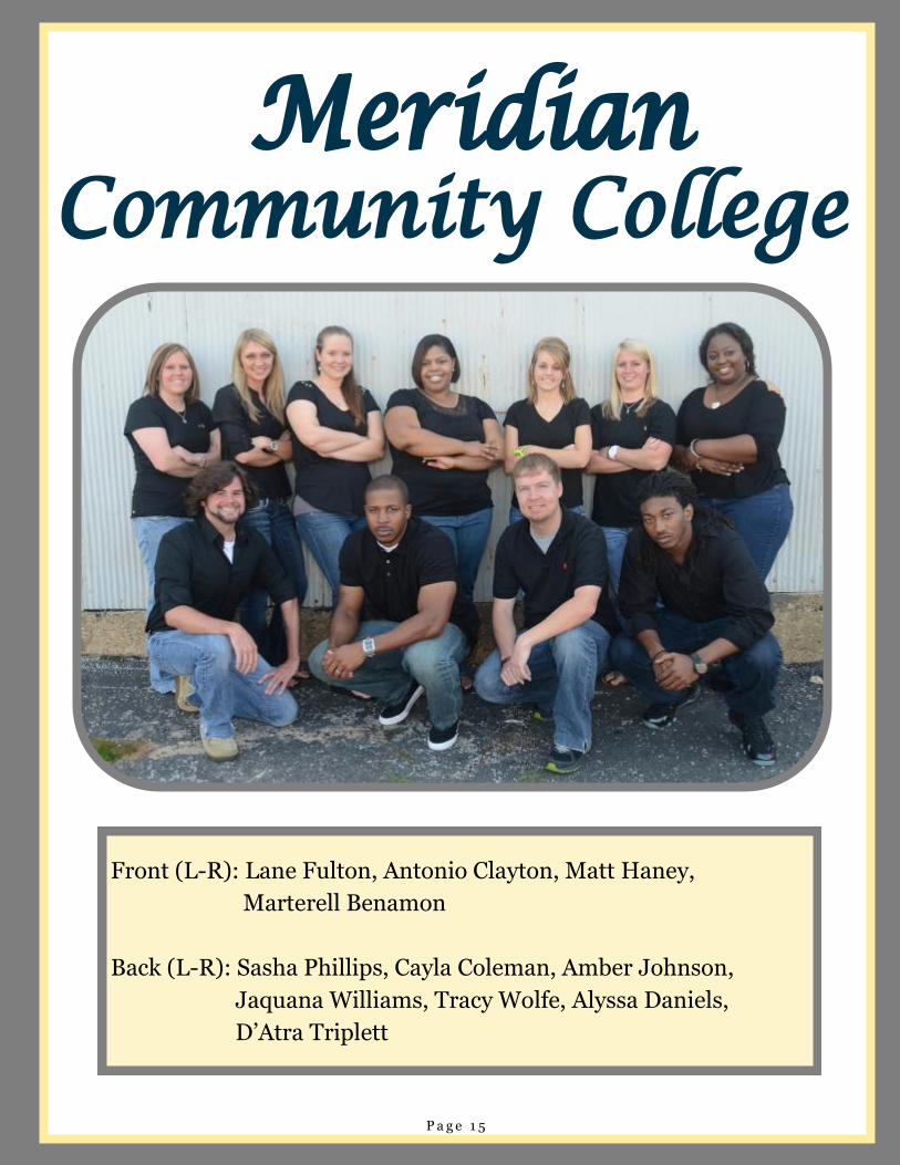
Front (L-R): Lane Fulton, Antonio Clayton, Matt Haney,
Marterell Benamon
Back (L-R): Sasha Phillips, Cayla Coleman, Amber Johnson,
Jaquana Williams, Tracy Wolfe, Alyssa Daniels,
D’Atra Triplett
P a g e 1 5
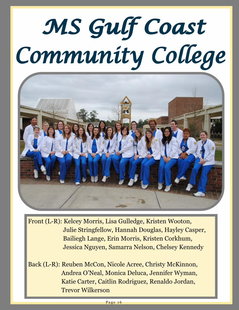
Front (L-R): Kelcey Morris, Lisa Gulledge, Kristen Wooton,
Julie Stringfellow, Hannah Douglas, Hayley Casper,
Bailiegh Lange, Erin Morris, Kristen Corkhum,
Jessica Nguyen, Samarra Nelson, Chelsey Kennedy
Back (L-R): Reuben McCon, Nicole Acree, Christy McKinnon,
Andrea O’Neal, Monica Deluca, Jennifer Wyman,
Katie Carter, Caitlin Rodriguez, Renaldo Jordan,
Trevor Wilkerson
P a g e 1 6

Front (L-R): Brittany Olmi, Stefanie Finley, Kristy Farmer,
Allison Williams, Brittany Avant, Jordan Bienvenu
Back (L-R): Madison Williford, Brittnee McCool, Ryan Watson,
Whitney Misner, Betsy Skender
P a g e 1 7
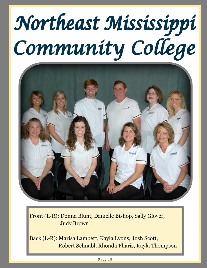
Front (L-R): Donna Blunt, Danielle Bishop, Sally Glover,
Judy Brown
Back (L-R): Marisa Lambert, Kayla Lyons, Josh Scott,
Robert Schnabl, Rhonda Pharis, Kayla Thompson
P a g e 1 8
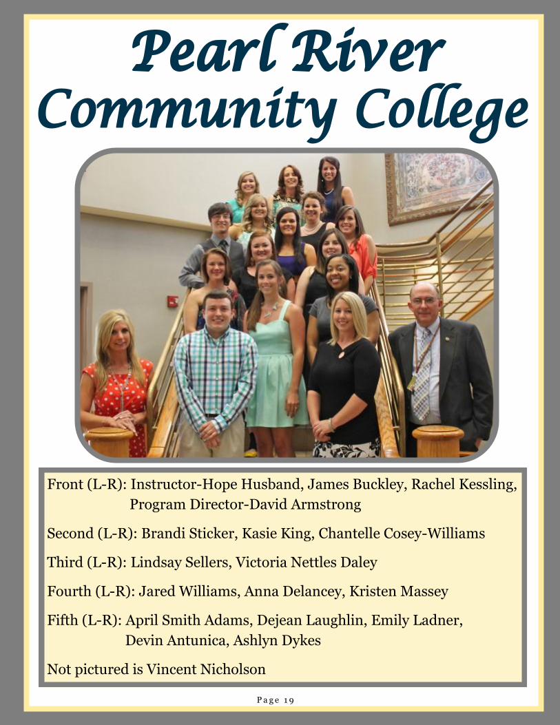
Front (L-R): Instructor-Hope Husband, James Buckley, Rachel Kessling,
Program Director-David Armstrong
Second (L-R): Brandi Sticker, Kasie King, Chantelle Cosey-Williams
Third (L-R): Lindsay Sellers, Victoria Nettles Daley
Fourth (L-R): Jared Williams, Anna Delancey, Kristen Massey
Fifth (L-R): April Smith Adams, Dejean Laughlin, Emily Ladner,
Devin Antunica, Ashlyn Dykes
Not pictured is Vincent Nicholson
P a g e 1 9
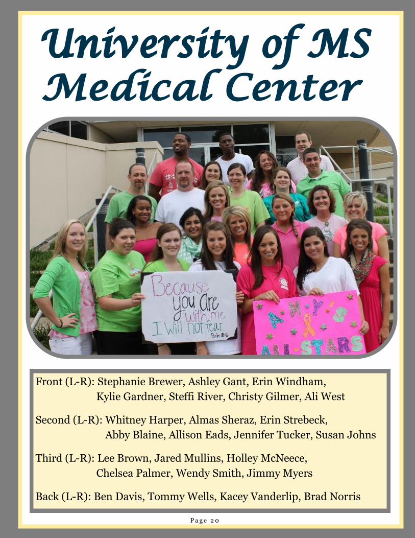
Front (L-R): Stephanie Brewer, Ashley Gant, Erin Windham,
Kylie Gardner, Steffi River, Christy Gilmer, Ali West
Second (L-R): Whitney Harper, Almas Sheraz, Erin Strebeck,
Abby Blaine, Allison Eads, Jennifer Tucker, Susan Johns
Third (L-R): Lee Brown, Jared Mullins, Holley McNeece,
Chelsea Palmer, Wendy Smith, Jimmy Myers
Back (L-R): Ben Davis, Tommy Wells, Kacey Vanderlip, Brad Norris
P a g e 2 0

MSRT
Scholarship
Recipients
Each of these students has demonstrated outstanding
academic and clinical performance throughout their
education. We salute them and wish them well in
their future endeavors.
MSRT Board of Directors
P a g e 2 1

Katie Adcox
Co-Lin Community College
Judy Brown
Northeast MS Community College
Beth Standridge
Hinds Community College
Kristin Powell
Jones County Junior College
P a g e 2 2
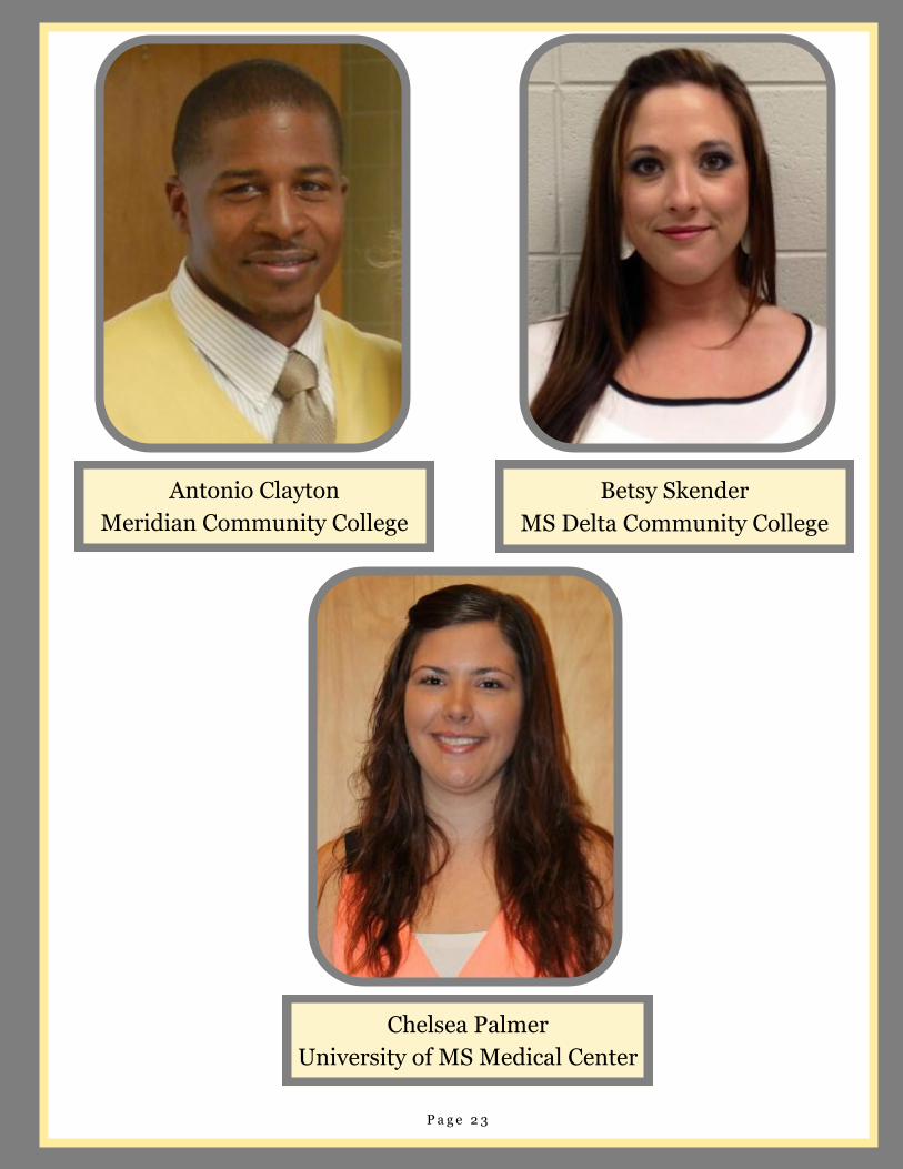
Antonio Clayton
Meridian Community College
Betsy Skender
MS Delta Community College
Chelsea Palmer
University of MS Medical Center
P a g e 2 3

Contact Information
Hard Rock Hotel & Casino
777 Beach Boulevard
Biloxi, MS 39530
Phone: 228-374-ROCK (7625
Email: http://
www.hardrockbiloxi.com/
MSRT 72nd
Annual Conference
October 22-24, 2013
Hard Rock Hotel & Casino
Biloxi, MS
Please continue to check the
MSRT website (www.msrt.biz) for
updated Conference information.
P a g e 2 4

MSRT 72nd
Annual Conference
October 22-24, 2013
Conference Registration
We prefer you register for Conference online at www.msrt.biz
when it is available; however, if you prefer to mail in your
registration, there will be an avenue for that as well. Please check
the website in August for a finalized agenda and registration
information.
P a g e 2 5

MSRT Prep Bowl
School #1
School #2
Where: Hard Rock Hotel & Casino
When: Wednesday, October 23, 2013
from 7:00 pm until...
Please see the rules beginning on the next page
P a g e 2 6

MSRT Central District Prep Bowl
Rules and Regulations
Purpose:
To review and increase knowledge of radiologic technology among students who should be
preparing themselves for the ARRT Registry. This will be an excellent form of registry review.
Eligibility:
Participants in the MSRT Central District Prep Bowl must be enrolled in a JRCERT approved
radiologic technology program. Each member of a team shall be in the final year of the program
and all team members shall be from the same program. The students participating in the prep bowl
must be a member of their state affiliate and registered for Conference in order to participate.
Team Roster:
Each school will be represented by only one (1) team. Each team will be represented by no more
than five (5) senior level students from the same approved program of Radiologic Technology. Only
three (3) team members may serve on the panel at any one time. Students will be allowed to rotate
members during scheduled breaks only.
Officials:
Each official shall be a registered radiologic technologist or a radiologist. No faculty member or
clinical instructor of a participating school shall serve as an official unless approved by the MSRT
Central District.
The Moderator: Shall serve as competition coordinator. It shall be the duty of the moderator to
present all questions, repeat each answer, and call official breaks or time-out. The moderator
must read the question only and may not elaborate in any way which might aid in the
answering of the question.
The Panel of Judges: Shall be available to verify all challenged questions using text references.
The decision of the judges is final. If the question cannot be verified, the question will be thrown
out and a new question asked.
The Timekeeper: Shall keep the official response time during competition.
The Scorekeeper and Backup Scorekeeper: Shall maintain a comprehensive score record of the
schools in competition. The scorekeeper will keep score on a board visible to the audience, while
the backup scorekeeper will keep score independently.
P a g e 2 7
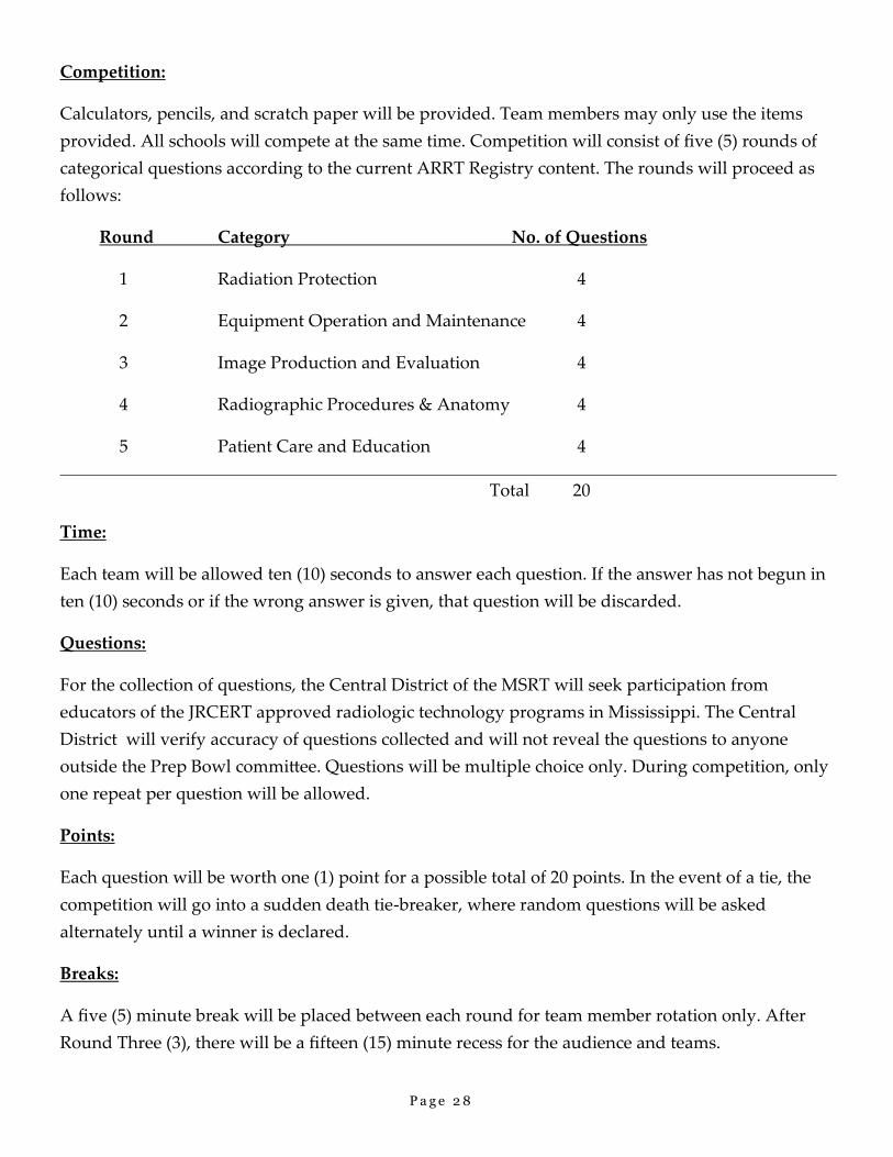
Competition:
Calculators, pencils, and scratch paper will be provided. Team members may only use the items
provided. All schools will compete at the same time. Competition will consist of five (5) rounds of
categorical questions according to the current ARRT Registry content. The rounds will proceed as
follows:
Round Category No. of Questions
1 Radiation Protection 4
2 Equipment Operation and Maintenance 4
3 Image Production and Evaluation 4
4 Radiographic Procedures & Anatomy 4
5 Patient Care and Education 4
Total 20
Time:
Each team will be allowed ten (10) seconds to answer each question. If the answer has not begun in
ten (10) seconds or if the wrong answer is given, that question will be discarded.
Questions:
For the collection of questions, the Central District of the MSRT will seek participation from
educators of the JRCERT approved radiologic technology programs in Mississippi. The Central
District will verify accuracy of questions collected and will not reveal the questions to anyone
outside the Prep Bowl committee. Questions will be multiple choice only. During competition, only
one repeat per question will be allowed.
Points:
Each question will be worth one (1) point for a possible total of 20 points. In the event of a tie, the
competition will go into a sudden death tie-breaker, where random questions will be asked
alternately until a winner is declared.
Breaks:
A five (5) minute break will be placed between each round for team member rotation only. After
Round Three (3), there will be a fifteen (15) minute recess for the audience and teams.
P a g e 2 8
Challenge:
A question may only be challenged by a member of the three person team participating at that
time. The question must be challenged prior to the reading of the next question.
The Judge’s ruling is final.
Penalties:
Any coaching or yelling of answers from the audience will disqualify the question from
competition and a new question will be asked. Continued disruption will result in removal from
the competition area.
Awards:
Plaques will be awarded to First, Second, and Third place teams. The First place team will also
receive a $100 cash award from the Central District of the MSRT.
Additional Rules:
Alcoholic beverages are not allowed and persons with alcohol/alcoholic beverages in their
possession shall be considered disruptive and removed from the competition area.
All electronic devices (i.e. cell phones, pagers, Bluetooth, etc.) must have the power turned off and
stowed away during competition.
P a g e 2 9
P a g e 3 0
Student Manuscript: 2nd Place Recipient — Steffi River (UMMC)
Tarsal Coalition
Tarsal coalition is the “fusion of two or more bones of the hind foot or, less commonly the midfoot,
resulting in absent or restricted movement between these bones” (Claridge & Sakellariou, 1999, p. 1066).
There are three various forms of tarsal coalition. They are cartilaginous, fibrous, or bony. These unifications
usually occur between different tarsal bones. The two most occurring tarsal coalitions seen in patients are
talocalcaneal and calcaneonavicular. The talocalcaneal coalition is where the talus and calcaneus fuse
together in the hind foot, whereas the calcaneonavicular coalition is the fusing together of the calcaneus and
navicular in the midfoot. Even though there are various forms of tarsal coalition, these two forms are the
most prevalent. Tarsal coalition can be corrected with either an operative or nonoperative treatment,
depending on the severity of the case.
There are different understandings as to where tarsal coalition originated. One case study shows that
“early literature suggested that this syndrome was attributable primarily to the nervous system disease” (Kelo
& Riddle, 1998, p. 519). This was later disproved because it does not have anything to do with the nervous
system, but rather the skeletal system of the body. This statement made tarsal coalition a misunderstood
disease of the tarsal bones. Also, this theory made tarsal coalition take on the name “spastic flat foot.” It took
many years later for researchers to understand the disease more thoroughly.
Several physicians have played an important role in the development of research to further understand
tarsal coalition. In 1769, “tarsal coalition was first described by Buffon” (Claridge & Sakellariou, 1999, p.
1066). Cruveilhier described a calcaneonavicular coalition, and then Zuckerkandl portrayed a talocalcaneal
coalition through their studies in 1877. Another researcher by the name of Holl “suggested that there was a
relationship between tarsal coalition and the so-called peroneal spastic flatfoot” (Claridge & Sakellariou,
1999, p. 1066). It was not until 1921 that a tarsal coalition was portrayed unto a radiograph. Slomann was
the first to image this disease on an oblique radiograph. Through this radiograph many observations were
made to better understand the extent of the disease. Harris and Beath are the researchers who are credited
P a g e 3 1
with popularizing a special view just to image the posterior and middle facets of the talocalcaneal joint in 1948
in order to diagnosis tarsal coalition. This particular view is called the “coalition” view. Radiographs are still
used in the preliminary diagnosis of tarsal coalition (Claridge & Sakellariou, 1999).
Tarsal coalition can either be congenital or acquired through time. Many studies have been conducted
to explain the origin of tarsal coalition and how it occurs. One explanation is that “tarsal coalitions are
inherited, most probably as a unifactorial disorder of the autosomal dominant inheritance” (Leonard, 1974, p.
520). This theory is the only rationalization for the congenital form of tarsal coalition. The congenital form
would be present at birth and found usually when the patient is a child. Tarsal coalition is an inherited
condition in which parents can pass the gene onto the child (Tarsal Coalition, 2012). Acquired tarsal coalition
“may occur secondary to trauma, inflammatory arthropathy, neoplasm or ever osteonecrosis, resulting in a
rigid painful flatfoot” (Claridge & Sakellariou, 1999, p. 1067). The acquired form is found in adults, rather
than children, and does not occur until the patient is older. Although it does not occur until the patient is older,
this factor does not affect the severity of the coalition in the patient (Tarsal Coalition, 2009).
Tarsal coalition is rare and not prevalent among the population. From a case study, tarsal coalition’s
“overall prevalence in the general population is reported to be about 1%” (Claridge & Sakellariou, 1999, p.
1067). This number does not account for the population that has “flat feet” because that is not always
associated with tarsal coalition. It does not happen in males more than females or vice versa. Tarsal coalition
also does not occur in one race more than another. “Of those affected, 50%-60% have the condition
bilaterally” (Claridge & Sakellariou, 1999, p. 1067). Bilateral tarsal coalition is common among those who are
affected. This can result in further problems and may have a longer recovery period for the patient if operative
treatment was the result.
Of all the different forms of tarsal coalition, two are represented fairly equal throughout the population
that was observed in this particular case study. Those affected with talocalcaneal were found to be 48.1% of
the population and calcaneonavicular, 43.6% (Claridge & Sakellariou, 1999). There are two more forms that
are seen, but are extremely rare among patients with tarsal coalition. Talonavicular coalitions and
P a g e 3 2
calcaneocuboid account for approximately 1.3% of the population (Claridge & Sakellariou, 1999). Even
though these forms are extremely rare, they are still seen in different patients with tarsal coalition.
Many patients who have tarsal coalition do not realize they have the condition until they reach young
adolescence. Usually they will experience pain while beginning to interact in physical activity. Another
common indication is reoccurring sprains in that particular foot. This may be caused by a tarsal coalition. It
can also limit the amount of mobility in that foot, which can lead to other types of injuries. “Abnormal
stresses, along with a reduced adaptive ability, result in a predisposition to ankle instability as well as pain,
inflammation, and eventually joint degeneration” (Claridge & Sakellariou, 1999, p. 1067). Symptoms may
include, but are not limited to, the following: pain when walking or standing, tired or fatigued legs, muscle
spasms in the leg, walking with a limp, and stiffness of the foot and ankle (Tarsal Coalition, 2009). Another
common symptom of patients with this syndrome is a painful arch, or around the arch area, of the midfoot
(Tarsal Coalition, 2012). Most patients, by the time they are adults, will experience severe arthritis in the foot
that is affected.
There are different means for doctors to diagnose tarsal coalition. The reason for patients consulting a
doctor is pain in the hind foot. The pain is “often aggravated by activity or prolonged standing” (Claridge &
Sakellariou, 1999, p. 1067). There can be many different deformities in the hind foot that may be palpable.
The doctor must also keep in mind that tarsal coalition is not always associated with a flat foot. These are
clinical findings for which the doctor would recommend the patient have radiographs of the foot in order to
further diagnose the condition.
The first assessment the patient may have would be a plain radiograph or an x-ray. If the doctor were
to order an imaging series for tarsal coalition, the radiographer would perform “a basic foot series such as
standing AP and lateral views, as well as a 45 degree oblique view, before progressing to more complex
imaging studies” (Claridge & Sakellariou, 1999, p. 1068). This would be a standing routine foot series. The
radiographer would need to consult with the doctor before making any further images.
P a g e 3 3
The different views in the standing routine foot series can show various signs to the radiologist in
diagnosing tarsal coalition. “The AP view of the foot is the least useful, but it can demonstrate the rare
talonavicular and calcaneocuboid coalitions” (Claridge & Sakellariou, 1999, p. 1068). The lateral is more
important for the radiologist because it shows a special sign in the diagnosis of tarsal coalition. This
radiographic sign is identified as “narrowing of the posterior subtalar joint space, failure to visualize the
middle subtalar joint, and the ‘C-sign,’ which is a c-shaped line formed by the medial outline of the talar dome
and the inferior outline of the sustentaculum tali” (Claridge & Sakellariou, 1999, p. 1068). In another study,
the lateral view of the talus is called a halo sign. This particular sign is caused by a ring-like trabecular bone
density below the talus (Kelo & Riddle, 1998). The oblique is the most useful in the imaging series. This can
show bony coalitions that are also palpable on the skin.
Computed Tomography, or CT, is the most accurate in diagnosing tarsal coalition. “The coronal
section will demonstrate the nature and cross-sectional area of the coalition as well as the presence and extent
of any degenerative arthritis present in the joints” (Claridge & Sakellariou, 1999, p. 1069). CT is then used so
the doctor can decide what to do during surgery and how to go about repairing the tarsal coalition. Magnetic
Resonance Imaging, or MRI, can also be used to obtain cross-sectional images, but CT is more highly
recommended.
The doctor may suggest nonoperative or operative treatments. Nonoperative treatments may include
restricting the patient’s mobility. The patient may not be able to continue various activities. Orthopedic shoes
and shoe alterations can help the patient perform daily actions. “A medial heel wedge, Thomas heel, or medial
arch support designed to help decrease subtalar motion can be helpful” (Claridge & Sakellariou, 1999, p.
1070). Other tactics in nonoperative treatment include oral medications. These oral medications are
nonsteriodal anti-inflammatory drugs, such as ibuprofen (Tarsal Coalition, 2009). Different approaches can be
taken in order to avoid surgery.
The alternate approach is an operative treatment. Operative treatment is carried out when nonoperative
is unsuccessful for the patient. This intensive surgery is used so that the tarsal bones can move freely and the
P a g e 3 4
motion can become normal. “Many times surgery may involve fusing the affected joint or surrounding
joints” (Tarsal Coalition, 2012). After surgery the patient would need to restrain from using that foot or
putting any weight on it.
I have a personal interest in this topic because I was born with congenital tarsal coalition. I was not
diagnosed until July 31, 2000, at age ten. Most patients with tarsal coalition develop symptoms usually during
adolescence (Kelo & Riddle, 1998). My case started with a simple sprain at a gymnastic class I had been
attending. My mother, being a nurse, knew that something was obviously wrong with my ankle. Below my
ankle there was a protruding mass that was not normal. My mother took me to the doctor and I was
immediately sent to an orthopedic surgeon for x-rays. The orthopedic surgeon was not certain from the x-rays
if it was tarsal coalition, so I was sent to Baptist Memorial Hospital in Oxford, Mississippi. I was then
scheduled to have a CT lower extremity without contrast of the right and left foot.
Soon after the CT, the radiologist, T. Sneed, conducted a report on what he saw through this test. Sneed
noted the following in the report: “There are no fractures. There is marked narrowing of the space between the
talus and calcaneus at the middle facet. There is some bony over growth seen about the middle facet in this
location. Complete fusion of the middle facet has not occurred. Otherwise the bone alignment is normal.”
This was good news for me as the patient because I would not require immediate surgery.
T. Sneed’s diagnosis stated “talocalcaneal coalition at the middle facet with incomplete fusion. This
probably explains palpable abnormality on clinical exam.” After this conclusion, I was referred back to my
orthopedic surgeon, Dr. Cooper Terry. He wanted to meet with me and my mother to decide a plan of action.
The surgeon offered that there would not be any need for immediate surgery, which was a relief for my family.
Although immediate surgery was not an option, eventually there would be a need for surgery. Dr. Terry
suggested that, by the age of forty, I have the corrective surgery. The surgery includes going into the
talocalcaneal joint and opening the joint space so that the sinus tarsi will no longer be blocked. Talocalcaneal
coalition “restricts subtalar motion to a greater degree” (Claridge & Sakellariou, 1999, p. 1071). This means
that most patients with talocalcaneal coalition are required to have surgery even though it may not be
P a g e 3 5
instantaneous. The reason for the pain of tarsal coalition is the blockage of the sinus tarsi and the nerves being
pinched in the middle facet of the foot.
Since being diagnosed, my affected ankle has been sprained multiple times. Sprains are common in
patients with tarsal coalition, and sometimes this leads to a misdiagnosis (Claridge & Sakellariou, 1999).
Throughout Junior High and High School I participated in multiple activities. Cheerleading was challenging
because of my tarsal coalition. Many times I had to ice the foot after practice. Stabilizing my ankle throughout
cheerleading and track was a necessity. Learning which shoes to wear was also essential. Today I wear special
insoles in most of the athletic shoes I wear everyday to make living with tarsal coalition more tolerable.
Tarsal coalition is the fusing together of the tarsal bones in specific forms throughout the hind foot and
the midfoot. These forms can be cartilaginous, fibrous, or bony. The two most prevalent coalitions are
talocalcaneal and calcaneonavicular. Several physicians and radiologists have performed research to further
understand this syndrome. Tests that can be completed to diagnose the patient include: radiographs, computed
tomography, and magnetic resonance imaging. Then the doctor will decide whether to require nonoperative or
operative treatment for the patient. After being diagnosed with tarsal coalition in 2000, I have found ways to
deal with the condition.
P a g e 3 6
References
Claridge, R. J., & Sakellariou, A. (1999). Tarsal coalition. Orthopedics. 22, 11, 1066-1074.
Kelo, M. J., & Riddle, D. L. (1998). Examination and management of a patient with tarsal coalition. Physical
Therapy. 78, 5, 518-525.
Leonard, M. A. (1974). The inheritance of tarsal coalition and its relationship to spastic flat foot. Journal of
Bone & Joint Surgery. 56-B, 3, 520-526.
Tarsal coalition. (2009). Retrieved January 25, 2012, from http://www.foothealthfacts.org/print.aspx?
keepthis=true&
Tarsal coalition. (2012). Retrieved January 25, 2012, from American Health Network, http://www.ahni.com/
specialties/foot-and-ankle/common-disorders/tarsal-coalition/
Tarsal coalition. (2012). Retrieved January 25, 2012, from Seattle’s Children’s Hospital, http://
www.seattlechildrens.org/medical-conditions/bone-joint-muscle-conditions
P a g e 3 7
Student Manuscript: 3rd Place Recipient — Jennifer Tucker (UMMC)
Radiography and Progressive Supranuclear Palsy
Progressive supranuclear palsy (PSP) is another name for Steele-Richardson-Olszewski syndrome, a
rare, degenerative neurological disease that affects approximately 5% of the population. Even though it is not
considered a life-threatening syndrome, people who have PSP will eventually die of complications from the
disease. It is considered to be a type of Parkinsonian syndrome. Serial MRI is used to help differentiate
among Parkinson’s disease, progressive supranuclear palsy, and multiple symptom atrophy Parkinsonian’s
variant (MSA-P). Currently there is no cure for PSP, and there are no effective drug treatments for this illness.
Progressive supranuclear palsy affects all regions of the brain that control movement. These regions
include the cerebral cortex, basal ganglia, brain stem, and cerebellum. The brainstem is composed of the
medulla oblongata, pons, reticular formation, and midbrain. It is the oldest part of the brain, and each part is
responsible for different survival functions. The medulla oblongata regulates such functions as heart rate,
respiration, blood pressure, vomiting, swallowing, and sneezing. The reticular formation manages sleep and
attentiveness. The pons and the midbrain connect different brain structures and are where ten of the twelve
cranial nerves originate. The cranial nerves control eye-movement and facial movement. The cerebellum is
the portion of the brain that helps the body move. After receiving signals from other parts the brain, the
cerebellum helps the body move fluidly and precisely, and also assists in maintaining posture and balance by
controlling muscle tone (Dubuc, 2002).
Other parts of the brain that are damaged by progressive supranuclear palsy include the basal ganglia,
especially the globus pallidus, subthalamic nuclei, and the dentate nucleus of the cerebellum (Hain, 2002).
The basal ganglia are involved in receiving information from other parts of the brain and are thought to
facilitate movement by using that information (Dubuc, 2002). In addition to the basal ganglia, the cerebral
cortex is affected, and the decreased metabolism of the cerebral cortex contributes to the dementia that patients
of PSP develop. The cerebral cortex controls many functions, but it is the motor functions that are directly
affected by PSP. However, cortical degeneration is minimal (Hain, 2002). The midbrain has nuclei that
P a g e 3 8
control eye movement, and the term “supranuclear” comes from lesions that occur above the nuclei of the
midbrain. As the disease progresses, there is a loss of brain cells, not only in the midbrain, but also in the
cerebral cortex, cerebellum, and basal ganglia, all of which control movement.
The causes of progressive supranuclear palsy are not clear. Some speculate that it could be caused by
environmental factors, free radicals, genetic mutations, or a virus that takes many years to manifest itself
(“Causes,” n.d.). Recent studies suggest that there could be a mutation in the tau gene. Hain (2002) states that
Tau is a microtubule-binding protein that is normally abundant in neurons. There are six different forms of tau
in the normal human brain. However, in typical PSP, pathological tau is composed of aggregated 4-repeat
forms that accumulate in cells and glia in the brain (para. 12). Whether or not PSP is a genetic mutation or
caused by environmental factors is not clear, but the possibility exists that it could be caused by a combination
of the two. What scientists do know is that PSP is caused by deterioration of the midbrain, and it is this
deterioration that causes an early symptom of PSP called gaze palsy, which is defined as the inability for a
patient with PSP to look down.
Although testing for gaze palsy is the most diagnostic exam for progressive supranuclear palsy (PSP),
magnetic resonance imaging (MRI) has been used in diagnosing PSP and to distinguish it from other
degenerative neurological diseases, such as Parkinson’s disease and multiple symptom atrophy Parkinson’s
variant (MSA-P). Through research, which has specifically used serial MRI, scientists have determined that as
the disease progresses, specific areas of the brain atrophy, which causes the symptoms. Since PSP so closely
resembles Parkinson’s disease and is so often misdiagnosed due to similarities, MRI is becoming a radiologic
modality often used to make a determination among Parkinson’s disease, PSP, and MSA-P (Oba et al., 2005).
One such study published in Brain: A neurological journal, involving longitudinal magnetic resonance
imaging (MRI) in progressive supranuclear palsy (PSP) and multiple system atrophy (MSA-P), was performed
on several patients over a number of years. The patients were either diagnosed with progressive supranuclear
palsy, Parkinson’s disease, or multiple-system atrophy of the Parkinson type. The purpose of the study was to
determine, through serial MRI scans, the amount of atrophy that occurred over many years to certain regions
P a g e 3 9
of the brain. Findings included that, in patients with PSP, there is significant regional atrophy of the
brainstem, specifically the midbrain, and these neurodegenerative diseases have a predilection for brainstem
structures. The study also revealed that regional atrophy rates were better markers for the progression of
these diseases rather than the lobes of the brain (Paviour, Prive, Jahanshahi, Lees, Fox, 2006). In both
Parkinson’s disease as well as MSA-P, there is atrophy in regions of the brain, but there is not the regional
atrophy of the midbrain that occurs in PSP patients. The study concluded that serial MRI can be used to help
distinguish PSP from both Parkinson’s disease and MSA-P through the regional atrophy of the midbrain and
shrinkage of the brain stem. Also, MRI could be useful in the early diagnosis of neurodegenerative diseases,
which could lead to drug treatment that is normally more successful in the first stages of these diseases
(Paviour et al., 2006).
As stated earlier, progressive supranuclear palsy (PSP) can be difficult to diagnose and is often
misdiagnosed as Parkinson’s disease due to similar symptoms, such as unexplained falling and change of gait,
which affects both Parkinson’s and PSP patients. However, patients who develop PSP do not have the
tremors associated with Parkinson’s disease, and they also respond poorly to levodopa treatments, a drug
treatment that often helps those with Parkinson’s disease. PSP resembles multiple symptom atrophy
Parkinson's variant (MSA-P) as well, but those with MSA-P have trouble controlling blood pressure, and with
PSP there is supranuclear gaze palsy and increased age at the onset of the disease (Hain, 2002).
Early symptoms of progressive supranuclear palsy (PSP) include imbalance, poor postural reflexes,
axial rigidity, dysathria, the slowing and slurring of speech, and bradykinesia, which is the inability to move
fluidly. In a year’s time, patients may have trouble sitting down. Rather than sitting down, they will often
“fall into a chair.” Patients will begin to have difficulty initiating swallowing. They will also drop food on
themselves because they are no longer able to look down to eat (Hain, 2002). Other symptoms include
masked facies, a condition that causes those who suffer from PSP to have an astonished, worried expression
(Eggenberger, 2012). In addition to the physical changes associated with PSP, there are also psychological
changes that include cantankerousness, increased irritability, and forgetfulness. However, the most telling
P a g e 4 0
symptom of PSP is gaze palsy, where the patient has trouble looking downward and increased difficulty in
blinking or controlling the eyelids (“PSP Fact Sheet,” 2011).
As PSP progresses, the brain cells continue to degenerate and the symptoms worsen. The reason for
the degeneration of the brain cells is unknown. The patient’s face may become rigid, causing difficulty in
smiling or speaking. He will often drool due to his mouth gaping open. He also loses the ability to drive and
eventually will lose the ability to walk. Midway through the disease progression, he becomes more rigid.
Those who suffer from PSP will not be able to go up and down stairs or drive. Since falling becomes such a
danger and the rigidity continues to increase through the progression of the disease, patients with PSP are
often confined to a wheel chair. Swallowing becomes more and more difficult as the disease progresses due
to the degeneration of brain cells in the cerebellum. As swallowing becomes more difficult, the risk of liquids
going down the trachea and into the lungs increases, as does the risk of pneumonia. There is also cognitive
degeneration in PSP patients. Unlike Alzheimer’s where cognitive degeneration affects the memory, PSP
cognitive degeneration is evident in the patient’s inability to process thoughts quickly and the difficulty of
combining different ideas into a new idea or plan (Progressive supranuclear palsy, n.d., para 46).
In the final stages of the disease, both horizontal and vertical eye movements are lost, as is the ability
to blink, and it may become necessary to give the patient eye drops frequently. The rigidity of the patient
further increases, and there is no mobility. When complete loss of mobility occurs, the patient will eventually
become bed-ridden. Eating becomes even more difficult due to increased difficulty in swallowing. Most
patients with PSP will aspirate small amounts of fluid as they eat and drink. When a PSP patient begins to
cough after every meal, this usually indicates that there is a danger in developing pneumonia due to aspiration
(Hain, 2002). When this finally occurs, a decision to insert a feeding tube into the stomach might be made in
order to provide nutrition (National Institute of Neurological Disorders and Stroke, 2011, para. 21). The most
common cause of death for a patient with PSP is pneumonia; other causes of death are sepsis and choking.
The prognosis for those with PSP is a life span of approximately five years after diagnosis. There is no
proven effective treatment for PSP. There are no drug therapies that have significant impact on the disease.
P a g e 4 1
Drugs used to treat Parkinson’s disease have been used to treat patients with PSP, but have been only mildly
helpful in 50% of the patients with PSP. Other drugs such as Mirapex, Idazoxan, Ambian, and Physiostig-
mine have had either a transient effect or no effect at all. However, there are some unproven treatments that
might help, including a drug called seligiline that could be helpful in slowing the progression of the disease,
but formal studies have not yet been performed. Some think that physical therapy might also help, but
ultimately it will not slow the progression of the disease. Other drugs, such as Elavil, an anti- depressant,
calcium channel blockers, and vitamin E might help slow the progression of PSP (Hain, 2002). Ultimately,
there is currently no effective treatment for PSP (NINDS, 2011).
In conclusion, the facts exist that progressive nuclear palsy is a neurodegenerative disease that is
caused by the degeneration of brain cells primarily in the midbrain, a part of the brainstem. PSP affects about
5% of the population and the cause is unknown. Diagnosis is difficult since the early stages may mimic
Parkinson’s disease as well as other neurodegenerative diseases. Since progressive nuclear palsy affects eye
movement, it is most often diagnosed through eye tests and the patient’s inability to look downward. Serial
MRI has been used to help diagnose PSP by showing the atrophy of the brainstem and midbrain regions of the
brain, which help differentiate PSP from other Parkinsonian syndromes. MRI can also be useful in early
diagnosis where drug treatment, should any be tried, might be most effective. Currently, there is no effective
drug treatment and no cure.
P a g e 4 2
References
Dubuc, B. (2002). The brain from top to bottom. Retrieved from http://thebrain.mcgill.ca/flash/i/i_01/i_01_cr/
i_01_cr_ana/i_01_cr_ana.html
Eggenberger, E.R. (2012). Progressive supranuclear palsy clinical presentation. Retrieved from http://
emedicine.medscape.com/article/1151430-clinical
Hain, T.C. (2002). Progressive supranuclear palsy. Retrieved from http://www.tchain.com/otoneurology/
disorders/central/movement/psp.htm
National Institute of Neurological Disorders and Stroke. (2011). Progressive supranuclear palsy fact sheet.
Retrieved from http://www.ninds.nih.gov/disorders/psp/detail_psp.htm:css=print
Oba, H., Yagishita, A., Terada H., Barkovich, A.J., Kutomi, K., Yamauchi, T., …Suzuki, S. (2005). New and
reliable MRI diagnosis for progressive supranuclear palsy [Abstract]. Neurology. 64(12), 2050-2055.
Paviour, D.C., Price, S.L., Jahanshahi, M., Lees, A.J., & Fox, N.C. (2006). Longitudinal MRI in progressive
supranuclear palsy and multiple symptom atrophy: rates and regions of atrophy. Brain: A journal of
neurology. 129, 1040-1049. doi:10.1093/brain/aw1021
Progressive supranuclear palsy (PSP). (n.d.). Retrieved February 25, 2012 from http://
www.pspinformation.com/disease/psp/pspinfo.shtml
P a g e 4 3
Student Paper: Shelby Harrell (Student Technologist of the Year Award Candidate)
Radiography Aids Diagnosis of Child Abuse
In the United States, child abuse is responsible for approximately 1400 deaths per year. Some injuries
are typically produced as a result of inappropriate force on the tender skeleton of a child. Most child abuse
related injuries can be detected through diagnostic imaging. Without diagnostic images to support the
findings in the abused children some children would have to return to the abusive environment. X-ray
technologists working in the pediatric department of most hospitals come in contact with abused children.
The radiologist may be the first to find signs of abuse if the patient has unexplained bone markings. The
radiologist has strict medical and legal code to follow in cases dealing with child abuse. Dr. Paul Kleinman
constructed the most frequently used classification of fractures related to abuse. The system classifies the
fractures obtained into categories of high, moderate, and low specifically for the ultimate diagnoses of child
abuse (Gellar, E.).
For children under the age of two, a skeletal survey should be performed when child abuse is being
considered. The skeletal survey consists of a series of images collimated down to the specific body part. The
series includes frontal and lateral views of the skull, frontal and lateral views of the spine, frontal view of the
chest (ribs) and pelvis, and frontal views of the extremities. The advantage of the skeletal survey is its high
sensitivity to acute and healing fractures and a relatively low radiation dose compared to other modalities.
All abdominal areas should be viewed on at least two projections. A babygram, in which the entire skeleton
is depicted on one image, is not a substitute for a skeletal survey. The babygram produces limitation such as
geometric distortion and varying exposures across the image. For skeletal surveys, a screen film system with
good spatial resolution is needed (Gellar, E.). Computed Tomography (CT) is the best image modality when
the child is suspected to have acute neurological findings or retinal hemorrhage. A head CT scan should be
performed on all patients one year of age or younger and in all children with neurological symptoms (Dwek,
R.).
Battered child syndrome, shaken infant syndrome, stress-related infant abuse and non-accidental
P a g e 4 4
trauma are all terms to describe the complexity of non-accidental injuries in infants and young children as a
result of abuse. The child develops shaken infant syndrome from being held around the chest and violently
shaken back and forth. This causes the child’s extremities to swing back in fourth in a whiplash motion. Rib
fractures are a result from being held tight around the chest. The ribs are compressed from front to back and
literally almost fold in half. The whiplash motion of the extremities cause a ‘corner’ or ‘bucket-handle’
fracture in the metaphyseal region. The ‘corner’ fracture is described as a small piece of bone avulsed due to
shearing forces on the fragile growth plate. The ‘bucket-handle’ fracture is essentially the same as the ‘corner’
fracture, except the avulsed bone fragment is larger and shaped like a bucket handle. The corner and bucket-
handle fractures are most commonly found in the tibia, distal femur, and proximal humerus (Robben, S.).
Radiography plays a major role in the diagnoses of child abuse. The quality of images the technologist
produces help determine the diagnoses of the child. This is why it is always important to take quality images,
because in the case of child abuse it could be a life or death situation. Unfortunately, child abuse will never
end but I am proud to say I have chosen a profession that makes a difference in the number of child abuse
victims out there.
References
Dwek, R. (n.d.). The Radiologist Approach to Child Abuse. Retrieved from http://www.ncbi.nlm.nih.gov/pmc/
articles/PMC3032862/.
Gellar, E. (n.d.). Medscape. Retrieved from http://emedicine.medscape.com/article/407144-overview.
Robben, S. (n.d.). Radiologist Assistant. Retrieved from http://www.radiologyassistant.nl/en/p43c63c41ef792.
P a g e 4 5
Student Paper: Brittany Carruth (Student Technologist of the Year Award Candidate)
Pregnancy + X-ray = ?
When treating pregnant women in a hospital setting, many of the traditional forms of health care
become controversial. X-ray immediately comes to mind. This has always been a controversial subject
among health care workers and the public, alike. One thing is for sure, though. An unborn child is at a
heightened state of sensitivity due to the rapid rate of cell division and development, and things like
prescription and OTC medications, cigarette smoke, alcohol, and infection should be avoided when possible
due to the risks that are involved. In some instances, though, medication and diagnostic exposure to radiation
have benefits that outweigh the risks. As with any patient, radiation protection measures should be utilized
when possible, and radiologic technologists should always retrieve diagnostic images using as little radiation
as possible.
Mayo Clinic obstetrician and medical editor-in-chief, Roger W. Harms, M.D., goes as far as to say
that, in reality, most radiologic exams pose no threat to an unborn child due to the location of the
exposure. Lead aprons and collars may be worn to block scatter radiation produced during the exam, and, in
these cases, x-ray has caused the embryo or fetus no harm. In instances of an abdominal, pelvic, or kidney
exam, radiation shielding may not be an option, but it is important to remember that typical doses of radiation
–such as doses acquired during an abdominal exam- do not put an unborn child in any danger of the effects of
high doses of radiation. While long-term exposure to high doses of radiation could cause genetic changes that
have the potential to cause birth defects or cancers such as leukemia later in life, the American College of
Radiology states that single diagnostic x-rays do require doses significant enough to cause these adverse
effects in an embryo or fetus.
Proper communication with health care providers may actually reveal options that do not involve x-ray
such as ultrasound or magnetic resonance imaging (MRI). Also, women who are-or suspect that they might
be-pregnant should not hold a child in position for an x-ray. In this case, someone else should be asked to
hold the child for their exam. Women who received an x-ray before they were aware of their pregnancy
P a g e 4 6
should not worry. Dr. Roger Harms explains that risk concerning an embryo or fetus due to radiation exposure
is, in his words, “exceedingly remote.” Conditions requiring radiation treatment might pose larger threats and
should be discussed with a health care provider. Any measures that can be reasonably taken to avoid a
pregnant woman’s exposure to radiation should, in fact, be taken.
An important point for pregnant women to remember, also, is that sometimes the benefits surrounding
diagnostic imaging far outweigh the dangers of the small doses of x-ray required to produce these images. X-
rays have the potential to reveal life-saving information. When proper radiation protection methods are
utilized and communication between a pregnant woman and her health care providers is open, there is no
reason for necessary health care to not be obtained. There is much speculation surrounding this topic, though.
Personal research and professional opinion should be observed while making the decision in whether or not to
allow diagnostic images to be acquired. Women should express any questions or concerns to their health care
provider in order to resolve any doubt she may have concerning her pregnancy.
P a g e 4 7
Nominations
It is time for nominations for the elected offices of the MSRT.
If you have someone you would like to nominate, please place your
nominee’s name in the appropriate space and mail or email to:
Robbie Nettles
415 Catherine Street
Brookhaven, MS 39601
President: __________________________________________________
Vice President: ______________________________________________
Secretary: __________________________________________________
Affiliate Delegate: ____________________________________________
P a g e 4 8

Change of Information &
Membership Renewal
We prefer you edit information on your profile and renew
your membership online at www.msrt.biz; however, if you
prefer to mail in the actual forms, they are located on the next
pages.
P a g e 4 9

MISSISSIPPI SOCIETY OF RADIOLOGIC TECHNOLOGISTS
CHANGE OF INFORMATION OR ADDRESS FORM
MSRT MEMBER #_____________________
Name:____________________________________________
OLD INFORMATION:
Address:_________________________________________
City:______________________________ State_____ ZIP____________
Telephone # : ( ) ______-_________
Email:__________________________________________________
NEW INFORMATION:
Address:_________________________________________
City:______________________________ State_____ ZIP____________
Telephone # : ( ) ______-_________
Email:__________________________________________________
**** This form can either be mailed or returned via email to the following ****
Kristi Moore
122 French Branch
Madison, MS 39110
P a g e 5 0
MISSISSIPPI SOCIETY OF RADIOLOGIC TECHNOLOGISTS
MEMBERSHIP APPLICATION
MEMBERS WILL RECEIVE AN AUTOMATED EMAIL PROMPTING ONLINE RENEWAL. IF YOU HAVE NOT
SUBMITTED PAYMENT WITHIN 30 DAYS OF THE DUE DATE, YOUR NAME WILL BE REMOVED FROM THE
MEMBERSHIP ROSTER.
Annual Fees: Student - $10.00, RT - $30.00, Associate - $35.00
Please make checks or money order payable to MSRT and mail to:
Kristi Moore
MSRT Executive Secretary
122 French Branch
Madison, MS 39110
Preferred membership and/or renewal is online. However, this form is accepted. Complete the following
form and return with payment.
MSRT MEMBER #_____________________
Name:________________________________________________________________
Address:____________________________________________________
City:______________________________ State_____ ZIP_____________
Telephone # : ( ) ________-___________
Email:______________________________________________________
**MSRT is now only sending the BEAM electronically, so it is essential to provide us with an email address**
Check one: Student _____ Associate _____ ARRT certified _____
*** If applying as a student, please give the name of the Radiologic Technology program you are enrolled in.***
School: ________________________________________________________________________ ____
ARRT certified technologists: Please provide the following information: ARRT #_________________
Primary Modality (Please Circle)
Radiography Education Sonography CT MRI Bone densitomery
CIT Mammography Dosimetry Radiation therapy Nuclear medicine
Quality management Military Management RA RPA
P a g e 5 1
P a g e 5 2
Kristi
See ya’ll soon…
Conference 2013 (Biloxi, MS)
Please be sure to check out the MSRT
website in December for the next issue of
The BEAM!!!
L e t t e r f r o m t h e E d i t o r :
I hope everyone has had a great summer so far! I am looking forward to Conference in
October...I want to encourage senior students to participate in the Prep Bowl. This is a
great way to prepare for the Registry. I also want to encourage students and
technologists to participate in the exhibit competitions. Exhibit forms can be found on
the website. Forms must be submitted to both the MSRT President and Conference
Coordinator if you choose to compete. I hope you enjoyed this edition of The BEAM!
The deadline for the next issue of The BEAM is tentatively set for November 15, 2013.