supplementary data identification of small molecules that ... fileidentification of small molecules...
TRANSCRIPT

Supplementary data
Identification of small molecules that suppress microRNA pathway and reverse tumorigenesis
Koichi Watashi, Man Lung Yeung, Matthew F. Starost, Ramachandra S. Hosmane, and Kuan-Teh Jeang
Supplementary Figures S1-8 and legends Supplementary Table
1
Supplementary Fig. S1.
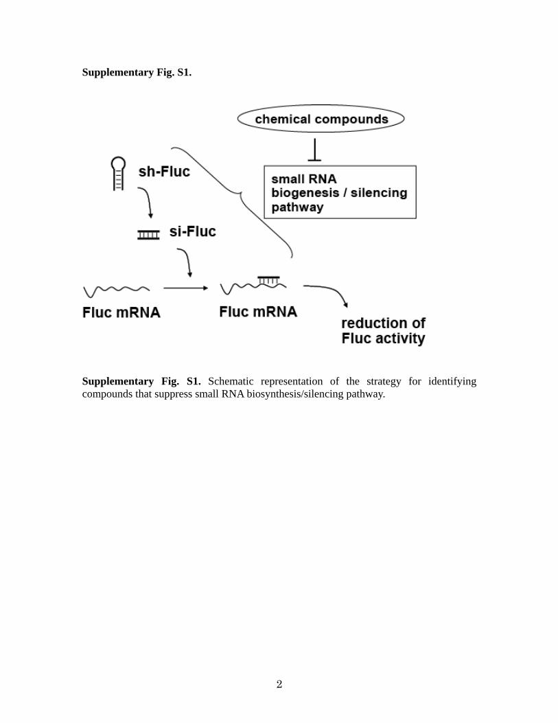
Supplementary Fig. S1. Schematic representation of the strategy for identifying compounds that suppress small RNA biosynthesis/silencing pathway.
2
Supplementary Fig. S2.
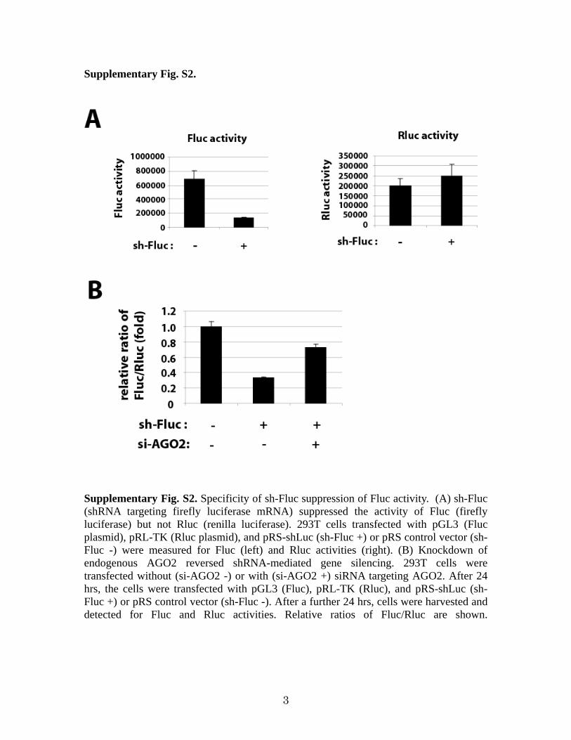
Supplementary Fig. S2. Specificity of sh-Fluc suppression of Fluc activity. (A) sh-Fluc (shRNA targeting firefly luciferase mRNA) suppressed the activity of Fluc (firefly luciferase) but not Rluc (renilla luciferase). 293T cells transfected with pGL3 (Fluc plasmid), pRL-TK (Rluc plasmid), and pRS-shLuc (sh-Fluc +) or pRS control vector (sh-Fluc -) were measured for Fluc (left) and Rluc activities (right). (B) Knockdown of endogenous AGO2 reversed shRNA-mediated gene silencing. 293T cells were transfected without (si-AGO2 -) or with (si-AGO2 +) siRNA targeting AGO2. After 24 hrs, the cells were transfected with pGL3 (Fluc), pRL-TK (Rluc), and pRS-shLuc (sh-Fluc +) or pRS control vector (sh-Fluc -). After a further 24 hrs, cells were harvested and detected for Fluc and Rluc activities. Relative ratios of Fluc/Rluc are shown.
3

Supplementary Fig. S3.
Supplementary Fig. S3. The various screening steps and their outcomes. 1) In the first screening, 293T cells were transfected with SV40 promoter-driven Fluc, Rluc, and sh-Fluc, and then treated with individual compounds from the library. 2) The second screening employed a Fluc cDNA driven from a CMV promoter instead of the SV40 promoter. This screening is designed to identify compounds that affect Fluc activity irrespective of the promoter that drives Fluc expression. 3) In the third screening, sh-GFP, which recognizes GFP mRNA, but not Fluc mRNA was used as a negative-control shRNA to rule out non-specific shRNA effects that might affect Fluc activity. In this figure, Rluc is not shown to simplify the schematic presentation. Treatment of transfected cells with PLL and TPF increased the Fluc/Rluc ratio in the first and second screening procedures, but not in the third screening (right).
4

Supplementary Fig. S4.
Supplementary Fig. S4. The structures of PLL (poly-L-lysine) and TPF (trypaflavine), two moieties identified from our screening of 530 compounds
5

Supplementary Fig. S5.
Supplementary Fig. S5. Assays for PLL and TPF activities in cell lysates and cultured cells. (A) In vitro Dicer assay. Pre-let-7a RNA was labeled with 32P and incubated with (lanes 2-5) or without (lane 1) Dicer in the presence of PLL (8 and 4 μM; lanes 3 and 4) or TPF (100 μM; lane 5). (B) In vitro RNA precipitation assays employing biotinylated pre-let-7a RNA in 293T cells over expressing myc-Drosha were performed as in Fig. 2D. The amount of Drosha protein associated with biotinylated RNA was quantified from untreated(-), PLL, or TPF- treated cells. (C) 293T cells overexpressing myc-Dicer were individually transfected with FLAG-AGO1, 2, 3, or 4, and the cells were treated with TPF or untreated (-). Each of the individually transfected cells was immunoprecipitated with anti-myc antibody followed by immunoblot detection with anti-FLAG or anti-myc antibody. The bottom myc-Dicer immunoblot panel is a representative result reflective of the anti-myc IP followed by anti-myc IB seen with each of the FLAG-AGO1, 2, 3, 4 transfected cells. (D) snoRNAs, ACA17 and ACA19 were quantified by real time RT-PCR analysis in 293T cells treated with PLL, TPF, or untreated. (E) The stability of endogenous AGO2 in TPF-treated or untreated (-) 293T cells was detected by immunoblotting with anti-AGO2 after cycloheximide treatment for 1, 3, 6, 12, and 24 hrs.
6

Supplementary Fig. S6.
Supplementary Fig. S6. PLL and TPF reversed the silencing activity of let-7a. A plasmid encoding firefly luciferase (Fluc) mRNA carrying three repeats of the target sequence for let-7a positioned in its downstream untranslated sequences was transfected into HeLa cells together with an expression plasmid for renilla luciferase (Rluc) and the indicated amounts (μg) of let-7a expression plasmid. After 4 hours, cells were treated with PLL or TPF. After another 48 hours, cells were lysed and luciferase activities were quantified. The relative activities of Fluc/Rluc are shown after setting the value in lane 1 as 1.
7

Supplementary Fig. S7.
A B
Supplementary Fig. S7. A cell-mouse model for miR-93 and -130b-dependant tumorigenesis. (A) NIH3T3 cells stably over expressing miR-93 (3T3-miR-93), -130b (3T3-miR-130b), or Ras (3T3-Ras), or control (3T3-control) cells with an empty vector (3T3-control) were assayed for focus formation in soft agar. Microscopy (upper) and graphic tabulation of colonies (bottom) are shown. 3T3-miR-93 and 3T3-miR-130b as well as 3T3-Ras produced significantly higher numbers of colonies than control. (B) 3T3-control, 3T3-miR-93, 3T3-miR-130b, and 3T3-Ras cells were implanted into nude mice subcutaneously in the back of the neck and observed for tumor formation. Pictures of the mice (upper) and the average weights of tumor masses dissected from the animals after necropsy (bottom) are shown.
8

Supplementary Fig. S8.
9

Supplementary Fig. S8. PLL and TPF treatment reduced the tumorigenic activity of miR-93 and -130b. (A) miR-93, miR-130b, and miR-16 (as a control) were quantified in 3T3-control, 3T3-miR-93, 3T3-miR-130b, and 3T3-Ras cells by real time RT-PCR analyses. (B) 3T3-miR-130b cells transfected with FLAG-AGO2 were treated with PLL or TPF, or were untreated (-) for 3 days, and then the cells were recovered for small RNAs as described in Fig. 6A. MiR-130b in the miRNA(T) and the miRNA(A) fractions were quantified, and the ratios of miRNA(A)/miRNA(T) are shown. (C) 3T3-miR-130b cells treated with 2 μM PLL, 1 μM TPF, or untreated (-), were observed in soft agar growth assays as described in Fig. 6B. Representative images are shown. (D) Colony formation assays were performed with 3T3-control, 3T3-miR-93, 3T3-miR-130b, and 3T3-Ras cells treated with PLL, TPF, or untreated, as described in Fig. 6B. Numbers of colonies were counted and shown. (E) 3T3-miR-93 cells transfected with antagonizing oligonucleotide for miR-93 (miR-93i) or a randomized oligonucleotide (control miRNAi) were subjected to colony formation assay as described in Fig. 6B. Numbers of colonies were counted and shown. (F) MTT assays of 3T3-miR-93 and 3T3-miR-130b cells treated with 2 μM PLL, 1 μM TPF, or untreated at days 3, 5, and 7 post treatment. (G) The average weights of tumor masses for the results in Fig. 6C are shown.
10

Supplementary Table. Names of chemical compounds screened in this study.
A23187 AB-RSH-5 Baicalein Cisplatin 17-AAG AB-RSH-6 Bafilomycin A1 Clofibrate AB-112 AB-RSH-7 BAPTA-AM CPA AB-120 AB-RSH-9 BBL-1 9cRA AB-122 ABR-1 BBL-116 Cucurbitacin I AB-124 ABR-6 BBL-128 Cycloheximide AB-13 ABR-8 BBL-132 Cyclosporin A AB-150 ABRSH-5 BBL-135 Cytochalasin D AB-160-OMe ABRSH-6 BBL-13 Damnacanthal AB-161 ABRSH-10H BBL-159 Daunorubicin AB-162 Aclarubicin BBL-161 Decylubiquinone AB-166 acetic acid BBL-19 Deoxynojirimycin AB-166B Actinomycin D BBL-228 Dephostatin AB-171 Actinonin BBL-32 Deprenyl AB-173 ADP BBL-37 Dequalinium AB-174 AG1024 BBL-37-1R Dexamethasone AB-175 AG1296 BBL-38-18R DFMO AB-176 AG1478 BBL-43 Diazoxide AB-18 AG490 BBL-44 2',5'-dideoxyadenosine AB-180 AG825 BBL-5 DIDS AB-185 AG957 BBL-59 Diltiazem AB-186 ALLN BBL-65 DMSO AB-2 a-Amanitin BBL-66 Doxorubicin AB-200 Amastatin BBL-67 E-64d AB-203 AMD3100 BBL-78 EGF AB-213 Amiloride BBL-RSH-1 ethanol AB-214 1-aminoanthracene BBL-RSH-2 Etoposide AB-221 2-aminoanthracene Benzamide ETYA AB-222 Aminoglutethimide Benzylguanine Finasteride AB-260 Aminoguanidine Bestatin FK-506 AB-3 AMT bFGF Flutamide AB-31 Anacardic acid Bisindolymaleimide I fluvastatin AB-32 Antimycin A1 Bisphenol A diglycidyl ether Formestane AB-43 Aphidicolin BKA FTI-276 AB-49 AR-1 blasticidin 5-FU AB-5 AR-104 Bleomycin Fumagillin AB-53 AR-122 BMP4 Fumitremorgin C AB-55 AR-19 t-Butylhydroquinone Fumonisin B1 AB-57 AR-2 C75 geneticin AB-61 AR-20 CA-074 Genistein AB-63 AR-3 calpeptin gentamicin AB-64 AR-4 Camptothecin GGTI-286 AB-66 AR-5 Cantharidin Glibenclamide AB-67 AR-6 CAT GM 6001 AB-68 AR-61 CB-10 Go6976 AB-82 AR-RSH-1 CB-129 H-7 AB-85 arachidonic acid CB-45 H-89, HCl AB-87A arsenate 2CB-202 HA 14-1 AB-9 arsenite 3-CB-85 HMC-RSH-12 AB-RSH-1 3-ATA 6-CB-59 HMC-RSH-16 AB-RSH-11 ATRA 6-CB-79 HMC-RSH-2 AB-RSH-2 Azacytidine 7-CB-178 HMC-RSH-26 AB-RSH-4 AZT Cerulenin HMC-RSH-27
11

Supplementary Table (continued).
HMC-RSH-29 L-NMMA Phenelzine SP-131 HMC-RSH-3 Lonidamine N-phenylanthranilic acid SP-133 HMC-RSH-30 Lovastatin Pifithrin-a (cyclic) SP-136 HMC-RSH-31 LY294002 PMA SP-137 HMC-RSH-32 LY 83583 polyarginine SP-138 HMC-RSH-33 Manumycin A polylysine SP-139 HMC-RSH-36 Methotrexate PRIMA-1 SP-141 HMC-RSH-37 methylene blue proflavin SP-144 HMC-RSH-41 MG-132 puromycin SP-146 HMC-RSH-42 Mifepristone Purvalanol A SP-159 HMC-RSH-44 Mitomycin C quinacrine SP-196 HMC-RSH-45 MK 886 R59022 SP-197 HMC-RSH-5 ML-7 Radicicol SP-199* HMC-RSH-51 Monastrol Rapamycin SP-212 HMC-RSH-53 Monensin RHC80267 SP-217 HMC-RSH-57 MPA riboflavin SP-225 HMC-RSH-58 MST-312 RK-05 SP-227 HMC-RSH-61 N-Acetyl-L-cysteine RK-06 SP-3* HMC-RSH-62 Nalidixic acid RK-07 SP-35 HMC-RSH-66 NB-DNJ RK-33 SP-36* HMC-RSH-67 NEM RK-40 SP-39* HMC-RSH-71 Nifedipine RK-48 SP-43 HMC-RSH-72 Nigericin RK-52 SP-45 HMC-RSH-75 Nocodazole RKP-04 SP-47 HMC-RSH-76 Nordihydroguaiaretic acid RKP-08 SP-48 HMC-RSH-77 NS-398 RKP-09 SP-51 HMC-RSH-78 NS-1 RKP-09a SP-6 HMC-RSH-79 NS-1' Ro-20-1724 SP600125 HMC-RSH-8 NS-2 Ro 5-4864 Staurosporine HMC-RSH-9 NS-3 Rotenone SU1498 4-OH tamoxifen NS-3' Rp-8-CPT-cGMPS Sulindac Hydroxyurea NS-4 RSH-004 Swainsonine hygromycin B NS-6 RSH-005 Tamoxifen IBMX Nutlin-3 RSH-009 TBB IFNa NZ28 RSH-012 TFP IFNb NZ48 RSH-019 TGF-b2 IFNg NZ49 RSH-020 Thapsigargin IGF1 NZ50 RSH-027 Theophylline IL-1b NZ51 RSH-117 thymidine IL-6 NZ-53 RSH-161A TNFa insulin ODQ RSH-6101 To-901317 Ionomycin Oligomycin salicylate TOFA IPTG Olomoucine Sanguinarine TR-079 juglone Ouabain SB 203580 TR-175 kanamycin Paclitaxel SB 431542 TR-191 Kenpaullone PAO SC560 TR-196 KN93 PD169316 SP-102* TR-197 KT 5823 PD 98059 SP-116 TR-251 Lactacystin PDGF-AA SP-119 TR-253 Leptomycin B PDGF-BB SP-120 TR-255 LFM-A13 Pepstatin A SP-121 TR-264 Lidocaine PGA1 SP-122 TR-267 lithium chloride PHA SP-128 TR-268
12

13
Supplementary Table (continued).
TR-269 VK-5 TR-270 VK-5dimer TR-272 VK-5NH2 TR-275 VK-50 TR-280 VK-52 TR-281 VK-53 TR-291 VK-54 TR-601 VK-6 TR-609 VK-6A TR-610 VK-6B TR-90 VK-6C TR-91 VK-6X TR-94 VK-7 TR-96 VK-7C Trichostatin A VK-75 Troglitazone VK-8 trypafravine VK-RSH-1 Tunicamycin VK-RSH-10 U0126 VK-RSH-12 UMR-101a VK-RSH-16 UMR-101b VK-RSH-18 UMR-103 VK-RSH-2 UMR-104a VK-RSH-20 UMR-104b VK-RSH-22 UMR-118 VK-RSH-3 UMR-120 VK-RSH-6 UMR-121 VK-RSH-7 UMR-126 1400W, HCl UMR-127 Wortmannin UMR-164 Xanthohumol UMR-20 Zaprinast UMR-22 ZP-110 UMR-38 ZP-121 UMR-63 ZP-122 Valeryl salicylate ZP-147 Valinomycin Z-VAD-FMK Verapamil Vinblastine VK-1 VK-100 VK-102 VK-103 VK-106 VK-120 VK-2 VK-24 VK-25 VK-26 VK-27 VK-3 VK-32 VK-4 VK-4-OPFP