supplementation with achyrocline satureioides
TRANSCRIPT
biomedicines
Article
Supplementation with Achyrocline satureioidesInflorescence Extracts to Pregnant and BreastfeedingRats Induces Tissue-Specific Changes in EnzymaticActivity and Lower Neonatal Survival
Karla Suzana Moresco 1,2,*, Alexandre Kleber Silveira 1, Carlos Eduardo Schnorr 1,Fares Zeidán-Chuliá 1, Rafael Calixto Bortolin 1, Leonardo da Silva Bittencourt 1,Moara Mingori 1, Luana Heimfarth 1, Thallita Kelly Rabelo 1, Maurilio da Silva Morrone 1,Juliana Poglia Carini 3, Daniel Pens Gelain 1, Valquiria Linck Bassani 3 ID
and José Cláudio Fonseca Moreira 1
1 Departamento de Bioquímica, Universidade Federal do Rio Grande do Sul (UFRGS), 90035-000 Porto Alegre,RS, Brazil; [email protected] (A.K.S.); [email protected] (C.E.S.);[email protected] (F.Z.-C.); [email protected] (R.C.B.); [email protected] (L.d.S.B.);[email protected] (M.M.); [email protected] (L.H.); [email protected] (T.K.R.);[email protected] (M.d.S.M.); [email protected] (D.P.G.); [email protected] (J.C.F.M.)
2 Faculdade de Ciências da Saúde, Centro Universitário Ritter dos Reis (UniRitter), 90840-440 Porto Alegre,RS, Brazil
3 Faculdade de Farmácia, Universidade Federal do Rio Grande do Sul (UFRGS), 90035-000 Porto Alegre, RS,Brazil; [email protected] (J.P.C.); [email protected] (V.L.B.)
* Correspondence: [email protected]; Tel.: +519-848-334-19
Received: 29 July 2017; Accepted: 24 August 2017; Published: 29 August 2017
Abstract: Achyrocline satureioides (AS, family Asteraceae) is a plant widely used in traditional medicinefor stomach, digestive, and gastrointestinal disorders during pregnancy. Studies regarding theindiscriminate use of plant infusions during pregnancy are limited. Recent reports have shownthat chronic flavonoid supplementation induces toxicity in vivo and raises the mortality rates ofhealthy subjects. Therefore, we investigated whether supplementation of pregnant and lactatingWistar rats with two AS inflorescence extracts, consisting of an aqueous (AQ) extract similar toa tea (47 mg·kg−1·day) and a hydroethanolic (HA) extract (35 mg·kg−1·day−1) with a higherflavonoid content, could induce redox-related side effects. Total reactive antioxidant potential(TRAP), thiobarbituric reactive species (TBARS), and total reduced thiol (SH) content were evaluated.Superoxide dismutase (SOD) and catalase (CAT) activities were additionally quantified. Our datasuggest that both AQ and HA of AS inflorescence extracts may induce symptoms of toxicity inconcentrations of (47 mg·kg−1·day) and (35 mg·kg−1·day−1), respectively, in mothers regarding thedelivery index and further decrease of neonatal survival. Of note, significant tissue-specific changes inmaternal (liver, kidney, heart, and hippocampus) and pups (liver and kidney) biochemical oxidativeparameters were observed. Our findings provide evidence that may support the need to controlsupplementation with the AQ of AS inflorescence extracts during gestation due to potential toxicityin vivo, which might be related, at least in part, to changes in tissue-specific redox homeostasis andenzymatic activity.
Keywords: Achyrocline satureioides; toxicity; gestation; neonatal mortality
1. Introduction
Medicinal plants, which have contributed extensively to the development of modern medicine,have been used for centuries to treat several diseases and continue to play a significant role in
Biomedicines 2017, 5, 53; doi:10.3390/biomedicines5030053 www.mdpi.com/journal/biomedicines

Biomedicines 2017, 5, 53 2 of 14
drug discovery [1]. During recent decades, interest in identifying metabolites from plants thatexert beneficial effects on human health has increased. Among these metabolites, antioxidants orfree radical scavengers have received particular attention for their pharmacological potential [2].Achyrocline satureioides (AS, family Asteraceae), popularly known as “marcela”, is one of the 25Achyrocline spp. described in the Brazilian territory [2]. AS is a medium-sized aromatic annual herb,commonly found in tropical and subtropical America [3]. This plant is collected before sunrise, andthe naturally dried flowers are used throughout the year to treat several gastrointestinal disorders [4].
AS is considered a promising medicinal plant, which has been used for a long time in folkmedicine, and is also a designated official vegetable drug in the Brazilian Pharmacopeia [3]. In fact,previous in vivo and in vitro studies have provided evidence supporting the traditional use of AS asan anti-inflammatory, hepatoprotective, antioxidant, immunomodulatory, antimicrobial, antitumoral,and photoprotective agent [3,5–7]. Furthermore, in vitro analysis have shown that AS is cytotoxic athigher concentrations (588–653 µg·mL−1) [8]. Investigations of chemical composition revealed theflavonoids quercetin, 3-O-methylquercetin, and luteolin as the main compounds in AS inflorescenceextracts [9]. These isolated compounds have demonstrated, in vitro, some pharmacological activities,such as scavenging of reactive oxygen species (ROS) [4,5,9]. This scavenger property is very importantconsidering that ROS and other reactive species have been implicated in the pathology of over100 human diseases [10].
The potential health benefits and general assumption that natural products are safe have increasedthe consumption of dietary flavonoid-based supplements by the general population, includingpregnant women [11]. Pregnancy is a condition associated with large physiological changes resulting innumerous pregnancy-related symptoms, including nausea, vomiting, constipation, and heartburn [12].Many women resort to the use of medicinal plants to alleviate these symptoms and one of the mostwidely consumed is A. satureoides infusion [3].
Herbal products are preferred over prescription medications in pregnant women because theyare believed to be safer for the fetus than modern medicines are [13]. However, unlike conventionaldrugs, the use of herbal medications is not strictly regulated and, unfortunately, the potentially toxiceffects of excessive flavonoid intake are largely ignored [14]. At higher doses, flavonoids may actas mutagens, pro-oxidants that generate free radicals, and inhibitors of key enzymes in hormonemetabolism, such as kinases [15,16] and topoisomeras [17,18]. Concentrations of 50 µM of quercetincan inhibit the mitochondrial respiratory chain [19]. Unrepaired or misrepaired oxidative DNA damagecan result in DNA strand breaks and mutations [20] that may lead to irreversible preneoplastic lesions.Furthermore, high intakes of these compounds may potentiate other deleterious effects due to theirdiverse pharmacological properties, which may alter drug and amino acid metabolism, modulate theactivity of environmental genotoxicants, and alter the activity of other key metabolizing enzymes [14].
Flavonoids also act as powerful antioxidants in vitro and in vivo by scavenging diverse reactiveoxygen species (ROS) or inhibiting their formation [21]. In vitro studies also showed that treatmentwith components of energy drinks (caffeine, taurine, and guarana) with higher doses of flavonoidsexerts cytotoxic effects on human neuronal SH-SY5Y cells by decreasing ROS production [22].Furthermore, fetuses exposed to a flavonoid-rich diet, especially during the third trimester ofpregnancy, show higher ductal velocities, lower pulsatility indices, and larger right ventricles thanthose exposed to minimal amounts of these substances do [21]. Thus, in high doses, the adverseeffects of flavonoids may outweigh their benefits and caution should be exercised in ingesting themat higher levels than would be obtained from a typical diet [14]. Any medication used duringpregnancy, including medicinal plants, should always have its cost-effectiveness and benefit versusharm considered in every situation. The scarcity of data on the use of medication during pregnancymakes it even more critical. Several flavonoids have been shown to cross the hemato-placental barrierto accumulate in fetal tissue [23], and adaptations made by the fetus to cope with inappropriatenutrition may lead to morphological and physiological changes that persist into postnatal life [24].

Biomedicines 2017, 5, 53 3 of 14
Therefore, the aim of the present study was to evaluate the effect of supplementation with AS extractduring pregnancy and lactation on redox parameters in Wistar rat dams and their offspring.
2. Experimental Section
2.1. Plant Material
AS inflorescences were purchased from Centro de Pesquisas Químicas, Biológicas e Agronômicas(CPQBA, Universidade de Campinas, Campinas, Brazil). The plant samples were collected, dried atroom temperature in May 2013, and subsequently identified as cultivar CPQBA/2 registered at theMinistério Agricultura, Pecuária e Abastecimento (MAPA-Brazil) as number 22975.
2.2. Chemicals
The following chemical compounds were used: methanol (J.T., Baker, CA, USA), acetonitrile(Tedia, Aparecida de Goiânia-GO, Brazil), and phosphoric acid (Merck, Kenilworth, NJ, USA)were high-performance liquid chromatography (HPLC) grade. The standards quercetin, luteolin,and 3-O-methylquercetin were purchased from Sigma, Alfa Aesar (Karlsruhe, Germany), andExtrasynthese (Genay, France), respectively. The standard of achyrobichalcone was isolated frominflorescences of A. satureoides according to the method in [25].
2.3. Preparation of AS Extracts
Two AS extracts were prepared and the medicinal plant to solvent proportion used was7.5:100 (w/v) for each extractive solution. The aqueous extract (AQ) was prepared by decoction,and the freeze-dried hydroalcoholic (HA) extract was prepared by macerating the inflorescences inethanol 80% (v/v). The extraction time was eight days, and the extraction mixture was constantlystirred [4]. The extract obtained was filtered before use, frozen at −80 ◦C, and was subsequently driedin a freeze-dryer (Edwards Modulyo 4K, Irvine, CA, USA) at a temperature and pressure of −60 ◦Cand −10−2 Bar, respectively.
2.4. Flavonoid Content Determination
Approximately 20 mg AS was dissolved in 20 mL 80.0% ethanol and placed in an ultrasoundbath (Unique, São Paulo, Brazil) for 10 min. This solution was appropriately diluted with amethanol and 16 mM phosphoric acid solution (1:1, v/v), filtered through a 0.45-µm membrane filter(Millipore-HVHP, MA, USA), and evaluated in triplicate. Liquid chromatography (LC) analysis of theAS was carried out following a method described previously [26]. The Shimadzu LC-10A system usedfor the analysis was equipped with an LC-10 AD pump and a CBM-10A system controller; the systemwas controlled at 30 ± 1 ◦C, and the programmed injection volume was 20 µL. Method specificitywas evaluated using a Shimadzu LC-20A system, equipped with an LC-20 AT pump, a CBM-20Asystem controller, an SIL-20A autosampler, and an SPD-M20A diode array detector. The limits ofdetection and quantitation were determined using the equations described in the International Councilfor Harmonization guidelines. The results are expressed as the mean of flavonoid (g) in 100 g dryextract of three analyses.
2.5. Animal Model and Experimental Design
The Federal University of Rio Grande do Sul Ethical Committee for Animal Experimentationreviewed and approved the study protocol (project number 21563, 19 July 2013). All experimentalprocedures were performed in accordance with the recommendations of the Brazilian Society forScience in Laboratory Animals. Male and female Wistar rats (90-day-old) were obtained fromour breeding colony. The animals were housed in groups of four with free access to water andstandard commercial food and were maintained on a 12-h light-dark cycle at a constant temperature

Biomedicines 2017, 5, 53 4 of 14
(22 ± 4 ◦C) and relative humidity (30–40%). These standard conditions were maintained throughoutthe experiments.
The pregnant rats were obtained from nulliparous females (90-day-old, weighing 200–250 g)caged with a single mature male (1 female:1 male (1F:1M)) overnight. Prior to mating, all females werechecked daily for two weeks to determine their estrous cycles by direct vaginal smear examinationusing light microscopy and selected during the sexual receptive phase of their estrous cycles(proestrus) [27]. In the morning, the presence of a vaginal plug, viable sperm, or both in the vaginalsmear was regarded as successful mating. This day was designated as gestation day 0 (GD0). The damswere allowed to litter naturally, the delivery day was defined as postnatal day 0 (PND0), and the damswere housed with their litter until euthanasia at PND42. The pregnant females were randomly dividedinto three groups, which were treated during pregnancy and lactation (21 days each of gestation andlactation) with of the AQ and AH extracts at concentrations equivalent to 150 mL tea from day one(47 mg·kg−1·day−1) and 35 mg·kg−1·day−1, respectively, while the control received water (Figure 1).
Biomedicines 2017, 5, 53 4 of 14
commercial food and were maintained on a 12-h light-dark cycle at a constant temperature (22 ± 4 °C) and relative humidity (30–40%). These standard conditions were maintained throughout the experiments.
The pregnant rats were obtained from nulliparous females (90-day-old, weighing 200–250 g) caged with a single mature male (1 female:1 male (1F:1M)) overnight. Prior to mating, all females were checked daily for two weeks to determine their estrous cycles by direct vaginal smear examination using light microscopy and selected during the sexual receptive phase of their estrous cycles (proestrus) [27]. In the morning, the presence of a vaginal plug, viable sperm, or both in the vaginal smear was regarded as successful mating. This day was designated as gestation day 0 (GD0). The dams were allowed to litter naturally, the delivery day was defined as postnatal day 0 (PND0), and the dams were housed with their litter until euthanasia at PND42. The pregnant females were randomly divided into three groups, which were treated during pregnancy and lactation (21 days each of gestation and lactation) with of the AQ and AH extracts at concentrations equivalent to 150 mL tea from day one (47 mg·kg·−1day−1) and 35 mg·kg·−1day−1, respectively, while the control received water (Figure 1).
Figure 1. Workflow timeline and experimental design.
All female rats were observed for clinical symptoms of toxicity and mortality once a day throughout the study. The body weights of the dams were assessed on GD0, 7, 14, and 20 and lactation day (LD) 0, 7, 14, and 21, and the body weight gain was calculated. Rats that died during the administration period were necropsied and simply examined. On PND0, pups of both sexes were counted, weighed, and checked for the presence of external malformations and stillbirth. During the lactation period, the pups were examined daily for clinical signs and mortality. Litter sizes were determined on PND0; the litters were weighed on PND0, 7, 14, and 20; and the body weight gain was calculated on PND15 for eye-opening of the pups. The viability indices of the pups were calculated for each litter on PND0, 7, 14, and 21 and at the terminal necropsy; the females were confirmed for gestation by counting the number of uterine implantation sites.
2.6. Antioxidant Enzymes and Glutathione S-Transferase (GST)
All animals (dams and offspring rats) were euthanized by decapitation 24 h after the last extract administration, the tissues were immediately collected, and then they were frozen at −80 °C. The total protein was quantified using the Lowry assay [28] and used to normalize all the data. The catalase, superoxide dismutase (SOD), glutathione (GSH) peroxidase (GPx), and GSH S-transferase (GST) activities were quantified in the tissue homogenates of the liver, heart, kidney, cortex,
Figure 1. Workflow timeline and experimental design.
All female rats were observed for clinical symptoms of toxicity and mortality once a daythroughout the study. The body weights of the dams were assessed on GD0, 7, 14, and 20 andlactation day (LD) 0, 7, 14, and 21, and the body weight gain was calculated. Rats that died duringthe administration period were necropsied and simply examined. On PND0, pups of both sexes werecounted, weighed, and checked for the presence of external malformations and stillbirth. During thelactation period, the pups were examined daily for clinical signs and mortality. Litter sizes weredetermined on PND0; the litters were weighed on PND0, 7, 14, and 20; and the body weight gain wascalculated on PND15 for eye-opening of the pups. The viability indices of the pups were calculatedfor each litter on PND0, 7, 14, and 21 and at the terminal necropsy; the females were confirmed forgestation by counting the number of uterine implantation sites.
2.6. Antioxidant Enzymes and Glutathione S-Transferase (GST)
All animals (dams and offspring rats) were euthanized by decapitation 24 h after the last extractadministration, the tissues were immediately collected, and then they were frozen at −80 ◦C. The totalprotein was quantified using the Lowry assay [28] and used to normalize all the data. The catalase,superoxide dismutase (SOD), glutathione (GSH) peroxidase (GPx), and GSH S-transferase (GST)activities were quantified in the tissue homogenates of the liver, heart, kidney, cortex, hippocampus,

Biomedicines 2017, 5, 53 5 of 14
and cerebellum of the dam and offspring rats. SOD activity was measured by quantifying the inhibitionof superoxide-dependent adrenaline auto-oxidation to adrenochrome [29]. CAT activity was evaluatedby following the rate of decrease in hydrogen peroxide (H2O2) absorbance at 240 nm [30]. GPx activitywas measured by following the decrease of NADPH at 340 nm (37 ◦C) [31]. GST was measured bythe to produce a colored of dinitrophenyl thioether monitored at 340 nm [32]. To better understandthe effect of AS extract supplementation on these free radical-detoxifying enzymes, we determinedthe ratio of SOD and CAT activities (SOD/CAT), two enzymes that act in sequence to reduce thesuperoxide anion to water.
2.7. Oxidative Damage Markers
All protein oxidative damage and effects on lipids in dams and offspring rats were analyzed intissue samples of the liver, kidney, heart, and cortex, hippocampus, and cerebellum. The oxidativestatus of the thiol groups was assessed by quantification of the total reduced sulfhydryl (SH) groups.Samples were reacted with 5,5′-dithionitrobis 2-nitrobenzoic acid (10 mM) during a 60-min incubationat room temperature, and the absorbance of the solution was read using a spectrophotometer at412 nm [33]. The carbonyl groups were determined as an index of the oxidative protein damage,based on the reaction with 2,4-dinitrophenylhydrazine (DNPH), as previously described [34].Lipoperoxidation was determined by the quantification of TBARS generated from the reaction ofthe thiobarbituric acid with lipoperoxides in an acid-heating medium. After precipitation with10% trichloroacetic acid (TCA), the supernatant was mixed with 0.67% TBA and heated in a boilingwater bath for 20 min. TBARS was determined by measuring the absorbance using a spectrophotometerat 532 nm [35].
2.8. Statistical Analysis
The statistical analysis was performed using Statistical Package for the Social Sciences software(IBM), and the results were expressed as the means ± standard error of the mean (SEM). The datawere evaluated using univariate analysis of variance (ANOVA) followed by Bonferroni’s post-hoc test.Differences were considered significant when p < 0.05 for all the data.
3. Results
3.1. AS Extract Composition
The components of the AS HA and AQ extracts were identified using LC separation.The flavonoids quercetin, luteolin, 3-o-methylquercetin, and achyrobichalcone were the maincomponents of both extracts, and their content in the HA (12.4 g 100 g−1 extract) andAQ (5.6 g 100 g−1 extract) are shown in Table 1. The HA contained 22.44 and 14.5% of quercetinand luteolin, respectively, which collectively corresponded to 36.9% of the total flavonoids present inthe extract. The AQ contained 12.3% and 6.5% quercetin and luteolin, respectively, which constitutedapproximately half of the flavonoid content of the HA extract, demonstrating that ethanol had a higherextraction capacity than water did for the flavonoids in the inflorescences.
Table 1. Total flavonoids content of Achyroclines satureoides (AS) extracts (µg·mg−1 DW).
Samples Quercetin(g/100 g Extract)
3-O-methylquercetin(g/100 g Extract)
Luteolin(g/100 g Extract)
Achyrobichalcone(g/100 g Extract)
Total Flavonoid(g/100 g Extract)
Freeze-driedhydroalcoholic 2.77 ± 0.6 b 6.23 ± 0.5 a 1.80 ± 0.01 a 1.60 ± 0.8 a 12.4 a
Aqueous extract 1.68 ± 0.3 b 2.70 ± 0.1 a 0.50 ± 0.02 b 0.60 ± 0.2 b 5.6 b
Data are means ± standard deviation SD. Different letters in the same column indicate significant differences in thetotal flavonoids content between of Achyroclines satureoides extracts (* p < 0.05). n = 3.

Biomedicines 2017, 5, 53 6 of 14
3.2. Reproductive, Maternal, and Litter Data in Pups
The litter sizes of the AQ and HA groups were significantly different (both p < 0.01) comparedto that of the control group (Table 2). The change in the delivery index (relation between thenumber of pups delivered and the number of pups implanted multiplied by 100, p < 0.01 and 0.001,respectively) suggest a possible toxic effect of the AQ and HA treatment compared to the controltreatment. The delivery index of the AQ group was also significantly different from that of theHA group (p < 0.01). During pregnancy, no differences in weight gain were observed between thegroups and no malformations were observed in the pups (Table 2). The treatments did not modifythe sex ratio of the litters between the groups. During lactation, the pups exhibited no intoxicationsymptoms related to the treatments and no treatment-induced reduction in body weights. However, weobserved treatment-related differences (p < 0.05) in the time of eye-opening of the pups. The AQ- andHA-treated pups opened their eyes three to four days before the control group pups did (Table 2).The number of pups in the AQ group was lower (p < 0.05, 11 animals) than that in the control andthe HA groups (24 and 20 animals, respectively), demonstrating that the AQ extract may containteratogenic compounds.
Table 2. Reproductive data.
Achyrocline Satureioides (mg·kg−1·day−1)
Reproductive Parameters Control(Water) Aqueous (AQ) Extract (47) Hydroalcoholic (HA) Extract (35)
Gestation weight gain (%) 15.8 ± 5.1 13.8 ± 7.1 14.8 ± 4.4Lactation weight gain (%) 13.8 ± 6.1 11.5 ± 2.1 11.8 ± 2.4
Gestation length (days) 21 ± 1 23 ± 1 22 ± 1No. of implantations 10 ± 4.1 8.5 ± 5.1 11.8 ± 2.4Delivery index (%) 98 ± 5.1 73 ± 6.1 80 ± 5.5
Days before eye opening 13 ± 1 10 ± 2 9 ± 1*No. of pups 24 ± 1 11 ± 6.1 * 20 ± 2.1 *,#
Viability index (%)Day 0 98.3 ± 1.6 70.3 ± 1.3 * 90.4 ± 3.6Day 7 99.3 ± 2.1 88.2 ± 2.3 * 98.4 ± 2.9
Day 14 88.3 ± 3.1 85.3 ± 1.3 87.4 ± 3.3Day 21 98.5 ± 1.6 97.3 ± 5.3 90.4 ± 5.6Day 42 98.3 ± 5.6 96.3 ± 3.5 98.4 ± 3.7
Pup weight (g)Day 5 9.8 ± 1.1 8.2 ± 1.3 8.4 ± 1.9
Day 14 18.3 ± 2.2 15.3 ± 1.0 17.4 ± 2.4Day 21 30.5 ± 5.2 29.3 ± 2.3 30.4 ± 2.6Day 42 48.3 ± 3.1 44.3 ± 2.3 42.4 ± 3.7
* p < 0.05 and # p < 0.05 compared to control and AQ treatment, respectively; Gestation weight gain (%) = ((weighton PND0 − weight on GD0)/weight on GD0)) × 100. Lactation weight gain (%) = ((weight on PND21 − weighton PND0)/weight on PND0)) × 100; Delivery index (%) = (no. of pups delivered/No. of implantations) × 100;Viability index on postnatal day 0 (%) = (no. of live pups delivered/total no. of pups delivered) × 100; Viabilityindex on postnatal day 7 (%) = (no. of live pups on postnatal day 7/no. of live pups delivered) × 100; Viabilityindex on postnatal day 14 (%) = (no. of live pups on postnatal day 14/no. of live pups on postnatal day 7 afterdelivery) × 100; Viability index on postnatal day 21 (%) = (no. of live pups on postnatal day 21/no. of live pups onpostnatal day 14 after delivery) × 100.
3.3. Maternal Oxidative Parameters
The biochemical data showed increased SOD, CAT, and GST activities in the liver and kidneysof dams treated with AQ and HA compared with the levels in the control group (Table 3). In theheart, we observed only a significant increase in GST activity in the treated groups compared with thecontrol group. Oxidative lipid and protein damage was determined by assaying TBARS and proteincarbonylation levels, which showed no significant difference in any of the analyzed tissues. The resultsof tissue sample analysis of the cerebellum, frontal cortex, and hippocampus are presented in Table 3.

Biomedicines 2017, 5, 53 7 of 14
No significant differences occurred in the enzyme activities, and no oxidative damage was observed incentral nervous system (CNS) structures.
Table 3. Biochemical data of tissues from dams.
Achyrocline Satureioides (mg·kg−1·day−1)
Biochemical Parameters Control (Water) AQ (47) HA (35)
LiverTBARS level (nmol MDA/mg protein) 7.21 ± 1.4 7.54 ± 1.1 6.11 ± 1.1
Carbonyl level (nmol carbonyl/mg protein) 1.52 ± 0.5 1.66 ± 0.5 1.71 ± 0.3Total thiol content (mmol SH/mg protein) 52.6 ± 5.2 53.61 ± 2.8 55.51 ± 5.2
TRAP (under curve area) 354.3 ± 18.5 357.76 ± 61.1 338.7 ± 27.7GST activity (U GST/mg protein) 0.217 ± 0.5 0.261 ± 0.3 * 0.278 ± 0.2 *CAT activity (U CAT/mg protein) 42.6 ± 1.8 47.62 ± 2.5 * 49.79 ± 1.7 *SOD activity (U SOD/mg protein) 32.76 ± 9.4 38.32 ± 1.5 * 39.09 ± 2.5 *SOD/CAT ratio (arbitrary units) 0.76 ± 0.3 0.80 ± 0.5 * 0.78 ± 0.9 *
KidneyTBARS level (nmol MDA/mg protein) 5.34 ± 1.8 3.28 ± 1.6 4.3846 ± 1.5
Carbonyl level (nmol carbonyl/mg protein) 1.77 ± 0.2 1.73 ± 0.5 1.38 ± 0.3Total thiol content (mmol SH/mg protein) 44.96 ± 5.9 45.53 ± 6.2 47.34 ± 5.3
TRAP (under curve area) 176.3 ± 27.8 224.3 ± 22.7 178.8 ± 21.79CAT activity (U CAT/mg protein) 31.87 ± 4.1 37.48 ± 3.1 * 36.43 ± 3.4 *SOD activity (U SOD/mg protein) 18.16 ± 2.5 19.81 ± 6.5 * 20.52 ± 1.2 *GST activity (U GST/mg protein) 0.217 ± 0.5 0.251 ± 0.3 * 0.248 ± 0.2 *SOD/CAT ratio (arbitrary units) 0.56 ± 0.6 0.52 ± 0.7 * 0.56 ± 0.7
HeartTBARS level (nmol MDA/mg protein) 2.0379 ± 0.2 2.2703 ± 0.5 2.1881 ± 0.6
Carbonyl level (nmol carbonyl/mg protein) 2.0249 ± 0.3 1.9941 ± 0.2 1.6428 ± 0.4Total thiol content (mmol SH/mg protein) 35.01 ± 5.8 36.91 ± 8.5 43.95 ± 7.4
TRAP (under curve area) 488.45 ± 48.0 401.53 ± 22.55 407.7 ± 60.39CAT activity (U CAT/mg protein) 6.42 ± 0.5 6.43 ± 1.6 6.3 ± 2.0SOD activity (U SOD/mg protein) 5.25 ± 1.2 6.75 ± 2.7 6.5 ± 1.7GST activity (U GST/mg protein) 0.217 ± 0.8 0.251 ± 0.2 * 0.248 ± 0.3 *SOD/CAT ratio (arbitrary units) 0.81 ± 0.8 1.04 ± 0.8 * 1.03 ± 0.8 *
CerebellumTBARS level (nmol MDA/mg protein) 6.81 ± 5.4 6.54 ± 1.6 6.01 ± 1.1
Carbonyl level (nmol carbonyl/mg protein) 1.2 ± 0.3 1.6 ± 0.9 1.0 ± 0.7Total thiol content (mmol SH/mg protein) 50.6 ± 2.2 52.61 ± 8.8 49.51 ± 7.2
CAT activity (U CAT/mg protein) 4.66 ± 1.8 4.62 ± 3.5 6.79 ± 1.7SOD activity (U SOD/mg protein) 6.76 ± 1.4 10.32 ± 4 * 11.09 ± 2.5SOD/CAT ratio (arbitrary units) 1.45 ± 1.3 2.23 ± 0.8 * 1.63 ± 0.9
HippocampusTBARS level (nmol MDA/mg protein) 5.38 ± 1.8 3.08 ± 1.6 4.46 ± 1.5
Carbonyl level (nmol carbonyl/mg protein) 1.77 ± 0.2 1.76 ± 0.5 1.28 ± 0.3Total thiol content (mmol SH/mg protein) 44.96 ± 5.9 45.53 ± 6.2 47.34 ± 5.3
CAT activity (U CAT/mg protein) 11.87 ± 4.1 12.48 ± 2.1 10.43 ± 3.4SOD activity (U SOD/mg protein) 8.16 ± 2.5 5.81 ± 1.5 * 5.52 ± 3.2 *SOD/CAT ratio (arbitrary units) 0.68 ± 0.1 0.46 ± 0.1 * 0.52 ± 0.2
CortexTBARS level (nmol MDA/mg protein) 2.0 ± 0.2 2.03 ± 0.5 2.81 ± 0.6
Carbonyl level (nmol carbonyl/mg protein) 2.49 ± 0.3 1.99 ± 0.2 1.68 ± 0.4Total thiol content (mmol SH/mg protein) 35.01 ± 5.8 36.91 ± 8.5 43.95 ± 7.4
CAT activity (U CAT/mg protein) 6.42 ± 0.5 6.43 ± 1.6 6.51 ± 0.2SOD activity (U SOD/mg protein) 5.25 ± 1.2 6.75 ± 2.7 6.31 ± 0.8SOD/CAT ratio (arbitrary units) 0.81 ± 1.8 1.04 ± 2.8 0.96 ± 1
* Significantly different from control; n = 6. TBARS, thiobarbituric reactive species; TRAP, total reactive antioxidantpotential; GST, glutathione S-transferase; CAT, catalase; SOD, superoxide dismutase; MDA, malondialdehyde.

Biomedicines 2017, 5, 53 8 of 14
3.4. Pups’ Oxidative Parameters
The biochemical data of the dams showed no increases in the levels of oxidative damage markersin the studied tissues (the liver, heart, and kidney). The liver showed significant alterations in theGST, CAT, and SOD activity in the AQ and HA extract-treated groups compared to that in the controlgroup. SOD enzymatic activity in the AQ and HA extract-treated groups were significantly higher(25.81 and 25.52 units SOD·mg−1 protein, respectively, p < 0.01) than that in the control group. In theheart tissue, no significant difference in enzymatic activities was observed (Table 4). We also analyzedthe oxidative parameters in the cerebellum, frontal cortex, and hippocampus tissues, and no significantdifferences were observed in the enzymes’ activities, and no oxidative damage was observed in theseCNS structures.
Table 4. Biochemical data of the tissues of the pups.
Achyrocline satureioides (AS, mg·kg−1·day−1)
Control (Water) AQ (47) HA (35)No. of Pups Examined 24 11 20
LiverTBARS level (nmol MDA/mg protein) 6.81 ± 1.4 6.54 ± 1.6 6.01 ± 1.1
Carbonyl level (nmol carbonyl/mg protein) 1.62 ± 0.3 1.66 ± 0.8 1.80 ± 0.7Total thiol content (mmol SH/mg protein) 55.6 ± 2.2 58.61 ± 8.8 53.51 ± 6.2
TRAP (under curve area) 324.3 ± 55.25 317.76 ± 29.9 318.37 ± 7.1CAT activity (U CAT/mg protein) 42.66 ± 1.8 54.62 ± 3.5 * 56.79 ± 1.7 *SOD activity (U SOD/mg protein) 36.76 ± 1.4 40.32 ± 1.5 * 46.09 ± 2.5 *SOD/CAT ratio (arbitrary units)GST activity (U GST/mg protein) 0.217 ± 0.5 0.251 ± 0.3 * 0.248 ± 0.2 *
KidneyTBARS level (nmol MDA/mg protein) 5.38 ± 1.8 3.08 ± 1.6 4.46 ± 1.5
Carbonyl level (nmol carbonyl/mg protein) 1.77 ± 0.2 1.76 ± 0.5 1.28 ± 0.3Total thiol content (mmol SH/mg protein) 44.96 ± 5.9 45.53 ± 6.2 47.34 ± 5.3
TRAP (under curve area) 176.6 ± 27.82 124.33 ± 32.76 128.78 ± 21.79CAT activity (U CAT/mg protein) 31.87 ± 4.1 37.48 ± 3.1 36.43 ± 3.4SOD activity (U SOD/mg protein) 18.16 ± 2.5 25.81 ± 1.5 * 25.52 ± 3.2 *SOD/CAT ratio (arbitrary units) 0.56 ± 0.5 0.68 ± 0.2 0.70 ± 1.0GST activity (U GST/mg protein) 0.05 ± 0.7 0.08 ± 0.5 0.09 ± 0.6
HeartTBARS level (nmol MDA/mg protein) 2.0 ± 0.2 2.03 ± 0.5 2.81 ± 0.6
Carbonyl level (nmol carbonyl/mg protein) 2.49 ± 0.3 1.99 ± 0.2 1.68 ± 0.4Total thiol content (mmol SH/mg protein) 35.01 ± 5.8 36.91 ± 8.5 43.95 ± 7.4
TRAP (under curve area) 488.5 ± 48.57 401.5 ± 22.57 407.89 ± 60.4CAT activity (U CAT/mg protein) 6.42 ± 0.5 6.43 ± 1.6 6.5 ± 1.3SOD activity (U SOD/mg protein) 5.25 ± 1.2 6.75 ± 2.7 6.3 ± 2.3SOD/CAT ratio (arbitrary units) 0.81 ± 1.7 1.04 ± 1.2 0.96 ± 2.9GST activity (U GST/mg protein) 0.06 ± 0.7 0.05 ± 0.7 0.06 ± 0.5
CerebellumTBARS level (nmol MDA/mg protein) 6.81 ± 1.4 6.54 ± 1.6 6.01 ± 1.1Carbonyl (nmol carbonyl/mg protein) 1.62 ± 0.3 1.66 ± 0.8 1.80 ± 0.7
Total thiol (mmol SH/mg protein) 55.6 ± 2.2 58.61 ± 8.8 53.51 ± 6.2CAT activity (U CAT/mg protein) 2.66 ± 1.8 4.2 ± 3.5 6.79 ± 1.7SOD activity (U SOD/mg protein) 36.76 ± 1.4 40.2 ± 1.5 46.09 ± 2.5SOD/CAT ratio (arbitrary units) 0.86 ± 1.8 0.74 ± 1.2 0.81 ± 1.6

Biomedicines 2017, 5, 53 9 of 14
Table 4. Cont.
Achyrocline satureioides (AS, mg·kg−1·day−1)
Control (water) AQ (47) HA (35)No. of Pups Examined 24 11 20
HippocampusTBARS level (nmol MDA/mg protein) 3.38 ± 1.8 3.08 ± 1.6 4.46 ± 1.5Carbonyl (nmol carbonyl/mg protein) 1.77 ± 0.2 1.76 ± 0.5 1.28 ± 0.3
Total thiol (mmol SH/mg protein) 44.96 ± 5.9 45.53 ± 6.2 47.34 ± 5.3CAT activity (U CAT/mg protein) 4.87 ± 4.1 7.48 ± 5.1 6.43 ± 3.4SOD activity (U SOD/mg protein) 5.16 ± 2.5 5.81 ± 5.5 5.52 ± 3.2SOD/CAT ratio (arbitrary units) 1.05 ± 1.5 0.77 ± 2.3 0.85 ± 0.5
CortexTBARS level (nmol MDA/mg protein) 2.0 ± 0.2 2.03 ± 0.5 2.81 ± 0.6Carbonyl (nmol carbonyl/mg protein) 2.49 ± 0.3 1.99 ± 0.2 1.68 ± 0.4
Total thiol (mmol SH/mg protein) 15.01 ± 5.8 16.91 ± 8.5 13.95 ± 7.4CAT activity (U CAT/mg protein) 3.2 ± 0.5 3.43 ± 1.6 3.5 ± 1SOD activity (U SOD/mg protein) 3.25 ± 1.2 3.75 ± 2.7 3.31 ± 2SOD/CAT ratio (arbitrary units) 1.01± 3.5 1.09 ± 2.2 0.94 ±3.6
* Significantly different from the control. TBARS, thiobarbituric reactive species; TRAP, total reactive antioxidantpotential; GST, glutathione S-transferase; CAT, catalase; SOD, superoxide dismutase; MDA, malondialdehyde.
4. Discussion
In this study, we supplemented pregnant and lactating rats with AS extracts in doses equivalentto the consumption of 150 mL of tea per day according to the extraction yield (AQ and HA,47 and 35 mg·kg−1·day−1, respectively). The doses used were equivalent to the mean doses ingestedin tea beverages by pregnant women [13]. Equivalent doses may be obtained by applying uncertaintyfactors of 10-fold each for species and interspecies differences [36]. At these doses and conditions,reproductive and developmental toxicity endpoints were observed with treatment-related clinicalsigns of maternal and offspring toxicity in the delivery index. Furthermore, treatment-related effects,including a slight delay in the eye-opening completion rate, were found in both AS extract-treatedgroups. Flavonoids are extensively and rapidly metabolized by the liver by methylation, sulfonation,glucuronidation, or a combination of these processes, which likely modulates the cellular bioavailabilityof these compounds [37]
Our results showed that AS extracts may induce symptoms of toxicity in dams in the deliveryindex; however, the exact mechanism has not yet been determined. Our results seems to be differentto those of another study that reported no negative effects on fertility, fetal weight, or prenataldevelopment when CD-1 mice were gavaged with 400 or 800 mg·kg−1·day−1 green tea extract (GTE)alone (containing epigallo-catechin-gallate (EGCG) flavonoid) from GD6 to 13 [38]. The numberof pups in the AQ group was lower than that in the control and the HA groups, demonstratingthat the AQ may contain compounds able to modify the oxidative biochemical parameters in damand pups, and to have a negative impact on the delivery index and neonatal survival. Levels ofenzymatic activity are considered an important factor that protects organs against the deleterious effectof potential toxicants [39], and our results showed that GST, CAT, and SOD activity in maternal liverswere significantly increased. In addition, GST also detoxifies endogenous electrophiles, which areusually the consequence of free-radical damage and may be an important participant in the mechanismmediating the repair of free-radical damage [40]. Alterations in GST activity likely altered the redoxstate and the antioxidant defenses of the tissue [41]. Finally, GST is also an endogenous switch for thecontrol of signaling cascade pathways, and alterations in its activity may alter the regulatory balanceof numerous kinase pathways [42].
SOD is a key antioxidant enzyme implicated in the regulation of ROS-mediated tissue damage.SOD plays a key role in detoxifying superoxide anions into H2O2 and oxygen, and CAT may degrade

Biomedicines 2017, 5, 53 10 of 14
H2O2 into water and oxygen [43]. A poor defense system allows the formation of superoxide anionsand H2O2. The superoxide radical can react with NOx, generating the highly reactive peroxynitriteanion, which can induce lipid oxidation and inactivate several key SH-bearing enzymes, depleting theSH protein content [44].
GST is a detoxification enzyme [42], which acts to detoxify endogenous compounds, such asperoxidized lipids, and thereby enables the breakdown of xenobiotics. GST may also bind toxinsand function as a transport protein, which explains the earlier term for GST, ligandin. These resultscorroborate those of another study that reported the hepatoprotective activity of achyrocline extract [45].The major natural antioxidative components in AS extracts are flavonoids [3] and also phenoliccompounds. The antioxidant activities of AS extracts have been reported by other studies andflavonoids, phenolic compounds, and achyrobichalcone were found to be the most powerful radicalscavenging compounds in the extracts.
The cerebellum, frontal cortex, and hippocampus were chosen based on their critical role in themaintenance of basic brain activities. The cerebellum is thought to have a primary role in motor controland coordination, and this complex structure, which contains the majority of the brain’s neurons, has aconsiderable role in cognition [46]. The frontal cortex has a crucial role in brain homeostasis duringadaptive behavior through its involvement in decision-making [47]. The acquisition of new memoriesof events and places depends on the optimal functioning of the hippocampus [48]. In humans,the development of CNS connections occurs mainly during the intrauterine phase; however, in rats,it occurs mainly during the period from the last third of the gestation until approximately the firsttwo weeks of the suckling phase [49].
Maternal nutrition has a significant effect on developmental processes during pregnancy andlactation. Many women resort to the use of AS infusions to alleviate symptoms of pregnancy [13] andthe potential health benefits are attributed to flavonoids and phenolic compounds [45,50]. The analysisof the flavonoid composition of the two extracts verified that content of the HA extract was twotimes that of the AQ extract based on the affinity of the compounds to the polar solvent used in theextraction [8]. This finding corroborates the result of two studies on AS freeze-dried extracts [25,26].The rationale for focusing on the flavonoids in the extracts is the current evidence of the metabolism andtransfer of flavonoids to the fetus during pregnancy. The flavanols, or more accurately their metabolites,can reach the fetal tissues, where they could potentially interact with molecules in the developmentalprocesses [51]. Studies have reported that flavonoids, such as quercetin, induce DNA double-strandbreaks and prenatal exposure to these substances slightly increases the incidence of malignancies inDNA repair-deficient mice [51]. This phenomenon is implicated in the development of cancer [21,51,52],and may pose a serious threat to the safe reproductive development. However, the evidence of theeffects of AS supplementation in women during pregnancy and lactation are still limited.
Therefore, no consensus has been reached on the safety of AS supplementation during gestation inhumans. Furthermore, the use of herbal medications is not strictly regulated, unlike other conventionaldrugs and, unfortunately, the potentially toxic effects of excessive intake are still largely ignored.
5. Conclusions
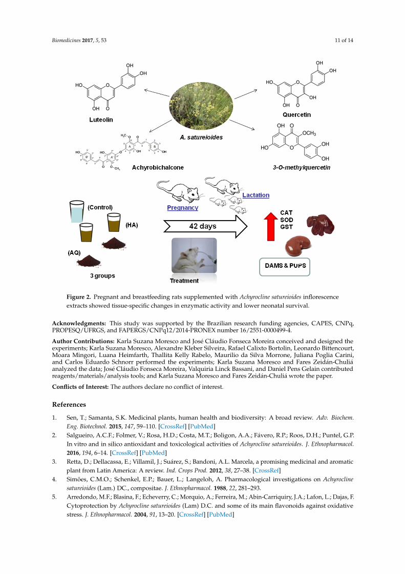
We conclude AQ in concentrations (47 mg·kg−1·day−1) of AS (35 mg·kg−1·day−1) inflorescenceextracts during gestation could lead to in vivo toxicity, reflected in a decreased delivery index andneonatal survival. These effects could be related, at least in part, to variations in tissue-specific redoxhomeostasis and enzymatic activity, especially as the liver and kidney were affected in both damsand pups (Figure 2). It was not possible to determine whether these events could be predominantlyattributed to pre- or early post-natal treatments with AQ of AS inflorescence extracts in pregnant orbreastfeeding rats, respectively. This represents the greatest limitation of our study, which deservesfurther investigation.
Biomedicines 2017, 5, 53 11 of 14Biomedicines 2017, 5, 53 11 of 14
Figure 2. Pregnant and breastfeeding rats supplemented with Achyrocline satureioides inflorescence extracts showed tissue-specific changes in enzymatic activity and lower neonatal survival.
Acknowledgments: This study was supported by the Brazilian research funding agencies, CAPES, CNPq, PROPESQ/UFRGS, and FAPERGS/CNPq12/2014-PRONEX number 16/2551-0000499-4.
Author Contributions: Karla Suzana Moresco and José Cláudio Fonseca Moreira conceived and designed the experiments; Karla Suzana Moresco, Alexandre Kleber Silveira, Rafael Calixto Bortolin, Leonardo Bittencourt, Moara Mingori, Luana Heimfarth, Thallita Kelly Rabelo, Maurilio da Silva Morrone, Juliana Poglia Carini, and Carlos Eduardo Schnorr performed the experiments; Karla Suzana Moresco and Fares Zeidán-Chuliá analyzed the data; José Cláudio Fonseca Moreira, Valquiria Linck Bassani, and Daniel Pens Gelain contributed reagents/materials/analysis tools; and Karla Suzana Moresco and Fares Zeidán-Chuliá wrote the paper.
Conflicts of Interest: The authors declare no conflict of interest.
References 1. Sen, T.; Samanta, S.K. Medicinal plants, human health and biodiversity: A broad review. Adv. Biochem.
Eng. Biotechnol. 2015, 147, 59–110, doi:10.1007/10-2014-273. 2. Salgueiro, A.C.F.; Folmer, V.; Rosa, H.D.; Costa, M.T.; Boligon, A.A.; Fávero, R.P.; Roos, D.H.; Puntel, G.P.
In vitro and in silico antioxidant and toxicological activities of Achyrocline satureioides. J. Ethnopharmacol. 2016, 194, 6–14.
3. Retta, D.; Dellacassa, E.; Villamil, J.; Suárez, S.; Bandoni, A.L. Marcela, a promising medicinal and aromatic plant from Latin America: A review. Ind. Crops Prod. 2012, 38, 27–38, doi:10.1016/j.indcrop.2012.01.006.
4. Simões, C.M.O.; Schenkel,E.P.; Bauer, L.; Langeloh, A. Pharmacological investigations on Achyrocline satureioides (Lam.) DC., compositae. J. Ethnopharmacol. 1988, 22, 281–293.
5. Arredondo, M.F.; Blasina, F.; Echeverry, C.; Morquio, A.; Ferreira, M.; Abin-Carriquiry, J.A.; Lafon, L.; Dajas, F. Cytoprotection by Achyrocline satureioides (Lam) D.C. and some of its main flavonoids against oxidative stress. J. Ethnopharmacol. 2004, 91, 13–20, doi:10.1016/j.jep.2003.11.012.
6. Cosentino, M.; Bombelli, R.; Carcano, E.; Luini, A.; Marino, F.; Crema, F.; Dajas, F.; Lecchini, S. Immunomodulatory properties of Achyrocline satureioides (Lam.) D.C. infusion: A study on human leukocytes. J. Ethnopharmacol. 2008, 116, 501–507, doi:10.1016/j.jep.2007.12.014.
Figure 2. Pregnant and breastfeeding rats supplemented with Achyrocline satureioides inflorescenceextracts showed tissue-specific changes in enzymatic activity and lower neonatal survival.
Acknowledgments: This study was supported by the Brazilian research funding agencies, CAPES, CNPq,PROPESQ/UFRGS, and FAPERGS/CNPq12/2014-PRONEX number 16/2551-0000499-4.
Author Contributions: Karla Suzana Moresco and José Cláudio Fonseca Moreira conceived and designed theexperiments; Karla Suzana Moresco, Alexandre Kleber Silveira, Rafael Calixto Bortolin, Leonardo Bittencourt,Moara Mingori, Luana Heimfarth, Thallita Kelly Rabelo, Maurilio da Silva Morrone, Juliana Poglia Carini,and Carlos Eduardo Schnorr performed the experiments; Karla Suzana Moresco and Fares Zeidán-Chuliáanalyzed the data; José Cláudio Fonseca Moreira, Valquiria Linck Bassani, and Daniel Pens Gelain contributedreagents/materials/analysis tools; and Karla Suzana Moresco and Fares Zeidán-Chuliá wrote the paper.
Conflicts of Interest: The authors declare no conflict of interest.
References
1. Sen, T.; Samanta, S.K. Medicinal plants, human health and biodiversity: A broad review. Adv. Biochem.Eng. Biotechnol. 2015, 147, 59–110. [CrossRef] [PubMed]
2. Salgueiro, A.C.F.; Folmer, V.; Rosa, H.D.; Costa, M.T.; Boligon, A.A.; Fávero, R.P.; Roos, D.H.; Puntel, G.P.In vitro and in silico antioxidant and toxicological activities of Achyrocline satureioides. J. Ethnopharmacol.2016, 194, 6–14. [CrossRef] [PubMed]
3. Retta, D.; Dellacassa, E.; Villamil, J.; Suárez, S.; Bandoni, A.L. Marcela, a promising medicinal and aromaticplant from Latin America: A review. Ind. Crops Prod. 2012, 38, 27–38. [CrossRef]
4. Simões, C.M.O.; Schenkel, E.P.; Bauer, L.; Langeloh, A. Pharmacological investigations on Achyroclinesatureioides (Lam.) DC., compositae. J. Ethnopharmacol. 1988, 22, 281–293.
5. Arredondo, M.F.; Blasina, F.; Echeverry, C.; Morquio, A.; Ferreira, M.; Abin-Carriquiry, J.A.; Lafon, L.; Dajas, F.Cytoprotection by Achyrocline satureioides (Lam) D.C. and some of its main flavonoids against oxidativestress. J. Ethnopharmacol. 2004, 91, 13–20. [CrossRef] [PubMed]
Biomedicines 2017, 5, 53 12 of 14
6. Cosentino, M.; Bombelli, R.; Carcano, E.; Luini, A.; Marino, F.; Crema, F.; Dajas, F.; Lecchini, S.Immunomodulatory properties of Achyrocline satureioides (Lam.) D.C. infusion: A study on human leukocytes.J. Ethnopharmacol. 2008, 116, 501–507. [CrossRef] [PubMed]
7. Morquio, A.; Rivera-Megret, F.; Dajas, F. Photoprotection by topical application of Achyrocline satureioides(‘Marcela’). Phytother. Res. 2005, 19, 486–490. [CrossRef] [PubMed]
8. Sabini, M.C.; Cariddi, L.N.; Escobar, F.M.; Mañas, F.; Comini, L.; Reinoso, E.; Sutil, S.B.; Acosta, A.C.;Núñez Montoya, S.; Contigiani, M.S.; et al. Evaluation of the cytotoxicity, genotoxicity and apoptoticinduction of an aqueous extract of Achyrocline satureioides (Lam.) DC. Food Chem. Toxicol. 2013, 60, 463–470.[CrossRef] [PubMed]
9. Carini, J.P.; Klamt, F.; Bassani, V.L. Flavonoids from Achyrocline. Satureioides: Promising biomolecules foranticancer therapy. RSC Adv. 2013, 4, 3131–3144. [CrossRef]
10. Halliwell, B. Free radicals and other reactive species in disease. eLS 2005, 2005. [CrossRef]11. Barenys, M.; Masjosthusmann, S.; Fritsche, E. Is intake of flavonoid-based food supplements during
pregnancy safe for the developing child–A literature review. Curr. Drug Targets 2017, 18, 196–231.[CrossRef] [PubMed]
12. Lindzon, G.; Sadry, S.; Sharp, J. Toronto Notes for Medical Students, 27th ed.; Type & Graphics Inc.: North York,ON, Canada, 2011.
13. John, L.J.; Shantakumari, N. Herbal medicines use during pregnancy: A review from the middle east.Oman Med. J. 2015, 30, 229–236. [CrossRef] [PubMed]
14. Skibola, C.F.; Smith, M.T. Potential health impacts of excessive flavonoid intake. Free Radic. Biol. Med. 2000,29, 375–383. [CrossRef]
15. Walker, E.H.; Pacold, M.E.; Perisic, O.; Stephens, L.; Hawkins, P.T.; Wymann, M.P.; Williams, R.L. Structuraldeterminants of phosphoinositide 3-kinase inhibition by wortmannin, LY294002, quercetin, myricetin, andstaurosporine. Mol. Cell 2000, 6, 909–919.
16. Baby, B.; Antony, P.; Halabi, W.; Homedi, Z.; Vijayan, R. Structural insights into the polypharmacologicalactivity of quercetin on serine/threonine kinases. Drug Des. Dev. Ther. 2016, 10, 3109–3123.
17. Castelli, S.; Coletta, A.; D’Annessa, I.; Fiorani, P.; Tesauro, C.; Desideri, A. Interaction between naturalcompounds and human topoisomerase I. Biol. Chem. 2012, 393, 1327–1340. [CrossRef] [PubMed]
18. Russo, M.; Spagnuolo, C.; Tedesco, I.; Bilotto, S.; Russo, G.L. The flavonoid quercetin in disease preventionand therapy: Facts and fancies. Biochem. Pharmacol. 2012, 83, 6–15. [CrossRef] [PubMed]
19. Dorta, D.J.; Pigoso, A.A.; Mingatto, F.E.; Rodrigues, T.; Prado, I.M.; Helena, A.F.; Uyemura, S.A.; Santos, A.C.;Curti, C. The interaction of flavonoids with mitochondria: Effects on energetic processes. Chem.-Biol. Interact.2005, 152, 67–78. [CrossRef] [PubMed]
20. Breimer, L.H. Molecular mechanisms of oxygen radical carcinogenesis and mutagenesis: The role of DNAbase damage. Mol. Carcinog. 1990, 3, 188–197. [CrossRef] [PubMed]
21. Hahn, M.; Baierle, M.; Charão, M.F.; Bubols, G.B.; Gravina, F.S.; Zielinsky, P.; Arbo, M.D.; Cristina Garcia, S.Polyphenol-rich food general and on pregnancy effects: A review. Drug Chem. Toxicol. 2016, 545, 1–7.[CrossRef] [PubMed]
22. Zeidan-Chulian, F.; Gelain, D.P.; Kolling, E.A.; Rybarczyk-Filho, J.L.; Ambrosi, P.; Resende Terra, S.; Pires, A.S.;Da Rocha, J.B.T.; Behr, G.A.; Fonseca Moreira, J.C. Major components of energy drinks (caffeine, taurine, andguarana) exert cytotoxic effects on human neuronal SH-SY5Y cells by decreasing reactive oxygen speciesproduction. Oxid. Med. Cell Longev. 2013, 152, 67–78. [CrossRef]
23. Schrö Der-Van Der Elst, J.P.; van Der Heide, D.; Rokos, H.; Morreale De Escobar, G.; Kö Hrle, J. Syntheticflavonoids cross the placenta in the rat and are found in fetal brain. APS 1998, 49209, 253–256.
24. Vanhees, K.; Godschalk, R.W.; Sanders, A.; van Waalwijk van Doorn-Khosrovani, S.B.; van Schooten, F.J.Maternal quercetin intake during pregnancy results in an adapted iron homeostasis at adulthood. Toxicology2011, 290, 351–359. [CrossRef] [PubMed]
25. Carini, J.P.; Leitão, G.G.; Schneider, P.H.; Santos, C.C.; Costa, F.N.; Holzschuh, M.H.; Klamt, F.; Bassani, V.L.Isolation of achyrobichalcone from Achyrocline satureioides by high-speed countercurrent chromatography.Curr. Pharm. Biotechnol. 2015, 16, 66–71. [CrossRef] [PubMed]
26. Bidone, J.; Bica, V.C.; Petrovick, P.R.; Simões, C.M.O.; Koester, L.S.; Bassani, V.L.; Teixeira, H.F. Simultaneousquantification of flavonoids from Achyrocline satureioides by a polar-reversed phase lc method-application toskin permeation/retention studies. Pharmazie 2014, 69, 5–9. [CrossRef] [PubMed]

Biomedicines 2017, 5, 53 13 of 14
27. Marcondes, F.K.; Bianchi, K.; Tanno, A. Determination of the estrous cycle phases of rats: Some helpfulconsiderations. Braz. J. Biol. 2002, 62, 609–614. [CrossRef] [PubMed]
28. Schagger, H.; Cramer, W.A.; Vonjagow, G. The folin by oliver. Anal. Biochem. 1994, 217, 220–230.[CrossRef] [PubMed]
29. Misra, H.P.; Fridovich, I. The role of superoxide anion in the autoxidation of epinephrine and a simple assayfor superoxide dismutase the role of superoxide anion in the epinephrine and a simple assay for superoxidedismutase. J. Biol. Chem. 1972, 247, 3170–3175. [PubMed]
30. Aebi, H. Catalase in vitro. Methods Enzymol. 1984, 105, 121–126. [PubMed]31. Mannervik, B.; Alin, P.; Guthenberg, C.; Jensson, H.; Tahir, M.K.; Warholm, M.; Jörnvall, H. Identification
of three classes of cytosolic glutathione transferase common to several mammalian species: Correlationbetween structural data and enzymatic properties. Proc. Natl. Acad. Sci. USA 1985, 82, 7202–7206. [CrossRef]
32. Habig, W.H.; Jakoby, H.B. Assays for differentiation of glutathione S-Transferases. Methods Enzymol. 1981,77, 398–405. [PubMed]
33. Ellman, G.L. Tissue sulfhydryl groups. Arch. Biochem. Biophys. 1959, 82, 70–77. [CrossRef]34. Levine, R.L.; Garland, D.; Oliver, C.N.; Amici, A.; Climent, I.; Lenz, A.G.; Ahn, B.W.; Shaltiel, S.;
Stadtman, E.R. Determination of carbonyl content in oxidatively modified proteins. Methods Enzymol.1990, 186, 464–478. [PubMed]
35. Draper, H.H.; Hadley, M. Malondialdehyde determination as index of lipid peroxidation. Methods Enzymol.1990, 186, 421–431. [CrossRef] [PubMed]
36. Dankovic, D.A.; Naumann, B.D.; Maier, A.; Dourson, M.L.; Levy, L.S. The scientific basis of uncertainty factorsused in setting occupational exposure limits. J. Occup. Environ. Hyg. 2015, 12, S55–S68. [CrossRef] [PubMed]
37. Day, A.J.; Williamson, G. Biomarkers for exposure to dietary flavonoids: A review of the current evidencefor identification of quercetin glycosides in plasma. Br. J. Nutr. 2001, 86, S105–S110. [CrossRef] [PubMed]
38. Logsdon, A.L.; Herring, B.J.; Lockard, J.E.; Miller, B.M.; Kim, H.; Hood, R.D.; Bailey, M.M. Exposure to greentea extract alters the incidence of specific cyclophosphamide-induced malformations. Birth Defects Res. 2012,95, 231–237. [CrossRef] [PubMed]
39. Schnorr, C.E.; Morrone, M.D.S.; Weber, M.H.; Lorenzi, R.; Behr, G.A.; Moreira, J.C.F. The effects of vitaminA supplementation to rats during gestation and lactation upon redox parameters: Increased oxidativestress and redox modulation in mothers and their offspring. Food Chem. Toxicol. 2011, 49, 2645–2654.[CrossRef] [PubMed]
40. Wu, G.; Fang, Y.Z.; Yang, S.; Lupton, J.R.; Turner, N.D. Glutathione metabolism and its implications forhealth. J. Nutr. 2004, 134, 489–492. [PubMed]
41. Sahu, S.C.; Gray, G.C. Pro-oxidant activity of flavonoids: Effects on glutathione and glutathione S-transferasein isolated rat liver nuclei. Cancer Lett. 1996, 104, 193–196. [CrossRef]
42. Townsend, D.M.; Tew, K.D.; Tapiero, H. The importance of glutathione in human disease. Biomed. Pharmacother.2003, 57, 145–155. [CrossRef]
43. Fridovich, I. Oxygen toxicity: A radical explanation. J. Exp. Biol. 1098, 201, 1203–1209. [CrossRef]44. Bannister, J.V.; Calabarese, L. Assays for SOD. Methods Biochem. 1987, 32, 279–312.45. Kadarian, C.; Broussalis, A.M.; Miño, J.; Lopez, P.; Gorzalczany, S.; Ferraro, G.; Acevedo, C. Hepatoprotective
activity of Achyrocline satureioides (Lam) D.C. Pharmacol. Res. 2002, 45, 57–61. [CrossRef] [PubMed]46. Knowlton, B.J. Introduction to the special section on new ideas about cerebellar function. Behav. Neurosci.
2016, 130, 545–546. [CrossRef] [PubMed]47. Rushworth, M.F.S.; Noonan, M.P.; Boorman, E.D.; Walton, M.E.; Behrens, T.E. Frontal cortex and
reward-guided learning and decision-making. Neuron 2011, 70, 1054–1069. [CrossRef] [PubMed]48. Cheng, S.; Frank, L.M. New experiences enhance coordinated neural activity in the hippocampus. Neuron
2008, 57, 303–313. [CrossRef] [PubMed]49. Morgane, P.J.; Austinlafrance, E.; Bronzino, J.; Tonkiss, J.; Diazcintra, S.; Cintra, L.; Kemper, T.; Galler, J.R.
Prenatal malnutrition and development of the brain. Neurosci. Biobehav. Rev. 1993, 17, 91–128. [CrossRef]50. Desmarchelier, C.; Coussio, J.; Ciccia, G. Antioxidant and free radical scavenging effects in extracts of the
medicinal herb Achyrocline satureioides (Lam.) DC. (“marcela”). Braz. J. Med. Biol. Res. 1998, 31, 1163–1170.[CrossRef] [PubMed]

Biomedicines 2017, 5, 53 14 of 14
51. Chu, K.O.; Wang, C.C.; Chu, C.Y.; Choy, K.W.; Pang, C.P.; Rogers, M.S. Uptake and distribution of catechinsin fetal organs following in utero exposure in rats. Hum. Reprod. 2007, 22, 280–287. [CrossRef] [PubMed]
52. Vanhees, K.; de Bock, L.; Godschalk, R.W.L.; van Schooten, F.J.; van Waalwijk van Doorn-Khosrovani, S.B.Prenatal exposure to flavonoids: Implication for cancer risk. Toxicol. Sci. 2011, 120, 59–67. [CrossRef][PubMed]
© 2017 by the authors. Licensee MDPI, Basel, Switzerland. This article is an open accessarticle distributed under the terms and conditions of the Creative Commons Attribution(CC BY) license (http://creativecommons.org/licenses/by/4.0/).