surface roughness of titanium disks influences the
TRANSCRIPT
RESEARCH Open Access
Surface roughness of titanium disksinfluences the adhesion, proliferation anddifferentiation of osteogenic propertiesderived from humanMaria Alejandra Frias Martinez1, Ísis de Fátima Balderrama1,2*, Paula Stephania Brandão Hage Karam1,Rodrigo Cardoso de Oliveira3, Flávia Amadeu de Oliveira3, Carlos Roberto Grandini4, Fábio Bossoi Vicente4,Andreas Stavropoulos2, Mariana Schutzer Ragghianti Zangrando1 and Adriana Campos Passanezi Sant’Ana1
Abstract
Purpose: The aim of this study was to investigate the response of osteogenic cell lineage and gingival fibroblasticcells to different surface treatments of grade IV commercially pure Titanium (cpTi) disks.
Material and methods: Grade IV cpTi disks with different surfaces were produced: machined (M), sandblasting (B),sandblasting and acid subtraction (NP), and hydrophilic treatment (ACQ). Surface microtopography characteristicsand chemical composition were investigated by scanning electron microscopy (SEM) and energy dispersive x-rayspectrometry (EDS). Adhesion and proliferation of SC-EHAD (human surgically-created early healing alveolar defects)and HGF-1 (human gingival fibroblasts) on Ti disks were investigated at 24 and 48 h, and osteogenic differentiationand mineralization were evaluated by assessing alkaline phosphatase (ALP) activity and alizarin red staining, respectively.
Results: No significant differences were found among the various surface treatments for all surface roughnessparameters, except for skewness of the assessed profile (Rsk) favoring M (p = 0.035 ANOVA). M disks showed a slightlyhigher (p > 0.05; Kruskal-Wallis/Dunn) adhesion of HGF-1 (89.43 ± 9.13%) than SC-EHAD cells (57.11 ± 17.72%). ACQshowed a significantly higher percentage of SC-EHAD (100%) than HGF-1 (69.67 ± 13.97%) cells adhered at 24 h. SC-EHAD cells expressed increased ALP activity in osteogenic medium at M (213%) and NP (235.04%) surfaces, but highermineralization activity on ACQ (54.94 ± 4.80%) at 14 days.
Conclusion: These findings suggest that surface treatment influences the chemical composition and the adhesion anddifferentiation of osteogenic cells in vitro.
Clinical relevance: Hydrophilic surface treatment of grade IV cpTi disks influences osteogenic cell adhesion anddifferentiation, which might enhance osseointegration.
Keywords: Fibroblasts, Titanium, Surface analysis, Cell adhesion, cell proliferation
© The Author(s). 2020 Open Access This article is licensed under a Creative Commons Attribution 4.0 International License,which permits use, sharing, adaptation, distribution and reproduction in any medium or format, as long as you giveappropriate credit to the original author(s) and the source, provide a link to the Creative Commons licence, and indicate ifchanges were made. The images or other third party material in this article are included in the article's Creative Commonslicence, unless indicated otherwise in a credit line to the material. If material is not included in the article's Creative Commonslicence and your intended use is not permitted by statutory regulation or exceeds the permitted use, you will need to obtainpermission directly from the copyright holder. To view a copy of this licence, visit http://creativecommons.org/licenses/by/4.0/.
* Correspondence: [email protected] of Prosthodontics and Periodontics, Discipline of Periodontics,School of Dentistry at Bauru, University of São Paulo, Bauru, SP 17012-901,Brazil2Department of Periodontology, Faculty of Odontology, Malmö University,Malmö, SwedenFull list of author information is available at the end of the article
International Journal ofImplant Dentistry
Martinez et al. International Journal of Implant Dentistry (2020) 6:46 https://doi.org/10.1186/s40729-020-00243-5
IntroductionOsseointegration was originally defined as a direct unionbetween vital bone and a functioning metal implant atoptical microscopy [1]. Since then, dental implants havesignificantly evolved, especially in surface treatment,aiming at improving the quality and/or speed of osseointe-gration [2, 3]. Osseointegration is influenced by varying pa-rameters, including material, design, surface properties,surgical technique, and bone quality [1, 4]. A modificationof implant surface topography has been considered as anessential parameter contributing to the success of dentalimplants [2]. A major part of implantology research focuseson the development of surface modifications that would beable of improving the biologic characteristics of titanium [5,6]. Considering that, new surfaces of dental implants weredeveloped to improve biological cell responses, guiding thedifferentiation of stem cells in osteoblasts and enhancingosseointegration [7]. Material biocompatibility is intimatelyrelated to cell behavior [8, 9]. Implant surface microtopo-graphy influences adhesion, proliferation, differentiation,and extracellular matrix synthesis by osteoblasts and othercells [9–15]. Surface roughness also influences the behaviorof osteoprecursor cells by stimulating proliferation and in-ducing differentiation into osteoblasts [16–19] and bonegrowth at implant threads, which may affect the process ofosseointegration [6, 20–26]. Popular treatments of implantsurfaces, increasing roughness, include blasting and/or acidetching, as well as addition of nanoparticles and risingunder protection with N2, followed by storage in NaClsolution [7, 27]. These modifications affect cell behavior,improving the adsorption of proteins, and favoring osteo-blastic cells differentiation [7, 28, 29]. The effects of titan-ium surface topography in the behavior of osteoblasts areassociated to adhesion-related cell function [7, 30]. Re-cently, the granulation tissue present in surgically cre-ated bone defects in the jaws of humans after 21 days ofhealing was isolated and characterized in vitro for thefirst time, and this granulation tissue removed from hu-man surgically created early healing alveolar defects(SC-EHAD cells) demonstrated osteogenic properties[31]. Cells exhibited a spindle-shaped morphology atearlier passages, changing to a cuboidal one at laterpassages. Alkaline phosphatase (ALP) activity andmineralization were observed both in conventional andosteogenic medium. Fresh samples of SC-EHAD tissue ex-hibited CD34− and CD45−phenotypes, while SC-EHADcells at later passages exhibited, besides that, a CD105−,CD166−, and collagen type I+ phenotype. These findingssuggest that SC-EHAD is a possible source of progenitorcells [31]. However, the properties of this human celllineage when cultivated on different implant surfaces areyet unknown. Considering that, the aim of this study is toinvestigate the response of SC-EHAD cells to differentgrade IV cpTi disks surface treatments.
Material and methodsTitanium discs and surface preparationGrade IV cp Ti disks, 6.0 mm × 2.0 mm (Neodent®,Curitiba, Brazil) with the following surface treatments,were used:
1) Machined (M)2) Sandblasted (B—subtraction with silicon,
aluminum, and titanium oxide creating abrasion ondisks surface)
3) Sandblasted and acid etched (combination:hydrofluoric, nitric, and sulfuric acid)(NP—Neoporos®, surface)
4) Sandblasted, acid etched, and immersed in 0.9%sodium chloride (ACQ—Acqua®, surface), were used.
Surface analysisDisks surfaces were examined by scanning electron mi-croscopy, at × 500 magnification in a high resolutionscanning electron microscopy (Machining TechnologyLaboratory, School of Engineering, São Paulo StateUniversity. Bauru, Brazil). Roughness characteristicswere examined in SEM photomicrographs by SurfCharJplugin (available for download at: http://imagej.nih.gov/ij/), which measures roughness parameters according toISO 4287/2000: Ra (arithmetical mean deviation), Rq(root mean square deviation), Rku (kurtosis of theassessed profile), Rsk (skewness of the assessed profile),Rv (lowest valley), Rp (highest peak), and Rt (total heightof the profile). The chemical composition of titaniumdisks was investigated by energy dispersive x-ray de-tector (EDS). One sample of each group was analyzed inthree different locations to detect the chemical compos-ition in order to detect possible differences betweenregions.
Cell cultureAfter approval of the Committee of Ethics in Research(CAAE 32274414900005417), SC-EHAD cells were ob-tained from two systemically healthy individuals, non-smoking, 40 and 45 years old males who signed the consentform for newly forming bone graft technique during peri-odontal treatment of deep infrabony periodontal pocketsand furcation lesions, as described the treatment protocolby Passanezi and co-authors in 1989 [32]. Briefly, a 10-mmheight × 3.5mm diameter surgical defect was created at thealveolar ridge with a cylindrical diamond bur under copiousirrigation, as previously method described [31–33]. After21 days [32], defects were re-opened, the granulation tissuepresent in the healing defects was collected with a Lucascurette, and a portion of the material was transferred to aFalcon tube containing Dulbecco’s minimal essentialmedium (DMEM, Sigma-Aldrich, USA), 20% fetal bovineserum (FBS, Sigma-Aldrich, USA), 200U/mLG potassium
Martinez et al. International Journal of Implant Dentistry (2020) 6:46 Page 2 of 11
penicillin, 200mg/mL streptomycin sulfate, and 20 μg/mLamphotericin B (Sigma-Aldrich, USA), allowing the estab-lishment of primary culture of osteoblasts after centrifuga-tion of the fine dissected fragments for 3min andpositioning in 25 cm2 tissue flasks containing DMEM sup-plemented with 20% FBS and 2% antibiotic-antimycotic so-lution. The option to collection of SC-EHAD after 21 daysafter creation of defect was based on the clinical applica-tions already proposed [32].Human gingival fibroblasts (HGF-1), used as positive
controls for comparisons, were obtained from two sys-temically healthy, non-smoking volunteers submitted toa gingivectomy by internal bevel incision (n = 1) and afree gingival graft (n = 1) procedure. The tissue removedduring surgical procedures was positioned in Falcontubes containing DMEM (Eagles minimal essentialmedium) supplemented with 20% FBS (fetal bovineserum) and 2% antibiotic-antimycotic solution, andtransported to cell culture lab to the establishment ofprimary culture. Tissue fragments were finely dissectedin Petri dishes containing saline buffer (PBS) and 2%antibiotic-antimycotic solution. Cell separation was per-formed by mechanical-enzymatic process by submersionof tissue fragments in trypsin solution at 37 °C followedby centrifugation for 3 min. This process was repeatedonce, and the resultant cell pellet was re-suspended inDMEM supplemented with 20% FBS and 2% antibiotic-antimycotic solution. Cells were allowed to expand inhumidified atmosphere containing 5% CO2 at 37
°C untilreaching subconfluency (approximately 80% of cultivablearea covered by cells); when cells were detached withtrypsin solution (Sigma-Aldrich, USA) and transferred toprogressively greater tissue flasks until experimental pro-cedures were performed, the cell sources were pooled toHGF-1 lineage group.
Adhesion and proliferation assays5 × 104 cells in 50 μl of DMEM were platted on 5 disks/group (SC-EHAD or HGF-1) in 96-well plates. Fourhours after platting, 200 μl of culture medium was addedin each well, completely covering the disks. Sampleswere fixated after 24 h (adhesion assay) and 48 h (prolifer-ation assay) with Karnovsky solution (6% glutaraldehydeand 4% paraphormaldehyde in 0.2M cadodylate buffer)and post-fixated with 2% osmium tetroxide in cacodylatebuffer at 4 °C for 2 h. After dehydration in graded alcohols,samples were immersed in 100% hexametildisilazane(HMDS) at room temperature for 24 h, air-dried, andsputter-coated with gold for examination by scanningelectron microscopy (SEM). From each specimen, 2 pho-tomicrographs were obtained (central and a randomlyselected peripheric area) at × 500 magnification.Each photomicrograph was coded and analyzed by a
blinded examiner for assessing the area covered by cells.
After calibration of the image size, a grid was super-posed on SEM images in an image analysis software(ImageJ®, NIH, Bethesda, USA) software, and the areawithout cells was determined and expressed as % of thetotal disk area. Additionally, cell adhesion and prolifera-tion were analyzed on each image, by a blinded exam-iner, using an index from 0–5 (Table 1).
Differentiation and mineralization assaysSC-EHAD and HGF-1 cells were cultured on the disksand plastic (negative control) at an initial density of 7 ×10³ cells in 24-well plates. After initial adhesion (4 h) instandard medium, cells were cultivated in standard andosteogenic medium. The osteogenic medium was ob-tained by supplementation of standard medium with 10mM β-glicerophosphate (Sigma, USA) and 50 μg/ml as-corbic acid. The assays were performed in triplicates.Alkaline phosphatase (ALP) was determined in poolafter 14 days. The culture medium was discarded, andcells were twice washed with saline solution and lysed byadding 100× Triton solution. ALP was observed at lysedcells using 25 μl of the sample added in 200 μl of p-nitrophenol phosphate. Total protein measurementswere performed according to Bradford method. The op-tical density was read at 405 nm in a spectrophotometer(FLUOstar Optima microplate reader, BMG, Leicester,UK). ALP activity (U/ml) was normalized according toprotein measurements for each sample.Alizarin red staining was performed after 14 and 28
days of culture of SC-EHAD and HGF-1 cells on the dif-ferent substrates in triplicates at initial density of 5 × 10⁴
cells in osteogenic and standard culture media, as de-scribed before. The culture medium was discarded; con-fluent cell layers were washed with PBS at 37 °C, fixatedwith 4% formalin solution, and again washed with PBS.The disks were stained with alizarin red S (2%, pH 4.2,Merck). Images of the disks were captured with stereo-microscope and processed in Image J to determine thepercentage of area showing mineralized nodules stainedby alizarin red.
Statistical analysisData were analyzed in Graph Pad Prism 6.0 for Mac,adopting a significance level of 5% (α = 0.05) for all tests.Comparisons of roughness characteristics among groups
Table 1 Index of cell adhesion and proliferation
Score Characteristics
0 No cells adhered
1 < 50% area covered by cells
2 50–<75% area covered by cells
3 75–100% area covered by cells
4 100% area covered by cells + formation of a second cell layer
Martinez et al. International Journal of Implant Dentistry (2020) 6:46 Page 3 of 11
were performed by ANOVA post hoc Tukey. The per-centage of area covered by cells and index of cell adhe-sion and proliferation were compared by non-parametricKruskal-Wallis post hoc Dunn at the different periods.Inter-group analysis of alizarin red staining was per-formed by non-parametric Kruskal-Wallis post hocDunn, and intra-group analysis was performed bynon-parametric Wilcoxon test. Differences in ALP ac-tivity and mineralization assay were assessed with theWilcoxon test.
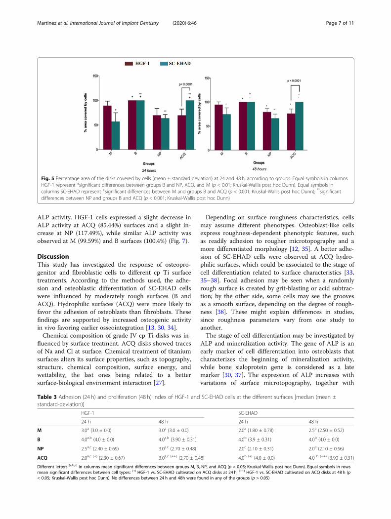
ResultsSurface characteristicsNo differences in roughness were found among groupsregarding all parameters, except for Rsk (p < 0.05;ANOVA post hoc Tukey; Table 2). Surface characteris-tics are presented in SEM photomicrographs (Fig. 1),while the 3D-plot surface characteristics of the differentsurfaces are illustrated in Fig. 2. The chemical compos-ition performed by EDS was resulted in weight percent-age (weight %), and Fig. 3 showed each surface found. Mand NP surface were composed by titanium only (100%);B surface showed the presence of titanium (62.13%),aluminum (10.78%), and oxygen (27.09%); and ACQ sur-face showed presence of titanium (61.99%), sodium(15.84%), and Chlorum (22.17%).
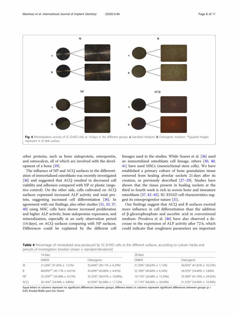
Adhesion and proliferation assays24 hoursACQ and B surfaces were covered at 100% by SC-EHADcells (Figs. 4 and 5). M surfaces showed 57.11% ±17.72%, and NP surfaces showed 63.87% ± 7.23% areacovered by SC-EHAD. ACQ surfaces showed 69.67% ±13.97% area covered by HGF-1, and B surfaces were cov-ered at 100%. M surfaces showed 89.43% ± 9.13% andNP surfaces showed 69.77% ± 13.97% area covered byHGF-1.
48 hoursSignificant differences in the area covered by cells werefound only for SC-EHAD on ACQ surfaces (p = 0.03;Wilcoxon) when compared with areas covered by HGF-1 (Fig. 5). Significant differences between the area occu-pied by cells at 24 and 48 h were observed only for SC-EHAD on ACQ surfaces (p = 0.03; Wilcoxon). A signifi-cant smaller area occupied by HGF-1 (p < 0.01; Kruskal-Wallis) and SC-EHAD (p < 0.005; Kruskal-Wallis) wasobserved in B than in NP and ACQ (Fig. 5).
Adhesion and proliferation indexSC-EHADA higher prevalence of score 4 was observed for groupsB and ACQ compared to groups M (median 2.0) and NP(median 2.0) at 24 h. At 48 h, significant differences werefound between M (median 2.5) and NP (median 2.0)compared to B (median 4.0) and ACQ (median 4.0) sur-faces. Significant differences between scores of HGF-1and SC-EHAD cells cultivated on ACQ surfaces werefound at 24 and 48 h. No significant intra-group differ-ences were found between 24 and 48 h (Table 3).
HGF-1A higher prevalence of score 4 was observed at B com-pared to NP (median: 2.5) and ACQ (median: 2.0) sur-faces at 24 h. No significant differences were foundbetween groups B and M (median: 3.0). Similar resultswere observed after 48 hours and there were no signifi-cant intra-group differences between 24 and 48 hours(Table 3).
Alizarin red stainingMineralized nodules were observed at all surfaces at thedifferent periods of investigation (Fig. 6). Comparativeanalysis between groups by Kruskal-Wallis post hocDunn showed significant differences in SC-EHAD culti-vated on M (31.45% ± 1.51%) and ACQ (54.94% ±4.80%) at 14 days. No significant differences were ob-served between groups at 28 days. Intra-group analysisby Wilcoxon test showed no differences in the percent-age of mineralization observed at 14 and 28 days for anysurface (Table 4).
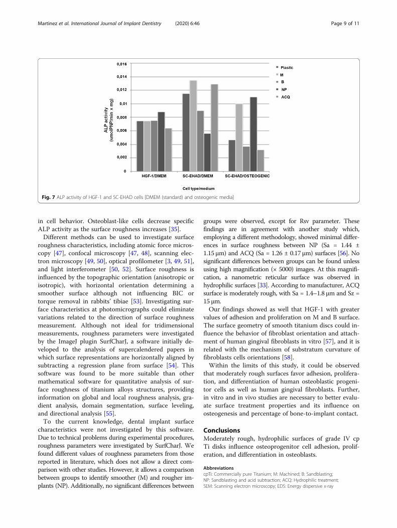
ALP activitySC-EHAD and HGF-1 cells were expressed in all sur-faces as well as on the plastic (negative control) (Fig. 7).SC-EHAD cells expressed increased ALP activity inosteogenic medium at M (213%) and NP (235.04%) sur-faces, while decreased activity was observed at B(79.05%) and ACQ (68.37%) surfaces (Fig. 7). SC-EHADcells cultivated on standard medium expressed a slightlyincreased ALP activity only at ACQ (112.17%), while M(90%), B (78.26%), and NP (48.86%) showed decreased
Table 2 Roughness parameters analyzed by SurfCharJ plugin(ImageJ, NIH, USA) [Sample length 100 μm, surface leveling]
M B NP ACQ
Rq 0.1289 0.1482 0.1956 0.1616
Ra 0.0983 0.1104 0.1574 0.1252
Rsk 1.9698* 1.5975 0.6909 1.3361
Rkv 13.4801 3.5427 0.1222 2.4683
Rv – 0.2893 – 0.2174 – 0.4170 – 0.2591
Rp 1.9496 0.8084 0.6484 1.1277
Rt 2.2388 1.0258 1.0653 1.3867
Rc – 0.0046 0.0012 0.0009 – 0.0030
*Significant differences between M, B, NP, and ACQ (p = 0.035)
Martinez et al. International Journal of Implant Dentistry (2020) 6:46 Page 4 of 11
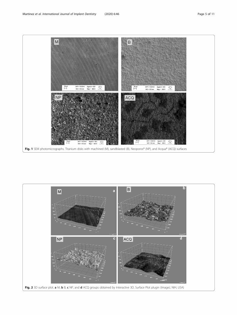
Fig. 1 SEM photomicrographs. Titanium disks with machined (M), sandblasted (B), Neoporos® (NP), and Acqua® (ACQ) surfaces
Fig. 2 3D surface plot. a M, b B, c NP, and d ACQ groups obtained by Interactive 3D, Surface Plot plugin (ImageJ, NIH, USA)
Martinez et al. International Journal of Implant Dentistry (2020) 6:46 Page 5 of 11
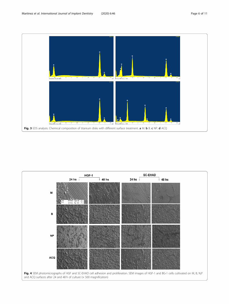
Fig. 3 EDS analysis. Chemical composition of titanium disks with different surface treatment. a M. b B. c NP. d ACQ
Fig. 4 SEM photomicrographs of HGF and SC-EHAD cell adhesion and proliferation. SEM images of HGF-1 and BG-1 cells cultivated on M, B, N,Pand ACQ surfaces after 24 and 48 h of culture (× 500 magnification)
Martinez et al. International Journal of Implant Dentistry (2020) 6:46 Page 6 of 11
ALP activity. HGF-1 cells expressed a slight decrease inALP activity at ACQ (85.44%) surfaces and a slight in-crease at NP (117.49%), while similar ALP activity wasobserved at M (99.59%) and B surfaces (100.4%) (Fig. 7).
DiscussionThis study has investigated the response of osteopro-genitor and fibroblastic cells to different cp Ti surfacetreatments. According to the methods used, the adhe-sion and osteoblastic differentiation of SC-EHAD cellswere influenced by moderately rough surfaces (B andACQ). Hydrophilic surfaces (ACQ) were more likely tofavor the adhesion of osteoblasts than fibroblasts. Thesefindings are supported by increased osteogenic activityin vivo favoring earlier osseointegration [13, 30, 34].Chemical composition of grade IV cp Ti disks was in-
fluenced by surface treatment. ACQ disks showed tracesof Na and Cl at surface. Chemical treatment of titaniumsurfaces alters its surface properties, such as topography,structure, chemical composition, surface energy, andwettability, the last ones being related to a bettersurface-biological environment interaction [27].
Depending on surface roughness characteristics, cellsmay assume different phenotypes. Osteoblast-like cellsexpress roughness-dependent phenotypic features, suchas readily adhesion to rougher microtopography and amore differentiated morphology [12, 35]. A better adhe-sion of SC-EHAD cells were observed at ACQ hydro-philic surfaces, which could be associated to the stage ofcell differentiation related to surface characteristics [33,35–38]. Focal adhesion may be seen when a randomlyrough surface is created by grit-blasting or acid subtrac-tion; by the other side, some cells may see the groovesas a smooth surface, depending on the degree of rough-ness [38]. These might explain differences in studies,since roughness parameters vary from one study toanother.The stage of cell differentiation may be investigated by
ALP and mineralization activity. The gene of ALP is anearly marker of cell differentiation into osteoblasts thatcharacterizes the beginning of mineralization activity,while bone sialoprotein gene is considered as a latemarker [30, 37]. The expression of ALP increases withvariations of surface microtopography, together with
Fig. 5 Percentage area of the disks covered by cells (mean ± standard deviation) at 24 and 48 h, according to groups. Equal symbols in columnsHGF-1 represent *significant differences between groups B and NP, ACQ, and M (p < 0.01; Kruskal-Wallis post hoc Dunn). Equal symbols incolumns SC-EHAD represent +significant differences between M and groups B and ACQ (p < 0.001; Kruskal-Wallis post hoc Dunn); **significantdifferences between NP and groups B and ACQ (p < 0.001; Kruskal-Wallis post hoc Dunn)
Table 3 Adhesion (24 h) and proliferation (48 h) index of HGF-1 and SC-EHAD cells at the different surfaces [median (mean ±standard-deviation)]
HGF-1 SC-EHAD
24 h 48 h 24 h 48 h
M 3.0a (3.0 ± 0.0) 3.0a (3.0 ± 0.0) 2.0a (1.80 ± 0.78) 2.5a (2.50 ± 0.52)
B 4.0a,b (4.0 ± 0.0) 4.0a,b (3.90 ± 0.31) 4.0b (3.9 ± 0.31) 4.0b (4.0 ± 0.0)
NP 2.5a,c (2.40 ± 0.69) 3.0a,c (2.70 ± 0.48) 2.0c (2.10 ± 0.31) 2.0a (2.10 ± 0.56)
ACQ 2.0a,c (+) (2.30 ± 0.67) 3.0a,c (++) (2.70 ± 0.48) 4.0b (+) (4.0 ± 0.0) 4.0 b (++) (3.90 ± 0.31)
Different letters (a,b,c) in columns mean significant differences between groups M, B, NP, and ACQ (p < 0.05; Kruskal-Wallis post hoc Dunn). Equal symbols in rowsmean significant differences between cell types: (+) HGF-1 vs. SC-EHAD cultivated on ACQ disks at 24 h; (++) HGF-1 vs. SC-EHAD cultivated on ACQ disks at 48 h (p< 0.05; Kruskal-Wallis post hoc Dunn). No differences between 24 h and 48h were found in any of the groups (p > 0.05)
Martinez et al. International Journal of Implant Dentistry (2020) 6:46 Page 7 of 11
other proteins, such as bone sialoprotein, osteopontin,and osteocalcin, all of which are involved with the devel-opment of a bone [39].The influence of NP and ACQ surfaces in the differenti-
ation of immortalized osteoblasts was recently investigated[36] and suggested that ACQ resulted in decreased cellviability and adhesion compared with NP or plastic (nega-tive control). On the other side, cells cultivated on ACQsurfaces expressed increased ALP activity and total pro-tein, suggesting increased cell differentiation [36]. Inagreement with our findings, also other studies [31, 33, 37,40] using MSC cells have shown increased proliferationand higher ALP activity, bone sialoprotein expression, andmineralization, especially at an early observation period(14 days), on ACQ surfaces comparing with NP surfaces.Differences could be explained by the different cell
lineages used in the studies. While Soares et al. [36] usedan immortalized osteoblasts cell lineage, others [30, 40,41] have used MSCs (mesenchymal stem cells). We haveestablished a primary culture of bone granulation tissueremoved from healing alveolar sockets 21 days after itscreation, as previously described [27–29]. Studies haveshown that the tissue present in healing sockets at thethird to fourth week is rich in woven bone and immatureosteoblasts [37, 42–45]. SC-EHAD cell characteristics sug-gest its osteoprogenitor nature [31].Our findings suggest that ACQ and B surfaces exerted
more influence in cell differentiation than the additionof β-glicerophosphate and ascorbic acid in conventionalmedium. Pivodova et al. [46] have also observed a de-crease in the expression of ALP activity after 72 h, whichcould indicate that roughness parameters are important
Fig. 6 Mineralization activity of SC-EHAD cells at 14 days in the different groups. a Standard medium. b Osteogenic medium. *Squared imagesrepresent ¼ of disk surface
Table 4 Percentage of mineralized area produced by SC-EHAD cells at the different surfaces, according to culture media andperiods of investigation [median (mean ± standard-deviation)]
14 days 28 days
DMEM Osteogenic DMEM Osteogenic
M 31.56%a (31.45% ± 1.51%) 35.64%a (38.11% ± 4.29%) 37.20%a (36.63% ± 1.12%) 36.05%a (41.65% ± 10.23%)
B 48.69%a,b (45.17% ± 6.61%) 45.69%a (45.80% ± 4.45%) 52.18%a (49.40% ± 6.54%) 54.92%a (54.40% ± 3.84%)
NP 35.33%a,b (34.38% ± 4.57%) 35.35%a (36.97% ± 10.08%) 19.11%a (26.68% ± 13.39%) 33.30%a (41.59% ± 24.42%)
ACQ 56.16%b (54.94% ± 4.80%) 62.93%a (65.68% ± 17.52%) 57.71%a (64.00% ± 20.69%) 51.55%a (54.96% ± 10.49%)
Equal letters in columns represent no significant differences between groups; different letters in columns represent significant differences between groups (p <0.05; Kruskal-Wallis post hoc Dunn)
Martinez et al. International Journal of Implant Dentistry (2020) 6:46 Page 8 of 11
in cell behavior. Osteoblast-like cells decrease specificALP activity as the surface roughness increases [35].Different methods can be used to investigate surface
roughness characteristics, including atomic force micros-copy [47], confocal microscopy [47, 48], scanning elec-tron microscopy [49, 50], optical profilometer [3, 49, 51],and light interferometer [50, 52]. Surface roughness isinfluenced by the topographic orientation (anisotropic orisotropic), with horizontal orientation determining asmoother surface although not influencing BIC ortorque removal in rabbits’ tibiae [53]. Investigating sur-face characteristics at photomicrographs could eliminatevariations related to the direction of surface roughnessmeasurement. Although not ideal for tridimensionalmeasurements, roughness parameters were investigatedby the ImageJ plugin SurfCharJ, a software initially de-veloped to the analysis of supercalendered papers inwhich surface representations are horizontally aligned bysubtracting a regression plane from surface [54]. Thissoftware was found to be more suitable than othermathematical software for quantitative analysis of sur-face roughness of titanium alloys structures, providinginformation on global and local roughness analysis, gra-dient analysis, domain segmentation, surface leveling,and directional analysis [55].To the current knowledge, dental implant surface
characteristics were not investigated by this software.Due to technical problems during experimental procedures,roughness parameters were investigated by SurfCharJ. Wefound different values of roughness parameters from thosereported in literature, which does not allow a direct com-parison with other studies. However, it allows a comparisonbetween groups to identify smoother (M) and rougher im-plants (NP). Additionally, no significant differences between
groups were observed, except for Rsv parameter. Thesefindings are in agreement with another study which,employing a different methodology, showed minimal differ-ences in surface roughness between NP (Sa = 1.44 ±1.15 μm) and ACQ (Sa = 1.26 ± 0.17 μm) surfaces [56]. Nosignificant differences between groups can be found unlessusing high magnification (× 5000) images. At this magnifi-cation, a nanometric reticular surface was observed inhydrophilic surfaces [33]. According to manufacturer, ACQsurface is moderately rough, with Sa = 1.4–1.8 μm and Sz =15 μm.Our findings showed as well that HGF-1 with greater
values of adhesion and proliferation on M and B surface.The surface geometry of smooth titanium discs could in-fluence the behavior of fibroblast orientation and attach-ment of human gingival fibroblasts in vitro [57], and it isrelated with the mechanism of substratum curvature offibroblasts cells orientations [58].Within the limits of this study, it could be observed
that moderately rough surfaces favor adhesion, prolifera-tion, and differentiation of human osteoblastic progeni-tor cells as well as human gingival fibroblasts. Further,in vitro and in vivo studies are necessary to better evalu-ate surface treatment properties and its influence onosteogenesis and percentage of bone-to-implant contact.
ConclusionsModerately rough, hydrophilic surfaces of grade IV cpTi disks influence osteoprogenitor cell adhesion, prolif-eration, and differentiation in osteoblasts.
AbbreviationscpTi: Commercially pure Titanium; M: Machined; B: Sandblasting;NP: Sandblasting and acid subtraction; ACQ: Hydrophilic treatment;SEM: Scanning electron microscopy; EDS: Energy dispersive x-ray
Fig. 7 ALP activity of HGF-1 and SC-EHAD cells [DMEM (standard) and osteogenic media]
Martinez et al. International Journal of Implant Dentistry (2020) 6:46 Page 9 of 11
spectrometry; SC-EHAD: Human surgically created early healing alveolardefects; HGF-1: Human gingival fibroblasts; ALP: Alkaline phosphatase;Rsk: Skewness of the assessed profile; Ra: Arithmetical mean deviation;Rq: Root mean square deviation; Rku: Kurtosis of the assessed profile;Rv: Lowest valley; Rp: Highest peak; Rt: Total height of the profile;DMEM: Eagle’s minimal essential medium; PBS: Phosphate buffered saline;FBS: Fetal bovine serum; HMDS: Hexametildisilazane; Ti: Titanium; Al: Aluminum;O: Oxygen; Na: Sodium; Cl: Chlorine; MSCs: Mesenchymal stem cells
AcknowledgementsThe authors thank Neodent (Curitiba, Brazil) for the donation of Ti disks usedin this study. The authors declare no conflict of interest.
Authors’ contributionsAll authors discussed the results and contributed to the final manuscript.Maria Alejandra Frias Martinez performed the analysis, collected the data,and wrote the paper. Ísis de Fátima Balderrama performed the analysis,collected the data, and wrote the paper. Paula Stephania Brandão HageKaram proposed and performed the cell culture analysis. Rodrigo Cardoso deOliveira proposed and performed the cell culture analysis. Flávia Amadeu deOliveira proposed and performed the cell culture analysis. Carlos RobertoGrandini proposed and performed the surface analysis. Fábio Bossoi Vicenteproposed and performed the surface analysis. Andreas Stavropoulos verifiedthe analytical methods and provided critical feedback and helped the shapethe research. Mariana Schutzer Ragghianti Zangrando contributed data andanalysis tools. Adriana Campos Passanezi Sant’Ana conceived and designedthe analysis and idea, verified the analytical methods, and supervised thefindings of this work. The authors read and approved the final manuscript.
FundingThis work was supported by the JJGC Industry and Commerce of DentalMaterials–NEODENT (Curitiba, Paraná, Brazil), which has donated the titaniumdisks used in this research.
Availability of data and materialsThe authors from this work are available to support data.
Ethics approval and consent to participateThis paper describes the response of osteogenic and fibroblastic cells todifferent surface treatments of cpTi disks, and this research was approved bythe Committee of Ethics in Research in Human (CAAE 32274414900005417),Bauru School of Dentistry/University of São Paulo.
Consent for publicationAll authors are aware for the publication of this work.
Competing interestsThe authors declare no conflict of interest.
Author details1Department of Prosthodontics and Periodontics, Discipline of Periodontics,School of Dentistry at Bauru, University of São Paulo, Bauru, SP 17012-901,Brazil. 2Department of Periodontology, Faculty of Odontology, MalmöUniversity, Malmö, Sweden. 3Department of Biological Sciences, School ofDentistry at Bauru, University of São Paulo, Bauru, SP, Brazil. 4Anelasticity andBiomaterials Laboratory, São Paulo State University, Bauru, SP, Brazil.
Received: 18 March 2020 Accepted: 2 July 2020
References1. Albrektsson T, Branemark PI, Hansson HA, Lindstrom J. Osseointegrated
titanium implants. Acta Orthop Scand. 1981;52:155–70.2. Albrektsson T, Wennerberg A. The impact of oral implants – past and future,
1966 – 2042. J Can Dent Assoc. 2005;71(5):327.3. Liu R, Lei T, Dusevich V, Yao X, Liu Y, Walker MP, Wang Y, Ye L. Surface
characteristics and cell adhesion: a comparative study of four commercialdental implants. J Prosthod. 2013;22:641–51.
4. Albrektsson T, Jacobsson M. Bone–metal interface in osseointegration. J ProsthetDent. 1987;57:597–607.
5. Wennerberg A, Albrektsson T. Effects of titanium surface topography onbone. Impl Res. 2009;20(Suppl 4):172–84.
6. Wennerberg A, Albrektsson T. On implants surfaces: a review of currentknwoledge and opinions. Int J Oral Maxillofac Implants. 2009;24:63–74.
7. Mendonça G, Mendonça DBS, Simões LGP, Araújo AL, Leite ER, Duarte WR,Aragão FJL, Cooper LF. The effects of implant surface nanoscale features onosteoblastspecific gene expression. Biomaterials. 2009;30:4053–62.
8. Anselme K, et al. The relative influence of the topography and chemistry ofTiA16V4 surfaces on osteoblastic cell behaviour. Biomaterials. 2000;21:1567–77.
9. Anselme K. Osteoblast adhesion on biomaterials. Biomaterials. 2000;21(7):667–81.
10. Lange R, Luthen F, Beck U, Rychly J, Baumann A, Nebe B. Cell–extracellularmatrix interactions and physiochemical characteristics of titanium surfacesdepend on the roughness of the material. Biomol Eng. 2002;19:255–61.
11. Magnani A, Priamo A, Pasqui D, Barbucci R. Cell behavior on chemicallymicrostructured surfaces. Mater Sci Eng C. 2003;23:315–28.
12. Boyan BD, Lossdorfer S, Wang L, Zhao G. Osteoblasts generate anosteogenic microenvironment when grown on surfaces with roughmicrotopographies. Eur Cells Mat. 2003;5(2):11–2.
13. Rupp F, Scheideler L, Rehbein D, Axmann D, Geis-Gerstorfer J. Roughnessinduced dynamic changes of wettability of acid etched titanium implantmodification. Biomaterials. 2004;25:1429–38.
14. Le Guehennec L, Lopez-Heredia MA, Enkel B, Weiss P, Amouriq Y, Layrolle P.Osteoblastic cell behaviour on different titanium implant surfaces. ActaBiomater. 2008;4(3):535–43.
15. Elias CN, Meirelles L. Improving osseointegration of dental implants. Exp RevMed Dev. 2010;7(2):241–56.
16. Boyan BD, Schwartz Z, Lohmann CH, Sylvia VL, Cochran DL, Dean DD, PuzasJE. Pretreatment of bone with osteoclasts affects phenotypic expression ofosteoblast-like cells. J Orthop Res. 2003;21:638–47.
17. Lüthen F, Lange R, Becker P, et al. The influence of surface roughness oftitanium on b1 and b3-integrin adhesion and the organization offibronectin in human osteoblastic cells. Biomaterials. 2005;26:2423–40.
18. Vallés G, Gil-Garay E, Munuera L, et al. Modulation of the cross-talk betweenmacrophages and osteoblasts by titanium based particles. Biomaterials.2008;29:2326–35.
19. Nebe B, Lüthen F, Lange R, et al. Topography-induced alterations inadhesion structures affect mineralization in human osteoblasts on titanium.Mater Sci Eng. 2004;24:619–24.
20. Bagno A, Di Bello C. Surface treatments and roughness properties of Tibased biomaterials. J Mater Sci Mater Med. 2004;15:935–49.
21. Hazan R, Brener R, Oron U. Bone growth to metal implants is regulated bytheir surface chemical properties. Biomaterials. 1993;14:570–4.
22. Larsson C, et al. Bone response to surface-modified titanium implants:studies on the early tissue response to machined and electropolishedimplants with different oxide thicknesses. Biomaterials. 1996;17:605–16.
23. Giavaresi G, Fini M, Cigada A, Chiesa R, Rondelli G, Rimondini L, Vicoli AldiniN, Martini L, Giardino R. Histomorphometric and microhardness assessmentsof sheep cortical bone surrounding titanium implants with different surfacetreatments. J Biomed Mater Res. 2003;67A:112–20.
24. Kim MJ, Choi MU, Kim CW. Activation of phospholipase D1 by surface roughnessof titanium in MG63 osteoblast-like cell. Biomaterials. 2006;27:5502–11.
25. Albrektsson T, Sennerdy L, Wennerberg A. State of the art of oral implants.Periodontology 2000. 2008;47:15–26.
26. Albrektsson T, Buser D, Sennerdy L. On crestal/marginal bone loss arounddental implants. Int J Periodont Rest Dent. 2013;33(1):9–11.
27. Schwarz F, Wieland M, Schwartz Z, Zhao G, Rupp F, Geis-Gerstorfer J,Schedle A, Broggini N, Bornstein M, Buser D, Ferguson SJ, Becker J, BoyanBD, Cochran DL. Potential of chemically modified hydrophilic surfacecharacteristics to support tissue integration of titanium dental implants. JBiomed Mater Res Part B: Appl Biomater. 2009;88B:544–57.
28. Bang S-M, Moon H-J, Kwon Y-D, Yoo J-Y, Pae A, Kwon IK. Osteoblastic andosteoclastic differentiation on SLA and hydrophilic modified SLA titaniumsurfaces. Clin Oral Impl Res. 2014;25:831–7.
29. Zhao GZO, Schwartz Z, Wieland M, Landolt D, Boyan BD. Osteoblastlike cellsare sensitive to submicron-scale surface structure. Clin Oral Implants Res.2006;17:258–64.
30. Mendonça G, Mendonça DBS, Aragão FJL, Cooper LF. The combination ofmicron and nanotopography by H2SO4/H2O2 treatment and its effects onosteoblast-specific gene expression of hMSCs. J Biomed Mater Res. 2010;94A:169–79.
Martinez et al. International Journal of Implant Dentistry (2020) 6:46 Page 10 of 11
31. Sant’Ana ACP, Damante CA, Frias Martinez MA, Valdivia MAM, Karam PSBH,de Oliveira FA, et al. Isolation and characterization of progenitor cells fromsurgically created early healing alveolar defects in humans: a preliminarystudy. J Periodontol. 2018;89(11):1326–33.
32. Passanezi E, Janson WA, Nahás D, Campos A Jr. Newly forming boneautografts to treat periodontal infrabony pockets: clinical and histologicalevents. Int J Periodontics Rest Dent. 1989;9(2):140–51.
33. Sant'ana AC, Ferraz BF, de Rezende ML, Greghi SL, Damante CA, Passanezi E.Newly forming bone graft: a novel surgical approach to the treatment ofdenuded roots. J Appl Oral Sci. 2012;20(3):392–8.
34. Mendonça G, Mendonça DBS, Oliveira LS, Arújo CA. Effects of humanmesenchymal stem cells on hydrophilic surfaces. ImplantNews 2013. 10(6Part A):111–6 [In Portuguese].
35. Martin JY, Schwartz Z, Hummert TW, Schraub DM, Simpson J, Lankford J,Dean DD, Cochran DL, Boyan BD. Effect of titanium surface roughness onproliferation, differentiation, and protein synthesis of human osteoblast-likecells (MG63). J Biomed Mater Res. 1995;29:389–401.
36. Soares PBF, Moura CCG, Coró CGC, Reis MVP, Zanetta-Barbosa D, Soares CJ.Biological characterization of implant surfaces – in vitro study. Dental Mat.2013;29(Suppl):e28. https://doi.org/10.1016/j.dental.2013.08.059.
37. Bryington M, Mendonc AG, Nares S, Cooper LF. Osteoblastic and cytokinegene expression of implant-adherent cells in humans. Clin Oral Impl Res.2014;25:52–8.
38. Lincks J, Boyan BD, Blanchard CR, Lohmann CH, Liu Y, Cochran DL, DeanDD, Schwartz Z. Response of MG63 osteoblast-like cells to titanium andtitanium alloy is dependente on surface roughness and composition.Biomaterials. 1998;19:2219–32.
39. Cooper LF, Zhou Y, Takebe J, Guo J, Abron A, Holmen A, et al. Fluoridemodification effects on osteoblast behavior and bone formation at TiO2grit-blasted c.p. titanium endosseous implants. Biomaterials. 2006;27:926–36.
40. Olivares-Navarrete R, Hyzy SL, Hutton DL, Erdman CP, Wieland M, Boyan BD,Schwartz Z. Direct and indirect effects of microstructured titaniumsubstrates on the induction of mesenchymal stem cell differentiationtowards the osteoblast lineage. Biomaterials. 2010;31:2728–35.
41. Bradford MM. A rapid and sensitive method for the quantitation ofmicrogram quantities of protein utilizing the principle of protein-dyebinding. Anal Biochem. 1976;72:248–54.
42. Evian CI, Rosenberg ES, Coslet JG, Corn H. The osteogenic activity of boneremoved from healing extraction sockets in humans. J Periodontol. 1982;53:81–5.
43. Cardaropoli G, Araújo M, Hayacibara R, Sukekava F, Lindhe J. Healing ofextraction sockets and surgically produced – augmented and non-augmented – defects in alveolar ridge. An experimental study in dogs. JClin Periodontol. 2005;32:435–40.
44. Cardaropoli G, Araújo M, Lindhe J. Dynamics of bone tissue formation intooth extraction sites. J ClinPeriodontol. 2003;30:809–18.
45. Penteado R, Romito GA, Pustiglioni FE, Marques MM. Morphological andproliferative analysis of the healing tissue in human alveolar sockets covered ornot by an e-PTFE membrane: a preliminary immunohistochemical andultrastructural study. Braz J Oral Sci. 2005;4:664–9.
46. Pivodova F, Frankova J, Dolezel P, Ulrichova J. The response of osteoblast-like Saos-2 cells to modified titanium surfaces. Int J Oral Maxillofac Implants.2013;28:1386–94.
47. Wennerberg A. On surface roughness and implant incorporation. Tese(Doutorado). Departmant of Biomaterial/Handicap Research, University ofGötenborg. Götenborg, Sweden, 1996.
48. Wennerberg A, Hallgren C, Johansson C, Danelli S. A histomorphometricevaluation of screw-shaped implants each prepared with two surfaceroughnesses. Clin Oral Implants Res. 1998;9:11–9.
49. Mustafa K, Wennerberg A, Wroblewski J, Hultenby K, Lopez BS, Arvidson K.Determining optimal surface roughness of TiO(2) blasted titanium implantmaterial for attachment, proliferation and differentiation of cells derived fromhuman mandibular alveolar bone. Clin Oral Implants Res. 2001;12:515–25.
50. Calvo-Guirado JL, Satorres-Nieto M, Aguilar-Salvatierra A, Delgado-Ruiz RA,de Val JEM-S, Gargallo-Albiol J, Gómez-Moreno G, Romanos GE. Influence ofsurface treatment on osseointegration of dental implants: histological,histomorphometric and radiological analysis in vivo. Clin Oral Invest. 2015;19:509–17.
51. Elias CN, Fernandes DJ, Resende CRS, Roestelb J. Mechanical properties,surface morphology and stability of a modified commercially pure highstrength titanium alloy for dental implants. Dental Mat. 2015;31:e1–e13.
52. Gasik M, Braem A, Chaudhari A, Duyck J, Vleugels J. Titanium implants withmodified surfaces: meta-analysis of in vivo osteointegration. Mat Sci EnginC. 2015;49:152–258.
53. Hallgren C, Sawase T, Ortengren U, Wennerberg A. Histomorphometric andmechanical evaluation of the bone-tissue response to implants preparedwith different orientation of surface topography. Clin Impl Dent Rel Res.2001;3:194–203.
54. Chinga G, Johnsen PO, Dougherty R, Berli EL, Walter J. Quantification of the3D microstructure of SC surfaces. J Microscopy. 2007;227(Part 3):254–65.
55. Safdar A. Microstructures and surface roughness of EBM produced Ti-6Al-4V. Dissertation.Material Science, Malmö University, Malmö, Sweden, 2010.
56. Sartoretto SC, Alves ATNN, Resende RFB, Calasans-Maia J, Granjeiro JM,Calasans-Maia MD. Early osseointegration driven by the surface chemistryand wettability of dental implants. J Appl Oral Sci. 2015;23:279–87.
57. Inoue T, Cox JE, Pilliar RM, Melcher AH. Effect of the surface geometry ofsmooth and porous-coated titanium alloy on the orientation of fibroblastsin vitro. J Biomed Mater Res. 1987;21:107–26.
58. Dunn GA, Heath JP. A new hypothesis of contact guidance in tissue cells.Exp Cell Res. 1976;101:1–14.
Publisher’s NoteSpringer Nature remains neutral with regard to jurisdictional claims inpublished maps and institutional affiliations.
Martinez et al. International Journal of Implant Dentistry (2020) 6:46 Page 11 of 11