survival patients with pulmonary metastases in testicular ... · patients with metastases will have...
TRANSCRIPT

Survival patients with pulmonary metastases in testicular cancer
MAN MILENA*, ANTIGONA TROFOR**, DANA ALEXANDRESCU***, OVIDIU BALANESCU****,
IOANA NEAGOE*, MONICA POP*, RUXANDRA RAJNOVEANU*,
Pneumology, Medical Informatics Department
*University of Medicine and Pharmacy “Iuliu HaŃieganu” Cluj-Napoca, ** University of Medicine and
Pharmacy “Gr.T.Popa” Iaşi, *** University “Transilvania” Braşov, **** Oncologic Institute Cluj
Caraiman 3 Street, Cluj-Napoca
ROMANIA
Abstract:
Testicular cancer is a rare disease reaching only 1% of all cancer types characteristic
to men with ages between 15 and 34 years and has shown an increasing worldwide incidence
over the past 30 years. In contrast, the mortality rate has decreased. Hematogen metastases are
responsible for the apparition of pulmonary nodules. Determining the pulmonary relapse
model, studying prognosis factors for defining risk groups and applying different therapeutic
strategies with the evaluation of survival represent the study’s main objectives. We conducted a survey from January 2000 to December 2005 on 17 patients admitted in Cluj-
Napoca Oncology Institute. We introduced in the study patients diagnosed with testicular carcinoma
and pulmonary metastases, we analyzed risk factor and evolution of the diseases with survival
function calculated since cancer diagnosis, and the other calculated since pulmonary metastases. We
evaluated the risk factors correlated with survival: the average age ,place of origin, histology
tumor markers for our batch: Beta HCG, AFP (alpha-fetoprotein), LDH ,types of pulmonary
metastases, the presence of other metastases, the number of metastatic locations, the time span
between the apparition of symptoms and diagnosis, the free interval the diagnosis of the
primary tumor and metastasis, and value of tumor markers at the time of diagnosis. Based on
these prognosis factors, the patients were divided into a good prognosis category (good) and
unfavorable (poor) . Because the risk factors usually available are not sufficient to identify
the subgroups of patients with an unfavorable prognosis, we tried to evaluate new genetic
markers which could prove their prognosis value. We also sought to evaluate the involvement
of the hTERT gene in testicular pathology.
Patients with metastases will have a different prognosis depending on the relapse
model and will require individual strategies selected based on the risk factors by combining
available therapeutic modalities. Early diagnosis and treatment of metastasis may lead to an
improvement in the survival rate of cancer patients
.Key-Words: prognostic factor, survival, pulmonary metastases hTERT Gene, testicular carcinomas, risk factor
WSEAS TRANSACTIONS on BIOLOGY and BIOMEDICINE
Man Milena, Antigona Trofor, Dana Alexandrescu, Ovidiu Balanescu, Ioana Neagoe, Monica Pop, Ruxandra Rajnoveanu
ISSN: 1109-9518 82 Issue 3, Volume 7, July 2010
Introduction
Testicular cancer is a rare disease reaching
only 1% of all cancer types characteristic to men
with ages between 15 and 34 years (1,2) The
incidence of testicular cancer in Europe is rising
with doubling every 20 years. The current incidence
is 63/100 000/year, with the highest rate in Northern
European countries (68/100 000/year) The death
rate is very low (3.8 cases/100 000/year) (3)
Therapeutic improvements emerged at the same
time with the introduction of chemotherapy in the
70’s make it so that 95% of patients with testicular
cancer (1) and 70-80% of patients with metastases
become treatable (1).
The etiology of germinal tumors is not
known. An increase in the frequency among
patients with development anomalies and testicle
descent was described (4) and this signaled the
existence of genetic components(5).
The histology of testicular tumors divides
these cancers into seminomas (50%), teratomas or
non-seminomas and mixed tumors (6). Tumor
markers [AFP, β-HCG and lactate dehydrogenase
(LDH)] are needed for risk assessment according to
UICC/IGCCCG stage and prognostic index.
Markers are determined before orchiectomy and
repeated a minimum of 7 days after orchiectomy
(for differentiation of stage and IGCCCG
prognostic group). HCG must be followed until
normalization.
The metastasis pattern is predictable.(3)
The first metastases to appear are retroperitoneal adenopathies (regional metastases). Hematogen
metastases are responsible for the apparition of
pulmonary nodules and left side over-clavicle
ganglion metastases. Hepatic bony metastases are
rare and represent an unfavorable prognosis factor.
The presence of pulmonary metastases does not
necessarily imply an unfavorable risk group if the
patients have not presented a previous therapeutic
failure and if they are not accompanied by other
visceral metastases (liver, brain, bone).
The identification of prognostic factors is
valuable due to the following three reasons:
1. Optimum treatment may be selected for each
patient
2. Various therapeutic strategies could be compared
among groups of patients with similar recurrence
risks and treatments
3. The knowledge that allows the identification
of recurrence patterns may be improved and new
treatment strategies established. Why spend money
on inefficient therapies that are sub-optimally dosed
in high-risk patients or excessive in low risk
patients? The selection of the optimum therapy is a
challenge for each team involved in the treatment of
cancer patients with lung metastases (7)
2 Problem Formulation
THE WORK HYPOTHESIS Pulmonary metastases are frequently met
among cases of testicular cancers. Determining the
pulmonary relapse model, studying prognosis
factors for defining risk groups and applying
different therapeutic strategies with the evaluation
of survival represent the study’s main objectives.
Knowing these prognosis factors and evaluating
them allows us to identify metastasizing risk factors
(vascular invasion, histology of the primary tumor,
precocious relapse) Patient division into high or low
risk categories according to prognostic factors may allow differential approaches, individualized follow-up for the early detection of recurrences and efficient treatments
aimed at increasing the patients’ quality of life and survival chances.
MATERIAL
Between 2000 and 2006 we studied 17
patients diagnosed with testicular cancer who
presented pulmonary, pleural and mediastinal
metastases. The patients were admitted at the “Leon
Daniello” Clinical Pneumonology Hospital, the
Oncology Institute from Cluj or the Oncology
Department of the County Clinical Hospital.
Inclusion criteria: patients with testicular cancer
(tumors with germinal cells diagnosed from a
histological point of view and/or serological one)
which presented pulmonary metastases on the chest
– pleural pulmonary x-ray and/or CT as well as the
presence of Beta HCG and AFP markers. The
histopathology exam of pulmonary metastases was
not necessary for the inclusion in the study.
Exclusion criteria: patients without follow up
WSEAS TRANSACTIONS on BIOLOGY and BIOMEDICINE
Man Milena, Antigona Trofor, Dana Alexandrescu, Ovidiu Balanescu, Ioana Neagoe, Monica Pop, Ruxandra Rajnoveanu
ISSN: 1109-9518 83 Issue 3, Volume 7, July 2010
METHOD
Evaluation of the patients was made: trough
anamnesis, clinical exam, biological samples and
imagistic explorations. The histopathology exam of
the testicular tumor was made before acceptance
into the study as it was established as an inclusion
criterion. We analyzed the main prognosis factors:
age, histology, position of the primary tumor,
localization of metastases (regional or remote), the
number of metastatic localizations, the type of
pulmonary metastasis, the time span between the
apparition of symptoms and diagnosis, the free
interval the diagnosis of the primary tumor and
metastasis, and value of tumor markers at the time
of diagnosis. Based on these prognosis factors, the
patients were divided into a good prognosis
category (good) and unfavorable (poor)(1) :
-Good risk: HCG and ATP increase, metastasized
cervical ganglions, minimal pulmonary metastases
(less than 5 on the pulmonary field and under 3 cm),
mediastinal masses under 50% of the intra-thoracic
diameter, solitary over 2 cm metastases with no
abdominal masses;
-Poor risk: extended pulmonary metastases (more
than 10 on the pulmonary field), pulmonary
metastases bigger than 3 cm with/without
abdominal masses, mediastinal masses greater than
50% of the intra/thoracic diameter, abdominal
masses, non/pulmonary visceral metastases
(hepatic, bone, cerebral).
The Multidisciplinary Board (oncologist,
radiotherapist, surgeon, thoracic surgeon) decided
the best therapy adapted to every case in part. The
type of treatment applied was orchiectomy (before
start of chemotherapy) associated with
chemotherapy. The initial chemotherapy used
Cisplatin in various combinations (bleomycin,
vinblastin, etoposid, ifosfamid) with curative intent.
Evaluation of treatment was made at 28 days after
the first cycle and the last chemotherapeutic cycle.
In case of remission we performed radiotherapy (20
Gy with 2.0 Gy for each session, 5 days a week).
3 Results
The diagnosis of testicular cancer was made
before entering the study due to the apparition of
symptoms on a local level in 15 from the 17 patients
included in the batch. The pulmonary metastasis
diagnosis was initially made only for two patients
and we tried to find the starting point. In the
hereditary collateral antecedents of 2 patients we
emphasized first degree relatives with neoplasia
(mother and grand mother with cancer without
finding any men with testicular cancer). We only
emphasized the presence of an ectopic testicle as
risk factor in one patient.
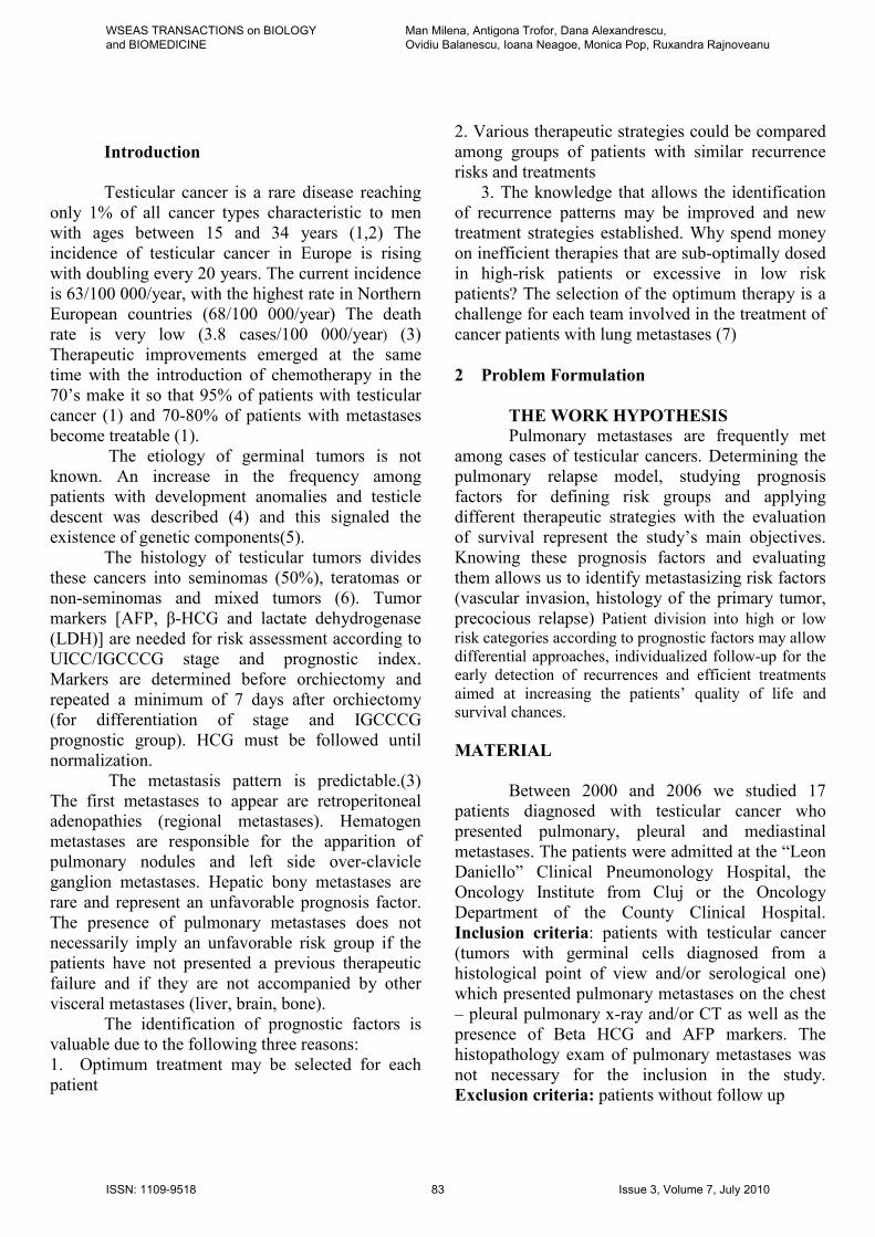
From the survival table for testicular cancer
one can notice the 6 months survival rate was 0.93
(93%), the 13 months survival rate was 0.78 (78%),
at 26 months it was 0.64 (64%), that at 39 months
was 0.55 (55%) and the survival rate at 72 months
equaled 0.38 (38%). These survival rates can also
be seen in the diagram below:
Figure 1.Survival rate
The clinical pathological characteristics of
patients with testicular tumors and evaluation of
risk factors associated to the host.
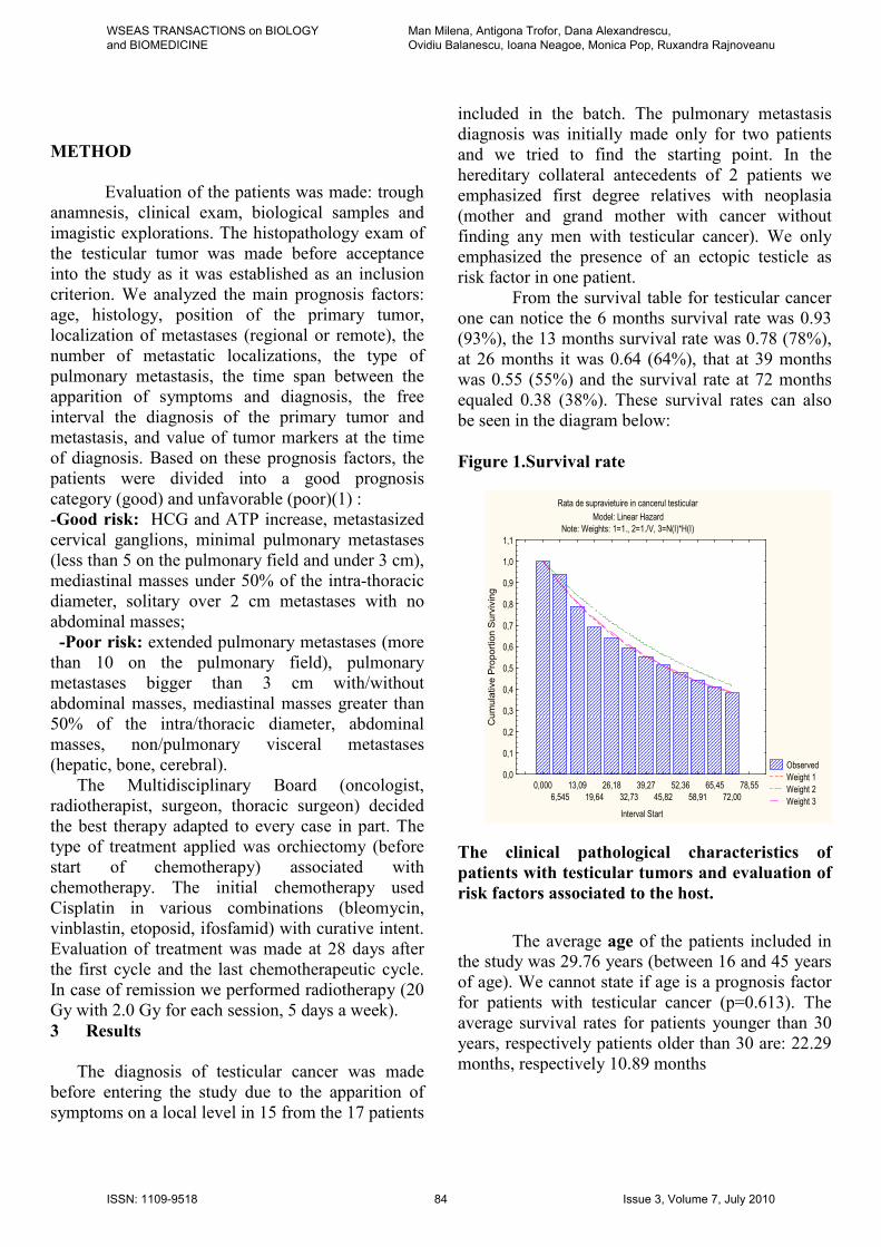
The average age of the patients included in the study was 29.76 years (between 16 and 45 years
of age). We cannot state if age is a prognosis factor
for patients with testicular cancer (p=0.613). The
average survival rates for patients younger than 30
years, respectively patients older than 30 are: 22.29
months, respectively 10.89 months
Rata de supravietuire in cancerul testicular
Model: Linear Hazard
Note: Weights: 1=1., 2=1./V, 3=N(I)*H(I)
Observed
Weight 1
Weight 2
Weight 3
0,000
6,545
13,09
19,64
26,18
32,73
39,27
45,82
52,36
58,91
65,45
72,00
78,55
Interval Start
0,0
0,1
0,2
0,3
0,4
0,5
0,6
0,7
0,8
0,9
1,0
1,1
Cumulative Proportion Surviving
WSEAS TRANSACTIONS on BIOLOGY and BIOMEDICINE
Man Milena, Antigona Trofor, Dana Alexandrescu, Ovidiu Balanescu, Ioana Neagoe, Monica Pop, Ruxandra Rajnoveanu
ISSN: 1109-9518 84 Issue 3, Volume 7, July 2010
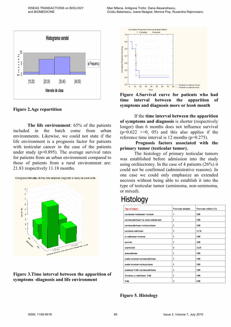
Figure 2.Age repartition
The life environment: 65% of the patients
included in the batch come from urban
environments. Likewise, we could not state if the
life environment is a prognosis factor for patients
with testicular cancer in the case of the patients
under study (p=0.895). The average survival rates
for patients from an urban environment compared to
those of patients from a rural environment are:
21.83 respectively 11.18 months.
Figure 3.Time interval between the apparition of
symptoms -diagnosis and life environment
Figure 4.Survival curve for patients who had
time interval between the apparition of
symptoms and diagnosis more or less6 month
If the time interval between the apparition
of symptoms and diagnosis is shorter (respectively
longer) than 6 months does not influence survival
(p=0.622 >>0, 05) and this also applies if the
reference time interval is 12 months (p=0.275).
Prognosis factors associated with the
primary tumor (testicular tumor). The histology of primary testicular tumors
was established before admission into the study
using orchiectomy. In the case of 4 patients (26%) it
could not be confirmed (administrative reasons). In
one case we could only emphasize an extended
necrosis without being able to establish it into the
type of testicular tumor (seminoma, non-seminoma, or mixed).
5,885,8811YolkYolk
5,885,8811Teratom, Teratom, cccc embrionar. embrionar. YolkYolk
5,885,8811seminomseminom++YolkYolk++carcinoembrionarcarcinoembrionar
5,885,8811seminsemin+teratom++teratom+coriocarcinomcoriocarcinom
5,885,8811seminsemin+teratom++teratom+carcinoembrionarcarcinoembrionar
5,885,8811nonseminomnonseminomaa
23,5323,5344unprecisedunprecised
5,885,8811necrnecrosisosis
5,885,8811cccc embrionar+teratomembrionar+teratom
11,7611,7622carcinom embrionarcarcinom embrionar
5,885,8811carcinoembrionarcarcinoembrionar++coriocarcinomcoriocarcinom
5,885,8811carcinoembrionarcarcinoembrionar+tu. sinus +tu. sinus endodermalendodermal
5,885,8811carcincarcinomaoma++seminseminomaoma+ teratom+ teratom
FrecvenFrecvenŃŃe relative (%)e relative (%)FrecvenFrecvenŃŃe absolutee absoluteTipTipe of tumorse of tumors
HistologyHistology
Figure 5. Histology
Cumulative Proportion Surviving (Kaplan-Meier)
Complete Censored
Pacienti cu interval <6 luni
Pacienti cu interval 6 luni;0 10 20 30 40 50 60 70 80
Time
0,3
0,4
0,5
0,6
0,7
0,8
0,9
1,0
Cumulative Proportion Surviving
Histograma varstei
0
5
10
[10;20) [20;30) [30;40) [40;50)
Intervale de clasa
Fre
cvente
Frequency
WSEAS TRANSACTIONS on BIOLOGY and BIOMEDICINE
Man Milena, Antigona Trofor, Dana Alexandrescu, Ovidiu Balanescu, Ioana Neagoe, Monica Pop, Ruxandra Rajnoveanu
ISSN: 1109-9518 85 Issue 3, Volume 7, July 2010
Our batch comprises cases of mixed tumors
(65%) and non-seminomas (6%) but lacks cases of
pure seminomas. This can explain the favorable
evolution at a reduced number of patients
(compared to data from literature). The histology of
our batch does not influence survival, p= 0.078 >>0,
05.
The tumor markers for our batch: Beta
HCG, AFP (alpha-fetoprotein), LDH does not
influence survival (p= 0.786), (p= 0.345)
respectively (p= 0.153).
Figure 6. Value of tumor markers
Prognosis factors associated with the
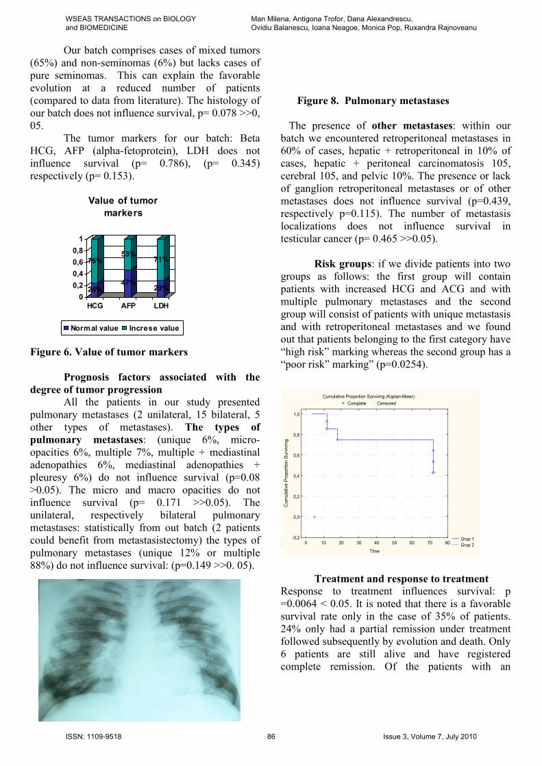
degree of tumor progression All the patients in our study presented
pulmonary metastases (2 unilateral, 15 bilateral, 5
other types of metastases). The types of
pulmonary metastases: (unique 6%, micro-
opacities 6%, multiple 7%, multiple + mediastinal
adenopathies 6%, mediastinal adenopathies +
pleuresy 6%) do not influence survival (p=0.08
>0.05). The micro and macro opacities do not
influence survival (p= 0.171 >>0.05). The
unilateral, respectively bilateral pulmonary
metastases: statistically from out batch (2 patients
could benefit from metastasistectomy) the types of
pulmonary metastases (unique 12% or multiple
88%) do not influence survival: (p=0.149 >>0. 05).
Figure 8. Pulmonary metastases
The presence of other metastases: within our
batch we encountered retroperitoneal metastases in
60% of cases, hepatic + retroperitoneal in 10% of
cases, hepatic + peritoneal carcinomatosis 105,
cerebral 105, and pelvic 10%. The presence or lack
of ganglion retroperitoneal metastases or of other
metastases does not influence survival (p=0.439,
respectively p=0.115). The number of metastasis
localizations does not influence survival in
testicular cancer (p= 0.465 >>0.05).
Risk groups: if we divide patients into two
groups as follows: the first group will contain
patients with increased HCG and ACG and with
multiple pulmonary metastases and the second
group will consist of patients with unique metastasis
and with retroperitoneal metastases and we found
out that patients belonging to the first category have
“high risk” marking whereas the second group has a
“poor risk” marking” (p=0.0254).
Figure 9.Survival curve in risk group
Treatment and response to treatment Response to treatment influences survival: p
=0.0064 < 0.05. It is noted that there is a favorable
survival rate only in the case of 35% of patients.
24% only had a partial remission under treatment
followed subsequently by evolution and death. Only
6 patients are still alive and have registered
complete remission. Of the patients with an
Cumulative Proportion Surviving (Kaplan-Meier)
Complete Censored
Grup 1
Grup 20 10 20 30 40 50 60 70 80
Time
-0,2
0,0
0,2
0,4
0,6
0,8
1,0
Cumulative Proportion Surviving
24%
76%
47%
53%
29%
71%
0
0,2
0,4
0,6
0,8
1
HCG AFP LDH
Value of tumor
markers
Normal value Increse value
WSEAS TRANSACTIONS on BIOLOGY and BIOMEDICINE
Man Milena, Antigona Trofor, Dana Alexandrescu, Ovidiu Balanescu, Ioana Neagoe, Monica Pop, Ruxandra Rajnoveanu
ISSN: 1109-9518 86 Issue 3, Volume 7, July 2010
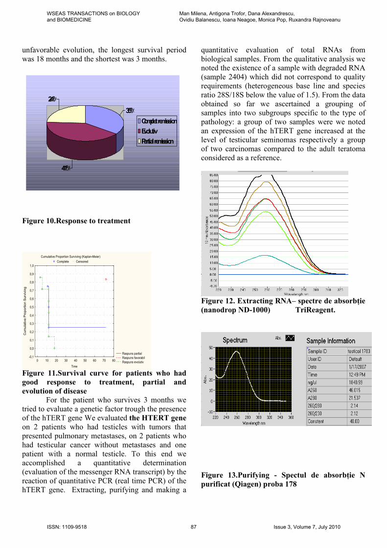
unfavorable evolution, the longest survival period
was 18 months and the shortest was 3 months.
Figure 10.Response to treatment
Figure 11.Survival curve for patients who had
good response to treatment, partial and
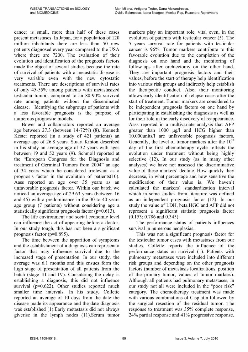
evolution of disease For the patient who survives 3 months we
tried to evaluate a genetic factor trough the presence
of the hTERT gene We evaluated the HTERT gene
on 2 patients who had testicles with tumors that
presented pulmonary metastases, on 2 patients who
had testicular cancer without metastases and one
patient with a normal testicle. To this end we
accomplished a quantitative determination
(evaluation of the messenger RNA transcript) by the
reaction of quantitative PCR (real time PCR) of the
hTERT gene. Extracting, purifying and making a
quantitative evaluation of total RNAs from
biological samples. From the qualitative analysis we
noted the existence of a sample with degraded RNA
(sample 2404) which did not correspond to quality
requirements (heterogeneous base line and species
ratio 28S/18S below the value of 1.5). From the data
obtained so far we ascertained a grouping of
samples into two subgroups specific to the type of
pathology: a group of two samples were we noted
an expression of the hTERT gene increased at the
level of testicular seminomas respectively a group
of two carcinomas compared to the adult teratoma
considered as a reference.
Figure 12. Extracting RNA– spectre de absorbŃie
(nanodrop ND-1000) TriReagent.
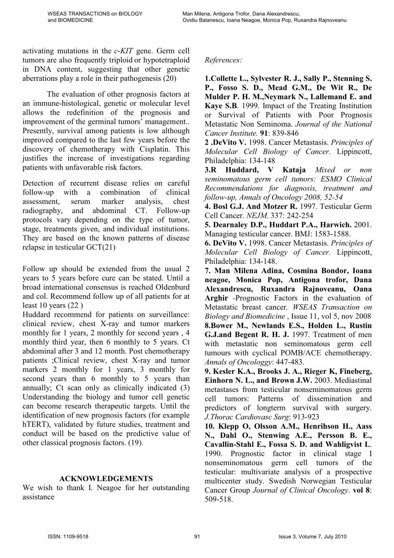
Figure 13.Purifying - Spectul de absorbŃie N
purificat (Qiagen) proba 178
Cumulative Proportion Surviving (Kaplan-Meier)
Complete Censored
Raspuns partial
Raspuns favorabil
Raspuns evolutiv0 10 20 30 40 50 60 70 80
Time
-0,1
0,0
0,1
0,2
0,3
0,4
0,5
0,6
0,7
0,8
0,9
1,0
Cumulative Proportion Surviving
41%
24%
35%
Complet remission
Evolutiv
Partial remission
WSEAS TRANSACTIONS on BIOLOGY and BIOMEDICINE
Man Milena, Antigona Trofor, Dana Alexandrescu, Ovidiu Balanescu, Ioana Neagoe, Monica Pop, Ruxandra Rajnoveanu
ISSN: 1109-9518 87 Issue 3, Volume 7, July 2010
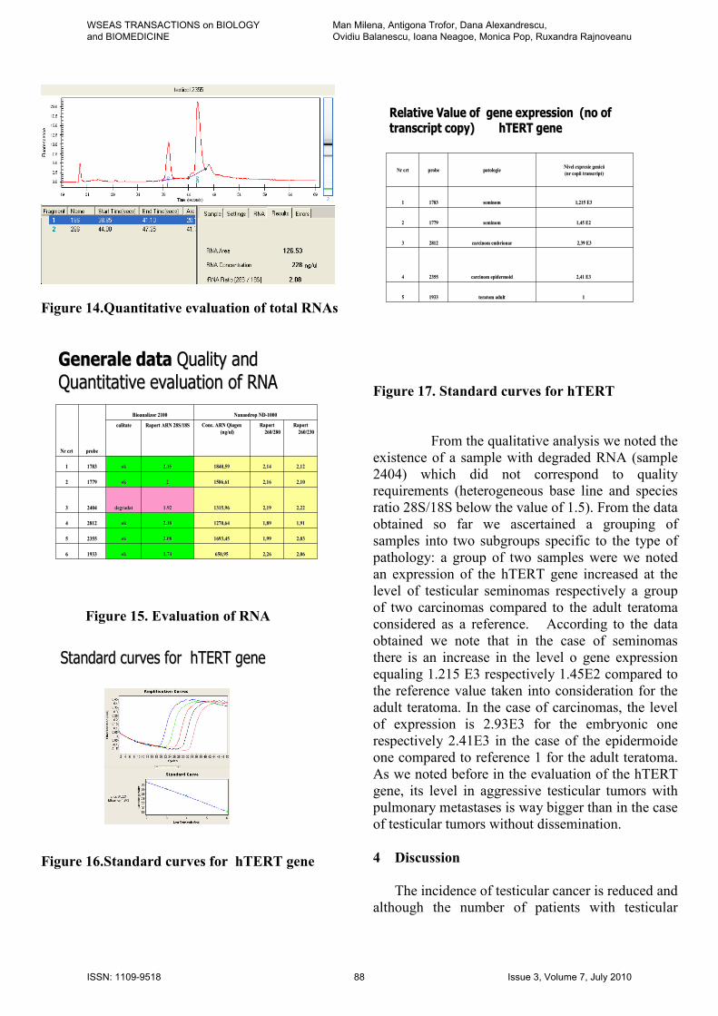
Figure 14.Quantitative evaluation of total RNAs
GGeneraleenerale data data QQualityuality andand
QQuantitativeuantitative evaluation of RNAevaluation of RNA
2,062,062,262,26650,95650,951,741,74okok1933193366
2,032,031,991,991693,451693,452.082.08okok2355235555
1,911,911,891,891270,641270,642.182.18okok2812281244
2,222,222,192,191315,961315,961.921.92degradatdegradat2404240433
2,102,102,162,161586,611586,6122okok1779177922
2,122,122,142,141840,591840,592,152,15okok1783178311
Raport Raport
260/230260/230
Raport Raport
260/280260/280
ConcConc. ARN . ARN QiagenQiagen
((ngng/ul)/ul)Raport ARN 28S/18SRaport ARN 28S/18Scalitatecalitate
NanaodropNanaodrop NDND--10001000BioanalizorBioanalizor 21002100
probeprobeNrNr crtcrt
Figure 15. Evaluation of RNA
SStandardtandard curves forcurves for hTERThTERT genegene
Figure 16.Standard curves for hTERT gene
Relative Value of Relative Value of gene expression gene expression ((nono ofof
transcripttranscript copycopy) ) hTERThTERT genegene
11teratom adultteratom adult1933193355
2,41 E32,41 E3carcinom carcinom epidermoidepidermoid2355235544
2,39 E32,39 E3carcinom embrionarcarcinom embrionar2812281233
1,45 E21,45 E2seminomseminom1779177922
1,215 E31,215 E3seminomseminom1783178311
Nivel expresie genică Nivel expresie genică
((nrnr copii transcript)copii transcript)patologiepatologieprobeprobeNrNr crtcrt
Figure 17. Standard curves for hTERT
From the qualitative analysis we noted the
existence of a sample with degraded RNA (sample
2404) which did not correspond to quality requirements (heterogeneous base line and species
ratio 28S/18S below the value of 1.5). From the data
obtained so far we ascertained a grouping of
samples into two subgroups specific to the type of
pathology: a group of two samples were we noted
an expression of the hTERT gene increased at the
level of testicular seminomas respectively a group
of two carcinomas compared to the adult teratoma
considered as a reference. According to the data
obtained we note that in the case of seminomas
there is an increase in the level o gene expression
equaling 1.215 E3 respectively 1.45E2 compared to
the reference value taken into consideration for the
adult teratoma. In the case of carcinomas, the level
of expression is 2.93E3 for the embryonic one
respectively 2.41E3 in the case of the epidermoide
one compared to reference 1 for the adult teratoma.
As we noted before in the evaluation of the hTERT
gene, its level in aggressive testicular tumors with
pulmonary metastases is way bigger than in the case
of testicular tumors without dissemination.
4 Discussion
The incidence of testicular cancer is reduced and
although the number of patients with testicular
WSEAS TRANSACTIONS on BIOLOGY and BIOMEDICINE
Man Milena, Antigona Trofor, Dana Alexandrescu, Ovidiu Balanescu, Ioana Neagoe, Monica Pop, Ruxandra Rajnoveanu
ISSN: 1109-9518 88 Issue 3, Volume 7, July 2010
cancer is small, more than half of these cases
present metastases. In Japan, for a population of 120
million inhabitants there are less than 50 new
patients diagnosed every year compared to the USA
where there are 7200. The evaluation of their
evolution and identification of the prognosis factors
made the object of several studies because the rate
of survival of patients with a metastatic disease is
very variable even with the new cytostatic
treatments. There are descriptions of survival rates
of only 45-55% among patients with metastasized
testicular tumors compared to an 80-90% survival
rate among patients without the disseminated
disease. Identifying the subgroups of patients with
a less favorable prognosis is the purpose of
numerous prognostic models.
Bower and collaborators reported an average
age between 27.3 (between 14-72%) (8). Kenneth
Kester reported (in a study of 421 patients) an
average age of 26.8 years. Stuart Kinton described
in his study an average age of 32 years with ages
between 19 and 52 years (9). Schmoll reported at
the “European Congress for the Diagnosis and
treatment of Germinal Tumors from 2004” an age
of 34 years which he considered irrelevant as a
prognosis factor in the evolution of patients(10).
Aass reported an age over 35 years as an
unfavorable prognosis factor. Within our batch we
noticed an average age of 29.63 years (between 16
and 45) with a predominance in the 30 to 40 years
age group (7 patients) without considering age a
statistically significant prognosis factor (p=0.613).
The life environment and social economic level
can influence the act of appearing before a doctor.
In our study tough, this has not been a significant
prognosis factor (p=0.895).
The time between the apparition of symptoms
and the establishment of a diagnosis can represent a
factor that may influence survival due to the
increased stage of presentation. In our study, the
average was 6.1 months and this ensues form the
high stage of presentation of all patients from the
batch (stage III and IV). Considering the delay is
establishing a diagnosis, this did not influence
survival (p=0.622). Other studies reported much
smaller time intervals. In his study, Collette
reported an average of 10 days from the date the
disease made its appearance and the date diagnosis
was established (1).Early metastasis did not always
giverise in the lymph nodes (11).Serum tumor
markers play an important role, vital even, in the
evolution of patients with testicular cancer (5). The
5 years survival rate for patients with testicular
cancer is 96%. Tumor markers contribute to this
favorable evolution due to the completion of the
diagnosis on one hand and the monitoring of
follow-ups after orchiectomy on the other hand.
They are important prognosis factors and their
values, before the start of therapy help identification
into various risk groups and indirectly help establish
the therapeutic conduct. Also, their monitoring
allows early identification of relapse cases after the
start of treatment. Tumor markers are considered to
be independent prognosis factors on one hand by
participating in establishing the diagnosis as well as
for their role in the early discovery of reappearance.
Aass reported in a multivariate analysis that AFP
greater than 1000 µg/l and HCG higher than
10.000units/l are unfavorable prognosis factors.
Generally, the level of tumor markers after the 10th
day of the first chemotherapy cycle reflects the
evolution under treatment without being highly
selective (12). In our study (as in many other
analyses) we have not assessed the discriminative
value of these markers’ decline. How quickly they
decrease, in what percentage and how sensitive the
modification of their value is. We haven’t
calculated the markers’ standardization interval
which in some studies from literature was defined
as an independent prognosis factor (12). In our
study the value of LDH, beta HGC and AFP did not
represent a significant statistic prognosis factor
(0.153; 0.786 and 0.345).
The performance status of patients influences
survival in numerous neoplasias.
This was not a significant prognosis factor for
the testicular tumor cases with metastases from our
studies. Collette reports the influence of the
performance status on survival (1). Patients with
pulmonary metastases were included into different
risk groups and depending on the other prognosis
factors (number of metastasis localizations, position
of the primary tumor, values of tumor markers).
Although all patients had pulmonary metastases, in
our study not all were included in the “poor risk”
category. The chemotherapy treatment was made
with various combinations of Cisplatin followed by
the surgical resection of the residual tumor. The
response to treatment was 35% complete response,
24% partial response and 41% progressive response.
WSEAS TRANSACTIONS on BIOLOGY and BIOMEDICINE
Man Milena, Antigona Trofor, Dana Alexandrescu, Ovidiu Balanescu, Ioana Neagoe, Monica Pop, Ruxandra Rajnoveanu
ISSN: 1109-9518 89 Issue 3, Volume 7, July 2010

Motzer(13) reported a ratio of complete response
among “good risk” patients (treated with four cycles
of Etoposid + Cisplatin or three cycles of BEP) of
90%.
20 to 30% of patients with advanced testicular
cancer suffered either from relapse or had an
incomplete response to chemotherapy. These
patients can be identified however during the initial
presentation into “poor risk” groups (histology non-
seminoma, pre-therapeutic high levels of markers,
hepatic, bone, cerebral metastases, mediastinal
localizations of primary tumors). Half of all “poor
risk” patients will die. Patients who obtained only a
partial response with the first BEP line and who
were progressive were considered to be refractory
and a second line of treatment was administered.
Few of these patients will have a complete response
with the rescue therapy (vinblastine, ifosfamide,
cisplatin). These patients have a low chance of
survival despite the high performance treatments
(13).
The survival rate of patients with testicular
cancer is generally very good as this is considered a
type of curable cancer with a 5 year survival rate of
approximately 90%. In the US and Europe this
survival rate can reach even 95%. In Japan,
although the survival rate has improved it could not
be reported statistically due to the rarity of the
disease (12,14). Bower reported in a study
conducted on 339 a rate of survival at 5 years of
82%. For patients with an unfavorable prognosis,
the 3 year survival period was 75% (8). Schmoll
and Kollmannsberger (Eupean Consensus) reported
a 90% survival rate for patients with a favorable
prognosis (56% of them with a favorable
prognosis), 80% for patients with intermediary risk
(28% of them) and a 50% survival rate at 5 years for
patients with an unfavorable prognosis (16% of
them) (5). Kenneth Kesler noted in a study of
mediastinal metastases a survival rate at 5 years of
86% +/- 2% and a 74% +/- 4% survival rate at 10
years. De Vita reported survival within localized
stages at 95.1% in cases with regional invasion
69.4% and with remote invasion 33.1 %(1,9).
Stuart Kinton published in his study (trial of the
Eastern cooperative oncology group) the favorable
response of 80% of cases and a lasting response
among 73% of patients with tumors of the germinal
cells(15). In his study, Motzer published a complete
response at 77%, incomplete response at 20%,
partial response but with a standardization of tumor
markers at 3% and relapse at 6%. He also reported a
complete response based on prognosis factors 86%,
50% and 25%. Of the patients who had an
unfavorable response and received rescue therapy
with increased doses of chemotherapy medicines
associated with autolog Stem cell transplant, 57%
had a complete response and 35% a durable
response for more than 18 months. We observe the
significant differences of evolution and survival
between various reported studies(4,9 ,16,17). This is
explained on one hand by the different experience
that various oncology centers have, by the diagnosis
conducts and non-standardized therapeutic
conducts, varied access to last generation
medicines, application of chemotherapies with large
dosages followed by autolog bone marrow
transplant only in few centers and on a reduced
number of patients.
Generally, patients with metastases will have a
different prognosis depending on the relapse model
and will require individual strategies selected based
on the risk factors by combining available
therapeutic modalities (irradiation, chemotherapy,
surgery of residual tumor masses). This entire
arsenal can lead to a very high healing rate.
Nevertheless, testicular cancer cases without a
complete remission are some of the most
disseminative cancers. Early diagnosis and
treatment of metastasis may lead to an improvement
in the survival rate of cancer patients. The diagnosis
of metastasis using molecular biological techniques
has been attempted with various tissues including
blood, pancreatic juice, ascites, lymph nodes, but
the methods is still controversial. Micrometastasis,
which is not detectable by routine histological
examinations, can now be identified by genetic
methods(18). Long term survival (over 5 years) and
the curability rate of patients with testicular cancer,
although in literature is above 90%, our study
revealed only a 35.29% survival rate. (19)
5.CONCLUSIONS
Germ cell tumors are characterized by the
acquisition of extra
copies of chromosome 12p,
most commonly through an isochromosome (i12p).
Several candidate genes have been localized to 12p
and may be important to the pathogenesis of GCT.
In addition, 10% to 20% of seminomas may harbor
WSEAS TRANSACTIONS on BIOLOGY and BIOMEDICINE
Man Milena, Antigona Trofor, Dana Alexandrescu, Ovidiu Balanescu, Ioana Neagoe, Monica Pop, Ruxandra Rajnoveanu
ISSN: 1109-9518 90 Issue 3, Volume 7, July 2010
activating mutations in the c-KIT gene. Germ cell
tumors are also frequently triploid or hypotetraploid
in DNA content, suggesting that other genetic
aberrations play a role in their pathogenesis (20)
The evaluation of other prognosis factors at
an immune-histological, genetic or molecular level
allows the redefinition of the prognosis and
improvement of the germinal tumors’ management..
Presently, survival among patients is low although
improved compared to the last few years before the
discovery of chemotherapy with Cisplatin. This
justifies the increase of investigations regarding
patients with unfavorable risk factors.
Detection of recurrent disease relies on careful
follow-up with
a combination of clinical
assessment, serum marker analysis,
chest
radiography, and abdominal CT. Follow-up
protocols vary depending on the type of tumor,
stage, treatments given, and individual institutions.
They are based on the known patterns of disease
relapse in testicular GCT(21)
Follow up should be extended from the usual 2
years to 5 years before cure can be stated. Until a
broad international consensus is reached Oldenburd
and col. Recommend follow up of all patients for at least 10 years (22 )
Huddard recommend for patients on surveillance:
clinical review, chest X-ray and tumor markers
monthly for 1 years, 2 monthly for second years , 4
monthly third year, then 6 monthly to 5 years. Ct
abdominal after 3 and 12 month. Post chemotherapy
patients ;Clinical review, chest X-ray and tumor
markers 2 monthly for 1 years, 3 monthly for
second years than 6 monthly to 5 years than
annually; Ct scan only as clinically indicated (3)
Understanding the biology and tumor cell genetic
can become research therapeutic targets. Until the
identification of new prognosis factors (for example
hTERT), validated by future studies, treatment and
conduct will be based on the predictive value of
other classical prognosis factors. (19).
ACKNOWLEDGEMENTS We wish to thank I. Neagoe for her outstanding
assistance
References:
1.Collette L., Sylvester R. J., Sally P., Stenning S.
P., Fosso S. D., Mead G.M., De Wit R., De
Mulder P. H. M.,Neymark N., Lallemand E. and Kaye S.B. 1999. Impact of the Treating Institution
or Survival of Patients with Poor Prognosis
Metastatic Non Seminoma. Journal of the National
Cancer Institute. 91: 839-846
2 .DeVito V. 1998. Cancer Metastasis. Principles of
Molecular Cell Biology of Cancer. Lippincott,
Philadelphia: 134-148
3.R Huddard, V Kataja Mixed or non
seminomatous germ cell tumors: ESMO Clinical
Recommendations for diagnosis, treatment and
follow-up, Annals of Oncology 2008, 52-54
4. Bosl G.J. And Motzer R. 1997. Testicular Germ
Cell Cancer. NEJM. 337: 242-254
5. Dearnaley D.P., Huddart P.A., Harwich. 2001.
Managing testicular cancer. BMJ: 1583-1588.
6. DeVito V. 1998. Cancer Metastasis. Principles of
Molecular Cell Biology of Cancer. Lippincott,
Philadelphia: 134-148.
7. Man Milena Adina, Cosmina Bondor, Ioana
neagoe, Monica Pop, Antigona trofor, Dana
Alexandrescu, Ruxandra Rajnoveanu, Oana Arghir -Prognostic Factors in the evaluation of
Metastatic breast cancer. WSEAS Transaction on
Biology and Biomedicine , Issue 11, vol 5, nov 2008
8.Bower M., Newlands E.S., Holden L., Rustin G.J.and Begent R. H. J. 1997. Treatment of men
with metastatic non seminomatous germ cell
tumours with cyclical POMB/ACE chemotherapy.
Annals of Oncologgy: 447-483.
9. Kesler K.A., Brooks J. A., Rieger K, Fineberg, Einhorn N. L., and Brown J.W. 2003. Mediastinal
metastases from testicular nonseminomatous germ
cell tumors: Patterns of dissemination and
predictors of longterm survival with surgery.
J.Thorac Cardiovasc Surg: 913-923
10. Klepp O, Olsson A.M., Henrihson H., Aass
N., Dahl O., Stenwing A.E., Persson B. E., Cavallin-Stahl E., Fossa S. D. and Wahligvist L.
1990. Prognostic factor in clinical stage I
nonseminomatous germ cell tumors of the
testicular: multivariate analysis of a prospective
multicenter study. Swedish Norwegian Testicular
Cancer Group Journal of Clinical Oncology. vol 8:
509-518.
WSEAS TRANSACTIONS on BIOLOGY and BIOMEDICINE
Man Milena, Antigona Trofor, Dana Alexandrescu, Ovidiu Balanescu, Ioana Neagoe, Monica Pop, Ruxandra Rajnoveanu
ISSN: 1109-9518 91 Issue 3, Volume 7, July 2010

11 Man MilenaAdina, Cosmina Bondor, Ioana
Neagoe, Dana Alexandrescu, Pop Monica, Ruxandra Survival patients with pulmonary
metastases in breast cancer, 10thWSEAS
International on Acustic and Music Theory and
Applications 23-25, 2009, Prague, Chech Republic.
Proceeding of the 10th WSEAS International on
Acustic and Music Theory and Applications
12. Miyazaki J., Kawai K., Hitashi, Onozawa M,
Tsukamoto, Miyanaga S. N., Hinotsu S, Shimuzui T. and Akaza H. 2003. The limited
efficacy of Methotrexate, Actinomycin D and
Cisplatin (Map) for Patients with Advanced
Testicular Cancer. Japonese Journal of Clinical
Oncology. 33: 391-395
13. Motzer R. J., Sheinfeld J., Mazumdar M.,
Bains M., Mariani T., Bacik J., Bajorin D. and Bosl G. J. 2000. Paclitaxel, Ifosfamide and
Cisplatin Second Line therapy for Parients with
relapsed Testicular Germ Cell Cancer. Journal of
Clinical Oncology. 18: 2413-2418
14. Ajiki W., Tsukuma H. and Oshima O. 2004.
Survival Rates of Childhood Cancer Patients in
Osaka Japan. Japonese Journal of Clinical
Oncology. 34: 50-54.
15. Kinton S., Catalano P., Einhorn L. H.,
Loehrer P., Kuzel T, Vaughn D. and Wilding G. 2002. Phase II Study of Paclitaxel Plus Gencitabine
in Refractory Getm Cell Tumotrs (E 98970): A trial
of the Eastern cooperative Oncology. 20: 1859-
1863.
16. Man MilenaAdina, Cosmina Bondor, Ioana
Neagoe, Dana Alexandrescu, Pop Monica, Ruxandra Survival patients with pulmonary
metastases in breast cancer, 10thWSEAS
International on Acustic and Music Theory and
Applications 23-25, 2009, Prague, Chech Republic.
Proceeding of the 10th WSEAS International on
Acustic and Music Theory and Applications
17. Vaeth M., Schultz H. P., Von Der Maase H.,
Engelholm S. A., Jacobsen G. K. and Norgaard-Pedersein B. 1984. Prognostic factors in testicular
germ cell tumours. Experiences from 1058
consecutive cases. Acta radiol Onco. 23 (4): 85-
271.
18. Komatsubara H., Umedo M., and Oku N.
2002. Establichment of in vivo Metastasis Model of
human adenoid cystic carcinoma. Detection of
metastasis by CR with human beta globin gene.
Kobe J Med. Sci. 48: 145-152
19 Man Milena Adina, Dana Alexandrescu, Ioana Neagoe, Antigona Trofor- The prognosis
value of the hTERT gene in evaluation of
pulmonary metastasized testicular carcinomas on a
reduced number of cases-Recent advances in
clinical medicine-Proceeding of WSEAS
International Conference on Oncology University of
Cambridge 23-25 february 2010
20 . Darre Fedman , George Bosl, Joel Sheinfeld,
Robert Motzet, Medical treatment of advanced
testicular cancer JAMA, 2008, 672-684
21 Aslan Sohaib, Dow Mu Koh and Janet E Husband The role of imaging in the diagnosis,
staging and management of testicular cancer AJR
2008, 387-395
22. Jan Oldenburg, Jarad M Martin, Sophie D
Fossa- Late relapse of germ cell
malignancies:incidence, management and prognosis
–Journal of clinical oncology 2006;5503-5511
WSEAS TRANSACTIONS on BIOLOGY and BIOMEDICINE
Man Milena, Antigona Trofor, Dana Alexandrescu, Ovidiu Balanescu, Ioana Neagoe, Monica Pop, Ruxandra Rajnoveanu
ISSN: 1109-9518 92 Issue 3, Volume 7, July 2010