suspicious liver lesion - university of virginia school of
TRANSCRIPT

Suspicious Liver LesionDANIEL PHADKE, MS4
RAD-PATH CORRELATION CASE PRESENTATION

Patient Presentation
79 year old female with a past medical history of open heart transplant, ischemic cardiomyopathy, HTN, and HLD who presented to the ED with a chief complaint of bilateral cervical lymphadenopathy.
Further workup revealed AST 80, ALT 81, alk phos 429, bili 3.4 Decision made to perform liver US, CT chest, abdomen, pelvis

Liver Ultrasound
Large heterogenous left hepatic lobe mass. Measures up to 8cm.

Liver Ultrasound
Slow flow in L portal venous system on doppler, and dilation of left biliary system
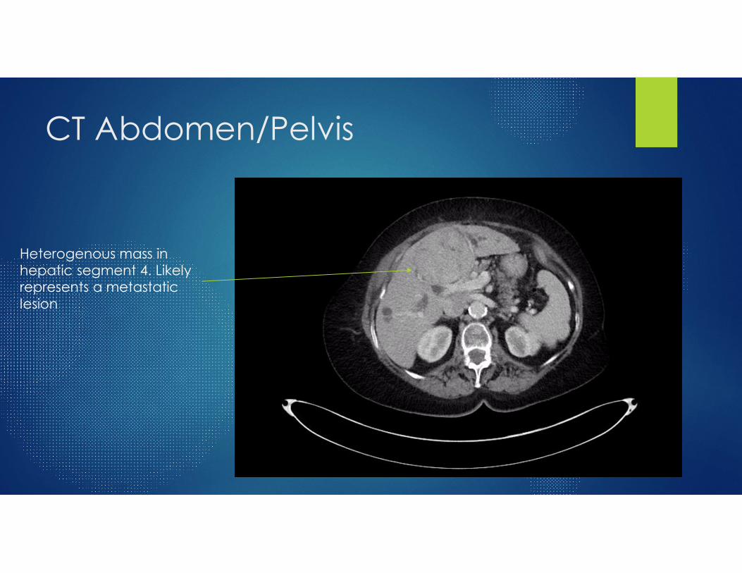
CT Abdomen/Pelvis
Heterogenous mass in hepatic segment 4. Likely represents a metastatic lesion

CT Abdomen/Pelvis, additional findings
Significant intrahepatic biliary ductal dilation in both the right and left hepatic lobe secondary to mass effect from the hepatic mass.

CT Chest
Posterior base segment right lower lobe mass. Measures 5x3x6cm.

Liver Biopsy
Liver biopsy was performed with 1x 22g fine needle aspiration, followed by 3x 18g core needle biopsy
When to biopsy solid liver lesions? If there is no evidence of chronic liver disease then benign conditions,
such as hepatic adenoma, and focal nodular hyperplasia are more likely
If patient has liver disease, risk of malignancy is higher and greater consideration to biopsy is necessary.
Given presence of other disseminated lesions in this patient, metastasis is likely
Primary risks of US guided liver biopsy are tract seeding and hemorrhage

Liver Biopsy
Needle tip visible in center of lesion
Care taken to ensure path of needle avoids vascular structures and biliary structures

Basic Steps of Procedure
• Supine positioning
• Prep site
• Ultrasound with Doppler to find target and biopsy angle• Away from hepatic vasculature (both venous and arterial, bile ducts
• Mark site
• Numb skin and liver capsule
• Get cores (2 to 3)
• Assess under light microscopy
• Scan for post-biopsy perihepatic or intraparenchymal hemorrhage

Pathology Results
Cytology (FNA)- atypical hepatocytes
Core Needle Biopsy- Distorted liver architecture with plate thickening and acinar formations. Findings are consistent with well differentiated hepatocellular carcinoma
Examples of cellular histopathology demonstrating features consistent with HCC

Extra info: Imaging findings in HCC
Classic radiologic criteria for HCC include arterial enhancement
Washout (loss of enhancement compared to the surrounding liver) in the portal venous, delayed phases, and capsular enhancement in the delayed phase.
Diagnosis of HCC can be made for all lesions that are larger than 2 cm. Lesions smaller than 2cm are extremely difficult to characterize with high accuracy

References
https://www.ncbi.nlm.nih.gov/books/NBK470567/
https://www.uptodate.com/contents/approach-to-liver-biopsy
Grant, A., & Neuberger, J. (1999). Guidelines on the use of liver biopsy in clinical practice. Gut, 45(suppl 4), IV1-IV11.