sutton 9 limfoma
TRANSCRIPT

Text Book Reading David Sutton
Radiology and Imaging 7th
ed vol.1
Section 2, page 521
LYMPHOMA
Presentan : Reni Indrastuti
Konsulen : dr. Yana Supriatna, PhD., Sp.Rad
INTRODUCTION
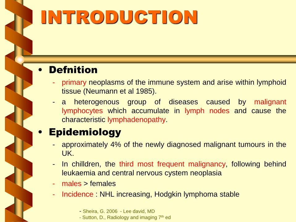
• Defnition
- primary neoplasms of the immune system and arise within lymphoid
tissue (Neumann et al 1985).
- a heterogenous group of diseases caused by malignant
lymphocytes which accumulate in lymph nodes and cause the
characteristic lymphadenopathy.
• Epidemiology
- approximately 4% of the newly diagnosed malignant tumours in the
UK.
- In chilldren, the third most frequent malignancy, following behind
leukaemia and central nervous cystem neoplasia
- males > females
- Incidence : NHL increasing, Hodgkin lymphoma stable
- Sheira, G. 2006 - Lee david, MD
- Sutton, D., Radiology and imaging 7th ed

PREDISPOSING
DISEASE
1- HTLV-1 ( Human T-cell lymphoma/leukaemia virus) is causative
agent in T-cell leukaemia/lymphoma.
2- Immunodeficiency, acquired or inherited, predispose to B-cell
lymphoma.
3- EBV, underlies the endemic form of Burkitt s lymphoma.
4- Helicobacter pylori has been implicated as a predisposing factor
in MALT lymphoma( mucosa associated lymphoid tissue).
5- Hepatitis C virus has been suggested as a risk factor for the
development of NHL.
- Sheira, G. 2006

CLINICAL
MANIFESTATION
• Variable• severity: asymptomatic to extremely ill
• time course: evolution over weeks, months, or years
• Systemic manifestations• fever, night sweats (HL>NHL), weight loss, anorexia,
pruritis (HL>NHL), LBP
• Local manifestations• lymphadenopathy, splenomegaly most common
• any tissue potentially can be infiltrated
• Lymph node swelling, often in the upper body area but it can be
in almost any node or related organ. The node is usually NOT
painful (HL, NHL)- Covell, Bruce Dr

Lymphoma classification
(2001 WHO)
• B-cell neoplasms
– precursor
– mature
• T-cell & NK-cell neoplasms
– precursor
– mature
• Hodgkin lymphoma
Non-
Hodgkin
Lymphomas
60%
40%
- Lee, David., MD

DIAGNOSIS
• Diagnosis should be biopsy-proven before treatment
is initiated
• Need enough tissue to assess cells and architecture
– open bx vs core needle bx vs FNA
• This has a large impact on radiology, with imaging being used
over large periods of time to finesse management
- This is particularly important in the case of Hodgkin's disease where 15-20
years after treatment the cumulative mortality from the complications of
treatment is greater than the disease itself.
- important to reduce the complications of all stages of management including
radiology
- Sheira, G. 2006
- Sutton, D., Radiology and imaging 7th ed
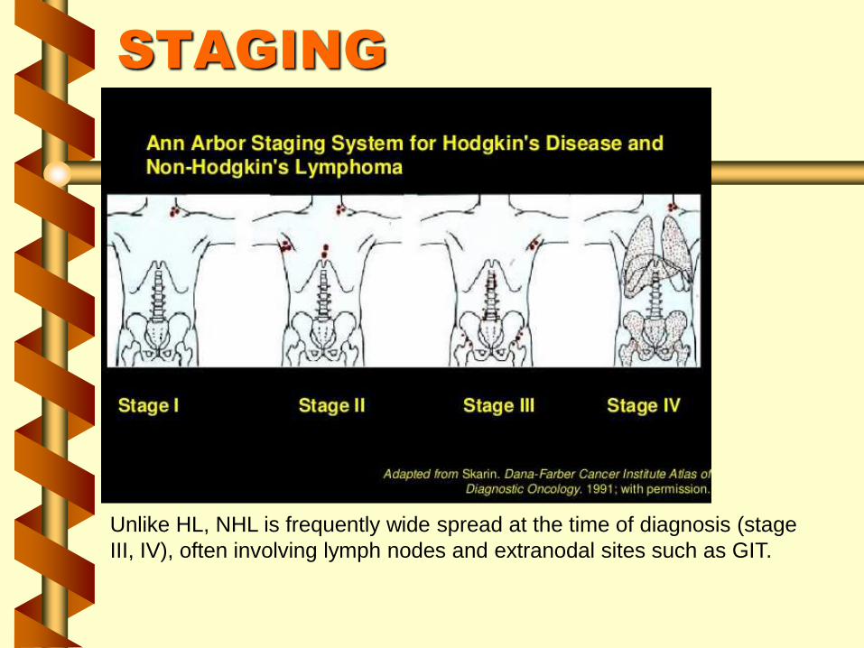
STAGING
Unlike HL, NHL is frequently wide spread at the time of diagnosis (stage
III, IV), often involving lymph nodes and extranodal sites such as GIT.

IMAGING MODALITY
CT SCAN
• Staging
• Follow up
FDG-PET
• Staging
• Follow up
MRI
• Staging
• Follow up
USG
• Initial diagnosis for organs involvement
• Guided biopsy
J Ultrasound Med 2007; 26:791–796

ORGANS INVOLVEMENT
Liver Spleen Kidney
Adrenal
glands
Pancreas Stomach
Small bowel
and
mesentery
Colon
A. Canelas, ESGAR Congress 2008

Sonographics Findings
Sonogram of the liver showing round, well-
defined hypoechoic focal lesions of 20 mm
in maximal diameter
A hypoechoic ring is clearly visible in
the periphery of a liver lesion.
Fine-needle aspiration biopsy confirmed the diagnosis of infiltration of the liver by a diffuse large B-cell lymphoma.
J Ultrasound Med 2007; 26:791–796

Sonographics Findings
Single focal liver lesion with the sonographic appearance of a cyst with
echogenic content, as seen in axial (left) and longitudinal (right) scans.
Echinococcal disease was suspected. Nevertheless, FNAB showed liver
infiltration by non-Hodgkin lymphoma.
J Ultrasound Med 2007; 26:791–796
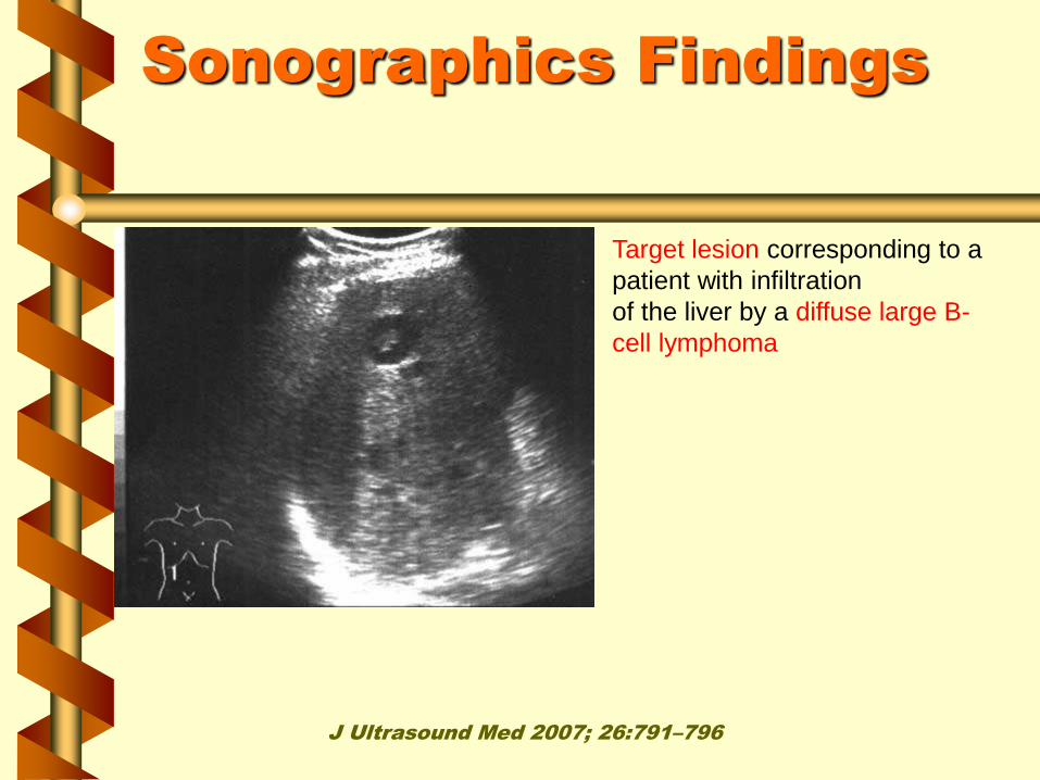
Sonographics Findings
Target lesion corresponding to a
patient with infiltration
of the liver by a diffuse large B-
cell lymphoma
J Ultrasound Med 2007; 26:791–796

RESUME
INTRODUCTION CLASSIFICATION
CLNICAL
MANIFESTATION
DIAGNOSIS STAGING
IMAGING
MODALITY
ORGANS
INVOLVEMENT
SONOGRAPHIC
FINDINGS

QUIZ
Sonography was performed for 54
years old man with nonspecific liver
biochemical anomalies.
a. Please describe the
sonographics findings !
b. What are the differential
diagnosis could be taken?

TERIMAKASIH DAN MOHON ASUPAN
“Many realities hidden behind wall
of perception.”