sutures and techniques...the ideal suture moynihan 1912 •monofilament •absorbable •predictable...
TRANSCRIPT

Understanding Sutures and Basic Flap Design
Solon Kao DDS FICDVice Chair of Oral and Maxillofacial Surgery
School of Dentistry, University of Missouri Kansas CityKansas City, Missouri USA

Objectives
• Wound healing
• Sutures
• Equipment
• Flap design
• Surgical Principles
• Suturing techniques

Before we suture, Let’s learn more about the injury/wound

THE WOUND

What Happens to the Wound?
• Inflammatory response– Outpour of tissue fluids
– Accumulation of cells and fibroblast
– Increased blood supply
– Leukocyte production of protelolytic enzymes
– Dissolve and remove damaged tissue debris

What Happens to the Wound?
• Fibroblasts begin to form collagen fiber

Wound Healing

Not all wounds are healing equally…

Factors Affecting Wound Healing
• Age
– Slow metabolism and impaired circulation
• Weight
– Excess fat makes tissue vulnerable to trauma and infection
• Nutritional status
– Essential to support cellular activity and collagen synthesis

Factors Affecting Wound Healing
• Dehydration
– Results in electrolyte imbalance affects cellular metabolism, oxygenation, cardiac function , kidney function, hormone function impaired healing process
• Inadequate blood supply
– Poor oxygenation / circulation will impair healing process
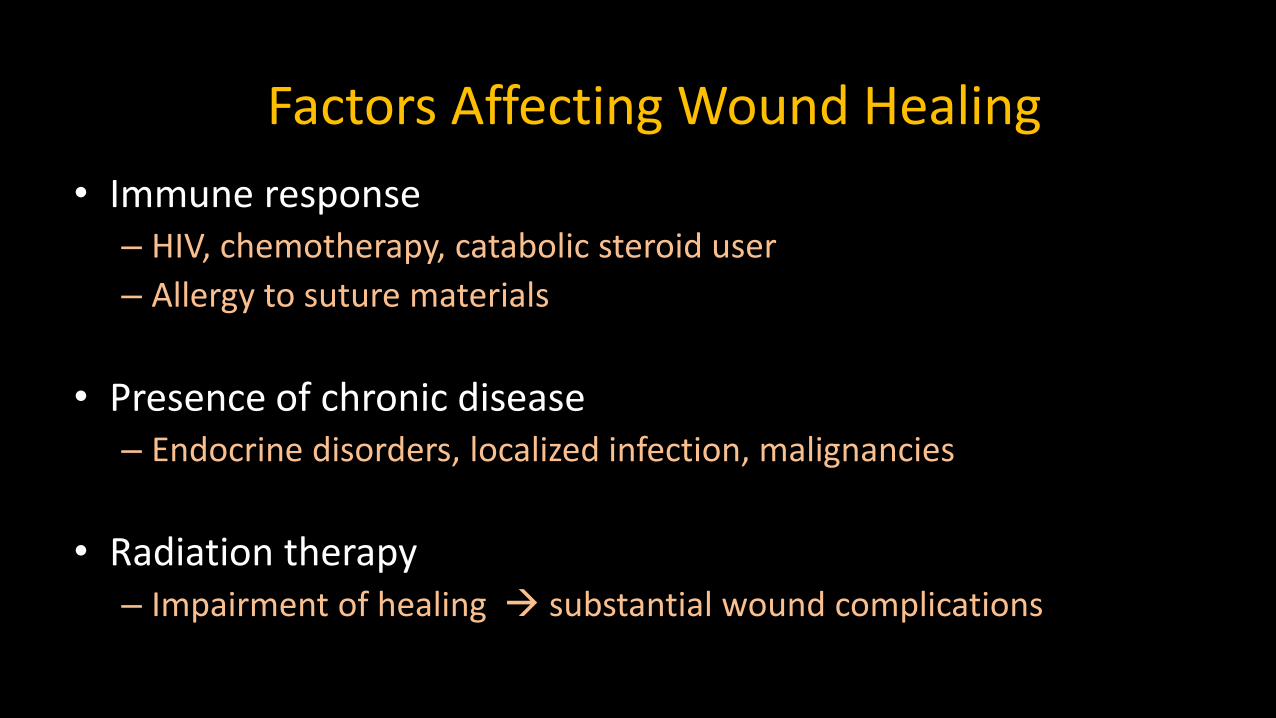
• Immune response– HIV, chemotherapy, catabolic steroid user
– Allergy to suture materials
• Presence of chronic disease– Endocrine disorders, localized infection, malignancies
• Radiation therapy– Impairment of healing substantial wound complications
Factors Affecting Wound Healing

WOUND CLASSIFICATION

Wound Classification
• Wounds can be divided into two groups– Clean or contaminated
– Probability of contamination increases rapidly and
directly related to the length of time since the injury
• Contamination of a clean wound is usually by Streptococcus
• Multiple bacteria become involved if mucosal layers are violated

Wound Classification
• When is the wound “contaminated”
– Wounds that involve the mucosal linings of the oral cavity
• Saliva may carry normal oral flora to deeper structures and lead to development of a wound infection
• Simple lacerations and abrasions have a lower bacterial content

Wound Contamination
• Factors that ↑ chances for infection:– Crushing of tissue, embedding of foreign bodies, and contamination of saliva
• The total number of bacteria present within the wound is a major concern in the development of an infection– Inoculum must exceed 10^5 organisms per gram of tissue

HEALING CHARACTERISTICSThe wound

Tensile Strength

Types of Healing
• Primary intention
• Second intention
• Tertiary intention

Types of Wound Healing
• Primary intention
– Phase I : Inflammatory response ( 1-5 days)
– Phase II : Migration/proliferation (5-14 days)
– Phase III : Maturation/remodeling (14 – done)

Types of Healing
• PRIMARY – “Golden Period” of closure is within first 6 hours
– Approximation of wound edges
– Minimum scar

Types of Healing• SECONDARY
– Granulation with increased scar formation– Prolonged healing– Due to infection, excessive trauma, tissue loss, poor
approximation
• What to do?– Allow time for healing from inner layer to outer surface– Peridex and let it heal

Types of Healing
• TERTIARY (Delayed primary closure) – Candidate for closure after cleaning, debridement
– Leave it open with 4-5 days of antibiotics
– Precise and secure approximation of edges needed
– Not common in dentistry

Complications in Wound Healing
• Infection
– Bacterial, viral, fungal
• Dehiscence
– Old/debilitated patients
– Tissue failure
– No difference in dehiscence rate between vertical versus transverseincisions

SUTURES

HISTORY OF SUTURES
Ancient Indian doctor : beetle head
Edwin Smith Papyrus 1600 BC
Galen AD 150 uses catgut, silk
Joseph Lister 19th century soaked catgut in phenol

SUTURE IS A STRAND OF ANY MATERIAL USED FOR
• Ligating blood vessels
• Transfixing
• Approximating tissues

What Materials Have Been Used?
• Hemp• Gold• Silver• Linen• Thorn• Cotton• Kangaroo tendons• Animal intestine• Human Hair……….

THE IDEAL SUTUREMoynihan 1912
• Monofilament
• Absorbable
• Predictable absorption
• Use for any Procedure
• Easy to handle
• Minimal Tissue Reaction
• High Breaking Strength
• Holds Knots Securely
• Sterile
Sir Berkeley Moynihan Famous British abdominal Surgeon 1865-1936

Choice of Suture
• Familiarity
• Ease of handling
• Tissue characteristics
• Knowledge of physical and biological characteristics of the suture
• Patient factors-infection, debility, obesity

Suture Materials
• Primary purpose for suture– Approximate wound margins
– Enhance tissue healing
• Early role of suture– Wounds do not gain strength until 4-6 days after injury
– Approximating of tissues depending on suture strength
• The relationship between gradual loss of suture strength and slow increase of wound strength must be considered

Suture Material Size
• Refers to the diameter of material
• The more zeroes in the number, the smaller the diameter i.e. 5-0 < 3-0
• Smaller the size, less tensile strength
– 9-0 or 10-0 for microsurgery
– 5-0 or 6-0 facial skin closure
– 3-0 or 4-0 for muscle, deep skin, intra oral mucosa

Different Types of Suture
Nonabsorbable Absorbable
Monofilament Braided

Nonabsorbable Sutures
ADVANTAGE
– Permanent Wound Support
DISADVANTAGE
– Foreign body left
– Suture Extrusion

Absorbable Sutures
ADVANTAGE
– Broken down by body
– No Foreign body left
DISADVANTAGE
– Time of Wound Support

Monofilament
ADVANTAGE
– Smooth surface
– Low friction
– Less drag
– Less tissue Trauma
– Less tissue Infection
– No capillarity action
DISADVANTAGE
– Handling and knotting
– Stretch ability and bending-PLIABILTY

Braided
ADVANTAGE
– Strength
– Soft and pliable
– Good handling
DISADVANTAGE
-Capillary action
-Tissue trauma
-Tissue drag
-Tissue cutting
-Harbors bacteria

Classification of Sutures
Absorbable
Nat
ura
lSy
nth
etic
SilkNatural
Syn
thet
ic
Nylon
Propylene
Polyester
SS
TitaniumN
atu
ral
Syn
thet
ic
Plain Gut
Chromic Gut
Vicryl
PDS
Non-Absorbable Absorbable

NON-ABSORBABLESuture Type


Silk
• Natural Nonabsorbable – Braided
• Made from Silkworm Cocoons

Silk
• Multifilaments of silk are twisted or braided together to form a strand
• Superior in handling, ties down smoothly
• Natural elasticity insures a secure knot
• Good intraorally for areas of tension

Silk
• Do not use for face• Due to amount of bacteria contained within filaments
• Wicking effect
• Never be used in area of wound infection

Summary for Silk
• Very Good Handling
• Very Good Knotting
• STANDARD for many years

Nylon
• First synthetic nonabsorbable monofilament
• Replaced monofilament silk
• Minimum acute inflammatory reaction
• Biologically inert

Nylon
• General soft tissue approximation
• Memory Effect– Pass through fingers when unpacking– Need more knot throw to counter
• Good for skin closure– Due to elastic nature, high tensile strength

Nylon
• Advantage– Easy tissue pass through
– No capillary effect
– Strong elasticity
– Long duration wound support
• Disadvantage– Absorbs water

Prolene
• Synthetic
• Nonabsorbable
• Monofilament

Prolene
• Synthetic linear monofilament
• Nonabsorbable with blue or clear color
• Replaced monofilament nylon

Prolene
• Minimum acute inflammatory reaction
• No tissue adherence, good for “pull-out” suture
• Memory Effect
• Good for general soft tissue closure

Prolene
• Advantage– Better control
• Up to 30% extension w/o breaking
– Indefinite tensile strength
– Excellent handling
– Less thrombogenic
• Disadvantage– Minimum oral cavity application

Gore-Tex Suture

Gore-Tex Suture
• Polytetrafluoroethylene (ePTFE) biomaterial
– Best known as Teflon (discovered in 1938)
• Monofilament, Non-absorbable
• Reduce blood loss
– Needle approximate thread diameter
• Allow suture to fill needle hole reduce bleeding better hemostasis
• Reduce tissue trauma
– Minimal suture friction
• Excellent handling characteristic

Gore-Tex Suture
• Procedures using Gore-Tex
• Common Usage
– Vascular surgery
• Chordae tendineae / mitral valve repair
– Hernia repair
– Oral Health
• Soft, flexible surface
• No snagging or knotting of the thread

RESORBABLESuture Type

Resorbable Suture
• Trend:
– Increasing popularity for all surgery
• Less post operative inflammation
• Easier post operative appointment
– No obligatory suture removal

Resorbable Suture
• Natural Resorbable
– Broken down by body enzymes
• Synthetic Resorbable
– Broken down by hydrolysis


Tensile Strength Retention
Absorption Time

NATURAL ABSORBABLEAbsorbable Suture Type

CATGUT
• 99% purified collagen – From submucosa of sheep or cow intestine
• Essentially monofilament type
• Available types– Plain gut or Chromic gut

Fast Absorbing Gut
• All strength lost by 7th day
• Absorption in 21-42 days
• Moderate tissue reaction
• Should be wet before using but NEVER soaked
• Dermal suture or low tension area ONLY

Plain Gut
• Tensile strength 7-10 days
• Absorption ~70 days
• Causes more tissue reaction
• Clinical usage– Ligate superficial vessels
– Subcutaneous tissue lip
– Oral mucosa

Chromic Gut
• Tensile strength last 10 – 14 days
• Absorption in 90-110 DAYS
• Moderate tissue reaction
• Treated with chromium salt – Brown color
– Resist digestive enzyme
– Prolong absorption
• Used intraorally, no to be used on skin

Cat Gut Suture
• Package contained fluids during storage
– Keep it supple and hydrated
• Sterilized by Gamma irradiation
– 2.5mega rads
• Body reaction to impurities or mucopolysaccharides rare
• Suture should be kept wet but not soaked

Cat Gut Absorption
PLAIN CHROMIC
WOUND SUPPORT 7 – 10 days 10-14 days
MASS ABSORPTION 60 – 90 days 90 – 110 days

SYNTHETIC ABSORBABLEAbsorbable Suture Type

Synthetic Absorbable
• Prepared from carbohydrates
• 2x stronger compared to natural absorbable
• Available types– Monofilament– Braided
• Dependable absorption rate

Synthetic Absorbable
• Fully absorbed without foreign residues
• Absorption by hydrolysis reaction
• Ease of handling
Sutures
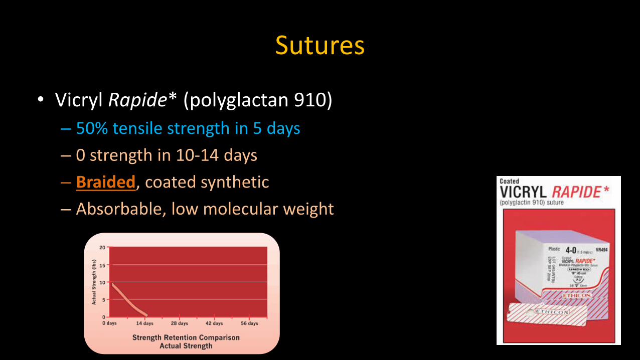
• Vicryl Rapide* (polyglactan 910)
– 50% tensile strength in 5 days
– 0 strength in 10-14 days
– Braided, coated synthetic
– Absorbable, low molecular weight

Vicryl Rapide*
• Elicits lower tissue reaction than chromic gut
• Ideal for dental procedures

Coated Vicryl
• Coated Vicryl (Polyglactan 910)– 75% tensile strength at 2 weeks, 40% at 3 weeks
– Absorption at 56-70 days
– Synthetic absorbable

Coated Vicryl
• Minimal acute inflammatory reaction when buried in subcutaneous tissues
• Braided coated Vicryl behaved like monofilament– Reduced surface tension
• Indicated for general soft tissue approximation and/or ligation

Coated Vicryl
• Frequent Uses
– General tissue/muscle approximation
– Vicryl PLUS has popular oral mucosa usage. Ligatures, reporductivetract, ophthalmic..
– Otherwise…. reproductive tract, orthopedics
Vicryl PLUS contains triclosan

Monocryl
• Monocryl (Poliglecaprone 25)
– 60-70% tensile strength at 7 days, 30-40% at 14 days
– Absorption at 91-119 days
– Monofilament synthetic absorbable

Monocryl
• Highest strength among absorbables
• Most pliable with excellent handling properties
• Minimal acute inflammatory reaction
• Frequent use
– Bladder, GI surgery…..
– Not much indication for dental use

PDS
• PDS II (Polydioxanone)
– 70% tensile strength at 2 weeks,
50% at 4 weeks, 25% at 6 weeks
– Absorption 180-210 days
– Monofilament synthetic absorbable
– Non-antigenic, only slight tissue reaction

PDS
• PDS
– Absorption complete at 6 months
– Indicated for all types of soft tissue closure
– Significant memory, poor knot security
• Frequent Uses
– For slow healing patient/compromised tissue
Fascia, orthopedics…

March, 2008

NEEDLEArmamentarium

Suture Needle
• Three basic components
– The attachment end (swaged or eyed)
– The body
– The point

The Attachment End
Virtually all needles used today are swaged type

Suture Needles
• Swaged
– Needle are permanently attached to the suture material
– Eliminating the need for threading
• Allows needle to be drawn through tissue easier and with less tissue trauma

Needle Body Curvature
1/4 3/8 1/2 5/8
STRAIGHT HALF-CURVED
1/4 3/8 1/2 5/8

Types of Suture Needles
• Reverse cutting
• Conventional cutting
• Tapercut
• Taper

Reverse Cutting Needle
• Most commonly used in Oral Surgical procedures
• Two opposing cutting edges with the third cutting edge on the outer curvature of the needle
• Reduces danger of “cut-out”

Conventional Cutting Needle
• Two opposing cutting edges with the third on the inside curvature of the needle
• The needle changes from a triangular cutting tip to a flattened body
• Not commonly used in Oral Surgery – because cutting edge tends to pull through the edge of the flap with limited access

Taper Cut Needle
• Specifically designed to use on tough and/or delicate tissues
• The point of the needle has sharp reverse cutting tip
• All three edges of the tip are sharpened to provide uniform cutting
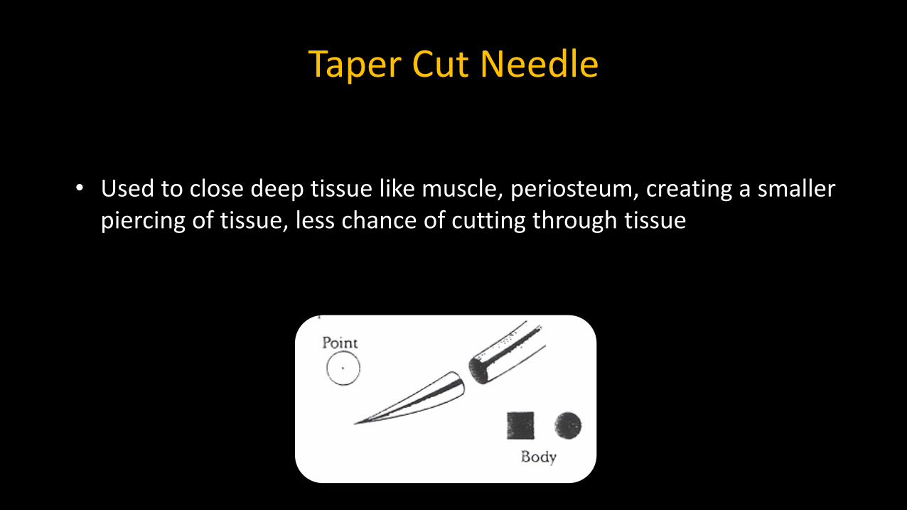
Taper Cut Needle
• Used to close deep tissue like muscle, periosteum, creating a smaller piercing of tissue, less chance of cutting through tissue

Common Needle Types

Summary for Surgical Needles
• Manufactured from stainless steel wire– Heat treated
• Provide: strength, hardness, malleability and sharpness
• Most common for closure of facial and oral mucosa lacerations– Reverse cutting needle
• Most commonly used curved needle is– 3/8 circle needle
• Easily manipulated in relatively large and superficial wounds

OTHER INSTRUMENTSArmamentarium

Suturing Instruments
• Tissue Pick-Ups
• Needle Holders
• Hemostats
• Scissors
• Scalpel

Tissue Pick-Ups
These instruments only have fine delicate fingers so they HOLD, not PIERCE or crush the tissues to be sutured.
Adson BrownAdson w teethAdson w/o teeth


Needle Holders
• Many different needle holders– Various beak size, shape, texture (smooth or serrated)
• Most common have locking handles and short beaks


Hemostats
• Used primarily to remove foreign bodies, pick up tissue to be discarded
• Not to be used as a needle holder
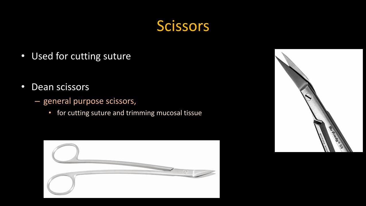
Scissors
• Used for cutting suture
• Dean scissors
– general purpose scissors,• for cutting suture and trimming mucosal tissue

How to hold Surgical Scissor?

Scalpel #15

FLAP DESIGNHow do I make / Design a flap?

Perfect Surgeon must have the heart of the lion and
the hands of a lady, not the claws of a lion and the heart of a sheep
Sir Berkeley Moynihan, 19th century British Abdominal Surgeon

Indications for Mucoperiosteal Flap
• Myth
– Flap is elevated only in the most extraordinary circumstances
• Reality is…
– Utilize flap whenever there is the slightest indication that it might be helpful

Principles of Flap Design
• Broad base: to assure adequate blood supply
• Large enough to provide both visual and instrument access– Large flap heals as rapidly as a small flap
– Flap does not heal from end-to-end by side-to-side
– Post surgical pain does not correlate with size of the flap but more to the amount of bone removed.
• Repositioned and sutured over solid bone
• Avoids major anatomical structures (nerve and blood vessels)

Common Design of Flaps
• Envelope Flaps– 1 tooth distal and 2 teeth mesial
• Flaps with vertical releasing incision– 1 tooth distal and 1 tooth mesial
• Includes papilla
– Vertical release incision must be originating from either• Mesial or distal line angle

Contraindications for Placement of Vertical Release Incision lines
• Canine Prominence
– Soft tissue defect due to frequent bone fenestration
• Mental Foramen
– Avoid vertical incision near proximity of mental foramen
• Palate
– Avoid vertical incision on the palate to void severing greater palatine vasculature and nerve

Contraindications for Placement of Vertical Release Incision lines
• Incisive Papillae– Avoid unnecessary incision through incisive papillae
• Bony lesion– Avoid placing incisions over bony lesion since dehiscence would result
with delayed healing
• Major Frena– Avoid incision through major frena
• Lingual side of mandibular arch– Never Ever place vertical incision on lingual side of the mandibular
arch

Flap Design
• Flap should be designed
– When repositioned, it needs to be over an adequate margin of solid bone• ↑healing rate & ↓wound dehiscence
• Margin of flap should be at least 5mm from the margins of a bony defect!

Mandibular Flaps
• An incision can be made from tip of one coronoid process down the anterior border of the ramus and along the crest of the alveolus to the tip of the coronoid process on the opposite without cutting any major structures except buccinators artery and long buccal nerve

Mandibular Flaps
• Hazard areas for mandibular flap
– Lingual region of 3rd molars• Lingual nerve damage
– Permanent anesthesia and loss of taste
– Premolar buccal vestibular region• Sever mental nerve/vessel
– Permanent loss of labial sensation
• Recovery of sensation maybe possible by proliferation of collateral innervation from C2,C3 and contralateral mental nerve
Mental nerve/vessel
Lingual nerve & its proximityTo #17

Mandibular Flap
• Hazard areas for mandibular flap
– Improper incision / Scalpel Slip to depth of the vestibule near 2nd molar • Facial artery/ vein
– Cross mandible at the anterior edge of masseter muscle
– If cut, needs to ligate the vessel and/or prolonged firm pressure
• Prevention:
– Buccal releasing incision needs to be directed “upward toward” the crest of the alveolar ridge
• Summary:
– Lingual Nerve, Facial Artery, Mental Nerve, Long Buccal Nerve, Buccinator ( Buccal) artery.

Maxillary Flaps
• An incision running from one tuberosity to the other along the alveolar crest will sever……………………………NOTHING larger than a capillary.
Maxillary flaps
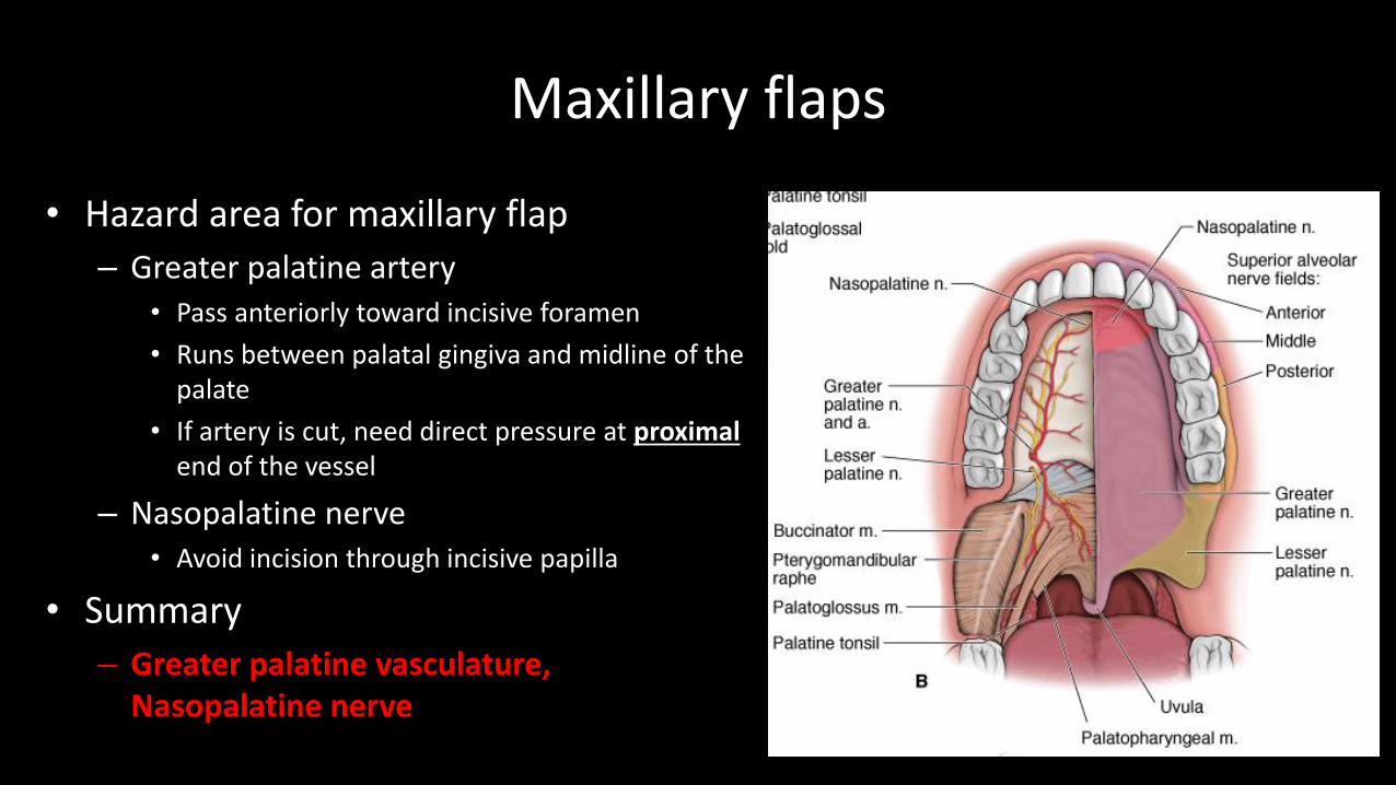
• Hazard area for maxillary flap
– Greater palatine artery• Pass anteriorly toward incisive foramen
• Runs between palatal gingiva and midline of the palate
• If artery is cut, need direct pressure at proximalend of the vessel
– Nasopalatine nerve• Avoid incision through incisive papilla
• Summary
– Greater palatine vasculature, Nasopalatine nerve

NEVER PERFORM ANY VERTICAL INCISION(S) ON THE MANDIBULAR LINGUAL AREA

Type of Flap
• Full thickness
– Mucosal tissue + Periosteum
– Preserve periosteum
– Most popular flap in dentistry
• Partial thickness (split thickness)
– Periosteum is left attached to bone
– Able to apically reposition flap• Increase amount of attached gingiva
– Special OMS/Perio procedures
Full thickness flap
Partial thickness flap

Flap Nomenclature
• Envelop flaps
– Flap of choice for most procedures
– Full thickness flaps• sulcular incision without vertical
releasing incision
– At least one tooth distal to two teeth mesial• Extend the “coverage” as clinically
necessary
• Add on one or two vertical release incision(s)

Modification of Basic Envelope FlapVertical Release Incision
• Important Principles about Vertical Release Incision
– Mesial or distal line angle of a tooth• NEVER cut
– In the middle of the papillae
– At the most apical point of facial gingiva.
» Likely to create mid facial gingival defect.

Modification of Basic Envelope Flap
• Important Principles about Vertical Release Incision
– Apical portion must be wider than coronal portion
– Beware of the blood supply pattern

Modification of Basic Envelope Flap
• Important Principles about Vertical Release Incision
– Incision must be over “sound” bone• Surgeon must anticipate the outcome of
the surgery before placing incision!!!!

Modification of Basic Envelope Flap
• Envelope flap with 1 Vertical Release incision
– Next most useful flap for exodontia
– Provide even greater access• Proximity to apex
• Deeply impacted tooth
Notice the Papilla at the mesial extentOf the incision is included

Modification of Basic Envelope Flap
• Envelope Flap with 2 Vertical Release Incisions ( rectangular flap)
– 2 vertical releasing incisions added to a basic envelop flap
– Basic flap with releasing incision design
• 1 distal and 1 mesial from surgical site

Modification of Basic Envelope Flap
• Curved Flaps (Semi-lunar)
– Full thickness
– Not involving gingival sulcus
– Placed partly in attached gingiva and extend into mucosal tissue
– Utilization• Periapical endodontic surgery
• Retrieval of small root tips
– At least 2mm apical to the base of the gingival sulcus• Periodontal probing should precede incision

Modification of Basic Envelope Flap
• Pedicle Flap
– Long, narrow flap for complete tissue coverage over osseous cavity
– Periodontology• Correct gingival recession
– OMS• Closure of oro-antral fistula
– High potential for necrosis and ejection• Technique sensitive to maintain
adequate blood flow in the flap
A pedicle flap
Closure of oronasal fistula

How to Reflect a Full-Thickness Mucoperiosteal Flap
• 1) Grasping the Knife Handle– Hold like a pen and not in you palm
• 2) Making the Incision– #15 knife blade applied at right angle to tissue and
underlying bone– Firm pressure– ONLY ONE pass incise tissue all the way to bone
• Multiple passes will create ragged margins
• Edentulous area• Right angle to the ridge crest
• Dentate area• Into periodontal sulcus to the height of crestal bone• Incise all the way through periosteum on the first incision

How to Reflect a Full-Thickness Mucoperiosteal Flap
• 3)Flap Reflection
– Begin with sharp-pointed end of the elevator• Pry the interdental papilla free
• Free the attached crestal gingiva
• Complete for the entire length of incision
– Use Broad end of the elevator• Continue reflect attached gingiva & alveolar
mucosa to the desired apical depth

How to Reflect a Full-Thickness Mucoperiosteal Flap
• 4) Flap Retraction
– Proper use of retractor is needed• Small flaps: use periosteal elevator
• Larger flaps: use Minnesota, Austin or others
– Place the tip of the retractor ON BONE
• Flap should be enlarged using knife & periosteal elevator followed by passive retraction
– Aggressive retraction will tear flap
Closure of Surgical Wound
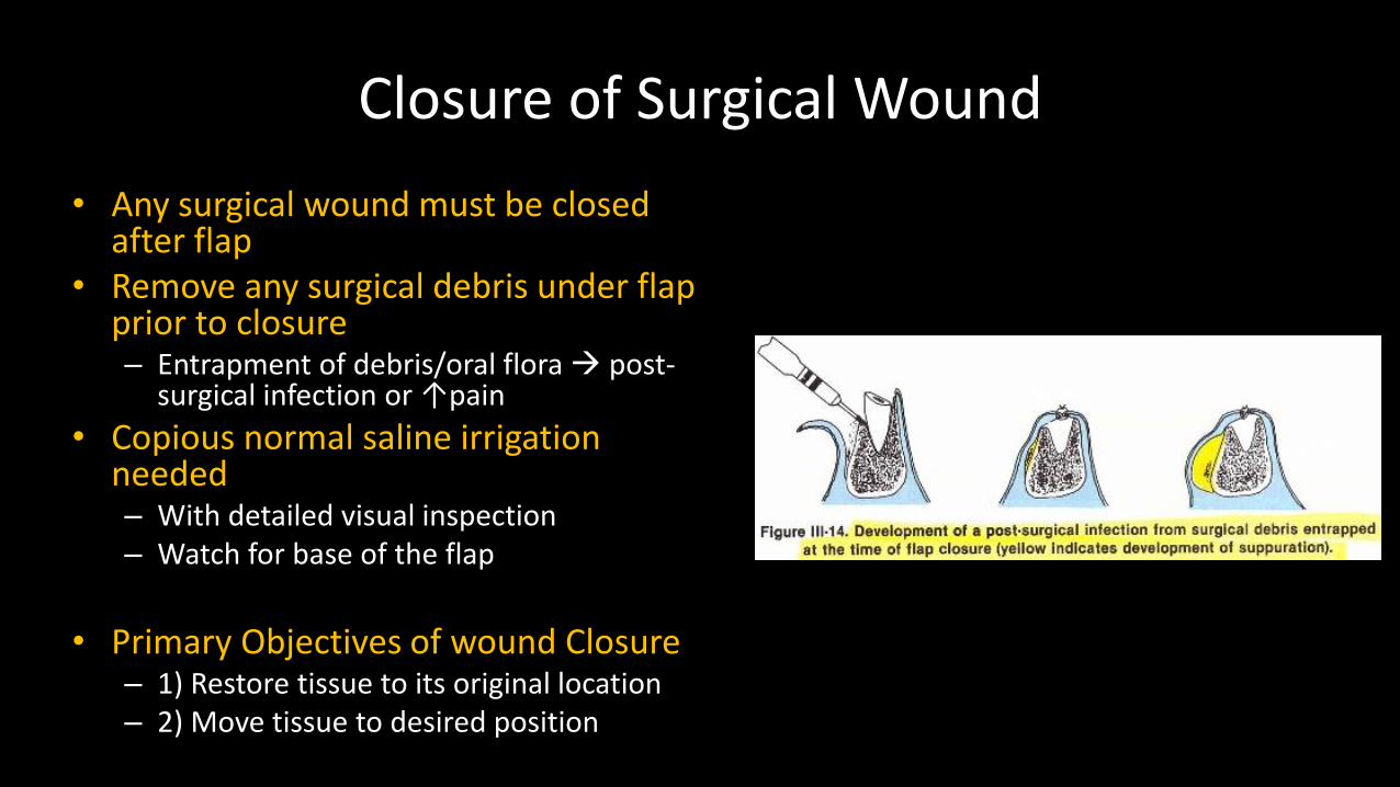
• Any surgical wound must be closed after flap
• Remove any surgical debris under flap prior to closure– Entrapment of debris/oral flora post-
surgical infection or ↑pain
• Copious normal saline irrigation needed– With detailed visual inspection– Watch for base of the flap
• Primary Objectives of wound Closure– 1) Restore tissue to its original location– 2) Move tissue to desired position

Suturing
• General Guidelines– Hemorrhaging should not be controlled by
suturing of flaps• Must manage bleeding before suture
– Intraoral suture should be left in place for 4-7 days• Extraoral is left for 3-5 days
– Suture needle should pass from mobile tissue to non-mobile tissue• Suture needle pass through reflected flap tissue
FIRST, then to the margin of unreflected tissue
– Suture needle should not be too close to wound margin• Minimum 3mm from flap margins

COMMON SURGICAL PRINCIPLES Suture Techniques

Surgical Principles
• Length and direction of the incision– Wound heals from side-to-side NOT end-to-end
– Make incision parallel to the direction of fiber
• Dissection technique– Clean incision with even pressure stroke
• Tissue handling– Minimize tissue trauma, excess tension

Surgical Principles
• Hemostasis
– Hematoma, seroma will interfere with wound closure / healing leading to infection
• Maintain moisture
– Reduce desiccation of tissue
• Removal of foreign materials
– Presence of fragments increase infection

Suturing Principles
• Elimination of dead space
– Dead space cause separation of wound edge
– Critical to healing
• Closing with sufficient tension
– Proximate NOT strangulate !!
• Match flap heights absolutely, with flap edges everted

Suturing Principles
• Suture must be placed so that depth is greater than width
• The more tension on a wound, the closer the sutures to each other and the wound margins
• Cut out crushed tissue

Suture Sequence
1. The needle should enter the tissue at a 90 degree angle to the skin surface, approximately 2mm from wound margin
2. The needle should then be passed into the wound by rotation of the wrist along the arc of the needle
3. Pass the needle through the dermal layer / submucosal layer to assist in eversion of the wound margin

Suture Sequence
4. The needle should then be passed through the tissue of the opposite wound margin at the same level in the dermal layer and should exit the flap at the same distance from the wound margin as that of the insertion
5. Suture is tied without undue tension
6. Wound is cleansed
7. Proper dressings are placed on the wound if necessary

SUTURE TECHNIQUES

Maybe helpful at posterior maxillary region WITH buccal fat pad exposed

PRINCIPLE OF not HALVING!!
Maybe helpful at posterior maxillary region WITH buccal fat pad exposed


How to Deal with Stellate Laceration?

Make sure depth of the stitch is greater than width Eversion of margin
Needle enter tissue at 90 degreethen roll your wrist!

Inversion vs Eversion
Deeper than Wide

Notice suture spacingvs suture width
Mandibular anterior regionmay benefit from layered closure, if deep tissues are present!
Position of needle entrance &depth penetrance must be equal on both side of tissue

Multiple Interrupted Continuous locking
Horizontal Matrices Continuous

DETAILED ILLUSTRATIONSHelpful oral suturing techniques

Horizontal Mattress Sutures
A B C
D E F

Continuous Mattress Sutures

Continuous Locking Sutures

Figure Eight Sutures

Securing Membrane

