swenoteca group: swedish & norwegian testicular cancer … · swenoteca group: swedish &...
TRANSCRIPT
SWENOTECA group: Swedish & Norwegian Testicular Cancer group
SWENOTECA VIII
A revised continuation of SWENOTECA IV and VI
A cancer care program
Non-Seminomatous Germ Cell Tumours (NSGCT) (including testicular, retroperitoneal and mediastinal tumours)

Regional Tumour Registry, Lund, Sweden Drawing on front page: Eva Henriksson ISBN Lund 2012

SWENOTECA WORKING GROUP
The SWENOTECA VIII cancer care program is developed by the working group of SWENOTECA in
collaboration with Lennart Blomqvist, Karolinska University Hospital, Stockholm for the imaging part and
Gunilla Chebil, Unilabs, Helsingborg for the histopathology part.
Members of the SWENOTECA working group
Anders Angelsen, St Olavs University Hospital, Trondheim
Roy Bremnes, University Hospital of Nord-Norge,Tromsö
Eva Cavallin-Ståhl, Skåne University Hospital, Lund
Gabriella Cohn Cedermark, Karolinska University Hospital, Stockholm
Olav Dahl, Haukeland University Hospital, Bergen
Aleksander Giwercman, Skåne University Hospital, Malmö
Hege S. Haugnes, University Hospital of Nord-Norge, Tromsö
Ulf Håkansson, Skåne University Hospital, Malmö
Anders Kjellman, Karolinska University Hospital, Stockholm
Carl Wilhelm Langberg, Ullevål University Hospital, Oslo
Anna Laurell, Uppsala University Hospital, Uppsala
Jan Oldenburg, The Norwegian Radium Hospital, Oslo
Arne Solberg, St Olavs University Hospital, Trondheim
Ulrika Stierner, Sahlgrenska University Hospital, Göteborg
Olof Ståhl, Skåne University Hospital, Lund
Karin Söderström, Norrlands University Hospital, Umeå
Martin Söderberg, Skåne University Hospital, Malmö
Torgim Tandstad, St Olavs University Hospital, Trondheim
Rolf Wahlqvist, Aker University Hospital, Oslo
Najme Wall, Linköping University Hospital, Linköping
Hans Wijkström, Karolinska University Hospital, Stockholm
Monika Andersson, Skåne University Hospital, Lund
Sectretarial work and layout of the protocol: Maria Rejmyr Davis, Regional Tumour Registry, Lund
Page 3

Page 4

TABLE OF CONTENTS
TABLE OF CONTENTS .............................................................................................. 5
ABBREVIATIONS ............................................................................................... 11
Purpose of the SWENOTECA VIII cancer care program ......................................... …12
Testicular tumours ......................................................................................... 13
1. Background .................................................................................................. 13
1.1 General information................................................................................................... 13
1.2 International guidelines for the treatment of NSGCT ....................................................... 13
2. Diagnosis, pre- and postorchiectomy examinations, clinical staging ..................... 14
2.1 Diagnosis of Testicular Cancer .................................................................................... 14
2.1.1 Clinical examination of the testes ........................................................................... 14
2.1.2 Serum tumour markers ........................................................................................ 14
2.1.3 Fertility measures and hormonal analyses ............................................................... 15
2.1.4 Tests to be performed before orchiectomy .............................................................. 15
2.1.5 Inguinal exploration and orchiectomy ..................................................................... 15
2.1.6 Organ sparing surgery .......................................................................................... 16
2.1.7 Biopsy of the contralateral testis ............................................................................ 16
2.1.8 Pathological examination of the testis ..................................................................... 16
2.2 Staging investigations ................................................................................................. 16
2.2.1 Tests to be performed directly after orchiectomy - Clinical staging procedure ............... 16
2.2.2 The 6–8 weeks observation period before definitive clinical staging in CS I................... 17
2.2.3 Definitive clinical staging ....................................................................................... 17
3. Imaging ....................................................................................................... 17
3.1 Imaging at diagnosis and staging .................................................................................. 17
3.2 Imaging during treatment ............................................................................................ 18
3.3 Imaging during follow-up ............................................................................................. 18
4. Clinical stage I – Treatment ............................................................................ 19
4.1 Experiences in SWENOTECA III+VI ................................................................... 19
4.2 Comments on treatment in CSI ..................................................................................... 19 Page 5

5. Metastatic disease - Treatment ...................................................................... 20
5.1 General comments to treatment of metastatic disease ..................................................... 20
5.2 Short description of SWENOTECA IV .............................................................................. 20
5.3 Good Prognosis ........................................................................................................... 20
5.3.1 Background .......................................................................................................... 20
5.3.2 Experiences in SWENOTECA IV – good prognosis ...................................................... 20
5.3.3 CS IIA marker negative (CS IIA Mk-) disease, treatment ........................................... 21
5.3.4 Comments to registration of CS IIA Mk- disease ....................................................... 21
5.3.5 Treatment of good prognosis disease (except CS IIA Mk-).......................................... 21
5.3.6 Comments to the treatment of good prognosis disease .............................................. 21
5.4 Intermediate prognosis ................................................................................................ 22
5.4.1 Background .......................................................................................................... 22
5.4.2 Experience in SWENOTECA IV – intermediate prognosis ............................................. 22
5.4.3 Treatment of intermediate prognosis disease ............................................................ 23
5.5 Poor prognosis ............................................................................................................ 23
5.5.1 Background .......................................................................................................... 23
5.5.2 Experience in SWENOTECA IV – poor prognosis ........................................................ 24
5.5.3 Treatment of poor prognosis disease ....................................................................... 25
5.5.4 Comments to the treatment of poor prognosis disease .............................................. 25
5.6 Chemotherapy, comments regarding administration and specific toxicity ........................... 26
5.7 CNS metastases .......................................................................................................... 26
6. Surgery (other than orchiectomy) ................................................................... 27
6.1 Retroperitoneal Lymph Node Dissection ......................................................................... 27
6.1.1 Indications and techniques ..................................................................................... 27
6.1.2 Indications ........................................................................................................... 27
6.1.3 RPLND Templates ................................................................................................. 27
6.1.4 Definitions of templates ......................................................................................... 27
6.1.5 Pathology results .................................................................................................. 28
6.1.6 Retrograde ejaculation........................................................................................... 28
Page 6

6.2 CS I .......................................................................................................................... 28
6.2.1 RPLND ................................................................................................................. 28
6.3 CS II-IV ..................................................................................................................... 28
6.3.1 Primary RPLND in CS IIA Mk- ................................................................................. 28
6.3.2 PC-RPLND ............................................................................................................ 29
6.3.3 Extra-retroperitoneal resections .............................................................................. 29
6.4 Surgery at relapse ...................................................................................................... 29
6.4.1 Marker negative (Mk-) and late relapses .................................................................. 29
6.4.2 Surgery after 2nd line or later chemotherapy ........................................................... 29
7. High-dose chemotherapy with stem cell support ............................................... 30
7.1 Background ................................................................................................................ 30
7.2 The SWENOTECA IV experience .................................................................................... 30
7.3 High-dose regimens and number of courses ................................................................... 30
7.4 Stem cell harvest and practical considerations ................................................................ 31
8. Follow-up principles ....................................................................................... 31
8.1 General comments ...................................................................................................... 31
9. Treatment of relapse ..................................................................................... 32
9.1 Background ................................................................................................................ 32
9.1.1 Relapse in CS I ..................................................................................................... 32
9.1.2 Relapse in metastatic disease ................................................................................. 32
9.1.3 Conventional dose salvage therapy ......................................................................... 33
9.1.4 HDCT used as salvage therapy in relapsed patients ................................................... 33
9.2 Treatment of relapses .................................................................................................. 33
9.2.1 Salvage treatment CS I ......................................................................................... 34
9.2.2 Salvage treatment metastatic disease ..................................................................... 34
9.2.3 Surgery ............................................................................................................... 34
10. Information to the patient ............................................................................ 34
Page 7
Extragonadal retroperitoneal and mediastinal non seminomatous germ cell tumours……36
1. Background .................................................................................................................. 36
2. Classification and prognosis ........................................................................................... 36
3. Diagnosis and treatment principles ................................................................................. 37
4. Treatment .................................................................................................................... 37
4.1 Chemotherapy......................................................................................................... 37
4.2 Surgery .................................................................................................................. 37
4.3 Salvage treatment ................................................................................................... 38
5. Registration and follow-up ............................................................................................. 38
Monitoring and reporting the results of the SWENOTECA VIII program .................... 39
References ....................................................................................................... 41
Background ..................................................................................................................... 41
Diagnosis, pre- and postorchiectomy examinations, clinical staging ........................................ 41
Imaging .......................................................................................................................... 42
Clinical stage I - Treatment ............................................................................................... 42
Metastatic disease - Treatment .......................................................................................... 42
Surgery ........................................................................................................................... 44
High-dose chemotherapy with stem cell support ................................................................... 48
Follow-up principles .......................................................................................................... 49
Treatment of relapse ......................................................................................................... 49
Extragonadal retroperitoneal and mediastinal non seminomatous germ cell tumours ................. 50
Addendum ........................................................................................................... 53
Page 8

ADDENDUM Flow sheets
1. Testicular tumours - staging and treatment principles ............................ 55 2. Clinical stage II A marker negative (CS IIA Mk-) at definitive staging ....... 56 3. Good prognosis (except CS IIA Mk-) .................................................... 57 4. Intermediate prognosis and poor prognosis due to “poor markers” only .... 58 5. Poor prognosis patients with non-pulmonary visceral metastasis ............. 59 6. Treatment of patients with brain metastases at diagnose ........................ 60 7. Treatment of patients with brain metastases at relapse after CR .............. 61 8. Treatment of relapse .......................................................................... 62 9. Extragonadal tumours - staging and treatment principles ....................... 63
Clinical staging according to Royal Marsden ............................................ 65
Prognostic risk group classification ......................................................... 66 AFP, plot – halveringstid ............................................................................ 67
β-HCG, plot – halveringstid......................................................................... 68 Chemotherapy regimens
BEP .......................................................................................................... 69 EP ............................................................................................................ 71 BEP-IF ...................................................................................................... 73 PEI ........................................................................................................... 75 TIP ........................................................................................................... 77 EMA-CO .................................................................................................... 79 GOP .......................................................................................................... 81 High-dose chemotherapy CE ........................................................................ 83
High-dose chemotherapy PEC ...................................................................... 85 Patient information Swedish Stadium I non-seminomatös testikelcancer utan kärlinväxt .............................. 87 Stadium I non-seminomatös testikelcancer med kärlinväxt .............................. 91 Metastatisk sjukdom, non-seminomatös testikelcancer.................................... 95 Norwegian Stadium I non-seminom testikkelkreft uten karinnvekst .................................. 99 Stadium I non-seminom testikkelkreft med karinnvekst ................................ 103 Metastatisk sykdom, non-seminom testikkelkreft ......................................... 107
Page 9

Follow-up schedules – Uppföljningsschemata Clinical stage I (CSI): surveillance .............................................................. 111
Clinical stage I (CSI): adjuvant 1 BEP ......................................................... 113 Metastatic disease, recurrence ................................................................... 115 Patient care plan to be given to the patient at last follow-up at the oncology unit Swedish version ....................................................................................... 117 Norwegian version .................................................................................... 119 English version ......................................................................................... 121 Carcinoma-in-situ of the testis .............................................................. 123 Radiotherapy of CIS - Behandling av kvarvarande testikel med elektroner ... 128 Abdominal MRI protocol for follow-up ................................................... 129 KVAST document Testicular cancer ...................................................................................... 130 Retroperitoneal lymph node dissection ........................................................ 135 TNM - Pathological (p) and Clinical classification……………………………………………….138
Late effects ........................................................................................... 139 Forms .................................................................................................... 143 Anmälan / Registreringsblankett Behandling – kemoterapi Behandling – kirurgi Behandling – radioterapi Uppföljning Högdos Extragonadal germinalcellscancer, Registreringsblankett Homepage- updated information available: www.rccsyd.se
Page 10

ABBREVIATIONS
AFP Alpha fetoprotein BEP Bleomycin, etoposide, cisplatin BEP-IF Bleomycin, etoposide, cisplatin, ifosfamide BIP Bleomycin-induced pneumonitis CE Carboplatin, etoposide CIS Carcinoma in situ CR Complete remission CS Clinical stage CSS Cancer-specific survival CT Computed tomography EAU European Association of Urology EGCC Extragonadal germ cell cancer EGCCCG European Germ Cell Cancer Collaboration Group EMACO Etoposide, methotrexate, actinomycin D, cyclofosfamide and vincristine EORTC European Organization for Research and Treatment of Cancer EP Etoposide, cisplatin ESMO European Society of Medical Oncology FSH Follicle-stimulating hormone GOP Gemcitabin, oxaliplatin, paclitaxel HCG Human chorionic gonadotropin HDCT High-dose chemotherapy IGCCCG International Germ Cell Cancer Collaboration Group LBB Liver, bone, brain LH Luteinizing hormone LDH Lactate dehydrogenase MRI Magnetic resonance imaging Mk Marker (tumour marker) Mk- Marker negative NSGCT Non-seminoma germ cell tumour OS Overall survival PC Post-chemotherapy PEI Cisplatin, etoposide, ifosfamide PET Positron emission tomography PLAP Placental-like alkaline phosphatase PFS Progression-free survival RPLND Retroperitoneal lymph node dissection SHBG Sex hormone-binding globulin SWENOTECA Swedish Norwegian Testicular Cancer TIP Paclitaxel, ifosfamide, cisplatin VASC +, VASC - Vascular infiltration, no vascular infiltration WHO World Health Organization
Page 11

SWENOTECA VIII Treatment Program for Non-Seminomatous Germ Cell Tumours (NSGCT)
PURPOSE OF THE SWENOTECA VIII CANCER CARE PROGRAM FOR PATIENTS WITH NSGCT
General purposes:
• To establish a complete register including all male adolescent and adult patients with non-seminomatous germ cell testicular, retroperitoneal and mediastinal cancer in Norway and Sweden.
• To standardise diagnostic procedures, staging, treatment and follow up in order to: • improve patient outcome • assure high quality prospective population-based clinical research • reduce the radiation burden inflicted by CT in the follow-up of patients, by recommending
MRI as standard abdomino-pelvic imaging modality
Specific foci in clinical stage I:
• Risk-adapted treatment: adjuvant one course of BEP treatment, or close surveillance. • The relapse rate and pattern of relapse for: the presumed low-risk patients
and high-risk patients, respectively. • The early and late toxicity after short adjuvant chemotherapy versus full treatment in case of
relapse, respectively.
Specific foci in metastatic disease:
• Individualized treatment of metastatic disease according to risk group and initial tumour marker decline
• Reducing overtreatment where possible, and intensifying treatment in those with poor prognostic disease or in poor responders.
• To evaluate treatment outcome, time to relapse, the histological type of the recurrence, and the response to salvage chemotherapy and/or surgery.
• To evaluate early and late side effects after treatment for advanced disease.
Page 12

TESTICULAR TUMOURS
1. BACKGROUND
1.1 General information
Testicular cancer accounts only for 1–2% of all male malignancies. It is, however, the most common cancer affecting young men in their second to fourth decade of life. The incidence is increasing in most Western countries, and Scandinavia is a high incidence area. In Sweden and Norway about 600 new cases are diagnosed each year. In Norway the age-standardised incidence is 12 per 100,000 males and in Sweden, the age-standardised incidence is 6 per 100,000 males in 2007. Testicular cancers are in 95% germ cell tumours. About 50% are pure seminomas, while the remaining cancers are non-seminomatous germ cell tumours (NSGCT). About 50% of the NSGCT patients have clinically detectable metastases at the time of diagnosis, mainly in the retroperitoneal lymph nodes and/or the lungs. However, in large population-based series about 27–38% of the patients originally in clinical stage 1 (CS1) will also have metastases revealed during surveillance or at pathological staging by surgery. More than 90% of the relapses will be detected during the first 18–24 months. The most valid predictor of subclinical disease and later metastases in CS I NSGCT is vascular invasion in the primary tumour (VASC+). The presence and percentage of embryonic elements and the size of the primary tumour are other factors with predictive value. Vascular invasion in the primary tumour is found in about 30% of CS I patients. The rate of subclinical metastases is in the order of 50%, for CS1 patients with VASC+, and 15-20% for CS1 patients without vascular invasion (VASC-). Testicular tumours with VASC+ are defined as minimum T2, and CS1 patients with VASC+ are classified as having stage IB. If patients with CS I and VASC+ are treated with 2 courses of adjuvant cisplatin-based combination chemotherapy, the relapse rate is reduced to less than 2%. The few patients that relapse after adjuvant chemotherapy are nearly always salvaged by secondary chemotherapy and/or surgery1-4. There are recent data using only one adjuvant course of BEP chemotherapy in CS I NSGCT. A non risk-adapted randomised trial on 382 patients, with a median follow-up of 56 months comparing one BEP versus RPLND showed a relapse rate of 1.1% versus 7.5% respectively4. Similar results were published by the SWENOTECA group2. Since the introduction of cisplatin the cure rate of metastatic disease has been excellent and steadily improving. The International Germ Cell Consensus (IGCCC) Classification (see Addendum) which is based on clinical parameters immediately prior to chemotherapy, in patients treated between 1975 and 1990, distinguishes patients with non-seminomatous germ cell tumours (NSGCT) in good, intermediate or poor prognosis risk group. The reported 5-year overall survival is 92%, 80% and 48%, respectively5. In a more recent meta-analysis 1775 patients treated after 1989 with NSGCT with good (n = 1087), intermediate (n = 232), or poor (n = 456) prognosis were included. Pooled 5-year survival estimates were 94%, 83% and 71%, respectively6. This increase is most likely due to both more effective treatment strategies including improvement of chemotherapy, surgery, better diagnostic imaging, as well as better supportive care.
1.2 International guidelines for the treatment of NSGCT
According to recent international reviews and guidelines for the treatment of CSI NSGCT, retroperitoneal lymphadenectomy, close surveillance, or adjuvant chemotherapy result in the same ultimate cure rate in the order of 98%. However, the relapse rate as well as the morbidity differs7. The EAU, ESMO and EGCCCG have treatment recommendations according to the three different prognostic
Page 13
risk groups in metastatic disease. The SWENOTECA recommendations are similar but not identical in all aspects8-10.
2. DIAGNOSIS, PRE- AND POSTORCHIECTOMY EXAMINATIONS, CLINICAL STAGING
See Flow sheet 1.
2.1 Diagnosis of Testicular Cancer
2.1.1 Clinical examination of the testes Testicular cancer usually presents as a painless, unilateral intrascrotal mass and is in the majority of cases diagnosed by palpation. Some patients will present clinical symptoms mimicking epididymitis, less than 10%. Ultrasound of both the testicles should be performed, and exploration should be performed in all cases when clinical or ultrasound investigations cannot exclude a tumour. Trans-scrotal fine needle aspiration or biopsy from the tumour should not be performed. 2.1.2 Serum tumour markers In non-seminomas about 40% of patients with clinical stage I, and up to 85% of metastatic patients have an elevation of either one or both serum tumour markers AFP and β-HCG1. Marker concentration is dependent on histological subtype and tumour burden. An elevated level of LDH occur in testicular cancer and is also used as a prognostic marker. The determination of AFP and β-HCG are used in order to:
• identify occult spread in CSI • identify seminoma that biologically are a non-seminoma; • assess prognostic risk group classification • follow treatment effect • identify early relapse
β-HCG (human chorionic gonadotropin) The syncythiotrophoblastic cells are responsible for production of β-HCG. All patients with choriocarcinoma and 40-60% of the patients with embryonic carcinoma have elevated serum levels of β-HCG. The serum half-life of β-HCG should as a rule be ≤ 3 days. However, the rate of reduction in the concentration of β-HCG following chemotherapy may follow a more complex pattern, with longer apparent half-life during later stages of chemotherapy, even in patients treated successfully2. Cross reactivity with the beta unit of the LH might occur resulting in a false positive test. Furthermore, hypogonadism can induce LH as well as β-HCG production by the pituitary gland. Short course of testosterone replacement therapy suppresses pituitary LH and β-HCG secretion allowing for a true measure of serum-HCG of germ cell origin. β-HCG can also be produced by tumours of other origin such as liver, pancreas, stomach, kidney and bladder cancer3. AFP (α-Fetoprotein) In germ cell tumours AFP is secreted by embryonic cell carcinoma and yolk sac tumour but not by pure choriocarcinoma or pure seminoma. The metabolic half-life of AFP should be ≤ 7 days. One should be aware that reparative and infectious/viral processes of the liver as well as cirrhosis and trauma also may induce an increase in AFP, sometimes as high as > 500 ng/ml. Rarely patients constitutionally may have an AFP level slightly above the normal range (≤ 1.5 upper limit). A slightly elevated and stable AFP level might thus not necessarily be associated with the testicular cancer disease. Slightly elevated levels of AFP after completed chemotherapy may also be explained by a slow leakage of fluid from cystic teratoma and should, if this is the case, normalize after post chemotherapy surgery4.
Page 14

AFP can also be elevated in hepatocellular carcinoma as well as pancreatic cancer, gastric cancer, colorectal and bronchial cancer. LDH (lactate dehydrogenase) LDH is a cytoplasmic enzyme in all living cells and elevated values are seen in all kinds of tissue destruction and cell death. Elevated level of LDH is seen in 40-60% of all patients with germinal cell cancer and is correlated to tumour burden. Typically it is the elevation of LDH isoenzyme number 1 that is seen. LDH elevation is taken in consideration in the classification in prognostic groups but is less specific for germ cell tumours than AFP or β-HCG. Insignificant elevated levels of LDH are commonly seen at patient visits during follow-up. PLAP Placental alkaline phosphatase (PLAP) is elevated in 50% of the patients with pure seminoma and thus also in patients with mixed non-seminomatous tumours but is only analysed in a few laboratories in Sweden. The use of this marker is optional. PLAP may be falsely elevated in smokers3. 2.1.3 Fertility measures and hormonal analyses Cryopreservation of sperm should preferably be offered before orchiectomy. If not performed before orchiectomy it should always be offered before start of any therapy although the adjuvant chemotherapy with 1 or 2 BEP most probably has no long-lasting detrimental effect on spermatogenesis5. Patients receiving multiple cycles of chemotherapy or operated with RPLND are at risk of subfertility/ infertility. Sexual hormones (LH/FSH, testosterone and SHBG) should be analysed before and after orchiectomy and during follow up. The serum for the hormone analyses should preferentially be sampled in the morning or at least before noon (due to their circadian variations). It is important to detect and treat hormonal insufficiency both with regard to short- and long-term morbidity of hypogonadism. 2.1.4 Tests to be performed before orchiectomy
• Ultrasound examination of both testicles • General physical examination • Serum levels of AFP, β-HCG, LDH Mandatory! • Serum levels of PLAP, optional • Serum levels of LH, FSH, testosterone and SHBG • All patients should be offered pre-orchiectomy sperm count with cryopreservation
2.1.5 Inguinal exploration and orchiectomy An incision similar to that performed in patients with inguinal hernia is done. The anterior wall of the inguinal canal is divided, and the vas and spermatic vessels are dissected free at the internal opening of the inguinal canal. In most cases there is no doubt of the diagnosis and the spermatic vessels and the vas are divided immediately. The testis and epididymis with their surrounding tunica vaginalis are pushed out of the scrotum and dissected free from the scrotal wall. The vas and the spermatic vessels are ligated and divided separately close to the peritoneal fold. The specimen is immediately sent for definitive histology. If possible, the specimen should be sent fresh on ice to the pathology department, otherwise placed in formalin. The urologist should not incise the specimen. It is recommended to offer all patients a testicular prostheses before orchiectomy6. If the patient would like to have a testicular prostheses it is recommended to close tunica vaginalis with an absorbable tobacco-pouch suture above the prosthesis to prevent migration to the inguinal canal.
If any doubt of the diagnosis, the spermatic cord is clamped before mobilization and inspection of the testis. In some cases the tunica albuginea of the testis is incised and a frozen section is sent for histology. If the result of the frozen section is a benign condition (for example adenomatoid tumour or epidermoid cyst) it is recommended to perform a local resection instead of an orchiectomy.
Page 15
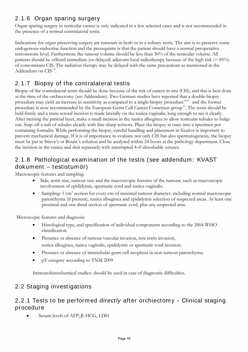
2.1.6 Organ sparing surgery Organ sparing surgery in testicular cancer is only indicated in a few selected cases and is not recommended in the presence of a normal contralateral testis. Indications for organ preserving surgery are tumours in both or in a solitary testis. The aim is to preserve some endogenous endocrine function and the prerequisite is that the patient should have a normal preoperative testosterone level. Furthermore the tumour volume should be less than 30% of the testicular volume. All patients should be offered immediate (or delayed) adjuvant local radiotherapy because of the high risk (> 85%) of concomitant CIS. The radiation therapy may be delayed with the same precautions as mentioned in the Addendum on CIS7-9. 2.1.7 Biopsy of the contralateral testis Biopsy of the contralateral testis should be done because of the risk of cancer in situ (CIS), and this is best done at the time of the orchiectomy (see Addendum). Two German studies have reported that a double-biopsy procedure may yield an increase in sensitivity as compared to a single-biopsy procedure10,11 and the former procedure is now recommended by the European Germ Cell Cancer Consensus group12. The testis should be held firmly and a trans scrotal incision is made laterally on the tunica vaginalis, long enough to see it clearly. After incising the parietal layer, make a small incision in the tunica albuginea to allow testicular tubules to bulge out. Snip off a tuft of tubules cleanly with fine sharp scissors. Place the biopsy at once into a specimen pot containing formalin. While performing the biopsy, careful handling and placement in fixative is important to prevent mechanical damage. If it is of importance to evaluate not only CIS but also spermatogenesis, the biopsy must be put in Stieve’s or Bouin´s solution and be analysed within 24 hours at the pathology department. Close the incision in the tunica and skin separately with interrupted 4–0 absorbable sutures. 2.1.8 Pathological examination of the testis (see addendum: KVAST dokument – testistumör) Macroscopic features and sampling:
• Side, testis size, tumour size and the macroscopic features of the tumour, such as macroscopic involvement of epididymis, spermatic cord and tunica vaginalis.
• Sampling: 1 cm2 section for every cm of maximal tumour diameter, including normal macroscopic parenchyma (if present), tunica albuginea and epididymis selection of suspected areas. At least one proximal and one distal section of spermatic cord, plus any suspected area.
Microscopic features and diagnosis:
• Histological type, and specification of individual components according to the 2004 WHO classification.
• Presence or absence of tumour vascular invasion, rete testis invasion, tunica albuginea, tunica vaginalis, epididymis or spermatic cord invasion.
• Presence or absence of intratubular germ cell neoplasia in non-tumour parenchyma. • pT category according to TNM 2009
Immunohistochemical studies: should be used in case of diagnostic difficulties.
2.2 Staging investigations
2.2.1 Tests to be performed directly after orchiectomy - Clinical staging procedure
• Serum levels of AFP, β-HCG, LDH
Page 16

• CT of thorax, abdomen and pelvis with iv and oral contrast should be performed as soon as possible after orchiectomy. If there is clinical indication of advanced metastatic disease the CT should be done before orchiectomy.
• MRI of the brain is required in patients with clinical symptoms or signs indicating brain metastases, in patients with HCG > 50 000, choriocarcinoma and massive pulmonary metastases as well as in patients with non-pulmonary visceral metastases.
• If in doubt of metastases (e.g. clinical stage IIA and early B) follow tumour markers weekly until nadir/normalisation, as long as the half-life is maintained.
If there is evidence of metastatic disease, the patient should be referred immediately to an oncology department for further evaluation and treatment. Prognostic group classification should be performed immediately prior to chemotherapy. 2.2.2 The 6–8 weeks observation period before definitive clinical staging in CS I (For patients without obvious metastases in presumed clinical stage I) Serum levels of AFP and β-HCG must be monitored weekly, during this observation interval before the definitive clinical staging (every other week if normal levels postoperatively). The results must be evaluated in regard to the expected decrease according to the half time values of 3 days for β-HCG and 7 days for AFP. The values should be plotted against the semi-logarithmic standard plots shown in figures in addendum. Any clear deviation from these plots indicates metastatic disease, thus ending the observation period. 2.2.3 Definitive clinical staging (For patients without obvious metastases in presumed clinical stage I after the first clinical staging procedure)
Clinical staging procedure 2 at 6–8 weeks from orchiectomy should include:
• Repeated clinical examination of potential metastatic sites, and the remaining testis. • CT of thorax, abdomen and pelvis, with special emphasis on the retroperitoneal, iliac and mediastinal
lymph nodes. Note that CT is the main imaging modality for the final clinical classification! • Magnetic resonance imaging (MRI) is presently not recommended as routine staging procedure. However,
MRI can be very helpful when abdominal CT is inconclusive (or in cases of allergies to iodinated contrast media).
• Serum levels of AFP, β-HCG, LDH • Serum levels of LH, FSH, testosterone and SHBG
Other investigations that may be indicated on an individual basis.
3. IMAGING
3.1 Imaging at diagnosis and staging
Ultrasound of the testes should be performed using high frequency (>7.5-MHz) transducers. Other imaging procedures, such as magnetic resonance imaging (MRI) or positron emission tomography/Computed tomography (PET/CT) of the testes, should not routinely be performed since the results of these examinations will not alter the clinical management of the patients.
Page 17
Computed tomography (CT) of the chest, abdomen, and pelvis is required as part of the initial staging procedure. Oral and intravenous contrast media is mandatory at baseline. If solitary or multiple pulmonary nodules are found, the decision whether to biopsy, follow-up or to leave the lesions without follow-up must be taken in consideration individually for each patient also considering the recommendations in Fleischner Society guidelines1.
When interpreting retroperitoneal lymph nodes on CT, irrespective of size criteria for metastases used, the limited sensitivity and specificity for characterisation of lymph nodes should be considered in the clinical management. Therefore, the differentiation between clinical stages I and IIA is unreliable, if both AFP and b-HCG are normal. A detailed description of the location, number, and size of lymph nodes with measures of the two perpendicular axial diameters should be provided in the radiology report.
MRI of the abdomen and pelvis is associated with similar limitations in sensitivity and specificity in the staging situation2, and has not proven to provide additional information in this disease. MRI is a yet good option in patients in whom intravenous contrast media cannot be given.
On the basis of available data, PET has not demonstrated to improve sensitivity of staging compared with CT scanning alone. Not even in high-risk stage I patients was PET sensitive enough to predict early metastatic disease in a statistically significant proportion of patients3,4. PET scans are not recommended outside clinical trials as part of routine initial staging procedures.
Imaging of the brain, preferably by magnetic resonance tomography, is required in patients with clinical symptoms or signs indicating brain metastases, in patients with HCG > 50 000, choriocarcinoma and massive lung metastases as well as in patients with non pulmonary visceral metastases.
MRI is the preferred method of investigation to elucidate if bone metastases are present in patients with symptoms
3.2 Imaging during treatment
The standard modality for response evaluation is CT. MRI should be used in patients with contraindications to CT. A detailed description of the location, number, and size of metastatic sites with measures of the two perpendicular axial diameters should be provided in the radiology report.
Image guided response evaluation during treatment for metastatic disease is a challenge. This should always be performed by a multidisciplinary team consisting of radiologists, oncologists and surgeons all with experience in treating patients with germ cell tumours PET-CT during treatment has currently no proven role outside clinical trials in this disease. 3.3 Imaging during follow-up
It is desirable to reduce the total radiation dose from repeated diagnostic imaging procedures to the patient without compromising the quality of follow-up. This is of particular concern in patients below 35 years at diagnosis. Magnetic resonance imaging (MRI) of the abdominal and pelvic lymph node areas is the preferred method to investigate the retroperitoneum during follow-up.
Ultrasonography may also be performed if the necessary expertise is available. However, ultrasonography of the retroperitoneum is usually less sensitive in the screening situation to detect retroperitoneal lymph nodes than MRI or CT. Therefore, if there is any ambiguity, an MRI examination must be performed. Since CT is associated with undesirable total radiation dose to young patients if repeated many times during follow up it is advisable to perform MRI at least once yearly if ultrasound is used in the follow-up.
Page 18

If a centre does not have access to MRI the patient should be referred to a more specialized centre. MRI should be performed according to the principles of the imaging protocol in Addendum. A dialogue with the responsible radiologist is necessary to make sure that the principles of the protocol and the reasons for the follow-up are fully understood. 4. CLINICAL STAGE I – TREATMENT
4.1 EXPERIENCES IN SWENOTECA III+VI
The SWENOTECA recently published population-based data on risk-adapted treatment in CS I NSGCT where 745 patients were included during the period of 1998-2005. The aim was to reduce the risk of relapse and thereby reducing the need for later salvage chemotherapy while maintaining high cure rates. VASC+ patients were treated with one course of BEP and VASC- patients had the choice between surveillance or one course of BEP. At a median follow-up of 4.7 years one course of BEP reduced the relapse rate by 90% in both VASC+ and VASC- patients resulting in a relapse rate of 3.2% and 1.4%, respectively 1.
4.2 Comments on treatment in CSI
If adjuvant chemotherapy is given, it should be started as soon as possible after the definitive clinical staging. The standard five-day BEP-regimen is used, (see Addendum). When the bleomycin is given on day 15, full blood counts for toxicity evaluation should be taken. The SWENOTECA “Behandlingsblankett” is filled in and sent to the national/regional SWENOTECA secretariat. Registration of toxicity is especially important.
Treatment of patients in CS I See Flow sheet 1
One BEP course to all VASC+ patients. For VASC- patients there are two options, either surveillance or BEP x1. Both written and oral information should be given to the patient.
Page 19
5. METASTATIC DISEASE - TREATMENT
5.1 General comments to treatment of metastatic disease
For unequivocal metastatic disease chemotherapy should start as soon as possible after staging is completed. Prognostic risk group assessment according to the International Germ Cell Consensus Classification (see Addendum) should be done. However, in widespread life-threatening poor prognosis disease orchiectomy must not delay the initiation of curative chemotherapy.
To maintain the highest chance of cure, the patient with poor prognosis should always be transferred to one of the main university centres with experience to benefit from optimal interdisciplinary management and supportive care.
The patients are to be treated according to the prognostic risk group they belong. Flow sheets for each prognostic group are available and are based on the SWENOTECA experiences paired with international recommendations (see Addendum).
5.2 Short description of SWENOTECA IV
In SWENOTECA IV (1995-2010) the treatment of patients with metastatic disease was guided by tumour marker decline. All patients initially received 2 courses of BEP. Subsequent treatment was determined by rate of tumour marker decline. Patients with satisfactory marker decline continued with BEP while those with unsatisfactory decline got intensified treatment. The treatment was intensified in 2 steps: 1st step with the addition of ifosfamide and the 2nd step was high-dose chemotherapy with stem cell rescue, HDCT. 77% of the patients were treated with BEP alone, median 4 courses, 18% received intensification step I (addition of ifosfamide) and 5% intensification step II (high-dose chemotherapy). The results were favourable with a 10-year overall survival for good, intermediate and poor prognosis group (according to the IGCCC) of 95 %, 90% and 67% respectively 1. 5.3 Good Prognosis 5.3.1 Background Standard treatment in metastatic non-seminoma was for long 4 courses of BEP. However recent data has shown that 3 BEP chemotherapy courses yield excellent results in patients belonging to the good prognosis risk group 2,3. In good prognosis patients with absolute contraindication to bleomycin (decreased lung function, lung fibrosis, diffusion capacity <60%) 4 courses of EP chemotherapy can be given. However, if more advanced disease still in the good prognosis risk group, three courses of PEI chemotherapy can be given instead of three courses of BEP 4,5. If GFR < 40 ml/min neither bleomycin nor cisplatin should be given. 5.3.2 Experiences in SWENOTECA IV – good prognosis In the period of July 1995 to Dec 2003, there were 395/603 (65%) patients belonging to the good prognosis group treated within the SWENOTECA IV protocol. Most of the patients with an adequate tumour marker decline received 4 BEP courses (63%). There were about 8% relapses (50% of the relapses were treated with surgery only). Median time to relapse was 11 months (2-118). 28% of the relapses occurred after 2 years (4.5-9.8), so called late relapses (LR). All of these LR were cured. The 10 year cancer specific survival at a median follow-up of 8.2 years was 96% 1.
Page 20

5.3.3 CS IIA marker negative (CS IIA Mk-) disease, treatment Slightly enlarged retroperitoneal lymph nodes <2 cm in patients without elevated tumour markers offer a diagnostic problem. Such modestly enlarged lymph nodes may be benign or, on the other hand, represent metastases containing mature teratoma, or cancer.
5.3.4 Comments to registration of CS IIA Mk- disease The patients should be registered as CS IIA Mk- disease according to first staging, irrespective of further findings. This will enable identification in the registry of the fraction of patients with incorrect clinical staging as CS IIA Mk- disease at definitive staging, but in reality being in another stage. 5.3.5 Treatment of good prognosis disease (except CS IIA Mk-)
5.3.6 Comments to the treatment of good prognosis disease
1. As long as tumour markers decline according to their scheduled halftimes, the patients belonging to the good risk group should be treated with 3xBEP. Tumour markers should be measured at day 1, 5 and 15 each course. GCS-F is not routinely to be given, but in case of neutropenic fever or delayed recovery of neutrophil count, it should be administered in adjunction to the following course in order to maintain dose intensity.
2. Response evaluation (tumour markers and radiological assessment) should be performed after 2 and 3 courses, tumour marker decline plotted in graphs (see Addendum). As long as the tumour markers decrease according to their respective half-life, treatment with chemotherapy should be stopped after three courses. Also, a slightly increased but stable tumour marker is no reason to continue with a fourth course, but rather to continue with resection of residual disease. If tumour marker decline is delayed, the patient should at that point (after 2 or 3 BEP) be treated according to the flow sheet of the intermediate risk group intensification step instead.
3. Radiological assessment with CT of metastatic lesions should be performed after 2 and 3 courses to evaluate tumour regression and the need for post chemotherapy surgery in the retroperitoneum or elsewhere after completed chemotherapy. In MK+ patients both CT of the thorax and retroperitoneum should be performed as sometimes the metastases are diagnosed in retrospect at response evaluation.
4. If the patient is tumour marker negative at final staging and radiological regression at response evaluation is less than 25% (tumour volume defined as the products of two perpendicular axial diameters measured on CT), surgery is recommended.
5. If tumour marker progression occurs during treatment (not due to surge at day 15 in each course) the patient must be re-evaluated for sanctuary metastases in brain or bone, other testis must be examined and surgery must be considered to better specify what tumour components are present to adequately
Treatment of patients with good prognosis disease (except CS IIA Mk-Disease) See Flow sheet 3
In good prognosis patients responding with adequate tumour marker decline, 3 courses of BEP chemotherapy are given, and no retroperitoneal post chemotherapy surgery is recommended if residuals are < 1 cm. When tumour marker decline is unsatisfactory, intensification is prescribed according to the intermediate prognosis schedule. An exception is CS IIA marker negative disease - see above.
Treatment of patients in CS IIA Mk-disease See Flow sheet 2
Further evaluation is necessary to establish “true” clinical stage in order to allocate these patients to appropriate therapy and follow-up. Patients in CSI at first clinical staging with unequivocal progression to CS IIA Mk- at definitive clinical staging should be operated without further observation.
Page 21

change treatment accordingly.
6. Our general principle is that patients in complete remission regarding retroperitoneal lymph node metastases, in this protocol defined as < 1 cm in transversal diameter, do not need to be operated with RPLND. However, there might be exceptions and therefore this decision should be discussed with the radiologist and urologist. There are certain conditions to be aware of that may affect the decision (see chapter 6.1 regarding surgery and RPLND).
7. Residual tumours outside of the retroperitoneum should be resected if possible as there is not a 100% concordance of tumour residuals of the retroperitoneum and lungs for example 6. In case pathological examination of the residuals from the first lung show necrosis, resection of contralateral pulmonary lesions are not mandatory 7. For further information see chapter 6.4.
8. After post chemotherapy surgery, patients are followed according to the follow-up schedule (see Addendum) if necrosis or teratoma was found in the pathological specimen. If "vital" cancer is found, two courses of chemotherapy (PEI) are to be given to consolidate earlier treatment. However, if only a minimal focus of vital cancer cells is found in a radically operated patient, one may refrain from consolidating chemotherapy 8,9.
9. The treatment of progressive disease should be discussed within the network.
5.4 Intermediate prognosis
5.4.1 Background Clinical trials on intermediate prognosis patients have with few exceptions included both intermediate and poor prognosis patients. A meta-analysis by van Dijk et al10, including twelve studies with a total of 1775 patients treated between 1989 and 2001, has data on 232 patients with intermediate prognosis according to the IGCCCG classification. These patients were pooled from three hospital registry reports and three phase II studies and have a 5-year estimated OS of 83%. Still BEP x4 seems to be the standard treatment. 5.4.2 Experience in SWENOTECA IV – intermediate prognosis In SWENOTECA IV 114 patients with intermediate prognosis were included until Dec 2003. Of these 77% were treated with BEP only, 20% with intensification step 1 (BEP-if/PEI) and 3% went on to intensification step 2 (HDCT). After primary treatment 95% were disease free. Nine % of the patients relapsed. The 10-year OS, CSS and PFS were 90.0%, 91.7% and 84.9% respectively1. These results are favourable in comparison to previously reported studies10 and we consider the individual intensification of treatment based on delayed marker decline to be a feasible strategy not to over- or undertreat these patients.
Page 22

5.4.3 Treatment of intermediate prognosis disease
5.5 Poor prognosis 5.5.1 Background Patients with metastatic NSGCT and poor prognosis are defined by the IGCCC group 11. The 5-year overall survival of this group, treated during the period 1975-1990 with cisplatin-based chemotherapy, was reported to be around 50%. Numerous attempts have been made to improve the outcome for poor prognosis patients by intensifying the primary drugs to standard BEP, adding high-dose chemotherapy. Many phase II trials have reported promising results with cure rates of 70-75%, see Table 1.
Table 1. Treatment of patients with poor prognosis. Phase I-II trials.
Author Treatment No of patients
Survival % (95% CI)
Fizazi et al 2002 12
BOP+CISCA+BOMP+ACE 38 OS at 3years 67 (53-84)
Christian et al 2003 13
CBOP/BEP 54 OS at 5years 88 (71-95)
Germà-Lluch et al 1999 14
BOMP/EPI 96 OS at 2years 64 (49-80)
Schmoll et al 2003 15
Sd VIPx1 + Hd VIPx 3-4 182 DSS at 5 years 73 (70-77)
Bhala et al 2004 16
POMB-ACE 33 OS at 5years 57
Fosså et al 2005 17
CBOP/BEP 27 PFS at 2years 56
Hartmann et al 2007 18
Sd VIP+ HdVIP+Paclitaxel x 3 52 OS at 5years 75 (63-88)
OS=Overall survival; DSS=Disease specific survival; PFS=Progression free survival; Sd=Standard-dose; Hd=High-dose with stem cell rescue There are also several randomized trials performed during the last 20 years. In studies, reported before the IGCCCG risk classification 1997, the definition of ”poor risk” varies and the results cannot be applied to today’s ”poor risk” patients ( BEP vs. BEP200 19,BEP vs. BEP/PVB 20,BEP+EP vs. BOP+VIP-B 21). None of these studies showed improved efficacy of the more intense regimen compared to standard BEP.
Treatment of intermediate prognosis patients See Flow sheet 4
The treatment for intermediate prognosis patients is the same as for patients classified as poor prognosis due to tumour marker levels only.
Page 23

There are a few more recent randomized trials in which the IGCCCG risk classification is used, see Table 2. There are 3 trials using high-dose CT with stem cell rescue in the experimental arm, Droz et al, Motzer et al and Daugaard et al (EORTC). A major limitation of the Droz study is that the total cisplatin dose in the high-dose arm is lower than in the standard dose-arm. Of great interest is the result of the study reported by Motzer in which patients with slow marker decline during initial CT had a significant benefit of high-dose CT but not patients with satisfactory decline. The EORTC trial was closed prematurely before planned patient accrual was reached. The numerical difference in OS at 2 years (65 vs. 73%) and PFS at 2 years (45 vs. 61%) might have reached statistical significance if the originally planned study population had been included. In summary: there are few randomized studies performed on intensified initial treatment in poor prognosis patients, defined according to IGCCC classification and any benefit of intensification compared to standard BEP has not been unequivocally shown. Table 2. Randomized trials of intensified treatment for patients with poor prognosis. Poor prognosis defined according to IGCCC prospectively or retrospectively. TREATMENT No. of patients RESULT
Hinton et al 2003 22 Updated Intergroup Trial
BEP x 4 vs VIP x 4
181 retrospectively
assessed in IGCCC
OS/PFS at 7 years not sign. different. More tox w VIP
Droz et al 2007 23 *
PveBV x 4 vs PveBV x 2 (modified)
+ Hd PEC x 1
71 retrospectively
assessed in IGCCC
OS at 5 years PveBV 69%, Hd arm 49% (p=0.045). More tox in Hd arm.
Motzer et al 2007 24 **
BEP x 4 vs BEP x 2
+ HDCT x 2
174 OS at 2 years BEP 69% BEP + HDCT 67, n.s. **
Culine et al 2008 25 A GETUG trial ***
BEP x 4 vs CISCA x 2-4
alternating with VB x 2
115 retrospectively
assessed in IGCCC
OS at 5 years BEP 58%, CISCA/VB 45%, n.s. More tox in CISCA/VB arm
Daugaard et al (EORTC) 26 ****
BEP x 4 vs SdVIP x 1
+ Hd VIP x 3
131 OS at 2 years BEP 65% HdVIP 73%, n.s.
* In PveBV regimen and Hd PEC regimen the cisplatin dose is the same, 40 mg/m2 d 1-5 , and thus the cumulative dose of cisplatin is lower in the high-dose arm than in PveBV arm.
** Patient with unsatisfactory marker decline during the initial 2 BEP courses were analysed separately and in this subgroup patients treated with HDCT (38 pats) had a significantly better 2-y OS, 74%, compared to those treated with standard dose BEP, 31 pats, 2-y OS 58%.
*** CISCA=cyclophosphamide 400 mg/m2 d 1-2, doxorubicin 35 mg/m2 d 1-2, cisplatin 100 mg/m2 d 3; VB=vinblastin 2.5 mg/m2/d + bleomycin 25 mg/d d 1-5.
**** The study was closed prematurely due to slow accrual. 5.5.2 Experience in SWENOTECA IV – poor prognosis From 1995-07-01 to 2003-12-31 94 patients, fulfilling the IGCCC criteria for poor prognosis, were treated according to SWENOTECA IV. 56/94 had non-pulmonary visceral metastasis and 38/94 had ”poor markers” only and no non-pulmonary visceral metastatic site. 10 % were treated with BEP only, 56% with BEP and the addition of ifosfamide and 34 % got HDCT. OS at 10 years was 64%. Patients with ”poor markers” only had a
Page 24

significantly better OS compared to patients with non-pulmonary visceral metastasis, 83% vs 58%. The patient number is limited but an expansion of the patient material is on-going. 5.5.3 Treatment of poor prognosis disease
5.5.4 Comments to the treatment of poor prognosis disease 1. The poor prognosis group represents a rather small proportion of metastatic NSGCT, about 15%. The clinical presentation is variable and in this care program all situations that might occur cannot be covered. Therefore it is most important that these patients are treated at centres with great experience in treatment of advanced metastatic NSGCT and that difficult treatment decisions are discussed within the SWENOTECA network. 2. Some patients are in a serious condition when diagnosed, e.g. respiratory distress due to extensive lung metastases, and the start of chemotherapy should not be delayed by orchiectomy, if the diagnosis is unequivocal. 3. All patients, except those with brain metastases, are treated with the BEP regimen initially but if the there are contraindications to treatment with bleomycin the regimen may be exchanged to the PEI regimen. In patients with brain metastases at diagnosis PEI is given instead of BEP. 4. Tumour lysis syndrome might occur (very rarely) and the treating centre should be prepared for this. 5. When massive pulmonary metastases or cerebral metastases are present, especially in patients with choriocarcinoma, pulmonary bleeding or cerebral bleeding might occur during initial treatment and the treatment centre should be prepared to handle the situation. Patients may have to be treated at the intensive care unit. 6. During the first 10 days after start of chemotherapy tumour markers (AFP, HCG) might increase (= surge) and if the increase is not recognized the calculation of marker t1/2 will falsely be found delayed. Therefore the calculation of t1/2 should be based on the marker values on day 14/15 in the first BEP and days 1, 5 and 14/15 of the second BEP. 7. In some patients, mostly in those with a very high marker level before chemotherapy (HCG>100 000 IU/L, AFP> 50 000ug/L), the rate of marker decline can be satisfactory after the initial 2 or 3 chemotherapy courses, but the decline rate slows down after the 3rd or 4th course to remain slightly increased, often around 30-40-50, and only slowly decreasing. This phenomenon, a ” marker tail”, should not be regarded a treatment failure and thus treatment intensification should not be performed. If in doubt, discuss within the network. 8. There will be patients in whom response to treatment, either not intensified (BEP) or intensified (BEP-IF/PEI or TIP), have been satisfactory (T1/2 on time), but the evaluation prior to RPLND or other surgery infers remaining active disease such as significantly elevated markers (>200-300) declining with definitely prolonged T1/2. For these groups of patients SWENOTECA recommends:
- further chemotherapy with TIPx2 (and stem cell harvest after first TIP) before surgery for patients treated only with BEP (not intensified)
- high-dose chemotherapy should be considered for patients already treated with PEI/TIP (intensified)
Treatment of poor prognosis patients See Flow sheet 4 and 5.
In SWENOTECA VIII treatment is different for patients with ” poor markers” only and patients with non-pulmonary visceral metastases. For the latter group the first intensification step will include both ifosfamide and paclitaxel, TIP. HdCT will be given as intensification step 2 for both groups. Patients with brain metastases at diagnosis are treated with PEI instead of BEP.
Page 25
5.6 Chemotherapy, comments regarding administration and specific
toxicity
Chemotherapy should be given without dose reductions at 22-d intervals. Dose reductions are highly discouraged. Postponing treatment, (maximum 3 days), should only rarely be done. If serious neutropenic infectious complications have occurred during one preceding chemotherapy cycle, prophylactic administration of G-CSF is recommended in subsequent cycles. Because dose reductions due to neutropenia should be avoided, prophylactic G-CSF should also be used if prolonged neutropenia occurs for maintenance of the required dose intensity. For body surface area above 2.2, individual consideration must be undertaken (fat/muscle/length). A body surface area exceeding 2.4 should not be used. Cisplatin To prevent cisplatin-induced nephrotoxicity saline loading alone is recommended rather than saline loading with Mannitol 27. Cisplatin is not to be given if GFR < 40 (normal range 80-125 for ages 18-50). However, if GFR is reduced due to tumour obstruction cisplatin is to be given without dose-reduction. A nephrostomy or a stent should be considered. Bleomycin Bleomycin should not be given to patients with decreased lung function, lung fibrosis, diffusion capacity <60% or if GFR < 40 ml/min (normal range 80-125 ml/min for ages 18-50). A cumulative dose > 300 000 units is associated with increased toxicity and the SWENOTECA therefore recommends a cumulative maximum dose of bleomycin of 300 000 units 28. Bleomycin during treatment The risk for bleomycin-induced pneumonitis (BIP) is increased in heavy smokers, in those with decreased kidney-function and in elderly >60 years and for these patients close observation for BIP should be undertaken during treatment. Although a negative effect of high inspired-oxygen fractions within days or weeks after bleomycin exposure (as shown in several animal studies) there is little reason to withhold oxygen administration when clinically needed, always bearing in mind to give as little as possible 29. Bleomycin and surgery There is no unequivocal evidence that the level of oxygenation is of major importance for pulmonary complications during/after surgery in patients having been treated with bleomycin due to metastatic germ cell cancer. Another possible mechanism is fluid overload. Therefore perioperative oxygen restriction in patients earlier treated with bleomycin is not necessary. However, oxygen concentration during surgery is to be maintained at the lowest level possible providing adequate oxygenation (average 40% fractional inspired oxygen) and fluid balance has to be monitored closely 30. Bleomycin and scuba diving Extensive clinical experience in patients resuming diving after bleomycin-containing chemotherapy, combined with the data from surgery in these patients, concludes that resuming scuba diving 6-12 months following uncomplicated therapy with 3-4 courses of bleomycin-containing chemotherapy is acceptable 31. 5.7 CNS metastases
See Flow sheet 6 and 7.
Page 26
6. SURGERY (OTHER THAN ORCHIECTOMY)
6.1 Retroperitoneal Lymph Node Dissection
6.1.1 Indications and techniques Retroperitoneal lymph node dissection as described in SWENOTECA IV was used for post-chemotherapy residual tumour resection in non-seminoma (PC-RPLND) or in recurrent disease, but only rarely as a staging procedure. However, in SWENOTECA VIII a staging procedure (primary RPLND) is recommended in Marker negative (Mk-) CS2A non-seminomas. Thus, the indications for surgery have been expanded to include not only PC-RPLND, but also primary RPLND. A multidisciplinary approach is mandatory and it is also recommended to include all these patients in our prospective population-based study RETROP (http://www.ocsyd.se/Blanketter/Retropstudien20100201.pdf). 6.1.2 Indications • A primary RPLND is recommended in marker negative non-seminomas CS IIA only (see Flow sheet
2).
• A post chemotherapy (PC) RPLND is mandatory in all cases of visible residual mass ≥1 cm (largest transverse diameter) and marker normalization after chemotherapy for non-seminoma.
In case of residual lesions < 1 cm (largest transverse diameter) after chemotherapy, a PC-RPLND is not mandatory. However, there is a risk of residual cancer or teratoma. A PC-RPLND may be considered if 1) teratoma was present in the primary histology, 2) there is a low degree of shrinking or 3) there is a cystic component in the residual mass1-8. 6.1.3 RPLND Templates Primary RPLND Primary RPLND is a staging procedure and a modified unilateral resection is recommended. A nerve-sparing approach is mandatory to reduce the risk of retrograde ejaculation8-11. PC-RPLND A midline incision is recommended and the minimum template is a modified unilateral resection, when low tumour burden has been present (clinical stage IIA-B)8, 12. A lumpectomy is inadequate. In persistent retroperitoneal disease, retroperitoneal surgery should include all areas of initial metastatic sites13. PC-RPLND should be carried out within 4–6 weeks after completion of chemotherapy. A bilateral infrahilar resection is recommended in patients with initially large tumour masses (stage IIC-D) or bilateral residual masses8, 12. Resections above the infrahilar regions should only be performed in selected cases with a suprahilar residual mass8. If the residual mass is located interaortocavally an infrahilar bilateral resection should be considered, irrespectively of the clinical stage8, 12, 14, 15. 6.1.4 Definitions of templates The different templates are defined in the RETROP protocol as follows (http://www.ocsyd.se/Blanketter/Retropstudien20100201.pdf): • right modified unilateral resection: station 1+2+3+4+5+7+9 • left modified unilateral resection: station 2+3+6+8+10 • infrahilar bilateral resection: station 1-8 + 9 (right primary tumour) or 10 (left primary tumour)
Page 27

Templates 1-12 according to RETROP protocol 6.1.5 Pathology results Pathology results should be reported according to the pathological examination of the lymph nodes (see Addendum KVAST document – RPLND) and the RETROP protocol (http://www.ocsyd.se/Blanketter/Retropstudien20100201.pdf). In recent reports it has been suggested that the total number of lymph nodes is an independent predictor of disease recurrence after PC-PRLND16, 17. The pathology report should therefore not only include histology, but also the number of lymph nodes. 6.1.6 Retrograde ejaculation With a nerve-sparing approach it is possible to reduce the risk of retrograde ejaculation. It is mandatory in primary RPLND, and a nerve-sparing technique preserve ejaculation in more than 95% of patients8, 9. In PC-RPLND it is recommended to perform a nerve-sparing technique, but it is technically feasible in only 20-50% of cases due to fibrosis8. Importantly, a nerve-sparing technique should not compromise radicality9, 18, 19. Ejaculation is preserved in 85% and 25% of patients undergoing modified unilateral or full bilateral PC-RPLND, respectively12. 6.2 CS I
6.2.1 RPLND RPLND in stage I non-seminoma will detect retroperitoneal metastases (PS2) in approximately 30% of cases20-
22. However, the risk of relapse depends on how thorough the RPLND has been23-25 and regardless of a negative RPLND procedure, about 10% will relapse outside the retroperitoneum26-30. On the other hand, two adjuvant courses of cisplatin-based chemotherapy in PS2 disease reduce the relapse risk to less than 2%28, 31, 32. A recent SWENOTECA study showed that one course adjuvant BEP reduced the relapse risk to 3.2% and 1.3% in VASC+ and VASC- CS1 patients, respectively33. A German study randomizing between RPLND and one course of adjuvant BEP in CS1 patients showed that BEP was superior with less relapses (1.3 vs 7.9%)34. As a consequence, RPLND is not recommended in stage I non seminoma. 6.3 CS II-IV
6.3.1 Primary RPLND in CS IIA Mk- Recommended as a staging procedure in marker negative non-seminomas CS IIA within two weeks after treatment decision has been made according to the flow chart (see Flow sheet 2).
11
Page 28

6.3.2 PC-RPLND After completed BEP induction therapy, about 15% of residual tumours are viable cancer, 35% mature teratoma and 50% necrosis or fibrotic tissue. Since no non-invasive methods, including PET, or prognostic models can predict histology or differentiation in residual masses, RPLND of residual tumour tissue post chemotherapy (largest transverse diameter ≥1 cm, marker normalization) is mandatory 5, 8, 35-39. RPLND should be performed within 4-6 weeks after completed chemotherapy. It is essential that all involved locations are surgically examined (se templates 6.1.4). If technically feasible and without jeopardizing the oncological results, a nerve-sparing procedure should be performed8, 13, 37. Since a complete resection is important, all residual tissue should be resected whenever surgically possible40. RPLND should be done as regular “open” surgery. Laparoscopic RPLND is considered experimental8, 41-43. The surgical procedures should be carried out at centres with adequate combined competence and experience with testicular cancer treatment and post chemotherapy surgery. 6.3.3 Extra-retroperitoneal resections Patients with multiple sites of residual disease post chemotherapy mostly require resection of masses also outside the retroperitoneum. The order of resection should be decided on an individual basis and is mainly dependent on the size of the residual tumours. Large residual masses should be resected first due to the risk of growth and complication1. In most studies, necrosis in resected retroperitoneal masses is discordant with necrosis in lung masses in more than 25% of cases44-47. Hence, pulmonary resection is advised despite necrosis in the retroperitoneal residual masses. Besides, in predominantly pulmonary disease a RPLND has to be performed regardless of fibrosis/necrosis in the lung since the concordance rate is only about 40%48. For lung resection, a recent study showed a good concordance rate of 95% between the two lungs, which indicates no need to operate the contralateral lung if there is only necrosis/fibrosis in the first44. Due to a high rate of viable cancer and teratoma post chemotherapy, surgical resections may be necessary in other metastatic sites as well (e.g. liver, brain)49. The surgical aggressiveness has to be based on relapse risk vs. quality of life for the individual patient50. Meticulous metastatic workup is mandatory to assure that no disease will be left outside the surgical field. For teratomas, surgical resections are the only effective treatment. The surgical procedures should be carried out at centres with adequate combined competence and experience with testicular cancer treatment and post chemotherapy surgery. 6.4 Surgery at relapse
6.4.1 Marker negative (Mk-) and late relapses Patients with marker negative relapses are primarily candidates for surgical resections. Vital germ cell cancer will be treated according to marker positive relapse, whereas patients with teratomas will be followed without any further treatment. In patients with late relapses (>2 years after initial treatment followed by complete remission), surgery is considered the most important part of treatment, and increases the chances of cure51-55. 6.4.2 Surgery after 2nd line or later chemotherapy All tumour tissue remaining after 2nd or later line chemotherapy should be resected if possible45, 49, 56. The completeness of the surgical procedure and the tissue histology are strong predictors for survival 40, 56-58. The prognosis will be significantly impaired by the finding of viable undifferentiated tumour tissue. At progression of tumour markers after “salvage” chemotherapy and in lack of chemotherapy alternatives (chemorefractory disease), surgical resection of tumours (“desperation surgery”) may be an option, provided that a complete resection appears feasible54, 56, 59-69. Such surgery can lead to long-term survival in up to 25% of cases. The prognosis is best in patients with late relapses, moderately elevated AFP, and limited metastases. Surgery is not indicated in patients with aggressively progressing disease and high HCG.
Page 29

In the case of multiple residual mature teratomas (e.g., lung) and sudden elevated marker(s) (suspected transformation to vital cancer): A PET examination and, if negative, expectancy with new CT/MRI after 4-6 weeks is advised to identify possible site of tumour growth. This will be valuable in order to direct surgical resections. 7. HIGH-DOSE CHEMOTHERAPY WITH STEM CELL SUPPORT
7.1 Background
High-dose chemotherapy (HDCT) has been used in selected patients with germ cell cancer for two decades. However, still there is no consensus regarding the selection of patients for HDCT and the benefit compared to standard dose chemotherapy. Three randomized studies have not been able to show any significant survival benefit for HDCT in primary treatment1,2,3. However, several phase I/II and retrospective studies has been published indicating a possible role for the high dose concept. The data are heterogeneous and difficult to compare due to different indications, patient selection, HDCT regimens and numbers of HDCT cycles. 7.2 The SWENOTECA IV experience
In the period September 1995 to June 2007, 55 patients were treated with HDCT according to the SWENOTECA IV cancer care program4. SWENOTECA IV has used two different high-dose regimens based on carboplatin/cyclophosphamide in combination with either etoposide or thiotepa. Three patient groups were selected for HDCT: A) insufficient response to standard-dose intensified chemotherapy (BEP with addition of iphosphamide, n=36), B) finding of vital cancer at surgery after intensified chemotherapy (n=7), C) relapse after intensified chemotherapy (n=12). In situation A and C two HDCT cycles and in situation B one HDCT cycle was recommended. The patients in situation A belonged in 92% (33/36) of the cases to the poor prognosis risk group while two were classified as intermediate prognosis. 27/36 (75%) received both of the intended HDCT cycles. In the relapse setting, 4/12 (33%) received both of the intended HDCT cycles. Overall survival after median 7.5 years follow-up was 72%, 100% and 58% in patient groups A, B and C, respectively, while failure-free survival was 64%, 71% and 42%. Three men (5.5%) died during high-dose treatment at three different institutions, all treated before the year 2000. Nephrotoxicity was the most common non-haematological grade 4 toxicity, affecting 5 (9.1%). Haematological toxicity was not more pronounced during the second vs. the first HDCT cycle. The time interval between cycle one and cycle two was median 55 days (range 30-84). The inward time was median 23 (range 12-54) in both cycles. The recovery time was median 10 days (range 8-17) for the neutrophils in both cycles, and 11 days (range 6-35) for the platelets in both cycles. 7.3 High-dose regimens and number of courses
Carboplatin and etoposide is the backbone in most high-dose regimens. The majority of studies have also incorporated cyclophosphamide or ifosfamide. Concern has been raised regarding the increased risk for developing secondary acute leukemia after high-dose etoposide. Several studies have shown that the risk is acceptably low, with a cumulative incidence in the range 0.5%-2.6%, for cumulative doses of more than 2 gram per meter square5,6,7. We recommend a modified Einhorn regimen, which is the regimen with best results and most widely used to date8. A single HDCT cycle is probably inadequate to provide optimal cell kill as indicted by the results presented by Pico et al9. Thus, most studies recommend two HDCT cycles. Some studies have included triplet HDCT, but these studies have used intermediate dosage of the active compunds10,11. Based on literature and our own experience, we recommend two cycles of carboplatin and etoposide as described below. The second cycle is
Page 30

scheduled to start as soon as the patient has recovered from the first, usually within 6-8 weeks from start of the first CE. 7.4 Stem cell harvest and practical considerations
The BEP-IF, PEI and TIP regimens effectively mobilize stem cells when followed by granulocyte colony stimulating factor (G-CSF), filgrastim (Neupogen®), starting 24 hours after the end of chemotherapy and continues until the harvest is completed. The standard dose of G-CSF(filgrastim) is 10 ug/kg for harvesting to enhance the outcome. Pegylated G-CSF (Neulasta®) is not recommended due to lack of substantial data regarding the mobilizing efficacy. A practical approach is to start the chemotherapy (BEP-IF, PEI or TIP) on a Thursday and the CD34 counting is started on a Monday or Tuesday (day 12 or 13 after start of chemotherapy). The harvesting is generally performed between 8-13 days after the last day of chemotherapy. If the outflow of CD34+ progenitor cells is delayed there will be at least three more working days to harvest before the weekend. However, the chemotherapy scheduling should not be delayed in an urgent clinical situation. We recommend that at least 7x10⁶ CD34+/kg are harvested for two treatments. The cell dose might have an impact on time to take, and a higher dose may shorten the critical period with difficult complications. However, the lower dose limit for one autologous stem cell support is 2x10⁶ CD34+/kg and thus >4x10⁶ CD34+/kg is the minimum that should be harvested. In patients with insufficient harvest after chemotherapy, the mobilizing agent plerixafor (Mozobil®) is an alternative although very expensive12. This substance is a CXCR4 antagonist which disconnects the progenitor cells from the bone marrow niche and the CD34+ cells surge into the peripheral blood. Filgrastim has to be used concomitantly.
8. FOLLOW-UP PRINCIPLES
See follow-up schedules in Addendum.
8.1 General comments
All NSGCT patients are to be followed closely, and according to the follow-up schedules, see Addendum. Due to the risk of second cancers induced by CT1,2, MRI is the preferred method for the follow-up of the retroperitoneum. The MRI should be performed according to protocol specified in Addendum.
Treatment with high-dose chemotherapy Two courses of carboplatin and etoposide (a modified Einhorn regimen) are given, see addendum for details:
• Carboplatin: 8x(absolute GFR+25) mg on days -6, -5, -4 and -3 prior to infusion of stem cells (day 0 = at least 72 hours after the termination of infusion of chemotherapy), to a total dose of 32AUC. MAXIMUM DOSE 1240 mg per day Absolute GFR is to be used for dosing, based on iohexol clearance or CrEDTA clearance (multiple measure method). The maximum level of absolute GFR to be used is 130 ml/min (concordant with a body surface area of 2.4).
• Etoposide: 560 mg/m2 on days -6, -5, -4 and -3 prior to infusion of stem cells (day 0 = at least 72 hours after the termination of infusion of chemotherapy), to a total dose of 2240/ m2. MAXIMUM DOSE 1340 mg per day
See high-dose chemotherapy regimen (CE) for further precautions on dosing and GFR measurement
Page 31

The SWENOTECA follow-up form must be filled in and sent to the national (Norway)/regional (Sweden) secretariat, preferably after each visit. If some of the follow-up consultations are delegated to a local hospital, the responsible SWENOTECA clinician must ensure that good compliance to follow-up can be continued, follow-up reported, and that any relapse is reported promptly to the SWENOTECA secretariat. The purpose of follow-up is to diagnose and treat recurrences and to diagnose and treat early and late side effects caused by the disease and the treatment. For late effects see Addendum. See Addendum for Patient information at end of follow-up. 9. TREATMENT OF RELAPSE
See Flow sheet 8.
The SWENOTECA Follow-up form must be filled in, and sent to the national SWENOTECA secretariat immediately if a relapse is detected.
9.1 Background
9.1.1 Relapse in CS I Published data from SWENOTECA III+VI show 13% of patients relapsing following surveillance1. In patients receiving one course of adjuvant BEP 3.5% relapsed in VASC+ and 1.4% in VASC- patients. At relapse patients were treated according to the clinical stage of the relapse. Patients with marker negative retroperitoneal relapses were treated with primary RPLND. The relapses following surveillance occurred within two years in 90% of the patients. Over 80% of the patients had abdominal relapse. Of 745 patients there were no deaths due to progressive testicular cancer. Patients relapsing after initial CS I disease have a good prognosis, even late relapses2.
9.1.2 Relapse in metastatic disease In SWENOTECA IV, the relapse rate was about 9%3. The relapses in the good prognosis group occurred after a median of 11 months (2-118 m) with about 30% of the relapses occurring more than 2 years (median 4.5 years) after completion of therapy (a so called late relapse). About 70% of the relapses were located in the retroperitoneum and half of these were cured by surgery alone. In the intermediate risk group, the median time to relapse was 6 months (2-56) also about 30% as late relapses, and in the poor prognostic risk group the median time to relapse was as short as 2 months (median 1-12 months).
A recent publication from the International Prognostic Factors Study Group, using data from 1984 relapsing patients, identified prognostic variables in patients relapsing after conventional dose chemotherapy4. Patients from the SWENOTECA group were included into this analysis. These variables form the IGCCCG-2 score which can help classify patients into prognostic categories with regard to PFS and OS. Patients in very low (only seminoma) or low prognostic category have a 2-year PFS >50%, and 3-year OS of >65%. The intermediate, high and very high prognostic score have 2-year PFS of 40%, 26% and 6%, respectively, and 3-year OS of 58%, 27% and 6%, respectively.
A retrospective study, from the same group, looking at the outcome of salvage treatment in 1594 patients was also recently presented5. The analysis indicated that high dose carboplatin based salvage treatment may benefit patients with regard to both PFS and OS. The benefit in OS was seen in the intermediate, high and very high prognostic groups.
Page 32

9.1.3 Conventional dose salvage therapy The currently most favoured salvage regimen is paclitaxel-based standard-dose chemotherapy (TIP). In patients with favourable prognostic features about 70% of patients can be cured by this regimen, in combination with surgery6.
Several other regimens have shown curable potential in relapsing germ cell cancer. These include regimens contain platinum/etopisode/ifosfamide7, gemcitabine/oxaliplatin8,9, gemcitabine/paclitaxel10, gemcitabine/oxaliplatin/paclitaxel11,12, oxaliplatin/irinotecan13 and gemcitabine/cisplatin/paclitaxel14 (see GOP regimen in Addendum). In patients refractory to platinum and with hCG producing choriocarcinoma, salvage therapy with EMA-CO may be used.
9.1.4 HDCT used as salvage therapy in relapsed patients HDCT has been increasingly used as salvage treatment for patients with relapse after primary cisplatin-based chemotherapy. Several phase I/II studies and retrospective studies have evaluated the effect of HDCT in patients with relapse or cisplatin-refractory disease. There is considerable variation in study design, dose-intensity, patient selection and characteristics, and thus outcome; the reported failure-free survival range from 12% to 63%.
Einhorn et al. have published the largest retrospective series until now, including 184 patients treated with salvage tandem HDCT (carboplatin and etoposide) during 1996 to 200415. Resection of residual masses was performed whenever technically feasible. After a median follow-up of 48 months, 63% were continuously disease-free. This is a higher proportion than previously reported in phase II studies, and may in part be explained by the exclusion of patients with primary mediastinal tumours or those with late relapse. In addition, 45% of patients that were refractory to cisplatin remained disease-free, confirming that HDCT can overcome cisplatin resistance in a considerable number of patients.
9.2 Treatment of relapses
See flow sheet 8.
Table 3. Prognostic Score for Patients with Relapsing Vital Germ-cell Tumours4 Score Points
Parameter -1 0 1 2 3
Primary site Gonadal Extragonadal Mediastinal
NSGCT
Prior response CR/PRm- PRm+/SD PD
PFI, months > 3 ≤ 3
AFP salvage Normal ≤ 1000 > 1000
hCG salvage ≤ 1000 > 1000
LBB* No Yes
Primary
histology
Pure SGCT Non SGCT
Regroup score sum into categories: -1 = very low risk; 0 = low risk; (1-2) = intermediate risk: (3-4) = high-risk; (5-) = very high-risk *LBB=Liver, bone or brain metastases
Page 33

• All patients relapsing after initial chemotherapy for metastatic disease should immediately be referred to a centre experienced in treating metastatic germ cell tumours.
• If a pathological level of AFP and/or β-hCG is detected without evident metastasis on CT thorax/abdomen/pelvis, additional MRI imaging of the brain and spine should be performed.
• Ultrasound of the contralateral testicle should also be performed. • Repeated tumour markers should be performed to exclude false positives. • If pathological levels of the specific markers AFP and/or HCG-beta (but not LDH) are confirmed, with
or without clinical or radiological evidence of metastases, salvage chemotherapy should be instituted as soon as possible.
• Biopsy of any evident metastatic/tumour lesions is advisable, but not mandatory if there is clear and persistent serum tumour marker elevation.
9.2.1 Salvage treatment CS I Relapses in CS1 following surveillance or adjuvant BEP is according to the clinical stage of the relapse. Patients with marker negative retroperitoneal relapses are to be treated with primary RPLND. Remember the maximum accumulated dose of Bleomycin 300 000 units = 300 mg (including any dose given previously in an adjuvant setting). When maximum accumulated dose of bleomycin is reached, BEP should be substituted by PEI. The indication for surgery is according to principles described for primary metastatic disease.
9.2.2 Salvage treatment metastatic disease In SWENOTECA VIII the IGCCCG-2 score will be used and treatment with salvage chemotherapy is determined by prognostic group and earlier treatment. Details are presented in Flow sheet 6. Patients with a favourable prognostic score will most likely be cured by conventional dose taxane-based regimen (TIP)5. Patients with intermediate prognostic score or worse have only a 2-year PFS of 40% and hence, high-dose chemotherapy is recommended as primary salvage chemotherapy.
If there are indications of metastatic disease on imaging without elevation of serum AFP or β-hCG, a growing teratoma should be suspected. If feasible, surgery according to the principles of post-chemotherapy surgery for metastatic disease should be performed to obtain histological verification. If a mature teratoma has been completely removed and there is no serum tumour marker elevation pre- or postoperatively, no postoperative chemotherapy is necessary. Chemotherapy should be instituted only when viable cancer is found, and treatment should be given according to histology.
9.2.3 Surgery Post-chemotherapy surgery is a pillar in the treatment of relapsing germ-cell cancer. Survival is closely correlated to the ability to obtain radical surgical resections of remaining lesions following completed chemotherapy. All patients relapsing in the abdomen following initial metastatic disease in the retroperitoneum should have a bilateral RPLND, even in the case of CR. In patients progressing during salvage treatment, and in lack of alternative and effective chemotherapy regimens, surgical resection of tumour tissue may be an option, provided complete resection is technically possible.
Patients still have a chance of cure following subsequent relapses, but the prognosis worsens and treatment will be on an individual patient basis and should be discussed with other experienced SWENOTECA clinicians. See also chapter 6.4, Surgery at relapse.
10. INFORMATION TO THE PATIENT
The written information (see addendum) regarding treatment options in clinical stage I must be given to the patient, with adequate time for remaining questions. The patient should always be offered a new consultation within a short time.
Page 34

There is also written patient information for those with metastatic disease to be given to the patient. Furthermore, both oral and written information should be given about the registration in the SWENOTECA data base. In Sweden this is done in the Swedish Testicular Cancer Registry which is an official National Quality Registry, and of external (SWENOTECA) monitoring of the case records. In Norway the patients are registered at the Kreftregister. In Norway the written information must be signed by the patient. The patient should be treated and followed according to the same principles if he does not consent to be registered with full name in the database, but in such case an anonymous registration form, with only a registration number and code made by the responsible clinician must be sent to the SWENOTECA secretariat. Immediately after the informed consent has been given, the SWENOTECA “Registreringsblankett” should be sent to:
In Norway: Kontor for klinisk kreftforskning, Haukeland Universitetssykehus, 5021 Bergen In Sweden: To the Regional Tumour Registry in the region where the patient is nationally registered, see www.cancercentrum.se.
Page 35

EXTRAGONADAL RETROPERITONEAL AND MEDIASTINAL NON SEMINOMATOUS GERM CELL TUMOURS
1. Background
Germ cell cancer is the most common cancer in young men aged 15-35 years. However, only 2-5 % of these tumours are extragonadal germ cell cancers (EGCC)1. The histology is similar to testicular cancers but the tumour arises outside the testicles, often in midline structures from the brain to the sacrum and is now considered a separate entity2. It is likely that EGCC arises in primordial germ cells that have not completed the migration from the yolk sac via the hindgut to the gonadal fold during foetal organogenesis. This mirrors the typical location in the pineal body, mediastinum, retroperitoneum, bladder, sacrum, and prostate. The most common localization is in the mediastinum and retroperitoneum. EGCC accounts for about 15 % of all tumours in the anterior mediastinum in adults3. Extragonadal germ cell cancers of the CNS in adults are not covered by this care program. In distinction from testicular germ cell cancers, EGCC generally present with a larger tumour burden, more frequent localization in the mediastinum and a larger proportion of non-seminomas (75-80%). There is no evidence of inheritance, but there is an association with Kleinfelter’s syndrome. Approximately one in 17 patients with mediastinal EGCC is affected by malignant blood diseases, such as acute myelogen leukemia and myelodysplastic syndrome4. These malignant blood diseases are not caused by treatment, and they carry a very grave prognosis. The same chromosomal aberration as seen in testicular germ cell tumours (i-12p), is often present in the leukemic blasts5. The median time from diagnosis of EGCC to the diagnosis of these malignant blood diseases are six months.
2. Classification and prognosis
The histopathological classification into seminomas and non-seminomas are in common with testicular cancers and mirrors the different levels of maturation in the normal embryogenisis3,6,7. According to the IGCCCG criteria (Addendum), retroperitoneal non-seminomatous EGCC can belong to any of the three prognostic groups while mediastinal non-seminomatous EGCC are always classified as poor prognosis5. In a meta-analysis of 635 patients with EGCC 83 % had non-seminomas5. More than 50 % were classified as poor prognosis according to the IGCCCG criteria. 54 % were localized in the mediastinum and 45 % in the retroperitoneum5. Median age at diagnosis was 28 and 30 years for mediastinal and retroperitoneal non-seminomas, respectively. Metastatic disease was present at diagnosis in 50% of the patients with primary mediastinal EGCC and in 76% of the patients presenting with retroperitoneal tumour, respectively. The prognosis of all seminomatous EGCC is good with a 5-year OS close to 90%. In non-seminomatous EGCC, the reported 5-year OS is 62% and 45% in primary retroperitoneal and mediastinal, respectively1.
Page 36

3. Diagnosis and treatment principles
See Flow sheet 7.
As there is no primary testicular tumour, most patients are diagnosed due to symptoms from growing tumour masses in the mediastinum or retroperitoneum. The distinction between a primary testicular tumour and EGCC has implications both for the treatment and prognosis, and the diagnostic staging should be thorough to reveal possible pathology in the testis. Both testicles should be assessed with ultrasound scanning to reveal possible pathology. In addition to an evident primary tumour in the testicles, a pathological ultrasound result may include signs of a burnt out tumour. Bilateral biopsy is recommended in all patients, both due to the possibility of an undetected primary tumour, and CIS which may result in the later risk of metachronous testicular cancer, which has been reported to be about 10% after treatment for EGCC8,9. Many EGCC tumours earlier diagnosed, in particular retroperitoneal EGCC, may in fact have originated from a primary testicular cancer, in part explaining the better prognosis of retroperitoneal EGCC10,11. If there is clinical suspicion of Kleinfelter’s syndrome in patients with mediastinal tumours, a chromosomal analysis may be considered.
4. Treatment
For all EGCC the treatment flow sheet for the respective prognostic group should be used. Primary non-seminomatous mediastinal EGCC tumours are classified and treated as poor-prognosis testicular cancer with non-pulmonary visceral metastasis. Patients with EGCC should only be treated by a centre with experience in advanced germ-cell tumours.
4.1 Chemotherapy Most studies on poor-prognosis metastatic germ cell tumours have included mediastinal EGCC. The standard treatment of all poor prognosis patients has been four courses of BEP. As with testicular GCC data show that a favourable decline of serum tumour- markers strongly predicts improved treatment outcomes. There are data indicating a benefit of treatment-intensification in the case of unsatisfactory marker decline in poor-prognosis patients, including EGCC12.
As the SWENOTECA protocols so far have not included EGCC, we lack own data to support the treatment of this group based on tumour-marker kinetics. The SWENOTECA VIII guideline for patients in the poor-prognosis group with non-pulmonary visceral metastasis represents a treatment intensification compared to the former SWENOTECA IV protocol. We therefore choose to treat poor-prognosis EGCC as poor-prognosis testicular cancer with non-pulmonary visceral metastasis.
4.2 Surgery In patients with primary non-seminomatous EGCC arising from the mediastinum, surgical resection of all residual masses is particularly important. The residual mediastinal tumour is often large, and there are often other residual masses. Histological analyses of surgically removed residual masses in these patients have revealed viable cancer in 66% of patients and teratoma in 22%13. More often than with other germ-cell tumours there is teratoma with malignant transformation in residual masses. This is an entity with particular poor prognosis, and where surgery is the only chance of cure. If complete resection is technically feasible, surgery seems to be beneficial even in patients with elevated tumour-markers.
In patients with primary non-seminomatous EGCC arising from the retroperitoneum, resection of residual masses have shown viable cancer in 25% of patients and teratoma in 16% of patients. This is a higher proportion than in patients with residual masses following treatment of primary gonadal metastatic disease. A full bilateral RPLND is therefore warranted in these patients.
Page 37

The indications for adjuvant chemotherapy in the case of viable cancer in residual lesions in patients within the intermediate and poor prognosis are uncertain, as there are no strong evidence-based data. Both immediate post-surgery adjuvant chemotherapy and close surveillance may be options. The most important focus should, however, be to obtain radical surgery in all residual lesions. 4.3 Salvage treatment Salvage therapy in retroperitoneal EGCC follows the general guidelines in SWENOTECA VIII with 30% long-time remissions. For mediastinal tumours, salvage chemotherapy leads to long-term remissions in less than 10 % of the patients and therefore one should consider surgical resection even if tumour markers are elevated5. 5. Registration and follow-up
There is a separate registration form for the patients with extra-gonadal germ cell cancer but the other forms are the same for all patients in this care program irrespective of primary testicular or primary extra-gonadal location of the tumour. The follow-up schedule is the same as for metastatic non-seminomas. In mediastinal EGCC imaging with MRI of the mediastinum should be scheduled at the same time as imaging of the retroperitoneum.
Page 38
MONITORING AND REPORTING THE RESULTS OF THE
SWENOTECA VIII PROGRAM
The SWENOTECA registration form, for testicular or extra-gonadal tumour, must be sent/registered by web to the national (Norway)/regional (Sweden) SWENOTECA secretariat immediately after inclusion, the treatment form for patients receiving treatment at the first follow-up visit after the chemotherapy, and the follow-up form should preferably be sent in after each follow-up visit, and at least yearly, if there is no relapse. In cases of relapse, it is very important that a complete follow-up form, with all details regarding the relapse reaches the national (Norway)/regional (Sweden) SWENOTECA secretariat as soon as possible. The treatment form must be forwarded to the secretariat after completion of the salvage therapy. The SWENOTECA secretariats, together with the Program Coordinators are responsible for monitoring all the incoming forms, and to perform an update regarding the actuarial relapse rates and evaluation of the other main purposes of the program, as detailed in paragraph 2.2 above, at least yearly. Missing data must be retrieved from the participating clinicians, and if necessary on-site monitoring and retrieval of all pertinent data, based upon the patient’s case record should be performed, provided that the patients have given permission to such on-site monitoring at inclusion into the program.
Page 39

Page 40
REFERENCES
Background
1. Cullen MH, Stenning SP, Parkinson MC, et al: Short-course adjuvant chemotherapy in high-risk stage i nonseminomatous germ cell tumors of the testis: A medical research council report. J Clin Oncol 14:1106-13, 1996
2. Tandstad T, Cohn-Cedermark G, Dahl O, et al: Long-term follow-up after risk-adapted treatment in clinical stage 1 (cs1) nonseminomatous germ-cell testicular cancer (nsgct) implementing adjuvant cvb chemotherapy. A SWENOTECA study. Ann Oncol 21:1858-63, 2010
3. Tandstad T, Dahl O, Cohn-Cedermark G, et al: Risk-adapted treatment in clinical stage i nonseminomatous germ cell testicular cancer: The SWENOTECA management program. J Clin Oncol 27:2122-8, 2009
4. Albers P, Siener R, Krege S, et al: Randomized phase iii trial comparing retroperitoneal lymph node dissection with one course of bleomycin and etoposide plus cisplatin chemotherapy in the adjuvant treatment of clinical stage i nonseminomatous testicular germ cell tumors: Auo trial ah 01/94 by the german testicular cancer study group. J Clin Oncol:JCO.2007.12.0899, 2008
5. International germ cell consensus classification: A prognostic factor-based staging system for metastatic germ cell cancers. International germ cell cancer collaborative group. J Clin Oncol 15:594-603, 1997
6. van Dijk MR, Steyerberg EW, Habbema JD: Survival of non-seminomatous germ cell cancer patients according to the igcc classification: An update based on meta-analysis. Eur J Cancer 42:820-6, 2006
7. Krege S, Beyer J, Souchon R, et al: European consensus conference on diagnosis and treatment of germ cell cancer: A report of the second meeting of the European Germ Cell Cancer Consensus Group (EGCCCG): Part i. Eur Urol 53:478-96, 2008
8. Krege S, Beyer J, Souchon R, et al: European consensus conference on diagnosis and treatment of germ cell cancer: A report of the second meeting of the European Germ Cell Cancer Consensus Group (EGCCCG): Part II. Eur Urol 53:497-513, 2008
9. Schmoll HJ, Jordan K, Huddart R, et al: Testicular non-seminoma: Esmo clinical recommendations for diagnosis, treatment and follow-up. Ann Oncol 20 Suppl 4:89-96, 2009
10. Albers P, Albrecht W, Algaba F, et al: Eau guidelines on testicular cancer: 2011 update. Eur Urol 60:304-19, 2011
Diagnosis, pre- and post orchiectomy examinations, clinical staging
1. Gilligan TD, Seidenfeld J, Basch EM, et al: American society of clinical oncology clinical practice guideline on uses of serum tumor markers in adult males with germ cell tumors. J Clin Oncol 28:3388-404, 2010
2. Christensen TB, Engbaek F, Marqversen J, et al: 125i-labelled human chorionic gonadotrophin (HCG) as an elimination marker in the evaluation of hcg decline during chemotherapy in patients with testicular cancer. Br J Cancer 80:1577-81, 1999
3. Ehrlich Y, Beck SD, Foster RS, et al: Serum tumor markers in testicular cancer. Urol Oncol, 2010
Page 41

4. Beck SD, Patel MI, Sheinfeld J: Tumor marker levels in post-chemotherapy cystic masses: Clinical implications for patients with germ cell tumors. J Urol 171:168-71, 2004
5. Eberhard J, Stahl O, Giwercman Y, et al: Impact of therapy and androgen receptor polymorphism on sperm concentration in men treated for testicular germ cell cancer: A longitudinal study. Hum. Reprod. 19:1418-1425, 2004
6. Skoogh J, Steineck G, Cavallin-Stahl E, et al: Feelings of loss and uneasiness or shame after removal of a testicle by orchiectomy: A population-based long-term follow-up of testicular cancer survivors. Int J Androl 34:183-92, 2011
7. Giannarini G, Dieckmann KP, Albers P, et al: Organ-sparing surgery for adult testicular tumours: A systematic review of the literature. Eur Urol 57:780-90, 2010
8. Heidenreich A, Weissbach L, Holtl W, et al: Organ sparing surgery for malignant germ cell tumor of the testis. J Urol 166:2161-5, 2001
9. Petersen PM, Giwercman A, Daugaard G, et al: Effect of graded testicular doses of radiotherapy in patients treated for carcinoma-in-situ in the testis. J Clin Oncol 20:1537-43, 2002
10. Kliesch S, Thomaidis T, Schutte B, et al: Update on the diagnostic safety for detection of testicular intraepithelial neoplasia (TIN). APMIS 111(1):70-4, Jan 2003
11. Dieckmann KP, Kulejewski M, Pichlmeier U, Loy V: Diagnosis of contralateral testicular intraepithelial neoplasia (TIN) in patients with testicular germ cell cancer: systematic two-site biopsies are more sensitive than a single random biopsy. Eur Urol 51(1):175-83, 2007 Jan
12. Krege S, Beyer J, Souchon R, et al: European consensus conference on diagnosis and treatment of germ cell cancer: A report of the second meeting of the European Germ Cell Cancer Consensus Group (EGCCCG): Part i. Eur Urol 53:478-96, 2008
Imaging
1. MacMahon H, Austin JH, Gamsu G, et al: Guidelines for management of small pulmonary nodules detected on ct scans: A statement from the fleischner society. Radiology 237:395-400, 2005
2. Sohaib SA, Koh DM, Barbachano Y, et al: Prospective assessment of mri for imaging retroperitoneal metastases from testicular germ cell tumours. Clin Radiol 64:362-7, 2009
3. de Wit M, Brenner W, Hartmann M, et al: [18f]-fdg-pet in clinical stage i/ii non-seminomatous germ cell tumours: Results of the german multicentre trial. Ann Oncol 19:1619-23, 2008
4. Huddart RA, O'Doherty MJ, Padhani A, et al: 18fluorodeoxyglucose positron emission tomography in the prediction of relapse in patients with high-risk, clinical stage i nonseminomatous germ cell tumors: Preliminary report of mrc trial te22--the ncri testis tumour clinical study group. J Clin Oncol 25:3090-5, 2007
Clinical stage I – Treatment
1. Tandstad T, Dahl O, Cohn-Cedermark G, et al: Risk-adapted treatment in clinical stage i nonseminomatous germ cell testicular cancer: The swenoteca management program. J Clin Oncol 27:2122-8, 2009
Metastatic disease – Treatment
1. Olofsson SE, Dahl O, Jerkeman G, et al: Individualized intensification of treatment based on tumour marker decline in metastatic nonseminomatous germ cell testicular cancer (nsgct): A report from the swedish norwegian testicular cancer group, swenoteca. J Clin Oncol 27:suppl; abstr 5015, 2009
Page 42

2. Saxman SB, Finch D, Gonin R, et al: Long-term follow-up of a phase III study of three versus four cycles of bleomycin, etoposide, and cisplatin in favorable-prognosis germ-cell tumours: The indian university experience. J Clin Oncol 16:702-6, 1998
3. de Wit R, Roberts JT, Wilkinson PM, et al: Equivalence of three or four cycles of bleomycin, etoposide, and cisplatin chemotherapy and of a 3- or 5-day schedule in good-prognosis germ cell cancer: A randomized study of the european organization for research and treatment of cancer genitourinary tract cancer cooperative group and the medical research council. J Clin Oncol 19:1629-40, 2001
4. Culine S, Kerbrat P, Kramar A, et al: Refining the optimal chemotherapy regimen for good-risk metastatic nonseminomatous germ-cell tumours: A randomized trial of the genito-urinary group of the french federation of cancer centers (getug t93bp). Ann Oncol 18:917-24, 2007
5. Kondagunta GV, Bacik J, Bajorin D, et al: Etoposide and cisplatin chemotherapy for metastatic good-risk germ cell tumours. J Clin Oncol 23:9290-4, 2005
6. Hartmann JT, Candelaria M, Kuczyk MA, et al: Comparison of histological results from the resection of residual masses at different sites after chemotherapy for metastatic non-seminomatous germ cell tumours. Eur J Cancer 33:843-7, 1997
7. Besse B, Grunenwald D, Flechon A, et al: Nonseminomatous germ cell tumours: Assessing the need for postchemotherapy contralateral pulmonary resection in patients with ipsilateral complete necrosis. J Thorac Cardiovasc Surg 137:448-52, 2009
8. Fizazi K, Tjulandin S, Salvioni R, et al: Viable malignant cells after primary chemotherapy for disseminated nonseminomatous germ cell tumours: Prognostic factors and role of postsurgery chemotherapy--results from an international study group. J Clin Oncol 19:2647-57, 2001
9. Fizazi K, Oldenburg J, Dunant A, et al: Assessing prognosis and optimizing treatment in patients with postchemotherapy viable nonseminomatous germ-cell tumours (nsgct): Results of the scr2 international study. Ann Oncol 19:259-64, 2008
10. van Dijk MR, Steyerberg EW, Habbema JD: Survival of non-seminomatous germ cell cancer patients according to the IGCC classification: An update based on meta-analysis. Eur J Cancer 42:820-6, 2006
11. International germ cell consensus classification: A prognostic factor-based staging system for metastatic germ cell cancers. International germ cell cancer collaborative group. J Clin Oncol 15:594-603, 1997
12. Fizazi K, Prow DM, Do KA, et al: Alternating dose-dense chemotherapy in patients with high volume disseminated non-seminomatous germ cell tumours. Br J Cancer 86:1555-60, 2002
13. Christian JA, Huddart RA, Norman A, et al: Intensive induction chemotherapy with cbop/bep in patients with poor prognosis germ cell tumours. J Clin Oncol 21:871-7, 2003
14. Germa-Lluch JR, Garcia del Muro X, Tabernero JM, et al: Bomp/epi intensive alternating chemotherapy for igccc poor-prognosis germ-cell tumours: The spanish germ-cell cancer group experience (gg). Ann Oncol 10:289-93, 1999
15. Schmoll HJ, Kollmannsberger C, Metzner B, et al: Long-term results of first-line sequential high-dose etoposide, ifosfamide, and cisplatin chemotherapy plus autologous stem cell support for patients with advanced metastatic germ cell cancer: An extended phase I/II study of the german testicular cancer study group. J Clin Oncol 21:4083-91, 2003
16. Bhala N, Coleman JM, Radstone CR, et al: The management and survival of patients with advanced germ-cell tumours: Improving outcome in intermediate and poor prognosis patients. Clin Oncol (R Coll Radiol) 16:40-7, 2004
17. Fossa SD, Paluchowska B, Horwich A, et al: Intensive induction chemotherapy with c-bop/bep for intermediate- and poor-risk metastatic germ cell tumours (EORTC trial 30948). Br J Cancer 93:1209-14, 2005
Page 43

18. Hartmann JT, Gauler T, Metzner B, et al: Phase I/II study of sequential dose-intensified ifosfamide, cisplatin, and etoposide plus paclitaxel as induction chemotherapy for poor prognosis germ cell tumours by the german testicular cancer study group. J Clin Oncol 25:5742-7, 2007
19. Nichols CR, Williams SD, Loehrer PJ, et al: Randomized study of cisplatin dose intensity in poor-risk germ cell tumours: A southeastern cancer study group and southwest oncology group protocol. J Clin Oncol 9:1163-72, 1991
20. de Wit R, Stoter G, Sleijfer DT, et al: Four cycles of BEP versus an alternating regime of PVB and BEP in patients with poor-prognosis metastatic testicular non-seminoma; a randomised study of the EORTC genitourinary tract cancer cooperative group. Br J Cancer 71:1311-4, 1995
21. Kaye SB, Mead GM, Fossa S, et al: Intensive induction-sequential chemotherapy with bop/vip-b compared with treatment with bep/ep for poor-prognosis metastatic nonseminomatous germ cell tumour: A randomized medical research council/european organization for research and treatment of cancer study. J Clin Oncol 16:692-701, 1998
22. Hinton S, Catalano PJ, Einhorn LH, et al: Cisplatin, etoposide and either bleomycin or ifosfamide in the treatment of disseminated germ cell tumours: Final analysis of an intergroup trial. Cancer 97:1869-75, 2003
23. Droz JP, Kramar A, Biron P, et al: Failure of high-dose cyclophosphamide and etoposide combined with double-dose cisplatin and bone marrow support in patients with high-volume metastatic nonseminomatous germ-cell tumours: Mature results of a randomised trial. Eur Urol 51:739-46; discussion 747-8, 2007
24. Motzer RJ, Nichols CJ, Margolin KA, et al: Phase III randomized trial of conventional-dose chemotherapy with or without high-dose chemotherapy and autologous hematopoietic stem-cell rescue as first-line treatment for patients with poor-prognosis metastatic germ cell tumours. J Clin Oncol 25:247-56, 2007
25. Culine S, Kramar A, Theodore C, et al: Randomized trial comparing bleomycin/etoposide/cisplatin with alternating cisplatin/cyclophosphamide/doxorubicin and vinblastine/bleomycin regimens of chemotherapy for patients with intermediate- and poor-risk metastatic nonseminomatous germ cell tumours: Genito-urinary group of the french federation of cancer centers trial t93mp. J Clin Oncol 26:421-7, 2008
26. Daugaard G, Skoneczna I, Aass N, et al: A randomized phase III study comparing standard dose bep with sequential high-dose cisplatin, etoposide, and ifosfamide (VIP) plus stem-cell support in males with poor-prognosis germ-cell cancer. An intergroup study of EORTC, GTCSG, and grupo germinal (EORTC 30974). Ann Oncol, 2010
27. Leu L, Baribeault D: A comparison of the rates of cisplatin (CDDP)--induced nephrotoxicity associated with sodium loading or sodium loading with forced diuresis as a preventative measure. J Oncol Pharm Pract 16:167-71, 2010
28. O'Sullivan JM, Huddart RA, Norman AR, et al: Predicting the risk of bleomycin lung toxicity in patients with germ-cell tumours. Ann Oncol 14:91-6, 2003
29. Sleijfer S: Bleomycin-induced pneumonitis. Chest 120:617-24, 2001
30. Donat SM, Levy DA: Bleomycin associated pulmonary toxicity: Is perioperative oxygen restriction necessary? J Urol 160:1347-52, 1998
31. de Wit R, Sleijfer S, Kaye SB, et al: Bleomycin and scuba diving: Where is the harm? Lancet Oncol 8:954-5, 2007
Surgery
1. Albers P, Melchior D, Muller SC: Surgery in metastatic testicular cancer. Eur Urol 44:233-44, 2003
Page 44
2. Albers P, Weissbach L, Krege S, et al: Prediction of necrosis after chemotherapy of advanced germ cell tumours: Results of a prospective multicenter trial of the german testicular cancer study group. J Urol 171:1835-8, 2004
3. de Wit R: Optimal management of retroperitoneal metastatic nonseminomatous testicular cancer: Toward a better selection between scalpel and needle. J Clin Oncol 25:5550-2, 2007
4. Oldenburg J, Alfsen GC, Lien HH, et al: Postchemotherapy retroperitoneal surgery remains necessary in patients with nonseminomatous testicular cancer and minimal residual tumour masses. J Clin Oncol 21:3310-7, 2003
5. Stomper PC, Fung CY, Socinski MA, et al: Detection of retroperitoneal metastases in early-stage nonseminomatous testicular cancer: Analysis of different ct criteria. AJR Am J Roentgenol 149:1187-90, 1987
6. Vergouwe Y, Steyerberg EW, Foster RS, et al: Predicting retroperitoneal histology in postchemotherapy testicular germ cell cancer: A model update and multicentre validation with more than 1000 patients. Eur Urol 51:424-32, 2007
7. Williams SB, Kacker R, Steele GS, et al: Primary vs. Post-chemotherapy retroperitoneal lymph node dissection (rplnd) in patients with presence of teratoma at orchiectomy. Urol Oncol 30:60-3, 2012
8. Risk MC, Foster RS: Postchemotherapy retroperitoneal lymph node dissection for testis cancer. Expert Rev Anticancer Ther 11:95-106, 2011
9. Donohue JP, Foster RS: Retroperitoneal lymphadenectomy in staging and treatment. The development of nerve-sparing techniques. Urol Clin North Am 25:461-8, 1998
10. Foster RS: Early-stage testis cancer. Curr Treat Options Oncol 2:413-9, 2001
11. Jacobsen NE, Foster RS, Donohue JP: Retroperitoneal lymph node dissection in testicular cancer. Surg Oncol Clin N Am 16:199-220, 2007
12. Heidenreich A, Pfister D, Witthuhn R, et al: Postchemotherapy retroperitoneal lymph node dissection in advanced testicular cancer: Radical or modified template resection. Eur Urol 55:217-24, 2009
13. Krege S, Beyer J, Souchon R, et al: European consensus conference on diagnosis and treatment of germ cell cancer: A report of the second meeting of the european germ cell cancer consensus group (EGCCCG): Part II. Eur Urol 53:497-513, 2008
14. Heidenreich A, Thuer D, Polyakov S: Postchemotherapy retroperitoneal lymph node dissection in advanced germ cell tumours of the testis. Eur Urol 53:260-72, 2008
15. Rabbani F, Goldenberg SL, Gleave ME, et al: Retroperitoneal lymphadenectomy for post-chemotherapy residual masses: Is a modified dissection and resection of residual masses sufficient? Br J Urol 81:295-300, 1998
16. Carver BS, Cronin AM, Eggener S, et al: The total number of retroperitoneal lymph nodes resected impacts clinical outcome after chemotherapy for metastatic testicular cancer. Urology 75:1431-5, 2010
17. Thompson RH, Carver BS, Bosl GJ, et al: Evaluation of lymph node counts in primary retroperitoneal lymph node dissection. Cancer 116:5243-50, 2010
18. Luz MA, Kotb AF, Aldousari S, et al: Retroperitoneal lymph node dissection for residual masses after chemotherapy in nonseminomatous germ cell testicular tumour. World J Surg Oncol 8:97, 2010
19. Pettus JA, Carver BS, Masterson T, et al: Preservation of ejaculation in patients undergoing nerve-sparing postchemotherapy retroperitoneal lymph node dissection for metastatic testicular cancer. Urology 73:328-31; discussion 331-2, 2009
Page 45

20. Hendry WF, Norman A, Nicholls J, et al: Abdominal relapse in stage I nonseminomatous germ cell tumours of the testis managed by surveillance or with adjuvant chemotherapy. BJU Int 86:89-93, 2000
21. Klepp O, Olsson AM, Ous S, et al: Early clinical stages of nonseminomatous testis cancer. Evaluation of the primary treatment and follow-up procedures of the swenoteca project. Scand J Urol Nephrol 25:179-90, 1991
22. Spermon JR, Roeleveld TA, van der Poel HG, et al: Comparison of surveillance and retroperitoneal lymph node dissection in stage i nonseminomatous germ cell tumours. Urology 59:923-9, 2002
23. Donohue JP, Thornhill JA, Foster RS, et al: Clinical stage b non-seminomatous germ cell testis cancer: The indiana university experience (1965-1989) using routine primary retroperitoneal lymph node dissection. Eur J Cancer 31A:1599-604, 1995
24. Pizzocaro G, Monfardini S: No adjuvant chemotherapy in selected patients with pathologic stage ii nonseminomatous germ cell tumours of the testis. J Urol 131:677-80, 1984
25. Richie JP, Kantoff PW: Is adjuvant chemotherapy necessary for patients with stage b1 testicular cancer? J Clin Oncol 9:1393-6, 1991
26. Albers P, Siener R, Kliesch S, et al: Risk factors for relapse in clinical stage i nonseminomatous testicular germ cell tumours: Results of the german testicular cancer study group trial. J Clin Oncol 21:1505-12, 2003
27. Klepp O, Olsson AM, Henrikson H, et al: Prognostic factors in clinical stage i nonseminomatous germ cell tumours of the testis: Multivariate analysis of a prospective multicenter study. Swedish-norwegian testicular cancer group. J Clin Oncol 8:509-18, 1990
28. Lashley DB, Lowe BA: A rational approach to managing stage i nonseminomatous germ cell cancer. Urol Clin North Am 25:405-23, 1998
29. Pizzocaro G, Salvioni R, Zanoni F: Unilateral lymphadenectomy in intraoperative stage i nonseminomatous germinal testis cancer. J Urol 134:485-9, 1985
30. Foster RS, Roth BJ: Clinical stage i nonseminoma: Surgery versus surveillance. Semin Oncol 25:145-53, 1998
31. Williams SD, Birch R, Einhorn LH, et al: Treatment of disseminated germ-cell tumours with cisplatin, bleomycin, and either vinblastine or etoposide. N Engl J Med 316:1435-40, 1987
32. Tandstad T, Dahl O, Cohn-Cedermark G, et al: Risk-adapted treatment in clinical stage i nonseminomatous germ cell testicular cancer: The swenoteca management program. J Clin Oncol 27:2122-8, 2009
33. Albers P, Siener R, Krege S, et al: Randomized phase III trial comparing retroperitoneal lymph node dissection with one course of bleomycin and etoposide plus cisplatin chemotherapy in the adjuvant treatment of clinical stage i nonseminomatous testicular germ cell tumours: Auo trial ah 01/94 by the german testicular cancer study group. J Clin Oncol:JCO.2007.12.0899, 2008
34. De Santis M, Becherer A, Bokemeyer C, et al: 2-18fluoro-deoxy-d-glucose positron emission tomography is a reliable predictor for viable tumour in postchemotherapy seminoma: An update of the prospective multicentric sempet trial. J Clin Oncol 22:1034-9, 2004
35. Fossa SD, Ous S, Lien HH, et al: Post-chemotherapy lymph node histology in radiologically normal patients with metastatic nonseminomatous testicular cancer. J Urol 141:557-9, 1989
36. Murphy AM, McKiernan JM: Reoperative retroperitoneal lymph-node dissection for testicular germ cell tumour. World J Urol 27:501-6, 2009
Page 46

37. Toner GC, Panicek DM, Heelan RT, et al: Adjunctive surgery after chemotherapy for nonseminomatous germ cell tumours: Recommendations for patient selection. J Clin Oncol 8:1683-94, 1990
38. Napier MP, Naraghi A, Christmas TJ, et al: Long-term follow-up of residual masses after chemotherapy in patients with non-seminomatous germ cell tumours. Br J Cancer 83:1274-80, 2000
39. Fizazi K, Tjulandin S, Salvioni R, et al: Viable malignant cells after primary chemotherapy for disseminated nonseminomatous germ cell tumours: Prognostic factors and role of postsurgery chemotherapy--results from an international study group. J Clin Oncol 19:2647-57, 2001
40. Janetschek G, Peschel R, Hobisch A, et al: Laparoscopic retroperitoneal lymph node dissection. J Endourol 15:449-53; discussion 453-5, 2001
41. Rassweiler JJ, Frede T, Lenz E, et al: Long-term experience with laparoscopic retroperitoneal lymph node dissection in the management of low-stage testis cancer. Eur Urol 37:251-60, 2000
42. Steiner H, Peschel R, Janetschek G, et al: Long-term results of laparoscopic retroperitoneal lymph node dissection: A single-center 10-year experience. Urology 63:550-5, 2004
43. Besse B, Grunenwald D, Flechon A, et al: Nonseminomatous germ cell tumours: Assessing the need for postchemotherapy contralateral pulmonary resection in patients with ipsilateral complete necrosis. J Thorac Cardiovasc Surg 137:448-52, 2009
44. Hartmann JT, Candelaria M, Kuczyk MA, et al: Comparison of histological results from the resection of residual masses at different sites after chemotherapy for metastatic non-seminomatous germ cell tumours. Eur J Cancer 33:843-7, 1997
45. McGuire MS, Rabbani F, Mohseni H, et al: The role of thoracotomy in managing postchemotherapy residual thoracic masses in patients with nonseminomatous germ cell tumours. BJU Int 91:469-73, 2003
46. Steyerberg EW, Keizer HJ, Habbema JD: Prediction models for the histology of residual masses after chemotherapy for metastatic testicular cancer. Rehit study group. Int J Cancer 83:856-9, 1999
47. Gerl A, Clemm C, Schmeller N, et al: Sequential resection of residual abdominal and thoracic masses after chemotherapy for metastatic non-seminomatous germ cell tumours. Br J Cancer 70:960-5, 1994
48. Herr HW: Does necrosis on frozen-section analysis of a mass after chemotherapy justify a limited retroperitoneal resection in patients with advanced testis cancer? Br J Urol 80:653-7, 1997
49. Baniel J, Foster RS, Gonin R, et al: Late relapse of testicular cancer. J Clin Oncol 13:1170-6, 1995
50. Dieckmann KP, Albers P, Classen J, et al: Late relapse of testicular germ cell neoplasms: A descriptive analysis of 122 cases. J Urol 173:824-9, 2005
51. George DW, Foster RS, Hromas RA, et al: Update on late relapse of germ cell tumour: A clinical and molecular analysis. J Clin Oncol 21:113-22, 2003
52. Gerl A, Clemm C, Schmeller N, et al: Late relapse of germ cell tumours after cisplatin-based chemotherapy. Ann Oncol 8:41-7, 1997
53. Oldenburg J, Wahlqvist R, Fossa SD: Late relapse of germ cell tumours. World J Urol 27:493-500, 2009
54. Hartmann JT, Rick O, Oechsle K, et al: Role of postchemotherapy surgery in the management of patients with liver metastases from germ cell tumours. Ann Surg 242:260-6, 2005
55. Albers P, Ganz A, Hannig E, et al: Salvage surgery of chemorefractory germ cell tumours with elevated tumour markers. J Urol 164:381-4, 2000
Page 47

56. Fox EP, Weathers TD, Williams SD, et al: Outcome analysis for patients with persistent nonteratomatous germ cell tumour in postchemotherapy retroperitoneal lymph node dissections. J Clin Oncol 11:1294-9, 1993
57. Spiess PE, Brown GA, Liu P, et al: Predictors of outcome in patients undergoing postchemotherapy retroperitoneal lymph node dissection for testicular cancer. Cancer 107:1483-90, 2006
58. Cassidy J, Lewis CR, Kaye SB, et al: The changing role of surgery in metastatic non-seminomatous germ cell tumour. Br J Cancer 65:127-9, 1992
59. Coogan CL, Foster RS, Rowland RG, et al: Postchemotherapy retroperitoneal lymph node dissection is effective therapy in selected patients with elevated tumour markers after primary chemotherapy alone. Urology 50:957-62, 1997
60. Eastham JA, Wilson TG, Russell C, et al: Surgical resection in patients with nonseminomatous germ cell tumour who fail to normalize serum tumour markers after chemotherapy. Urology 43:74-80, 1994
61. Fossa SD, Stenning SP, Gerl A, et al: Prognostic factors in patients progressing after cisplatin-based chemotherapy for malignant non-seminomatous germ cell tumours. Br J Cancer 80:1392-9, 1999
62. Foster RS, Donohue JP: Can retroperitoneal lymphadenectomy be omitted in some patients after chemotherapy? Urol Clin North Am 25:479-84, 1998
63. Kisbenedek L, Bodrogi I, Szeldeli P, et al: Results of salvage retroperitoneal lymphadenectomy in the treatment of patients with nonseminomatous germ cell tumours remaining marker positive after inductive chemotherapy. Int Urol Nephrol 27:325-9, 1995
64. Murphy BR, Breeden ES, Donohue JP, et al: Surgical salvage of chemorefractory germ cell tumours. J Clin Oncol 11:324-9, 1993
65. Nichols CR: Treatment of recurrent germ cell tumours. Semin Surg Oncol 17:268-74, 1999
66. Ravi R, Ong J, Oliver RT, et al: Surgery as salvage therapy in chemotherapy-resistant nonseminomatous germ cell tumours. Br J Urol 81:884-8, 1998
67. Tiffany P, Morse MJ, Bosl G, et al: Sequential excision of residual thoracic and retroperitoneal masses after chemotherapy for stage III germ cell tumours. Cancer 57:978-83, 1986
68. Wood DP, Jr., Herr HW, Heller G, et al: Distribution of retroperitoneal metastases after chemotherapy in patients with nonseminomatous germ cell tumours. J Urol 148:1812-5; discussion 1815-6, 1992
High-dose chemotherapy with stem cell support
1. Droz JP, Kramar A, Biron P, et al: Failure of high-dose cyclophosphamide and etoposide combined with double-dose cisplatin and bone marrow support in patients with high-volume metastatic non-seminomatous germ-cell tumours: Mature results of a randomised trial. Eur Urol 51:739-46; discussion 747-8, 2007
2. Motzer RJ, Nichols CJ, Margolin KA, et al: Phase III randomized trial of conventional-dose chemotherapy with or without high-dose chemotherapy and autologous hematopoietic stem-cell rescue as first-line treatment for patients with poor-prognosis metastatic germ cell tumors. J Clin Oncol 25:247-56, 2007
3. Daugaard G, Skoneczna I, Aass N, et al: A randomized phase III study comparing standard dose BEP with sequential high-dose cisplatin, etoposide, and ifosfamide (VIP) plus stem-cell support in males with poor-prognosis germ-cell cancer. An intergroup study of EORTC, GTCSG, and grupo germinal (EORTC 30974). Ann Oncol, 2010
Page 48
4. Haugnes S H, Laurell A, Stierner U, et al: High-dose chemotherapy with autologous stem cell support in patients with metastatic non-seminomatous testicular cancer - a report from the Swedish Norwegian Testicular Cancer Group (SWENOTECA). Acta Oncologica 51: 168-76, 2012
5. Kollmannsberger C, Beyer J, Droz JP, et al: Secondary leukemia following high cumulative doses of etoposide in patients treated for advanced germ cell tumors. J Clin Oncol 16:3386-91, 1998
6. Houck W, Abonour R, Vance G, et al: Secondary leukaemia in refractory germ cell tumor patients undergoing autologous stem-cell transplantation using high-dose etoposide. J Clin Oncol 22:2155-8, 2004
7. Wierecky J, Kollmannsberger C, Boehlke I, et al: Secondary leukemia after first-line high-dose chemotherapy for patients with advanced germ cell cancer. J Cancer Res Clin Oncol 131:255-60, 2005
8. Einhorn LH, Williams SD, Chamness A, et al: High-dose chemotherapy and stem-cell rescue for metastatic germ-cell tumors. N Engl J Med 357:340-8, 2007
9. Pico JL, Rosti G, Kramar A, et al: A randomised trial of high-dose chemotherapy in the salvage treatment of patients failing first-line platinum chemotherapy for advanced germ cell tumours. Ann Oncol 16:1152-9, 2005
10. Feldman DR, Sheinfeld J, Bajorin DF, et al: Ti-ce high-dose chemotherapy for patients with previously treated germ cell tumors: Results and prognostic factor analysis. J Clin Oncol 28:1706-13, 2010
11. Lotz JP, Bui B, Gomez F, et al: Sequential high-dose chemotherapy protocol for relapsed poor prognosis germ cell tumors combining two mobilization and cytoreductive treatments followed by three high-dose chemotherapy regimens supported by autologous stem cell transplantation. Results of the phase II multicentre TAXIF trial. Ann Oncol 16:411-8, 2005
12. Li J, Hamilton E, Vaughn L, et al: Effectiveness and cost analysis of "just-in-time" salvage plerixafor administration in autologous transplant patients with poor stem cell mobilization kinetics. Transfusion: Vol 51 Issue 10: 2175-2182, October 2011
Follow-up principles
1. Brenner DJ, Hall EJ: Computed tomography--an increasing source of radiation exposure. N Engl J Med 357:2277-84, 2007
2. Tarin TV, Sonn G, Shinghal R: Estimating the risk of cancer associated with imaging related radiation during surveillance for stage I testicular cancer using computerized tomography. J Urol 181:627-32; discussion 632-3, 2009
Treatment of relapse
1. Tandstad T, Dahl O, Cohn-Cedermark G, et al: Risk-adapted treatment in clinical stage I nonseminomatous germ cell testicular cancer: The swenoteca management program. J Clin Oncol 27:2122-8, 2009
2. Sharp DS, Carver BS, Eggener SE, et al: Clinical outcome and predictors of survival in late relapse of germ cell tumor. J Clin Oncol 26:5524-9, 2008
3. Olofsson SE, Dahl O, Jerkeman G, et al: Individualized intensification of treatment based on tumour marker decline in metastatic nonseminomatous germ cell testicular cancer (nsgct): A report from the swedish norwegian testicular cancer group, swenoteca. J Clin Oncol 27:suppl; abstr 5015, 2009
4. Lorch A, Beyer J, Bascoul-Mollevi C, et al: Prognostic factors in patients with metastatic germ cell tumors who experienced treatment failure with cisplatin-based first-line chemotherapy. J Clin Oncol 28:4906-11, 2010
Page 49
5. Lorch A, Mollevi A, Kramar A, et al: Conventional-dose versus high-dose chemotherapy in relapsed or refractory male germ-cell tumors: A retrospective study in 1,594 patients. J Clin Oncol 28:(suppl: abstr 4513), 2010
6. Kondagunta GV, Bacik J, Donadio A, et al: Combination of paclitaxel, ifosfamide, and cisplatin is an effective second-line therapy for patients with relapsed testicular germ cell tumors. J Clin Oncol 23:6549-55, 2005
7. McCaffrey JA, Mazumdar M, Bajorin DF, et al: Ifosfamide- and cisplatin-containing chemotherapy as first-line salvage therapy in germ cell tumors: Response and survival. J Clin Oncol 15:2559-63, 1997
8. Kollmannsberger C, Beyer J, Liersch R, et al: Combination chemotherapy with gemcitabine plus oxaliplatin in patients with intensively pretreated or refractory germ cell cancer: A study of the german testicular cancer study group. J Clin Oncol 22:108-14, 2004
9. De Giorgi U, Rosti G, Aieta M, et al: Phase ii study of oxaliplatin and gemcitabine salvage chemotherapy in patients with cisplatin-refractory nonseminomatous germ cell tumor. Eur Urol 50:1032-8; discussion 1038-9, 2006
10. Einhorn LH, Brames MJ, Juliar B, et al: Phase ii study of paclitaxel plus gemcitabine salvage chemotherapy for germ cell tumors after progression following high-dose chemotherapy with tandem transplant. J Clin Oncol 25:513-6, 2007
11. Bokemeyer C, Oechsle K, Honecker F, et al: Combination chemotherapy with gemcitabine, oxaliplatin, and paclitaxel in patients with cisplatin-refractory or multiply relapsed germ-cell tumors: A study of the german testicular cancer study group. Ann Oncol 19:448-53, 2008
12. De Giorgi U, Rosti G, Papiani G, et al: Weekly gemcitabine, paclitaxel, oxaliplatin combination chemotherapy in patients with cisplatin-refractory germ cell tumor: Preliminary experience. Am J Clin Oncol 27:457-60, 2004
13. Pectasides D, Pectasides M, Farmakis D, et al: Oxaliplatin and irinotecan plus granulocyte-colony stimulating factor as third-line treatment in relapsed or cisplatin-refractory germ-cell tumor patients: A phase ii study. Eur Urol 46:216-21, 2004
14. Nicolai N, Necchi A, Gianni L, et al: Long-term results of a combination of paclitaxel, cisplatin and gemcitabine for salvage therapy in male germ-cell tumours. BJU Int 104:340-6, 2009
15. Einhorn LH, Williams SD, Chamness A, et al: High-dose chemotherapy and stem-cell rescue for metastatic germ-cell tumors. N Engl J Med 357:340-8, 2007
Extragonadal retroperitoneal and mediastinal non seminomatous germ
cell tumours
1. Schmoll HJ: Extragonadal germ cell tumours. Ann Oncol 13 Suppl 4:265-72, 2002
2. Utz DC, Buscemi MF: Extragonadal testicular tumours. J Urol 105:271-4, 1971
3. Ueno T, Tanaka YO, Nagata M, et al: Spectrum of germ cell tumours: From head to toe. Radiographics 24:387-404, 2004
4. Hartmann JT, Nichols CR, Droz JP, et al: Hematologic disorders associated with primary mediastinal nonseminomatous germ cell tumours. J Natl Cancer Inst 92:54-61, 2000
5. Bokemeyer C, Nichols CR, Droz JP, et al: Extragonadal germ cell tumours of the mediastinum and retroperitoneum: Results from an international analysis. J Clin Oncol 20:1864-73, 2002
Page 50

6. Schneider DT, Schuster AE, Fritsch MK, et al: Multipoint imprinting analysis indicates a common precursor cell for gonadal and nongonadal pediatric germ cell tumours. Cancer Res 61:7268-76, 2001
7. McKenney JK, Heerema-McKenney A, Rouse RV: Extragonadal germ cell tumours: A review with emphasis on pathologic features, clinical prognostic variables, and differential diagnostic considerations. Advances in anatomic pathology 14:69-92, 2007
8. Hartmann JT, Fossa SD, Nichols CR, et al: Incidence of metachronous testicular cancer in patients with extragonadal germ cell tumours. J Natl Cancer Inst 93:1733-8, 2001
9. Hashimoto K, Fujimoto H, Kouno T, et al: The incidence and management of metachronous testicular germ cell tumours in patients with extragonadal germ cell tumours. Urologic oncology, 2010
10. Scholz M, Zehender M, Thalmann GN, et al: Extragonadal retroperitoneal germ cell tumour: Evidence of origin in the testis. Ann Oncol 13:121-4, 2002
11. Bokemeyer C, Hartmann JT, Fossa SD, et al: Extragonadal germ cell tumours: Relation to testicular neoplasia and management options. APMIS : acta pathologica, microbiologica, et immunologica Scandinavica 111:49-59; discussion 59-63, 2003
12. Motzer RJ, Nichols CJ, Margolin KA, et al: Phase III randomized trial of conventional-dose chemotherapy with or without high-dose chemotherapy and autologous hematopoietic stem-cell rescue as first-line treatment for patients with poor-prognosis metastatic germ cell tumours. J Clin Oncol 25:247-56, 2007
13. Vuky J, Bains M, Bacik J, et al: Role of postchemotherapy adjunctive surgery in the management of patients with nonseminoma arising from the mediastinum. Journal of clinical oncology : official journal of the American Society of Clinical Oncology 19:682-8, 2001
Page 51

Page 52

Addendum
Page 53

Page 54

Flow sheet 1
Testicular tumour staging and treatment principles SWENOTECA VIII
Testicular tumour Ultrasound both testicles Physical examination History of prior testicular disorders
Tumour markers: AFP, β-HCG, LDH (PLAP optional) Hormone levels: Testosterone, LH, FSH, SHBG Sperm count & cryopreservation (should be offered) Offer the patient testicular prosthesis
Inguinal orchiectomy and biopsy contralateral testicle
CIS → See Addendum Non-seminomatous testicular cancer (non-seminoma) Risk factor: Vascular invasion (VASC+)
Clinical staging procedure (within a week of orchiectomy) Tumour markers post-op: β-HCG, AFP, LDH Sperm count & cryopreservation, (consider if not done earlier). CT scan of thorax, abdomen and pelvis No metastases Metastatic disease If marker negative: tumour markers every second week
If marker positive: follow weekly until normalisation, as long as the half-life is maintained
If in doubt of metastases (e.g. clinical stage IIA and early B) tumour markers also to be followed to nadir
Clinical staging procedure 2 (6–8 weeks after orchiectomy): CT of thorax, abdomen, pelvis. Hormone levels as above.
Prognostic group classification immediately prior to chemotherapy.
Clinical stage 1 Clinical stage IIA Mk -:
Clinical stage Mk+, IIA Mk+, CS IIB-D, III, IV:
VASC-Surveillance or BEP x 1
VASC+ BEP x 1
See flow sheet Treat according to flow sheet for good, intermediate and poor prognosis respectively
Send in the SWENOTECA registration form. After treatment, send in the treatment form. Follow-up according to schedule. Send in FU forms after each follow-up.
Definitive staging
Page 55

SWENOTECA VIII Clinical stage IIA marker negative at definitive staging
Mk+
CS IIA Marker negative
Follow-up 6 weeks CT and markers
Regression
Markers after 3 weeks
Patients should be registered as CS IIA
Stable
Follow-up 6 weeks
CT and markers
Treatment and follow-up as CS I If VASC+: BEP x 1 If VASC :Surveillance or BEP x 1
Regression
Stable or
Progression Mk-
RPLND
Treatment and follow-up according to prognostic
group
Normal lymphnodes
Teratoma
Vital cancer
Follow-up
as CSII
BEP x 2
Follow-up
as CSI
Mk+
CT
Progression Mk-
Mk+
Flow sheet 2
Page 56

SWENOTECA VIII Non-seminoma clinical stages Mk+, IIA Mk+, III, IV
Good prognosis
BEP x 2
BEP x 1
±RPLND and other tumour resection
PEI x 2
t½ delayed → go to intermediate t½ on time
t½ on time
Response evaluation*
Vital cancer No vital cancer
FU
*Primarily Mk- at final staging: If radiological regression after 2 cycles is < 25%, surgery is recommended
Response evaluation
t½ delayed → go to intermediate Marker progress: see protocol for alternative strategies Reevaluate- Sanctuary met? (brain, bone) Other testis? Consider surgery
RPLND if not CR (<1 cm) Other residual tumours should be resected if possible.
Marker t ½ on time: AFP ≤7 days ßHCG ≤3 days Marker t ½ delayed: AFP >7 days ßHCG >3 days
Flow sheet 3
Page 57

“Poor markers”: AFP >10 000ng/ml ßHCG >50 000 IU/L LD >10x upper limit of normal
SWENOTECA VIII Patients with intermediate prognosis and patients with poor prognosis due to “poor markers” only and no non-pulmonary visceral metastasis
Marker t ½ on time: AFP ≤7 days ßHCG ≤3 days
Marker t ½ delayed: AFP >7 days ßHCG >3 days
delayed
±RPLND ±resection of other rest tumors (see page xx)
No vital germ cell
cancer
FU
Vital germ cell cancer
TIPx2
FU
Stem cell harvest
Progress: Individual treatment Discussion within SWENOTECA
Surgery: PR marker neg Marker pos and t½ on time Marker slightly increased and stable (so called tail) No surgery: CR PR marker pos and t½ delayed: TIPx2 or HD, see page xx PD: individual treatment, discussion within SWENOTECA
PEI x 1 response evaluation
t½ on time
delayed
Stem cell harvest (if not performed)
* *
BEP x 2 response evaluation
t½ on time
BEP x 1 marker evaluation
BEP x 1 response evaluation
t½ on time
BEP-if x 1
PEI x 1
Response evaluation
HD 1 w stem cell support
HD 2 w stem cell support
± RPLND ± resection of other rest tumors
FU
delayed
*
Flow sheet 4
Page 58

SWENOTECA VIII Non-seminoma poor prognosis
Patients with non-pulmonary visceral metastasis
Marker t ½ on time: AFP ≤7 days ßHCG ≤3 days Marker t ½ delayed: AFP >7 days ßHCG >3 days
delayed
±RPLND ±resection of other rest tumors (see page xx)
No vital germ cell
cancer
FU
Vital germ cell cancer
Stem cell harvest
Progress: Individual treatment Discussion within SWENOTECA
Surgery: PR marker neg Marker pos and t½ on time Marker slightly increased and stable (so called tail) No surgery: CR PR marker pos and t½ delayed: TIPx2 or HD, see page xx PD: individual treatment, discussion within SWENOTECA
TIP x 1 response evaluation
t½ on time
delayed
Stem cell harvest (if not performed)
* *
# BEP x 2 response evaluation
t½ on time
BEP x 1 marker evaluation
BEP x 1 response evaluation
t½ on time
TIP x 1
TIP x 1
Response evaluation
HD 1 w stem cell support
HD 2 w stem cell support
± RPLND ± resection of other rest tumors
FU
delayed
Individual treatment Discussion within
SWENOTECA
Vital germ cell cancer
TIPx2
FU
Pats. treated with BEP
Pats. treated with TIP
*
Start with PEI when brain metastases
#
Flow sheet 5
Page 59

If resectable brain metastasis and a short delay of start of chemotherapy is possible, consider primary brain surgery
Chemotherapy according to poor-prognosis with extrapulm visceral metastases except that PEI is given instead of BEP (ifosfamide better penetration to CNS than bleomycin)
CR brain
No further therapy to brain
PR brain
If resectable If not resectable, surgery radiotherapy
PD brain
Radiotherapy wbr
Wb RT=whole brain radiotherapy, 40 Gy, fraction dose 1.8 Gy. Consider boost to tumour up to max 54 Gy
Systemic extracerebral disease under control
(if the tumour is a chorion- carcinoma, consider POMB-ACE CHT instead of RT)
If vital cancer wb RT
SWENOTECA VIII Treatment of patients with brain metastases at diagnosis
Flow sheet 6
Page 60
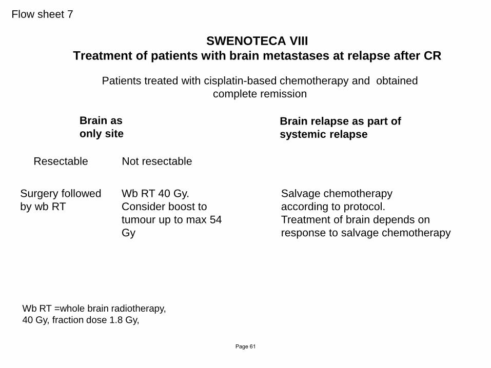
SWENOTECA VIII Treatment of patients with brain metastases at relapse after CR
Patients treated with cisplatin-based chemotherapy and obtained complete remission
Brain as only site
Brain relapse as part of systemic relapse
Resectable Not resectable
Surgery followed by wb RT
Wb RT 40 Gy. Consider boost to tumour up to max 54 Gy
Salvage chemotherapy according to protocol. Treatment of brain depends on response to salvage chemotherapy
Wb RT =whole brain radiotherapy, 40 Gy, fraction dose 1.8 Gy,
Flow sheet 7
Page 61

SWENOTECA VIII Salvage treatment metastatic non-seminoma
Marker negative relapse
Surgery
TIP x 1
Stem cell harvest, if not done
HD 1
Marker positive relapse
Vital germ cell cancer
Teratoma only
t½ delayed t½ on time
Follow-up
If initial treatment BEP/BEP-if/PEI and IGCCCG-2-score = 0
If initial treatment Taxane based or
IGCCCG-2 score ≥ 1
If initial treatment included HD
TIP x 1
TIP x 2
Surgery
TIP x 1* for stem cell harvest
if not done, otherwise treat
immediately with
HD 2
Surgery
Discuss within SWENOTECA
As marker positive relapse
If the patient has received several courses of TIP in the primary treatment, other mobilization chemotherapy could be chosen.
Response evaluation
Progression: Individual treatment Discussion within SWENOTECA
*
Flow sheet 8
Page 62
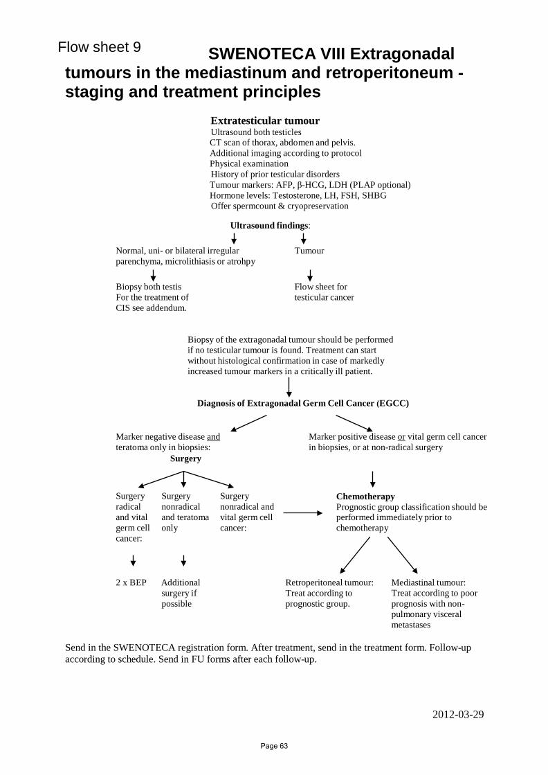
SWENOTECA VIII Extragonadal tumours in the mediastinum and retroperitoneum - staging and treatment principles
Extratesticular tumour Ultrasound both testicles CT scan of thorax, abdomen and pelvis. Additional imaging according to protocol Physical examination History of prior testicular disorders Tumour markers: AFP, β-HCG, LDH (PLAP optional) Hormone levels: Testosterone, LH, FSH, SHBG Offer spermcount & cryopreservation
Ultrasound findings:
Normal, uni- or bilateral irregular parenchyma, microlithiasis or atrohpy
Tumour
Biopsy both testis For the treatment of CIS see addendum.
Flow sheet for testicular cancer
Biopsy of the extragonadal tumour should be performed if no testicular tumour is found. Treatment can start without histological confirmation in case of markedly increased tumour markers in a critically ill patient.
Diagnosis of Extragonadal Germ Cell Cancer (EGCC)
Marker negative disease and teratoma only in biopsies:
Surgery
Marker positive disease or vital germ cell cancer in biopsies, or at non-radical surgery
Surgery radical and vital germ cell cancer:
Surgery nonradical and teratoma only
Surgery nonradical and vital germ cell cancer:
Chemotherapy Prognostic group classification should be performed immediately prior to chemotherapy
2 x BEP Additional surgery if possible
Retroperitoneal tumour: Treat according to prognostic group.
Mediastinal tumour: Treat according to poor prognosis with non- pulmonary visceral metastases
Send in the SWENOTECA registration form. After treatment, send in the treatment form. Follow-up according to schedule. Send in FU forms after each follow-up.
2012-03-29
Flow sheet 9
Page 63

Page 64

Clinical staging according to Royal Marsden, modified CS I No evidence of metastases
CS Mk+ Tumour markers AFP/ß-HCG persistently elevated (not declining according to
half-life), but no macroscopic metastatic disease demonstrated
CS II Metastatic disease restricted to abdominal nodes:
A Maximal transverse diameter <2 cm
B Maximal transverse diameter 2–5 cm
C Maximal transverse diameter >5–10 cm
D Maximal transverse diameter >10 cm
CS III Supradiaphragmatic node involvement
For abdominal lymph-nodes: 0 No metastases; A-D According to CS II.
CS IV Extra-lymphatic metastases
For abdominal lymph-nodes: 0 No metastases; A-D According to CS II.
H+ Liver metastases, Br+ Brain metastases, Bo+ Bone metastases
Page 65

Prognostic risk group classification according to IGCCCG
GOOD-PROGNOSIS GROUP
Primary testis/retroperitoneum and No non-pulmonary visceral metastases (for example liver, bone, brain) and all good markers; ß-HCG < 5000 IU/L (1000 µg/L) and AFP < 1000 µg/L and LDH < 1,5 X N
INTERMEDIATE-PROGNOSIS
Primary testis/retroperitoneum and No non-pulmonary visceral metastases and any intermediate marker; ß-HCG ≥ 5000 and ≤ 50000 IU/L or AFP ≥ 1000 and ≤ 10000 µg/L or LD ≥ 1,5 x N and ≤ 10 x N
POOR-PROGNOSIS
Mediastinal primary or Non- pulmonary visceral metastasis (for example liver, bone, brain) or any poor marker: ß-HCG > 50000 IU/L or AFP > 10000 µg/L or LD > 10 x N
Page 66

AFP, plot – halveringstid
Page 67
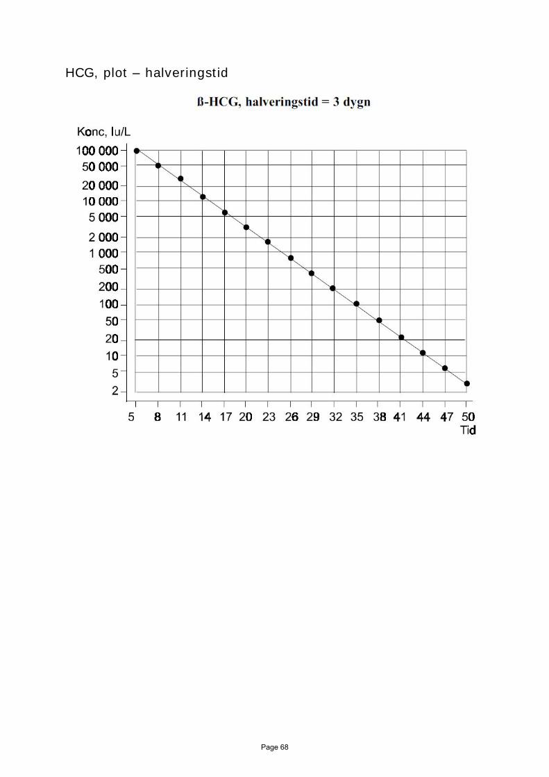
HCG, plot – halveringstid
Page 68
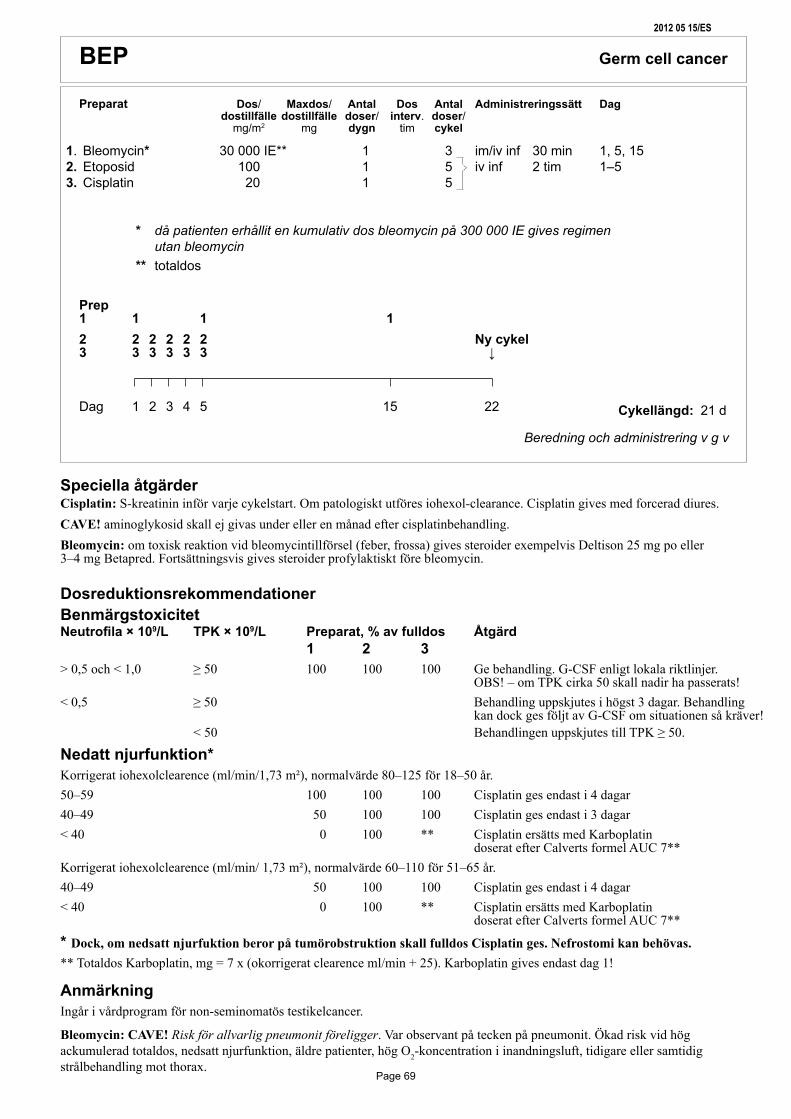
Preparat Dos/ Maxdos/ Antal Dos Antal Administreringssätt Dag dostillfälledostillfälle doser/ interv. doser/ mg/m2 mg dygn tim cykel
Cykellängd:
Beredning och administrering v g v
BEP Germcellcancer
1. Bleomycin* 30000IE** 1 3 im/ivinf 30min 1,5,152. Etoposid 100 1 5 ivinf 2tim 1–53. Cisplatin 20 1 5
* då patienten erhållit en kumulativ dos bleomycin på 300 000 IE gives regimen utan bleomycin ** totaldos
21d
Prep 1 1 1 1 2 2 2 2 2 2 Nycykel 3 3 3 3 3 3 ↓
Dag 1 2 3 4 5 15 22
SpeciellaåtgärderCisplatin: S-kreatinin inför varje cykelstart. Om patologiskt utföres iohexol-clearance. Cisplatin gives med forcerad diures. CAVE! aminoglykosid skall ej givas under eller en månad efter cisplatinbehandling.Bleomycin: om toxisk reaktion vid bleomycintillförsel (feber, frossa) gives steroider exempelvis Deltison 25 mg po eller 3–4 mg Betapred. Fortsättningsvis gives steroider profylaktiskt före bleomycin.
DosreduktionsrekommendationerBenmärgstoxicitet Neutrofila × 109/L TPK × 109/L Preparat,%avfulldos Åtgärd 1 2 3 > 0,5 och < 1,0 ≥ 50 100 100 100 Ge behandling. G-CSF enligt lokala riktlinjer. OBS! – om TPK cirka 50 skall nadir ha passerats!< 0,5 ≥ 50 Behandling uppskjutes i högst 3 dagar. Behandling kan dock ges följt av G-CSF om situationen så kräver! < 50 Behandlingen uppskjutes till TPK ≥ 50.
Nedattnjurfunktion*Korrigerat iohexolclearence (ml/min/1,73 m²), normalvärde 80–125 för 18–50 år. 50–59 100 100 100 Cisplatin ges endast i 4 dagar40–49 50 100 100 Cisplatin ges endast i 3 dagar< 40 0 100 ** Cisplatin ersätts med Karboplatin doserat efter Calverts formel AUC 7**Korrigerat iohexolclearence (ml/min/ 1,73 m²), normalvärde 60–110 för 51–65 år.40–49 50 100 100 Cisplatin ges endast i 4 dagar< 40 0 100 ** Cisplatin ersätts med Karboplatin doserat efter Calverts formel AUC 7**
*Dock, om nedsatt njurfuktion beror på tumörobstruktion skall fulldos Cisplatin ges. Nefrostomi kan behövas.** Totaldos Karboplatin, mg = 7 x (okorrigerat clearence ml/min + 25). Karboplatin gives endast dag 1! AnmärkningIngår i vårdprogram för non-seminomatös testikelcancer.
Bleomycin: CAVE! Risk för allvarlig pneumonit föreligger. Var observant på tecken på pneumonit. Ökad risk vid hög ackumulerad totaldos, nedsatt njurfunktion, äldre patienter, hög O2-koncentration i inandningsluft, tidigare eller samtidig strålbehandling mot thorax.
20120515/ES
Page 69

Blandning och administrering
Preparat Blandas i Administrering Sköljdropp Spoldropp Kemiskt Kommentar ml sätt tid NaCl, ml NaCl, ml stabil
250Cisplatin 1000 NaCl iv inf 2 tim 72 tim Etoposid rumstemp
Bleomycin 250 NaCl iv inf 30 min 7 dygn, kallt
BEP
Prehydrering: 1 000 NaCl under 2 tim.
Hydrering under behandlingen:Under behandlingsdygnen gives ytterligare minst 2 000 ml vätska po el iv.
Posthydrering: Dygnet efter sista cisplatininfusion minst 2 000 ml; om patienten ej själv kan dricka denna mängd, skall vätska givas iv. Diuresen under behandlingsdygnet samt dygnet efter sista cisplatinbehandlingen skall vara > 400 ml/4 tim. Mätning startar samtidigt med start av prehydrering.
20120515/ES
Page 70

Preparat Dos/ Maxdos/ Antal Dos Antal Administreringssätt Dag dostillfälledostillfälle doser/ interv. doser/ mg/m2 mg dygn tim cykel
Cykellängd:
Beredning och administrering v g v
EP Germcellcancer
1. Etoposid 100 1 5 ivinf 2tim 1–52. Cisplatin 20 1 5
21d
Prep 1 1 1 1 1 1 Nycykel 2 2 2 2 2 2 ↓
Dag 1 2 3 4 5 22
SpeciellaåtgärderCisplatin: S-kreatinin inför varje cykelstart. Om patologiskt utföres iohexol-clearance. Cisplatin gives med forcerad diures. CAVE! aminoglykosid skall ej givas under eller en månad efter cisplatinbehandling.
DosreduktionsrekommendationerBenmärgstoxicitet Neutrofila × 109/L TPK × 109/L Preparat,%avfulldos Åtgärd 1 2 > 0,5 och < 1,0 ≥ 50 100 100 Ge behandling. G-CSF enligt lokala riktlinjer. OBS! – om TPK cirka 50 skall nadir ha passerats!< 0,5 ≥ 50 Behandling uppskjutes i högst 3 dagar. Behandling kan dock ges följt av G-CSF om situationen så kräver! < 50 Behandlingen uppskjutes till TPK ≥ 50.
Nedattnjurfunktion*Korrigerat iohexolclearence (ml/min/1,73 m²), normalvärde 80–125 för 18–50 år. 50–59 100 100 Cisplatin ges endast i 4 dagar40–49 100 100 Cisplatin ges endast i 3 dagar< 40 100 ** Cisplatin ersätts med Karboplatin doserat efter Calverts formel AUC 7**Korrigerat iohexolclearence (ml/min/ 1,73 m²), normalvärde 60–110 för 51–65 år.40–49 100 100 Cisplatin ges endast i 4 dagar< 40 100 ** Cisplatin ersätts med Karboplatin doserat efter Calverts formel AUC 7**
*Dock, om nedsatt njurfuktion beror på tumörobstruktion skall fulldos Cisplatin ges. Nefrostomi kan behövas.** Totaldos Karboplatin, mg = 7 x (okorrigerat clearence ml/min + 25). Karboplatin gives endast dag 1!
AnmärkningIngår i vårdprogram för non-seminomatös testikelcancer.
20120507/ES
Page 71

Blandning och administrering
Preparat Blandas i Administrering Sköljdropp Spoldropp Kemiskt Kommentar ml sätt tid NaCl, ml NaCl, ml stabil
250 Cisplatin 1000 NaCl iv inf 2 tim 72 tim Etoposid rumstemp
EP
Prehydrering:1000 NaCl under 2 tim.
Hydrering under behandlingen:Under behandlingsdygnen gives ytterligare minst 2 000 ml vätska po el iv.
Posthydrering: Dygnet efter sista cisplatininfusion minst 2 000 ml; om patienten ej själv kan dricka denna mängd, skall vätska givas iv.
Diuresen under behandlingsdygnet samt dygnet efter sista cisplatinbehandlingen skall vara > 400 ml/4 tim. Mätning startar samtidigt med start av prehydrering.
20120507/ES
Page 72

Preparat Dos/ Maxdos/ Antal Dos Antal Administreringssätt Dag dostillfälledostillfälle doser/ interv. doser/ mg/m2 mg dygn tim cykel
Cykellängd:
Beredning och administrering v g v
1.Bleomycin* 30000**IE 1 3 im/ivinf 30min 1,5,152.Etoposid 75 1 5 ivinf 2tim 1–53.Cisplatin 20 1 54. Ifosfamid 1200 1 5 ivinf 30min 1–55. Mesna 240(20%avifosfdos)1 5 Mesna 480(40%avifosfdos) 2 10 po*** 2o6timefterifosfamid *då patienten erhållit en kumulativ dos bleomycin på 300 000 IE gives regimen utan bleomycin **totaldos *** Ompatientenintesäkertfårisigmesnapo(kräks)givessamtliga3doseriv. 20%avifosfamiddosengivesdåtimme4och8.
BEP-Ifosfamid Germcellcancer
21d
Prep 1 1 1 1 2 2 2 2 2 2 3 3 3 3 3 3 4 4 4 4 4 4 Nycykel 5 5 5 5 5 5 ↓
Dag 1 2 3 4 5 15 22 SpeciellaåtgärderCisplatin: S-kreatinin inför varje cykelstart. Om patologiskt utföres iohexol-clearance. Cisplatin gives med forcerad diures. CAVE! aminoglykosid skall ej givas under eller en månad efter cisplatinbehandling.Bleomycin: om toxisk reaktion vid bleomycintillförsel (feber, frossa) gives steroider exempelvis Deltison 25 mg po eller 3–4 mg Betapred. Fortsättningsvis gives steroider profylaktiskt före bleomycin.Ifosfamid: Observerans på cystitbesvär. Hematuristicka vid behov. Om 3+ så avbryts ifosfamidbehandlingen.
DosreduktionsrekommendationerBenmärgstoxicitet Neutrofila × 109/L TPK × 109/L Preparat,%avfulldos Åtgärd 1 2 3 4+5 > 0,5 och < 1,0 ≥ 50 100 100 100 100 Ge behandling. G-CSF enligt lokala riktlinjer. OBS! – om TPK cirka 50 skall nadir ha passerats< 0,5 ≥ 50 Behandling uppskjutes i högst 3 dagar. Behandling kan dock ges följt av G-CSF om situationen så kräver! < 50 Behandlingen uppskjutes till TPK ≥ 50.Nedattnjurfunktion*Korrigerat iohexolclearence (ml/min/1,73 m²), normalvärde 80–125 för 18–50 år. 50–59 100 100 100 100 Cisplatin ges endast i 4 dagar40–49 50 100 100 100 Cisplatin ges endast i 3 dagar. Ifosfamid och Mesna ges endast i 4 dagar< 40 0 100 ** 100 Cisplatin ersätts med Karboplatin doserat efter Calverts formel AUC 7**. Ifosfamid och Mesna ges endast i 4 dagarKorrigerat iohexolclearence (ml/min/1,73 m²), normalvärde 60–110 för 51–65 år.40–49 50 100 100 100 Cisplatin ges endast i 4 dagar< 40 0 100 ** 100 Cisplatin ersätts med Karboplatin doserat efter Calverts formel AUC 7** Ifosfamid och Mesna ges endast i 4 dagar
*Dock, om nedsatt njurfuktion beror på tumörobstruktion skall fulldos Cisplatin ges. Nefrostomi kan behövas.** Totaldos Karboplatin, mg = 7 x (okorrigerat clearence ml/min + 25). Karboplatin gives endast dag 1!
AnmärkningIngår i vårdprogram för non-seminomatös testikelcancer.Bleomycin: CAVE! Risk för allvarlig pneumonit föreligger. Var observant på tecken på pneumonit. Ökad risk vid hög ackumulerad totaldos, nedsatt njurfunktion, äldre patienter, hög O2-koncentration i inandningsluft, tidigare eller samtidig strålbehandling mot thorax.
20120515/ES
Page 73

Blandning och administrering
Preparat Blandas i Administrering Sköljdropp Spoldropp Kemiskt Kommentar ml sätt tid NaCl, ml NaCl, ml stabil
BEP-Ifosfamid
250Cisplatin 1000 NaCl iv inf 2 tim 72 tim Etoposid rumstemp
Ifosfamid 250 NaCl iv inf 30 min 72 tim, kylMesna 1:a dos
Mesna dos 2 och 3 gives om möjligt po
Bleomycin 250 NaCl iv inf 30 min 7 dygn, kallt
Prehydrering: 1 000 NaCl under 2 tim.
Hydrering under behandlingen:Under behandlingsdygnen gives ytterligare minst 2 000 ml vätska po el iv.
Posthydrering: Dygnet efter sista cisplatininfusion minst 2 000 ml; om patienten ej själv kan dricka denna mängd, skall vätska givas iv. Diuresen under behandlingsdygnet samt dygnet efter sista cisplatinbehandlingen skall vara > 400 ml/4 tim. Mätning startar samtidigt med start av prehydrering.
20120515/ES
Page 74

Preparat Dos/ Maxdos/ Antal Dos Antal Administreringssätt Dag dostillfälle dostillfälle doser/ interv. doser/ mg/m2 mg dygn tim cykel
Cykellängd:
Beredning och administrering v g v
PEI Germcellcancer
1. Etoposid 100 1 5 ivinf 2tim 1–52. Cisplatin 20 1 53. Ifosfamid 1200 1 5 ivinf 30min 1–54. Mesna 240(20%avifosfdos) 1 5 Mesna 480(40%avifosfdos) 2 10 po* 2o6timefterifosfamid *Ompatientenintesäkertfårisigmesnapo(kräks)givessamtliga3doseriv. 20%avifosfamiddosengivesdåtimme4och8.
21d
Prep 1 1 1 1 1 1 2 2 2 2 2 2 3 3 3 3 3 3 Nycykel 4 4 4 4 4 4 ↓
Dag 1 2 3 4 5 22
SpeciellaåtgärderCisplatin: S-kreatinin inför varje cykelstart. Om patologiskt utföres iohexol-clearance. Cisplatin gives med forcerad diures. CAVE! aminoglykosid skall ej givas under eller en månad efter cisplatinbehandling.Ifosfamid: Observerans på cystitbesvär. Hematuristicka vid behov. Om 3+ så avbryts ifosfamidbehandlingen.
DosreduktionsrekommendationerBenmärgstoxicitet Neutrofila × 109/L TPK × 109/L Preparat,%avfulldos Åtgärd 1 2 3+4 > 0,5 och < 1,0 ≥ 50 100 100 100 Ge behandling. G-CSF enligt lokala riktlinjer. OBS! – om TPK cirka 50 skall nadir ha passerats!< 0,5 ≥ 50 Behandling uppskjutes i högst 3 dagar. Behandling kan dock ges följt av G-CSF om situationen så kräver! < 50 Behandlingen uppskjutes till TPK ≥ 50.
Nedattnjurfunktion*Korrigerat iohexolclearence (ml/min/1,73 m²), normalvärde 80–125 för 18–50 år. 50–59 100 100 100 Cisplatin ges endast i 4 dagar40–49 100 100 100 Cisplatin ges endast i 3 dagar Ifosfamid och Mesna ges endast i 4 dagar< 40 100 ** 100 Cisplatin ersätts med Karboplatin doserat efter Calverts formel AUC 7** Ifosfamid och Mesna ges endast i 4 dagarKorrigerat iohexolclearence (ml/min/ 1,73 m²), normalvärde 60–110 för 51–65 år.40–49 100 100 100 Cisplatin ges endast i 4 dagar Ifosfamid och Mesna ges endast i 4 dagar< 40 100 ** 100 Cisplatin ersätts med Karboplatin doserat efter Calverts formel AUC 7** Ifosfamid och Mesna ges endast i 4 dagar
*Dock, om nedsatt njurfuktion beror på tumörobstruktion skall fulldos Cisplatin ges. Nefrostomi kan behövas.** Totaldos Karboplatin, mg = 7 x (okorrigerat clearence ml/min + 25). Karboplatin gives endast dag 1!
AnmärkningIngår i vårdprogram för non-seminomatös testikelcancer.
2012 05 07/ES
Page 75

Blandning och administrering
Preparat Blandas i Administrering Sköljdropp Spoldropp Kemiskt Kommentar ml sätt tid NaCl, ml NaCl, ml stabil
250Cisplatin 1000 NaCl iv inf 2 tim 72 tim Etoposid rumstemp
Ifosfamid 250 NaCl iv inf 30 min 72 tim, kalltMesna 1:a dos
Mesna dos 2 och 3 gives om möjligt po
PEI
Prehydrering: 1 000 NaCl under 2 tim.
Hydrering under behandlingen:Under behandlingsdygnen gives ytterligare minst 2 000 ml vätska po el iv.
Posthydrering: Dygnet efter sista cisplatininfusion minst 2 000 ml; om patienten ej själv kan dricka denna mängd, skall vätska givas iv.
Diuresen under behandlingsdygnet samt dygnet efter sista cisplatinbehandlingen skall vara > 400 ml/4 tim. Mätning startar samtidigt med start av prehydrering.
20120507/ES
Page 76

Preparat Dos/ Maxdos/ Antal Dos Antal Administreringssätt Dag dostillfälledostillfälle doser/ interv. doser/ mg/m2 mg dygn tim cykel
Cykellängd:
Beredning och administrering v g v
TIP Germcellcancer
1. Paclitaxel 250 1 1 ivinf 24tim 12. Ifosfamid 1500 1 4 ivinf 2tim 2–53.Mesna 300 1 4 Mesna* 300 2 8 ivinjtim4och8efteravslutadifosfamidinf4. Cisplatin 25 1 4 ivinf 2tim 2–5
*Mesna:Kanävengesperoraltmendåidubbeldos(40%avifosfamiddosen).Förstadosengivesivtillsammansmedifosfamid,deföljandeperoraladoserna2och6timefteravslutadifosfamidinf.
21d
SpeciellaåtgärderPaclitaxel premedicinering: 30 min före start av infusion gives inj. Betametason 12 mg iv, inj. Clemastin 2 mg iv, inj Ranitidin 50 mg iv.Kontroll av puls och blodtryck före och 15 min efter start av infusion. Akutbricka + PM för åtgärder vid akuta allergiska reaktioner skall vara tillgängliga. Läkare skall finnas nåbar på personsökare. Se även ”Handläggning av lindrig reaktion vid Taxolinfusion”.Cisplatin: S-kreatinin inför varje cykelstart. Om patologiskt utföres iohexol-clearance. Cisplatin gives med forcerad diures. CAVE! aminoglykosid skall ej givas under eller en månad efter cisplatinbehandling.G-CSF: 5 µg/kg gives från och med dag 7.Ifosfamid: Observerans på cystitbesvär. Hematuristicka vid behov. Om 3+ så avbryts ifosfamidbehandlingen.
AnmärkningIngår i vårdprogram för non-seminomatös testikelcancer. JCO 2005, 23, 6549
Prep 1 1 2 2 2 2 2 3 3 3 3 3 4 4 4 4 4 Nycykel ↓
Dag 1 2 3 4 5 22
DosreduktionsrekommendationerBenmärgstoxicitet Neutrofila×109/L TPK×109/L Preparat,%avfulldos Åtgärd 1 2+3 4 ≥ 0,5 ≥ 50 100 100 100 OBS! – om TPK cirka 50 skall nadir ha passerats!< 0,5 eller < 50 Behandling uppskjutes kortast möjliga tid.
Nedattnjurfunktion*Korrigerat iohexolclearence (ml/min/1,73 m²), normalvärde 80–125 för 18–50 år. 50–59 100 100 80 Dag 2–540–49 100 100 80 Dag 2–4< 40 100 100 ** Cisplatin ersätts med Karboplatin doserat efter Calverts formel AUC 7**Korrigerat iohexolclearence (ml/min/ 1,73 m²), normalvärde 60–110 för 51–65 år.40–49 100 100 80 Dag 2–5< 40 100 100 ** Cisplatin ersätts med Karboplatin doserat efter Calverts formel AUC 7**
*Dock, om nedsatt njurfuktion beror på tumörobstruktion skall fulldos Cisplatin ges. Nefrostomi kan behövas.** Totaldos Karboplatin, mg = 7 x (okorrigerat clearence ml/min + 25). Karboplatin gives endast dag 1!
20120507/ES
Page 77

Blandning och administrering
Preparat Blandas i Administrering Sköljdropp Spoldropp Kemiskt Kommentar ml sätt tid NaCl, ml NaCl, ml stabil
250Paclitaxel 1000 NaCl iv inf 24 tim 48 tim, rumstemp
250Ifosfamid 1000 NaCl iv inf 2 tim 72 tim, kalltMesna 1:a dos
Mesna följande doser iv inj/po
Cisplatin 1000 NaCl iv inf 2 tim 72 tim, rumstemp
TIP–Testikelcancer
Prehydrering: 1 000 NaCl under 2 tim.
Hydrering under behandlingen:Under behandlingsdygnen gives ytterligare minst 2 000 ml vätska po el iv.
Posthydrering: Dygnet efter sista cisplatininfusion minst 2 000 ml; om patienten ej själv kan dricka denna mängd, skall vätska givas iv. Diuresen under behandlingsdygnet samt dygnet efter sista cisplatinbehandlingen skall vara > 400 ml/4 tim. Mätning startar samtidigt med start av prehydrering.
20120507/ES
Page 78

Preparat Dos/ Maxdos/ Antal Dos Antal Administreringssätt Dag dostillfälledostillfälle doser/ interv. doser/ mg/m2 mg dygn tim cykel
Cykellängd:
Beredning och administrering v g v
EMA-CO
1. Etoposid 100 1 2 ivinf 1tim 1,22. Daktinomycin 0.5* 1 2 ivinf 30min 1,23. Metotrexat 100 1 1 ivinf 30min 14. Metotrexat 200 1 1 ivinf 12tim 15. Kalciumfolinat 15* 4 8 po/iv var6:etim 2,3 medstart24timefterstartavMTX6. Vinkristin 1.0 2.0 1 1 ivinf 30min 87. Cyklofosfamid 600 1 1 ivinf 30min 8
*totaldos
14d
SpeciellaåtgärderOBS! cykelintervallet skall ej överstiga 15 dagar.
Nedsatt njurfunktion, pleuravätska, ascites är relativa kontraindikationer för Mtx. Om det trots detta gives bör plasmakoncentrationen följas; förlängd kalciumfolinatrescue kan vara aktuell. Pat skall ha skriftliga instruktioner angående kalciumfolinatrescue. S-kreatinin följes under behandlingen.
Dosreduktionsrekommendationer Preparat,%avfulldosGranulocyter×109/L TPK×109/L 1 2 3 4 5 6 7
<1.0 <100 75 100 100 100 100 100 75
Om även efter dosreduktion granulocyter <1.0 eller TPK ×109/L <100 vid cykelstart gives i fortsättningen: 50 100 100 100 100 100 50
Anmärkning
Prep 1 1 1 2 2 2 3 3 4 4 5 5 5 6 6 Nycykel 7 7 ↓
Dag 1 2 3 8 15
20120507/ES
RecidivavHCGproducerandegermcellcancer
Page 79

Blandning och administrering
Preparat Blandas i Administrering Sköljdropp Spoldropp Kemiskt Kommentar ml sätt tid NaCl, ml NaCl, ml stabil
500
Etoposid 1000NaCl ivinf 1tim 72tim,kallt Konc≤0,4mg/ml Daktinomycin 250 NaCl iv inf 30 min 72 tim, kallt Metotrexat 250 NaCl iv inf 30 min 72 tim, kallt
Metotrexat 500 NaCl iv inf 12 tim
250
Vinkristin 250 NaCl iv inf 30 min 72 tim, kallt
Cyklofosfamid 250 NaCl iv inf 30 min 72 tim, kallt
EMA-CO20120507/ES
Page 80

Preparat Dos/ Maxdos/ Antal Dos Antal Administreringssätt Dag dostillfälledostillfälle doser/ interv. doser/ mg/m2 mg dygn tim cykel
Cykellängd:
Beredning och administrering v g v
GOP Germcellcancer
1. Gemcitabin 800 1 2 ivinf 30min 1+82.Oxaliplatin 130 1 1 ivinf 2tim 13.Paclitaxel 80 1 2 ivinf 1tim 1+8
SpeciellaåtgärderOxaliplatin: Vid polyneuropati gives substitution med calcium och magnesium (se omstående sida).
Paclitaxel premedicinering: 30 min före infusion gives inj Betametason 6 mg iv. Gives endast dag 1 och 8 i cykel 1 om inga oönskade reaktioner inträffat. Inj. Clemastin 2 mg iv inj. Ranitidin 50 mg iv gives samtliga cykler.
Kontroll av puls och blodtryck före och 15 min efter start av infusion dag 1 och 8 cykel 1.
Akutbricka + PM för åtgärder vid akuta allergiska reaktioner skall vara tillgängliga. Läkare skall finnas nåbar på personsökare. Se även ”Handläggning av lindrig reaktion vid Taxolinfusion”.
Dosreduktionsrekommendationer Preparat,%avfulldos Preparat,%avfulldos dag1 dag8Granulocyter×109/L TPK×109/L 1 2 3 1 3
≥ 1,0 och < 1,5 ≥ 50 och < 75 100 100 100 75 75
≥ 0,5 och < 1,0 ≥ 50 75 75 75 50 50
< 0,5 < 50 Behandlingen uppskjutes.
G-CSF gives enligt lokala riktlinjer.Om bestående neuropati WHO grad 2 (svåra parestesier och/eller lätt svaghet), dosreduceras oxaliplatin och paclitaxel till 75 % i följande cykler. Om grad 3–4 toxicitet gives inte denna behandling.
AnmärkningIngår i vårdprogram SWENOTECA VIII.Ref. European Urology 2011, 60, 850-835.
Prep 1 1 1 2 2 3 3 3 Nycykel ↓
Dag 1 8 22
20120507/ES
21d
Page 81
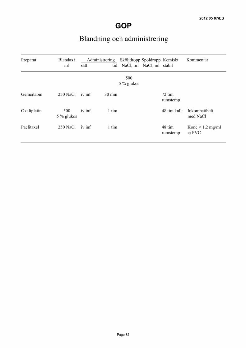
Blandning och administrering
Preparat Blandas i Administrering Sköljdropp Spoldropp Kemiskt Kommentar ml sätt tid NaCl, ml NaCl, ml stabil
500 5 % glukos
Gemcitabin 250 NaCl iv inf 30 min 72 tim rumstemp
Oxaliplatin 500 iv inf 1 tim 48 tim kallt Inkompatibelt 5 % glukos med NaCl
Paclitaxel 250 NaCl iv inf 1 tim 48 tim Konc < 1,2 mg/ml rumstemp ej PVC
GOP20120507/ES
Page 82
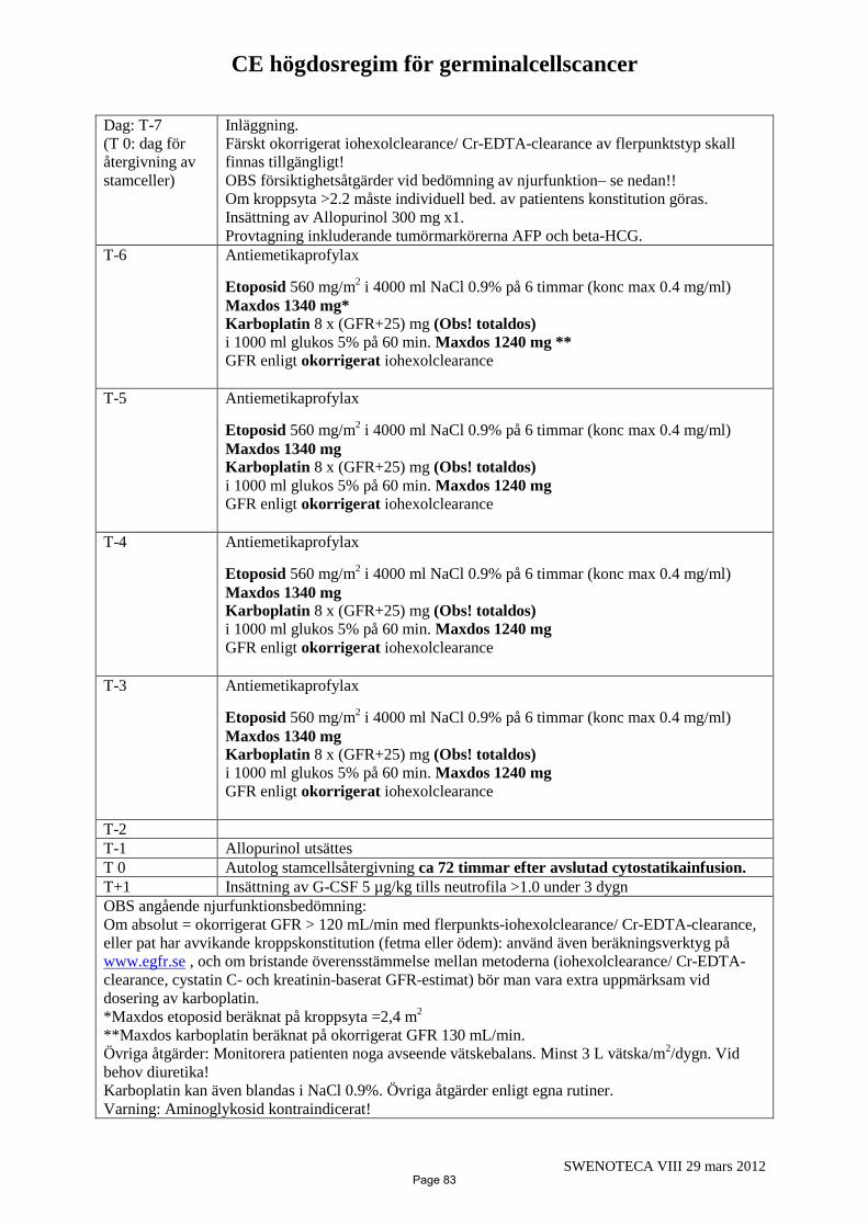
CE högdosregim för germinalcellscancer
SWENOTECA VIII 29 mars 2012
Dag: T-7
(T 0: dag för
återgivning av
stamceller)
Inläggning.
Färskt okorrigerat iohexolclearance/ Cr-EDTA-clearance av flerpunktstyp skall
finnas tillgängligt!
OBS försiktighetsåtgärder vid bedömning av njurfunktion– se nedan!!
Om kroppsyta >2.2 måste individuell bed. av patientens konstitution göras.
Insättning av Allopurinol 300 mg x1.
Provtagning inkluderande tumörmarkörerna AFP och beta-HCG.
T-6 Antiemetikaprofylax
Etoposid 560 mg/m2 i 4000 ml NaCl 0.9% på 6 timmar (konc max 0.4 mg/ml)
Maxdos 1340 mg* Karboplatin 8 x (GFR+25) mg (Obs! totaldos)
i 1000 ml glukos 5% på 60 min. Maxdos 1240 mg **
GFR enligt okorrigerat iohexolclearance
T-5 Antiemetikaprofylax
Etoposid 560 mg/m2 i 4000 ml NaCl 0.9% på 6 timmar (konc max 0.4 mg/ml)
Maxdos 1340 mg Karboplatin 8 x (GFR+25) mg (Obs! totaldos)
i 1000 ml glukos 5% på 60 min. Maxdos 1240 mg
GFR enligt okorrigerat iohexolclearance
T-4 Antiemetikaprofylax
Etoposid 560 mg/m2 i 4000 ml NaCl 0.9% på 6 timmar (konc max 0.4 mg/ml)
Maxdos 1340 mg Karboplatin 8 x (GFR+25) mg (Obs! totaldos)
i 1000 ml glukos 5% på 60 min. Maxdos 1240 mg
GFR enligt okorrigerat iohexolclearance
T-3 Antiemetikaprofylax
Etoposid 560 mg/m2 i 4000 ml NaCl 0.9% på 6 timmar (konc max 0.4 mg/ml)
Maxdos 1340 mg Karboplatin 8 x (GFR+25) mg (Obs! totaldos)
i 1000 ml glukos 5% på 60 min. Maxdos 1240 mg
GFR enligt okorrigerat iohexolclearance
T-2
T-1 Allopurinol utsättes
T 0 Autolog stamcellsåtergivning ca 72 timmar efter avslutad cytostatikainfusion.
T+1 Insättning av G-CSF 5 µg/kg tills neutrofila >1.0 under 3 dygn
OBS angående njurfunktionsbedömning:
Om absolut = okorrigerat GFR > 120 mL/min med flerpunkts-iohexolclearance/ Cr-EDTA-clearance,
eller pat har avvikande kroppskonstitution (fetma eller ödem): använd även beräkningsverktyg på
www.egfr.se , och om bristande överensstämmelse mellan metoderna (iohexolclearance/ Cr-EDTA-
clearance, cystatin C- och kreatinin-baserat GFR-estimat) bör man vara extra uppmärksam vid
dosering av karboplatin.
*Maxdos etoposid beräknat på kroppsyta =2,4 m2
**Maxdos karboplatin beräknat på okorrigerat GFR 130 mL/min.
Övriga åtgärder: Monitorera patienten noga avseende vätskebalans. Minst 3 L vätska/m2/dygn. Vid
behov diuretika!
Karboplatin kan även blandas i NaCl 0.9%. Övriga åtgärder enligt egna rutiner.
Varning: Aminoglykosid kontraindicerat!
Page 83

Page 84
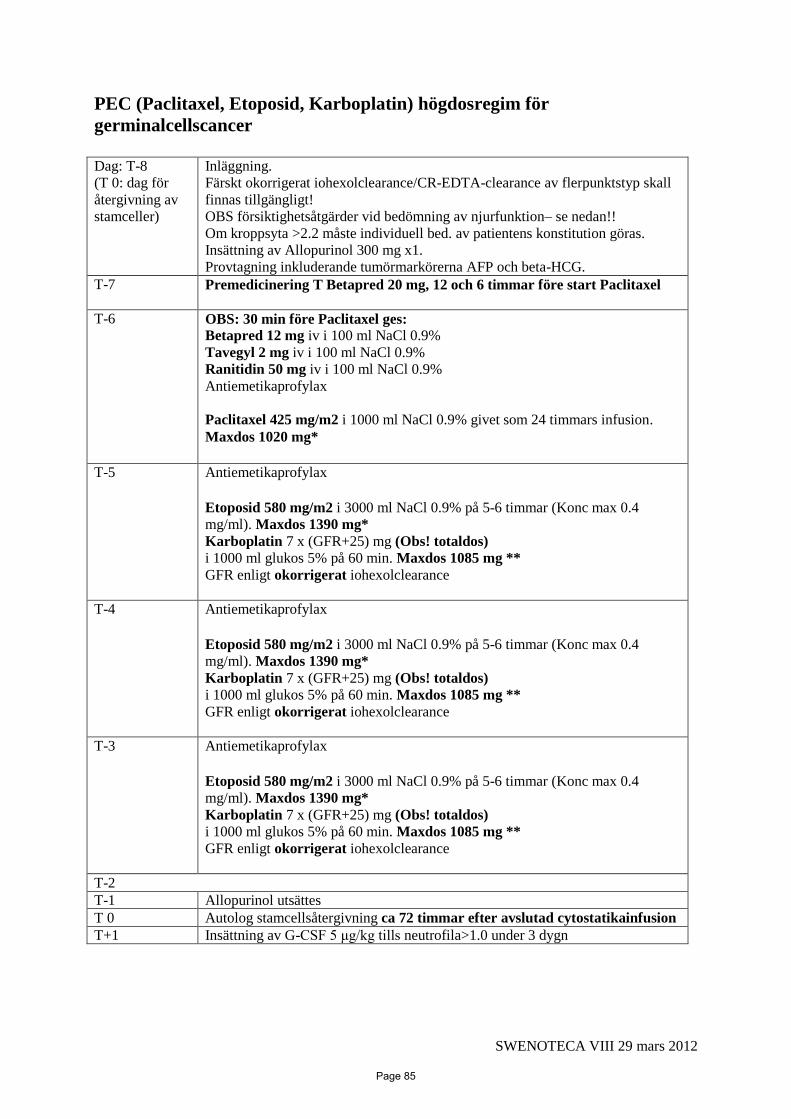
SWENOTECA VIII 29 mars 2012
PEC (Paclitaxel, Etoposid, Karboplatin) högdosregim för
germinalcellscancer
Dag: T-8
(T 0: dag för
återgivning av
stamceller)
Inläggning.
Färskt okorrigerat iohexolclearance/CR-EDTA-clearance av flerpunktstyp skall
finnas tillgängligt!
OBS försiktighetsåtgärder vid bedömning av njurfunktion– se nedan!! Om kroppsyta >2.2 måste individuell bed. av patientens konstitution göras. Insättning av Allopurinol 300 mg x1. Provtagning inkluderande tumörmarkörerna AFP och beta-HCG.
T-7 Premedicinering T Betapred 20 mg, 12 och 6 timmar före start Paclitaxel
T-6 OBS: 30 min före Paclitaxel ges: Betapred 12 mg iv i 100 ml NaCl 0.9%
Tavegyl 2 mg iv i 100 ml NaCl 0.9%
Ranitidin 50 mg iv i 100 ml NaCl 0.9%
Antiemetikaprofylax
Paclitaxel 425 mg/m2 i 1000 ml NaCl 0.9% givet som 24 timmars infusion.
Maxdos 1020 mg*
T-5 Antiemetikaprofylax
Etoposid 580 mg/m2 i 3000 ml NaCl 0.9% på 5-6 timmar (Konc max 0.4
mg/ml). Maxdos 1390 mg*
Karboplatin 7 x (GFR+25) mg (Obs! totaldos)
i 1000 ml glukos 5% på 60 min. Maxdos 1085 mg **
GFR enligt okorrigerat iohexolclearance
T-4 Antiemetikaprofylax Etoposid 580 mg/m2 i 3000 ml NaCl 0.9% på 5-6 timmar (Konc max 0.4
mg/ml). Maxdos 1390 mg*
Karboplatin 7 x (GFR+25) mg (Obs! totaldos)
i 1000 ml glukos 5% på 60 min. Maxdos 1085 mg **
GFR enligt okorrigerat iohexolclearance
T-3 Antiemetikaprofylax Etoposid 580 mg/m2 i 3000 ml NaCl 0.9% på 5-6 timmar (Konc max 0.4
mg/ml). Maxdos 1390 mg*
Karboplatin 7 x (GFR+25) mg (Obs! totaldos)
i 1000 ml glukos 5% på 60 min. Maxdos 1085 mg **
GFR enligt okorrigerat iohexolclearance
T-2
T-1 Allopurinol utsättes
T 0 Autolog stamcellsåtergivning ca 72 timmar efter avslutad cytostatikainfusion
T+1 Insättning av G-CSF 5 μg/kg tills neutrofila>1.0 under 3 dygn
Page 85

SWENOTECA VIII 29 mars 2012
OBS angående njurfunktionsbedömning:
Om absolut = okorrigerat GFR > 120 mL/min med flerpunkts- iohexolclearance/CR-EDTA-
clearance, eller pat har avvikande kroppskonstitution (fetma eller ödem): använd även
beräkningsverktyg på www.egfr.se , och om bristande överensstämmelse mellan metoderna
(iohexolclearance/CR-EDTA-clearance, cystatin C- och kreatinin-baserat GFR-estimat) bör man
vara extra uppmärksam vid dosering av karboplatin.
*Maxdos Paclitaxel och Etoposid beräknat på kroppsyta =2,4 m2
**Maxdos Karboplatin beräknat på okorrigerat GFR 130 mL/min.
Övriga åtgärder: Monitorera patienten noga avseende vätskebalans. Minst 3 L vätska/m
2/dygn. Vid
behov diuretika!
Karboplatin kan även blandas i NaCl 0.9%. Övriga åtgärder enligt egna rutiner.
Varning: Aminoglykosid kontraindicerat!
PACLITAXEL premedicinering:
30 min före start av infusion gives inj. Betapred 12 mg iv, inj Tavegyl 2 mg samt inj Ranitidin
50 mg.
En toxisk reaktion kan uppstå under de första minuterna av Paclitaxel-infusionen varför
patienten skall observeras kontinuerligt avseende ev. plötsligt påkommande ansiktsrodnad,
andningspåverkan samt ryggvärk. Ofta ses även BT stegring.
Vid denna typ av reaktion ska droppet stängas av varpå reaktionen vanligtvis snabbt avklingar.
Behandlingen återupptas efter 10-15 min. med en reducerad infusionshastighet de första 15
min. därefter enl. ordination. Enstaka fall av anafylaktisk överkänslighetsreaktion finns
beskrivet, vilket motiverar en skärpt övervakning.
Syrgas, akut bricka, inhaleringsutrustning skall finnas tillgängligt.
Under den första behandlingen ska patienten observeras varje kvart under 1 timme och under
påföljande behandlingar under första kvarten.
Särskilda aggregat som ej innehåller PVC-plast MÅSTE användas för Paclitaxel
Page 86

2013-03-18
Patientinformation Stadium I non-seminomatös testikelcancer utan kärlinväxt
Idag botas nästan 100 % av patienter med testikelcancer i stadium I (=utan spridning av
tumören). Det finns olika behandlingsalternativ, med sina respektive för och nackdelar. Vi vill
därför att du läser igenom denna information, och därefter funderar över vilket
behandlingsalternativ du tror skulle passa dig bäst.
Du har opererats för testikelcancer av så kallad non-seminom typ. Vid den utredning som
gjorts har det inte påvisats spridning av tumören. Ändå kan det hos en del patienter
förekomma spridning av enstaka tumörceller och då oftast till lymfkörtlar i buken som leder
till återfall. Risken är relativt liten, cirka 15 %. Spridningen visar sig i regel inom de första 18
månaderna efter testikeloperationen. I sällsynta fall kan återfall uppkomma senare.
Man kan idag inte på förhand veta vilka patienter, som utan förebyggande tilläggsbehandling,
kommer att förbli friska (ca 85 %), eller vilka som kommer få återfall av sin sjukdom (ca
15%). Med förebyggande behandling, s.k. adjuvant cytostatikabehandling, kan man minska
risken för återfall men samtidigt behandlas de som ändå inte skulle fått återfall (85 %) i
onödan.
Om man får återfall finns mycket effektiv behandling som botar de flesta, men man måste då
få flera kurer cytostatika och också ofta opereras.
I Norge och Sverige, som sedan länge har ett nära samarbete vad gäller behandling av
testikelcancer används för närvarande två behandlingsprinciper (A eller B) för patienter med
non-seminomatös testikelcancer i stadium I.
A) Enbart regelbundna kontroller med blodprov och röntgenundersökningar under cirka fem
års tid. Behandling ges om sjukdomen skulle återkomma. Detta är en vedertagen princip
som används på många håll i Europa och USA.
Fördelen är att ingen behandlas i onödan. Nackdelen är att de patienter (ca 15 %) som får
återfall, måste behandlas med cytostatika i 3-4 månader samt i vissa fall genomgå en
operation, oftast i buken, för att avlägsna eventuella tumörrester som kan finnas kvar efter
cytostatikabehandlingen. Sådan behandling leder i de allra flesta fall till bot, men
behandlingen är omfattande och medför risk för bestående biverkningar.
B) En kort förebyggande cytostatikabehandling, samt därefter kontroller med blodprov och
röntgenundersökningar under cirka fem års tid.
Vi vet att risken för återfall efter en cytostatikakur är liten (ca 1,5 %). Fördelarna med att
ge en förebyggande cytostatikakur är att de flesta återfall förhindras samtidigt som risken
för bestående biverkningar efter en cytostatikakur är liten. Nackdelen är att cirka 85 % av
patienterna behandlas i onödan. Kontroller sker regelbundet, men med mindre behov av
röntgenundersökningar än om ingen förebyggande behandling har givits.
Page 87

2013-03-18
Förebyggande cytostatikabehandling
All cytostatikabehandling ger akuta biverkningar som någon enstaka gång kan vara allvarliga.
Biverkningarna av en enda cytostatikakur med de för testikelcancer aktuella medicinerna
(bleomycin, etoposid, platinol, s.k. BEP-kur), kommer i de allra flesta fall att vara måttliga,
och mindre uttalade än vid den mer långvariga behandling som måste ges om man får ett
återfall. En BEP-kur ges som dropp av medicinerna etoposid och platinol under fem dagar
samt en injektion av bleomycin dag 1, 5 och 15. Man vårdas inne på sjukhus under de fem
dagar då droppen ges. De mest besvärande biverkningarna av BEP-kuren är illamående och
kräkningar under och några dagar efter behandlingen. Dessa besvär kan dock i allmänhet
förhindras effektivt med hjälp av mediciner mot illamående. Oftast kommer håravfall ca 2-4
veckor efter man fått sin 5-dagarsbehandling. Håret börjar växa igen efter några veckor.
Antalet vita blodkroppar kommer att sjunka de första två veckorna efter start av behandlingen
och man kan på grund av detta vara mer känslig för infektioner. Även andra blodvärden kan
påverkas, men efter cirka tre veckor har de i allmänhet normaliserats. En del patienter kan
känna en besvärande trötthet, som kan vara under 3-4 veckor. Det är också känt att
biverkningar bl.a i form av hörsel och njurfunktionsskador kan uppstå efter
cytostatikabehandlingen, men sällan efter bara en kur.
Vi ber Dig noggrant tänka igenom de två behandlingsalternativen, A och B, och ta ställning
till vilket som skulle passa Dig bäst.
__________________________________________________________________________
Vi vill be om ditt tillstånd att få registrera uppgifter som rör din sjukdom, behandlingen och
förloppet av sjukdomen i det Svenska Testikelcancerregistret. Det är ett av sekretess skyddat
nationellt kvalitetsregister med stöd från Sveriges Kommuner och Landsting,
Denna registrering har betydelse för att möjliggöra kontinuerlig utvärdering av behandlingen,
samt för att framöver kunna dra slutsatser som skall leda till att bästa tänkbara behandling ges
till patienter med testikelcancer.
Denna registrering ger oss även möjlighet att försäkra oss om att alla patienter får behandling
enligt de nationella vårdprogram som gäller.
Vi kan också vid behov behöva gå igenom dina journalhandlingar och tumörpreparat, för att
komplettera uppgifter i registret, om de uppgifter som skickats in på förtryckta blanketter inte
varit kompletta eller varit oklara.
Om du inte samtycker till denna registrering skall du meddela detta till oss.
…………………………………………………… …………………………………
kontaktperson telefonnummer
___________________________________________________________________________
Page 88
Patientinformation om medverkan i kvalitetsregister För att kunna förbättra vården registrerar vi uppgifter om klinikens patienter i kvalitetsregister. Kvalitetsregister är till för att säkra och utveckla vårdens kvalitet och detta görs genom att jämföra resultat mellan olika vårdenheter i regionen och i landet. Det övergripande syftet är att främja god vård för alla, oavsett bostadsort, kön och ålder. Uppgifterna i registret kan också användas för att framställa statistik och i forskningen för att vinna ny kunskap och bidra till en förbättrad vård för alla som drabbas av samma sjukdom. Vad samlas in och hur hanteras uppgifterna? I kvalitetsregistret finns uppgifter om din sjukdom och den utredning och behandling som du fått
vid denna vårdenhet. Informationen sammanställs med uppgifter från andra patienter och statistik
sammanställs på gruppnivå. Det innebär att det inte går att identifiera eller spåra enskilda
individer i det sammanställda materialet. Används uppgifterna för forskning måste varje
forskningsprojekt godkännas av en etikprövningsnämnd. Hur skyddas dina uppgifter? Uppgifterna som finns om dig i kvalitetsregistret skyddas av flera lagar. Detta betyder att
informationen har samma skydd som de uppgifter som finns i patientjournalen.
Sekretess
Dina uppgifter omfattas av sekretess enligt Offentlighets- och sekretesslagen. Det innebär som
huvudregel att uppgifter om dig endast får lämnas ut från registret om det står klart att varken du
eller någon närstående till dig lider men om uppgiften lämnas ut.
Säkerhet och åtkomst
Dina uppgifter skyddas mot obehöriga. Det finns särskilda krav som bl. a. innebär att bara den
som har rätt till uppgifterna får ha tillgång till dem, att det skall kontrolleras att ingen obehörig
tagit del av informationen, att uppgifterna skyddas genom kryptering samt att inloggning för att ta
del av uppgifterna bara får ske på ett säkert sätt. Endast sjukvårdspersonal och registerpersonal
med tystnadsplikt har tillgång till dina uppgifter.
Gallring
Dina uppgifter tas bort när de inte längre behövs för att utveckla och säkra kvaliteten i vården.
Vem är ansvarig för uppgifterna som registreras i kvalitetsregister?
För varje kvalitetsregister finns en centralt personuppgiftsansvarig myndighet, oftast ett landsting. Dina rättigheter som patient
Din medverkan i registret är frivillig och påverkar inte den vård du får. Om du inte vill att dina uppgifter registreras, vänd dig till den vårdgivare du besökt
Du har när som helst rätt att få dina uppgifter utplånade ur registret
Du har rätt att få information om vid vilken vårdenhet och tidpunkt någon tagit del av dina uppgifter
Du har en gång per år, kostnadsfritt, rätt att få veta vilka uppgifter som har registrerats om dig
(registerutdrag). En sådan ansökan skall vara skriftlig, undertecknad och skickas till centralt
personuppgiftsansvarig (se nedan)
Du bidrar till en bättre vård Genom registrering av dina uppgifter i kvalitetsregistret är du med och förbättrar vården. Ju fler
som är med desto statistiskt säkrare blir resultaten.
__________________________________________________________________________
Vill du ha mer information om kvalitetsregister- se hemsidan för Regionalt cancercentrum www.cancercentrum.se ___________________________________________________________ För att få ett utdrag på vilka uppgifter som registrerats i kvalitetsregistret eller om du vill ha dina
uppgifter borttagna kontakta Centralt Personuppgiftsansvarig. Vårdenheten eller Regionalt cancer
centrum i din region kan lämna besked om vilken myndighet som är Centralt
Personuppgiftsansvarig för det kvalitetsregister som är aktuellt för din del.
Page 89

Page 90
2013-03-18
Patientinformation Stadium I non-seminomatös testikelcancer med kärlinväxt
Idag botas nästan 100 % av patienter med testikelcancer i stadium I (=utan spridning av
tumören). Det finns olika behandlingsalternativ, med sina respektive för och nackdelar. Vi vill
därför att du läser igenom denna information.
Du har opererats för testikelcancer av så kallad non-seminom typ. Vid den utredning som
gjorts har det inte påvisats spridning av tumören. Ändå kan det hos en del patienter
förekomma spridning av enstaka tumörceller, och då oftast till lymfkörtlar i bukhålan. Risken
är hög, cirka 50 %. Spridningen visar sig i regel inom de första 18 månaderna efter
testikeloperationen. I sällsynta fall kan återfall uppkomma senare.
Man kan idag inte på förhand veta vilka patienter som utan förebyggande tilläggsbehandling
kommer att förbli friska (50 %), eller vilka som kommer få återfall av sin sjukdom (50 %).
Med tilläggsbehandling, s.k. adjuvant cytostatikabehandling, kan man minska risken för
återfall, men samtidigt behandlas alla som ändå inte skulle fått återfall (50 %) i onödan.
Om man får återfall finns mycket effektiv behandling som botar de flesta, men man måste då
få flera kurer cytostatika och också ofta opereras.
Det finns olika behandlingsprinciper för patienter med non-seminomatös testikelcancer med
kärlinväxt. I Norge och Sverige, som sedan länge har ett nära samarbete vid behandling av
testikelcancer rekommenderas behandlingsprincip A.
A) En kort förebyggande cytostatikabehandling, samt därefter kontroller med blodprov och
röntgenundersökningar under cirka fem års tid.
Vi vet att risken för återfall efter en cytostatikakur är liten, (ca 3,5 %). Fördelarna med att
ge förebyggande cytostatika är att många återfall förhindras, samtidigt som risken för
bestående biverkningar efter en cytostatikakur är liten. Kontroller sker regelbundet, men
med mindre behov av röntgenundersökningar än om ingen förebyggande behandling har
givits
På en del ställen i Europa och USA används alternativ B, dvs ingen adjuvant behandling. I
Norge och Sverige rekommenderas inte detta alternativ.
B) Enbart regelbundna kontroller med blodprov och röntgenundersökningar under cirka fem
års tid. Fler röntgenundersökningar behövs än om förebyggande behandling ges.
Behandling ges om sjukdomen skulle återkomma. Fördelen är att ingen behandlas i
onödan. Nackdelen är att de patienter som får återfall (ca 50 %), måste behandlas med
cytostatika i 3-4 månader samt i många fall genomgå en operation, oftast i buken, för att
avlägsna eventuella tumörrester, som kan finnas kvar efter cytostatikabehandlingen. Sådan
behandling leder i de allra flesta fall till bot men behandlingen är omfattande och medför
risk för bestående biverkningar.
Page 91

2013-03-18
Förebyggande cytostatikabehandling
All cytostatikabehandling ger akuta biverkningar som någon enstaka gång kan vara allvarliga.
Biverkningarna av en enda cytostatikakur med de för testikelcancer aktuella medicinerna
(bleomycin, etoposid, platinol, s.k. BEP-kur), kommer i de allra flesta fall att vara måttliga,
och mindre uttalade än vid den mer långvariga behandling som måste ges om man får ett
återfall. En BEP-kur ges som dropp av medicinerna etoposid och platinol under fem dagar
samt en injektion av bleomycin dag 1, 5 och 15. Man vårdas inne på sjukhus under de fem
dagar då droppen ges. De mest besvärande biverkningarna av BEP-kuren är illamående och
kräkningar under och några dagar efter behandlingen. Dessa besvär kan dock i allmänhet
förhindras effektivt med hjälp av mediciner mot illamående. Oftast kommer håravfall ca 2-4
veckor efter man fått sin 5-dagarsbehandling. Håret börjar växa igen efter några veckor.
Antalet vita blodkroppar kommer att sjunka de första två veckorna efter start av behandlingen
och man kan på grund av detta vara mer känslig för infektioner. Även andra blodvärden kan
påverkas, men efter cirka tre veckor har de i allmänhet normaliserats. En del patienter kan
känna en besvärande trötthet, som kan vara under 3-4 veckor. Det är också känt att
biverkningar bl.a i form av hörsel och njurfunktionsskador kan uppstå efter
cytostatikabehandlingen, men sällan efter bara en kur.
__________________________________________________________________________
Vi vill be om ditt tillstånd att få registrera uppgifter som rör din sjukdom, behandlingen och
förloppet av sjukdomen i det Svenska Testikelcancerregistret. Det är ett av sekretess skyddat
nationellt kvalitetsregister med stöd från Sveriges Kommuner och Landsting,
Denna registrering har betydelse för att möjliggöra kontinuerlig utvärdering av behandlingen,
samt för att framöver kunna dra slutsatser som skall leda till att bästa tänkbara behandling ges
till patienter med testikelcancer.
Denna registrering ger oss även möjlighet att försäkra oss om att alla patienter får behandling
enligt de nationella vårdprogram som gäller.
Vi kan också vid behov behöva gå igenom dina journalhandlingar och tumörpreparat, för att
komplettera uppgifter i registret, om de uppgifter som skickats in på förtryckta blanketter inte
varit kompletta eller varit oklara.
Om du inte samtycker till denna registrering skall du meddela detta till oss.
…………………………………………………… …………………………………
kontaktperson telefonnummer
___________________________________________________________________________
Page 92

Patientinformation om medverkan i kvalitetsregister För att kunna förbättra vården registrerar vi uppgifter om klinikens patienter i kvalitetsregister. Kvalitetsregister är till för att säkra och utveckla vårdens kvalitet och detta görs genom att jämföra resultat mellan olika vårdenheter i regionen och i landet. Det övergripande syftet är att främja god vård för alla, oavsett bostadsort, kön och ålder. Uppgifterna i registret kan också användas för att framställa statistik och i forskningen för att vinna ny kunskap och bidra till en förbättrad vård för alla som drabbas av samma sjukdom. Vad samlas in och hur hanteras uppgifterna? I kvalitetsregistret finns uppgifter om din sjukdom och den utredning och behandling som du fått
vid denna vårdenhet. Informationen sammanställs med uppgifter från andra patienter och statistik
sammanställs på gruppnivå. Det innebär att det inte går att identifiera eller spåra enskilda
individer i det sammanställda materialet. Används uppgifterna för forskning måste varje
forskningsprojekt godkännas av en etikprövningsnämnd. Hur skyddas dina uppgifter? Uppgifterna som finns om dig i kvalitetsregistret skyddas av flera lagar. Detta betyder att
informationen har samma skydd som de uppgifter som finns i patientjournalen.
Sekretess
Dina uppgifter omfattas av sekretess enligt Offentlighets- och sekretesslagen. Det innebär som
huvudregel att uppgifter om dig endast får lämnas ut från registret om det står klart att varken du
eller någon närstående till dig lider men om uppgiften lämnas ut.
Säkerhet och åtkomst
Dina uppgifter skyddas mot obehöriga. Det finns särskilda krav som bl. a. innebär att bara den
som har rätt till uppgifterna får ha tillgång till dem, att det skall kontrolleras att ingen obehörig
tagit del av informationen, att uppgifterna skyddas genom kryptering samt att inloggning för att ta
del av uppgifterna bara får ske på ett säkert sätt. Endast sjukvårdspersonal och registerpersonal
med tystnadsplikt har tillgång till dina uppgifter.
Gallring
Dina uppgifter tas bort när de inte längre behövs för att utveckla och säkra kvaliteten i vården.
Vem är ansvarig för uppgifterna som registreras i kvalitetsregister?
För varje kvalitetsregister finns en centralt personuppgiftsansvarig myndighet, oftast ett landsting. Dina rättigheter som patient
Din medverkan i registret är frivillig och påverkar inte den vård du får. Om du inte vill att dina uppgifter registreras, vänd dig till den vårdgivare du besökt
Du har när som helst rätt att få dina uppgifter utplånade ur registret
Du har rätt att få information om vid vilken vårdenhet och tidpunkt någon tagit del av dina uppgifter
Du har en gång per år, kostnadsfritt, rätt att få veta vilka uppgifter som har registrerats om dig
(registerutdrag). En sådan ansökan skall vara skriftlig, undertecknad och skickas till centralt
personuppgiftsansvarig (se nedan)
Du bidrar till en bättre vård Genom registrering av dina uppgifter i kvalitetsregistret är du med och förbättrar vården. Ju fler
som är med desto statistiskt säkrare blir resultaten.
__________________________________________________________________________
Vill du ha mer information om kvalitetsregister- se hemsidan för Regionalt cancercentrum www.cancercentrum.se ___________________________________________________________ För att få ett utdrag på vilka uppgifter som registrerats i kvalitetsregistret eller om du vill ha dina
uppgifter borttagna kontakta Centralt Personuppgiftsansvarig. Vårdenheten eller Regionalt cancer
centrum i din region kan lämna besked om vilken myndighet som är Centralt
Personuppgiftsansvarig för det kvalitetsregister som är aktuellt för din del.
Page 93

Page 94
Patientinformation Non-seminomatös testikelcancer med spridning
Idag botas majoriteten av patienter med testikelcancer, även när den spridit sig till andra delar av kroppen. Detta förutsätter dock behandling med cytostatika och ibland även operation med borttagande av tumörrester efter avslutad cytostatikabehandling. I en del situationer kan det förekomma att en operation med borttagande av förstorade lymfkörtlar i buken behöver göras för att klarlägga om det verkligen föreligger spridning av sjukdomen.
Norge och Sverige har sedan länge ett nära samarbete vad gäller behandling av testikelcancer och grundprinciperna är desamma som i övriga Europa och USA, med den skillnaden att vi anpassar behandlingen till varje individs sjukdomssituation och effekten av den första delen av behandlingen innan den slutgiltiga behandlingsplanen bestäms. Vi har i Sverige och Norge resultat som är bland de bästa i världen, med systematisk uppföljning sedan 1981. För cirka 75 % av alla patienter räcker standardbehandling, men cirka 25 % behöver få intensifierad behandling. Ett fåtal patienter kan behöva behandlas med så kallad högdoscytostatika för att uppnå tillräckligt effekt.
Omfattningen av behandlingen beror på hur mycket tumören har spridit sig, till vilka organ och hur höga tumörmarkörerna är. Efter två cytostatikabehandlingar (= 2 BEP kurer) görs en utvärdering av effekten. De flesta patienter behöver erhålla totalt 3-4 cytostatikabehandlingar. Om behandlingen behöver förändras efter de första två cytostatikabehandlingarna så kommer även så kallad skörd av blodstamceller att göras (leukaferes). Detta innebär att man från blodet samlar upp blodkroppsbildande celler och fryser in dessa för att ha i beredskap ifall man vid senare tillfälle (hos ett fåtal patienter) skulle behöva behandla med högre doser av cytostatika och därefter återföra stamcellerna till blodet.
I enstaka fall visar den primära utredningen lätt förstorade lymfkörtlar i buken som ger misstanke om spridning utan att detta kunnat fastslås med säkerhet. I dessa fall görs ny kontroll med datortomografi och blodprover efter 6 och ibland efter ytterligare 6 veckor. Ibland behöver man gå vidare med en operation och ta bort lymfkörtlarna i buken för att undersöka dem i mikroskop. Först därefter fattas beslut om tilläggsbehandling med cytostatika behövs. Om så är fallet räcker det med 2 BEP-kurer (se nedan).
Cytostatikabehandling En BEP-kur (bleomycin, etoposid, platinol), ges som dropp under fem dagar (dag 1-5) samt en injektion av bleomycin dag 15. Nästa behandlingsomgång börjar dag 22, dvs tre veckor efter att första behandlingen påbörjats. De mest besvärande biverkningarna av BEP-kuren är illamående och kräkningar under och några dagar efter behandlingen. Dessa besvär kan dock i allmänhet förhindras effektivt med hjälp av mediciner mot illamående. Oftast kommer håravfall ca 2-4 veckor efter man fått sin 5-dagarsbehandling. Håret börjar växa igen ca 6 veckor efter att sista behandlingen givits. Antalet vita blodkroppar kommer att sjunka de första två veckorna efter start av behandlingen och man kan på grund av detta vara mer känslig för infektioner. Även andra blodvärden kan påverkas, men efter cirka tre veckor har de i allmänhet normaliserats. En del patienter kan känna en besvärande trötthet, som kan fortsätta under 3-4 veckor efter att all behandling avslutats. Det är också känt att biverkningar bland annat i form av hörsel och njurfunktionsskador kan uppstå efter cytostatikabehandlingen. Andra biverkningar som är relativt vanliga är tinnitus (öronsus) samt så kallat Raynauds fenomen (fingrarna blir kalla och vita/blå/röda/vid kyla), domningar och stickningar i händer och fötter, men hos de flesta är dessa besvär övergående.
Page 95
Kirurgisk behandling Efter avslutad cytostatikabehandling kan det ibland bli aktuellt med bortoperation av eventuella resttumörer. Om du kommer att behöva genomgå kompletterande operation kommer detta oftast ske inom cirka 4-8 veckor efter cytostatikabehandlingen. En mindre andel av patienterna behöver sedan ytterligare cytostatikabehandling om man vid operation funnit kvarvarande tumör.
Uppföljning När all behandling är avslutad sker uppföljning med kontroll av blodprover och röntgenundersökningar samt läkarbesök, tätare i början men med glesare intervall efter att några år passerat.
Vi vill be om ditt tillstånd att få registrera uppgifter som rör din sjukdom, behandlingen och förloppet av sjukdomen i det Svenska Testikelcancerregistret. Det är ett av sekretess skyddat nationellt kvalitetsregister med stöd från Sveriges Kommuner och Landsting. Denna registrering har betydelse för att möjliggöra kontinuerlig utvärdering av behandlingen, samt för att framöver kunna dra slutsatser som skall leda till att bästa tänkbara behandling ges till patienter med testikelcancer. Denna registrering ger oss även möjlighet att försäkra oss om att alla patienter får behandling enligt de nationella vårdprogram som gäller. Vi kan också vid behov behöva gå igenom dina journalhandlingar och tumörpreparat, för att komplettera uppgifter i registret, om de uppgifter som skickats in på förtryckta blanketter inte varit kompletta eller varit oklara.
Om du inte samtycker till denna registrering skall du meddela detta till oss.
…………………………………………………… ………………………………… kontaktperson telefonnummer
Page 96

Patientinformation om medverkan i kvalitetsregister För att kunna förbättra vården registrerar vi uppgifter om klinikens patienter i kvalitetsregister. Kvalitetsregister är till för att säkra och utveckla vårdens kvalitet och detta görs genom att jämföra resultat mellan olika vårdenheter i regionen och i landet. Det övergripande syftet är att främja god vård för alla, oavsett bostadsort, kön och ålder. Uppgifterna i registret kan också användas för att framställa statistik och i forskningen för att vinna ny kunskap och bidra till en förbättrad vård för alla som drabbas av samma sjukdom. Vad samlas in och hur hanteras uppgifterna? I kvalitetsregistret finns uppgifter om din sjukdom och den utredning och behandling som du fått
vid denna vårdenhet. Informationen sammanställs med uppgifter från andra patienter och statistik
sammanställs på gruppnivå. Det innebär att det inte går att identifiera eller spåra enskilda
individer i det sammanställda materialet. Används uppgifterna för forskning måste varje
forskningsprojekt godkännas av en etikprövningsnämnd. Hur skyddas dina uppgifter? Uppgifterna som finns om dig i kvalitetsregistret skyddas av flera lagar. Detta betyder att
informationen har samma skydd som de uppgifter som finns i patientjournalen.
Sekretess
Dina uppgifter omfattas av sekretess enligt Offentlighets- och sekretesslagen. Det innebär som
huvudregel att uppgifter om dig endast får lämnas ut från registret om det står klart att varken du
eller någon närstående till dig lider men om uppgiften lämnas ut.
Säkerhet och åtkomst
Dina uppgifter skyddas mot obehöriga. Det finns särskilda krav som bl. a. innebär att bara den
som har rätt till uppgifterna får ha tillgång till dem, att det skall kontrolleras att ingen obehörig
tagit del av informationen, att uppgifterna skyddas genom kryptering samt att inloggning för att ta
del av uppgifterna bara får ske på ett säkert sätt. Endast sjukvårdspersonal och registerpersonal
med tystnadsplikt har tillgång till dina uppgifter.
Gallring
Dina uppgifter tas bort när de inte längre behövs för att utveckla och säkra kvaliteten i vården.
Vem är ansvarig för uppgifterna som registreras i kvalitetsregister?
För varje kvalitetsregister finns en centralt personuppgiftsansvarig myndighet, oftast ett landsting. Dina rättigheter som patient
Din medverkan i registret är frivillig och påverkar inte den vård du får. Om du inte vill att dina uppgifter registreras, vänd dig till den vårdgivare du besökt
Du har när som helst rätt att få dina uppgifter utplånade ur registret
Du har rätt att få information om vid vilken vårdenhet och tidpunkt någon tagit del av dina uppgifter
Du har en gång per år, kostnadsfritt, rätt att få veta vilka uppgifter som har registrerats om dig
(registerutdrag). En sådan ansökan skall vara skriftlig, undertecknad och skickas till centralt
personuppgiftsansvarig (se nedan)
Du bidrar till en bättre vård Genom registrering av dina uppgifter i kvalitetsregistret är du med och förbättrar vården. Ju fler
som är med desto statistiskt säkrare blir resultaten.
__________________________________________________________________________
Vill du ha mer information om kvalitetsregister- se hemsidan för Regionalt cancercentrum www.cancercentrum.se ___________________________________________________________ För att få ett utdrag på vilka uppgifter som registrerats i kvalitetsregistret eller om du vill ha dina
uppgifter borttagna kontakta Centralt Personuppgiftsansvarig. Vårdenheten eller Regionalt cancer
centrum i din region kan lämna besked om vilken myndighet som är Centralt
Personuppgiftsansvarig för det kvalitetsregister som är aktuellt för din del.
Page 97

Page 98
Version: Mars 2009
Pasientinformasjon Stadium I non-seminom testikkelkreft uten karinnvekst (CS1 VASC-)
I dag helbredes nesten 100 % av pasienter med testikkelkreft i stadium I (uten påvist spredning av svulsten). Det finnes ulike behandlingsprinsipper, med forskjellige fordeler og ulemper.
Vi ber derfor at du leser nøye gjennom denne informasjonen, og selv vurderer hvilket behandlingsopplegg du synes passer best for din situasjon.
Du er operert for testikkelkreft av såkalt non-seminom type. Etter grundig utredning er det ikke påvist spredning av kreftsvulsten, men vi vet at det likevel er en risiko for at enkelte kreftceller kan ha spredt seg; da oftest til lymfeknuter på bakre bukvegg. En slik mikroskopisk spredning gir senere dattersvulster (metastaser) i lymfeknutene, eventuelt også til lunger og andre organ. Dersom ingen tilleggsbehandling gis, er risikoen for spredning av din type testikkelkreft relativt lav, ca. 15 %. Eventuell spredning viser seg vanligvis innen de første to årene etter testikkeloperasjonen. Dessverre kan vi ikke forutsi hvem som får tilbakefall. Vi har i dag effektiv behandling som kan helbrede de aller fleste som får påvist tilbakefall fra testikkelkreft. Denne behandlingen innebærer vanligvis 3 til 4 intensive cellegiftkurer (cytostatika behandlinger) og operasjon.
I Norge og Sverige, som lenge har samarbeidet om behandling av testikkelkreft, anvendes nå to ulike behandlingsalternativer (A og B) for pasienter med non-seminom testikkelkreft i stadium I.
A) Regelmessige og tette etterkontroller med blodprøver og røntgenundersøkelser i minst 5 år. Pasienter med tilbakefall behandles straks dette påvises. Dette er et internasjonalt veletablert behandlingsopplegg. Fordelen er at ingen pasienter får unødvendig behandling. Ulempen er at pasienter som får tilbakefall i kontrollperioden (ca. 15 %), må gjennom cellegiftbehandling i 3 til 4 måneder. I de fleste tilfelle utføres deretter en operasjon for å fjerne eventuelle rester av dattersvulster som kan gjenstå etter cellegiftbehandlingen. Nesten alle pasienter med tilbakefall helbredes, men behandlingen er ganske intensiv og medfører en viss risiko for varige bivirkninger. Alternativ A (nøye etterkontroll og kun behandling ved tilbakefall) er i dag en etablert behandling ved den type testikkelkreft du har, og representerer ikke utprøvende behandling.
B) En forebyggende cellegiftkur, etterfulgt av nøye etterkontroller i 10 år. Vi vet at risiko for tilbakefall etter en forebyggende cellegiftkurer er meget liten (mindre enn 2 %). Fordelen med å gi en forebyggende cellegiftkur er at de fleste tilbakefall hindres, samtidig som risikoen for senbivirkninger sannsynligvis er liten. Ulempen er at ca. 85 % av pasienten får en unødvendig cellegiftkur.
Page 99

Version: Mars 2009
Forebyggende cellegiftbehandling
All cellegiftbehandling gir akutte bivirkninger, som i enkelte tilfelle kan være alvorlige. Bivirkningene av en enkelt cellegiftkur med de velprøvde medikamentene bleomycin, etoposid og cisplatin (såkalt BEP-kur) er i de fleste tilfeller begrensede og mindre uttalte enn ved den mer langvarige behandlingen som må gis ved tilbakefall. En BEP-kur gis som intravenøs infusjon (drypp i en vene på armen) av etoposid og cisplatin i 5 dager på rad, samt en injeksjon av bleomycin dag 1, 5 og 15. Den mest ubehagelige bivirkning av BEP-kuren er kvalme og brekninger under og noen dager etter behandlingen. Dette hindres eller dempes ved moderne kvalmeforebyggende medisiner. De fleste vil få håravfall 2 til 4 uker etter start av 5 dagers behandlingen. Etter noen uker vil håret begynne å vokse ut igjen. Antall hvite blodlegemer faller de 2 første ukene etter behandlingsstart og man kan da være mer utsatt for infeksjoner. Også andre blodverdier kan påvirkes, men etter 3 uker er de vanligvis normalisert. Noen pasienter vil merke plagsom trøtthet, som kan vare 3-4 uker. Det er kjent at bivirkninger som varig redusert hørsel og påvirket nyrefunksjon kan oppstå etter denne type cellegift. Sannsynligheten for slike langtids-bivirkninger etter kun en BEP-kur anser vi som liten, og vesentlig mindre enn etter 3 til 4 kurer som må gis ved påvist tilbakefall av testikkelkreft.
Konfidensialitet og dataregistrering
Forskningsmedarbeiderne har taushetsplikt på linje med de som behandler deg i sykehuset. Dataregisteret er sikret mot uvedkommendes innsyn etter retningslinjer i norske helselover. Vi ber om din tillatelse til å få registrere relevante opplysninger omkring din sykdom, behandlingen og det videre sykdomsforløp. Disse opplysningene lagres i den medisinske databasen som er opprettet ved Kontor for Kliniske Kreftforskning, Haukeland Universitetssykehus, Bergen. Vi ber også om din tillatelse til at forskningsmedarbeidere herfra kan gå gjennom din journal for å komplettere og kontrollere de registrerte opplysninger. Slik registrering og kontroll er nødvendig for kontinuerlig å vurdere fordeler og ulemper ved de ulike behandlingsalternativer, og dra erfaringer som kan lede til best mulig behandling for pasienter med testikkelkreft. Etter vår mening vil en slik løpende registrering og mulighet for ekstern kontroll innebære ekstra kvalitetssikring også for ditt eget etterkontroll-opplegg.
Du kan reservere deg for en slik person-identifiserbar registrering og ekstern kontrollmulighet av dine journalopplysninger. Vi ønsker i så fall å kunne registrere relevante opplysninger om deg i databasen kun via et løpenummer, og hvor din identitet bare er kjent av din behandlende sykehusavdeling.
Da din diagnose ble stilt påviste man sykdomstypen i tumorvevet som ble fjernet sammen med testikkelen eller ved en vevsprøve (biopsi). Vev som ikke er brukt til diagnostikk ligger lagret som en parafininnstøpt formalinfiksert vevsblokk. Det er nå utviklet metoder slik at vi til forskningsformål kan isolere genetisk informasjon (DNA, RNA) som kan gi oss informasjon om hva som styrer utviklingen i en testikkelkreft. Vi ber derfor om din tillatelse til at vi kan benytte overskytende deler av dette arkivmaterialet til forskning som vedrører tumorutvikling, klinisk presentasjon og respons på cytostatika. Det er helt frivillig å delta i dette prosjektet og du kan når som helst trekke tilbake din tillatelse uten å oppgi noen grunn.
Page 100

Version: Mars 2009
SAMTYKKEERKLÆRING • Jeg har lest ovenstående informasjon, og gir / gir ikke (stryk det som ikke passer) tillatelse til at medisinske opplysninger som er relevante for min behandling og etterkontroll av min sykdom registreres med mitt navn og personnummer i en medisinsk database ved Kontor for Klinisk Kreftforskning (KKK) ved Kreftavdelingen, Haukeland Universitetssykehus, Bergen.
• Ved behov kan / kan ikke (stryk det som ikke passer) forskningsmedarbeider fra KKK få kontrollere relevante opplysninger i min sykehusjournal.
• Jeg gir / gir ikke (stryk det som ikke passer) tillatelse til at vevsblokker kan brukes til forskningsformål som nevnt ovenfor.
................................................... den….../.... 20…….. sted…………………………………..
Navn............................................................................................Fødselsdato.............................. signatur
Navn...........................................................................................
blokkbokstaver Behandlende lege……………………………………………………..Dato …../……. 20…….
Page 101

Page 102
Version: Mars 2009
Pasientinformasjon Stadium I non-seminom testikkelkreft med karinnvekst (CS1 VASC+)
I dag helbredes nesten 100 % av pasienter med testikkelkreft i stadium I (uten påvist spredning av svulsten). Det finnes ulike behandlingsprinsipper, med forskjellige fordeler og ulemper. Vi ber derfor at du leser nøye gjennom denne informasjonen, og selv vurderer hvilket behandlingsopplegg du synes passer best for din situasjon.
Du er operert for testikkelkreft av såkalt non-seminom type. Etter grundig utredning er det ikke påvist spredning av kreftsvulsten. Det er imidlertid påvist innvekst av kreftceller i blod- eller lymfekar omkring primærsvulsten i testikkelen. Vi vet derfor at risikoen for spredning, oftest til lymfeknuter på bakre bukvegg, statistisk sett er relativt høy, ca.50 %, i slike tilfeller dersom ingen tilleggsbehandling gis. Eventuell spredning viser seg vanligvis innen de første 2 år etter testikkeloperasjonen. Dessverre kan vi ikke forutsi hvem som får tilbakefall.
Vi har i dag effektiv behandling som kan helbrede de aller fleste som får påvist tilbakefall av testikkelkreft. Denne behandling innebærer 3- 4 intensive cellegiftkurer (cytostatika kurer). I de fleste tilfelle utføres deretter en operasjon for å fjerne eventuelle rester av dattersvulster som kan gjenstå etter cellegiftbehandlingen.
Det finnes ulike behandlingsprinsipper for pasienter med testikkelkreft av non-seminom type med innvekst av kar. I Norge og Sverige, som lenge har samarbeidet om testikkelkreftbehandling, anbefaler vi behandlingsalternativ A.
A) Pasientene får én forebyggende cellegiftkur etterfulgt av etterkontroller i 10 år. Vi vet at risiko for tilbakefall etter en forebyggende cellegiftkurer er liten (ca. 3,5 %). Risiko for varige bivirkninger etter én cellegiftkur er meget liten, og man unngår langvarig behandling med stor sjanse for bivirkninger ved evt. tilbakefall. Ulempen med dette behandlingsalternativet er at ca. halvparten av pasientene får en unødvendig cellegiftkur.
B) Regelmessige og tette legekontroller med blodprøver og røntgenundersøkelser i minst 5 år. Dersom dattersvulster påvises, vil cellegiftbehandling etter internasjonale retningslinjer straks bli iverksatt. Fordelen med dette behandlingsalternativet er at ingen får unødvendig behandling. Ulempen er at de ca. halvparten av pasientene som får tilbakefall må cellegift behandles i 3 til 4 måneder, og i mange tilfelle også gjennomgå en omfattende operasjon. Nesten alle pasienter med tilbakefall helbredes, men behandlingen er ganske intensiv og medfører en viss risiko for varige bivirkninger.
Alternativ B benyttes noen steder i USA og Europa. I Norge og Sverige anbefales ikke dette alternativet.
Page 103
Version: Mars 2009
Forebyggende cellegiftbehandling
All cellegiftbehandling gir akutte bivirkninger, som i enkelte tilfelle kan være alvorlige. Bivirkningene av en enkelt cellegiftkur med de velprøvde medikamentene bleomycin, etoposid og cisplatin (såkalt BEP-kur) er i de fleste tilfeller begrensede og mindre uttalte enn ved den mer langvarige behandlingen som må gis ved tilbakefall. En BEP-kur gis som intravenøs infusjon (drypp i en vene på armen) av etoposid og cisplatin i 5 dager på rad, samt en injeksjon av bleomycin dag 1, 5 og 15. Den mest ubehagelige bivirkning av BEP-kuren er kvalme og brekninger under og noen dager etter behandlingen. Dette hindres eller dempes ved moderne kvalmeforebyggende medisiner. De fleste vil få håravfall 2 til 4 uker etter start av 5 dagers behandlingen. Etter noen uker vil håret begynne å vokse ut igjen. Antall hvite blodlegemer faller de 2 første ukene etter behandlingsstart og man kan da være mer utsatt for infeksjoner. Også andre blodverdier kan påvirkes, men etter 3 uker er de vanligvis normalisert. Noen pasienter vil merke plagsom trøtthet, som kan vare 3-4 uker. Det er kjent at bivirkninger som varig redusert hørsel og påvirket nyrefunksjon kan oppstå etter denne type cellegift. Sannsynligheten for slike langtids-bivirkninger etter kun en BEP-kur anser vi som liten, og vesentlig mindre enn etter 3 til 4 kurer som må gis ved påvist tilbakefall av testikkelkreft.
Konfidensialitet og dataregistrering
Forskningsmedarbeiderne har taushetsplikt på linje med de som behandler deg i sykehuset. Dataregisteret er sikret mot uvedkommende innsyn etter retningslinjer i norske helselover. Vi ber om din tillatelse til å få registrere relevante opplysninger omkring din sykdom, behandlingen og det videre sykdomsforløp. Disse opplysningene lagres i den medisinske databasen som er opprettet ved Kontor for Kliniske Kreftforskning, Haukeland Universitetssykehus, Bergen. Vi ber også om din tillatelse til at forskningsmedarbeidere herfra kan gå gjennom din journal for å komplettere og kontrollere de registrerte opplysninger. Slik registrering og kontroll er nødvendig for kontinuerlig å vurdere fordeler og ulemper ved de ulike behandlingsalternativer, og dra erfaringer som kan lede til best mulig behandling for pasienter med testikkelkreft. Etter vår mening vil en slik løpende registrering og mulighet for ekstern kontroll innebære ekstra kvalitetssikring også for ditt eget etterkontroll-opplegg.
Du kan reservere deg for en slik person-identifiserbar registrering og ekstern kontrollmulighet av dine journalopplysninger. Vi ønsker i så fall å kunne registrere relevante opplysninger om deg i databasen kun via et løpenummer, og hvor din identitet bare er kjent av din behandlende sykehusavdeling
Da din diagnose ble stilt påviste man sykdomstypen i tumorvevet som ble fjernet sammen med testikkelen eller ved en vevsprøve (biopsi). Vev som ikke er brukt til diagnostikk ligger lagret som en parafininnstøpt formalinfiksert vevsblokk. Det er nå utviklet metoder slik at vi til forskningsformål kan isolere genetisk informasjon (DNA, RNA) som kan gi oss informasjon om hva som styrer utviklingen i en testikkelkreft. Vi ber derfor om din tillatelse til at vi kan benytte overskytende deler av dette arkivmaterialet til forskning som vedrører tumorutvikling, klinisk presentasjon og respons på cytostatika. Det er helt frivillig å delta i dette prosjektet og du kan når som helst trekke tilbake din tillatelse uten å oppgi noen grunn.
Page 104

Version: November 2011
SAMTYKKEERKLÆRING
• Jeg har lest ovenstående informasjon, og gir / gir ikke (stryk det som ikke passer) tillatelse til at medisinske opplysninger som er relevante for min behandling og etterkontroll av min sykdom registreres med mitt navn og personnummer i en medisinsk database ved Kontor for Klinisk Kreftforskning (KKK) ved Kreftavdelingen, Haukeland Universitetssykehus, Bergen.
• Ved behov kan / kan ikke (stryk det som ikke passer) forskningsmedarbeider fra KKK få kontrollere relevante opplysninger i min sykehusjournal.
• Jeg gir / gir ikke (stryk det som ikke passer) tillatelse til at vevsblokker kan brukes til forskningsformål som nevnt ovenfor.
................................................... den….../.... 20…….. Sted…………………………………..
Navn............................................................................................Fødselsdato.............................. Signatur
Navn...........................................................................................
blokkbokstaver Behandlende lege……………………………………………………..Dato …../……. 20…….
Page 105

Page 106
Til pasienter med testikkelkreft av typen nonsemonom som har fått påvist spredning Informasjonen nedenfor sammenfatter behandlingen vi gir i Sverige og Norge. Behandlingen er individuell og vil avhenge av hvor utbredt sykdommen er, samt hvordan du responderer på behandlingen. Målsetting ved oppstart av behandling vil alltid være å kurere sykdommen. Med moderne behandling helbredelses over 95 % av pasienter med spredning av testikkelkreft. Hvor omfattende behandling som gies bestemmes av hvor omfattende spredning som foreligger på diagnosetidspunktet og hvilken effekt behandlingen har på sykdommen. Det er derfor ikke mulig å gi en detaljert beskrivelse som passer alle pasienter. Behandlingen er basert på internasjonale behandlingopplegg, men er i tillegg basert på erfaring fra systematisk oppfølging og behandling av pasienter i Sverige og Norge siden 1981. Behandlingsresultatene i Norge og Sverige er de beste i verden. Behandling med cellegift er sentralt ved påvist spredning. Som hovedregel starter behandlingen med to cellegiftkurer (kalles BEP kurer). BEP kur består av tre ulike cellegifter (cisplatin, etoposid og bleomycin). Hver BEP kur gis over 5 påfølgende dager pluss en ekstra dose avd den ene cellegiften (bleomycin) to uker etter oppstart. Kurene gjentaes med 3 ukers mellomrom. I noen tilfeller viser utredningen lett forstørrede lymfeknuter i magen som kan gi mistanke om spredning uten at det kan fastslås med sikkerhet. Før vi bestemmer behandling kontrolleres disse lymfeknutene med ny CT undersøkelse og blodprøver etter 6-12 uker før vi bestemmer behandling. Dersom man da fortsatt har mistanke om spredning til disse lymfeknutene og ikke har påvist spredning til andre organer anbefaler vi at disse lymfeknutene fjernes med operasjon. Hvis mistanken om spredning da bekreftes ved vevsundersøkelse av lymfeknutene er anbefalingen tilleggsbehandling med cellegift. I slike tilfeller er 2 BEP kurer tilstrekkelig behandling. Effekten av behandlingen blir vurdert med ny CT undersøkelse, vanligvis etter 2 BEP kurer, og ved måling av såkalte tumormarkører i blodet. Dette er blodprøver som er førhøyet hos 60-70 % av pasienter med spredning av testikkelkreft. Dersom tumormarkørene faller etter fastsatt skjema og sykdommen går tilbake på CT bildene, kan behandlingen avsluttes etter 3 BEP kurer hos de fleste pasienter, men de med mer omfattende spredning trenger minst 4 BEP kurer. Dersom det foreligger restsvulster etter cellegiftbehandling blir disse vanligvis fjernet med en operasjon. Hos noen pasienter som har normale tumormarkører før behandling kan det foreligger en spesiell form for testikkelkreft (teratom) som er lite følsom for cellegift men som kan fjernes med operasjon. Dersom spredningen ikke går tilbake etter to cellegiftkurer og tumormarkørene er normale kan operasjon derfor være aktuelt allerede etter to BEP kurer. Dette gjelder en liten andel av alle pasienter med spredning av tesikkelkreft. Dersom tumormarkørene ikke faller så raskt som forventet intensiveres behandlingen ved at man endrer cellegiftkurene og legger til nye typer cellegift. Samtidig høster man stamceller fra blod som fryses ned som reserve dersom det blir nødvendig med ytterligere intensivering av cellegiftbehandlingen (såkalt høydosebehandling) for å få kontroll med sykdommen. Hvor mange kurer som gies hos pasienter der man intensiverer behandling
Page 107
er individuelt, men sjelden mer enn fem. Dersom høydosebehandling blir nødvendig gis så kraftige doser av cellegift at det er fare for at beinmargen utryddes. Da er det nødvendig å tilbakeføre stamcellene som tidligere ble frosset ned for at kroppen skal lage ny beinmarg. Dette gjøres ved at stamcellene tines og tilbakeføres til benmargen via blodbanen (intravenøst) noen dager ettar at men har gitt cellegiften. I perioden før beinmargen tar seg opp igjen må man isoleres da man har økt risiko for infeksjoner som kan utvikles raskt på grunn av manglende immunforsvar. I tillegg vil man ha behov for overføring av røde blodlegemer og blodplater i denne perioden som vanligvis varer 10 -15 dager. Tidligere studier av kurerte pasienter har vist at nesten 80 % blir kurert uten å måtte intensivere behandlingen og kun 5 % av alle med spredning hadde behov for høydosebehandling. Behandlingingsopplegget for din sykdom vil altså bli nøye vurdert og bestemt ut fra spredningens omfang og nivå av tumormarkører på diagnosepunktet. Underveis vil effekt på spredningen målt med CT bilder og tumormarkører være styrende for videre behandlingsvalg. Vi evaluerer kontinuerlig behandlingen av testikkelkreft for å kunne gjøre den sikrere og bedre, vi ber derfor om din tillatelse til at opplysninger om din sykdom foruten å bli sendt til Kreftregisteret (dette er obligatorisk i Norge) også brukes til forskning innen SWENOTECA som er samarbeidsgruppen for testikkelkreft i Norge og Sverige. Dine papirskjema blir påført personnummer for å sikre korrekt håndtering, ved dataregistrering brukes fødselsdato og en kode. Det er helt frivillig å delta i dette prosjektet og du kan når som helst trekke tilbake din tillatelse uten å oppgi noen grunn.
Page 108

Version: November 2011
SAMTYKKEERKLÆRING
• Jeg har lest ovenstående informasjon, og gir / gir ikke (stryk det som ikke passer) tillatelse til at medisinske opplysninger som er relevante for min behandling og etterkontroll av min sykdom registreres med mitt navn og personnummer i en medisinsk database ved Kontor for Klinisk Kreftforskning (KKK) ved Kreftavdelingen, Haukeland Universitetssykehus, Bergen.
• Ved behov kan / kan ikke (stryk det som ikke passer) forskningsmedarbeider fra KKK
få kontrollere relevante opplysninger i min sykehusjournal.
• Jeg gir / gir ikke (stryk det som ikke passer) tillatelse til at vevsblokker kan brukes til
forskningsformål som nevnt ovenfor. ................................................... den….../.... 20……..
sted Navn............................................................................................Fødselsdato..............................
signatur Navn...........................................................................................
Blokkbokstaver Behandlende lege……………………………………………………..Dato …../……. 20…….
Page 109

Page 110

Uppföljningsschema för patienter med non-seminom stadium I, CSI: surveillance Namn: ______________________________________________________ Person-nr: _________________________________________ Orchidectomidatum: _______________________________ sida: hö / vä VASC+ / VASC- : __________________________________ Datum definitiv stadieindelning CS1: ______________________________
Kontroll typ Tm: Endast tumörmarkörer, AFP, β-HCG och LDH. (Sätt upp patienten för telefonsamtal på mottagn.lista) Kontroll typ A: Klinisk undersökning, AFP, β-HCG och LDH Kontroll typ B: Klinisk undersökning, AFP, β-HCG, LDH, kreatinin, rtg pulm samt MRT-retroperitoneum/(CT buk-bäcken). Kontroll typ C: Som B med tillägg av Testosteron, SHBG, LH, FSH. Kontroll typ D: Som C med tillägg av ultraljud testis. ”Slutkontrollinfolapp” till patienten.
Kontroll av BT, blodfetter, fasteblodsocker samt livskvalitetsenkät vid 1 och 5-årskontrollen. Tm Tm B Tm Tm C Kontroller första året 0 2 4 6 8 10 12 Månader från definitiv stadieindelning Tm B Tm B Kontroller andra året 12 15 18 21 24 Månader från definitiv stadieindelning A C Kontroller tredje året 24 30 36 Månader från definitiv stadieindelning Tm A Kontroller fjärde året 36 42 48 Månader från definitiv stadieindelning Tm D Kontroller femte året 48 54 60 Månader från definitiv stadieindelning
Page 111

Follow-up schedule for non-seminoma patients stage I, CSI: surveillance Name: ______________________________________________________ Civic registration number: _________________________________________ Orchiectomy, date: ____________________________________________ side: right / left VASC+ / VASC- Date definitive staging CSI: _____________________________________
Control type Tm: Tumour markers, AFP, β-HCG and LDH. (List the patient for a telephone appointment) Control type A: Clinical examination, AFP, β-HCG and LDH Control type B: Clinical examination, AFP, β-HCG, LDH, creatinin, pulmonary X-ray, MRI of the retroperitoneum/(abdomino-pelvic CT). Control type C: Like B with addition of Testosterone, SHBG, LH, FSH. Control type D: Like C with addition of scrotal ultrasound. Patient info: ”Slutkontrollinfolapp”.
Metabolic screening (lipids, fasting glucose), blood pressure and quality of life questionnaire at 1 and 5-year check-up. Tm Tm B Tm Tm C Check-ups year 1 0 2 4 6 8 10 12 Months from definitive staging Tm B Tm B Check-ups year 2 12 15 18 21 24 Months from definitive staging A C Check-ups year 3 24 30 36 Months from definitive staging Tm A Check-ups year 4 36 42 48 Months from definitive staging Tm D Check-ups year 5 48 54 60 Months from definitive staging
Page 112

SWENOTECA VIII
2013-03-18
Uppföljningsschema för non-seminom-patienter stadium I, CSI: adjuvant BEP x 1. Namn: ______________________________________________________ Person-nr: _________________________________________
Orchidectomidatum: ___________________________________________ sida: hö / vä VASC+ / VASC-
Datum definitiv stadieindelning CS1: ______________________________ Datum färdigbehandlad: ______________________________
Kontroll typ Tm: Endast tumörmarkörer, AFP, -HCG och LDH. (Sätt upp patienten för telefonsamtal på mottagn.lista)
Kontroll typ A: Klinisk undersökning, AFP, -HCG och LDH
Kontroll typ B: Klinisk undersökning, AFP, -HCG, LDH, kreatinin, rtg pulm samt MRT-retroperitoneum/(CT buk-bäcken).
Kontroll typ C: Som B med tillägg av Testosteron, SHBG, LH, FSH.
Kontroll typ D: Som C med tillägg av ultraljud testis.
Överlämna patientinformation ” Sammanfattning av sjukdomsförlopp och behandling”
Kontroll av BT, blodfetter, fasteblodsocker samt livskvalitetsenkät vid 1och 5-årskontrollen.
Tm Tm A Tm Tm C Kontroller första året
0 2 4 6 8 10 12 Månader från avslutad behandling
Tm A Tm B Kontroller andra året
12 15 18 21 24 Månader från avslutad behandling
A C Kontroller tredje året
24 30 36 Månader från avslutad behandling
Tm A Kontroller fjärde året
36 42 48 Månader från avslutad behandling
Tm D Kontroller femte året
48 54 60 Månader från avslutad behandling
Övriga undersökningar på indikation.
Page 113

Follow-up schedule for non-seminoma patients stage I, CSI: adjuvant BEP x 1. Name: ______________________________________________________ Civic registration number: _________________________________________ Orchiectomy, date: ____________________________________________ side: right / left VASC+ / VASC- Date definitive staging CSI: _____________________________________ Date end of treatment: ____________________________________ Control type Tm: Tumour markers, AFP, β-HCG and LDH. (List the patient for a telephone appointment) Control type A: Clinical examination, AFP, β-HCG and LDH Control type B: Clinical examination, AFP, β-HCG, LDH, creatinin, pulmonary X-ray, MRI of the retroperitoneum/(abdominopelvic CT). Control type C: Like B with addition of Testosterone, SHBG, LH, FSH. Control type D: Like C with addition of scrotal ultrasound and patient information “Slutkontrollinfo”.
Metabolic screening (lipids, fasting glucose), blood pressure and quality of life questionnaire at 1 and 5-year check-up.
.
Tm Tm A Tm Tm C Check-ups year 1 0 2 4 6 8 10 12 Months from end of treatment Tm A Tm B Check-ups year 2 12 15 18 21 24 Months from end of treatment A C Check-ups year 3 24 30 36 Months from end of treatment Tm A Check-ups year 4 36 42 48 Months from end of treatment Tm D Check-ups year 5 48 54 60 Months from end of treatment Other examinations when clinically indicated
Page 114

Uppföljningsschema för patienter med non-seminom, metastatisk sjukdom, samt efter recidivbehandling Namn: ______________________________________________________ Person-nr: _________________________________________ Orchidectomidatum: ________________ sida: hö / vä VASC+ / VASC- Förekomst av moget teratom i testikel eller metastas: Datum definitiv stadieindelning: __________________________________ Datum färdigbehandlad: ______________________________
Detta är ett minimiuppföljningsschema. Övriga undersökningar beroende på primära metastaslokaler, eventuella resttumörer. Individuellt kontrollintervall beroende på grad av recidivrisk. Kontroll typ Tm: Tumörmarkörer, AFP, β-HCG och LDH. (Sätt upp patienten för telefonsamtal på mottagn.lista) Kontroll typ A: Klinisk undersökning, AFP, β-HCG och LDH Kontroll typ B: Klinisk undersökning, AFP, β-HCG, LDH, kreatinin, rtg pulm samt MRT-retroperitoneum/(CT buk-bäcken). Kontroll typ C: Som B med tillägg av Testosteron, SHBG, LH, FSH. Kontroll typ D: Som C med tillägg av ultraljud testis.
Kontroll av BT, blodfetter, fasteblodsocker samt liskvalitetsenkät vid 1, 5-, och 10-årskontrollen.
A Tm B Tm Tm C Kontroller första året 0 2 4 6 8 10 12 Månader från avslutad behandling Tm B Tm B Kontroller andra året 12 15 18 21 24 Månader från avslutad behandling Tm A Tm C Kontroller tredje året 24 27 30 33 36 Månader från avslutad behandling Tm B Kontroller fjärde året 36 42 48 Månader från avslutad behandling Tm D Kontroller femte året 48 54 60 Månader från avslutad behandling År 6-9 från avslutad behandling kontrolleras patienten en gång årligen med kontrolltyp A. År 7 Bukradiologi – om moget teratom i RPLND. Övriga resttumörer följes vid behov med ytterligare radiologi. År 10 kontrolltyp D samt överlämnande av ”Slutkontrollinfo”.
Page 115
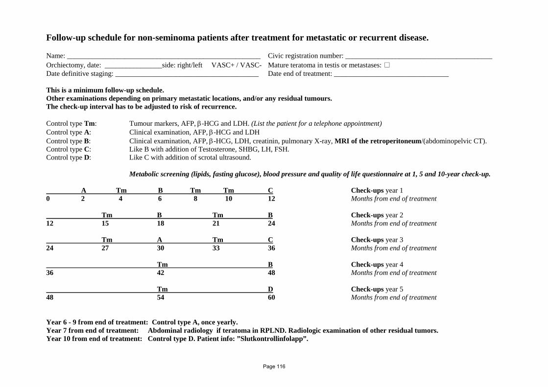
Follow-up schedule for non-seminoma patients after treatment for metastatic or recurrent disease. Name: ______________________________________________________ Civic registration number: _________________________________________ Orchiectomy, date: ________________side: right/left VASC+ / VASC- Mature teratoma in testis or metastases: Date definitive staging: ________________________________________ Date end of treatment: ________________________________
This is a minimum follow-up schedule. Other examinations depending on primary metastatic locations, and/or any residual tumours. The check-up interval has to be adjusted to risk of recurrence. Control type Tm: Tumour markers, AFP, β-HCG and LDH. (List the patient for a telephone appointment) Control type A: Clinical examination, AFP, β-HCG and LDH Control type B: Clinical examination, AFP, β-HCG, LDH, creatinin, pulmonary X-ray, MRI of the retroperitoneum/(abdominopelvic CT). Control type C: Like B with addition of Testosterone, SHBG, LH, FSH. Control type D: Like C with addition of scrotal ultrasound.
Metabolic screening (lipids, fasting glucose), blood pressure and quality of life questionnaire at 1, 5 and 10-year check-up.
A Tm B Tm Tm C Check-ups year 1 0 2 4 6 8 10 12 Months from end of treatment Tm B Tm B Check-ups year 2 12 15 18 21 24 Months from end of treatment Tm A Tm C Check-ups year 3 24 27 30 33 36 Months from end of treatment Tm B Check-ups year 4 36 42 48 Months from end of treatment Tm D Check-ups year 5 48 54 60 Months from end of treatment Year 6 - 9 from end of treatment: Control type A, once yearly. Year 7 from end of treatment: Abdominal radiology if teratoma in RPLND. Radiologic examination of other residual tumors. Year 10 from end of treatment: Control type D. Patient info: ”Slutkontrollinfolapp”.
Page 116

Sammanfattning av sjukdomsförlopp och behandling När man fått behandling för testikelcancer kan det efter lång tid dyka upp seneffekter av den givna behandlingen. Dessa seneffekter kan till exempel vara brist på manligt könshormon (testosteron), nedsatt fruktsamhet (fertilitet) samt påverkan på njurar.
Behandlingen kan även ge en liten ökad risk för hjärt-kärlsjukdom. Detta kan yttra sig som förhöjt blodtryck, kärlkramp och förhöjda blodfetter, och du bör avstå från rökning och också försöka undvika övervikt.
Det är bra att du vid kontakter med läkare i framtiden nämner att du varit behandlad för testikelcancer, och denna lapp med information om din genomgångna behandling kan vara till hjälp för dig att komma ihåg.
Namn __________________________________ Personnummer ______________________
Du opererades år _________ för testikelcancer av typen:
Seminom
Icke-seminom
Biopsiprov togs även från den friska testikeln
Ingen spridning konstaterades
Spridning konstaterades till _______________, ________________, _________________
Kompletterande behandling:
Ingen
Cytostatika (totalt antal behandlingar ______)
Strålbehandling
Kirurgi (förutom operation av den sjuka testikeln) _______________, _______________
Återfall under uppföljningsperioden:
Nej
Ja, år _________________
Sista kontrollen gjordes: datum _______________ Sjukhus___________________________
Behandlande läkare ___________________________________ Telefon _________________
Tag gärna med denna informationslapp vid kontakt med sjukvården i framtiden.
Page 117

Page 118
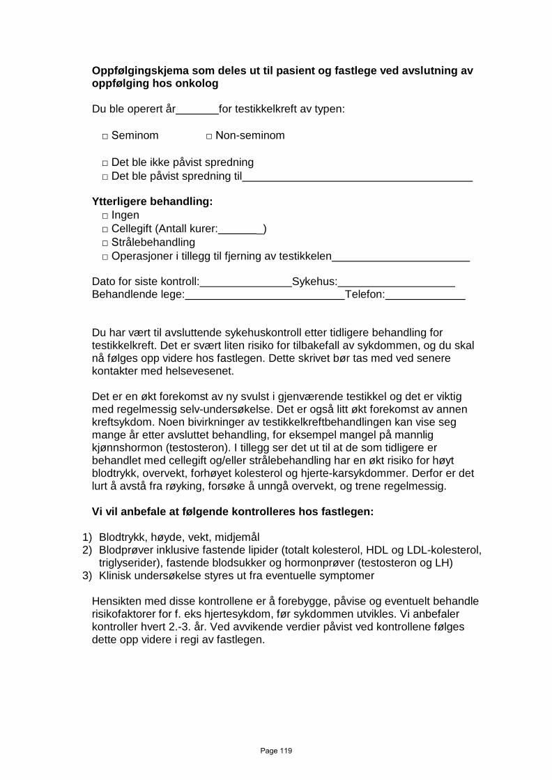
Oppfølgingskjema som deles ut til pasient og fastlege ved avslutning av oppfølging hos onkolog
Du ble operert år for testikkelkreft av typen:
□ Seminom □ Non-seminom
□ Det ble ikke påvist spredning □ Det ble påvist spredning til
Ytterligere behandling:
□ Ingen □ Cellegift (Antall kurer: _) □ Strålebehandling □ Operasjoner i tillegg til fjerning av testikkelen
Dato for siste kontroll: Sykehus: Behandlende lege: Telefon:
Du har vært til avsluttende sykehuskontroll etter tidligere behandling for testikkelkreft. Det er svært liten risiko for tilbakefall av sykdommen, og du skal nå følges opp videre hos fastlegen. Dette skrivet bør tas med ved senere kontakter med helsevesenet.
Det er en økt forekomst av ny svulst i gjenværende testikkel og det er viktig med regelmessig selv-undersøkelse. Det er også litt økt forekomst av annen kreftsykdom. Noen bivirkninger av testikkelkreftbehandlingen kan vise seg mange år etter avsluttet behandling, for eksempel mangel på mannlig kjønnshormon (testosteron). I tillegg ser det ut til at de som tidligere er behandlet med cellegift og/eller strålebehandling har en økt risiko for høyt blodtrykk, overvekt, forhøyet kolesterol og hjerte-karsykdommer. Derfor er det lurt å avstå fra røyking, forsøke å unngå overvekt, og trene regelmessig.
Vi vil anbefale at følgende kontrolleres hos fastlegen:
1) Blodtrykk, høyde, vekt, midjemål 2) Blodprøver inklusive fastende lipider (totalt kolesterol, HDL og LDL-kolesterol,
triglyserider), fastende blodsukker og hormonprøver (testosteron og LH) 3) Klinisk undersøkelse styres ut fra eventuelle symptomer
Hensikten med disse kontrollene er å forebygge, påvise og eventuelt behandle risikofaktorer for f. eks hjertesykdom, før sykdommen utvikles. Vi anbefaler kontroller hvert 2.-3. år. Ved avvikende verdier påvist ved kontrollene følges dette opp videre i regi av fastlegen.
Page 119

Page 120

Patient care plan to be delivered to the patient and general practitioner at termination of uro-oncological follow-up
You were operated year for testicular cancer, subtype:
□ Seminoma □ Non-seminoma
□ No dissemination of disease were confirmed □ Dissemination of disease were confirmed to
Additional treatment
□ No □ Chemotherapy (number of cycles : ) □ Radiotherapy □ Surgery in addition to removal of the testicle
Date for last follow-up: _ Hospital: Responsible doctor: Telephone:
You have completed the last oncological follow-up after previous treatment for testicular cancer. The risk for relapse of the disease is very low, and you will be taken care of at your general practitioner in the future. This patient care plan should be shown in case of future contact with the health services.
You are at risk of a new tumor in the remaining testicle and regular self-exams are important. Further, another cancer type may develop after treatment with chemotherapy and/or radiotherapy. Some side-effects from testicular cancer treatment may emerge several years after treatment, e.g. sub-normal values of male sex hormone (testosterone). In addition, men previously treated with chemotherapy and/or radiotherapy have an increased risk for hypertension, overweight, elevated cholesterol levels and cardiovascular disease. Thus, it is advisable to keep away from smoking, avoid overweight and exercise regularly.
We recommend that the following are controlled by the general practitioner:
1) Blood pressure, height, weight, waist circumference 2) Blood samples including fasting lipids (total cholesterol, HDL and LDL-cholesterol,
triglycerides), fasting glucose and hormones (testosterone, FSH and LH) 3) Clinical examination in case of any symptoms
The purpose of these controls is to prevent, identify and possibly treat risk factors which might lead to complications, e.g. cardiovascular disease. We recommend controls every 2.-3. years. If abnormal values are detected at these controls, further follow-up at the general practitioner is initiated.
Page 121

Page 122
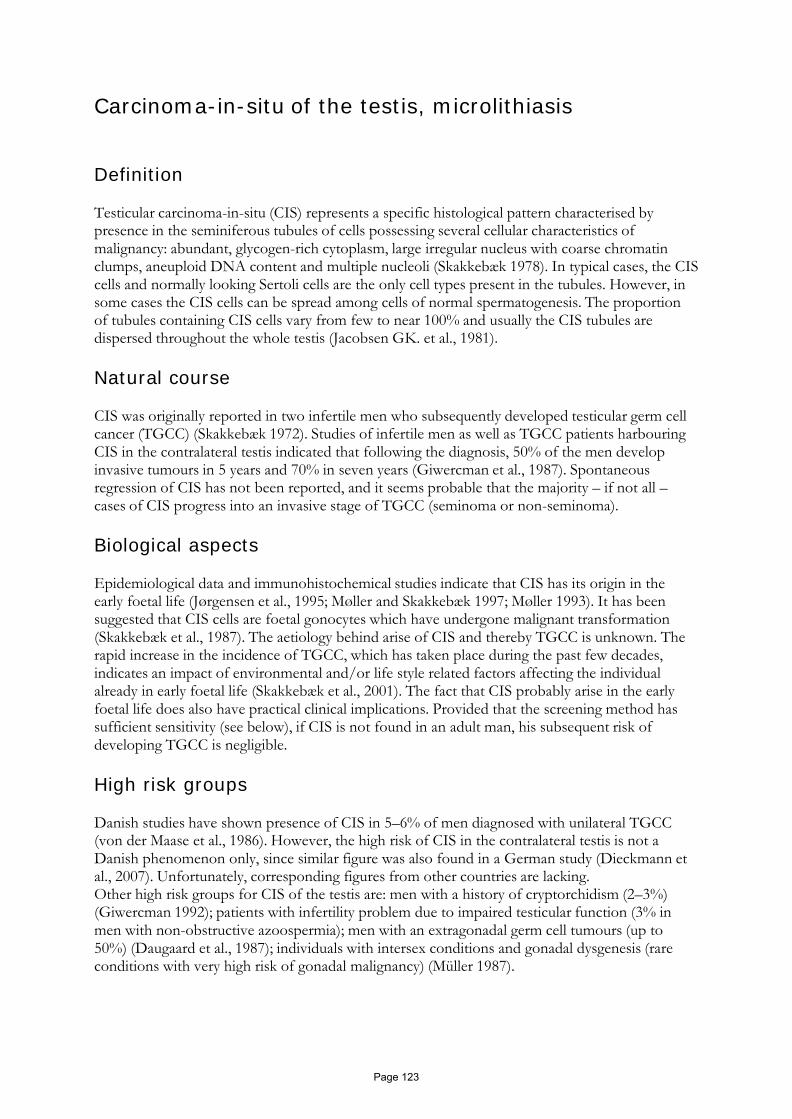
Carcinoma-in-situ of the testis, microlithiasis Definition
Testicular carcinoma-in-situ (CIS) represents a specific histological pattern characterised by presence in the seminiferous tubules of cells possessing several cellular characteristics of malignancy: abundant, glycogen-rich cytoplasm, large irregular nucleus with coarse chromatin clumps, aneuploid DNA content and multiple nucleoli (Skakkebæk 1978). In typical cases, the CIS cells and normally looking Sertoli cells are the only cell types present in the tubules. However, in some cases the CIS cells can be spread among cells of normal spermatogenesis. The proportion of tubules containing CIS cells vary from few to near 100% and usually the CIS tubules are dispersed throughout the whole testis (Jacobsen GK. et al., 1981).
Natural course
CIS was originally reported in two infertile men who subsequently developed testicular germ cell cancer (TGCC) (Skakkebæk 1972). Studies of infertile men as well as TGCC patients harbouring CIS in the contralateral testis indicated that following the diagnosis, 50% of the men develop invasive tumours in 5 years and 70% in seven years (Giwercman et al., 1987). Spontaneous regression of CIS has not been reported, and it seems probable that the majority – if not all – cases of CIS progress into an invasive stage of TGCC (seminoma or non-seminoma).
Biological aspects
Epidemiological data and immunohistochemical studies indicate that CIS has its origin in the early foetal life (Jørgensen et al., 1995; Møller and Skakkebæk 1997; Møller 1993). It has been suggested that CIS cells are foetal gonocytes which have undergone malignant transformation (Skakkebæk et al., 1987). The aetiology behind arise of CIS and thereby TGCC is unknown. The rapid increase in the incidence of TGCC, which has taken place during the past few decades, indicates an impact of environmental and/or life style related factors affecting the individual already in early foetal life (Skakkebæk et al., 2001). The fact that CIS probably arise in the early foetal life does also have practical clinical implications. Provided that the screening method has sufficient sensitivity (see below), if CIS is not found in an adult man, his subsequent risk of developing TGCC is negligible.
High risk groups
Danish studies have shown presence of CIS in 5–6% of men diagnosed with unilateral TGCC (von der Maase et al., 1986). However, the high risk of CIS in the contralateral testis is not a Danish phenomenon only, since similar figure was also found in a German study (Dieckmann et al., 2007). Unfortunately, corresponding figures from other countries are lacking. Other high risk groups for CIS of the testis are: men with a history of cryptorchidism (2–3%) (Giwercman 1992); patients with infertility problem due to impaired testicular function (3% in men with non-obstructive azoospermia); men with an extragonadal germ cell tumours (up to 50%) (Daugaard et al., 1987); individuals with intersex conditions and gonadal dysgenesis (rare conditions with very high risk of gonadal malignancy) (Müller 1987).
Page 123

Clinical features CIS of the testis does not present with any specific clinical features. Testes harbouring CIS may be atrophic but CIS can also be found in normal-size gonads. Testicular palpation can be associated with some tenderness but usually is not associated with any abnormal findings. There are no specific serum markers of CIS.
Testicular ultrasound
Recent data have indicated that testicular microlithiasis (TM) - detection of multiple, small hyperechogenic lesions in the testis – is associated with high risk of CIS (Lenz et al., 1987; von Eckardstein et al., 2001). Thus, in a study of 78 men with TGCC the predictive value of TM for the contralateral testis to contain CIS was 22.2% (Lenz et al., 1996). Although a single case of CIS was found in a testis without TM, in such case the predictive value that the testis would not contain CIS was 97.6% (Lenz et al., 1996). This result was recently confirmed in a Dutch study (Elzinga-Tinke et al., Int J Androl 2010) showing CIS in 26% of patients with TM, The men were selected for ultrasound if they belonged to one or more of following high-risk groups: unilateral TGCC, poor semen quality, testicular atrophy (< 15mL) and/or history of cryptorchidism. The negative predictive value of absence of TM was 100% as no CIS was found in testes without TM.
Recommendations for use of ultrasound and its implications:
1) TGCC patients: since testicular biopsy can easily be performed in these men at the time of
orchiectomy, ultrasound as a screening for CIS in the contralateral testis is not recommended. However, in rare cases where the biopsy performed at the time of orchiectomy fails ultrasound can be used for decision whether to repeat the biopsy (+TM) or not (- TM);
2) In men seeking for infertility and presenting with testicular atrophy (one or both testes with orchidometer measure below 15 ml) and/or history of cryptorchidism, testicular biopsy is indicated in case TM is found;
3) The clinical implications of finding TM in otherwise healthy men are yet unknown and the method is, therefore, not recommended as a general screening for early TGCC.
Diagnosis
The only reliable method of diagnosing CIS is performing open surgical biopsy (Dieckmann et al., 1999). It has been suggested that open biopsy could be replaced by a needle biopsy but, so far, there are no studies to prove the reliability of the latter method in diagnosis of CIS. In TGCC patients the biopsy can ideally be done at the time of orchiectomy. If not done at the time of orchiectomy, the biopsy can be performed, in local anaesthesia, at one of subsequent follow-up visits. The risk of serious complications following open surgical biopsy is very low and significantly less than the probability of developing contralateral TGCC (Bruun et al., 1987). The advantage of diagnosing the malignancy at a pre-invasive stage is the possibility of preserving the testis and thereby the endogenous androgen production (Giwercman et al., 1991) (see below). It is recommended that the biopsy is performed in all men undergoing orchiectomy, since no clinical or laboratory test are sufficiently reliable to exclude TGCC patients not being at risk of having CIS. A single biopsy, approximately 3 mm in diameter disclose CIS in 95% of the cases when this condition is present (Dieckmann et al., 1999). Therefore, provided that the risk of contra-lateral
Page 124

CIS is 5%, if no CIS is found the risk of subsequent second testicular tumour is less than 0.3%. However, such high sensitivity in the diagnosis of CIS can only be obtained if: a) the tissue is properly handled; b) the diagnosis is performed by a pathologists experienced in this area. Two German studies have reported that a double-biopsy procedure may yield an even higher disclosure rate up to 99% (Kliesch et al., 2003; Dieckmann et al, 2007). It is not recommended to use formalin as fixative, it leads to poor preservation of testicular morphology. Preferably, Stieve’s or Bouin’s fixative should be used.
For pathological investigation, routine haematoxylin-eosin staining is sufficient. However,
immunohistochemical staining with use of an antibody against Placental-like Alkaline Phosphatases can facilitate the diagnosis. It is important that the whole tissue block is thoroughly investigated since such procedure minimise the risk of missing the diagnosis of focal CIS.
Management of CIS
1) Unilateral TGCC and CIS in the contralateral testis:
a) In patients not receiving chemotherapy: CIS cells can be eradicated by local irradiation given as 8-9 daily doses of 2 Gy (total dose – 16-18 Gy). It has been suggested that the irradiation dose should be increased to 20 Gy. However, so far, there is no evidence to show that 20 Gy implies a lower risk of therapy failure but seems to be associated with a higher risk of androgen deficiency (Bang et al., 2009). Although some of the men subsequently develop androgen insufficiency; in more than 50% androgen replacement is not required, at least during the first years post-irradiation. For details of radiation technique see separate section.
b) In patients receiving chemotherapy: Platinum-containing chemotherapy may eradicate CIS. However, patients with CIS may develop invasive cancer in spite of chemotherapy (Kleinschmidt et al., 2009). The safest alternative is to give local irradiation as indicated under (a). However, an alternative is to repeat the biopsy, 1–2 years after completion of chemotherapy, and perform ultrasound every 6 months until biopsy. If CIS cells are present, irradiation should be offered. However, it should be kept in mind that following chemotherapeutic treatment the CIS cells may be reduced in number without being completely eradicated. A double biopsy is therefore recommended as the sensitivity of a single testicular biopsy is, expected to be lower than the figures given above and the risk of late contralateral TGCC exists. Even if the re-biopsy is negative, testicular ultrasound should be performed once yearly during the follow-up;
2) Patients with extragonadal disease and CIS in one testicle: Orchiectomy of the affected testicle is recommended;
3) Bilateral CIS: Irradiation as indicated under 1 a; 4) Unilateral CIS and no malignancy in the other testis: Orchiectomy.
Guidelines for follow-up after testicular irradiation for CIS:
• Control testicular double biopsy should be done 24 months after irradiation and should disclose Sertoli-cell only pattern. Presence of germ cells indicates failure of the radiotherapy;
• Serum levels of testosterone, SHBG, LH, and FSH should be checked prior to the radiation therapy, 6 and 12 months after. Subsequently the tests should be repeated with 12–24 months interval. Symptoms of hypogonadism combined with subnormal or low normal (even men with testosterone levels within the reference interval can be
Page 125

hypogonadal) total and free (adjusted for SHBG level) testosterone as well as high LH can be indicative of need for androgen substitution;
• Testicular ultrasound should be performed at 5 and 10 years after radiotherapy. Treatment of CIS and preservation of fertility potential
Testicular irradiation will lead to eradication of all germ cells and permanent sterility. Therefore, if the patient has a wish of future fertility following precautions should be taken: • If the patient has a partner, immediate wish of having a child and significant sperm output
(this issue needs to be discussed with an andrologist), some months to a few years of surveillance, while the couple is trying to obtain pregnancy, can be recommended. During this period testicular palpation and ultrasound should be performed every 6th months;
• In other cases cryopreservation of sperms prior to irradiation is recommended; • In case of azoospermia (no sperms in the ejaculate) and a strong wish of preservation of
fertility, multiple testicular biopsies, and if intratesticular elongated spermatids are found, subsequent cryopreservation is an option to be discussed with the patient.
References
1. Bruun,E., Frimodt-Møller,C., Giwercman,A., Lenz,S., and Skakkebæk,N.E. (1987) Testicular
biopsy as an outpatient procedure in screening for carcinoma-in-situ: complications and the patient's acceptance. Int.J.Androl., 10, 199–202.
2. Christensen,T.B., Daugaard,G., Geertsen,P.F., and von der Maase,H. (1998) Effect of
chemotherapy on carcinoma in situ of the testis. Ann.Oncol., 9, 657–660. 3. Daugaard,G., Olsen,J., von der Maase,H., Rørth,M., and Skakkebæk,N.E. (1987) Carcinoma-
in-situ testis in patients with assumed extragonadal germ-cell tumours. Lancet, 528–529. 4. Dieckmann,K.P., Souchon,R., Hahn,E., and Loy,V. (1999) False-negative biopsies for
testicular intraepithelial neoplasia. J Urol., 162, 364–368. 5. Dieckmann,K.-P. and Loy,V. (1996) Prevalence of contralateral testicular intraepithelial
neoplasia in patients with testicular germ cell neoplasms. J.Clin.Oncol., 14, 3121–3125. 6. Giwercman,A. (1992) Carcinoma-in-situ of the testis: screening and management.
Scand.J.Urol.Nephrol., 148 (suppl.), 1–47. 7. Giwercman,A., Berthelsen,J.G., Müller,J., von der Maase,H., and Skakkebæk,N.E. (1987)
Screening for carcinoma-in-situ of the testis. Int.J.Androl., 10, 173–180. 8. Giwercman,A., von der Maase,H., Berthelsen,J.G., Rørth,M., Bertelsen,A., and
Skakkebæk,N.E. (1991) Localized Irradiation of Testes with Carcinoma in Situ: Effects on Leydig Cell Function and Eradication of Malignant Germ Cells in 20 Patients. J.Clin.Endocrinol.Metab., 73, 596-603.
9. Jacobsen GK., Henriksen OB., and von der Maase,H. (1981) Carcinoma In Situ of Testicular
Tissue Adjacent to Malignant Germ-Cell Tumors: A study of 105 cases. Cancer, 47, 2660– 2662.
Page 126

10. Jørgensen,N., Rajpert-De Meyts,E., Græm,N., Müller,J., Giwercman,A., and Skakkebæk,N.E. (1995) Expression of immunohistochemical markers for testicular carcinoma in situ by normal human fetal germ cells. Lab.Invest., 72, 223–231.
11. Lenz,S., Giwercman,A., Skakkebæk,N.E., Bruun,E., and Frimodt-Møller,C. (1987)
Ultrasound in detection of early neoplasia of the testis. Int.J.Androl., 10, 187–190. 12. Lenz,S., Skakkebæk,N.E., and Hertel,N.T. (1996) Abnormal ultrasonic pattern in
contralateral testes in patients with unilateral testicular cancer. World J.Urol, 14, S55–S58. 13. Møller,H. (1993) Clues to the aetiology of testicular germ cell tumours from descriptive
epidemiology. Eur.Urol., 23, 8–15. 14. Møller,H. and Skakkebæk,N.E. (1997) Testicular cancer and cryptorchidism in relation to
prenatal factors: case-control studies in Denmark. Cancer Causes Contr., 8, 904–912. 15. Müller,J. (1987) Abnormal infantile germ cells and development of carcinoma-in-situ in
maldeveloped testes: a stereological and densitometric study. Int.J.Androl., 10, 543–567. 16. Skakkebæk,N.E. (1972) Possible carcinoma-in-situ of the testis. Lancet, 516–517.
17. Skakkebæk,N.E. (1978) Carcinoma in situ of the testis: frequency and relationship to
invasive germ cell tumours in infertile men. Histopathology, 2, 157–170. 18. Skakkebæk,N.E., Berthelsen,J.G., Giwercman,A., and Müller,J. (1987) Carcinoma-in-situ of
the testis: possible origin from gonocytes and precursor of all types of germ cell tumours except spermatocytoma. Int.J.Androl., 10, 19–28.
19. Skakkebæk,N.E., Rajpert-De Meyts,E., and Main,K.M. (2001) Testicular dysgenesis
syndrome: an increasingly common developmental disorder with environmental aspects. Hum.Reprod., 16, 972–978.
20. von der Maase,H., Rørth,M., Walbom-Jørgensen,S., Sørensen,B.L., Christophersen,I.S., Hald
T., Jacobsen GK., Berthelsen,J.G., and Skakkebæk,N.E. (1986) Carcinoma in situ of contralateral testis in patients with testicular germ cell cancer: study of 27 cases in 500 patients. Br.Med.J., 293, 1398–1401.
21. von Eckardstein,S., Tsakmakidis,G., Kamischke,A., and Nieschlag,E. (2001) Sonographic
testicular microlithiasis as an indicator of premalignant conditions in normal and infertile men. J Androl, 22, 818–824.
Page 127

Radiotherapy of CIS STRÅLDOS Targetdos skall vara 18 Gy, 2 Gy*9. Detta ger en minimidos i testikeln på 17.1 Gy.
Uppläggning
För uppläggningen behövs ett stöd för blyskyddet (t.ex. en trekant av frigolit), 1 cm tjockt vattenekvivalent bolus, 0.8–1 cm tjockt blyskydd samt ytterligare bolus (ej med på bilden). Blyskyddet och det bolus som skall placeras under testikeln har en halvmåneformad urfasning för bättre passform mot scrotum.
Patienten placeras i ryggläge med benen fixerade brett isär.
Placera blystödet (trekanten av frigolit) mellan benen. Närmast under testikeln placeras 1 cm vattenekvivalent bolus följt av ett 0.8–1 cm tjockt blyskydd. Blyskyddet kan med fördel bestå av flera tunnare skivor.
Tejpa undan penis uppåt. Kring testikeln placeras vattenekvivalent bolus med en utsträckning på minst 2 cm lateralt. Det är viktigt att bolusen har en så bra passform som möjligt.
Vid användandet av ”låga” elektronenergier kan det vara aktuellt att placera bolus även ventralt om testikeln.
Page 128

Abdominal Magnetic Resonance Imaging protocol for follow-up of patients operated for testicular cancer General imaging protocol recommendations for examinations at 1.5 T or 3T Examination is performed after at least four hours of fasting. A body phased-array coil is used. Pulse sequences are performed with 5-6 mm section thickness with maximized feasible spatial resolution depending on the available signal with the system used.
a .Coronal Half acquisition single shot turbo-spin echo sequence with limits for upper and lower abdominal transaxial sections outlined b and c. Transaxial T2-weighted respiratory triggered turbo spin-echo sequence d and e. Transaxial T1-weighted breath-hold spoiled gradient-echo sequence with fat and water in- (d) and opposed (e) phase. f and g. Transaxial T2 weighted echo planar imaging diffusion weighted sequences with b=50 (f) and b=800 (g) (optional) h Coronal Inverted Maximum Intensity Projection (MIP) diffusion weighted image reconstructed from the volume of b=800 sections (optional). May 15, 2012
Page 129

KVAST dokument testikeltumör Deltagare i KVAST-gruppen för urologi Tomasz Gorecki, [email protected], Länssjukhuset, 391 85 Kalmar, Tel 0480 448251 Gunilla Chebil, Unilabs, Bergaliden 11, 252 23 Helsingborg, Tel: 0734 22 20 16
Lars Egevad, [email protected], Avd för patologi och cytologi, Karolinska Universitetssjukhuset Solna, 171 76 Stockholm
Viktoria Gaspar, [email protected] Labmedicin Skåne, 251 85 Helsingborg Hans Hamberg, [email protected] Patologkliniken Centrallasarettet, 721 89 Västerås Carl-Gustaf Pihl, [email protected], Klinisk Patologi och Cytologi, Gula stråket 8, SU/Sahlgrenska, 413 45 Göteborg. Tel: 031 342128 Janos Vasko, [email protected], Klinisk patologi/cytologi, Laboratoriecentrum, NUS, 901 85 Umeå. Tel: 090 - 785 18 28
Introduktion Preoperativ diagnostik av testistumörer är huvudsakligen klinisk och radiologisk. Detta dokument behandlar histopatologisk undersökning av orchidektomipreparat med tumörfrågeställning samt biopsi som tages från kontralateral testis vid orchidektomi med frågeställning: Intratubulär germinalcellsneoplasi? Den diagnostiska bedömningen som är styrande för kontroll och fortsatt behandling är huruvida tumören är malign eller benign, av könscelltyp eller stromal. Avseende tumörer av germinal typ är det helt avgörande om tumören är ett rent seminom eller av annan histologisk typ. Däremot är andelen av andra tumörkomponenter icke behandlingsstyrande. Patologiskt tumörstadium samt tumörförekomst i rete testis är likaledes behandlingsstyrande. Beträffande teratomen kan dessa bestå av såväl mogen, omogen som intermediärt mogen vävnad och är hos prepubertala barn en icke metastaserande tumör. Postpubertalt är teratom en potentiellt metastaserande tumör, undantaget mogen dermoidcysta. Anvisningar för provtagarens hantering av provet 1. Operationpreparatet hanteras enligt lokala överenskommelser mellan patologavdelning och opererande klinik. 2. Som fixativ rekommenderas buffrad formalin 10% (formaldehyd 4%) som kan beställas från Apoteksbolaget. Använd minst fem gånger preparatvikten. 3. Biopsi från kontralateral testis vid orchidektomi hanteras varsamt för att undvika klämningsartefakter och bör fixeras i formalin (rutinmässiga immunhistokemiska metoder baseras närmast uteslutande på detta fixativ). Anamnestisk remissinformation 1. Korrekt namn och personnummer, inklusive de fyra sista siffrorna. Stämplade uppgifter skall vara läsliga och rätt placerade på remissen.
Page 130

2. Adekvata och utförliga uppgifter om sjukhistoria och undersökningsfynd inkluderande kända resultat av tumörmarköranalyser, klinisk diagnos och ultraljudsfynd 3. Uppgifter om vad operationsmaterialet i sin helhet omfattar med sidoangivelse. 4. Antalet burkar skall anges på remiss. Numrering eller annan märkning på preparatburk skall överensstämma med remissuppgifter. (OBS! Ej märkning på locket.) Utskärningsanvisningar Biopsi Efter makroskopisk bedömning inbäddas hela biopsimaterialet. Orchidektomi Om det intakta operationspreparatet skickas färskt till patologen kan en skiva från resektionsänden i funikel skäras ut INNAN testis delas. För optimal fixering av tumörvävnad klyv testikeln genom rete och epididymis. Fixera minst 24 timmar. Vid misstanke om engagemang av resektionsytor bör dessa färgmarkeras före utskärning. Mät funikeln. Beskriv parietala tunica vaginalis (notera ev hydrocele, adherenser). Mät testikeln, beskriv ytan. Notera tumörens (eller tumörernas) lokalisation, storlek och makroskopiska utseende på snittytan, avgränsning mot omgivande strukturer och relation till tunica, rete, epididymis och funikel. Skiva testikelvävnaden i tunna skivor (3-4 mm) Notera avvikande områden såsom ärrfibros eller nekros inom ”normal” testikelvävnad. Bitar till mikroskopisk undersökning: •Resektionsänden i funikel. •Tumören – bitar representerande samtliga makroskopiskt olika områden av tumören. Tumör < 3 cm paraffininbäddas i sin helhet, för större tumörer kan om tumregel anges 1 bit/cm av tumörens diameter. Gärna storsnitt genom rete och epididymis. •Bitar omfattande övergång mellan tumör och normal testis. •Bitar som visar tumörens relation till tunica, rete, epididymis och funikel. •Tumörfri testisvävnad. Analyser 1. Snitten färgas med valfri rutinfärgning. 2. Biopsi från kontralateral testis färgas med valfri rutinfärgning och vid behov immunhistokemi.
Page 131

3. Förslag till immunhistokemiska undersökningar (se tabell) PLAP OCT
4 AFP HCG CD
30 CD117 CK(Pan) CK7 Inhibin
IGCN(Intratubular Germinal Cell Neoplasia)
+ + -
Seminom + + - - - + -/+ - Spermatocytiskt Seminom
- - - - - -/+ -/+
Embryonal cancer +/- + -/+ - + - + + - Gulesäckstumör -/+ - +/- - ? ? + - Choriocarcinom +/- - - + - ? + ? + Leydigcellstumör - - - - ? + Sertolicellstumör - - - - ? +/- Granulosacellstumör - - - - ? + + : >90% av tumörerna är positiva +/- : 50-90% av tumörerna är positiva -/+ : 10-50% av tumörerna är positiva - : <10% av tumörerna är positiva ? : varierande uppgifter i litteraturen/uppgift saknas 4. Valfritt antal snittnivåer ska undersökas.
5. Analyser som särskilt bör kvalitetssäkras via externa kontrollprogram: relevanta immunfärgningar.
Information i remissens svarsdel A. Makroskopisk beskrivning: 1. Preparatbeskrivning 2. Beskrivning av förändringar (ex lokalisation, avgränsning, relation till prepytor/resektionsytor och omgivande vävnad samt måttangivelser) 3. Övrigt (ex väsentliga bifynd, söndertrasningar och annat som begränsar möjligheterna till adekvat undersökning) B. Mikroskopiutlåtande: 1. Tumörtyp enligt WHO med angivande av rent seminom eller annan tumörtyp (annan ren
histologisk typ eller blandad histologisk typ). Samtliga förekommande tumörkomponenter anges.
2. Tumörstadium pTNM 2009. Notera att frånvaro/förekomst av kärlinvasion skall anges. Vid behov bör immunhistokemisk undersökning göras avseende kärlinvasion.
3. Andra prognosvariabler: Tumörstorlek.
Tumörinfiltration (ja/nej) i rete testis. 4. Radikalitet. Rekommenderade klassifikationssystem WHO Classification of Tumours: Pathology and Genetics of Tumours of the Urinary System and Male Genital Organs 2004 (s 218)
Page 132
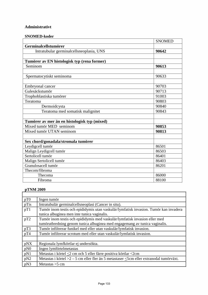
Administrativt SNOMED-koder SNOMED Germinalcellstumörer Intratubular germinalcellsneoplasia, UNS 90642 Tumörer av EN histologisk typ (rena former) Seminom 90613 Spermatocytiskt seminoma 90633 Embryonal cancer 90703 Gulesäckstumör 90713 Trophoblastiska tumörer 91003 Teratoma 90803 Dermoidcysta 90840 Teratoma med somatisk malignitet 90843 Tumörer av mer än en histologisk typ (mixed) Mixed tumör MED seminom 90853 Mixed tumör UTAN seminom 90813 Sex chord/gonadala/stromala tumörer Leydigcell tumör 86501 Malign Leydigcell tumör 86503 Sertolicell tumör 86401 Malign Sertolicell tumör 86403 Granulosacell tumör 86201 Thecom/fibroma Thecoma 86000 Fibroma 88100 pTNM 2009 pT0 Ingen tumör pTis Intratubulär germinalcellsneoplasi (Cancer in situ). pT1 Tumör inom testis och epididymis utan vaskulär/lymfatisk invasion. Tumör kan invadera
tunica albuginea men inte tunica vaginalis. pT2 Tumör inom testis och epididymis med vaskulär/lymfatisk invasion eller med
tumörutbredning genom tunica albuginea med engagemang av tunica vaginalis. pT3 Tumör infiltrerar funikel med eller utan vaskulär/lymfatisk invasion. pT4 Tumör infiltrerar scrotum med eller utan vaskulär/lymfatisk invasion. pNX Regionala lymfkörtlar ej undersökta. pN0 Ingen lymförtelmetastas pN1 Metastas i körtel <2 cm och 5 eller färre positiva körtlar <2cm pN2 Metastas i körtel >2 – 5 cm eller fler än 5 metastaser <5cm eller extranodal tumörväxt. pN3 Metastas >5 cm
Page 133

Klinisk organisation som granskat och godkänt dokumentet: SWENOTECA (Swedish - Norwegian Testicular Cancer Group) Rekommenderad litteratur: WHO Classification of Tumours. Pathology & Genetics, Tumours of the Urinary System and Male Genital Organs, Lyon 2004. Annals of Oncology 15: 1377–1399, 2004 European consensus on diagnosis and treatment of germ cell cancer: a report of the European Germ Cell Cancer Consensus Group (EGCCCG) Emerson R, Ulbright T: Morphological approach to tumour of the testis and paratestis, J Clin Pathol 2007;60:866-880 Emerson RE, Ulbright TM: The use of immunohistochemistry in the differential diagnosis of tumors of the testis and paratestis. Seminars in Diagnostic Pathology 2005 Krag Jacobsen G, Talerman A: Atlas of Germ Cell Tumours 1989 Raghavan D: Germ Cell Tumors: American Cancer Society Atlas of Clinical Oncology, 2003 Ulbright T: Germ cell tumors of the gonads, Mod Pathol 2005, 18, 61-79 Young RH: Testicular tumors – some new and a few perennial problems, Arch Pathol Lab Med 2008 132(4):548-64 För immunhistokemi se exempelvis http://www.e-immunohistochemistry.info/ http://www.nordiqc.org http://www.ipox.org/ http://www.dako.com/asp/algo/default.asp?get=table&table=20
Page 134

KVAST dokument retroperitoneal lymfkörteldissektion Klinisk bakgrundsinformation
Retroperitoneal lymfkörteldissektion (RPLND) utföres som diagnostisk och kurativ åtgärd dels för staging dels efter kemoterapi för abdominell metastasering. För närvarande kan radiologiska diagnostiska metoder ej skilja aktiv tumör från nekros/fibros.
Anvisningar för provtagarens hantering av provet. 1. Vid radikalitetsfrågeställning skall tydlig suturmarkering anbringas. 2. Som fixativ rekommenderas buffrad formalin 10% (formaldehyd 4%), använd minst fem gånger preparatvikten. 3. Fryssnitt bör begränsas p g a kvalitetsaspekter. IV Anamnestisk remissinformation. 1. Typ av operation/undersökning 2. Fraktionsbeteckningar enligt RETROP-protokoll (se länk nedan) 3. Klinisk bedömning/diagnos 4. Relevanta tidigare PAD/CD, rtg/labfynd, tidigare sjukdomar, statusfynd, fynd i samband med provtagningen V. Utskärningsanvisningar. 1. Alla lymfkörtlar utan makroskopiska avvikelser bäddas i sin helhet, eventuellt var för sig. 2. Från makroskopiskt avvikande körtlar tages representativa bitar för mikroskopi. VI. Analyser. 1. Snitten färgas med valfri rutinfärgning. 2. Immunhistokemi vid behov. 3. Minst 2 snittnivåer undersökes
VII. Information i remissens svarsdel. A. Makroskopisk beskrivning: 1. Preparatbeskrivning 2. Beskrivning av förändringar (ex avgränsning, relation till prepytor/resektionsytor, måttangivelser)
B. Mikroskopiutlåtande: 1. Alla fraktioner besvaras var för sig med angivande av lokalisation enligt RETROP-protokollet. 2. Ange antalet lymfkörtlar i varje fraktion med viabel cancer, teratom respektive benignt fynd. Ange största diameter på tumör i mm. 3. Ange förekomst av nekros. 4. Sammanställ i diagnos totalt antal lgl med angivande av hur många med viabel cancer, teratom samt nekros .
VIII. Rekommenderade klassifikationssystem Se ovan avseende testis. IX. Administrativt SNOMED-kodning och pTNM se ovan avseende testis.
Page 135
XII. Exempel på utlåtande RPLND: PM 2009 Inkom 12 fraktioner. Fraktion 1: Burk märkt ”Station 1” innehållande lymfkörtlar och fettvävnad. Histologiskt ses 7 stycken lymfkörtlar utan hållpunkter för kvarvarande viabel tumör eller teratom. Fraktion 2: Burk märkt ”Station 2” innehållande lymfkörtlar och fettvävnad. Histologiskt ses 6 stycken lymfkörtlar utan hållpunkter för kvarvarande viabel tumör eller teratom. Fraktion 3: Burk märkt ”Station 3” innehållande lymfkörtlar och fettvävnad. Histologiskt ses totalt 20 stycken lymfkörtlar utan hållpunkter för kvarvarande viabel tumör eller teratom. Fraktion 4: Burk märkt ”Station 4” innehållande lymfkörtlar och fettvävnad. Histologiskt ses endast 1 stycken lymfkörtlar utan hållpunkter för kvarvarande viabel tumör eller teratom. Fraktion 5: Burk märkt ”Station 5” innehållande lymfkörtlar och fettvävnad. Histologiskt ses 2 stycken lymfkörtlar utan hållpunkter för kvarvarande viabel tumör eller teratom. Fraktion 6: Burk märkt ”Station 6” innehållande lymfkörtlar och fettvävnad. Histologiskt ses i en av körtlarna en större nekros som mäter 17x14 mm. Därtill ses i samma körtel blödningspigment samt förekomst av makrofager och främmande kroppsreaktion. I övrigt ses ytterliggare 6 körtlar, även dessa fria från kvarvarande viabel tumör eller teratom. Totalt 7 körtlar. Fraktion 7: Burk märkt ”Station 7” innehållande lymfkörtlar och fettvävnad. Histologiskt ses endast 1 stycken lymfkörtel utan hållpunkter för kvarvarande viabel tumör eller teratom. Fraktion 8: Burk märkt ”Station 8” innehållande lymfkörtlar och fettvävnad. Histologiskt ses 5 stycken lymfkörtlar utan hållpunkter för kvarvarande viabel tumör eller teratom. Fraktion 9: Burk märkt ”Station 9” innehållande stroma och kärl. Ingen lymfkörtelvävnad, ingen viabel tumör, några nekroser eller teratomstrukturer. Fraktion 10: Burk märkt ”Station 10” innehållande stroma och kärl. Ingen lymfkörtelvävnad, ingen viabel tumör, några nekroser eller teratomstrukturer. Fraktion 11: Burk märkt ”Station 11” innehållande lymfkörtlar och fettvävnad. Histologiskt ses 7 stycken lymfkörtlar utan hållpunkter för kvarvarande viabel tumör eller teratom. Fraktion 12: Burk märkt ”Station 12” innehållande lymfkörtlar och fettvävnad. Histologiskt ses 8 stycken lymfkörtlar utan hållpunkter för kvarvarande viabel tumör eller teratom. Diagnos: 1-12) Totalt 64 stycken lymfkörtlar utan hållpunkter för kvarvarande viabel tumör eller teratom. PM 2009 Inkom 6 fraktioner. Fraktion 1: Burk märkt ”Station 2” innehållande lymfkörtlar och fettvävnad. Histologiskt ses 8 stycken lymfkörtlar. I en av dessa (Snitt 11A+B) ses relativt stora nekroser (som störst 8 mm i diameter), blödningspigment samt makrofager med riklig cytoplasma men utan hållpunkter för kvarvarande viabel tumör. I snitt 11A ses några körtelstrukturer i kanten av nekroserna med cylindriskt epitel - således tecken på kvarvarande teratom. Dessa strukturer mäter sammanlagt 3x2 mm. Fraktion 2: Burk märkt ”Station 3” innehållande lymfkörtlar och fettvävnad. Histologiskt ses 18 stycken lymfkörtlar utan hållpunkter för kvarvarande tumör eller teratom. Fraktion 3: Burk märkt ”Station 5” innehållande lymfkörtlar och fettvävnad. Histologiskt ses 4 stycken lymfkörtlar utan hållpunkter för kvarvarande tumör eller teratom. Fraktion 4: Burk märkt ”Station 6” innehållande lymfkörtlar och fettvävnad. Histologiskt ses 5 stycken lymfkörtlar. I en av dessa ses relativt stora nekroser, blödningspigment samt makrofager med riklig cytoplasma men utan hållpunkter för kvarvarande viabel tumör. Några kvarvarande teratom strukturer ses inte.
Page 136
Fraktion 5: Burk märkt ”Station 8” innehållande lymfkörtlar och fettvävnad. Histologiskt ses 7 stycken lymfkörtlar utan hållpunkter för kvarvarande tumör eller teratom. Fraktion 6: Burk märkt ”Station 12” innehållande lymfkörtlar och fettvävnad. Histologiskt ses 1 stycken lymfkörtel utan hållpunkter för kvarvarande tumör eller teratom. Kommentar: Enligt klinisk tilläggsinformation har patienten från Linköping fått diagnosen ”Embryonalt carcinom med syncytiotrofoblast inslag samt teratom”; därtill ”IGCNU i båda testis”. På snitt 11A och snitt 36 har gjorts immunhistokemisk undersökning avseende PLAP, OCT3/4, CD30, hCG samt AFP. Ingenstädes ses äkta positiva cellelement från patientens testistumör. I snitt 11A ses däremot CEA och EMA positiva körtlar som bekräftelse på kvarvarande teratomstrukturer. Diagnos: 1-6) Totalt 43 stycken lymfkörtlar varav 2 stycken visar nekroser utan kvarvarande vital tumör. I en av körtlarna påvisas kvarvarande teratomstrukturer, v.g. se kommentar ovan.
Page 137
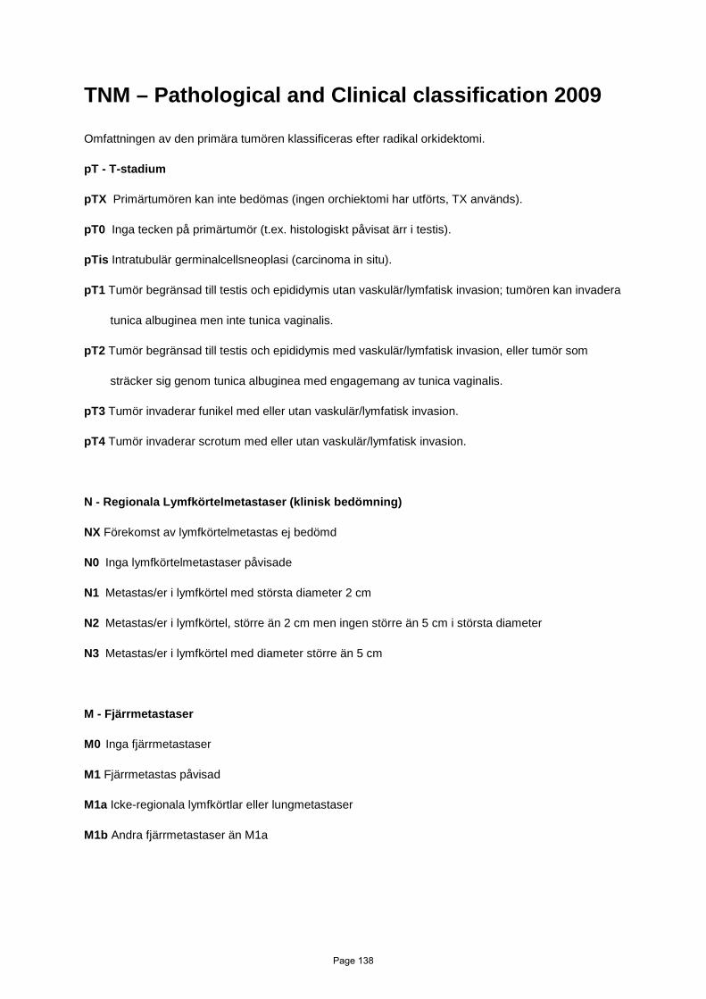
TNM – Pathological and Clinical classification 2009 Omfattningen av den primära tumören klassificeras efter radikal orkidektomi.
pT - T-stadium
pTX Primärtumören kan inte bedömas (ingen orchiektomi har utförts, TX används).
pT0 Inga tecken på primärtumör (t.ex. histologiskt påvisat ärr i testis).
pTis Intratubulär germinalcellsneoplasi (carcinoma in situ).
pT1 Tumör begränsad till testis och epididymis utan vaskulär/lymfatisk invasion; tumören kan invadera
tunica albuginea men inte tunica vaginalis.
pT2 Tumör begränsad till testis och epididymis med vaskulär/lymfatisk invasion, eller tumör som
sträcker sig genom tunica albuginea med engagemang av tunica vaginalis.
pT3 Tumör invaderar funikel med eller utan vaskulär/lymfatisk invasion.
pT4 Tumör invaderar scrotum med eller utan vaskulär/lymfatisk invasion.
N - Regionala Lymfkörtelmetastaser (klinisk bedömning)
NX Förekomst av lymfkörtelmetastas ej bedömd
N0 Inga lymfkörtelmetastaser påvisade
N1 Metastas/er i lymfkörtel med största diameter 2 cm
N2 Metastas/er i lymfkörtel, större än 2 cm men ingen större än 5 cm i största diameter
N3 Metastas/er i lymfkörtel med diameter större än 5 cm
M - Fjärrmetastaser
M0 Inga fjärrmetastaser
M1 Fjärrmetastas påvisad
M1a Icke-regionala lymfkörtlar eller lungmetastaser
M1b Andra fjärrmetastaser än M1a
Page 138
LATE EFFECTS 1. Long-term complications and follow-up after treatment for
testicular cancer Some side-effects from testicular cancer treatment may emerge several years after treatment. Thus, regular controls at the general practitioner are recommended after the oncological follow- up has been completed. At the last oncological follow-up, all patients should receive a patient care plan which summarizes the previous treatment, the most important long-term complications and recommendations for further follow-up at the caring physician (appendix, norsk og engelsk). In this section we will describe possible late complications after treatment for testicular cancer, with emphasis on complications that can be prevented or treated.
1.1 Cardiovascular disease (CVD) Mortality from CVD is higher in testicular cancer survivors (TCSs) than in the general population.1,2 Men treated with orchiectomy alone do not have an increased risk for CVD in comparison to the general population.3,4 Thus, the risk for CVD is associated with cytotoxic treatment and not testicular cancer itself. Men previously treated with cisplatin-based chemotherapy have a 2-3 fold risk for CVD in comparison to men treated with surgery only or the general population in several studies.3-5 Two large studies indicate that the risk for CVD is increased also after infradiaphragmatic irradiation,4,5 but other results are conflicting.3 The absolute risk for CVD several years after cytotoxic treatment is 6-10 %.4,5 Combination of both chemotherapy and radiotherapy is particularly harmful, with an absolute risk for CVD at 20% several years after treatment.4 Cisplatin-based chemotherapy is associated with an increased prevalence of hypertension4 and the metabolic syndrome,6,7 while radiotherapy is associated with an increased prevalence of diabetes.4 Accordingly, the increased risk for CVD is at least partly mediated by classical cardiovascular risk factors.
Endothelial and inflammatory markers, e.g. fibrinogen and von Willebrand factor, are increased in men treated with cisplatin-based chemotherapy,8 while high-sensitivity C-reactive protein (hs- CRP) is increased several years after treatment with radiotherapy.4,9 These findings indicate that endothelial dysfunction might be a possible link between cytotoxic treatment and atherosclerosis. Screening for cardiovascular risk factors10 such as hypertension, obesity, diabetes, unfavorable lipids, smoking, physical inactivity and an unhealthy diet is important among TCSs for the prevention of CVD.
1.2 Subfertility and hypogonadism Subfertility is common among men diagnosed with testicular cancer.11 Additionally, cytotoxic treatment may negatively affect both the fertility and the levels of sex hormones.12 Results from a large Norwegian follow-up study among TCSs have shown that fertility decreases with increasing treatment intensity.13 Still, nearly half of the males treated with large cumulative cisplatin doses had become fathers after testicular cancer treatment without using cryopreserved semen.
Retrograde ejaculation has been a rather frequent complication after RPLND, but the incidence has been reduced after the introduction of nerve-sparing surgery techniques.14 For men who desire to achieve fatherhood, treatment with α-sympathomimetics (Rinexin® or Tofranil®
Page 139

[imipranin, unregistered]) should be considered as these substances may reverse the retrograde ejaculation.
Up to 20 % of TCSs are diagnosed with endocrine hypogonadism/testosterone deficiency (testosterone <8 nmol/l and/or LH >12 U/l).15 Decreased libido, erectile dysfunction and loss of energy are common symptoms of endocrine hypogonadism, but these symptoms may also occur without accompanying testosterone deficiency. Endocrine hypogonadism is associated with hypertension, obesity, the metabolic syndrome and diabetes,16,17 and probably also with increased mortality rates.18,19 Thus, men with severe endocrine hypogonadism should be considered for testosterone substitution even if they lack clinical symptoms. If low levels of serum testosterone are detected, a new venipuncture should be performed in the morning to confirm the diagnosis.20
Men with considerable clinical symptoms (decreased libido, erectile dysfunction, loss of energy) but with testosterone levels within the normal range, may benefit from testosterone substitution, but there are so far no data available supporting this treatment strategy. After bilateral orchiectomy and with established testosterone deficiency after treatment for CIS, lifelong testosterone substitution is warranted.
1.3 Other long-term complications A considerable number of TCSs suffer from other long-term complications (nephrotoxicity, neurotoxicity, ototoxicity, pulmonary toxicity and psychosocial problems).21-23 Both treatment with large cisplatin doses (>850 mg) and smoking increase the risk for long-term ototoxicity, neurotoxicity and pulmonary toxicity. Men with treatment-induced ototoxicity (tinnitus, hearing impairment) should avoid noisy environments.24
There is an increased risk for second malignant neoplasms after cytotoxic treatment for testicular cancer.25 After chemotherapy or radiotherapy, the relative risk of a solid second cancer is approximately doubled, while combination of both techniques is associated with a three-fold increased risk. These second cancers are often diagnosed many years after treatment. Accordingly, all doctors involved in long-term follow-up of TCSs should timely initiate necessary examinations in the case of symptomatic patients.
1.4 Controls at the general practitioner We recommend regular examinations at the general practitioner every 2.-3. years after completion of oncological follow-up, and more often in the case of pathological findings. The purpose of these controls is to prevent, identify and possibly treat risk factors which may lead to complications, e.g. cardiovascular disease. These controls should include: • Anamnesis regarding cardiovascular risk profile and symptoms of cardiovascular disease • Advice about lifestyle-factors such as smoking cessation, healthy diet and physical activity • Measurement of blood pressure, height/weight (BMI) and waist circumference • Blood samples: Fasting lipid profile (total cholesterol, HDL-and LDL cholesterol,
triglycerides), glucose, testosterone and LH • The prophylaxis (primary and secondary) of cardiovascular disease should be according to the
general population recommendations • Consider testosterone substitution in case of endocrine hypogonadism, possibly in
cooperation with endocrinologist
Page 140
References
1. Fossa SD, Aass N, Harvei S, et al: Increased mortality rates in young and middle-aged patients with malignant germ cell tumours. Br J Cancer 90:607-612, 2004
2. Fossa SD, Gilbert E, Dores GM, et al: Noncancer Causes of Death in Survivors of Testicular
Cancer. J Natl Cancer Inst 99:533-544, 2007
3. van den Belt-Dusebout A, Nuver J, de Wit R, et al: Long-Term Risk of Cardiovascular Disease in 5- Year Survivors of Testicular Cancer. J Clin Oncol 24:467-475, 2006
4. Haugnes HS, Wethal T, Aass N, et al. Cardiovascular risk factors and morbidity in testicular cancer
survivors: A 20 year follow-up study. J Clin Oncol 28:4649-4657, 2010
5. Huddart RA, Norman A, Shahidi M, et al: Cardiovascular disease as a long-term complication of treatment for testicular cancer. J Clin Oncol 21:1513-1523, 2003
6. Haugnes HS, Aass N, Fossa SD, et al: Components of the metabolic syndrome in long-term
survivors of testicular cancer. Ann Oncol 18:241-248, 2007
7. Nuver J, Smit AJ, Wolffenbuttel BH, et al: The Metabolic Syndrome and Disturbances in Hormone Levels in Long-Term Survivors of Disseminated Testicular Cancer. J Clin Oncol 23:3718-3725, 2005
8. Nuver J, Smit AJ, Sleijfer DT, et al: Microalbuminuria, decreased fibrinolysis, and inflammation as
early signs of atherosclerosis in long-term survivors of disseminated testicular cancer. Eur J Cancer 40:701-706, 2004
9. Wethal T, Kjekshus J, Røislien J, et al: Treatment-related differences in cardiovascular risk factors in
long-term survivors of testicular cancer. J Cancer Surviv 1:8-16, 2007
10. Yusuf S, Hawken S, Èunpuu S, et al: Effect of potentially modifiable risk factors associated with myocardial infarction in 52 countries (the INTERHEART study): case-control study. The Lancet 364:937-952, 2004
11. Moller H, Skakkebaek NE: Risk of testicular cancer in subfertile men: case-control study. Bmj
318:559-562, 1999
12. DeSantis M, Albrecht W, Holtl W, et al: Impact of cytotoxic treatment on long-term fertility in patients with germ-cell cancer. Int J Cancer 83:864-865, 1999
13. Brydoy M, Fossa SD, Klepp O, et al: Paternity following treatment for testicular cancer. J Natl
Cancer Inst 97:1580-1588, 2005
14. Jacobsen KD, Ous S, Waehre H, et al: Ejaculation in testicular cancer patients after post- chemotherapy retroperitoneal lymph node dissection. Br J Cancer 80:249-255, 1999
15. Nord C, Bjoro T, Ellingsen D, et al: Gonadal hormones in long-term survivors 10 years after
treatment for unilateral testicular cancer. Eur Urol 44:322-328, 2003
16. Svartberg J, von Muhlen D, Schirmer H, et al: Association of endogenous testosterone with blood pressure and left ventricular mass in men. The Tromso Study. Eur J Endocrinol 150:65-71, 2004
Page 141

17. Laaksonen DE, Niskanen L, Punnonen K, et al: Testosterone and sex hormone-binding globulin predict the metabolic syndrome and diabetes in middle-aged men. Diabetes Care 27:1036-1041, 2004
18. Shores MM, Matsumoto AM, Sloan KL, et al: Low serum testosterone and mortality in male
veterans. Arch Intern Med 166:1660-1665, 2006
19. Laughlin GA, Barrett-Connor E, Bergstrom J: Low Serum Testosterone and Mortality in Older Men. J Clin Endocrinol Metab 93:68-75, 2008
20. Gupta SK, Lindemulder EA, Sathyan G: Modeling of circadian testosterone in healthy men and
hypogonadal men. J Clin Pharmacol 40:731-738, 2000
21. Fossa SD, Aass N, Winderen M, et al: Long-term renal function after treatment for malignant germ- cell tumours. Ann Oncol 13:222-228, 2002
22. Brydøy M, Oldenburg J, Klepp O, et al: Prevalence of long-term Raynaud-like phenomena and
neurological side-effects in survivors of testicular cancer. J Natl Cancer Inst 101:1682-1695, 2009
23. Haugnes HS, Aass N, Fossa SD, et al: Pulmonary Function in Long-Term Survivors of Testicular Cancer. J Clin Oncol 27:2779-2786, 2009
24. Widen SE, Erlandsson SI: Self-reported tinnitus and noise sensitivity among adolescents in Sweden.
Noise Health 7:29-40, 2004
25. Travis LB, Fossa SD, Schonfeld SJ, et al: Second cancers among 40,576 testicular cancer patients: focus on long-term survivors. J Natl Cancer Inst 97:1354-1365, 2005
Page 142

MRD 2012-02-27
Primärtumördata1 Orchiektomi
Nej Ja
Datum
år mån Dag
hö vä bilat. Om bilat:
non-seminom i båda testiklar nej ja (en registreringsblankett för varje tumör)2
Klinik, Sjukhus (där orchiektomi utfördes) …………………………………………
Patolog avd ………………………………………
PAD nr
–
Vaskulär invasion nej ja oklart ”Utbränd tumör” nej ja Kontralateral testisbiopsi Cis ej Cis ej utfört
Lämnat spermaprov nej ja vet ej vill ej
om ja: före orchiektomi efter orchiektomi
Tumörmarkörer/utredning
Före orchiektomi AFP ,
normalt förhöjt ej utfört
Datum år mån dag
β-HCG ,
normalt förhöjt ej utfört
LD ,
normalt förhöjt ej utfört Hormonstatus Datum
Testosteron mmol/l normalt förhöjt lågt ej utfört
SHBG normalt förhöjt lågt ej utfört
LH normalt förhöjt lågt ej utfört
FSH normalt förhöjt lågt ej utfört
Metastaser
Lymfkörtelmetastaser Största metastas (mm x mm) Extralymfatiska metastaser Inguinalt nej ja X
Lunga nej ja
Iliakalt nej ja X
Hjärna nej ja
Paraaortalt nej ja X
Lever nej ja
Mediastinalt nej ja X
Skelett nej ja
Supraklav nej ja X
Annan lokal nej ja , spec……………………………..
Definitiv klinisk stadieindelning3
Datum vid definitiv stadieindelning
CSI CSMk+ CSII CSIII CSIV
Abdominella lymfkörtlar 0 A B C D
Var patienten i CS 1 vid första utredningen nej ja oklart (Ifylles endast när två stadieindelningar utförts)
Datum för första utredning
Tumörmarkörer
(vid definitiv stadieindelning)
AFP ,
normalt förhöjt ej utfört
β-HCG ,
normalt förhöjt ej utfört LD ,
normalt förhöjt ej utfört
Prognos enl IGCCC4 god intermedär dålig
Behandling Pat remitterad till klinik/sjukhus…………………………………………………………………… CSI ingen adj.behandling BEP x 1 annan, spec…………………………………… CSIIA MK- expektans enl flödesschema annat, spec…………………………………… CS MK+, II–IV BEP annan, spec……………………………………
Kompletterande uppgifter för att gälla som canceranmälan SNOMED-kod5……………………
Diagnosgrund (flera alternativ kan ifyllas) Provexcision eller operation med histopatologisk undersökning Cytologisk undersökning Annan lab. undersökning (tumörmarkörer)
TNM T-primärtumör6 N-regionala lymfkörtelmetastaser M-fjärrmetastaser
pTX pT0 pTis pT1 pT2 pT3 pT4 NX N0 N1 N2 N3 M0 M1 M1a M1b
Nationellt kvalitetsregister SWENOTECA Non-Seminom testis Registreringsblankett Blanketten gäller fr o m 2012-01-01 som canceranmälan
Personnummer Namn
Blanketter skickas till Regionalt Cancercentrum Tidigare cancer i andra testikeln seminom non-seminom
år
Klinik, sjukhus Läkare
,
år mån dag
år mån dag
år mån dag
år mån dag
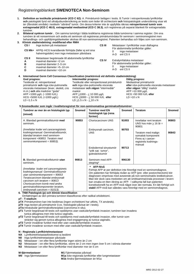
MRD 2012-02-27
Registreringsblankett SWENOTECA Non-Seminom
1. Definition av testikulär primärtumör (ICD C 62): A. Primärtumör belägen i testis. B.Tumör i retroperitoneala lymfkörtlar och patologiskt fynd vid ultraljudsundersökning av testis som leder till orchiectomi och histopatologisk undersökning visar ett
ärr (fibrotiskt område) i testikeln ”utbränd” tumör. Om dessa kriterier inte är uppfyllda räknas retroperitoneal tumör som extragonadal (ICD C 48.0), extragonadal mediastinal (ICD C 38.3), och registreras på separat blankett för extragonadala tumörer.
2. Bilateral synkron tumör . Om samma tumörtyp i båda testiklarna registreras båda tumörerna i samma register. Om ena tumören är ett nonseminom och andra ett seminom så registreras primärtumördata för seminom i seminomregistret men behandlings- och uppföljningsblanketter skickas till non-seminomregistret. Patienten behandlas och följes som non-seminom.
3. Klinisk stadieindelning: modifierad efter RMH CS I Inga tecken på metastaser
CS Mk+ AFP/β-HCG kvarstående förhöjda (faller ej enl sina halveringstider) men inga metastaser påvisbara
CS II Metastaser begränsade till abdominella lymfkörtlar A maximal diameter <2 cm B maximal diameter 2–5 cm C maximal diameter >5–10 cm D maximal diameter >10 cm
CS III Metastaser i lymfkörtlar ovan diafragma För abdominella lymfkörtlar gäller: 0 inga metastaser A-D enl CS II
CS IV Extralymfatiska metastaser
För abdominella lymfkörtlar gäller: 0 inga metastaser A-D enl CS II
4. International Germ Cell Consensus Classification (markörnivå vid definitiv stadieindelning)
God prognos: Intermediär prognos: Dålig prognos: Testikulär el. retroperitoneal primärtumör och inga icke-pulmonella viscerala metastaser (lever, skelett, cns m.m.) och alla markörer ”goda” AFP <1000 µg/L (~1000 IU/L) HCG <5000 IU/L (=1000 µg/L) LD <1.5 x N
Testikulär eller retroperitoneal primärtumör och inga icke-pulmonella viscerala metastaser och någon ”intermediär” markör AFP >1000 - < 10 000 µg/L HCG >5000 - < 50 000 IU/L eller LD >1.5 x N - < 10 x N
Mediastinal primärtumör eller icke-pulmonella viscerala metastaser eller någon “dålig” markör AFP >10 000 µg/L HCG >50 000 IU/L eller LD >10 x N
5.Snomedkoder som ingår i kvalitetsregistret för non-seminomatösa germinalcellstumörer:
Tumörer av mer än en histologisk typ (mixed)
Snomed Tumörer med EN histologisk typ (rena former)
Snomed Teratom Snomed
A. Blandad germinalcellstumör med seminom.
(Innefattar koder enl cancerregistrets kodningsmanual: Germinalcellstumör, blandad teratom med seminom-komponent =90853. Teratom med seminomkomponent = 90853).
90853 Choriocarcinom UNS
91003 Innefattar rent teratom UNS hos män > 16 år = 90801
90803
Embryonalt carcinom, UNS
90703
Teratom med malign somatisk komponent Finns ej i cancer-registrets kodnings-manual
90843
Endodermal sinustumör ”yolk sac tumor”, gulesäckstumör
90713
B. Blandad germinalcellstumör utan seminom. (Innefattar koder enl cancerregistrets kodningsmanual: Germinalcellstumör utan seminomkomponent = 90653 Teratocarcinom blandat embryonalt carcinom och teratom = 90813 Choriocarcinom komb med andra germinalcellskomponenter teratom, embryonalt carcinom = 91013).
90813
Seminom med AFP-stegring*
906130
* AFP-Nivå Förhöjt AFP är per definition inte förenligt med en seminomdiagnos. Om patienten har förhöjda nivåer av AFP (pre- eller postorchiectomi) bör diagnosen omprövas med avseende på non-seminomatös testikelcancer. Man bör dock vara medveten om att smittsamma/virala processer i levern kan orsaka en liten ökning av AFP. I sällsynta fall kan patienten konstitutionellt ha en AFP-nivå något över det normala. En lätt förhöjd och stabil AFP-nivå kan således vara förenligt med en seminomdiagnos.
6. TNM Patologisk (p) och klinisk klassifikation Omfattningen av den primära tumören klassificeras efter radikal orkidektomi. pT - T-stadium pTX Primärtumören kan inte bedömas (ingen orchiektomi har utförts, TX används). pT0 Inga tecken på primärtumör (t.ex. histologiskt påvisat ärr i testis). pTis Intratubulär germinalcellsneoplasi (carcinoma in situ). pT1 Tumör begränsad till testis och epididymis utan vaskulär/lymfatisk invasion; tumören kan invadera tunica albuginea men inte tunica vaginalis. pT2 Tumör begränsad till testis och epididymis med vaskulär/lymfatisk invasion, eller tumör som sträcker sig genom tunica albuginea med engagemang av tunica vaginalis. pT3 Tumör invaderar funikel med eller utan vaskulär/lymfatisk invasion. pT4 Tumör invaderar scrotum med eller utan vaskulär/lymfatisk invasion. N - Regionala Lymfkörtelmetastaser NX Lymfkörtelmetastasförekomst ej bedömt N0 Inga lymfkörtelmetastaser påvisade N1 Metastaser i en eller flera lymfkörtlar ingen större än 2 cm N2 Metastaser i en eller flera lymfkörtlar, större än 2 cm men ingen över 5 cm i största diameter N3 Metastaser i en eller flera lymfkörtlar med diameter mer än 5 cm M-fjärrmetastaser M1 Fjärrmetastas påvisad M0 Inga fjärrmetastaser M1a Icke-regionala lymfkörtlar eller lungmetastaser M1b Andra fjärrmetastaser än M1a

MRD 2012-02-02
Nationellt kvalitetsregister
SWENOTECA Non-Seminom Behandlingsblankett – Kemoterapi
Personnummer Namn
Blanketten skickas till Regionalt Cancercentrum
Klinik, sjukhus Läkare
Behandling (En blankett vid varje ny typ av cytostatikabehandling) Orsak till behandling
adjuvant CS I primärbehandling av metastatisk sjukdom
vital cancer vid kirurgi postkemoterapi
adjuvant CS II A Mk- bristande effekt av föregående behandling, t½ markör långsam
recidiv
annan bristande effekt
biverkningar av föregående behandling
annan orsak, spec………………………………………
………………………………………………………………
Start av behandling år mån dag
Avslutad behandling år mån dag
Antal cykler
Regim
BEP
EP
BEP-if/PEI
TIP
annan, spec……………………………………………………………..
Orsak till avslutad behandling
enligt program biverkningar annan, spec……………………………….…………
bristande effekt
biverkningar
annan, spec……………………………………………………………..
Toxicitet1 Grad 3–4
Hematol.
Hb Vita Trombocyter
nej ja
Perifer neuropati Obstipation Infektion
nej ja
Renal S-kreat Annan allvarlig toxicitet spec …………………...............
Effekt av behandling2
Ifylles ej om orsak till behandling = adjuvant CS I eller adjuvant CS II A Mk-
år mån dag
AFP (anges i heltal)
ß-HCG (anges i heltal)
normalt förhöjt ej utfört
normalt förhöjt ej utfört
Behandlingseffekt grundad på CT/MR och markörer
CR ej bedömbart PR SD PD
Fortsatt behandling
ingen ytterligare behandling
byte av kemoterapi
kirurgi
högdoskemoterapi med rescue
strålbehandling
annan beh, spec. ……………………………………………………………………………………………………………………………………………..
år mån dag

MRD 2012-02-02
Behandlingsblankett–Kemoterapi Non-Seminom
1. Gradering av toxicitet (WHO 1979)
Grad 3 Grad 4
Hematologisk (vuxna) Hemoglobin g/L 65–79 < 65 Vita x 109/L 1,0–1,9 < 1,0 Trombocyter x 109/L 25–49 < 25 Urinvägar S-Kreatinin 5–10 x N > l0 x N Neurotoxicitet Perifer Intolerabla parestesier Förlamning och/eller uttalad svaghet Obstipation* Uppspänd buk Uppspänd buk och kräkningar Infektion Svår infektion Svår infektion med blodtrycksfall N = Övre normalgränsen * = Obstipation, inkluderar ej obstipation p g a morfinpreparat 2. Effekt av behandling. Remissionsbedömning
Komplett remission: Fullständigt försvinnande av samtliga tumörmanifestationer på CT/MR eller motsvarande. Normala tumörmarkörer.
Partiell remission: Reduktion av mätbar tumör med >50 % (> 50 % reduktion av produkten av de största perpendikulära diametrarna) utan samtidig progress på andra lokaler. Tumörmarkörer normala eller faller enligt t1/2.
Stabil sjukdom: Effekt av behandling uppfyller inte kriterier för partiell remission och inte heller för progressiv sjukdom.
Progressiv sjukdom: Ökning av tumörmanifestationer skall vara >25 %, eller tillkomst av nya tumörmanifestationer, eller ökning av tumör- markörer >10 %.

MRD 2012-02-02
Nationellt kvalitetsregister SWENOTECA Non-Seminom Behandlingsblankett – Kirurgi
Personnummer Namn Blanketten skickas till Regionalt Cancercentrum
Klinik, sjukhus Läkare Denna blankett ifylles för varje typ av kirurgiskt ingrepp (förutom primär orchiectomi = registreringsblankett)
Kirurgi
Datum
år mån dag
sjukhus där kirurgi har utförts, spec…………………………………………………………... Orsak till kirurgi
postkemoterapi, som led i primärbehandling
recidiv
stagingoperation för CS II A Mk- annan, spec. ……………………………………………………..
Tumörstatus vid kirurgi Markörer (AFP, ß-HCG): normalt förhöjt ej utfört
Resttumör i buk, storlek <1 cm >1 cm
Resttumör i lunga
Resttumör på annan lokal, spec. ………………………………………………………………………………….
Annan tumör, spec. …………………………………………………………………………………………………
Typ av kirurgi
unilat RPLND
bilat RPLND
excision av resttumör i buk lunga annan lokal
annan operation, spec. ……………………………………............................................................................
Operation makroskopiskt radikal nej ja
Patolog avd ………………………………………………………………………………
PAD nr
år
–
PAD
nekros/fibros
teratom, förekomst av omogna komponenter nej ja vet ej
vital cancer
normala lymfkörtlar
annat, spec. ………………………………………………………………………………………………………..
Fria resektionsränder nej ja ej bedömbart
Kirurgisk livshotande komplikation nej ja, spec. ...........................................................................................................................
Fortsatt behandling
Ingen ytterligare behandling
Kemoterapi
Kirurgi
Strålbehandling
Högdoskemoterapi med rescue
Annan, spec. ……………………………………………………..……………………………………………….
år mån dag


MRD 2012-02-02
Nationellt kvalitetsregister
SWENOTECA Non-Seminom Behandlingsblankett – Radioterapi
Personnummer Namn
Blanketten skickas till Regionalt Cancercentrum
Klinik, sjukhus Läkare
Behandling (En blankett för varje angiven lokal vid varje ny strålbehandlingsomgång)
Orsak till strålbehandling
strålbehandling som del av kombinationsbehandling med kemoterapi / kirurgi
annan orsak, spec…………………………………………………………………………………………………………………………………………….
Start av behandling Avslutad behandling Fraktionsdos (Gy)
Targetdos (Gy)
år mån dag
år mån dag
Strålbehandling, lokal
abdomen
thorax
CNS
annan, spec. ………………………………
Effekt av behandling
1
år mån dag
CR PR SD PD ej bedömbart
Fortsatt behandling
ingen ytterligare behandling
kemoterapi
kirurgi
högdoskemoterapi med rescue
strålbehandling
annan beh, spec. ……………………………………………………………………………………………………………………………………
,
år mån dag
,

MRD 2012-02-02
1. Effekt av behandling. Remissionsbedömning
Komplett remission: Fullständigt försvinnande av samtliga tumörmanifestationer på CT/MR eller motsvarande. Normala tumörmarkörer.
Partiell remission: Reduktion av mätbar tumör med >50 % (> 50 % reduktion av produkten av de största perpendikulära diametrarna) utan samtidig progress på andra lokaler. Tumörmarkörer normala eller faller enligt t1/2.
Stabil sjukdom: Effekt av behandling uppfyller inte kriterier för partiell remission och inte heller för progressiv sjukdom.
Progressiv sjukdom: Ökning av tumörmanifestationer skall vara >25 %, eller tillkomst av nya tumörmanifestationer, eller ökning av tumör- markörer >10 %.

MRD 2012-02-13
Nationellt kvalitetsregister SWENOTECA Non-seminom Uppföljningsblankett
Personnummer Namn
Blankett skickas till Regionalt Cancercentrum
Klinik, sjukhus Läkare
Besöksdatum
år mån dag
Status
inga tecken på sjukdom stabil eller minskande resttumör recidiv/progress
kontralateral testikelcancer annan cancer, spec…………………..
Seneffekt av behandling anges 1, 3 och 5 år efter avslutad behandling normalt förhöjt lågt ej utfört
Retrograd ejakulation nej ja Testosteron ,
Minskad libido nej ja SHBG
Impotens nej ja LH
Annan nej ja, spec. FSH
…………………………………… Testosteron substitution nej ja Fortsatt behandling (ny beh blankett ifylles vid behov)
ingen kirurgi högdoskemoterapi med rescue kontroller avslutas kemoterapi strålbehandling annan behandling, spec …………………………
Recidiv/progress
Datum år mån dag
Lymfkörtelmetastaser Extralymfatiska metastaser
Inguinalt
ej nej ja evaluerbart
Största metastas (mm x mm)
ej nej ja evaluerbart
X Lunga
Iliakalt X Lever
Paraortalt Hjärna
Mediastinalt X
Skelett Supraklav X
Annan lokal spec……….......................
Tumörmarkörer
Markörer förhöjda nej ja ej utfört Enbart förhöjda tumörmarkörer som tecken på recidiv/progress
nej ja
Symtom eller undersökning(ar) som först signalerar recidiv/progress (flera alternativ kan anges) symptom CT/MR/UL tumörmarkörer annat, spec …………………………………………………….
Recidiv histologiskt undersökt nej ja
Dödsdatum år mån dag
Obduktion utförd nej ja
Dödsorsak
testikelcancer behandlingskomplikation, spec ………............. annan cancer annan orsak, spec……………………................
Kvarvarande testikelcancer (ifylles ej om dödsorsak=testikelcancer)
nej ja ej bedömbart Ev. kommentar
………………………………………………………………………………………………………………………………………………… …………………………………………………………………………………………………………………………………………………
år mån dag


MM
200
4 09
30
SWENOTECA Högdosterapi 4
Personnr Namn
Svenska patienter Norska patienter Swenoteca sekretariatet Kontor for klinisk kreftforskning Regionala tumörregistret Onkologisk avdeling Universitetssjukhuset i Lund Haukeland Sykehus 221 85 Lund 5021 Bergen Klinik, Sjukhus
Läkare
Stamcellsskörd
Perifer Benmärg Datum
Start av stamcellsskörd 1
Start av stamcellsskörd 2
Start av stamcellsskörd 3
år mån dag Antal leukafereser Antal CD34+celler x 106/kg Antal CFU-GM x 104/kg
Högdoskemoterapi
CYKEL 1 Startdatum (T–7)
Längd cm Vikt Given dos under behandlingstiden, mg/m2
år mån dag
. kg Cr-EDTA clearence ml/min
Etoposid Karboplatin Cyklofosfamid Mesna år mån dag
Infusion av stamceller (dag 0) Datum
Antal CD34+ celler givna x 106/ml Volym ml
år mån dag år mån dag
Behandling med G-CSF påbörjad, datum Avslutad, datum
Trombocyttransfusion given nej ja Antal
Benmärgsrestitution
ANC >1.0 x 10⁹/L Trombocyter >20 x 109/L Livshotande komplikation
nej ja, specif;.................................................................................. Vårdtid på avdelning dagar
CYKEL 2 Startdatum (T–7)
Längd cm Vikt Given dos under behandlingstiden, mg/m2
år mån dag
. kg Cr-EDTA clearence ml/min
Tiotepa Karboplatin Cyklofosfamid Mesna år mån dag
Infusion av stamceller (dag 0) Datum
Antal CD34+ celler givna x 106/ml Volym ml
år mån dag år mån dag
Behandling med G-CSF påbörjad, datum Avslutad, datum
Trombocyttransfusion given nej ja Antal
Benmärgsrestitution
ANC >1.0 x 10⁹/L Trombocyter >20 x 109/L Livshotande komplikation
nej ja, specif;.................................................................................. Vårdtid på avdelning dagar


2012 01 26
Primärtumördata1
Lokalisation
Retroperitoneal Mediastinal Tumörtyp
2
Nonseminom,
seminom Diagnosdatum
år mån Dag
Klinik, Sjukhus (där diagnosen fastställts) …………………………………………
Patolog avd ………………………………………
PAD/Cyt nr
År
–
Utredning Test Testis
Testisbiopsi hö Cis ej Cis ej utfört Testisbiopsi vä Cis ej Cis ej utfört
Orchiektomi hö nej ja PAD nr
Orchiektomi vä nej ja PAD nr
År
–
År
–
Lämnat spermieprov: nej ja vet ej vill ej
TumTumörmarkörer, vid slutförd utredning
AFP ,
normalt förhöjt ej utfört
Datum år mån dag
-HCG ,
normalt förhöjt ej utfört
LD ,
normalt förhöjt ej utfört PLAP ,
normalt förhöjt ej utfört
Hormonstatus Testosteron normalt förhöjt lågt ej utfört , mmol/l
Datum år mån dag
SHBG normalt förhöjt lågt ej utfört
LH normalt förhöjt lågt ej utfört
FSH normalt förhöjt lågt ej utfört
Tumörutbredning Lymfkörtlar Största tumör (mm x mm) Extralymfatisk lokal Inguinalt nej ja X
Lunga nej ja
Iliakalt nej ja X
Hjärna nej ja
Paraaortalt nej ja X
Lever nej ja
Mediastinalt nej ja X
Skelett nej ja
Supraklav nej ja X
Annan lokal nej ja,spec……………………………….
Prognosgruppering3
Datum
Prognos enl IGCCC
3
god intermediär dålig
Behandling Patient remitterad till klinik/sjukhus……………………………………………….
BEP Kirurgi strålbehandling Annan spec………………………
Kompletterande uppgifter för att gälla som canceranmälan
SNOMED-kod4……………………
Diagnosgrund (flera alternativ kan ifyllas) Provexcision eller operation med histopatologisk undersökning Cytologisk undersökning Annan lab undersökning
Nationellt kvalitetsregister SWENOTECA Extragonadal germinalcellscancer Registreringsblankett Blanketten gäller fr o m 2012 01 01 som canceranmälan
Personnummer Namn
Blanketter skickas till Regionalt Onkologiskt Centrum
Klinik, sjukhus
Läkare
år mån dag
år mån dag
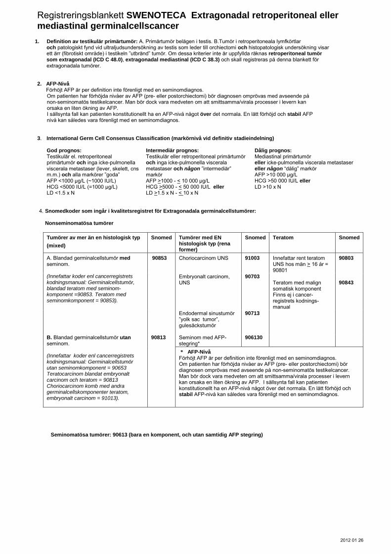
2012 01 26
Registreringsblankett SWENOTECA Extragonadal retroperitoneal eller mediastinal germinalcellscancer
1. Definition av testikulär primärtumör: A. Primärtumör belägen i testis. B.Tumör i retroperitoneala lymfkörtlar och patologiskt fynd vid ultraljudsundersökning av testis som leder till orchiectomi och histopatologisk undersökning visar
ett ärr (fibrotiskt område) i testikeln ”utbränd” tumör. Om dessa kriterier inte är uppfyllda räknas retroperitoneal tumör som extragonadal (ICD C 48.0), extragonadal mediastinal (ICD C 38.3) och skall registreras på denna blankett för extragonadala tumörer.
2. AFP-Nivå Förhöjt AFP är per definition inte förenligt med en seminomdiagnos. Om patienten har förhöjda nivåer av AFP (pre- eller postorchiectomi) bör diagnosen omprövas med avseende på
non-seminomatös testikelcancer. Man bör dock vara medveten om att smittsamma/virala processer i levern kan orsaka en liten ökning av AFP.
I sällsynta fall kan patienten konstitutionellt ha en AFP-nivå något över det normala. En lätt förhöjd och stabil AFP nivå kan således vara förenligt med en seminomdiagnos.
3. International Germ Cell Consensus Classification (markörnivå vid definitiv stadieindelning)
God prognos: Intermediär prognos: Dålig prognos: Testikulär el. retroperitoneal primärtumör och inga icke-pulmonella viscerala metastaser (lever, skelett, cns m.m.) och alla markörer ”goda” AFP <1000 µg/L (~1000 IU/L) HCG <5000 IU/L (=1000 µg/L) LD <1.5 x N
Testikulär eller retroperitoneal primärtumör och inga icke-pulmonella viscerala metastaser och någon ”intermediär” markör AFP >1000 - < 10 000 µg/L HCG >5000 - < 50 000 IU/L eller LD >1.5 x N - < 10 x N
Mediastinal primärtumör eller icke-pulmonella viscerala metastaser eller någon “dålig” markör AFP >10 000 µg/L HCG >50 000 IU/L eller LD >10 x N
4. Snomedkoder som ingår i kvalitetsregistret för Extragonadala germinalcellstumörer: Nonseminomatösa tumörer
Tumörer av mer än en histologisk typ
(mixed)
Snomed Tumörer med EN histologisk typ (rena former)
Snomed Teratom Snomed
A. Blandad germinalcellstumör med seminom.
(Innefattar koder enl cancerregistrets kodningsmanual: Germinalcellstumör, blandad teratom med seminom-komponent =90853. Teratom med seminomkomponent = 90853).
90853 Choriocarcinom UNS
91003 Innefattar rent teratom UNS hos män > 16 år = 90801
90803
Embryonalt carcinom, UNS
90703
Teratom med malign somatisk komponent Finns ej i cancer-registrets kodnings-manual
90843
Endodermal sinustumör ”yolk sac tumor”, gulesäckstumör
90713
B. Blandad germinalcellstumör utan seminom. (Innefattar koder enl cancerregistrets kodningsmanual: Germinalcellstumör utan seminomkomponent = 90653 Teratocarcinom blandat embryonalt carcinom och teratom = 90813 Choriocarcinom komb med andra germinalcellskomponenter teratom, embryonalt carcinom = 91013).
90813
Seminom med AFP-stegring*
906130
* AFP-Nivå Förhöjt AFP är per definition inte förenligt med en seminomdiagnos. Om patienten har förhöjda nivåer av AFP (pre- eller postorchiectomi) bör diagnosen omprövas med avseende på non-seminomatös testikelcancer. Man bör dock vara medveten om att smittsamma/virala processer i levern kan orsaka en liten ökning av AFP. I sällsynta fall kan patienten konstitutionellt ha en AFP-nivå något över det normala. En lätt förhöjd och stabil AFP-nivå kan således vara förenligt med en seminomdiagnos.
Seminomatösa tumörer: 90613 (bara en komponent, och utan samtidig AFP stegring)