synaptic+transmission
DESCRIPTION
synaptic transmissionTRANSCRIPT

LECTURE OBJECTIVES1. To understand the overall concept of synaptic transmission. 2. To understand the inputs into dendrites and the cell body.3. To analyze the differences between metabotropic and ionotropic
receptors.4. To understand the sequence of events in the presynaptic terminal
leading to neurotransmitter release.5. To list the methods of transmitter inactivation and discuss their
importance.6. To describe in the steps (synthesis, storage, release and inactivation
of acetylcholine) of neurotransmission at the neuromuscular junction.7. To analyze a disorder associated with the NMJ - Myasthenia Gravis.
Synaptic TransmissionRichard W. Keller, Jr., Ph.D.
Center for Neuropharmacology and NeuroscienceME-751 [email protected] 2-5253

Dendrites(inputs)
Cell Body(metaboliccenter)
Axon(conduction of
message)
Axon Terminals(output)
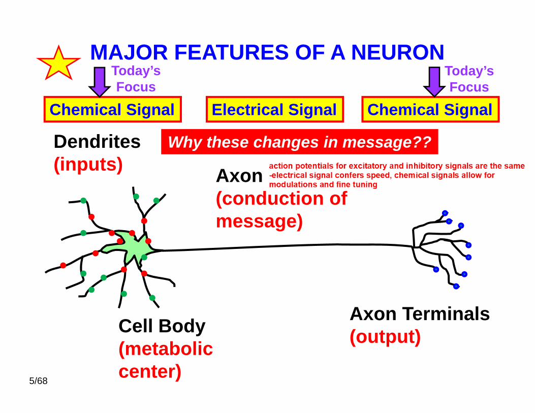
MAJOR FEATURES OF A NEURONThere are many different types of neurons but they almost all have similar major features.
Quick Review covered by Dr. Lindsley
Synaptictransmission
- AP – Dr. Shin

What does this really look like????
NeuromuscularJunction
Nerve – NerveConnections
See Figure 11-36 – Alberts, et al.http://education.vetmed.vt.edu/Curriculum/VM8054/Labs/Lab10/lab10.htm
Artist John Allison’s Rendering of the Cortex
No Exam Material!
Dendrites(inputs)
Cell Body(metaboliccenter)
Axon(conduction ofmessage)
Axon Terminals(output)
MAJOR FEATURES OF A NEURON
Chemical Signal Chemical SignalElectrical Signal
Why these changes in message??
Today’sFocus
Today’sFocus
5/68

Major features common to almost all nerve cells.
1. Cell Bodya. Basic machinery of life. The cell body of the
neuron contains all the same organelles that arefound in all animals cells.
[nucleus, rough endoplasmic reticulum (ER),smooth ER, Golgi apparatus, mitochondria - lots!]
b. The cell body functions as the metabolic center,the site of protein synthesis & packaging, etc.
c. Diameter 5 - 60 um - small compared to most cells
d. Some inputs from other neurons - most via dendrites
Quick Review covered by Dr. Lindsley

The cell body =~0.1% of entire
cell volume!
Nucleus
Fig 1.3 – Neuroscience; 2nd Ed.Purves, et al 2001; Sinauer
Quick Review covered by Dr. Lindsley
THE CELL BODY
No Exam Material!

2. Dendritesa. Communication: neurons receive inputs (messages)
from adjacent cells to pass on to other cells. Manyof these inputs (afferent signals) are onprojections from the cell body called dendrites.
1. "antennas" of the neuron2. branched in a "tree-like" fashion
b. Several dendrites - number and extent of thebranching varies dramatically
1. average neuron receives about 10,000inputs in its dendritic tree
2. Purkinje cells - very large dendritic treefrequently receiving 150,000 inputs
INPUTS = NEUROTRANSMITTERS interacting with RECEPTORS (Ligand-gated Ion Channels!)

A typical Neuron hasabout10,000 inputs into the dendrites
Thousands of excitatory and thousands of
inhibitory inputs!
Inputs
ExciteInhibit
Receptors

A cell potential exists across all parts of a neuronincluding the cell body and dendrites.
The resting membrane potential is ~ -80 mVFrom last lecture - primarily due to a K leak!
In fact, all living cells have membrane potentials!
so.... What’s the big deal about potentials in neurons?
Significant Changes in the cell potential result from NTsinteracting with & opening LIGAND-GATED Ion-Channels!

Really 2 types of Neurotransmitter-Activated Receptors
Ligand-gatedIon channels
Metabotropicreceptors
2nd messengers
Fig 5.16 – Neuroscience; 5th Ed.Purves, et al 2012; Sinauer
1. 2.
neurotransmitter = ligand
Direct!Indirect!
FAST!SLOW!

Lots of Inhibitory and Excitatory Inputs
Some inputs function to "excite" the cell, while others tend to "inhibit"the cell.
At a cell potential level what does “excite” mean?
"inhibit"?

Excitation Inhibition Excitation & Inhibition
DepolarizingLess (-) more (+)-50mv 0 mv(+) current in
HyperpolarizingMore (-)-100mv
(+) current outor (-) current in
InitialResting pot.-80 mv
-80 mV -80 mV
Fig 12-16 Principles of Neural Science4th Ed. Kandel, Schwartz & Jessell
Reality is a combination of the
two effects.
Reality is a combination of Excitation & Inhibition
15/68
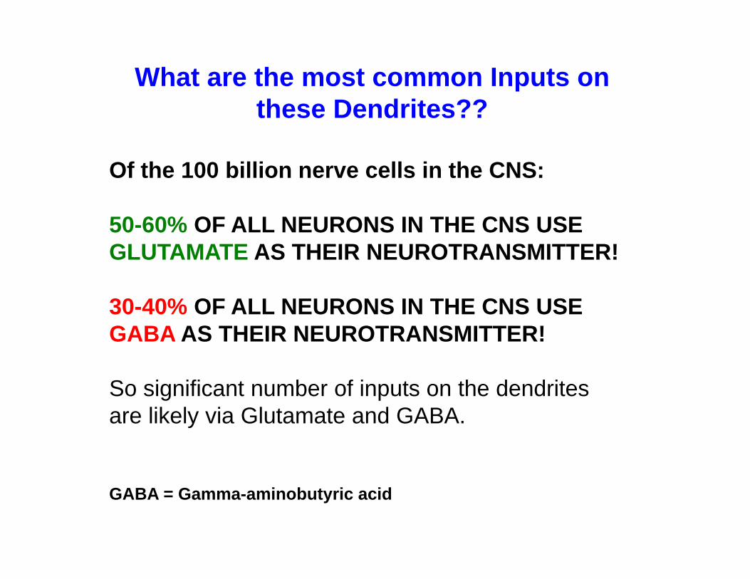
What are the most common Inputs on these Dendrites??
Of the 100 billion nerve cells in the CNS:
50-60% OF ALL NEURONS IN THE CNS USE GLUTAMATE AS THEIR NEUROTRANSMITTER!
30-40% OF ALL NEURONS IN THE CNS USE GABA AS THEIR NEUROTRANSMITTER!
So significant number of inputs on the dendritesare likely via Glutamate and GABA.
GABA = Gamma-aminobutyric acid

Na+ Cl-
GlutamateExcitation
GABAInhibition
Glu Glu GABA GABA
InitialResting
pot.-80 mv
MorePositive;
depolarizing
MoreNegative;
hyperpolarizing
Glutamate = major excitatory NT GABA = major inhibitory NT
In CNS Most Excitatory & Inhibitory Inputs are Glu & GABA

xxx
K+
Does “xxx” produceexcitatory orinhibitoryeffects viathis receptor???
xxx
Self StudyYou think about
this!inside
outside
No Exam Material!

DendriteApply Glutamate (Glu) into area with Glu receptors
Adapted from Fig 2.3 – Neuroscience; 5th Ed. Purves, et al 2012; Sinauer
a “graded” potential !!
Activation of Receptors Results in GradedPotentials in the Dendrites & Cell Body
signal decays over distance…..
ExcitatoryInput
ligand-gated ion channels…..

axon
Axon Hillock
B. There are thousands of excitatory and inhibitory inputs on dendrites & the cell body1. “cellular decision” has to be made to fire or
not to fire an action potential2. potential is summed over area (spatial
summation) & time (temporal summation)
3. the “trigger zone” is located in the “initialsegment” of the axon; this is where the “cellular decision” is made. [Adjacent to the axon hillock region of the cell body.]
If the cell potential in the initial segment is
sufficiently positive to lead to the opening of
voltage-gated Na+
channels – the cell fires an action potential.
Initial segment - very high density of voltage-
gated Na+ channels
20/68

Na+
Channel= AP
K+
Channel= recovery
Action Potential moves down the Axon!
Covered by Dr. Shin and in Membrane Transport
axon = neuron's output - the "wire" leading to where the message will be delivered
All or None?
No Exam Material!

Na+/K+ Pump
ENERGYREQUIRED!
Covered in Membrane TransportFrom Fig 4.11 – Neuroscience; 5th Ed. Purves, et al 2012; SinauerNo Exam Material!

TERMINAL FIELDCELL BODYand
DENDRITES
AXON
LIGAND-GATED ION CHANNELS(“Neurotransmitter-activated”)
METABOTROPIC RECEPTORS that modify ion channels
Excite - depolarizeInhibit - hyperpolarize
GRADED POTENTIALS!
AXON Terminalsor “boutons”
AXON HILLOCKthreshold - fire?spatial summationtemporal summation
VOLTAGE-GATED ION CHANNELSALL OR NONE ACTION POTENTIAL!INFO CODED IN FREQUENCY
Examine what happens here!
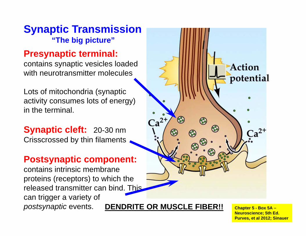
Presynaptic terminal:contains synaptic vesicles loaded with neurotransmitter molecules
Lots of mitochondria (synaptic activity consumes lots of energy) in the terminal.
Synaptic cleft: 20-30 nmCrisscrossed by thin filaments
Postsynaptic component:contains intrinsic membrane proteins (receptors) to which the released transmitter can bind. This can trigger a variety of postsynaptic events.
Synaptic Transmission“The big picture”
Chapter 5 - Box 5A –Neuroscience; 5th Ed. Purves, et al 2012; Sinauer
DENDRITE OR MUSCLE FIBER!!

Synthesis of Neurotransmitters
synthesis of Enzymes &
Vesicles
synthesis of Neuro-
transmitters
can be up to 2 meters in human!
Figure 5.5 – Neuroscience; 5th Ed. Purves, et al 2012; Sinauer
THE “NAME” OF A NEURON COMES FROM THE NEUROTRANSMITTER THAT IS RELEASED
25/68

The Synthesis, Packaging and Secretion of Neurotransmitters
Storage Vesicles
BiosyntheticEnzymes
Fig 5.3 – Neuroscience; 5th Ed.Purves, et al 2012; Sinauer

Synaptic Terminals / Synaptic Boutons -con’t• An action potential (AP) moves down the axon and
invades the terminal area. This AP opens voltage-dependent Na+ channels and then voltage-dependent Ca+2 channels leading to an influx of Ca+2.
• This influx of Ca+2 is the trigger for exocytosiswhich leads to the release of neurotransmitter.In this process, vesicles fuse with the plasma membrane of the terminal and dump their contents into the cleft of the synapse.
• Terminals conserve membrane by recycling it; afterrelease, vesicles are reformed and refilled.

EXOCYTOSIS - the simple version!
Fig 5.14 – Neuroscience; 5th Ed.Purves, et al 2012; Sinauer
“DockedVesicle”
Plasmamembrane

A REALVESICLE!
Takamori, et al.Cell 127: 831-846
Fig 5.10 – Neuroscience; 2nd Ed.Purves, et al 2001; Sinauer
More of the components involved in exocytosis
… and then a miracle
happens!
No Exam Material!

Fig 5.11 – Neuroscience; 5th Ed.Purves, et al 2012; Sinauer
Role of Ca+2 in Synaptic Transmission
1
2
Remove Ca+2 from Buffer!1
2
Control
Why this delay?
?
30/68

“PROJECTION AREA”OR
“TERMINAL FIELD”Those with AXON terminals
There are 2 major types of release sites.
Those with AXON “boutons”
AcetylcholineGlutamateGABA
DopamineNorepinephrineSerotonin
10,000terminals
500,000boutons

Associated with these two different fields are
1. AxonTerminals axon
axon
Extracellular levels – IMP!
Volume Transmission
2. Axon Boutons
ClassicalNeurotransmission
Synaptic cleft levels - IMP!
2 types of synaptic contacts that release NTs.
Catecholamines & Serotonin
GlutamateGABA
(ACh - NMJ)
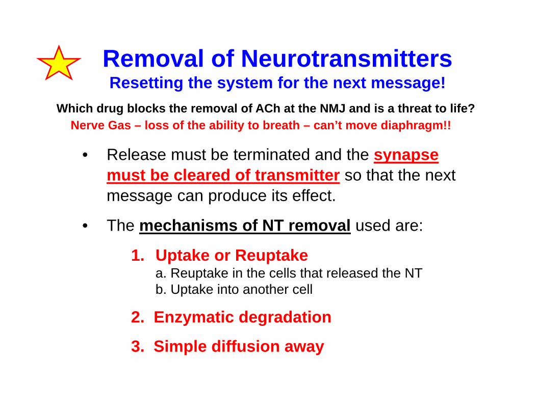
• Release must be terminated and the synapse must be cleared of transmitter so that the next message can produce its effect.
• The mechanisms of NT removal used are:
1. Uptake or Reuptakea. Reuptake in the cells that released the NTb. Uptake into another cell
2. Enzymatic degradation
3. Simple diffusion away
Removal of NeurotransmittersResetting the system for the next message!
Which drug blocks the removal of ACh at the NMJ and is a threat to life?Nerve Gas – loss of the ability to breath – can’t move diaphragm!!

DA
DADADA
DA
AP
DA-term.ppt
vesicular uptakeVMAT
DAT DAT
Other Cells
DA
COMTmetabolism
80%
Post SynapticTarget Cell
20%
1
2
1
2DA Uptake 1DA Uptake 2
DAT = Dopamine Transporter (Reuptake)
DAT MAOmetabolism
60%
40%
DA recp DA recpDA recp
DopamineNorepinephrineEpinephrineSerotonin
ReUptakeinto Boutons
Transporters (Trans) are protein carriers that move molecules through the plasma membrane
ECF
1 of the 500,000 boutons
35/68

Uptake intoAstrocytes
GlutamateGABA
Fig 7.2 – Fundamental Neuroscience;2nd Ed. Squire, et al 2003; Academic Press
Major removal Site! Glu & GABA
Transporters
(Astrocyte)
Nerve Terminal

1. Uptake
2. Enzymatic degradation1. In the ECF2. In other cells3. In the releasing terminal / bouton
3. Simple diffusion away
Removal of Neurotransmitters

Thanvi & Lo - Postgrad Med J 2004;80:690-700
Enzymatic Degradation
AChCholine+ acetate
NeuroMuscularJunction
AChHistamine
Acetylcholinesterase

1. Uptake
2. Enzymatic degradation
3. Simple diffusion away
Removal of Neurotransmitters
All NTs to some extent!
Fig 7.2 – Fundamental Neuroscience;
2nd Ed. Squire, et al 2003; Academic Press
Diffusion

Recovery from Depolarization of Release(Recovery of the Terminal)
Fig 4.9 – Neuroscience; 5th Ed.Purves, et al 2012; Sinauer

Receptor Binding in Post synaptic cell
Metabotrophic Receptor - Second
Messengers
Neurotransmitter Release
Ligand-gated Ion channelopening or closing
Postsynaptic cells – membrane potential change - excited or inhibited
Events from Neurotransmitter Release to Postsynaptic Excitation or Inhibition
Does binding to a receptor play a role
in NT removal?

NtNt Nt
Nt
NtNtNt
Nt
Nt2
Locations ofReceptors
Nt2
PostsynapticPresynaptic
autoreceptors(Feedback – enough NT! )
Presynapticheteroreceptors
Nt3
Nt3
Ion channel
Metabotropic receptor
Not much evidence for thesehetero -receptors at NMJ.
There are presynaptic auto-receptors at NMJ.
45/68
Nt = neurotransmitter

The Combined Effect of Neurotransmitters and their Receptors
Many different kinds of chemical messages
• Fast vs slow – Some neurotransmitters (NTs) work veryquickly and are rapidly removed (ACh at NMJ) vsothers that are more neuromodulators (DA in brain)
• Receptors also add variability. The same NT can beExcitatory at one receptor and Inhibitory at another.
• There are receptors with different affinities for the same NT; so low levels of NT can stimulate one set of receptors and high levels can stimulate additional receptors.

Same NT Different Responses at Different Acetylcholine Receptors
Excitatory
Inhibitory
see animation of inhibition @ http://www.blackwellpublishing.com/matthews/neurotrans.html
Nicotiniccholinergicion channelreceptor
Muscariniccholinergicmetabotropicreceptor
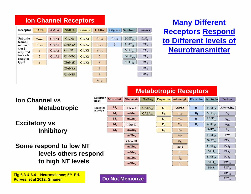
Many Different Receptors Respond to Different levels of
Neurotransmitter
Ion Channel Receptors
Metabotropic ReceptorsIon Channel vs
Metabotropic
Excitatory vsInhibitory
Some respond to low NTlevels others respondto high NT levels
Fig 6.3 & 6.4 – Neuroscience; 5th Ed.Purves, et al 2012; Sinauer Do Not Memorize

The Neuromuscular Junction (NMJ)Specialized chemical synapse between a motor neuron and striated muscle fibers. The NT used is acetylcholine(ACh)
Synthesis: from acetyl CoA and choline by choline-acetyl transferase (CAT) in the terminal
Storage & release: Uses mechanisms described previously
Receptors: NMJ uses nicotinic acetylcholine receptors. These are ligand-gated Na+ (some K+) ion channels that will generate a depolarization when bound to ACh. This depolarization is called the endplate potential (EPP).
Inactivation: ACh is inactivated enzymatically. The synaptic cleft in NMJ contains acetylcholinesterase (ACE) which breaks ACh into acetyl CoA and choline. Choline is brought into the terminal via a transporter for reuse in synthesis.
50/68

The Neuromuscular Junction (NMJ):
http://education.vetmed.vt.edu/Curriculum/VM8054/Labs/Lab10/lab10.htmhttp://demo.classontheweb.com
NMJ

Saladin – Anatomy and Physiology:The Unity of Form and Function 1996
McGraw-Hill
CNS Synapse
2,000-6,000 um2 2-10 um2
Surface Area of a Presynaptic TerminalThe Neuromuscular Junction (NMJ) A lot of what we
know was first worked out at the
NMJ!!
Not toScale!

Freeze Fracture EM
Heuser, et al. 1979J.Cell.Biol 81 275John Heuser = http://www.heuserlab.wustl.edu/v2.0/index.shtml
Unstimulated
Stimulated
NMJ Freeze
FractureSlam the tissue against a copper plate frozen in liquid nitrogen and then break it apart.
Do Not Memorize

Recordings in the junction reveal “graded” local potential changes (EPP) before an Action Potential is produced.
Stimulated ACh release ~150 MEPPs (vesicles) = EPP
An EPP involves ligand-gated ion channels – if the membrane potential change is sufficient (exceeds the voltage-gated Na+
channel threshold) then an AP that involves voltage-gated ion channels occurs in the muscle fiber.

The discovery of Miniature Endplate Potentials“mini’s” or MEPPs
Bernard Katz1950’s
spontaneous 0.5mV changes in potential(single vesicle leak)
evoked change with stimulation (AP-stimulated release of ~150 vesicles of ACh)
Nerve stimulation
Membrane potential of muscle fiber
EPP

Skeletal Muscle Action Potential• in the muscle, away from the neuromuscular junction
the AP is again all-or-nothing (no EPP!)
When the depolarization produced by the EPP is sufficient to activate voltage-gated Na+ channels, an action potential is generated in the muscle fiber.
This action potential results in opening of voltage-gated Ca+2 channelslocated both in the cell membrane and in endoplasmic reticulum membranes of the muscle fiber. The consequence is a massive entry of Ca+2 into the cytoplasm, both from outside the cell and from intracellular storage sites, which results in contraction of the muscle fiber.
EPP is playing a role similar to which region in
the neuron?
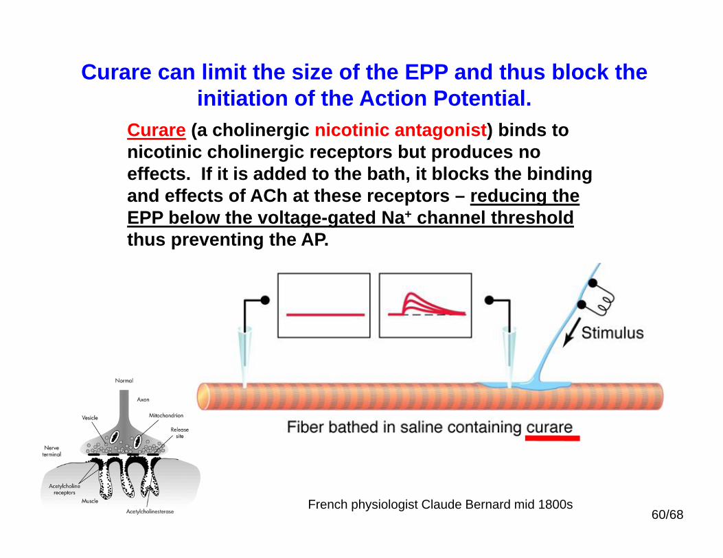
Curare (a cholinergic nicotinic antagonist) binds to nicotinic cholinergic receptors but produces no effects. If it is added to the bath, it blocks the binding and effects of ACh at these receptors – reducing the EPP below the voltage-gated Na+ channel thresholdthus preventing the AP.
Curare can limit the size of the EPP and thus block the initiation of the Action Potential.
60/68French physiologist Claude Bernard mid 1800s

Myasthenia Gravis• MG is an autoimmune disease that disrupts transmission at the
NMJ.• The body forms antibodies to nicotinic ACh receptors
- destroys some receptors and blocks others• There are less functional receptors and thus weak muscles.
Y Y
Receptorsblocked byantibodies!
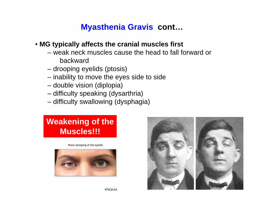
Myasthenia Gravis cont…
• MG typically affects the cranial muscles first– weak neck muscles cause the head to fall forward or
backward– drooping eyelids (ptosis)– inability to move the eyes side to side– double vision (diplopia)– difficulty speaking (dysarthria)– difficulty swallowing (dysphagia)
Weakening of theMuscles!!!

Myasthenia Gravis cont…• One of the most treatable neuromuscular disorders!• Administer acetylcholinesterase blockers
such as neostigmine or pyridostigmine(+ immunosuppressive drugs)
This reduces the ACh destruction by AChE & increases the accumulation of ACh at the neuromuscular junction. Hence there is more ACh to interact the remaining receptors.
Before Neostigmine
After Neostigmine
Walker MB (1934). Treatment of myasthenia
gravis with physostigmine. Lancet 1:1200-1201.

Sample Questions:
1. Neurotransmitters (ligands) activate ion channels in the dendrites resulting in membrane potential changes that are described as:
a. all or noneb. negativec. gradedd. positivee. integrated
2. A rabbit shot with a curare-tipped arrow would quickly lose its ability to run away, because at the neuromuscular junction
a. the breakdown of acetylcholine would be inhibited.b. the acetylcholine receptors would be blocked.c. acetylcholine could no longer be released from the presynaptic terminals.d. the voltage-dependent sodium channels would be blocked.e. synthesis of acetylcholine from choline would be inhibited.
The End!c b