synthesis of an analog of the thyroid-hormone-binding …€¦ · · 2001-05-14t-boc chemistry...
TRANSCRIPT

1
Synthesis of an analog of the thyroid-hormone-binding protein
transthyretin via regioselective chemical ligation
Jackie A Wilce1, Stephen G Love2, Samantha J Richardson3, Paul F Alewood2 and
David J Craik2
1Department of Biochemistry/Chemistry, University of Western Australia,
Nedlands Western Australia 6907, Australia
2Institute for Molecular Bioscience, University of Queensland,
Brisbane, Queensland 4072, Australia.
3Department of Biochemistry and Molecular Biology, The University of Melbourne,
Parkville Victoria 3010, Australia
Corresponding Author:
David J Craik
Ph: 61 7 3365 4945
Fax: 61 7 3365 2487
Email: [email protected]
Running title: Chemical synthesis and folding of TTR
Key words: peptide ligation, protein folding, transthyretin, ligand binding
Copyright 2001 by The American Society for Biochemistry and Molecular Biology, Inc.
JBC Papers in Press. Published on May 14, 2001 as Manuscript M101228200 by guest on June 3, 2018
http://ww
w.jbc.org/
Dow
nloaded from

2
Summary
Transthyretin is an essential protein responsible for the transport of thyroid hormones
and retinol in human serum and is also implicated in the amyloid diseases familial
amyloidotic polyneuropathy (FAP) and senile systemic amyloidosis (SSA). Its folding
properties and stabilization by ligands are of current interest due to their importance in
understanding and combating these diseases. Here we report the solid phase synthesis
of the monomeric unit of a transthyretin analog (equivalent to 127 amino acids) using
t-Boc chemistry and peptide ligation and its folding to form a functional 54 kDa
tetramer. The monomeric unit of the protein was chemically synthesized in three
parts: 1-51, 54-99 and 102-127 and ligated using a chemoselective thioether ligation
chemistry. The synthetic protein was folded and assembled to a tetrameric structure in
the presence of TTR’s native ligand, thyroxine, as shown by gel filtration
chromatography, native gel electrophoresis, transthyretin antibody recognition and
thyroid hormone binding. Other folding products included a high molecular weight
aggregate as well as a transient dimeric species. This represents one of the largest
macromolecules chemically synthesized to date and demonstrates the potential of
protein chemical synthesis for investigations of protein:ligand interactions.
by guest on June 3, 2018http://w
ww
.jbc.org/D
ownloaded from

3
Introduction
Transthyretin (TTR) is a 54 kDa tetramer which is present in human plasma (3.6 µM
tetramer) and transports thyroid hormones such as 3,5,3’-triiodo-L-thyronine (T3) and
L-thyroxine (T4) as well as retinol binding protein (1,2). Its structure and ligand
binding capabilities have been characterized by X-ray crystallography (3-5). Each
subunit contains extensive beta-sheet structure and is arranged within a dimer of
dimers to form a compact molecule with two funnel-shaped hormone ligand binding
sites, each defined by a dimer-dimer interface. The thyroid hormones bind deeply
within the hydrophobic binding channel, their iodinyl moieties residing in
hydrophobic pockets at two different binding sites within the channel (6). Upon
binding of one T4 ligand, the binding affinity for the second ligand is reduced from
nM to µM.
Although the TTR tetramer is inherently very stable (1,7), in some circumstances
transthyretin has a propensity to form amyloid fibrils. In diseases such as senile
systemic (SSA) and familial amyloidotic polyneuropathies (FAP), native and mutant
TTR, respectively have been found to form long beta-sheet based fibrillar structures
(8,9). These amyloid lesions accumulate in specific organs and are implicated in their
dysfunction and ultimately the death of the patients. More than 70 separate mutations
that appear to increase the propensity of TTR to form amyloid structures underlying
FAP have so far been identified (10-14). It is thought that the underlying mechanism
for TTR fibril formation involves tetramer dissociation to a monomeric
conformational intermediate which self assembles to form amyloid fibrils. Any
mutation or cellular condition (such as low pH) that tends to destabilize the tetramer,
can result in an increased propensity of the protein to form amyloid fibrils (9,11).
by guest on June 3, 2018http://w
ww
.jbc.org/D
ownloaded from

4
The ligand binding properties of TTR have become of major importance recently,
with the discovery that certain thyroid hormone competitors (e.g. 2,4,6-triiodophenol)
are able to decrease the tendency of TTR to form amyloid fibrils (15,16). They are
reported to act by binding deeply within the TTR binding channel at both ligand
binding sites and inhibiting the formation of amyloid by stabilizing the normal fold
against the pathogenic conformational change. A range of non-steroidal anti-
inflammatory drugs are currently being investigated for their ability to inhibit and
reverse amyloid formation (17). Whilst crystallographic information has revealed the
orientations of several of these drugs bound to TTR, there are many aspects of the
ligand binding that cannot be probed using crystallographic techniques. These include
dynamic aspects of protein:ligand binding in solution and in the presence of
competitors, or in the presence of other serum proteins, including the other thyroid
hormone carriers albumin and thyroxine binding globulin. For such studies it is
possible that nuclear magnetic resonance (NMR) spectroscopic methods could be
employed.
NMR can be used to observe the interaction of a ligand with particular sites in the
protein. Such studies currently depend upon the assignment of the specific resonances
that are perturbed by the ligand (18) which limits studies to systems in which the
protein signal of interest is either fortuitously distinct or has been fully assigned using
isotopic labeling and heteronuclear NMR techniques. In the latter case this also
requires that the protein is less than about 40 kDa due to the broad linewidths of the
NMR signals and large number of signals that have similar chemical shifts. The
problem can be somewhat reduced by using chemical ligation strategies in which just
by guest on June 3, 2018http://w
ww
.jbc.org/D
ownloaded from

5
one portion of the protein is isotopically labeled (19). Of even greater value would be
the ability to completely control the position of the isotopic label in a protein for
probing the ligand interaction.
The current study thus presents the first stage in the development of a spectroscopic
method for probing a protein:ligand interaction. The strategy involves the complete
synthesis of a TTR analog using solid-phase synthesis and chemical ligation
techniques, and refolding of the protein to a tertiary and quaternary structure that
approximates the native form. Since the structure of TTR is known to atomic
resolution from X-ray crystallographic studies, an NMR active probe (i.e. 15N- or 13C-
labelled amino acid) may be incorporated at any strategic position, to enable
subsequent ligand binding studies to be manifest in NMR spectra.
The chemical synthesis of proteins of the size of the monomeric unit of TTR (127
residues) represents a significant challenge. In the past, long peptides were
synthesized in a stepwise fashion, as exemplified by HIV-1 protease (20) and IL-8
(21) – but significant purification problems resulted in low yields of protein.
Currently, the synthesis techniques for proteins of this size rely on chemoselective
ligation techniques, where two or more non-protected peptides are joined through a
highly selective chemical reaction.
There have been several notable protein syntheses described using chemoselective
ligation techniques that incorporate thioester and thioether surrogate amide bonds.
These include the synthesis of linked heterodimeric b/HLH transcription factors (22)
and cpn10 (23) respectively. Recently, techniques for forming a native amide bond
by guest on June 3, 2018http://w
ww
.jbc.org/D
ownloaded from

6
have been applied to a range of protein types including serine protease inhibitors (24-
26), human II secretory phospholipase A2 (27), and barnase (28,29).
Native chemical ligation chemistry is only useful provided there are suitably
positioned cysteines. In order to synthesize a protein like transthyretin without
suitably located cysteines it becomes necessary to use chemically modified amino
acid substitutes or amino acid mutations. The thioether ligation strategy we use here
introduces –NH-CH2-CH2-S-CH2-CO-, which mimics a two amino acid subunit
closely resembling a glycyl-glycine (-NH-CH2-CO-NH-CH2-CO-). While the
thioether moiety closely resembles the spatial requirements for glycyl-glycine it may
lack potential hydrogen bond donation/acceptor behavior of the di-amino acid unit,
thereby potentially introducing some non-native structural characteristics.
Here we demonstrate the total chemical synthesis of an analog of human TTR through
the use of the thioether strategy for the sequential ligation of three peptides. We also
show that this synthetic TTR (henceforth referred to as sTTR) may be successfully
refolded and reconstituted to form a 54 kDa tetrameric structure able to bind the
thyroid hormone T4. This represents one of the largest active proteins made
synthetically, and provides methodology for future protein:ligand investigations using
NMR spectroscopic techniques.
Experimental Procedures
Chemicals and Reagents— trifluoroacetic acid (TFA), dichloromethane (DCM), N,N-
dimethyl formamide (DMF), and diisopropylethylamine (DIEA) were from Auspep
(Melbourne, Australia). O-benzotriazole-N,N,N’,N’-tetramethyl-uronium-
by guest on June 3, 2018http://w
ww
.jbc.org/D
ownloaded from
7
hexafluorophosphate (HBTU) was from Richelieu Biotechnologies (St Hyacinth,
Quebec Canada). Acetonitrile was from BDH Laboratory Supplies (Poole, UK)
Acetic acid and chloroacetic acid were from Ajax chemicals (Auburn, Australia),
diethyl ether from Fluka Biochemicals (Melbourne) and mercaptoethanol from Sigma
(St Louis Mo, USA). Ethanolamine, N,N-diisopropylcarbodiimide (DIC) and
bromoacetic acid were from Aldrich (Milwaukee, WI, USA). Hydrogen fluoride (HF)
was purchased from Boc Gases (Brisbane, Australia). The following N-Boc protected
L-amino acids Ala, Gly, Ile, Leu, Phe, Pro, Val, Arg(p-toluenesulphonyl), Asp(O-
cyclohexyl; OChx), Asn(xanthyl; Xanth), Glu (O-cyclohexyl; OChx),
His(dinitrophenyl; DNP), Lys(2-chlorobenzyloxycarbonyl; CIZ), Ser(benzyl; Bzl),
Thr(benzyl;Bzl), Trp(formyl; CHO), Tyr(2-bromobenzyloxycarbonyl; 2BrZ) were
purchased either from NovaBiochem (La Jolla, USA) or Bachem, (Switzerland).
Human serum was supplied by the Red Cross Blood Bank, Melbourne.
Equipment—Analytical and preparative HPLC was carried out using a Waters HPLC
system comprised of model 600 solvent delivery system 600E controller and model
484 detector. Vydac C18 columns, analytical (4.6 X 250 mm) at a flow rate of 1
ml/min, semi preparative (10 X 250 mm) at a flow rate of 3 ml/min, and preparative
(22 X 250 mm) at a flow rate of 8 ml/min were used. All peptides were purified using
linear gradients of 0.1% aqueous TFA (solvent A) and 90% aqueous acetonitrile
0.09% TFA (solvent B).
Mass spectral data were collected using a Perkin Elmer Sciex (Toronto, Canada) API
III Biomolecular Mass Analyser ion-spray mass spectrometer equipped with an ABI
140B solvent delivery system. Raw data were analyzed using the program MassSpec
by guest on June 3, 2018http://w
ww
.jbc.org/D
ownloaded from
8
(Perkin Elmer Sciex). Calculated masses were obtained using the program
MACROMASS (Sunil Vemuri and Terry Lee, City of Hope, Durate, CA).
Enhanced Chemiluminescence kit was from Amersham, X-ray film was from
Eastman-Kodak, methyl cellulose and activated charcoal (Norit PN.5) were from
BDH, L[125I]-thyroxine (1.2 Ci/mg) was from NEN Dupont, SepPak C-18 cartridges
were from Millipore Waters, and thin layer chromatography plates were from Merck.
All reagents were of analytical grade.
Native hTTR – Native hTTR was isolated from serum using an adapted version of the
method described by Dwulet and Benson (30).
Peptide Synthesis—Peptides were synthesized using the rapid manual HBTU in-situ
neutralization synthesis technique (31) or using the same technique on a modified
ABI 430A peptide synthesizer (32). The thioether resins were prepared according the
methods of Englebretsen et al (33), initially on amino methyl resins then subsequently
Boc-amino acid -Pam resins (ABI CA, USA). The bromoacetyl and chloroacetyl
groups at the amino termini of peptides were coupled using the symmetrical
anhydride formed from reaction with DIC. The dinitrophenyl (DNP) group was
removed using 20% mercaptoethanol in 10% DIEA/DMF solution for 2-3 x 30 min
treatments. The Trp formyl deprotection was carried out using ethanolamine prior to
HF cleavage.
Peptide resins were cleaved using HF with p-cresol and p-thiocresol as scavengers at
–5 to 0oC for 1-2 h. The HF was removed in vacuo, the peptide product triturated
by guest on June 3, 2018http://w
ww
.jbc.org/D
ownloaded from

9
with cold diethyl ether (3 X 50 ml), and the precipitated peptide collected and
dissolved in 50% acetonitrile with 0.1% TFA.
The crude peptides were purified by reverse phase (RP)-HPLC and fractions collected
and analyzed by analytical RP-HPLC and electrospray mass spectrometry (ESMS).
Fractions containing the purified peptide were combined and lyophilized.
Solid Phase Synthesis of Br-Ac-PRRYTIAALLSPYSYSTTAVVTNPKE-OH.
(Bromoacetyl-102-127 TTR) (I)—Peptide I was synthesized on a Boc-Glu(OBzl)-Pam
- polystyrene resin (ABI) on a 0.5 mmole scale. Amino acid couplings averaged
99.5% efficiency. N-Boc deprotection, coupling of bromoacetic acid followed by HF
cleavage gave the crude peptide, which was purified by preparative HPLC using a
linear gradient of 0-70% B. The peptide was then analyzed by HPLC and ESMS.
The purified peptide was characterized as the desired product (I) by ESMS [observed
mass = 3021±0.3 Calculated for C133 H210 N34 O41 Br1 =3021.24 (average isotope
composition) ].
Solid Phase Synthesis of Cl-Ac-ELHGLTTEEEFVEGIYKVEIDTKSYWKALGISPFHE-
HAEVVFTAND-NH-CH2-CH2-SH. (Chloroacetyl-54-99-NH-CH2-CH2-SH) (II)—The
C-terminal thiol peptide II was manually synthesized using the thiol linker attached to
Boc-Ala-Pam resin in the first synthesis then Boc-Gly-Pam resin for the second. The
average amino acid coupling for the syntheses was 99.5 and 99.6%, respectively. The
DNP protecting group was removed followed by N-Boc and CHO group removal.
The chloroacetyl group was coupled then the peptide HF cleaved. The crude peptide
was purified by preparative HPLC using a linear gradient of 0-70% B, then analyzed
by guest on June 3, 2018http://w
ww
.jbc.org/D
ownloaded from

10
by HPLC and ESMS. The purified peptide was characterized as the desired product
(II) by ESMS [observed mass = 5416.46±1.1 Calculated for C246 H364 N58 O76 S1 Cl1
= 5417.46 (average isotope composition) ].
Solid Phase Synthesis of H-GPTGTGESKAPLMVKVLDAVRGSPAINVAVHV-
FRKAADDTWEPFASGKTSE-NH-CH2-CH2-SH. (1-51-NH-CH2-CH2-SH) (III)—The
C-terminal thiol peptide III was synthesized using machine assisted synthesis. The
peptide was synthesized using the thiol linker attached to Boc-Gly-Pam resin. The
average amino acid coupling was 99.6% (1st coupling) for the synthesis, which was
routinely double coupled. The DNP protecting group was removed followed by N-
Boc and then the formyl group. The peptide was cleaved from the resin and the crude
peptide purified by preparative HPLC using a linear gradient of 0-70% B. The
peptide was then analyzed by HPLC and ESMS. The purified peptide was
characterized as the desired product (III) by ESMS [observed mass =5355±1.0
Calculated for C236 H379 N66 O72 S2 = 5357.14 (average isotope composition)].
Formation of Cl-Ac-ELHGLTTEEEFVEGIYKVEIDTKSYWKALGISPFHEHAEV-
VFTAND-NH-CH2-CH2-S-CH2-CO-PRRYTIAALLSPYSYSTTAVVTNPKE-OH.
(Chloroacetyl-54-99-ψ-102-127) (IV) —The ligation reaction was initiated by mixing
the two peptides, bromoacetyl-102-127 TTR (I) (5.44 mg 1.8 mmol) and
chloroacetyl-54-99-NH-CH2-CH2-SH (II) (6.84 mg 1.26 mmol), in 1 ml of 6 M urea
0.1 M NaHCO3 pH 8.3 under nitrogen. After mixing, the reaction mixture was left to
stand at room temperature for 24 h. Samples were withdrawn at 0, 1, 2 and 20 h
during this period for HPLC and ESMS analysis. The ligated peptide IV was isolated
from the reaction mixture by semipreparative HPLC using a linear gradient of 0-70%
by guest on June 3, 2018http://w
ww
.jbc.org/D
ownloaded from

11
B. The fractions were analyzed by HPLC and ESMS the fractions containing the
ligated peptide were then lyophilized. The purified peptide IV (6.01 mg) was
characterized as the desired product IV by ESMS [observed mass =8355±1.2.
Calculated for C379H572N92O117S1Cl1 = 8356.8 (average isotope composition)].
Chloro-Iodo exchange of (IV) to give I-Ac-ELHGLTTEEEFVEGIYKVEIDTKSYW-
KALGISPFHEHAEVVFTAND-NH-CH2-CH2-S-CH2-CO-PRRYTIAALLSPYSYSTTAV-
VTNPKE-OH. (Iodoacetyl-54-99-ψ-102-127) (V) — Peptide IV (5.46 mg, 0.654
mmol) was dissolved in 8M urea 0.01M NaOAc pH 7.5 (1 ml) and KI added to
saturation (~8M). After placing under nitrogen a sample was removed at 30 min,
purified by HPLC and analyzed by ESMS to check completion of the iodo exchange.
The iodoacetyl peptide V was then purified by semi preparative HPLC and
lyophilized to give 3.64 mg. The peptide was characterized as the desired product V
by ESMS [observed mass =8447±1.0. Calculated for C379H572N92O117S1Cl1 = 8448.2
(average isotope composition)].
Formation of H-GPTGTGESKAPLMVKVLDAVRGSPAINVAVHVFRKAADDTWEPF-
ASGKTSE-NH-CH2-CH2-S-CH2-COELHGLTTEEEFVEGIYKVEIDTKSYWKALGISP-
FHEHAEVVFTAND-NH-CH2-CH2-S-CH2-CO-PRRYTIAALLSPYSYSTTAVVTNPKE-
OH. (1-51-ψ-54-99-ψ-102-127 TR) (VI)—Ligation of the two peptides Iodoacetyl-54-
99-NH-CH2-CH2-SH V (3.46 mg 0.43 mmol) and 1-51-NH-CH2-CH2-SH III (3.58
mg 6.68 mmol) was initiated by mixing the two peptides in 6 M urea 0.2 M NaHCO3
pH 8.3 (500 ml) and placing under nitrogen. The reaction was mixed then left to
stand at room temperature for 5 h. Samples were withdrawn at 0, 1.5, and 4 h for
HPLC and ESMS analysis. The ligated peptide VI was isolated from the reaction
by guest on June 3, 2018http://w
ww
.jbc.org/D
ownloaded from

12
mixture by semi preparative HPLC using a linear gradient of 0-70% B. The fractions
were analyzed by HPLC and ESMS the fractions containing the ligated peptide were
then lyophilized to give VI (2.76 mg). The purified peptide was characterized as the
desired product VI by ESMS [observed mass =13,676 ± 1.3 Calculated for
C615H949N158O189S3 = 13,676.4 (average isotope composition)].
Formation of the Tetrameric complex.—The ligated peptide (1-51-ψ-54-99-ψ-102-
127) (VI) (0.25mg) was dissolved in 0.075 M NH4HCO3, pH 8.3 (100µl) then diluted
with 100µl of H2O. To this solution was added 5µl thyroxine T4 (5 mg/ml in 0.1M
NaOH). After equilibrating at RT for 18 h the tetrameric protein was isolated by gel
filtration on a Superdex 75 column (HR10/30 Pharmacia – calibrated with
phosphorylase, 97 kDa; bovine albumin, 66 kDa; native TTR, 54 kDa; ovalbumin, 45
kDa; carbonic anhydrase, 30 kDa; soya bean trypsin inhibitor, 20.1 kDa and α-
lactalbumin, 14.4 kDa) with 0.075 M NH4HCO3 10%CH3CN as the eluent at
0.3ml/min. The tetrameric protein was isolated at a retention time of 22 min as
determined by its equivalent retention time to that of native TTR. It was kept in
solution in the presence of excess T4 prior to further analysis.
Western analysis of synthetic TTR—2 µl human serum, 25 µl (0.5 µg) sTTR solution
and 50 µl (1.0 µg) sTTR were separated in a 0.1% SDS polyacrylamide gel, using a
stacking gel of 4.5% acrylamide, pH 6.8, and a resolving gel of 15% acrylamide, pH
8.6 (34). Proteins were transferred onto a nitrocellulose membrane following which
the membrane was blocked. The primary antibody was 1:5000 antiserum raised in a
rabbit against a mixture of TTRs purified from serum from human (Homo sapiens)
wallaby (Macropus eugenii) and chicken (Gallus gallus), and the secondary antibody
by guest on June 3, 2018http://w
ww
.jbc.org/D
ownloaded from

13
was 1:10 000 anti rabbit Ig raised in sheep (Silenus), as described previously (35).
Detection achieved was using enhanced chemiluminescence against X-ray film.
Preparation of L[125I]-thyroxine — Commercially available L[125I]-thyroxine was
found to contain up to 5% 125I- on the reference date. Therefore, 125I-thyroxine was
separated from 125I- and other degradation products by reversed phase chromatography
using a SepPak C-18 cartridge column (36). Purification was checked by thin layer
chromatography followed by autoradiography (37).
Analysis of thyroxine binding to synthetic human TTR — Commercially purchased
125I-thyroxine was purified from degradation products and 125I- as described above.
Methyl cellulose charcoal was prepared as described by Chang et al. (38). In order to
remove thyroxine from the solution containing sTTR, 40 µl of methylcellulose-
charcoal (1%) in Tris-HCl pH 8.9 was centrifuged and the supernatant was removed.
The methylcellulose-charcoal was resuspended in a 160 µl solution containing 3.2 µg
sTTR. The mixture was kept at 4oC for half an hour, with mixing each 10 minutes.
The solution was centrifuged and the supernatant removed for analysis of 125I-
thyroxine binding to sTTR.
10 µl human serum, and 80 µl solution containing sTTR (1.6 µg) was incubated with
1.1 fmol (2.4 nCi) purified 125I-thyroxine (r.t. 1 hr) and a second aliquot of 80 µl
solution containing sTTR (1.6 µg) was incubated with 4.4 fmol (9.6 nCi) purified 125I-
thyroxine (r.t. 1 hr). 5 µl human serum, and the total amounts of sTTR solutions were
analyzed by non-denaturing polyacrylamide gel electrophoresis, 10% acrylamide,
0.05 M Tris-HCl pH 8.9, 4oC (39) followed by autoradiography.
by guest on June 3, 2018http://w
ww
.jbc.org/D
ownloaded from

14
Results
Synthetic strategy
The choice of ligation sites for the preparation of sTTR was based on both the amino
acid sequence and the known tertiary structure of the TTR molecule (Fig. 1A,B) and
involved the ligation of three peptides (Fig. 1C). Whilst the ligation of two peptides,
each of 60-65 residues in length, is a possible alternative strategy, the degree of
difficulty of preparing and purifying peptides of this length is comparable with the
difficulty of a second ligation. In addition, since all of the residues considered for
future labeling studies occur within the last 30 residues it was desirable to prepare a
relatively small C-terminal fragment. It was anticipated that this approach would
increase the ease of preparing several sTTR molecules with selective labels since only
this fragment would need to be resynthesized.
The thioether linker spans a distance equivalent to two amino acids, is highly flexible
and non-functionalised. Whilst Gly-Gly sequences are thus ideally suited as ligation
points, the TTR sequence contains no Gly-Gly site, though several Ser-Gly sites are
present at convenient positions. Two of these Ser-Gly sites exist in loop positions
within the TTR structure, which were considered to be potentially more tolerant of
surrogate amide bonds than β-sheet or helical regions. Ser100-Gly101 occurs in the loop
between β-strands F and G, and Ser52-Gly53 in the loop between β-strands C and D.
Neither are close to the hormone binding site or at points of intersubunit contact. A
third Ser-Gly site (Ser46-Gly47) occurs in the centre of β-strand C and was ruled out as
a potential ligation point due to the likely disruption of the β-sheet by a surrogate
amide.
by guest on June 3, 2018http://w
ww
.jbc.org/D
ownloaded from

15
The selection of Ser52-Gly53 and Ser100-Gly101 ligation sites required the synthesis of
three peptides of 51, 46 and 26 residues long. The ligation strategy for the three
peptides is outlined in Fig. 2. The N-bromoacetylated-102-127 (peptide I) is ligated to
the N-chloroacetylated, C-thiolated-54-99 (peptide II). The ligation proceeds
preferentially between the thiol and the bromoacetyl groups, so that polymerization of
the peptide II should not occur. The purified chloroacetylated-54-127 (peptide IV)
may then undergo simultaneous or sequential exchange to the iodoacetylated form
(peptide V) and ligation with C-thiolated-1-51 (peptide III) to produce fully ligated 1-
127 (peptide VI).
In addition to non-native bonds, one other modification was made to sTTR. Cysteine
10 was replaced with alanine in order to avoid any problems of competition for the
iodoacetyl peptide fragment during the ligation reaction and to exclude the possibility
for oxidation when the protein was folded. Alanine was chosen due to its similarity in
bulk and hydrophobicity to cysteine. This was considered unlikely to have any
detrimental effect on the folding or activity of TTR.
Peptide Synthesis and purification
Trial syntheses of the peptides I, II and III were carried out to see how each of the
peptides would behave during the synthesis, cleavage and ligation reactions. This
preliminary work showed that all three peptides could be readily synthesized, cleaved
from the resin, and purified, excepting the middle fragment, which tended to retain the
methylphenoxyacetic acid (AMPA) linker at the C-terminal thiol after HF treatment.
This was detected as a result of cleavage at the C-terminal amino acid attached to the
by guest on June 3, 2018http://w
ww
.jbc.org/D
ownloaded from
16
resin on which the peptide-AMPA linker was synthesized (see experimental for
details). Despite this the three target peptides were obtained in good yield and purity.
Ligation of peptides I and II
Ligation of peptides I and II proceeded cleanly to give peptide IV (Fig. 2). The
purification and monitoring of the ligation reaction was complicated by the fact that
the ligated peptide IV co-elutes with the starting peptide II, thus the use of an excess
of peptide I was required to drive the reaction to completion. After 2 h the starting
peptide II could not be detected and the ligated peptide IV was present in high yield
as judged by HPLC and ESMS (Fig. 3A,B). The ligation was left overnight for
completion of the reaction. The broad HPLC profile of peptide II was attributed to the
chloroacetyl and the thiol functionalites at the N and C-termini of the peptide. The
absence of the chloroacetyl group, in particular, significantly reduces the broadness of
the peak shape.
Formation of the iodoacetyl peptide V
The initial chloro-iodo exchange reaction and the subsequent ligation were carried out
in-situ (peptide III was added to peptide IV in saturated KI solution). Most of the
thiol peptide III was found to form disulfide dimers, and the iodoacetyl peptide V also
lost a small percentage of its iodofunctionality. In subsequent ligations the iodoacetyl
peptide V was either purified by RP-HPLC and the ligation reaction carried out
immediately after lyophylisation or purified by rapid desalting using a PD10 column
(Pharmacia) followed by ligation.
by guest on June 3, 2018http://w
ww
.jbc.org/D
ownloaded from

17
Ligation of peptide III and V to give VI
After purification of the iodoacetyl peptide V its ligation to peptide III (1-51 TTR)
was carried out as rapidly as possible under nitrogen. Samples were removed from the
ligation mixture and analyzed by HPLC and ESMS. The rate of the ligation reaction,
affording the ligated 1-127 TTR VI, was monitored by a slow HPLC gradient (0.5%
/min B) as the ligated product eluted very close to the two starting peptides (III and
V), and the disulfide-linked peptide formed by the unreacted excess of peptide III
(Fig. 4A). The ligated 1-127 peptide VI was easily purified by RP-HPLC in excellent
yield, and its identity confirmed by ESMS (Fig 4B).
Folding of synthetic TTR to give the 54 kDa Tetrameric Complex.
The synthetic TTR (sTTR) spontaneously folded to its tetramer complex in the
presence of the ligand T4, in 0.075 M NH4HCO3. In addition to the formation of
tetramer (as initially determined by its equivalent retention time to that of native TTR
upon size exclusion chromatography) two other products were also formed. These
corresponded to species with an intermediate MW, likely to be a dimeric form of
sTTR, as well as a high molecular weight aggregated sTTR (Fig 5A). The ratio of
these products altered over time in the refolding buffer, with a gradual accumulation
of the high MW aggregate. After isolating the tetrameric complex it was equilibrated
at 37oC for 48 h in the presence of excess T4 ligand. Some re-equilibration between
the monomeric, tetrameric and the high molecular weight aggregates occurred in the
initial 2h but the ratios of tetramer to monomer and the high molecular weight
aggregates remained constant after that time (Fig 5B).
by guest on June 3, 2018http://w
ww
.jbc.org/D
ownloaded from
18
Western analysis of synthetic TTR
Western analysis was employed to determine the subunit molecular mass and confirm
the recognition of the sTTR by anti-TTR antiserum (Fig. 6). An aliquot of human
serum was analyzed as a positive control. TTR has a subunit molecular mass of
approximately 15 kDa, as estimated by SDS-polyacrylamide gel electrophoresis (39).
The interactions between monomers to form the dimer are very strong, and even after
boiling in the presence of 2% SDS for 20 minutes, some TTR still exists as a dimer
(38). Thus bands with molecular masses of about 15 and of about 34 kDa correspond
to the TTR monomer and dimer, respectively. Bands are also apparent at higher
molecular masses. These result from non-specific binding of the antibodies to other
proteins in serum as is commonly observed (35).
Synthetic TTR gave rise to bands corresponding to the molecular masses of the sTTR
monomer and dimer (Fig. 6, lanes 2 and 3). The bands were discrete, and no
indication of partially synthesized or partially degraded sTTR was apparent.
Analysis of thyroxine binding to synthetic TTR
The correct folding and formation of the tetramer was assessed by analyzing non-
denaturing polyacrylamide gel migration combined with a 125I-thyroxine binding
assay. The analysis of 125I-thyroxine binding to proteins in human serum was used as
the reference. This revealed the presence of thyroxine-binding globulin, albumin and
TTR (Fig. 7 lanes 1 and 4). The binding of 125I-thyroxine by sTTR was clearly
demonstrated in both the aliquots of 80 µl solution containing 1.6 µg sTTR, following
incubation with both 1 µl (1.1 fmol) and 4 µl (4.4 fmol) 125I-thyroxine (Fig. 7 lanes 2
and 3, respectively). The position of migration was almost identical to that in serum,
by guest on June 3, 2018http://w
ww
.jbc.org/D
ownloaded from

19
indicating that the tetrameric size, shape and charge distribution of sTTR were almost
identical to native TTR. There was no evidence for the existence of aggregates.
Discussion
We have shown that it is possible to chemically synthesize and correctly fold a
transthyretin analog (sTTR) from its monomeric unfolded state to produce the 54 kDa
tetrameric quaternary structure in the presence of TTR’s strongest-binding native
ligand T4. This is likely to be due to the stabilizing effect of T4, which has previously
been reported to stabilize native TTR against acid denaturation leading to the
formation of amyloid fibrils (15). The integrity of the final product was confirmed by
native TTR antibody recognition and ligand binding studies. Since the binding site for
thyroid hormone ligands is formed only upon the formation of the tetrameric species,
ligand binding also demonstrated tetramer formation.
Competing folding pathways involving the formation of high molecular weight
aggregate as well as the appearance of smaller molecular weight species, possibly
corresponding to dimeric and monomeric sTTR, were also apparent. The former
pathway was expected, considering the predisposition for TTR to form amyloid fibrils
and, in particular, the increased propensity of many mutant forms of TTR to do so.
The appearance of dimeric-like species of sTTR however, was unexpected, as this
species has never previously been reported. The current model of the unfolding
pathway of TTR involves a transition between correctly folded TTR to its monomeric
form via a perturbed tetramer and an extremely transient dimeric form (40). The
monomeric TTR is thought to be in equilibrium with a molten globule-like
monomeric structure or be sequestered to irreversible amyloid formation. Our
by guest on June 3, 2018http://w
ww
.jbc.org/D
ownloaded from

20
refolding studies of sTTR have revealed a species that appears to have the molecular
weight of the dimeric form of sTTR. It was not possible to isolate this dimeric species
for further identification. Rechromatographing the species using gel diffusion
chromatography only gave rise to the high molecular weight aggregate and some of
the monomeric species. This highly unstable form may be unique to sTTR, or
represent an alternative species that could be the basic unit of TTR-based amyloid
fibrils.
It is not surprising that the sTTR construct has a high propensity to form aggregates.
Investigations into mutant forms of TTR have shown that even in the absence of
apparent perturbations to the tertiary and quaternary structure of TTR, most mutant
forms of TTR display a higher propensity to form amyloid fibril than native TTR (41-
43). The mutations, rather than underlying an alternative TTR conformation, are
thought to cause subtle perturbations to the equilibrium between the different forms of
TTR. Any such movement towards the monomeric form of TTR thus results in the
increased opportunity for TTR monomer to be irreversibly incorporated into amyloid.
As observed for native TTR, the tetrameric form of sTTR is its most stable form.
Early studies of recombinant TTR and TTR isolated from serum showed that it is very
difficult to dissociate the tetrameric structure into its monomeric subunits. TTR was
reported as stable in chaotropic solutions of 8M urea and 6M guanadinium
hydrochloride (7). More recent studies of the denaturation pathway of recombinant
TTR showed that the tetrameric unit does not begin to dissociate into unfolded
monomer until the chaotrope concentration exceeds 4M guanadinium hydrochloride
(44). Fully unfolded recombinant TTR (denatured in 7M guanadinium hydrochloride)
by guest on June 3, 2018http://w
ww
.jbc.org/D
ownloaded from
21
does not refold until the denaturant concentration has dropped well below 2M
guanadinium hydrochloride – showing that the tetrameric TTR structure is kinetically
highly stable.
In the current study sTTR could not be refolded by slowly decreasing the denaturant
concentration by dialysis. This only led to the formation of high molecular weight
aggregates. sTTR did, however, fold to the tetrameric structure at a low salt
concentration in the presence of the native T4 ligand. Folding the protein using
ligands more soluble than thyroxine (T4) such as 3,5,3' triiodothyronine (T3), 3',5',3-
triiodothyronine (rT3), diiodothyronine (T2) and thyronine did not give rise to the
tetrameric form of the protein. This may have been due to the lower Kd of these
ligands to TTR relative to the T4 ligand thereby not stabilizing the sTTR complex to
the same degree as the T4 ligand.
It is not known whether the sTTR tetramer forms and then is stabilized by T4 or the
tetramer spontaneously forms about the T4. The stabilization of sTTR by a high
affinity ligand, however, appears analogous to the stabilization observed for TTR in
the presence of several non-steroidal anti-inflammatory drugs, currently under
investigation for their ability to inhibit human transthyretin amyloid disease (17).
These molecules, which include flufenamic acid, diclofenac and flurbiprofen, bind in
the T4 binding site of TTR (45), forming similar polar and non-polar contacts as do
the natural ligands. These interactions are thought to stabilize the native quaternary
structure of TTR against pH-mediated dissociation and conformational changes
associated with amyloid formation (16). It may be that some of these drugs would also
promote the folding of sTTR from its unfolded monomeric state.
by guest on June 3, 2018http://w
ww
.jbc.org/D
ownloaded from

22
In conclusion, the current study reports a methodology for the total chemical synthesis
of a TTR analog (sTTR) through use of a thioether strategy for the sequential ligation
of three peptides and successful folding and formation of the sTTR tetramer in the
presence of the native ligand T4. It is remarkable that this macromolecule can be
synthesized, however, since other multimeric forms of sTTR also formed readily, the
construct may not be not ideal for biophysical studies. It is possible that other TTR
analogs made using thioether linkages placed in alternative positions may provide
closer mimics of native structure. Such constructs potentially allow the incorporation
of 15N or 13C-isotopic labels at TTR ligand binding sites and may facilitate future
TTR:ligand studies using NMR spectroscopy. In particular, the kinetics of the
interaction between sTTR and TTR-binding drugs of current interest for their anti-
amyloidogenic capacity may be carried out in the presence of other competitive
binding proteins to provide a more complete picture of their mode of action in serum.
Acknowledgements
This work was supported by a grant (DC, JW) from the Australian Research Council.
DC is an Australian Research Council Senior Fellow. The Institute for Molecular
Bioscience is a Special Research Centre of the Australian Research Council.
by guest on June 3, 2018http://w
ww
.jbc.org/D
ownloaded from
23
References
1. Nilsson, S.F., Rack, L., and Peterson, P. (1975) J. Biol. Chem. 250, 8554-
8563.
2. Monaco, H.L., Rizzi, M., and Coda, A. (1995) Science, 268, 1039-1041.
3. Blake, C. C. F., Geisow, M. J., Oatley, S. J. Rerat, B., and Rerat, C. (1978) J.
Mol. Biol. 121, 339-356.
4. Ciszak, E., Cody, V., and Luft, J.R. (1992) Proc. Natl. Acad. Sci. USA 89,
6644-6648.
5. Wojtczak, A., Luft, J., and Cody, V. (1992) J. Biol. Chem. 267, 353-357.
6. Wojtczak, A., Cody, V., Luft, J.R., and Pangborn, W (1996) Acta. Crys. D52,
758-765.
7. Branch, W.T., Robbins, J., and Edelhoch, H. (1972) Arch. Biochim. Biophys.
152, 144-151.
8. Saraiva, M.J.M. (1996) J. Periph. Nerv. Syst. 1, 179-188.
9. Lai, Z., Colon, W., and Kelly, J.W. (1996) Biochemistry 35, 6470-6482.
10. Kelly, J.W. (1998) Current Opinions in Structural Biology 8, 101-106.
11. McCutchen, S.L., Lai, Z., Miroy, G.J., Kelly, J.W., and Colon, W. (1995)
Biochemistry, 34, 13527-13536.
12. Jacobson, R., and Buxbaum, J.N. (1991) Adv. Human Genetics 20, 69-123.
13. Sipe, J.D. (1994) Crit. Rev. Clin. Lab. Sci. 31, 325-354.
14. Benson, M.D., and Uemichi, T. (1996) Amyloid Int. J. Exp. Clin. Invest. 3, 44-
56.
15. Miroy, G.J., Lai, Z., Lashuel, H.A., Peterson, S.A., Strang, C., and Kelly, J.W.
(1996) Proc Natl. Acad. Sci. USA 93, 15051-15056.
by guest on June 3, 2018http://w
ww
.jbc.org/D
ownloaded from

24
16. Peterson, S.A., Klabunde, T., Lashuel, H.A., Purkey, H., Sacchettini, J.C., and
Kelly, J.W. (1998) Proc. Natl. Acad. Sci. USA 95, 12956-12960.
17. Klabunde, T., Petrassi, H.M., Oza, V.B., Raman, P., Kelly, J.W., and
Sacchettini, J.C. (2000) Nat. Struct. Biol. 7, 312 - 321.
18. Shuker, S.B., Hajduk, P.J., Meadows, R.P., and Fesik, S.W. (1996) Science
274, 1531-1534.
19. Xu, R., Ayers, B., Coburn, D., and Muir, T. W. (1999) Proc. Natl. Acad. Sci.,
96, 388-393.
20. Schneider, J., and Kent, S.B.H. (1988) Cell, 54;363-368.
21. Rajarathnam, K., Kay, C.M. Clark-Lewis I., and Sykes, B.D. (1997) Methods
Enzymol. 287, 89-105.
22. Canne, L.E., Ferre- D'Amare, A.R., Burley, S.K., and Kent, S.B.H. (1995) J.
Am. Chem. Soc. 117, 2998-3007.
23. Ball, H.L., Giuliani, P., Lucietto, P., Fossati, G., and Mascagni, P. (1994)
Biomed. Pept., Proteins Nucleic Acids 1, 39-44.
24. Lu, W. Qasim, M.A., and Kent, S.B.H. (1996) J. Am. Chem. Soc. 118, 8518-
8523.
25. Lu, W., Qasim, M.A., Laskowski, M. Jr, and Kent, S.B.H. (1997)
Biochemistry 36, 673-679.
26. Lu, W., Starovasnik, M.A., and Kent S.B.H. (1998) FEBS Lett. 429, 31-35.
27. Hackeng, T.M., Mounier, C.M., Bon C., Dawson, P.E., Griffin, J.H., and Kent,
S.B.H. (1997) Proc. Natl. Acad. Sci. USA 94, 7845-7850.
28. Dawson, P.E., Churchill, M., Ghadin M.R., and Kent, S.B.H. (1997) J. Am.
Chem. Soc. 119, 4325-4329.
by guest on June 3, 2018http://w
ww
.jbc.org/D
ownloaded from

25
29. Wilken, J., and Kent, B.H. (1998) Current Opinion In Biotechnology 9,412-
426.
30. Dwulet F.E., and Benson M.D. (1983) Biochem. Biophy.s Res. Comm. 114,
65-662.
31. Schnoelzer, M., Alewood, P., Jones, A., Alewood, D., and Kent, S B.H. (1992)
Int. J. Pept. Protein Res. 40, 180-93.
32. Alewood, P.F., Alewood, D., Miranda, L., Love, S., Meutermans, W., and
Wilson, D. (1997) Methods Enzymol. 289, 14-28.
33. Englebretsen, D.R., Garnham, B.G., Bergman, D.A., and Alewood, P.F.
(1995) Tetrahedron Lett. 36, 8871-4.
34. Laemmli, U.K., and Favre, M. (1973) J. Mol. Biol. 80,575-599.
35. Richardson, S.J., Wettenhall, R.E.H., and Schreiber, G. (1996) Endocrinology
137,3507-3512.
36. Mendel, C.M., Cavalieri, R.R., Gavin, L.A., Pettersson, T., and Inoue, M.
(1989) J. Clin. Invest. 83,143-148.
37. Pardridge, W.M., and Mietus, L.J. (1980) J. Clin. Invest. 66,367-374.
38. Chang, L., Munro, S.L.A., Richardson, S.J., and Schreiber, G. (1999) Eur. J.
Biochem 259, 534-542.
39. Richardson, S.J., Bradley, A.J., Duan, W., Wettenhall, R.E.H., Harms, P.J.,
Babon, J.J., Southwell, B.R., Nicol, S., Donnellan, S.C., and Schreiber, G.
(1994) Am. J. Physiol. 266, R1359-R1370.
40. Kelly, J.W. (1996) Current Opinion in Structural Biology 6, 11-17.
41. Hamilton, J.A., Steinrauf, L.K., Braden, B.C., Liepnieks, J., Benson, M.D.,
Holmgren, G., Sandgren, O., and Steen, L. (1993) J. Biol. Chem. 268, 2416-
2424.
by guest on June 3, 2018http://w
ww
.jbc.org/D
ownloaded from

26
42. Steinrauf, L.K., Hamilton, J.A., Braden, B.C., Murrel, J.R., and Benson, M.D.,
(1993) J. Biol. Chem. 268, 2425-2430.
43. Damas, A.M., Ribeiro, S., Lamzin, V.S., Palha, J.A., and Saraiva, M.J. (1996)
Acta. Crystallogr. D 52, 966-972.
44. Lai, Z., McCulloch, J., Lashuel, A., and Kelly, J.W. (1997) Biochemistry 36,
10230-10239.
45. Munro, S. L., Lim, C. F., Hall, J. G., Barlow, J. W., Craik, D. J., Topliss, D. J.,
and Stockigt, J. R. (1989) J. Clin. Endocrinol. Metab. 68, 1141-1147.
by guest on June 3, 2018http://w
ww
.jbc.org/D
ownloaded from

27
Figure legends
Fig. 1. A) Sequence of human transthyretin 1-127 and depiction of ligation points
within the monomeric sequence. The chemoselective thioether ligation sites are
underlined. The cysteine at position 10 was changed to Ala in the current synthesis.
B) ribbon diagram of the TTR monomer and tetramer showing the sites of chemical
ligation in the folded molecule. C) names and sequences of the three peptides
synthesized for the current study of sTTR.
Fig. 2. Ligation scheme for the synthesis of transthyretin showing the three step
process to give synthetic transthyretin.
Fig. 3. A). HPLC traces showing the peptide ligation mixture for peptides I and II at
time 0 (above) and 2 h (below). The resultant ligated peptide IV elutes at the same
position that the starting peptide II elutes. B). The mass spectrum of the peak for
peptide IV showing the formation of the ligated peptide. * = daughter ions of starting
peptide II still present after 2 h. Inset is the mass spectrum reconstruct showing the
correct mass for the ligated peptide IV.
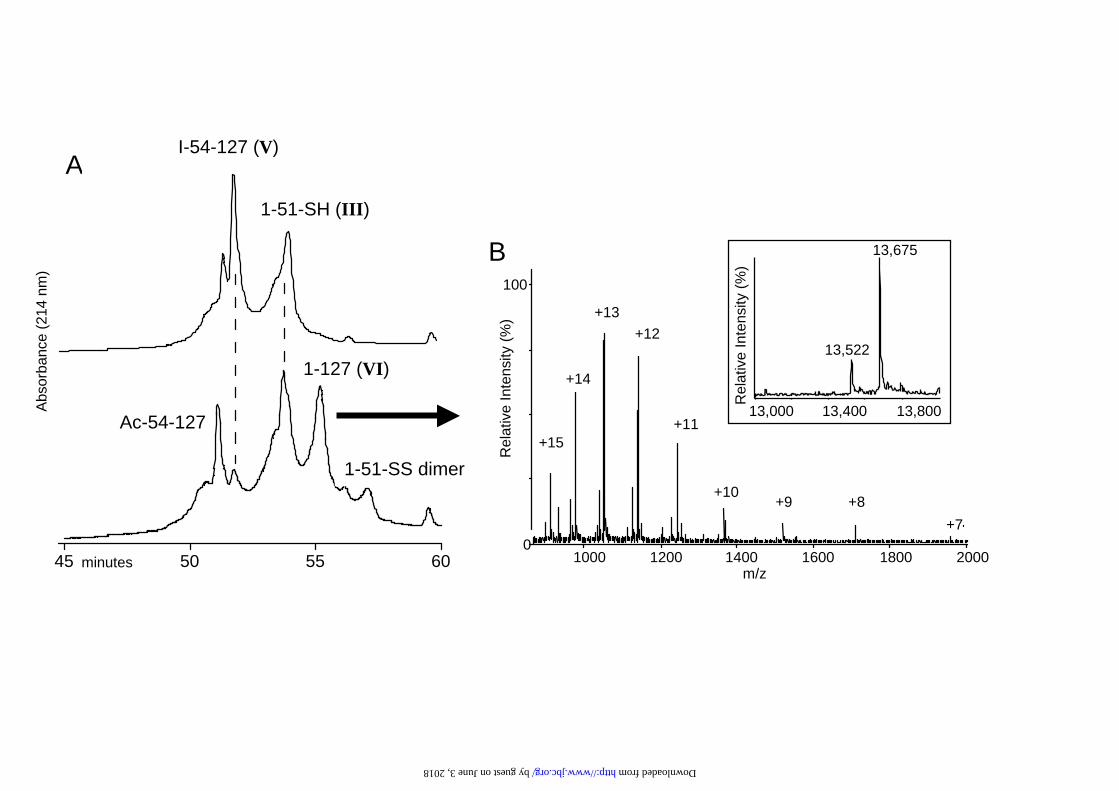
Fig. 4. A) HPLC traces showing the peptide ligation mixture for peptides III and V at
time 0 (above) and 2 h (below). B) The mass spectrum of a sample of the peak for the
ligated peptide VI 1-127. Inset is the mass spectrum reconstruct showing the correct
mass for the ligated synthetic TTR, peptide VI (13,675 Da) as well as the presence of
a deletion product (13,522 Da).
by guest on June 3, 2018http://w
ww
.jbc.org/D
ownloaded from
28
Fig. 5. Gel filtration profiles of the solution used for folding the ligated synthetic
transthyretin VI. Pharmacia Superdex 75 10/30 using 0.075 M NH4HCO3 pH 8.3 with
10% CH3CN and a flow rate of 0.3 mL/min. The protein was detected at 214 nm. The
top trace shows the protein VI (3.65 nM) after 18 h at room temperature in 0.375 M
NH4HCO3 with 0.5 equivalents of T4 (excess T4 precipitates from solution). The
bottom trace shows the isolated synthetic transthyretin VI tetrameric complex after
reequilibration at 37oC for 48 h. Monomeric and dimeric species, as ascertained by
their retention time, are indicated.
Fig. 6. Western analysis for the presence of transthyretin in human serum and in
solution containing synthetic transthyretin. Samples were separated in an SDS-
polyacrylamide gel, then proteins were transferred onto a nitrocellulose membrane
and probed with antiserum against a mixture of transthyretins purified from human,
wallaby and chicken sera (raised in a rabbit), and the secondary antibody was anti-
rabbit Ig (raised in sheep). Detection was using enhanced chemiluminescence (see
Materials and Methods for details). Lane 1: 2 µl human serum; lane 2: 25 µl solution
containing 0.5 µg synthetic transthyretin; lane 3: 50 µl solution containing 1.0 µg
synthetic transthyretin. Molecular weight markers (x 10-3) were “Mark 12” from
Novex: beta galactoside: 116.3; phosphorylase b: 97.4; serum albumin: 66.3; glutamic
dehydrogenase: 55.4; lactate dehydrogenase: 36.5; carbonic anhydrase: 31; trypsin
inhibitor: 21.5; lysozyme: 14.4; aprotinin: 6; insulin B chain: 3.5; and insulin A chain:
2. The positions of the origin, migration of transthyretin dimer, transthyretin monomer
and the electrophoretic front are indicated.
by guest on June 3, 2018http://w
ww
.jbc.org/D
ownloaded from

29
Fig. 7. Analysis of thyroxine binding to proteins in human serum and to synthetic
transthyretin. Aliquots of human serum (10µl) and of synthetic TTR solution (80 µl
containing 1.6 µg) were incubated with 125I-thyroxine prior to separation in a non-
denaturing polyacrylamide gel, pH 8.6. The gel was dried then exposed to
autoradiographic film for 7 days (see Materials and Methods). Lane 1: 5 µl human
serum; lanes 2 and 3: 80 µl solution containing 1.6 µg synthetic transthyretin with 1.1
fmol and 4.4 fmol 125I-thyroxine respectively; lane 4: 5 µl human serum. The
positions of migration of human thyroxine-binding globulin (TBG), albumin and
transthyretin (TTR) are indicated. The positions of the origin and front of the gel are
also indicated.
by guest on June 3, 2018http://w
ww
.jbc.org/D
ownloaded from

ADG
H
BC
E
F
51-54
102-99
10 20 30 40GPTGTGESKC PLMVKVLDAV RGSPAINVAV HVFRKAADDT
50 60 70 80WEPFASGKTS ESGELHGLTT EEEFVEGIYK VEIDTKSYWK
90 100 110 120ALGISPFHEH AEVVFTANDSGPRRYTIAAL LSPYSYSTTA
127VVTNPKE
Bromoacetyl-102-127 (I)Br-Ac -PRRYTIAALLSPYSYSTTAVVTNPKE-OH
Chloroacetyl-54-99-NH-CH2-CH2-SH (II )Cl-Ac-ELHGLTTEEEFVEGIYKVEIDTKSYWKAL-GISPFHEHAEVVFTAND -NH-CH2-CH2-SH
1-51-NH-CH2-CH2-SH (III )H-GPTGTGESKAPLMVKVLDAVRGSPAINVAVH-VFRKAADDTWEPFASGKTSE- NH-CH2-CH2-SH
B
C
A
T4
102- 99
51- 54
by guest on June 3, 2018http://w
ww
.jbc.org/D
ownloaded from

-OHBr-CH 2-CO- 102 - 127
Peptide I
-HN-CH2-CH2-SHCl-CH2-CO- 54 - 99
Peptide II
6M urea0.1 M NaHCO3 pH8.324 hr r.t.
-HN-CH2-CH2-S-CH2-CO-Cl-CH2-CO- 54 - 99 -OH102 - 127
Peptide IV
6 M urea0.2 M NaHCO3 pH8.35 hr r.t.
-HN-CH2-CH2-SHNH2- 1 - 51
Peptide III
-HN-CH2-CH2-S-CH2-CO-NH2- 1 - 51 -HN-CH2-CH2-S-CH2-CO-54 - 99 -OH102 - 127
Peptide VI = sTTR
8 M urea0.1 M NaOAc pH 7.5KI 30 min r.t.
-HN-CH2-CH2-S-CH2-CO-I-CH2-CO- 54 - 99 -OH102 - 127
Peptide V
by guest on June 3, 2018 http://www.jbc.org/ Downloaded from
35 50 65
Br 101-127
Cl-53-99-SH
Cl-53-127 lig
Br-101-127
Ac-101-127
1000 1250 1500 1750 2000m/z
100
0
Re
lativ
eIn
ten
sity
(%)
+9
+8+7
+6
+5
+4**
90008200 8600
8357
A
B
35 50 65
Br 101-127
Cl-53-99-SH
Cl-53-127 lig
Br-101-127
Ac-101-127
A
B
A
Br-102-127 (I )
Cl-54-99-SH (II)
Cl-54-127 (IV )Br-102-127 (I )
Ac-102-127
90008200 8600
8357
Rel
ativ
ein
tens
ity(%
)
minutes
Abs
orba
nce
(214
nm)
by guest on June 3, 2018 http://www.jbc.org/ Downloaded from
45 50 55 60
TTR 1-127
Iodo-53-127
1-51-SH
1-51-SS dimer
Ac-53-127
1000 1200 1400 1600 1800 2000m/z
100
0
Rel
ativ
eIn
tens
ity(%
)+9 +8
+7
+10
+11
+12
+13
+14
+1513,000 13,400 13,800
13,523
13,680
A
B
I-54-127 (V)
1-51-SH (III)
1-127 (VI )
1-51-SS dimer
Ac-54-127
B
A
Rel
ativ
eIn
tens
ity(%
)
13,000 13,400 13,800
13,522
13,675
Abs
orba
nce
(214
nm)
minutes
by guest on June 3, 2018 http://www.jbc.org/ Downloaded from

0 25 50 75
Tetrameric TTR
Dimer TTR
Monomer TTR
minutes
Abs
orba
nce
(214
nm)
by guest on June 3, 2018http://w
ww
.jbc.org/D
ownloaded from


J. CraikJackie A. Wilce, Stephen G. Love, Samantha J. Richardson, Paul F. Alewood and David
regioselective chemical ligationSynthesis of an analog of the thyroid hormone binding protein transthyretin via
published online May 14, 2001J. Biol. Chem.
10.1074/jbc.M101228200Access the most updated version of this article at doi:
Alerts:
When a correction for this article is posted•
When this article is cited•
to choose from all of JBC's e-mail alertsClick here
by guest on June 3, 2018http://w
ww
.jbc.org/D
ownloaded from