tetramer stability and functional regulation of tumor suppressor protein p53
DESCRIPTION
Tetramer Stability and Functional Regulation of Tumor Suppressor Protein p53TRANSCRIPT


Springer Theses
Recognizing Outstanding Ph.D. Research
For further volumes:http://www.springer.com/series/8790

Aims and Scope
The series ‘‘Springer Theses’’ brings together a selection of the very best Ph.D.theses from around the world and across the physical sciences. Nominated andendorsed by two recognized specialists, each published volume has been selectedfor its scientific excellence and the high impact of its contents for the pertinentfield of research. For greater accessibility to non-specialists, the published versionsinclude an extended introduction, as well as a foreword by the student’s supervisorexplaining the special relevance of the work for the field. As a whole, the serieswill provide a valuable resource both for newcomers to the research fieldsdescribed, and for other scientists seeking detailed background information onspecial questions. Finally, it provides an accredited documentation of the valuablecontributions made by today’s younger generation of scientists.
Theses are accepted into the series by invited nomination onlyand must fulfill all of the following criteria
• They must be written in good English.• The topic should fall within the confines of Chemistry, Physics, Earth Sciences
and related interdisciplinary fields such as Materials, Nanoscience, ChemicalEngineering, Complex Systems and Biophysics.
• The work reported in the thesis must represent a significant scientific advance.• If the thesis includes previously published material, permission to reproduce this
must be gained from the respective copyright holder.• They must have been examined and passed during the 12 months prior to
nomination.• Each thesis should include a foreword by the supervisor outlining the signifi-
cance of its content.• The theses should have a clearly defined structure including an introduction
accessible to scientists not expert in that particular field.

Rui Kamada
Tetramer Stabilityand Functional Regulationof Tumor SuppressorProtein p53
Doctoral Thesis accepted byHokkaido University, Japan
123

AuthorDr. Rui KamadaNational Institute of Child Health
& Human DevelopmentNational Institutes of HealthBuilding 6, Room 2A116 Center DriveBethesda, MD 20892USA
SupervisorProf. Kazuyasu SakaguchiDepartment of ChemistryFaculty of ScienceHokkaido UniversityHokkaidoJapan
ISSN 2190-5053 ISSN 2190-5061 (electronic)ISBN 978-4-431-54134-9 ISBN 978-4-431-54135-6 (eBook)DOI 10.1007/978-4-431-54135-6Springer Tokyo Heidelberg New York Dordrecht London
Library of Congress Control Number: 2012939122
� Springer Japan 2012This work is subject to copyright. All rights are reserved by the Publisher, whether the whole or part ofthe material is concerned, specifically the rights of translation, reprinting, reuse of illustrations,recitation, broadcasting, reproduction on microfilms or in any other physical way, and transmission orinformation storage and retrieval, electronic adaptation, computer software, or by similar or dissimilarmethodology now known or hereafter developed. Exempted from this legal reservation are briefexcerpts in connection with reviews or scholarly analysis or material supplied specifically for thepurpose of being entered and executed on a computer system, for exclusive use by the purchaser of thework. Duplication of this publication or parts thereof is permitted only under the provisions ofthe Copyright Law of the Publisher’s location, in its current version, and permission for use must alwaysbe obtained from Springer. Permissions for use may be obtained through RightsLink at the CopyrightClearance Center. Violations are liable to prosecution under the respective Copyright Law.The use of general descriptive names, registered names, trademarks, service marks, etc. in thispublication does not imply, even in the absence of a specific statement, that such names are exemptfrom the relevant protective laws and regulations and therefore free for general use.While the advice and information in this book are believed to be true and accurate at the date ofpublication, neither the authors nor the editors nor the publisher can accept any legal responsibility forany errors or omissions that may be made. The publisher makes no warranty, express or implied, withrespect to the material contained herein.
Printed on acid-free paper
Springer is part of Springer Science+Business Media (www.springer.com)

Parts of this thesis have been published in the following journal articles:
1. Kamada, R., Nomura, T., Anderson, C. W., Sakaguchi, K. (2011) Cancer-associated p53 tetramerization domain mutants: quantitative analysis reveals alow threshold for tumor suppressor inactivation, J. Biol. Chem. 286, 252–258.
2. Kamada, R., Yoshino, W., Nomura, T., Chuman, Y., Imagawa, T., Suzuki, T.,Sakaguchi, K. (2010) Enhancement of transcriptional activity of mutant p53tumor suppressor protein through stabilization of tetramer formation bycalix[6]arene derivatives. Bioorg. Med. Chem. Lett. 20, 4412–4415.
v

Supervisor’s Foreword
To understand the regulatory mechanism of a biological system, it is important toclarify the equilibrium of protein–protein interactions and the threshold for thesignal output of cells in response to input cellular stimuli and stresses. Quantitativeanalysis is essential for truly understanding the equilibrium and threshold. In thisthesis, we propose a novel mechanism of regulation of protein function, from theviewpoint of quantitative analysis of structural stability and protein function.
Using tumor suppressor protein p53 as a model protein, we demonstrated thatthe threshold for loss of tumor suppressor activity, in terms of the disruption ofp53’s tetrameric structure, could be extremely low. Our model revealed that p53could respond quickly to stimuli by activating and regulating its function via smallalterations in its tetrameric status. This is a novel and important discovery in theunderstanding of functional regulation mechanisms of proteins that play essentialroles in biological systems.
Furthermore, we developed two projects concerning the functional control ofp53 and reported a mutant p53 stabilizer and p53 inhibitor. We reported, for thefirst time, enhancement of the in vivo transcriptional activity of the most commonLi-Fraumeni p53 mutant, R337H, by a calixarene derivative that stabilized thetetramer formation. To develop an efficient method of generating induced plu-ripotent stem cells, we demonstrated p53 inhibition via hetero-oligomerizationwith a tetramerization domain-derived peptide.
The results of the quantitative analysis presented in this thesis suggest that p53is inactivated by amplification of destabilization in cells. Moreover, further studiesshould be directed toward the development of p53 stabilizers and inhibitors as newtypes of drugs.
Hokkaido, Japan, February 2012 Prof. Kazuyasu Sakaguchi
vii

Acknowledgments
I would like to express my sincere gratitude to my supervisor, Professor KazuyasuSakaguchi, for his superior guidance, considerable encouragement, and invaluablediscussion.
My heartfelt thanks go to Professor Takanori Suzuki (Laboratory of OrganicChemistry I, Hokkaido University) for his valuable suggestions and guidance onmy papers, especially in organic synthesis. I would also like to thank ProfessorYota Murakami (Laboratory of Bioorganic Chemistry) and Dr. Toshiaki Imagawa,Associate Professor, who gave me helpful and constructive suggestions andcomments throughout this study.
I am very grateful to those who collaborated with me in this work: Dr. Carl W.Anderson (Brookhaven National Laboratory, USA), who provided me withinvaluable comments and discussion; Professor Koichiro Ishimori (Laboratory ofStructural Chemistry, Hokkaido); Professor Keiji Tanino (Laboratory of OrganicChemistry II, Hokkaido); Professor James G. Omichinski (Université de Montréal,Canada); Professor Yuji Kobayashi (Osaka Pharmaceutical University, Osaka);and Dr. Koichiro Fukuda (Laboratory of Organic Chemistry II, Hokkaido).
I wish to express my warm and sincere thanks to Dr. Yoshiro Chuman,Assistant Professor, who gave me helpful comments and encouragement. I alsowish to thank the following people: Mr. Wataru Yoshino, for his essential assis-tance in organic synthesis; Dr. Takao Nomura, for his excellent advice and kindsupport throughout this study; Dr. Shunji Kaya, for his helpful advice and sug-gestions; and all members of the Laboratory of Biological Chemistry. In addition, Iwould like to thank the JSPS, Scholarship and Grant for Inter-laboratory ResearchProgram, ‘‘Initiatives for Attractive Education in Graduate Schools’’, and theHokkaido University Global COE Program for making my Ph.D. study possiblewith their financial support.
Finally, I extend my indebtedness to my parents for their support, under-standing, and encouragement throughout my study.
17 February 2012 Rui Kamada
ix

Contents
1 General Introduction . . . . . . . . . . . . . . . . . . . . . . . . . . . . . . . . . . 11.1 Tumor Suppressor Protein p53 . . . . . . . . . . . . . . . . . . . . . . . . 11.2 Primary Structure of p53 Protein . . . . . . . . . . . . . . . . . . . . . . . 11.3 Posttranslational Modifications of p53 . . . . . . . . . . . . . . . . . . . 21.4 3D Structure of p53 . . . . . . . . . . . . . . . . . . . . . . . . . . . . . . . . 41.5 Tetramerization and Function of p53 . . . . . . . . . . . . . . . . . . . . 51.6 Biophysical Methods . . . . . . . . . . . . . . . . . . . . . . . . . . . . . . . 7
1.6.1 Circular Dichroism (CD) . . . . . . . . . . . . . . . . . . . . . . . 71.6.2 The p53 Tetramerization Domain Peptides . . . . . . . . . . . 8
1.7 Aims of this Study. . . . . . . . . . . . . . . . . . . . . . . . . . . . . . . . . 9References . . . . . . . . . . . . . . . . . . . . . . . . . . . . . . . . . . . . . . . . . . 9
2 Quantitative Analysis for p53 Tetramerization Domain MutantsReveals a Low Threshold for Tumor Suppressor Inactivation . . . . 132.1 Introduction . . . . . . . . . . . . . . . . . . . . . . . . . . . . . . . . . . . . . 132.2 Experimental Procedures . . . . . . . . . . . . . . . . . . . . . . . . . . . . 15
2.2.1 Peptide Synthesis and Purification. . . . . . . . . . . . . . . . . 152.2.2 Gel Filtration Chromatography . . . . . . . . . . . . . . . . . . . 162.2.3 Thermal Denaturation by Circular Dichroism
Spectroscopy . . . . . . . . . . . . . . . . . . . . . . . . . . . . . . . 162.2.4 Structural Modeling of p53TD Mutants . . . . . . . . . . . . . 16
2.3 Results . . . . . . . . . . . . . . . . . . . . . . . . . . . . . . . . . . . . . . . . . 172.3.1 Oligomerization State of Mutant p53
Tetramerization Domains . . . . . . . . . . . . . . . . . . . . . . . 172.3.2 Secondary Structure of Mutant p53
Tetramerization Domains . . . . . . . . . . . . . . . . . . . . . . . 192.3.3 Thermal Stability of the Mutant p53
Tetramerization Domains . . . . . . . . . . . . . . . . . . . . . . . 192.3.4 Effects of Mutation on the Tetrameric Structure . . . . . . . 192.3.5 Modeling of Mutant p53TD Peptides. . . . . . . . . . . . . . . 30
xi
2.3.6 Correlation Between Stability of p53TD Peptidesand that of the Full-Length p53 Proteinand the Transcriptional Activity . . . . . . . . . . . . . . . . . . 30
2.4 Discussion . . . . . . . . . . . . . . . . . . . . . . . . . . . . . . . . . . . . . . 31References . . . . . . . . . . . . . . . . . . . . . . . . . . . . . . . . . . . . . . . . . . 41
3 Stabilization of Mutant Tetrameric Structuresby Calixarene Derivatives. . . . . . . . . . . . . . . . . . . . . . . . . . . . . . . 453.1 Introduction . . . . . . . . . . . . . . . . . . . . . . . . . . . . . . . . . . . . . 453.2 Experimental Procedures . . . . . . . . . . . . . . . . . . . . . . . . . . . . 47
3.2.1 Synthesis of the Calixarene Derivatives . . . . . . . . . . . . . 473.2.2 Thermodynamic Stability of the Peptide by CD . . . . . . . 483.2.3 Transcriptional Activity of p53-R337H in Cell . . . . . . . . 48
3.3 Results . . . . . . . . . . . . . . . . . . . . . . . . . . . . . . . . . . . . . . . . . 493.3.1 Design of Calixarene Derivatives . . . . . . . . . . . . . . . . . 493.3.2 Stabilization of Mutant Peptides
by Calixarene Derivatives . . . . . . . . . . . . . . . . . . . . . . 503.3.3 Transcriptional Activity of p53 Protein in Cell . . . . . . . . 52
3.4 Discussion . . . . . . . . . . . . . . . . . . . . . . . . . . . . . . . . . . . . . . 54References . . . . . . . . . . . . . . . . . . . . . . . . . . . . . . . . . . . . . . . . . . 58
4 Inhibition of the Transcriptional Activity of p53 ThroughHetero-Oligomerization . . . . . . . . . . . . . . . . . . . . . . . . . . . . . . . . 614.1 Introduction . . . . . . . . . . . . . . . . . . . . . . . . . . . . . . . . . . . . . 614.2 Experimental Procedures . . . . . . . . . . . . . . . . . . . . . . . . . . . . 62
4.2.1 Peptide Synthesis . . . . . . . . . . . . . . . . . . . . . . . . . . . . 624.2.2 CD Analysis of PTD-p53tet Peptide . . . . . . . . . . . . . . . 634.2.3 Hetero-Oligomerization of PTD-p53tet Peptide. . . . . . . . 634.2.4 Transcriptional Activity of p53 with
PTD-p53tet Peptide . . . . . . . . . . . . . . . . . . . . . . . . . . . 634.3 Results . . . . . . . . . . . . . . . . . . . . . . . . . . . . . . . . . . . . . . . . . 64
4.3.1 CD Spectra of PTD-p53tet Peptides . . . . . . . . . . . . . . . 644.3.2 Hetero-Oligomerization of PTD-p53tet Peptides
with p53(319-358) Peptide . . . . . . . . . . . . . . . . . . . . . . 644.3.3 Cellular Introducing and Localization
of PTD-p53tet Peptides . . . . . . . . . . . . . . . . . . . . . . . . 654.3.4 Transcriptional Activity of Venus-p53 Protein in Cell . . . 65
4.4 Discussion . . . . . . . . . . . . . . . . . . . . . . . . . . . . . . . . . . . . . . 67References . . . . . . . . . . . . . . . . . . . . . . . . . . . . . . . . . . . . . . . . . . 70
5 Conclusion . . . . . . . . . . . . . . . . . . . . . . . . . . . . . . . . . . . . . . . . . . 71
xii Contents

Abbreviations
ADC Adrenocortical carcinomaBD C-terminal basic domainCD Circular dichroismCp Het capacityDAPI 4,6-diamidino-2-phenylindoleDBD DNA binding domainDCM DichloromethaneDIEA N,N-diisopropylethylamineDMF DimethylformamideEGFP Enhanced green fluorescence proteinFBS Fetal bovine serumFmoc 9-Fluorenylmethoxycarbonylfu Fraction of unfoldedHOBt N-HydroxybenzotriazoleHPLC High performance liquid chromatographyHSQC Heteronuclear single quantum coherenceiPS Induced pluripotent stemIPTG Isopropyl b-D-1-thiogalactopyranosideKd Dissociation thrmodynamic constantMALDI Matrix-assisted laser desorption/ionizationTOF-MS Time of flying mass spectrometryMDM2 Murine double minute proteinNHS N-hydroxysulfosuccinimideNLS Nuclear localization signalNMR Nuclear magnetic resonanceOPTI-MEM Reduced serum modified Eagle’s mediump53tet p53(324–358)p53TD p53(319–358)PBS Phosphate buffered salinePDB Protein databankPRD Proline rich domain
xiii

PTD Protein transduction domainRPMI Roswell Park Memorial InstituteSNP Single nucleotide polymorphismTAD Transactivation domainTD Tetramerization domainTFA Trifluoroacetic acidTm Melting temperatureWT Wild-type
xiv Abbreviations

Chapter 1General Introduction
1.1 Tumor Suppressor Protein p53
Tumor suppressor protein p53 plays a central role in maintaining genomic integrity[1–3]. In unstimulated cells p53 is maintained at very low levels but in response toDNA damage or other stress stimuli, such as hypoxia or activation of oncogenes,p53 becomes stabilized and accumulates in the cell [4, 5]. p53 can exert its tumorsuppressor function in different ways (Fig. 1.1). It can function as a transcriptionfactor that binds to the promoters of many target genes, such as p21, mdm2, pumaand bax, thereby elevating or repressing their expression levels to induce cell cyclearrest and apoptosis. Nuclear and cytoplasmic p53 also physically interact withmany other proteins to promote apoptosis and other processes, such as homologousrecombination. In mitochondria, p53 contributes to transcription-independentapoptosis [6]. Moreover, in response to DNA damage, p53 enhances the post-transcriptional maturation of several microRNAs that have growth-suppressivefunctions [7]. Because p53 plays a prominent role in tumor suppression, it is a keytarget protein in cancer therapy. Recently, however, inactivation of p53 has alsoreceived attention because heart failure is associated with up-regulation of p53function [8]. In addition, temporary suppression of p53 has been suggested as anapproach to reduce side effects of cancer treatment and to increase the efficiency ofiPS (induced pluripotent stem) cell generation, which is inhibited by p53–p21pathways [9, 10].
1.2 Primary Structure of p53 Protein
p53 consists of five main domains: the N-terminal transactivation domain, thePro-rich domain, the central DNA binding domain, the tetramerization domain andthe C-terminal basic domain (Fig. 1.2). The N-terminal transactivation domain,
R. Kamada, Tetramer Stability and Functional Regulation of Tumor SuppressorProtein p53, Springer Theses, DOI: 10.1007/978-4-431-54135-6_1,� Springer Japan 2012
1

residues 1–42, is required for transactivation activity and interacts with manyproteins, including the transcription factors TFIID, TFIIH and several TAFs.The transactivation domain also mediates interaction with the histone acetyl-transferases CBP/p300 and PCAF and with the MDM2 E3 ubiquitin ligase. ThePro-rich domain, residues 61–92, is required for interaction with the Sin3 cor-epressor and is important for p53 stability, transactivation and for the induction oftranscription independent apoptosis [11]. The central DNA binding domain candirectly bind to the p53 consensus DNA binding site, which is composed of fourpentanucleotide repeats. Full-length p53 can reversibly form tetramers via thetetramerization domain, residues 326–356. The tetramerization domain regulatesthe oligomeric state of p53. The C-terminal basic domain interacts with a non-specific DNA sequence and negatively regulates the sequence-specific binding ofthe central DNA binding domain [12, 13]. Deletion of the C-terminal basic domainincreases sequence-specific DNA binding [14].
1.3 Posttranslational Modifications of p53
p53 can be modified by as many as 50 individual post-translational modifications,including phosphorylation, acetylation, mono- and di-methylation, glycosylation,ubiquitylation, neddylation, sumoylation and poly-ribosylation (Fig. 1.3).
Fig. 1.1 Tumor suppressor protein p53. p53 becomes activated, stabilized and forms tetramers inresponse to genotoxic stress. Activated p53 suppresses tumorigenesis by inducing cell cycle arrestand apoptosis in different ways. 1. p53 induces apoptosis in a transcription-independent mannerin mitochondria. 2. p53 transactivates or represses gene expression, leading to cell cycle arrestand apoptosis. 3. p53 enhances the maturation of microRNAs in response to DNA damage
2 1 General Introduction

These modifications, which occur mainly in the N- and C-terminal regions, regulatep53 activity and also tetramerization [15–17]. The N-terminal transactivationdomain contains seven serines (Ser6, 9, 15, 20, 33, 37 and 46) and two threonines(Thr18 and 81) that are phosphorylated or dephosphorylated in response to ionizingradiation or UV light [2, 18–23]. p53 is maintained at low levels in normal, unstressedcells mainly through the action of MDM2, a RING finger type E3 ligase that pro-motes the poly-ubiquitylation and proteasomal degradation of p53. The main p53targets of MDM2-mediated ubiquitylation are the six carboxy-terminal lysines(Lys370, 372, 373, 382, and 386). Phosphorylation of p53 Thr18, which requiresprior phosphorylation of Ser15, induces dissociation of MDM2 from p53, resulting inp53 stabilization [24]. Phosphorylation of Ser46 in response to severe DNA damageenhances p53 mediated apoptosis by inducing the expression of p53AIP1, a proteinassociated with mitochondrial membranes that induces the release of cytochrome c toinitiate apoptosis. Phosphorylation at Ser15, Thr18, and Ser20, in the N-terminaltransactivation domain, induces conformational changes and switches the transac-tivation domain to more open conformations, which can interact with transcriptionfactors, leading to enhancement of p53 transcriptional activity [25, 26]. Phosphor-ylation at Ser15 and Ser37 enhances p53-TFIID interaction and blocks MDM2binding [27]. In contrast, phosphorylation of Ser15 alone inhibits p53-TFIIDinteractions.
In the C-terminal basic domain, Ser313, 314, 315, 366, 376, 378 and 392 andThr377 and 387 can be phosphorylated, while Lys320, 372, 373, 371 and 382 can
Fig. 1.2 Schematic structure and amino acid sequence of p53. p53 consists of five maindomains: the N-terminal transactivation domain (TAD; 1–42, green), the Pro-rich domain (PRD;61–92, yellow), the central DNA binding domain (DBD; 101–300, light blue), the tetramerizationdomain (TD; 326–356, magenta), and the C-terminal basic domain (BD; 364–393, dark blue)
1.3 Posttranslational Modifications of p53 3

be acetylated and Lys370, 372 and 382 can be methylated [28]. C-terminalphosphorylation and acetylation also promote p53 tetramerization and binding toPTEN, a dual specificity phosphatase [29–31]. Over-expression of the histonedeacetylases HDAC-1, -2, or -3, or hSir2, which deacetylate p53, result in thetranscriptional inhibition of p53 target genes; therefore, acetylation of p53 isimportant for its transcriptional activation. SMAR1 interacts with Ser15 phos-phorylated p53 and MDM2 and recruits HDAC1 to p53, leading to the deacety-lation of p53 and to decreased DNA binding [32]. Three C-terminal p53 residues,Lys370, Lys372, and Lys382 can be monomethylated and these modifications alsomodulate p53 activity in response to DNA damage. The methylation at Lys372rapidly increases after DNA damage and promotes nuclear localization andincreased stability of the p53 protein. On the other hand, methylation of K370blocks p53 binding to DNA and represses transcriptional activation.
1.4 3D Structure of p53
The N-terminal region (TAD and PRD) and the C-terminal region (BD) areunfolded in their native states. However, when TAD binds to partner proteins, suchas MDM2 and TFIIH, it folds into an amphipathic a-helical structure [33, 34]. TheBD also changes its disordered structure to a folded structure upon binding to otherproteins or nonspecific DNA [35]. In contrast to the unstructured N- and C-ter-minal regions, the central DNA binding domain and the tetramerization domainexist as folded structures. The DNA binding core domain, residues 101–300,
Fig. 1.3 Post-translational modification sites of human p53. Post-translational modificationsoccur mainly in the N- and C-terminal regions to regulate p53 activity. P (yellow),phosphorylation of Ser residue; P (cyan), phosphorylation of Thr residue; Ac, acetylation; Ub,ubiquitination; N, neddylation; M, methylation; S, sumoylation
4 1 General Introduction

consists of a central immunoglobulin-like b-sandwich that provides a scaffold forthe DNA binding surface (Fig. 1.4) [36]. This surface is formed by two largeloops, L2 an L3, that are stabilized by a zinc ion and a loop-sheet helix motif. Thetetramerization domain, residues 326-356, exhibits dihedral symmetry and isdescribed as a dimer of dimers (Fig. 1.5) [37–43]. Each monomer is comprised ofa b-strand, a tight turn and an a-helix. Two monomers form a dimer via anti-parallel interaction of the b-strands and two dimers interact via a four-helix bundleto form a tetramer. The folding-unfolding process of the p53 tetramerizationdomain is in equilibrium between unfolded monomers and the folded tetramer[44]. Recently, the quaternary structure of full-length p53 has been revealedthrough electron microscopy, biophysical methods and computational modeling[45]. The model for p53 in solution, in the absence of DNA, is an elongated cross-shaped tetramer with extended N- and C-termini. When p53 binds to DNA, itwraps around the DNA helix with all four N-termini pointing away from the faceof the core domain DNA complex.
1.5 Tetramerization and Function of p53
p53 tetramer formation is essential for its function. p53 binds DNA as a tetramer andtetramerization mediated by the C-terminus tetramerization domain allostericallyregulates the DNA binding activity of p53 [15]. The DNA sequence of the p53response element contains four pentanucleotide repeats [46]. Because each p53 DNA
Fig. 1.4 Structure of the tetrameric p53DBD/DNA complex (pdb; 3EXJ). The four subunits ofp53DBD are represented by green, yellow, magenta, and cyan. The p53DBD consists of a centralimmunoglobulin-like b-sandwich
1.4 3D Structure of p53 5

binding domain recognizes one repeat, a polyvalent p53 tetramer has a 100-foldhigher affinity for the p53 DNA site than a p53 monomer [47]. Thus, p53tetramerization is required for sequence-specific DNA binding. In addition, protein–protein interactions and some post-translational modifications, such as phosphory-lation, acetylation, and ubiquitination, require tetramerization of p53 (Fig. 1.6).Many proteins bind directly to the tetramerization domain or have their interactionwith p53 influenced by its oligomeric status. Casein kinase 2, Ca2+-dependent proteinkinase C and adenovirus E4orf6 directly bind to the tetramerization domain [48].Other proteins, such as MDM2, HPV-16 E2, c-abl, and TBP, only interact withtetrameric p53. Several post-translational modifications are dependent on the
Fig. 1.5 Structure of the tetramerization domain (pdb; 3SAK). Cartoon model of p53TD (pdb;3SAK) prepared with MolFeat version 4.0 (FiatLux Corp.). The left and right tetramers were obtainedby rotating the structure in the center by 90� around the horizontal and vertical axes, respectively
Fig. 1.6 Correlation between tetramerization and function of p53. The post-translationalmodifications and protein–protein interactions are dependent on the oligomeric state of p53.Conversely, post-translational modifications, such as phosphorylation and acetylation, andprotein–protein interactions regulate tetramerization of p53
6 1 General Introduction

quaternary structure of p53 [49]. The phosphorylation by CHK1 or casein kinase 2and the ubiquitination of p53 require its tetramerization. The binding of S100 familyproteins depends on the oligomeric status of p53 and controls the balance betweenmonomer and tetramer [50]. The S100B protein interacts with the C-terminal regionof p53 (residues 319–393) and protects p53 from thermal denaturation [51].Moreover, because the nuclear export signal is located in the tetramerization domain,localization of p53 is dependent on its oligomeric status [52]. Post-translationalmodification affects tetramerization. The phosphorylation of Ser392 enhancestetramer formation [30, 53, 54]. Thus, tetramerization, post-translational modifica-tion, protein–protein interaction, and protein concentration regulate one another, andthey control p53 tumor suppressor activity (Fig. 1.7).
1.6 Biophysical Methods
1.6.1 Circular Dichroism (CD)
CD spectroscopy measures differences in absorbance of right and left circularlypolarized light. When the circularly polarized light passes through an opticallyactive sample, the right-handed and the left-handed components interact in adifferent manner with the optically active molecules. Thus, the right-handed and
Fig. 1.7 Correlation between tetramerization and function of p53. Tetramer formation of p53affects p53 activities, such as post-translational modifications, protein–protein interactions andDNA binding affinity. Conversely, post-translational modification and protein concentrationincrease or decrease p53 tetramer formation
1.5 Tetramerization and Function of p53 7
the left-handed components are differentially absorbed. After passing through thesample, the phase and amplitude of each component are changed. When they arecombined, the result is an elliptically distorted polarized light beam.
For proteins, the optically active absorption bands mainly arise from the amidesof the protein backbone that affect the far-UV or near-UV regions of the spectrum(n-p*, p-p* transitions). These transitions of the amide bond are split into multipletransitions, intrinsically asymmetric or periodical arrangements of the residues. Inthe protein or peptide structure, each secondary structure motif has characteristicCD bands. An a-helix displays two negative bands (208 and 222 nm) and onepositive band (190 nm). A b-sheet shows a less intense spectrum with two neg-ative bands (217 and 180 nm) and one positive band (195 nm). For a disorderedrandom coil structure, there is a negative band around 200 nm. Thermodynamicand kinetic information of the protein and peptide can be obtained by measuringCD changes as a function of temperature, concentration of denaturant or time.
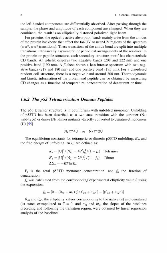
1.6.2 The p53 Tetramerization Domain Peptides
The p53 tetramer structure is in equilibrium with unfolded monomer. Unfoldingof p53TD has been described as a two-state transition with the tetramer (N4;wild-type) or dimer (N2; dimer mutants) directly converted to denatured monomers(U) [55].
N4� 4U or N2� 2U
The equilibrium constants for tetrameric or dimeric p53TD unfolding, Ku, andthe free energy of unfolding, DGu, are defined as:
Ku ¼ U½ �4= N4½ � ¼ 4P3t f 4
u = 1� fuð Þ Tetramer
Ku ¼ U½ �2= N2½ � ¼ 2Ptf2u = 1� fuð Þ Dimer
DGu ¼ �RT ln Ku
Pt is the total p53TD monomer concentration, and fu the fraction ofdenaturation.
fu was calculated from the corresponding experimental ellipticity value h usingthe expression:
fu ¼ h� hn0 þ mnTð Þ½ �= hu0 þ muT½ � � hn0 þ mnTð Þ½ �
hn0 and hu0, the ellipticity values corresponding to the native (n) and denatured(u) states extrapolated to T = 0, and mn and mu, the slopes of the baselinespreceding and following the transition region, were obtained by linear regressionanalysis of the baselines.
8 1 General Introduction

To calculate Tm and DHuTm, DGu values obtained for T values within the tran-
sition zone of the denaturation curve using the following equations were used to fitthe appropriate expanded form of the Gibbs equation:
DGu ¼DHTmu 1� T=Tmð Þ þ DCp T � Tm � T ln T=Tmð Þ½ � � RT ln P3
t =2� �
Tetramer
DGu ¼DHTmu 1� T=Tmð Þ þ DCp T � Tm � T ln T=Tmð Þ½ � � RT ln Pt Dimer
where DCp = 1.7 kcal/(K.mol of tetramer) or 0.85 kcal/(K.mol of dimer), whichcorrespond to 425 cal/(K.mol of monomer) [44].
1.7 Aims of this Study
p53 is a sequence-specific transcription factor that suppresses tumor development.In response to genotoxic stress, p53 induces cell cycle arrest and apoptosis bytransactivating expression of downstream target genes. Furthermore, p53 alsoregulates microRNA processing and apoptosis in mitochondria. Tetramer forma-tion of p53 is essential for its activity.
The aims of this study were to reveal the dysfunction threshold of p53 due tothe destabilization of its tetrameric structure. The effects of tumor-associatedmutations in the tetramerization domain on the tetrameric structure were analyzed.At endogenous p53 levels the mutations significantly decreased the fraction oftetramer, leading to dysfunction of p53 transcriptional activity. Based on thisstructural analysis, functional control of p53 via tetramer formation was alsoperformed; (1) the tumor-associated mutants were rescued by stabilization of thetetrameric structure using calixarene derivatives and (2) transcriptional activity ofp53 was inhibited by hetero-oligomerization. p53 plays important roles in cellcycle arrest, apoptosis, DNA repair, and microRNA maturation in response togenotoxic stress. Loss of p53 function, either directly through mutation or indi-rectly through several mechanisms, is an important step in tumorigenesis. Thus,the development of methods that enhance reduced p53 activity is important tocancer therapy. Recently, however, inactivation of p53 has also received attentionbecause heart failure is associated with up-regulation of p53 function. In addition,temporary suppression of p53 has been suggested as an approach to reduce sideeffects of cancer treatment and to increase the efficiency of iPS cell generation,which is inhibited by p53–p21 pathways.
References
1. Meek DW (2009) Tumour suppression by p53: a role for the DNA damage response? NatRev Cancer 9:714–723
2. Meek DW, Anderson CW (2009) Posttranslational modification of p53: cooperativeintegrators of function. Cold Spring Harb Perspect Biol 1:a000950
1.6 Biophysical Methods 9

3. Rodier F, Campisi J, Bhaumik D (2007) Two faces of p53: aging and tumor suppression.Nucleic Acids Res 35:7475–7484
4. Haupt Y, Maya R, Kazaz A, Oren M (1997) Mdm2 promotes the rapid degradation of p53.Nature 387:296–299
5. Picksley SM, Lane DP (1993) The p53-mdm2 autoregulatory feedback loop: a paradigm forthe regulation of growth control by p53? BioEssays 15:689–690
6. Speidel D (2010) Transcription-independent p53 apoptosis: an alternative route to death.Trends Cell Biol 20:14–24
7. Suzuki HI, Yamagata K, Sugimoto K, Iwamoto T, Kato S, Miyazono K (2009) Modulation ofmicroRNA processing by p53. Nature 460:529–533
8. Sano M, Minamino T, Toko H, Miyauchi H, Orimo M, Qin Y, Akazawa H, Tateno K,Kayama Y, Harada M, Shimizu I, Asahara T, Hamada H, Tomita S, Molkentin JD, Zou Y,Komuro I (2007) p53-induced inhibition of Hif-1 causes cardiac dysfunction during pressureoverload. Nature 446:444–448
9. Komarov PG, Komarova EA, Kondratov RV, Christov-Tselkov K, Coon JS, Chernov MV,Gudkov AV (1999) A chemical inhibitor of p53 that protects mice from the side effects ofcancer therapy. Science 285:1733–1737
10. Hong H, Takahashi K, Ichisaka T, Aoi T, Kanagawa O, Nakagawa M, Okita K, Yamanaka S(2009) Suppression of induced pluripotent stem cell generation by the p53-p21 pathway.Nature 460:1132–1135
11. Zhang Y, Xiong Y (2001) A p53 amino-terminal nuclear export signal inhibited by DNAdamage-induced phosphorylation. Science 292:1910–1915
12. Anderson ME, Woelker B, Reed M, Wang P, Tegtmeyer P (1997) Reciprocal interferencebetween the sequence-specific core and nonspecific C-terminal DNA binding domains ofp53: implications for regulation. Mol Cell Biol 17:6255–6264
13. Mazur SJ, Sakaguchi K, Appella E, Wang XW, Harris CC, Bohr VA (1999) Preferentialbinding of tumor suppressor p53 to positively or negatively supercoiled DNA involves the C-terminal domain. J Mol Biol 292:241–249
14. Hupp TR, Meek DW, Midgley CA, Lane DP (1992) Regulation of the specific DNA bindingfunction of p53. Cell 71:875–886
15. Balagurumoorthy P, Sakamoto H, Lewis MS, Zambrano N, Clore GM, Gronenborn AM,Appella E, Harrington RE (1995) Four p53 DNA-binding domain peptides bind natural p53-response elements and bend the DNA. Proc Natl Acad Sci U S A 92:8591–8595
16. Halazonetis TD, Kandil AN (1993) Conformational shifts propagate from the oligomerizationdomain of p53 to its tetrameric DNA binding domain and restore DNA binding to select p53mutants. EMBO J 12:5057–5064
17. Nagaich AK, Zhurkin VB, Durell SR, Jernigan RL, Appella E, Harrington RE (1999) p53-induced DNA bending and twisting: p53 tetramer binds on the outer side of a DNA loop andincreases DNA twisting. Proc Natl Acad Sci U S A 96:1875–1880
18. Anderson CW, Appella E, Sakaguchi K (1998) Posttranslational modifications involved inthe DNA damage response. J Protein Chem 17:527
19. Bulavin DV, Saito S, Hollander MC, Sakaguchi K, Anderson CW, Appella E, Fornace AJJ(1999) Phosphorylation of human p53 by p38 kinase coordinates N-terminal phosphorylationand apoptosis in response to UV radiation. EMBO J 18:6845–6854
20. Canman CE, Lim DS, Cimprich KA, Taya Y, Tamai K, Sakaguchi K, Appella E, Kastan MB,Siliciano JD (1998) Activation of the ATM kinase by ionizing radiation and phosphorylationof p53. Science 281:1677–1679
21. Higashimoto Y, Saito S, Tong XH, Hong A, Sakaguchi K, Appella E, Anderson CW (2000)Human p53 is phosphorylated on serines 6 and 9 in response to DNA damage-inducingagents. J Biol Chem 275:23199–23203
22. Lees-Miller SP, Sakaguchi K, Ullrich SJ, Appella E, Anderson CW (1992) Human DNA-activated protein kinase phosphorylates serines 15 and 37 in the amino-terminaltransactivation domain of human p53. Mol Cell Biol 12:5041–5049
10 1 General Introduction

23. Siliciano JD, Canman CE, Taya Y, Sakaguchi K, Appella E, Kastan MB (1997) DNA damageinduces phosphorylation of the amino terminus of p53. Genes Dev 11:3471–3481
24. Sakaguchi K, Saito S, Higashimoto Y, Roy S, Anderson CW, Appella E (2000) Damage-mediated phosphorylation of human p53 threonine 18 through a cascade mediated by a casein1-like kinase. Effect on Mdm2 binding. J Biol Chem 275:9278–9283
25. Kar S, Sakaguchi K, Shimohigashi Y, Samaddar S, Banerjee R, Basu G, Swaminathan V,Kundu TK, Roy S (2002) Effect of phosphorylation on the structure and fold oftransactivation domain of p53. J Biol Chem 277:15579–15585
26. Polley S, Guha S, Roy NS, Kar S, Sakaguchi K, Chuman Y, Swaminathan V, Kundu T,Roy S (2008) Differential recognition of phosphorylated transactivation domains of p53 bydifferent p300 domains. J Mol Biol 376:8–12
27. Pise-Masison CA, Radonovich M, Sakaguchi K, Appella E, Brady JN (1998)Phosphorylation of p53: a novel pathway for p53 inactivation in human T-celllymphotropic virus type 1-transformed cells. J Virol 72:6348–6355
28. Anderson CW, Appella E (2011) Signaling to the p53 tumor suppressor through pathwaysactivated by genotoxic and nongenotoxic stress. In: Bradshaw RA, Dennis EA (eds)Regulation in Organella and Cell Compartment Signaling, Chap 264, Elsevier BV, CA,pp 235–254
29. Sakaguchi K, Herrera JE, Saito S, Miki T, Bustin M, Vassilev A, Anderson CW, Appella E(1998) DNA damage activates p53 through a phosphorylation-acetylation cascade. GenesDev 12:2831–2841
30. Sakaguchi K, Sakamoto H, Lewis MS, Anderson CW, Erickson JW, Appella E, Xie D (1997)Phosphorylation of serine 392 stabilizes the tetramer formation of tumor suppressor proteinp53. Biochemistry 36:10117–10124
31. Li AG, Piluso LG, Cai X, Wei G, Sellers WR, Liu X (2006) Mechanistic insights intomaintenance of high p53 acetylation by PTEN. Mol Cell 23:575–587
32. Pavithra L, Mukherjee S, Sreenath K, Kar S, Sakaguchi K, Roy S, Chattopadhyay S (2009)SMAR1 forms a ternary complex with p53-MDM2 and negatively regulates p53-mediatedtranscription. J Mol Biol 388:691–702
33. Kussie PH, Gorina S, Marechal V, Elenbaas B, Moreau J, Levine AJ, Pavletich NP (1996)Structure of the MDM2 oncoprotein bound to the p53 tumor suppressor transactivationdomain. Science 274:948–953
34. Di Lello P, Jenkins LM, Jones TN, Nguyen BD, Hara T, Yamaguchi H, Dikeakos JD,Appella E, Legault P, Omichinski JG (2006) Structure of the Tfb1/p53 complex: Insights intothe interaction between the p62/Tfb1 subunit of TFIIH and the activation domain of p53. MolCell 22:731–740
35. Rustandi RR, Baldisseri DM, Weber DJ (2000) Structure of the negative regulatory domainof p53 bound to S100B(betabeta). Nat Struct Biol 7:570–574
36. Joerger AC, Fersht AR (2008) Structural biology of the tumor suppressor p53. Annu RevBiochem 77:557–582
37. Sakamoto H, Lewis MS, Kodama H, Appella E, Sakaguchi K (1994) Specific sequences fromthe carboxyl terminus of human p53 gene product form anti-parallel tetramers in solution.Proc Natl Acad Sci U S A 91:8974–8978
38. Clore GM, Omichinski JG, Sakaguchi K, Zambrano N, Sakamoto H, Appella E, GronenbornAM (1994) High-resolution structure of the oligomerization domain of p53 bymultidimensional NMR. Science 265:386–391
39. Clubb RT, Omichinski JG, Sakaguchi K, Appella E, Gronenborn AM, Clore GM (1995)Backbone dynamics of the oligomerization domain of p53 determined from 15 N NMRrelaxation measurements. Protein Sci 4:855–862
40. Miller M, Lubkowski J, Rao JKM, Danishefsky AT, Omichinski JG, Sakaguchi K, SakamotoH, Appella E, Gronenborn AM, Clore GM (1996) The oligomerization domain of p53: crystalstructure of the trigonal form. FEBS Lett 399:166–170
References 11

41. Clore GM, Ernst J, Clubb R, Omichinski JG, Kennedy WMP, Sakaguchi K, Appella E,Gronenborn AM (1995) Refined solution structure of the oligomerization domain of thetumour suppressor p53. Nat Struct Biol 2:321–333
42. Clore GM, Omichinski JG, Sakaguchi K, Zambrano N, Sakamoto H, Appella E, GronenbornAM (1995) Interhelical angles in the solution structure of the oligomerization domain of p53:correction. Science 267:1515–1516
43. Jeffrey PD, Gorina S, Pavletich NP (1995) Crystal structure of the tetramerization domain ofthe p53 tumor suppressor at 1.7 angstroms. Science 267:1498–1502
44. Johnson CR, Morin PE, Arrowsmith CH, Freire E (1995) Thermodynamic analysis of thestructural stability of the tetrameric oligomerization domain of p53 tumor suppressor.Biochemistry 34:5309–5316
45. Tidow H, Melero R, Mylonas E, Freund S, Grossmann JG, Carazo JM, Svergun DI, Valle M,Fersht AR (2007) Quaternary structures of tumor suppressor p53 and a specific p53–DNAcomplex. Proc Natl Acad Sci U S A 104:12324–12329
46. Malecka KA, Ho WC, Marmorstein R (2009) Crystal structure of a p53 core tetramer boundto DNA. Oncogene 28:325–333
47. McLure KG, Lee PW (1998) How p53 binds DNA as a tetramer. EMBO J 17:3342–335048. Chene P (2001) The role of tetramerization in p53 function. Oncogene 20:2611–261749. Ullrich SJ, Sakaguchi K, Lees-Miller SP, Fiscella M, Mercer WE, Anderson CW, Appella E
(1993) Phosphorylation at Ser-15 and Ser-392 in mutant p53 molecules from human tumorsis altered compared to wild-type p53. Proc Natl Acad Sci U S A 90:5954–5958
50. van Dieck J, Fernandez–Fernandez MR, Veprintsev DB, Fersht AR (2009) Modulation of theoligomerization state of p53 by differential binding of proteins of the S100 family to p53monomers and tetramers. J Biol Chem 284:13804–13811
51. Delphin C, Ronjat M, Deloulme JC, Garin G, Debussche L, Higashimoto Y, Sakaguchi K,Baudier J (1999) Calcium-dependent interaction of S100B with the C-terminal domain of thetumor suppressor p53. J Biol Chem 274:10539–10544
52. Stommel JM, Marchenko ND, Jimenez GS, Moll UM, Hope TJ, Wahl GM (1999) A leucine-rich nuclear export signal in the p53 tetramerization domain: regulation of subcellularlocalization and p53 activity by NES masking. EMBO J 18:1660–1672
53. Sakaguchi K, Sakamoto H, Xie D, Erickson JW, Lewis MS, Anderson CW, Appella E (1997)Effect of phosphorylation on tetramerization of the tumor suppressor protein p53. J ProteinChem 16:553–556
54. Sakamoto H, Kodama H, Higashimoto Y, Kondo M, Lewis MS, Anderson CW, Appella E,Sakaguchi K (1996) Chemical synthesis of phosphorylated peptides of the carboxy-terminaldomain of human p53 by a segment condensation method. Int J Pept Protein Res 48:429–442
55. Mateu MG, Fersht AR (1998) Nine hydrophobic side chains are key determinants of thethermodynamic stability and oligomerization status of tumour suppressor p53 tetramerizationdomain. EMBO J 17:2748–2758
12 1 General Introduction

Chapter 2Quantitative Analysis for p53Tetramerization Domain Mutants Revealsa Low Threshold for Tumor SuppressorInactivation
2.1 Introduction
Genome instability and DNA breakage are the hallmarks of cancer cells that arisein response to the activation of oncogenes through point mutations, gene ampli-fications, or gene translocations [1, 2]. Counterbalancing the effects of oncopro-teins are tumor suppressor proteins, the most important of which is p53, atranscription factor that modulates cell cycle arrest, senescence, apoptosis, andDNA repair largely via the direct or indirect induction or repression of hundreds ofgenes [3].
The p53 tumor suppressor monomer is a 393 amino acid protein with fivedomains: An N-terminal transactivation domain (91–42); a proline- rich domain(61–92); a central site-specific DNA-binding domain (101–300); a tetramerizationdomain (TD, 326–356); and a C-terminal basic domain (364–393). Severalstressors, including DNA damage, activate p53 partly through multiple post-translational modifications modulating its activity and stability [4]. However, wild-type p53 acts as a transcription factor only when it binds site-specific DNAresponse elements as a tetramer [5]. Furthermore, a number of the post-transla-tional modifications that are believed to be important regulators of p53 activitydepend on its quaternary structure [6–11]. The p53 protein also exhibits tran-scription-independent apoptogenesis, possibly contributing to its role in tumorsuppression, that is mediated through its interaction with BCL2 family members,including Bak. The efficient targeting to and oligomerization of Bak in themitochondrial membrane reportedly depends on p53 oligomerization [12]. Thus,tetramer formation by p53 is crucial to its tumor suppressive activity.
About half of human tumors carry inactivating mutations in the TP53 gene [13,14]. Unlike other tumor suppressor genes, such as RB1, APC, BRCA1, andCDKN2A that are inactivated primarily by deletion or nonsense mutations, 74% ofTP53 tumor-derived mutations are point mutations that change a single aminoacid. More than 95 % of these missense mutations occur in the DNA-binding
R. Kamada, Tetramer Stability and Functional Regulation of Tumor SuppressorProtein p53, Springer Theses, DOI: 10.1007/978-4-431-54135-6_2,� Springer Japan 2012
13

domain; they fall into two main categories, commonly termed DNA contact- andconformational-mutations (Fig. 2.1). In contrast, about 17 % of germ-line p53mutations in people with Li-Fraumeni syndrome and Li-Fraumeni-like ones affectamino acids in the TD even though it consists only of a short amino acid segment(&30 a.a.), while *80 % of germ-line mutations affect DNA-binding domainresidues, viz., six times as long as the TD [14]. This finding implies that germ-linemutations exist at similar frequencies in the tetramerization and DNA-bindingdomains, and both are essential for p53-mediated tumor suppressor activity.
The p53TD consists of a b-strand (Glu326-Arg333), a tight turn (Gly334), andan a-helix (Arg335-Gly356) [15]. The structure of the TD was determined byNMR spectroscopy [16] and X-ray crystallography [17]. Two monomers form adimer through their antiparallel b-sheets and a-helices, and two dimers become atetramer through the formation of an unusual four-helix bundle. Alanine (Ala)-scanning of p53TD revealed that nine hydrophobic residues constitute criticaldeterminants of its stability and oligomerization status [18]. An earlier study of
Fig. 2.1 Relative frequency of somatic (top) and germline (bottom) mutations along p53sequence. Codon 337 in the tetramerization domain is the most frequently affected position in p53germline mutations. From the IARC TP53 Mutation Database, release R14, November 2009)
14 2 Quantitative Analysis for p53 Tetramerization Domain Mutants

tumor-derived mutants R337C, R337H, and L344P from patients with Li-Frau-meni-like syndrome revealed a propensity for dramatic destabilization; the pres-ence of the R337H mutation entailed pH-dependent instability of the mutant p53tetramer [19]. Leu344 occurs in the a-helix, and after introducing a helix-breakingproline (L344P), p53 could not form tetramers. R337C forms dimers and tetramersat low temperature; however, even though its tetrameric structure is destabilizedsignificantly at physiological temperatures, it is only partially inactivated in sev-eral functional assays [20, 21]. The p53 proteins with these mutations, as withother p53TD mutations (e.g., L330H, R337L, R342P, E349D and G334V), exhibitan overall decrease in DNA-binding and transactivation activity [22, 23].
Because the p53 tetramer is in equilibrium with the monomer, the protein con-centration of p53 will affect its oligomeric status [18, 24]. In unstressed normal cells,p53 is maintained at low levels by continuous ubiquitylation and subsequent deg-radation by the 26S proteasome [25]. DNA damage-induced phosphorylation ofN-terminal residues of p53, and of Mdm2, an ubiquitin protein ligase, inhibits itsbinding to the latter and enables p53 stabilization and accumulation [4]. A highconcentration of p53 shifts the monomer-tetramer equilibrium toward the tetramerstate, thereby promoting increased DNA binding and interactions with proteinsimportant for p53 activation and function, and heightening post-translational mod-ifications that activate p53. Past research used only semi-quantitative analyses toassess the effects of mutations on the oligomeric structure and transcriptional activityof p53 [26–28]. Whilst this research determined the oligomeric status of the mutantp53 protein by cross-linking [27] or by Fluorescence Intensity Distribution Analysis(FIDA) [28], the abundance of the p53 protein was not controlled; thus, a destabilizedmutant might show wild-type stability under high concentrations of mutant p53.
In this study, I quantitatively analyzed the oligomeric structure and stability ofTD peptides from the reported cancer-associated, TD mutants of p53. Surprisingly,the abilities of these mutants to form tetramers spanned a broad, almost continuousdistribution. While mutants that changed the domain core drastically preventedtetramer formation and/or folding as previously reported, the effects of manymutants were much more subtle. Nevertheless, even for mutants that slightlydestabilized tetramer formation, at an endogenous concentration of p53, thefraction of tetramer is significantly decreased. The data further suggested thatadditional studies of the biochemical and biophysical properties of the TD mightbe required to explain why some p53 TD mutations are cancer-associated.
2.2 Experimental Procedures
2.2.1 Peptide Synthesis and Purification
WT- and mutant-p53TD peptides, comprising residues 319–358 of the extendedTD, were synthesized as described previously [29]. Peptide concentrations weremeasured spectrophotometrically using an extinction coefficient for mutant p53TD
2.1 Introduction 15

peptides, e280 = 1280 M-1 cm-1, corresponding to a single tyrosine; for G334Wand G356W, e280 = 6800 M-1 cm-1, corresponding to a single tyrosine and atryptophan. Because the peptides Y327D, Y327H, and Y327S have no Tyr or Trp,peptide concentrations were determined by the BCA method (Pierce Co.) using aWT peptide as the standard.
2.2.2 Gel Filtration Chromatography
The WT- and mutant-p53TD peptides were resolved using a Superdex 75 PC 3.2/30 (GE Healthcare) with a Precision Column Holder (GE Health) in 50 mMphosphate buffer pH 7.5, 100 mM NaCl [29]. Peptide concentrations were100 lM. The flow-rate was 0.1 mL/min at 15 �C, and the effluent at 214 nm wasmonitored. Each peak was quantified by calculating the peak area using IGORsoftware (Wavemetrics).
2.2.3 Thermal Denaturation by Circular Dichroism Spectroscopy
For the CD measurements, a Jasco-805 spectropolarimeter was employed using a1 mm path-length quartz cell. CD spectra were recorded in 50 mM sodiumphosphate buffer containing 100 mM NaCl, pH 7.5. For the thermal denaturationstudies, spectra were recorded at discrete temperatures from 4 to 96 �C with a scanrate of 1 �C/min; ellipticity was measured at 222 nm for the p53TD solutions(10 lM monomer in 50 mM phosphate buffer, pH 7.5). The unfolding process ofthe p53TD peptide was fitted to a two-state transition model wherein the nativetetramer directly converts to an unfolded monomer, as previously described [18,24]. The thermodynamic parameters of the peptides were determined by calcu-lation with the functions described by Mateu et al. [18]. The Tm and DHTm wasdetermined by fitting the fraction of monomer; we estimated the Kd value of thetetramer-monomer transition from Kd = ((1-Ku)/2)-1/3 [30]. For dimer mutants,Kd = Ku
-1 was used. The oligomeric states at 37 �C against the peptide concen-tration were assessed via the Kd value.
2.2.4 Structural Modeling of p53TD Mutants
The three-dimensional coordinates of p53TD wild-type (PDB: 3sak) were used asa template. Homology modeling of mutants was performed with Modellersoftware [31].
16 2 Quantitative Analysis for p53 Tetramerization Domain Mutants

2.3 Results
2.3.1 Oligomerization State of Mutant p53 TetramerizationDomains
Fifty distinct mutations in human cancers occur in 25 of the 31 residues thatcomprise the p53 core TD (amino acids 326–356) (Table 2.1, Figs. 2.2, 2.3).Wild-type (WT) and mutant p53TD peptides corresponding to residues 319–358were synthesized and their oligomeric state and thermodynamic stability wereanalyzed; I quantified this state from the peak areas corresponding to a monomerand tetramer during gel-filtration chromatography (Table 2.2). WT and most
Fig. 2.2 Amino acidsequences and the positionsof the missense mutations inthe TD of p53. Forty-ninedistinct mutations werereported in 23 residuesamong 31 residues of thetetramerization domain
Table 2.1 Missense mutations found in tetramerization domain
2.3 Results 17

mutant peptides eluted as tetramers, but five, L330P, L330R, R337P, L342P, andL344P, eluted as a single peak contemporaneously with the monomer mutantL330A. Interestingly, three mutants (F341C, L344R, and A347T) eluted betweenthe tetramer and monomer fractions. Accordingly, five mutants, L330R/P,R337P, L342P, and L344P, exist as monomers, three mutants, F341C, L344R,and A347T as dimers, and the others as tetramers under conditions used in thisstudy. Moreover, some mutants, such as L330H, R337C, and L348S, contained alower tetramer fraction (45.1, 54.8, and 40.5 %, respectively), and part of thesepeptides chromatographed as monomers, implying destabilization of their tetra-meric structure, thus favoring the monomer side of the monomer-tetramerequilibrium.
Table 2.2 The fraction of tetramer determined by gel filtration chromatography
No. mutant State Tetramer (%) No. mutant State Tetramer (%)
WT M-T 90.7 26 F338I M-T 65.91 E326G M-T 87.8 27 F338L M-T 75.22 Y327D M-T 85.0 28 E339 K M-T 83.23 Y327H M-T 87.1 29 E339Q M-T 84.04 Y327S M-T 81.1 30 F341C M-D 55.4*5 F328L M-T 88.2 31 R342L M-T 90.46 F328S M-T 74.4 32 R342P M 0.07 F328 V M-T 83.5 33 R342Q M-T 88.88 T329I M-T 84.5 34 E343G M-T 78.19 T329S M-T 89.7 35 L344P M 0.010 L330H M-T 45.1 36 L344R M-D 61.0*11 L330P M 0.0 37 E346A M-T 81.412 L330R M 0.0 38 A347G M-T 88.313 Q331H M-T 85.6 39 A347T M-D 62.1a
14 Q331P M-T 88.4 40 L348F M-T 76.315 Q331R M-T 90.9 41 L348S M-T 40.516 I332 V M-T 88.5 42 E349D M-T 89.517 G334 V M-T-A 84.3 43 K351 N n.d. n.d.18 G334 W M-T 82.0 44 D352H M-T 89.719 R335G M-T 62.2 45 A353T M-T 76.920 R335H M-T 89.5 46 Q354E M-T 85.221 R335L M-T 81.3 47 Q354 K M-T 82.222 R337C M-T-A 54.8 48 Q354R M-T 85.723 R337H M-T 80.9 49 G356A M-T 91.224 R337L M-T 80.4 50 G356 W M-T 88.625 R337P M 0.0
M-T, Monomer-Tetramer; M-D, Monomer–Dimer; M-T-A, Monomer-Tetramer-Aggregatea Fraction of dimer; n.d., not determined
18 2 Quantitative Analysis for p53 Tetramerization Domain Mutants

2.3.2 Secondary Structure of Mutant p53 TetramerizationDomains
The secondary structures of all mutant peptides were deduced from their CD spectra(Fig. 2.3). Five monomeric mutants (L330P, L330R, R337P, R342P, and L344P)showed a negative minimum near 200 nm, characteristic of a random coil, evenunder a high (10 lM) peptide concentration and low temperature of 4 �C. Seem-ingly, substitutions by Pro catastrophically affect tetramer formation. Five mutants(L330H, Q331P, R337C, F338I, and L348S) showed weaker negative CD spectrabetween*210 and 240 nm compared with the WT, pointing to destabilized WT-liketetrameric structures. The three dimer mutants (F341C, L344R, and A347T) dis-played the same spectra as the other tetrameric mutants, indicating that their a-helicalsegment and b-strand are structurally similar to those of the WT tetramer. Still othermutant peptides formed WT-like tetrameric structures under these same conditions.
2.3.3 Thermal Stability of the Mutant p53 TetramerizationDomains
The effects of temperature on the conformation of the WT and mutant peptideswere analyzed by calculating the thermal denaturation curves for each p53 peptidefrom changes in CD ellipticity at 222 nm, using a two-state transition mode.Figures 2.4, 2.5 and Table 2.3 show that the effects of amino acid changes on thetetramer’s stability elicited alterations in the DTms of the mutant peptides from 4.8to -46.8 �C. Changes to the TD hydrophobic core residues (F328, L330, R337,F338, F341, L344, and L348), except for I332V, dramatically lowered stability;modifications to solvent-exposed residues had less profound effects. The intro-duction of proline into the a-helix (R337P, R342P, and L344P) devastated tetramerformation; these peptides existed substantially as monomers only. No cancer-associated mutation has been reported in the codon for the hydrophobic coreresidue Met340. Four cancer-associated mutants changed amino acids such as toactually increase tetramer stability (T329I, Q331H, Q331R, and G356A;Table 2.3, Fig. 2.4). I noted a good correlation between the fraction of oligomersanalyzed by gel filtration and the Tm value of the mutants obtained by CD(Fig. 2.11), indicating that these thermodynamic parameters corresponded to thetetrameric state of the p53 peptides.
2.3.4 Effects of Mutation on the Tetrameric Structure
Especially, Pro mutations in the a-helix caused devastating effects on tetramerformation and these peptides existed only as the monomer (Fig. 2.3). Mutations of
2.3 Results 19

amino acid residues in the hydrophobic core of the p53 tetramerization domaininduced dramatic reduction in stability of the structures. On the other hand,mutations of the residues accessible to solvent were less effective in destabilizingthe tetrameric structures. Interestingly, the stability of the mutants was highlydispersed and there was no large stability gap between the mutants. These resultssuggested that similar to the case of the mutations (R337H, L344P) found in Li-Fraumeni syndrome, other mutations cause destabilization of the tetramericstructure and are associated with dysfunction of the tumor suppressor activity of
Fig. 2.3 Space-filling modelof p53TD (pdb; 3sak)prepared with MolFeatversion 4.0 (FiatLux Corp.)The amino acid residues ofthe mutation site in thep53TD and the location ofthese residues in thetetrameric structure areshown. The primary dimersare depicted, and the otherdimer is removed to give adirect view of the protein’sinterior. The bottom dimerwas obtained by rotating thestructure in the top picture by1808 around the vertical axis
20 2 Quantitative Analysis for p53 Tetramerization Domain Mutants

p53 protein. Thus, the threshold resulting in destabilization of the structure andhence loss of p53 tumor suppressor function could be extremely low.
Three mutants (F341C, L344R, and A347T), which formed dimers, were stronglydestabilized (Tm = 23.8–44.3 �C) (Fig. 2.6). F341C was the most destabilizedmutant out of the three (Tm = 23.8 �C). Phe341 is located in the hydrophobic coreand the hydrophobicity of the Phe residue stabilized the tetrameric structure of p53.The Phe341 residue from one peptide chain is located near another Phe341 residue
Fig. 2.4 CD spectra of WT and mutant p53TD peptides. CD spectra of WT and mutant p53TDpeptides in 50 mM phosphate buffer, pH 7.5, 100 mM NaCl at 4 �C. Peptide concentrationwas 10 lM
2.3 Results 21

from the opposite side-chain. The mutation of Phe341 to Cys might result in theability of C341 to form a disulfide bond in the tetrameric structure. Leu344 andAla347 are located at the dimer–dimer interface, and Leu344 also constitutes part ofthe hydrophobic core of the tetramer. It is clear that mutation of Leu344 to Argresulting in a charged polar side chain disrupts the tetrameric structure of p53. TheAla347 residue from one peptide chain and a residue from another peptide chain arein an opposite position. A347G could form tetramers although its stability wasmoderately destabilized. In contrast, A347T could not form tetramers because of thesubstitution with Thr, which has a more bulky side chain than Gly.
Fig. 2.4 continued
22 2 Quantitative Analysis for p53 Tetramerization Domain Mutants

Ten mutant peptides (F328V, F328S, L330H, G334V, R335G, R337C, R337L,R337H, F338I, and L348S) formed tetramers at 10 lM concentration of themonomer and at 4 �C, however, the stability of the tetrameric structures weresignificantly low (Tm = 21.6–49.9 �C) (Fig. 2.7). Phe328 and Phe338 are locatedin the hydrophobic core and these amino acid residues stabilized the tetramericstructure through a p-interaction. Mutation of Phe328 to Val or Phe338 to Ilestrongly destabilized the structure (Tm = 37.9 and 36.8 �C, respectively) even ifVal and Ile were hydrophobic amino acids. This suggests that the aromaticity ofPhe328 and Phe338 is important for the tetrameric structure. Mutation of Leu330,
Fig. 2.4 continued
2.3 Results 23

which is located in the center of the hydrophobic core, to Pro or Arg disrupted thetetramer and resulted in unfolded monomers. In contrast, L330H could form atetrameric structure at low temperature. Under physiological pH, the His residueswere not protonated because the basicity of the His residue was lower than the Argresidue. G334V and G334W induced a b-strand rich conformation and aggregation.Mutation of Gly334 to Val destabilized the turn and the G334V peptide formed anamyloid-like fibril under physiological conditions [32, 33]. Mutation of Arg335 toGly, which resulted in the loss of the methylene group, strongly destabilized thetetrameric structure. There is a hydrophobic interaction between methylene groupof Arg335 and the aromatic ring of Phe338. Mutations of Arg337 to His or Leuresulted in a strong destabilization (Tm = 36.9 and 37.6 �C, respectively) of thestructure. Arg337 forms a salt-bridge with Asp352, and the methylene group ofArg337 likely interacts with nearby hydrophobic residues. Mutation of Arg337 toLeu was more destabilizing than mutation to His. The stability of R337H has beenshown to be highly sensitive to pH within the physiological range [33]. Under theconditions used in the CD experiments, His had an overall positive charge like Arg.Therefore, R337H was slightly more stable than R337L. Mutation of the Leu348residue which is buried at the tetramer interface, to Ala resulted in a dimer not atetramer [18, 34]. This suggested that Leu348 stabilized the tetrameric structurethrough hydrophobic interactions. Mutation of Leu348 to Ser induced significantdestabilization of the structure. In contrast, mutation to a hydrophobic amino acidresidue Phe had little impact on the structure.
Fig. 2.5 Thermal denaturation of WT- and mutant-p53TD peptides. Thermal denaturation of thepeptides was analyzed by measuring the ellipticity at 222 nm for peptide solutions containing10 lM peptide in 50 mM sodium phosphate, pH 7.5, 100 mM NaCl over the range of 4–96 �C,with a scan rate of 1 �C/min
24 2 Quantitative Analysis for p53 Tetramerization Domain Mutants

Tab
le2.
3T
herm
odyn
amic
para
met
ers
for
the
mut
ant
pept
ides
No.
mut
ant
Sta
teT
m (8C
)D
HuT
m
(kca
l/m
ol)
DD
GuT
m
(kca
l/m
ol)
Kd
(nM
)W
TA
A
WT
M-T
68.4
±0.
316
6.0
±7.
00.
010
.21
E32
6GM
-T66
.3±
0.2
134.
0±
3.8
0.8
63.5
solv
ent-
expo
sed
2Y
327D
M-T
52.6
±0.
111
1.8
±2.
66.
178
7.3
inte
rmon
omer
wit
h33
1,33
33
Y32
7HM
-T61
.2±
0.3
135.
7±
6.3
3.0
113.
1in
term
onom
erw
ith
331,
333
4Y
327S
M-T
56.4
±0.
110
2.6
±2.
14.
164
7.2
inte
rmon
omer
wit
h33
1,33
35
F32
8LM
-T54
.5±
0.3
94.1
±3.
74.
510
20.0
p-in
tera
ctio
nw
ith
Phe
338,
dim
erco
re6
F32
8SM
-T39
.9±
0.4
81.0
±4.
99.
567
40.0
p-in
tera
ctio
nw
ith
Phe
338,
dim
erco
re7
F32
8V
M-T
39.7
±0.
211
5.8
±4.
812
.858
60.0
p-in
tera
ctio
nw
ith
Phe
338,
dim
erco
re8
T32
9IM
-T73
.2±
0.2
126.
4±
2.7
-1.
748
.0so
lven
t-ex
pose
d9
T32
9SM
-T60
.5±
0.2
108.
7±
2.3
2.7
347.
2so
lven
t-ex
pose
d10
L33
0HM
-T27
.2±
0.5
103.
4±
7.4
18.8
7210
0.0
cent
erof
the
hydr
opho
bic
core
,di
mer
core
11L
330P
Mce
nter
ofth
ehy
drop
hobi
cco
re,
dim
erco
re12
L33
0RM
cent
erof
the
hydr
opho
bic
core
,di
mer
core
13Q
331H
M-T
68.6
±0.
214
9.6
±6.
8-
0.1
22.6
inte
rmon
omer
wit
h32
714
Q33
1PM
-T60
.2±
0.2
133.
9±
4.8
3.4
137.
8in
term
onom
erw
ith
327
15Q
331R
M-T
72.7
±0.
315
6.9
±6.
6-
1.9
9.0
inte
rmon
omer
wit
h32
716
I332
VM
-T67
.9±
0.2
151.
3±
4.3
0.2
22.6
buri
edin
the
hydr
opho
bic
core
,di
mer
core
17G
334
VM
-T-A
49.9
±0.
212
8.5
±4.
18.
278
7.7
ati
ght
turn
18G
334
WM
-T53
.0±
0.2
110.
2±
2.5
5.8
777.
2a
tigh
ttu
rn19
R33
5GM
-T46
.4±
0.7
100.
5±
26.5
8.2
2290
.0so
lven
t-ex
pose
d20
R33
5HM
-T57
.8±
0.2
118.
5±
3.5
4.1
327.
2so
lven
t-ex
pose
d21
R33
5LM
-T64
.1±
0.2
137.
9±
3.0
1.8
70.0
solv
ent-
expo
sed
22R
337C
M-T
-A21
.6±
0.7
92.0
±21
.720
.619
6000
.0sa
lt-b
ridg
ew
ith
Asp
352,
hydr
opho
bic
core
23R
337H
M-T
36.9
±0.
210
4.0
±3.
713
.210
200.
0sa
lt-b
ridg
ew
ith
Asp
352,
hydr
opho
bic
core
24R
337L
M-T
37.6
±0.
481
.5±
4.6
10.6
9200
.0sa
lt-b
ridg
ew
ith
Asp
352,
hydr
opho
bic
core
25R
337P
Msa
lt-b
ridg
ew
ith
Asp
352,
hydr
opho
bic
core
2.3 Results 25

Tab
le2.
3(c
onti
nued
)N
o.m
utan
tS
tate
Tm
(8C
)D
HuT
m
(kca
l/m
ol)
DD
GuT
m
(kca
l/m
ol)
Kd
(nM
)W
TA
A
26F
338I
M-T
36.8
±0.
593
.0±
7.0
12.1
1040
0.0
p-i
nter
acti
onw
ith
Phe
328,
dim
erco
re27
F33
8LM
-T51
.3±
0.1
103.
3±
3.6
6.2
1140
.0p
-int
erac
tion
wit
hP
he32
8,di
mer
core
28E
339
KM
-T67
.4±
0.2
134.
9±
2.7
0.4
53.9
solv
ent-
expo
sed
29E
339Q
M-T
66.4
±0.
214
1.1
±4.
90.
845
.2so
lven
t-ex
pose
d30
F34
1CM
-D23
.8±
0.3
66.4
±3.
315
.464
400.
0hy
drop
hobi
cco
re,
tetr
amer
inte
rfac
e31
R34
2LM
-T62
.4±
0.2
137.
3±
4.9
2.5
90.2
dim
erco
re,
solv
ent-
expo
sed
32R
342P
Mdi
mer
core
,so
lven
t-ex
pose
d33
R34
2QM
-T62
.1±
0.2
134.
9±
4.1
2.6
102.
4di
mer
core
,so
lven
t-ex
pose
d34
E34
3GM
-T57
.9±
0.2
120.
7±
4.0
4.1
302.
3m
cin
terd
imer
H-b
ond
wit
hsc
351
35L
344P
Mhy
drop
hobi
cco
re,
tetr
amer
inte
rfac
e36
L34
4RM
-D39
.0±
0.2
71.4
±2.
89.
079
00.0
hydr
opho
bic
core
,te
tram
erin
terf
ace
37E
346A
M-T
64.6
±0.
114
5.1
±3.
41.
747
.7so
lven
t-ex
pose
d38
A34
7GM
-T55
.3±
0.2
100.
1±
2.8
4.4
793.
4te
tram
erin
terf
ace
39A
347T
M-D
44.3
±0.
462
.2±
4.7
6.2
4967
.5te
tram
erin
terf
ace
40L
348F
M-T
55.0
±0.
212
8.6
±4.
85.
734
7.3
hydr
opho
bic
core
,te
tram
erin
terf
ace
41L
348S
M-T
32.6
±0.
376
.4±
4.6
12.3
1850
0.0
hydr
opho
bic
core
,te
tram
erin
terf
ace
42E
349D
M-T
54.3
±0.
282
.6±
2.6
4.1
1450
.0sc
inte
rmon
omer
H-b
ond
wit
hm
c333
43K
351
NM
-T54
.0±
0.4
117.
9±
3.1
5.7
549.
5so
lven
t-ex
pose
d44
D35
2HM
-T60
.6±
0.2
133.
0±
4.3
3.3
138.
1fo
rms
asa
lt-b
ridg
ew
ith
Arg
337
45A
353T
M-T
63.0
±03
128.
5±
4.8
2.1
119.
3so
lven
t-ex
pose
d46
Q35
4EM
-T59
.3±
0.3
99.8
±4.
12.
953
9.6
solv
ent-
expo
sed
47Q
354
KM
-T64
.1±
0.2
113.
9±
3.1
1.5
197.
3so
lven
t-ex
pose
d48
Q35
4RM
-T66
.7±
0.2
117.
2±
10.0
0.6
134.
9so
lven
t-ex
pose
d49
G35
6AM
-T70
.3±
0.2
152.
4±
4.7
-0.
915
.6so
lven
t-ex
pose
d50
G35
6W
M-T
68.5
±0.
214
5.0
±4.
7-
0.1
28.6
solv
ent-
expo
sed
aT
hefr
acti
onof
tetr
amer
dete
rmin
edby
gel
filt
rati
onch
rom
atog
raph
y;as
teri
sks
indi
cate
the
frac
tion
ofdi
mer
.M-T
(Mon
omer
–Tet
ram
er);
M-D
(Mon
omer
–Dim
er);
M(M
onom
er);
Tm
(tra
nsit
ion
tem
pera
ture
);D
HT
m(v
aria
tion
inth
een
thal
pyof
unfo
ldin
gat
Tm
);D
DG
uTm
(the
diff
eren
cein
DG
betw
een
WT
and
mut
antp
epti
des
atth
eT
mof
the
WT
pept
ide)
;sc
(sid
ech
ain)
;mc
(mai
nch
ain)
.The
stan
dard
erro
rsof
fitt
ings
are
indi
cate
d.T
hedi
ssoc
iati
onco
nsta
ntat
37�C
isca
lcul
ated
byK
d=
((1-
Ku)/
2)-
1/3
.F
ordi
mer
mut
ants
,w
eus
edK
d=
Ku-
1
26 2 Quantitative Analysis for p53 Tetramerization Domain Mutants

Fig. 2.6 Thermal denaturation of WT- and three dimer mutant-p53TD peptides (F341C, L344R,and A347T). Thermal denaturation of the peptides was analyzed by measuring the ellipticity at222 nm for peptide solutions containing 10 lM peptide in 50 mM sodium phosphate, pH 7.5,100 mM NaCl over the range of 4–96 �C, with a scan rate of 1 �C/min
Fig. 2.7 Thermal denaturation of WT- and ten mutant-p53TD peptides (F328V, F328S, L330H,G334V, R335G, R337C, R337L, R337H, F338I, and L348S). Thermal denaturation of thepeptides was analyzed by measuring the ellipticity at 222 nm for peptide solutions containing10 lM peptide in 50 mM sodium phosphate, pH 7.5, 100 mM NaCl over the range of 4–96 �C,with a scan rate of 1 �C/min
2.3 Results 27

Twenty-two mutant peptides (Y327D/H/S, F328L, T329S, Q331P, G334W,R335H/L, F338L, R342L/Q, E343G, E346A, A347G, L348F, E349D, K351N,D352H, A353T, Q354E, and Q354K) were slightly or moderately destabilized(Tm = 51.3–64.6 �C) (Fig. 2.8). Mutation of Tyr327 to Asp, His, and Ser mod-erately destabilized the structure (Tm = 52.6, 61.2, and 56.4 �C, respectively). Thearomatic ring of Tyr327 might interact with methylene group of Arg333. Y327Hwas more stable than Y327D and Y327S because His with an imidazole group ismore hydrophobic than Asp or Ser. Mutation of Thr329 to Ser slightly destabilizedthe tetrameric structure. Effect of the mutation on the structure was expected to besmall because the side chain of Thr329 was exposed to solvent. Interestingly, theQ331P peptide could form tetramers and the mutation had only small effects on thestability of the structure even though the Pro mutation is in the b-strand. R342Land R342Q were only slightly destabilized because the side chain of Arg342 isexposed to solvent. The mutant E349D was slightly destabilized (Tm = 54.3 �C).The effect of removal of a methylene group by mutation to Asp was small becausethe Glu349 residue is exposed to solvent. Asp352 forms a salt-bridge with Arg337.Mutation of Asp352 to His moderately destabilized the structure, althoughmutation of Arg337 was strongly destabilizing. These results suggested that anelectrostatic interaction such as a salt-bridge was less crucial to the structure than ahydrophobic interaction. A353T, Q354E, and Q354K slightly destabilized thetetrameric structure, because Ala353 and Gln354 are exposed to solvent.
Fig. 2.8 Thermal denaturation of WT- and twenty-one mutant peptides (Y327D/H/S, F328L,T329S, Q331P, G334W, R335H/L, F338L, R342L/Q, E343G, E346A, A347G, L348F, E349D,K351 N, D352H, A353T, Q354E, and Q354K). Thermal denaturation of the peptides was analyzedby measuring the ellipticity at 222 nm for peptide solutions containing 10 lM peptide in 50 mMsodium phosphate, pH 7.5, 100 mM NaCl over the range of 4–96 �C, with a scan rate of 1 �C/min
28 2 Quantitative Analysis for p53 Tetramerization Domain Mutants

Seven mutants (E326G, I332V, Q331H, E339K/Q, Q354R, and G356W) showedalmost the same stability as the WT (Fig. 2.9). Glu326 is located at the amino edgeof a b-strand and exposed to the solvent. Mutation of Glu326 to Gly had little effecton the structure, although Gly has no side chain and does not tend to form a b-strand.I332V induced a limited change in the thermal stability of the tetrameric structures(DTm = -0.5 �C, DDGu
Tm = -0.2 kcal/mol), even though Ile332 is buried at thehydrophobic core of the tetramer. Interestingly, the destabilization observed(DGu
Tm = -0.2 kcal/mol) was smaller than the average found for the removal of aburied methylene group in monomeric proteins. E339K, E339Q, and G356Wshowed almost same stability as WT. Glu339 and Gly356 are exposed to solvent andthus, their mutations did not affect the stability of the tetramer.
Three mutants (T329I, Q331R, and G356A) were more stable than WT(Tm = 73.2, 72.7, and 70.3 �C, respectively) (Fig. 2.10). Mutation of Glu331 toHis or Arg slightly stabilized the tetrameric structure. It was expected thatmutation of Gln331 would have little effect on the structure because the side chainof Gln331 is exposed to solvent. Intriguingly, Arg substitution for Gln331 causedan increase in stability of &4.3 �C. There are two possible reasons why Q331Rstabilized the structure. One, the methylene group of Arg can stabilize the structurethrough hydrophobic interactions with amino acid residues around Gln331, such asthe aromatic ring of Tyr327. Two, the cationic group of Arg can interact with thearomatic ring of Tyr327 by cation-p interaction. Gly356 is located at the carboxyledge of an a-helix. Mutation of Gly356 to Ala results in a tendency to form an a-helix, which could in turn stabilize the tetrameric structure.
Fig. 2.9 Thermal denaturation of WT- and seven mutants (E326G, I332V, Q331H, E339K/Q,Q354R, and G356W). Thermal denaturation of the peptides was analyzed by measuring theellipticity at 222 nm for peptide solutions containing 10 lM peptide in 50 mM sodiumphosphate, pH 7.5, 100 mM NaCl over the range of 4–96 �C, with a scan rate of 1 �C/min
2.3 Results 29

2.3.5 Modeling of Mutant p53TD Peptides
Mutations that changed some solvent-exposed p53TD amino acid residues hadlittle or no significant affect on the tetramers’ thermal stability. To elucidate whythese mutations occur in human cancers, the TD of each mutant were modeled(Fig. 2.12), finding that changes in some solvent-exposed residues altered thecalculated electrostatic potential on the surface of the p53TD. This was especiallyso for E339K, E339Q, E343G, E346A, and Q354K. I suggest that these changesmight influence either the interdomain or the intermolecular interactions withbinding partners that thereby could account for their selection as cancer mutants.
2.3.6 Correlation Between Stability of p53TD Peptides andthat of the Full-Length p53 Proteinand the Transcriptional Activity
The stabilities of the tetrameric structures of the mutant p53TD peptides werecompared with the oligomeric state of full-length p53-EGFP fusion proteins car-rying TD mutations; FIDA was employed that yields a quantitative assessment ofthe fraction of protein oligomers in vivo at physiologically relevant concentrations
Fig. 2.10 Thermal denaturation of WT- and three mutants (T329I, Q331R, and G356A).Thermal denaturation of the peptides was analyzed by measuring the ellipticity at 222 nm forpeptide solutions containing 10 lM peptide in 50 mM sodium phosphate, pH 7.5, 100 mM NaClover the range of 4–96 �C, with a scan rate of 1 �C/min
30 2 Quantitative Analysis for p53 Tetramerization Domain Mutants

[28]. The clear correlation (r = 0.77) between the Tm measured here and the invivo oligomerization state (Fig. 2.13) strongly suggests that the quantitative dataon the tetrameric structure of p53 peptides is extendable to the full p53 protein.
Also, the stabilities of the tetrameric structures were compared with the tran-scriptional activity of full-length EGFP-p53 protein with TD mutations, whichwere analyzed by p53 reporter system in living cell [28]. There were strongcorrelation between the Tm and the in vivo transcriptional activity (Fig. 2.14).
2.4 Discussion
In this study, I represent the first comprehensive, quantitative biophysical analysisof the oligomeric state and thermal stability of the 50 TD mutants identified inhuman cancers. Most mutant p53TD peptides formed a WT-like tetramericstructure with diminished stability (Fig. 2.4). However, tetrameric mutants withaltered hydrophobic core residues (F328, L330, R337, F338, F341, L344, andL348), except I322V, exhibited dramatic reductions in stability and, in some cases,unfolding of the peptide (e.g. L330H, P, R; R337C, P; R342P, L344P) as deter-mined by CD measurements (Fig. 2.3). In particular, mutations that introducedproline in the a-helix devastated tetramer formation; some mutants could not formtetramers and existed as unfolded monomers (L330P/R, R337P, R342P, andL344P), or as folded dimers (F341C, L344R, and A347T). Indeed, the thermaldenaturation study predicted that several TD mutants (e.g. L330H, R337C/H/L,F338I, F341C, L344R, and L348S) are thermally unstable at or near body tem-perature. These results are consistent with an alanine-scanning study of the p53TDthat identified nine key hydrophobic resides important for TD thermal stability andoligomerization [18].
Fig. 2.11 Comparison ofthermodynamic stability byCD and oligomeric status bygel filtration analysis. The Tm
values of mutant peptides andthe fraction of oligomerswere obtained as shown inTable 2.2 and Table 2.3. Thedata for mutants are plotted assolid circles. Numbers referto mutants as listed inTable 2.2. Monomer mutants11, 12, 25, 32, and 35, anddimer mutants 30, 36, and 39are not shown
2.3 Results 31

In contrast to mutations that affect hydrophobic core residues, mutations that affectresidues accessible to solvent were less destabilizing. The WT p53TD is thermallyquite stable (Tm * 70 �C, Table 2.3) compared with the core DNA-binding domain
Fig. 2.12 The surface modeling of mutant p53TDs. A solvent exposed surface of the p53TDmutants, colored by electrostatic potential. The positively charged surface in blue and negative in red
32 2 Quantitative Analysis for p53 Tetramerization Domain Mutants

[15]; most mutations that affect TD domain residues, except as noted above, would notbe expected to unfold the domain structure. Nevertheless, for most thermally stableTDs, the change in amino acids significantly altered the disassociation constant fortetramer formation (Table 2.3). Importantly, because tetramers are essential for DNA
Fig. 2.12 continued
2.4 Discussion 33

binding and activating transcription [9], and the p53 tetramer is in equilibrium with themonomer, the intra-nuclear p53 concentration is a critical factor in determining p53function. In cultured, undamaged human embryonic skin fibroblasts (WS-1 cells), p53
Fig. 2.12 continued
34 2 Quantitative Analysis for p53 Tetramerization Domain Mutants

abundance was 7.8 9 103 molecules/cell, whereas after DNA damage from neo-carzinostatin, it rose* threefold to 21.8 9 103 molecules/cell [35]. The volume of thenucleus of a human fibroblast is about 10-12 L [36]. Correspondingly, the p53 con-centration in the nucleus of normal, unstressed human cells is &13 nM and increasesto &36 nM after DNA damage. At these concentrations, the fraction of tetramer forTD mutants at 37 �C can be assessed (Fig. 2.15). Thus, I predict that about 80 % of theaccumulated WT p53 protein (WT p53TD Kd = 10.2 nM) is in the tetrameric statefollowing DNA damage. These values might be somewhat high as the skin fibroblastswere cultured under normoxic conditions (*21 % oxygen), i.e., much higher that theoxygen concentration in most tissues (*5–8 %). Oxidative stress activates p53, andnormoxia causes oxidative stress in cultured cells [32]. Nevertheless, the dissociationconstant for the formation of the WT p53 tetramer seems tuned to accommodate amuch greater change in p53 function than the * threefold change in nuclear
Fig. 2.12 continued
2.4 Discussion 35

concentration. Hence, cancer mutations that affect the p53TD Kd by more than * 10-fold might not elicit a sufficient concentration of tetramers in cells to induce thetranscriptional responses important for tumor suppression [3]. Additionally, p53-mediated transcription-independent apoptosis, which reportedly depends on the abilityof p53 to form tetramers in the cytoplasm [12], might be diminished. Nevertheless, Iargue that even mild changes to the Kd for tetramer formation or in p53 stability couldsignificantly affect p53 function because of the sensitivity of tetramer formation to theKd and p53 concentration in the physiological range. Many previous studies involved a* 20-fold p53 overexpression (Fig. 2.16) that would not reveal the detrimental effectsof many p53TD mutations (Fig. 2.17). These results suggested that care should betaken when p53 function is analyzed under nonnative conditions, such as in a transientexpression system, where the protein concentration is very high.
Fig. 2.13 Correlationbetween the Tm and thefraction of monomers of WTand mutant p53 proteinestimated by FIDA. FIDAdata for the fraction ofmonomer of p53 protein asreported by Imagawa et al.[28]. Fraction of monomers isplotted as a function of thestability of p53TD mutantsobtained from the thermaldenaturation data (Table 2.3)
Fig. 2.14 Correlationbetween the Tm and thetranscriptional activity of themutant p53 protein estimatedby reporter assay. The datafor the transcriptional activityof p53 protein as reported byImagawa et al. [28].Transcriptional activity isplotted as a function of thestability of p53TD mutantsobtained from the thermaldenaturation data (Table 2.3)
36 2 Quantitative Analysis for p53 Tetramerization Domain Mutants

In addition to the direct effects of mutations on tetramer formation, severalindirect effects may further acerbate p53 function (Fig. 2.18). First, the p53TDcontains a nuclear export signal (NES, M340-K351) that is exposed in the monomerand dimer but not in the tetramer [33]. Except for those mutants in the a-helixbetween M340 and K351 that affect the interaction of the NES with CRM1 [33], TDmutants potentially could acerbate tetramer formation by increasing the cytoplasmicexport of p53 and preventing its nuclear accumulation to normal levels. Second,several post-translational modifications that potentially modulate p53 activity areinfluenced by its oligomeric state, and the oligomeric state can be modulated by post-translational modifications. The phosphorylation of Ser392 enhances tetramerformation [30]. The p53TD peptide in which His replaced L330 was thermallydestabilized (Table 2.3), and the phosphorylation of p53 Ser392 by casein kinase 2was diminished when L330 was mutated to His [37]. Deletion of the residues334–354 abolishes the ability of Chk1 to phosphorylate p53 [8]; sites that Chk1 canphosphorylate are believed to modulate p53 activity and stability [4]. The PCAFacetyltransferase, which acetylates Lys320, specifically recognizes p530s tetramericstructure [6]. In addition, Pirh2, an E3 ubiquitin protein-ligase that binds andubiquitylates p53 protein in vitro and in vivo, only acts on its tetrameric form [38].
More than 50 proteins reportedly interact with the C-terminal region of the p53protein, and several of them either require or influence tetramer formation. Tetramerformation of p53 is essential for its interaction with HPV-16 E2, c-Abl, and Mdm2[9, 21]. The binding affinity of p53 to MDM2 fell when p53 contained the mutationL344P or R337C found in Li-Fraumeni patients [21]. c-Abl binds directly to theC-terminal basic domain of p53, and this interaction requires a tetramer. c-Abl maystabilize the tetrameric conformation, resulting in a more stable p53-DNA complex[39]. In contrast, the interaction of ARC with the p53TD inhibits tetramer formationand increases nuclear export [40]. The binding of S100 family proteins depends onthe oligomeric status of p53 and controls the balance between monomer and tetramer[41]. Binding of the 14-3-3 protein to p53 enhances sequence-specific DNA bindingby inducing p53 to form tetramers at lower concentrations [42].
Fig. 2.15 Fraction oftetramer at 37 �C atconcentrations of 13 nM(endogenous p53 level inunstressed cell) and 36 nM(stressed cell) against thevalue of Kd. Each data pointrepresents the value of amutant at 13 nM (solidcircles) and 36 nM (opencircles). Monomer mutants11, 12, 25, 32, and 35, anddimer mutants 30, 36, and 39are not shown
2.4 Discussion 37

The p53TD from * 13 apparently cancer-associated mutants in eight residues,mostly in the a-helical region of the TD, only moderately affected, by *fivefold,the Kd of tetramer formation of E326G, T329I, R335L, E339 K, E339Q, andE346A, and very slightly affected, by twofold, that of Q331H, Q331R, I332 V,G356A and G356 W (Table 2.3). The apparently minimal effect of these changes
Fig. 2.17 Fraction oftetramer at 37 �C in cellswhen overexpressed. b Eachdata point represents thevalue of a mutant at 36 nM,endogenous p53 level instressed cell, (open circles)and 720 nM, p53 level in cellwhen overexpressed, (solidtriangles). Monomer mutants11, 12, 25, 32, and 35, anddimer mutants 30, 36, and 39are not shown
Fig. 2.16 Fraction of tetramer at 37 �C in cells when overexpressed. H1299 cells (p53 null) weretransiently transfected p53 expression vector. The H1299 cells and A549 cells (expressed wild-type p53) with or without UV (25 J/m2) treatment were stained with anti-p53 monoclonalantibody (DO-1) and Cy3-conjugated secondary antibody. The Cy3 signal intensity of each cellwas measured and the distributions of Cy3 signal intensity are shown; A549 (endogenous p53level without UV; solid circle, solid line), A549 (endogenous p53 level with UV; open circle,solid gray line), and H1299 (transient transfection; solid triangle, dotted line). The detailedmethods were described as previously [28]
38 2 Quantitative Analysis for p53 Tetramerization Domain Mutants

is particularly surprising for mutations that affect E326, I332, E339, and E346,because these are among the 12 most highly evolutionarily conserved residues inthe TD [9], and changes to conserved residues often are deleterious to function. Iquestioned why then do the mutations causing these changes exist among p53-associated cancer mutants? As Soussi et al. noted [43], mistakes occur in theliterature on p53, possibly due to errors in sequencing or PCR, so caution is neededabout accepting mutants that have been reported in cancers only once or a fewtimes; data on germline mutants should be more reliable. Of the 13 mutants notedabove, all but four (Q331H, Q331R, R342Q, G356W) occur only once in the IARCTP53 mutant database (http://www-p53.iarc.fr/), and none have been reported asgermline mutations. Thus, some of these mutants may be false reportings. Of theremaining four mutants (Fig. 2.12), two clearly are solvent exposed residues thateither change the surface charge (R342Q) or replace a small residue with onebearing a bulky, hydrophobic side chain at the surface (G356W). Althoughmutations affecting solvent exposed residues and altering the electrostatic potentialof p530s surface were less thermally destabilizing than core mutants were, thechange in surface charge potential might well affect intra-protein interactions orthe interaction of the TD with one or more of its many binding partners. R342Qrepresents such a change, and the G356W change might disrupt surface comple-mentarity that could affect protein interactions important for p530s function. Manymutants with greatly changed Kds also involve surface-exposed alterations in
Fig. 2.18 Amplification of the destabilization effects of mutations. Tumor suppressor activity ofp53 is regulated by many factors, including tetramerization, posttranslational modification,protein–protein interactions. Mutations of tetramerization domain directly destabilize thetetrameric structure and decrease p53 function (dark gray arrow). Because tetramer formationis crucial for posttranslational modification, DNA binding, and protein–protein interactions,destabilization of the tetrameric structure decreases them. Thus, mutations of tetramerizationdomain indirectly affect the posttranslational modification and protein–protein interactions andresult in more destabilization of the structure and decrease function
2.4 Discussion 39

charge that affect the predicted electrostatic potential of the p53TD0s surface(Fig. 2.3). In the crystal structure, E349D, a change that moderately increases theKd (to 1450 nM) is implicated in crystal contacts and, therefore, probably isimportant for such interactions [44].
Residue Q331 is in the short b-sheet that forms part of the monomer–monomerinterface, but Q331 is not involved in monomer–monomer interactions, and thechange to either His, Arg, or even Pro had only minor effects on p53TD thermalstability (Table 2.3). A recent yeast-based assay for transcriptional activationrevealed that almost any amino acid sufficed at this position [45]. Thus, bio-physical or biochemical measurements do not show why mutations that alter thisresidue appear among cancer-associated ones.
Analyses of SNPs in p53 and its pathway support the suggestion of the potentialimportance of the subtle effects of TD mutants on tetramer formation. The TP53gene reportedly contains nineteen exonic polymorphisms, among whichresearchers have validated four (R47S, R72P, V217M, and G360A). The codon47P/S and 72R/P polymorphisms subtly alter expression of p53 transcriptionaltargets. Although controversial [46], molecular evidence suggests that bothpolymorphisms alter cancer risk [47–49]. Additional evidence comes from SNPsin p53 pathways. Bond et al., working on the most intensively studied T/G SNP atnucleotide 309 in the first intron of the MDM2 gene, demonstrated that the 309Gvariant is bound more efficiently by the transcription factor SP1, therebyincreasing the efficiency of synthesizing MDM2, and consequently, slightly low-ering levels of p53 [50]. The estrogen receptor also binds the MDM2 promoter inthe region of SNP309 and also can increase MDM2 expression in response to thehormone. Several studies associated MDM2 SNP309 polymorphism and increasedcancer risk in males and females, although others saw no such connection [46].Thus, although further work is required, these studies support the hypothesis thatrelatively small changes in p53 concentration or the ability to form tetramers couldcontribute to cancer risk or progression. I suggest that the threshold for loss of
Fig. 2.19 Threshold for loss of tumor suppressor activity of p53 in terms of the disruption oftetrameric structure. The stability of tetrameric structure is correlated with the transcriptionalactivity of p53. From our results, it was suggested that the threshold for loss of tumor suppressoractivity in terms of the disruption of p530s tetrameric structure could be extremely low
40 2 Quantitative Analysis for p53 Tetramerization Domain Mutants

tumor suppressor activity in terms of the disruption of p530s tetrameric structurecould be extremely low (Fig. 2.19). Furthermore, the study of cancer-associatedmutants in the TD that minimally affect tetramer formation may reveal additionalfunctions for this domain in p53 biology. The main result in this chapter waspublished first in Journal of Biological Chemistry in 2011, and this chapter is itsexpanded version [51].
References
1. Hanahan D, Weinberg RA (2000) The hallmarks of cancer. Cell 100:57–702. Halazonetis TD, Gorgoulis VG, Bartek J (2008) An oncogene-induced DNA damage model
for cancer development. Science 319:1352–13553. Zilfou JT, Lowe SW (2009) Tumor suppressive functions of p53. Cold Spring Harb Perspect
Biol 2:a000935–a0009354. Meek DW, Anderson CW (2009) Posttranslational modification of p53: cooperative
integrators of function. Cold Spring Harb 1–16: 528–5365. Halazonetis TD, Kandil AN (1993) Conformational shifts propagate from the oligomerization
domain of p53 to its tetrameric DNA binding domain and restore DNA binding to select p53mutants. EMBO J 12:5057–5064
6. Sakaguchi K, Herrera JE, Saito S, Miki T, Bustin M, Vassilev A, Anderson CW, Appella E(1998) DNA damage activates p53 through a phosphorylation-acetylation cascade. GenesDev 12:2831–2841
7. Maki CG (1999) Oligomerization is required for p53 to be efficiently ubiquitinated byMDM2. J Biol Chem 274:16531–16535
8. Shieh SY, Ahn J, Tamai K, Taya Y, Prives C (2000) The human homologs of checkpointkinases Chk1 and Cds1 (Chk2) phosphorylate p53 at multiple DNA damage-inducible sites.Genes Dev 14:289–300
9. Chene P (2001) The role of tetramerization in p53 function. Oncogene 20:2611–261710. Warnock LJ, Knox A, Mee TR, Raines SA, Milner J (2008) Influence of tetramerisation on
site-specific post-translational modifications of p53: comparison of human and murine p53tumor suppressor protein. Cancer Biol Ther 7:1481–1489
11. Itahana Y, Ke H, Zhang Y (2009) p53 Oligomerization is essential for its C-terminal lysineacetylation. J Biol Chem 284:5158–5164
12. Pietsch EC, Perchiniak E, Canutescu AA, Wang G, Dunbrack RL, Murphy ME (2008)Oligomerization of BAK by p53 utilizes conserved residues of the p53 DNA binding domain.J Biol Chem 283:21294–21304
13. Hainaut P, Hollstein M (2000) p53 and human cancer: the first ten thousand mutations. AdvCancer Res 77:81–137
14. Petitjean A, Mathe E, Kato S, Ishioka C, Tavtigian SV, Hainaut P, Olivier M (2007) Impactof mutant p53 functional properties on TP53 mutation patterns and tumor phenotype: lessonsfrom recent developments in the IARC TP53 database. Hum Mutat 28:622–629
15. Joerger AC, Fersht AR (2010) The tumor suppressor p53: from structures to drug discovery.Cold Spring Harb Perspect 2(6):a000919–a000919
16. Clore GM, Ernst J, Clubb R, Omichinski JG, Kennedy WMP, Sakaguchi K, Appella E,Gronenborn AM (1995) Refined solution structure of the oligomerization domain of thetumour suppressor p53. Nat Struct Biol 2:321–333
17. Jeffrey PD, Gorina S, Pavletich NP (1995) Crystal structure of the tetramerization domain ofthe p53 tumor suppressor at 1.7 angstroms. Science 267:1498–1502
2.4 Discussion 41

18. Mateu MG, Fersht AR (1998) Nine hydrophobic side chains are key determinants of thethermodynamic stability and oligomerization status of tumour suppressor p53 tetramerizationdomain. EMBO J 17:2748–2758
19. DiGiammarino EL, Lee AS, Cadwell C, Zhang W, Bothner B, Ribeiro RC, Zambetti G,Kriwacki RW (2002) A novel mechanism of tumorigenesis involving pH-dependentdestabilization of a mutant p53 tetramer. Nat Struct Biol 9:12–16
20. Davison TS, Yin P, Nie E, Kay C, Arrowsmith CH (1998) Characterization of theoligomerization defects of two p53 mutants found in families with Li-Fraumeni andLi-Fraumeni-like syndrome. Oncogene 17:651–656
21. Lomax ME, Barnes DM, Hupp TR, Picksley SM, Camplejohn RS (1998) Characterization ofp53 oligomerization domain mutations isolated from Li-Fraumeni and Li-Fraumeni likefamily members. Oncogene 17:643–649
22. Atz J, Wagner P, Roemer K (2000) Function, oligomerization, and conformation of tumor-associated p53 proteins with mutated C-terminus. J Cell Biochem 76:572–584
23. Rollenhagen C, Chene P (1998) Characterization of p53 mutants identified in human tumorswith a missense mutation in the tetramerization domain. Int J Cancer 78:372–376
24. Johnson CR, Morin PE, Arrowsmith CH, Freire E (1995) Thermodynamic analysis of thestructural stability of the tetrameric oligomerization domain of p53 tumor suppressor.Biochemistry 34:5309–5316
25. Maki CG, Huibregtse JM, Howley PM (1996) In vivo ubiquitination and proteasome-mediated degradation of p53(1), Cancer Res 56:2649–2654
26. Kato S, Han SY, Liu W, Otsuka K, Shibata H, Kanamaru R, Ishioka C (2003) Understandingthe function-structure and function-mutation relationships of p53 tumor suppressor protein byhigh-resolution missense mutation analysis. Proc Natl Acad Sci USA 100:8424–8429
27. Kawaguchi T, Kato S, Otsuka K, Watanabe G, Kumabe T, Tominaga T, Yoshimoto T,Ishioka C (2005) The relationship among p53 oligomer formation, structure andtranscriptional activity using a comprehensive missense mutation library. Oncogene24:6976–6981
28. Imagawa T, Terai T, Yamada Y, Kamada R, Sakaguchi K (2009) Evaluation oftranscriptional activity of p53 in individual living mammalian cells. Anal Biochem387:249–256
29. Nomura T, Kamada R, Ito I, Chuman Y, Shimohigashi Y, Sakaguchi K (2009) Oxidation ofmethionine residue at hydrophobic core destabilizes p53 tetrameric structure. Biopolymers91:78–84
30. Sakaguchi K, Sakamoto H, Lewis MS, Anderson CW, Erickson JW, Appella E, Xie D (1997)Phosphorylation of serine 392 stabilizes the tetramer formation of tumor suppressor proteinp53. Biochemistry 36:10117–10124
31. Šli A, Blundell TL (1993) Comparative protein modelling by satisfaction of spatial restraints.J Mol Biol 234:779–815
32. Parrinello S, Samper E, Krtolica A, Goldstein J, Melov S, Campisi J (2003) Oxygensensitivity severely limits the replicative lifespan of murine fibroblasts. Nat Cell Biol5:741–747
33. Stommel JM, Marchenko ND, Jimenez GS, Moll UM, Hope TJ, Wahl GM (1999) A leucine-rich nuclear export signal in the p53 tetramerization domain: regulation of subcellularlocalization and p53 activity by NES masking. EMBO J 18:1660–1672
34. Fernandez-Fernandez MR, Veprintsev DB, Fersht AR (2005) Proteins of the S100 familyregulate the oligomerization of p53 tumor suppressor. Proc Natl Acad Sci USA102:4735–4740
35. Wang YV, Wade M, Wong E, Li YC, Rodewald LW, Wahl GM (2007) Quantitative analysesreveal the importance of regulated Hdmx degradation for p53 activation. Proc Natl Acad SciUSA 104:12365–12370
36. Swanson JA, Lee M, Knapp PE (1991) Cellular dimensions affecting the nucleocytoplasmicvolume ratio. J Cell Biol 115:941–948
42 2 Quantitative Analysis for p53 Tetramerization Domain Mutants

37. Chene P (2000) Fast, qualitative analysis of p53 phosphorylation by protein kinases.Biotechniques 28:240–242
38. Sheng Y, Laister RC, Lemak A, Wu B, Tai E, Duan S, Lukin J, Sunnerhagen M, Srisailam S,Karra M, Benchimol S, Arrowsmith CH (2008) Molecular basis of Pirh2-mediated p53ubiquitylation. Nat Struct Mol Biol 15:1334–1342
39. Nie Y, Li HH, Bula CM, Liu X (2000) Stimulation of p53 DNA binding by c-Abl requires thep53 C terminus and tetramerization. Mol Cell Biol 20:741–748
40. Foo RSY, Nam YJ, Ostreicher MJ, Metzl MD, Whelan RS, Peng CF, Ashton AW, Fu W,Mani K, Chin SF, Provenzano E, Ellis I, Figg N, Pinder S, Bennett MR, Caldas C, Kitsis RN(2007) Regulation of p53 tetramerization and nuclear export by ARC. Proc Natl Acad SciUSA 104:20826–20831
41. van Dieck J, Fernandez–Fernandez MR, Veprintsev DB, Fersht AR (2009) Modulation of theoligomerization state of p53 by differential binding of proteins of the S100 family to p53monomers and tetramers. J Biol Chem 284:13804–13811
42. Rajagopalan S, Jaulent AM, Wells M, Veprintsev DB, Fersht AR (2008) 14-3-3 activation ofDNA binding of p53 by enhancing its association into tetramers. Nucleic Acids Res 36:5983–5991
43. Soussi T, Kato S, Levy PP, Ishioka C (2005) Reassessment of the TP53 mutation database inhuman disease by data mining with a library of TP53 missense mutations. Hum Mutat 25:6–17
44. Miller M, Lubkowski J, Rao JKM, Danishefsky AT, Omichinski JG, Sakaguchi K, SakamotoH, Appella E, Gronenborn AM, Clore GM (1996) The oligomerization domain of p53: crystalstructure of the trigonal form. FEBS Lett 399:166–170
45. Merritt J, Roberts KG, Butz JA, Edwards JS (2007) Parallel analysis of tetramerizationdomain mutants of the human p53 protein using PCR colonies. Genomic Med 1:113–124
46. Whibley C, Pharoah PD, Hollstein M (2009) p53 polymorphisms: cancer implications. NatRev Cancer 9:95–107
47. Feng L, Hollstein M, Xu Y (2006) Ser46 phosphorylation regulates p53-dependent apoptosisand replicative senescence. Cell Cycle 5:2812–2819
48. Mantovani F, Tocco F, Girardini J, Smith P, Gasco M, Lu X, Crook T, Del Sal G (2007) Theprolyl isomerase Pin1 orchestrates p53 acetylation and dissociation from the apoptosisinhibitor iASPP. Nat Struct Mol Biol 14:912–920
49. Bergamaschi D, Samuels Y, Sullivan A, Zvelebil M, Breyssens H, Bisso A, Del Sal G, Syed N,Smith P, Gasco M, Crook T, Lu X (2006) iASPP preferentially binds p53 proline-rich regionand modulates apoptotic function of codon 72-polymorphic p53. Nat Genet 38:1133–1141
50. Bond GL, Hu W, Bond EE, Robins H, Lutzker SG, Arva NC, Bargonetti J, Bartel F, TaubertH, Wuerl P, Onel K, Yip L, Hwang SJ, Strong LC, Lozano G, Levine AJ (2004) A singlenucleotide polymorphism in the MDM2 promoter attenuates the p53 tumor suppressorpathway and accelerates tumor formation in humans. Cell 119:591–602
51. Kamada R, Nomura T, Anderson CW, Sakaguchi K (2011) Cancer-associated p53tetramerization domain mutants: quantitative analysis reveals a low threshold for tumorsuppressor inactivation. J Biol Chem 286:252–258
References 43

Chapter 3Stabilization of Mutant TetramericStructures by Calixarene Derivatives
3.1 Introduction
Li-Fraumeni syndrome (LFS) is a familial clustering of early onset tumorsincluding sarcomas, breast cancer, brain tumors and adrenocortical carcinomas(ADC) [1]. The TP53 and CHEK2 genes are associated with Li-Fraumenisyndrome. More than half of all families with Li-Fraumeni syndrome haveinherited mutations in the TP53 gene. In contrast to other tumor suppressor genesthat are mainly altered by truncating mutations, the majority of TP53 mutations aremissense mutations (75 %). Missense mutations located within the DNA-bindingloops of the protein were found associated with a higher prevalence of braintumors and an earlier age at onset of breast cancers. In contrast, mutations outsidethe DNA binding loops were associated with a higher prevalence of ADC. R337H(CGC to CAC), located within the tetramerization domain of p53 has been firstreported in Brazilian children with ADC [2]. R337H carriers have a high preva-lence of ADC, breast cancers, and sarcomas. Indeed, R337H is the most frequentgermline mutation for the TP53 gene. Structural study has identified that R337Hmutant had a pH-dependent instability of the mutant p53 tetramer in the physio-logical range [3]. This sensitivity correlates with the protonation state of themutated His337 residue. In functional studies, p53 protein with the R337Hmutation showed defect in oligomerization and decreased the transcriptionalactivity [4, 5].
The tumor suppressor protein p53 is a 393 amino acid phosphoprotein that iscomposed of five domains: an N-terminal transactivation domain, a proline richdomain, a central site-specific DNA binding domain, a tetramerization domain(TD), and a C-terminal basic domain. p53 controls apoptosis and cell cycle arrestin response to DNA damage through inducing or repressing the transcription of
R. Kamada, Tetramer Stability and Functional Regulation of Tumor SuppressorProtein p53, Springer Theses, DOI: 10.1007/978-4-431-54135-6_3,� Springer Japan 2012
45

several genes. Many structural and functional studies have revealed that the tet-ramer formation of p53 is essential for its tumor suppressor activity. The tetra-merization domain consists of a b-strand (Glu326-Arg333), a tight turn (Gly334),and an a-helix (Arg335-Gly356) [6]. Two monomers form a primary dimer viaantiparallel b-sheet and a-helices, and the two primary dimers associate into atetramer with an unusual four-helix-bundle [7, 8]. In the tetrameric structure, theguanidinium group of R337 side-chain forms a salt bridge (plus hydrogen-bond)with the carboxy group of D352 side-chain. Moreover, the methylene groupfrom the R337 side-chain stabilizes the tetrameric structure via hydrophobicinteractions.
Mutation of DNA binding domain can be mainly classified into two groups, aDNA contact mutation and a structural mutation. There are six mutation hot spotscluster in the DNA binding domain: two DNA contact mutation (R248 and R273)and four structural mutations (R175, G245, R249, and R282). These hot spotsmutants can not bind a p53-specific promoter sequences (two repeats of PuPu-PuC(A/T)(A/T)GPyPyPy, in which Pu is a purine and Py is a pyrimidine).Structural mutants can be rescued by stabilization of their thermodynamic sta-bility. One of the structural mutants in the DNA binding domain, V143A, hasbeen rescued by increasing the stability of the p53 core domain by 2 kcal/mol viatwo amino acid substitution (N239Y and N268D). Foster et al. [9] reported thatsmall molecules, CP-257042 and CP-31398, rescued mutant p53 function bystabilizing a native conformation of DNA core domain. In the presence of thetwo compounds, core domain mutants (V173A, R175S, R249S, and R273H)bound PAb1620, which can only recognize native conformation of the DNA coredomain. CP-31398 activates p53 transcription in V173A or R249S. Furthermore,CP31398 suppress the growth of colon carcinoma cells by 75 % in mice thatharbor tumors. There is another small molecule which rescues mutant p53function in living cancer cells, PRIMA-1, but its mechanism of action wasunknown [10]. Fersht et al. [11] reported that a peptide, CDB3 (REDEDEIEW),could bind to and stabilize the p53 core domain in vitro. For DNA contactmutants, R273H, which retains an intact native fold, can be activated by C-terminal binding peptide via stabilization of the p53 tetramer [12]. For tetra-merization domain mutants, destabilization of the tetrameric structure causesdysfunction of p53 activity. Recently, Gordo et al. [13] reported that guanidi-nium-calix[4]arene which have four guanidinium groups could stabilize the tet-rameric structure of mutant R337H and G334V. HSQC-NMR analysis hasrevealed that the compound can stabilize the mutant tetrameric structure throughinteraction with a hydrophobic pocket and Glu residue (Glu336 and Glu339) onthe tetrameric surface of p53. However, this compound could not stabilize thep53TD in physiological pH and salt concentration. In this study, we report sta-bilization of the mutant R337H tetrameric structure in physiological conditionsand rescue of the transcriptional activity of mutant p53-R337H protein bycalix[6]arene derivatives.
46 3 Stabilization of Mutant Tetrameric Structures

3.2 Experimental Procedures
3.2.1 Synthesis of the Calixarene Derivatives
3.2.1.1 37,38,39,40,41,41-Hexamethoxycalix[6]arene (1)
Compound 1 was synthesized as described by the method of Gutsche and Lin [14](Scheme 3.1).
3.2.1.2 5,11,17,23,29,35-Hexachloromethyl-37,38,39,40,41,41-Hexamethoxy-calix[6]arene (2)
Compound 2 was synthesized as described by the method of Almi et al. [15].
3.2.1.3 5,11,17,23,29,35-Hexa(1-pyrazolylmethyl)-37,38,39,40,-41,42-Hexamethoxycalix[6]arene (3)
Compound 3 was synthesized as described by the method of Zadmard et al. [16].
Scheme 3.1 Synthesis of calixarene derivatives
3.2 Experimental Procedures 47

3.2.1.4 5,11,17,23,29,35-Hexa(1-imidazolylmethyl)-37,38,39,40,41,41-Hexamethoxycalix[6]arene (4)
Chloromethyl-methoxycalix[6]arene 2 (1.65 g, 0.163 mmol) was treated withimidazole (23 mg, 0.978 mmol) and NaH (23 mg, 978 lmol) in DMF (60 ml).The resulting solution was refluxed for 7 days. The organic phase was separated,washed, dried, and evaporated. The residue was purified by column chromatog-raphy over silica gel (CH3Cl/acetone 1:1) to give 26 mg (11 %) of compound 4.1H NMR (300 MHz, CDCl3) d 3.50 (18H, s), 3.90 (12H, s), 4.53 (12H, br. s), 6.69(12H, s), 6.22 (6H, s), 7.00 (6H, s), 7.32 (6H, s). FD-MS m/z 1201 (M ? H+),1223 (M ? Na+) [calcd. C72H72O6N12; 1200.6].
3.2.1.5 5,11,17,23-Tetraguanidinemethyl-25,26,27,28-biscrown-3-calix[4]arene Tetrahydrochloride (5)
Compound 5 was synthesized as described by the method of Gordo et al. [13].
3.2.2 Thermodynamic Stability of the Peptide by CD
CD spectra were recorded using a Jasco-805 spectropolarimeter in 50 mM sodiumphosphate buffer containing 100 mM NaCl, pH 7.5. For thermal denaturationstudies, spectra were recorded at discrete temperatures from 4 to 95 �C with a scanrate of 1 �C/min, and the ellipticity was measured at 222 nm for the p53TDsolutions (10 lM monomer in 50 mM phosphate buffer, pH 7.5). The unfoldingprocess of the p53TD peptide was fitted to a two-state transition model in whichthe native tetramer directly converts to an unfolded monomer. The thermodynamicparameters of the peptides were determined by calculation with the functionsdescribed by Mateu et al. [17]. We calculated the Tm and DHu
Tm by fitting thefraction of monomer. Compound 3 was protonated by stirring with an excess of2 M hydrochloric acid for 1 h. Compound 3-(6-HCl) was used.
3.2.3 Transcriptional Activity of p53-R337H in Cell
The transcriptional activity of the p53 R337H mutant in the presence of calixarenewas measured by a modified method as previously described in Ref [4]. NCI-H1299 cells (p53-null mutant cell line) were cultured in a 35 mm dish in RPMI1640 medium containing 10 % fetal calf serum. Cells were transfected with 0.5 lgof pEGFP-p53 and 3 lg of p53RE-mCherry reporter plasmid DNA with Lipo-fectamine 2000 (Invitrogen, USA) in OPTI-MEM. After 1 h, the medium waschanged to RPMI-1640 medium with 10 % fetal calf serum and calixarene
48 3 Stabilization of Mutant Tetrameric Structures

derivatives were added to the medium. After 11 h incubation, cells were fixed with3.5 % formaldehyde, and EGFP (green) and mCherry (red) signals in the cellswere quantified with a BZ-9000 (Keyence).
3.3 Results
3.3.1 Design of Calixarene Derivatives
On the tetrameric surface of p53, six glutamic acid residues (Glu346, Glu339 andGlu346) from two different monomers are located as shown in Fig. 3.1. In thetetrameric structure, distances between each glutamic acid residue are: Glu336-Glu336, 9.7 Å; Glu339-Glu339, 16.4 Å; Glu343-Glu343, 13.7 Å. We designedand synthesized two calix[6]arene derivatives that possess six imidazole (imid-azole-calixarene 4) [18] or six pyrazole (pyrazole-calixarene 3) [16] groups at theupper rim to interact with the glutamic acid residues (Fig. 3.2, Scheme 3.1). Thedistance between each functional group of the calix[6]arenes is about 10–20 Å.The imidazole and pyrazole groups from the calix[6]arenes can interact with theglutamic acid residues and stabilize the mutant tetrameric structure. The guanid-inium-calix[4]arene 5 was also synthesized, as described by Gordo et al. [13].
Fig. 3.1 Structure of the p53TD. Space-filling model ofthe p53 TD (PDB code 3sak)prepared with MolFeatversion 4.0 (FiatLux Corp).Glutamic acid residues thatinteract with the calixarenederivatives (Glu336, Glu339and Glu343) from twodifferent monomers areshown in blue
3.2 Experimental Procedures 49

3.3.2 Stabilization of Mutant Peptides by Calixarene Derivatives
Mutant p53TD peptide (R337H, E339K, and E343G) corresponding to p53 TDresidues 319–358, was chemically synthesized as described previously [19]. Thethermal denaturation curves of the mutant p53TD peptide (10 lM monomer) in thepresence of calixarenes were calculated from changes in CD ellipticity at 222 nm,which corresponds to an a-helix (Fig. 3.3, Table 3.1). The results showed that
Fig. 3.2 Structures of the calixarene derivatives
Fig. 3.3 Thermal denaturation curves of the p53TD-R337H peptide in the presence of calixarenederivatives. The unfolding process of the p53 TD peptide was fitted to a two-state transitionmodel in which the native tetramer directly converts to an unfolded monomer. Solid circle, noderivative present; open circle, 40 lM imidazole-calixarene 4; solid triangle, 10 lM pyrazole-calixarene 3; open triangle, 40 lM guanidinium-calixarene 5. Because pyrazole-calixarene 3precipitated in phosphate buffer at the high concentrations used, the CD experiment wasperformed at 10 lM for pyrazole-calixarene 3
50 3 Stabilization of Mutant Tetrameric Structures

imidazole-calixarene 4 (40 lM) significantly stabilized tetramer formation byR337H, with the Tm value shifted from 36.6 to 40.7 �C. The DDGu value calcu-lated at 37 �C was DDGu
37 �C = -2.06 kcal/mol. It is reported that guanidinium-calix[4]arene 5 stabilized R337H tetrameric structure in the absence of salt(Fig. 3.4) [13]. However, compound 5 showed no stabilization for R337H in thepresence of 100 mM NaCl. For pyrazole-calixarene 3 (10 lM), the stability of thep53TD-R337H peptide was not significantly altered (DDGu
37 �C = 0.04 kcal/mol.).This difference in stabilization could be explained by the pKa values of theimidazole and pyrazole groups. The pKa of a 1-methylimidazole is 7.06 [20], sohalf of the imidazole groups of the calix[6]arene derivative could be protonatedunder the conditions used in the CD experiment. By contrast, the pyrazole groupwould be mainly unprotonated, because the pKa of a 1-methylpyrazole is 2.04 [21].The data suggested that imidazole-calixarene 4 stabilized the tetrameric structureof R337H under physiological conditions, possibly through interactions with theglutamic acid residues.
For the wild-type p53TD peptide, all calixarene derivatives did not enhance thestability of the tetrameric structure (Fig. 3.5, Table 3.1). This result indicated thatcalixarene derivatives could not bind the tetrameric structure of p53TD at high
Table 3.1 Thermodynamic parameters for the p53TD peptide in the presence of calixarenes
Mutants Calixarenes Tm (�C) DTm (�C) DHuTm
(kcal/mol)DDG37 �C
u
(kcal/mol)
R337H No calixarene 36.6 ± 0.1 – 116.9 ± 2.4 –Imidazole-calixarene 1 40.7 ± 0.2 4.1 167.6 ± 5.4 -2.06Pyrazolea-calixarene 2 36.6 ± 0.2 0.0 124.2 ± 3.4 0.04Guanidium-calixarene 3 36.3 ± 0.2 -0.3 108.2 ± 4.3 0.10
R337L No calixarene 36.2 ± 0.2 – 75.4 ± 2.9 –Imidazole-calixarene 1 39.8 ± 0.2 3.6 87.9 ± 2.7 -0.97Pyrazole-calixarene 2 38.6 ± 0.3 2.4 95.9 ± 4.4 -0.67Guanidium-calixarene 3 38.5 ± 0.3 2.3 66.0 ± 4.3 -0.50
WT No calixarene 68.7 ± 0.2 – 154.9 ± 3.7 –Imidazole-calixarene 1 68.9 ± 0.1 0.2 126.6 ± 2.4 2.59Pyrazole-calixarene 2 68.7 ± 0.1 0.0 149.1 ± 4.4 0.29Guanidium-calixarene 3 67.7 ± 0.2 -1.0 143.4 ± 2.0 0.78
E339K No calixarene 70.0 ± 0.2 – 153.8 ± 5.0 –Imidazole-calixarene 1 67.3 ± 0.1 -2.7 121.7 ± 3.2 3.55Pyrazole-calixarene 2 70.7 ± 0.2 0.7 145.6 ± 6.4 0.64Guanidium-calixarene 3 69.0 ± 0.2 -1.0 151.9 ± 4.8 0.42
E343G No calixarene 61.9 ± 0.2 – 151.9 ± 4.9 –Imidazole-calixarene 1 57.7 ± 0.2 -4.2 143.0 ± 4.2 1.83Pyrazole-calixarene 2 58.2 ± 0.2 -3.7 130.4 ± 4.0 2.49Guanidium-calixarene 3 62.2 ± 0.2 0.3 116.5 ± 4.5 2.57
Tm, transition temperature; DHuTm, variation in the enthalpy of unfolding at Tm; DDGu
37 �C, thedifference in DG between p53TD peptide in the absence of calixarene derivatives and the peptidein the presence of calixarene derivatives at 37 �C. The standard errors of fittings are indicated
3.3 Results 51

temperature such as the melting temperature of wild-type p53TD peptide becauseof the low binding affinity of derivatives for p53TD. Furthermore, the calixarenederivatives could not stabilized Glu residue mutants (E339K and E343G), andmoreover, these mutants were destabilized by the calixarene derivatives (Figs. 3.6,3.7, Table 3.1). Mutation of the solvent-exposed residues changed the electrostaticpotential on the surface of the p53TD, including potential interaction site of thecalixarene derivatives (Figs. 3.6, 3.7). We suggested that surface charge changemight result into charge/charge repulsion between the calixarene derivatives andp53TD. The data suggested that imidazole-calixarene 1 stabilized the tetramericstructure of the mutant peptide under physiological conditions, possibly throughinteractions with the glutamic acid residues.
3.3.3 Transcriptional Activity of p53 Protein in Cell
The imidazole-calixarene 4 stabilized the mutant tetrameric structure of R337H in invitro assays. We analyzed the effects of imidazole-calixarene 1 on the transcriptionalactivity of the p53-R337H protein in cells. The transcriptional activity of the p53-R337H mutant in the presence of calixarene was measured by a modified method aspreviously described [4, 22]. H1299 cells were transfected with the EGFP-p53-R337H expression vector and the reporter plasmid p53RE-mCherry(NLS) (Fig. 3.8).
Fig. 3.4 Thermal denaturation curves of the p53TD R337H peptide in the presence ofguanidinium-calix[4]arene 5. The unfolding process of the p53 TD peptide was fitted to a two-state transition model in which the native tetramer directly converts to an unfolded monomer.Solid circle, no compound present; open circle, 20 lM guanidinium-calixarene 5. Samples wereprepared in water and the pH was thoroughly adjusted to 7.0
52 3 Stabilization of Mutant Tetrameric Structures

In this study, we used p53RE-mCherry(NLS) plasmid as the reporter vector, insteadof p53RE-DsRed, to enhance the red fluorescence signal. The amount of p53expressed in each cell and its transcriptional activity were estimated from the greenand red fluorescence signals, respectively. Cells expressing EGFP-p53-R337Hmutant in the absence of calixarene derivatives showed weak mCherry signals(Fig. 3.9, left panels). The mCherry signal in each cell increased in the presence ofimidazole-calixarene 1 (Fig. 3.9, right panels). The red signal in each cell wasquantified, which expressed EGFP-p53 at the same level as endogenous p53 in A549,estimated by immunostain with anti-p53 monoclonal antibody [4]. Many humannormal cells and cancer cell lines with the wild-type p53 are reported to express p53protein mostly in the same range [23, 24]. Figure 3.10 shows that the transcriptionalactivity of EGFP-p53-R337H increased to *130 % in the presence of imidazole-calixarene 4 compared with the absence of the compound. On the other hand,guanidinium-calixarene 5, which showed no stabilization of the mutant tetramericstructure, did not enhance the transcriptional activity of EGFP-p53-R337H in cells(Fig. 3.10). For the wild-type p53 or an inactive mutant p53 with the mutation ofR273H in DNA binding domain, imidazole-calixarene 4 did not show anyenhancement of the transcriptional activities (Fig. 3.11). These results suggested thatimidazole-calixarene 4 could restore the transcriptional activity of p53-R337Hthrough stabilization of its tetrameric structure.
Fig. 3.5 Thermal denaturation curves of the p53TD-WT. The unfolding process of the p53 TDpeptide was fitted to a two-state transition model in which the native tetramer directly converts toan unfolded monomer. Solid circle, no derivative present; open circle, 40 lM imidazole-calixarene 4; solid triangle, 10 lM pyrazole-calixarene 3; open triangle, 40 lM guanidinium-calixarene 5. Because pyrazole-calixarene 3 precipitated in phosphate buffer at the highconcentrations used, the CD experiment was performed at 10 lM for pyrazole-calixarene 3
3.3 Results 53

3.4 Discussion
The calix[6]arene derivative with six imidazole groups could stabilize the p53tetrameric structure under physiological conditions. In the absence of salt (in H2Oat pH 7.0), guanidinium-calix[4]arene 5, which has four guanidinium groups,could induce significant thermal stabilization in mutant R337H (DTm = *10 �C,20 lM of compound 5 for 10 lM of R337H) [13]. However, guanidinium-calix[4]arene 5 does not show stabilization of the mutant p53 under physiologicalconditions. These data indicate one advantage of using calix[6]arene as a templatein restoring mutant p53 function. Furthermore, we have demonstrated that imid-azole-calixarene 4 could enhance the transcriptional activity of p53-R337H incells. The future optimization of this calix[6]arene derivative might lead to thedevelopment of a candidate for cancer therapy.
In Chap. 2, we demonstrated that relatively small destabilization of the tetra-meric structure by the missense mutation could induce significantly decrease of thetetramer in nucleus. In this chapter, I showed the possibility that small stabilizationby imidazole-calix[6]arene 4 could enhance the transcriptional activity of p53.This result might supports the hypothesis that destabilization (or stabilization) ofthe tetrameric structure, which is deemed to be relatively small change in vitro, isamplified and could cause decrease (or increase) the transcriptional activity of p53
Fig. 3.6 Thermal denaturation curves of the p53TD-E339K peptides. The unfolding process ofthe p53 TD peptide was fitted to a two-state transition model in which the native tetramer directlyconverts to an unfolded monomer. Black, no derivative present; open circle, 40 lM imidazole-calixarene 4; solid triangle, 10 lM pyrazole-calixarene 3; open triangle, 40 lM guanidinium-calixarene 5. Because pyrazole-calixarene 3 precipitated in phosphate buffer at the highconcentrations used, the CD experiment was performed at 10 lM for pyrazole-calixarene 3
54 3 Stabilization of Mutant Tetrameric Structures

Fig. 3.7 Thermal denaturation curves of the p53TD-E343G peptides. The unfolding process ofthe p53 TD peptide was fitted to a two-state transition model in which the native tetramer directlyconverts to an unfolded monomer. Black, no derivative present; open circle, 40 lM imidazole-calixarene 4; solid triangle, 10 lM pyrazole-calixarene 3; open triangle, 40 lM guanidinium-calixarene 5. Because pyrazole-calixarene 3 precipitated in phosphate buffer at the highconcentrations used, the CD experiment was performed at 10 lM for pyrazole-calixarene 3
Fig. 3.8 Analysis of the transcriptional activity of p53 protein. The expression of mCherry(NLS)was dependent on the transcriptional activity of EGFP-p53. The amount of EGFP-p53 expressedin each cell and its transcriptional activity were estimated from the green and red fluorescencesignals, respectively
3.4 Discussion 55

Fig. 3.9 Effect of imidazole-calixarene 4 on the transcriptional activity of the p53 R337Hmutant. H1299 cells were transfected with two plasmids (pEGFP-p53-Arg337His and p53RE-mCherry) using Lipofectamine 2000. Derivative 1 was added to H1299 cells (final 20 lM) 1 hafter transfection. After 11 h incubation, cells were fixed with 3.5 % formaldehyde, and greenand red fluorescence signals (arrows) were quantified in each cell
Fig. 3.10 Relative transcriptional activity of EGFP-p53 protein with calixarene derivatives.Relative transcriptional activity of EGFP-R337H with imidazole-calixarene 4 (20 lM) orguanidinium-calixarene 5 (20 lM). The mCherry (red) signals in each cell, which expressedEGFP-p53 at same level as endogenous p53 in A549 were quantified [4]. The averages of themCherry signals in each EGFP signal are shown. Solid circle, no derivative present; open circle,20 lM imidazole-calixarene 1; solid triangle, 20 lM guanidinium-calixarene 5. The standarderror is indicated
56 3 Stabilization of Mutant Tetrameric Structures

Fig. 3.12 Stabilization of mutant tetrameric structure by calixarene derivatives
Fig. 3.11 Relative transcriptional activity of the wild-type EGFP-p53 and EGFP-p53-R273Hwith imidazole-calixarene 4 (20 lM). The averages of the mCherry signals in each EGFP signalare shown. Solid circle solid line, wild-type EGFP-p53 without compound 4; open circle dottedline, wild-type EGFP-p53 with compound 4; solid triangle solid line, R273H without compound4; open triangle dotted line, R273H with compound 4. The standard error is indicated. The datawere averaged from three independent experiments
3.4 Discussion 57
(Fig. 3.12). The main result in this chapter was published first in Bioorganic &Medicinal Chemistry Letters in 2010, and this chapter is its expanded version [25].
References
1. Palmero EI, Achatz MI, Ashton-Prolla P, Olivier M, Hainaut P (2010) Tumor protein 53mutations and inherited cancer: beyond Li-Fraumeni syndrome. Curr Opin Oncol 22:64–69
2. Achatz MI, Olivier M, Le Calvez F, Martel-Planche G, Lopes A, Rossi BM, Ashton-Prolla P,Giugliani R, Palmero EI, Vargas FR, Da Rocha JC, Vettore AL, Hainaut P (2007) The TP53mutation, R337H, is associated with Li-Fraumeni and Li-Fraumeni-like syndromes inBrazilian families. Cancer Lett 245:96–102
3. DiGiammarino EL, Lee AS, Cadwell C, Zhang W, Bothner B, Ribeiro RC, Zambetti G,Kriwacki RW (2002) A novel mechanism of tumorigenesis involving pH-dependentdestabilization of a mutant p53 tetramer. Nat Struct Biol 9:12–16
4. Imagawa T, Terai T, Yamada Y, Kamada R, Sakaguchi K (2009) Evaluation oftranscriptional activity of p53 in individual living mammalian cells. Anal Biochem387:249–256
5. Kawaguchi T, Kato S, Otsuka K, Watanabe G, Kumabe T, Tominaga T, Yoshimoto T,Ishioka C (2005) The relationship among p53 oligomer formation, structure andtranscriptional activity using a comprehensive missense mutation library. Oncogene24:6976–6981
6. Sakamoto H, Lewis MS, Kodama H, Appella E, Sakaguchi K (1994) Specific sequences fromthe carboxyl terminus of human p53 gene product form anti-parallel tetramers in solution.Proc Natl Acad Sci USA 91:8974–8978
7. Clore GM, Ernst J, Clubb R, Omichinski JG, Kennedy WMP, Sakaguchi K, Appella E,Gronenborn AM (1995) Refined solution structure of the oligomerization domain of thetumour suppressor p53. Nat Struct Biol 2:321–333
8. Jeffrey PD, Gorina S, Pavletich NP (1995) Crystal structure of the tetramerization domain ofthe p53 tumor suppressor at 1.7 angstroms. Science 267:1498–1502
9. Foster BA, Coffey HA, Morin MJ, Rastinejad F (1999) Pharmacological rescue of mutant p53conformation and function. Science 286:2507–2510
10. Bykov VJ, Issaeva N, Shilov A, Hultcrantz M, Pugacheva E, Chumakov P, Bergman J,Wiman KG, Selivanova G (2002) Restoration of the tumor suppressor function to mutant p53by a low-molecular-weight compound. Nat Med 8:282–288
11. Friedler A, Hansson LO, Veprintsev DB, Freund SM, Rippin TM, Nikolova PV, Proctor MR,Rudiger S, Fersht AR (2002) A peptide that binds and stabilizes p53 core domain: chaperonestrategy for rescue of oncogenic mutants. Proc Natl Acad Sci USA 99:937–942
12. Selivanova G, Iotsova V, Okan I, Fritsche M, Strom M, Groner B, Grafstrom RC, Wiman KG(1997) Restoration of the growth suppression function of mutant p53 by a synthetic peptidederived from the p53 C-terminal domain. Nat Med 3:632–638
13. Gordo S, Martos V, Santos E, Menendez M, Bo C, Giralt E, de Mendoza J (2008) Stabilityand structural recovery of the tetramerization domain of p53-R337H mutant induced by adesigned templating ligand. Proc Natl Acad Sci USA 105:16426–16431
14. Gutsche CD, Lin LG (1986) Calixarenes 12: the synthesis of functionalized calixarenes.Tetrahedron 42:1633–1640
15. Almi M, Arduini A, Casnati A, Pochini A, Ungaro R (1989) Chloromethylation ofcalixarenes and synthesis of new water soluble macrocyclic hosts. Tetrahedron 45:2177–2182
16. Zadmard R, Junkers M, Schrader T, Grawe T, Kraft A (2003) Capsule-like assemblies inpolar solvents. J Org Chem 68:6511–6521
58 3 Stabilization of Mutant Tetrameric Structures

17. Mateu MG, Fersht AR (1998) Nine hydrophobic side chains are key determinants of thethermodynamic stability and oligomerization status of tumour suppressor p53 tetramerizationdomain. EMBO J 17:2748–2758
18. Atz J, Wagner P, Roemer K (2000) Function, oligomerization, and conformation of tumor-associated p53 proteins with mutated C-terminus. J Cell Biochem 76:572–584
19. Nomura T, Kamada R, Ito I, Chuman Y, Shimohigashi Y, Sakaguchi K (2009) Oxidation ofmethionine residue at hydrophobic core destabilizes p53 tetrameric structure. Biopolymers91:78–84
20. Casciola M, Costantino U, Calevi A (1993) Intercalation compounds of zirconiumphosphates with substituted pyrazoles and imidazoles and their ac conductivity. Solid StateIonics 61:245–250
21. Boulton BE, Coller BAW (1971) Kinetics, stoicheiometry, and mechanism in thebromination of aromatic heterocycles. I. Aust J Chem 24:1413–1423
22. Bergamaschi D, Samuels Y, Sullivan A, Zvelebil M, Breyssens H, Bisso A, Del Sal G, SyedN, Smith P, Gasco M, Crook T, Lu X (2006) iASPP preferentially binds p53 proline-richregion and modulates apoptotic function of codon 72-polymorphic p53. Nat Genet38:1133–1141
23. Wang YV, Wade M, Wong E, Li YC, Rodewald LW, Wahl GM (2007) Quantitative analysesreveal the importance of regulated Hdmx degradation for p53 activation. Proc Natl Acad SciUSA 104:12365–12370
24. Chen L, Lu W, Agrawal S, Zhou W, Zhang R, Chen J (1999) Ubiquitous induction of p53 intumor cells by antisense inhibition of MDM2 expression. Mol Med 5:21–34
25. Kamada R, Yoshino W, Nomura T, Chuman Y, Imagawa T, Suzuki T, Sakaguchi K (2010)Enhancement of transcriptional activity of mutant p53 tumor suppressor protein throughstabilization of tetramer formation by calix[6]arene derivatives. Bioorg Med Chem Lett20:4412–4415
References 59

Chapter 4Inhibition of the Transcriptional Activityof p53 Through Hetero-Oligomerization
4.1 Introduction
Induced pluripotent stem (iPS) cells can be generated from skin fibroblasts byexpressing pluripotency factors and oncogenes (KLF4, SOX2, OCT4, and c-MYC)[1]. Three to four weeks after viral vector-mediated transfection of these pluripo-tency factors, small numbers of transfected cells begin to become morphologicallyand biochemically similar to pluripotent stem cells. iPS cells can be differentiatedinto various cell types, in a similar manner to ES cells. However, the viral trans-fection systems used result in the random integration of the pluripotency genes inthe host’s genome. This raises issues for the potential therapeutic application of iPScells because such random retroviral integration might increase the risk of tumorformation [2]. To overcome these dangers, reprogramming was accomplishedusing a plasmid-based system, although the efficiency of reprogramming adultfibroblasts was low (\0.1 %) [3]. Recently, it was reported that over-expression ofreprogramming factors, including the oncogene c-Myc, can activate the p53-p21pathway [4], which reduces reprogramming efficiency. Thus, reducing p53 activityby expressing a dominant-negative p53 mutant or by deleting or knocking downp53 or p21 increases reprogramming efficiency (Fig. 4.1). Notably, however,suppression of p53 may increase the risk of cancer development. p53-deficientmice are extremely sensitive to radiation-induced tumorigenesis. Thus, temporarysuppression of p53 is necessary for the efficient generation of iPS cells.
The tumor suppressor protein p53 acts as a homotetramer and tetramer for-mation of p53 is essential for its tumor suppressor function. p53 induces tran-scription of cell cycle arrest- and apoptosis-inducing genes, such as p21 and bax.Approximately 50 % of human tumors carry inactivating mutations in the p53gene. p53 DNA binding domain mutants, which have lost their DNA bindingability, inactivate wild-type p53 by hetero-oligomerization through the tetramer-ization domains. The p53 tetramerization domain consists of a b-strand (Glu326-Arg333), a tight turn (Gly334), and an a-helix (Arg335-Gly356). Two monomers
R. Kamada, Tetramer Stability and Functional Regulation of Tumor SuppressorProtein p53, Springer Theses, DOI: 10.1007/978-4-431-54135-6_4,� Springer Japan 2012
61

form a dimer through interaction between antiparallel b-sheets and a-helices, andthe two dimers associate to form a tetramer with an unusual four-helix bundle. Thep53 tetramerization domain peptide, in the absence of other domains, including theDNA binding domain, might bind to full-length p53 and inhibit p53’s function. Totest this hypothesis, the tetramerization domain peptide was introduced into cells.
The plasma membrane of the cell forms an effective barrier, which restricts theintracellular uptake of macromolecules. Several proteins (containing a proteintransduction domain or PTD), such as the HIV-1 transcriptional activator Tatprotein and the herpes simplex virus structural protein, VP22, have been discov-ered which have the ability to efficiently pass through the plasma membrane ofeukaryotic cells [5]. Not only can these proteins pass through the plasma mem-brane but they can enable other proteins or peptides that are attached to beintroduced into cells. The transduction of these proteins does not appear to beaffected by cell type and they can be efficiently introduced into cells both in vitroand in vivo with no apparent toxicity. The efficiency of cellular uptake of thetransduction domain strongly correlates with the number of basic residues present,indicating that internalization is dependent on an interaction between the chargedside groups of the basic residues and lipid phosphates on the cell surface. Ho-mopolymers of L-arginine or L-lysine can also show efficient internalization.
In this study, tetramerization domain peptides fused with a PTD were synthe-sized to introduce the peptides into cells. The PTD-p53tet peptide could be effi-ciently introduced into cells and it significantly decreased the transcriptionalactivity of p53.
4.2 Experimental Procedures
4.2.1 Peptide Synthesis
PTDs attached to p53 tetramerization domain peptides were synthesized usingstandard Fmoc chemistry. To visualize the subcellular localization of PTD-p53tetpeptides, K10-p53tet peptide was labeled with fluorescein at the N-terminus. K10-
Fig. 4.1 Generation ofinduced pluripotent stem cells(iPS cells). Reprogrammingefficiency is reduced throughoncogene-mediatedactivation of the p53pathway. Thus, reducedsignaling of p53 byexpressing a dominant-negative mutant p53 or bydeleting or knocking downp53 or p21 increasesreprogramming efficiency
62 4 Inhibition of the Transcriptional Activity of p53

p53tet peptide resin was swollen in DMF, washed with NMP and then incubatedwith 5-(and 6-)carboxyfluorescein, succinimidyl ester (Thermo scientific Inc.,Bremen, Germany) in NMP for 6 h. Peptide resin cleavage and peptide depro-tection were accomplished in a single step using regent K (TFA:H2O:thioani-sole:ethanedithiol:phenol = 82.5:5:5:2.5:5) to yield a crude peptide amide. Allsynthetic peptides were purified by reverse-phase HPLC on a Vydac C-8 columnwith elution in a linear gradient of water containing 0.05 % TFA and acetonitrilecontaining 0.04 % TFA. The correct sequence of purified peptides was confirmedby matrix-assisted laser desorption/ionization time-of-flight mass spectrometry(MALDI-TOF–MS).
4.2.2 CD Analysis of PTD-p53tet Peptide
CD spectra were recorded on a JASCO J-805 spectropolarimeter (JASCO, Tokyo,Japan) using a 1 mm path length quartz cell. A Refrigerated/Heating Circulator(Julabo, Seelbach, Germany) was used to control the temperature of the cell. Thepeptide concentrations were 10 lM in 50 mM sodium phosphate buffer, pH 7.5,containing 100 mM NaCl. CD spectra were the average of six measurementsobtained by collecting data from 260 to 195 nm at a rate of 50 nm/min at 37 �C.
4.2.3 Hetero-Oligomerization of PTD-p53tet Peptide
K10- or R10-p53tet peptides were mixed with an equal concentration of biotin-ylated p53(319-358) in 50 mM sodium phosphate buffer, pH 7.5, containing100 mM NaCl. The peptide mixtures were incubated with Avidin beads (Thermoscientific Inc., Bremen, Germany) for 3 h at 4 �C and the beads were then washedwith 50 mM sodium phosphate buffer. The peptides were extracted with 80 %acetic acid and analyzed by analytical HPLC on a C-8 reverse-phase column.
4.2.4 Transcriptional Activity of p53 with PTD-p53tet Peptide
The transcriptional activity of wild-type p53 in the presence of PTD-p53tet peptidewas measured using a modification of the method previously described in Ref. [2].NCI-H1299 cells (a p53-null mutant cell line) were cultured in a 35 mm dish inRPMI 1640 medium containing 10 % fetal calf serum. Six hours before transfection,peptides were added to the cells. Cells were transfected with 0.8 lg of p53RE-mCherry-2-Venus-p53 with Lipofectamine 2000 (Invitrogen, USA) in OPTI-MEM.After 1 h, RPMI 1640 medium with 10 % FBS was added and cells were cultured.After 11 h, the cells were fixed with 3.5 % formaldehyde, stained with DAPI, and
4.2 Experimental Procedures 63

EGFP (green) and mCherry (red) signals in the cells were quantified with a BZ-9000fluorescence microscope (Keyence Corp., Osaka, Japan).
4.3 Results
4.3.1 CD Spectra of PTD-p53tet Peptides
The objective of this study was to inhibit p53 transcriptional activity by hetero-oligomerization of the p53 tetramerization domain peptide with full-length p53. Tointroduce the tetramerization domain peptide into cells, PTDs attached to tetra-merization domain peptides (PTD-p53tet) were designed. For PTD sequence,polyarginine and polylysine sequences were used and four PTD-p53tet peptides,K10-p53tet-WT, K10-p53tet-L344P, R10-p53tet-WT, and R10-p53tet-L344P,were chemically synthesized by standard Fmoc methodology (Table 4.1). Thesecondary structure of PTD-p53tet peptides was analyzed by CD. K10-p53tet-WTand R10-p53tet-WT showed negative minima at 208 and 222 nm, which ischaracteristic of the spectrum for the p53 tetramer structure (Fig. 4.2). This resultsuggested that polylysine and polyarginine sequences fused to the N-terminus didnot interrupt tetramer formation of p53. K10- and R10-p53tet-L344P peptidesexisted as a random coil. These results suggested that K10- and R10-p53tet-WTpeptides could form a wild-type p53-like tetrameric structure. K10- and R10-p53tet-L344P peptides, which have a Pro?Leu mutation in the a-helix, could notform a tetramer and existed as random-coil monomers.
4.3.2 Hetero-Oligomerization of PTD-p53tet Peptideswith p53(319-358) Peptide
We investigated whether the PTD-p53tet peptides could form a hetero-oligomerwith the p53(319-358) wild-type peptide. K10- and R10-p53tet-WT peptides, but
Table 4.1 Amino acid sequences of the PTD-p53tet peptides
64 4 Inhibition of the Transcriptional Activity of p53

not L344P mutants, were coprecipitated with the biotinylated p53(319-358) pep-tide (Fig. 4.3). This result indicated that the PTD-p53tet-WT peptides could formhetero-oligomers with the wild-type tetramerization domain sequence. This sug-gested that the PTD sequence, (K10 and R10) did not interfere with tetrameriza-tion of the peptide.
4.3.3 Cellular Introducing and Localization of PTD-p53tetPeptides
To visualize the introduction and the subcellular localization of PTD-p53tetpeptide, H1299 cells were exposed to fluorescein-labeled K10-p53tet-WT peptidefor 6 h. The H1299 cells were then stained with DAPI and observed using aBZ9000 microscope (Keyence Corp., Osaka, Japan). The Fluor-K10-p53tet-WTpeptide was observed in cells and was localized in the nucleus (Fig. 4.4).
4.3.4 Transcriptional Activity of Venus-p53 Protein in Cell
The effect of PTD-p53tet peptides on the transcriptional activity of p53 wasanalyzed using a p53 reporter system employing yellow fluorescent protein for p53
Fig. 4.2 CD spectra of PTD-p53tet peptides. CD spectraof K10- and R10-p53tetpeptides in 50 mM phosphatebuffer, pH 7.5, 100 mM NaClat 37�C. Peptideconcentration was 10 lM.The CD spectra wererecorded on a Jasco-805 s(JASCO) spectropolarimeterusing a 1 mm path lengthquartz cell. Solid circle solidline p53(319-358); opencircle solid line K10-p53tet-WT; solid triangle solid lineK10-p53tet-L344P; opentriangle dotted line R10-p53tet-WT; solid inversedtriangle dotted line R10-p53tet-L344P
4.3 Results 65

protein levels and red fluorescent protein for transactivation by p53. This systemcontains a Venus-p53 expression unit and an mCherry(NLS) expression unit with ap53-dependent promoter in a single plasmid (Fig. 4.5). The ratio between Venus-p53 expression and mCherry reporter expression is constant; therefore, tran-scriptional activity of p53 can be accurately measured. In the absence of the PTD-p53tet peptide, cells expressing Venus-p53 showed strong mCherry signals(Fig. 4.5). In the presence of K10- and R10-p53tet-WT peptides, which can formhetero-oligomers with wild-type p53(319-358), mCherry signals in cells weresignificantly decreased (Fig. 4.6). In contrast, mutant K10- and R10-p53tet pep-tides, which could not bind to wild-type p53(319-358) peptide, did not affectmCherry signals. The mCherry signals in each cell, which expressed EGFP-p53 atthe same level as endogenous p53 in A549 cells, were quantified. As shown in
Fig. 4.3 Hetero-oligomerization of PTD-p53tet peptides with p53(319-358) peptide. K10- orR10-p53tet were mixed with an equal concentration of biotinylated p53(319-358) in 50 mMsodium phosphate buffer, pH 7.5, containing 100 mM NaCl. The peptide mixtures were incubatedwith Avidin beads for 3 h at 4�C and the beads were washed with 50 mM sodium phosphatebuffer. The peptides were extracted with 80 % acetic acid and analyzed by analytical HPLC on aC-8 reverse-phase column
Fig. 4.4 Introduction and cellular localization of Fluor-K10-p53tet peptide. H1299 cells wereexposed to Fluor-K10-p53tet (10 lM) peptide for 24 h. The cells were fixed with 3.5 %formaldehyde, treated with 0.2 % Triton X-100 for 5 min at room temperature and then washedthree times with PBS. The cells were stained with DAPI. Scale bars indicate 50 lm
66 4 Inhibition of the Transcriptional Activity of p53

Fig. 4.7, the p53 reporter assay showed that introduction of the K10- and R10-p53tet-WT peptides into cells significantly decreased the transcriptional activity ofVenus-p53. In contrast, mutant K10- and R10-p53tet-L344P, which could not formtetramer and existed as unfolded monomer showed no effect on the transcriptionalactivity. These results suggested that the inhibitory activity of PTD-p53tet peptideswas dependent on oligomerization. Hetero-oligomerization of Venus-p53 proteinwith PTD-p53tet peptide inhibited p53 transcriptional activity.
4.4 Discussion
p53 research has largely focused on the reactivation of mutant p53 tumor sup-pressor function. Inactivation of p53 leads to the development of cancer; therefore,the rescue of inactive mutant p53 function is important in cancer therapy.
Fig. 4.5 Analysis of transcriptional activity of p53 protein. A p53 reporter system using yellowfluorescent protein for p53 levels and red fluorescent protein for transactivation by p53 was used.H1299 Cells were transfected with 0.8 lg of p53RE-mCherry-2-Venus-p53 in OPTI-MEM. After1 h, RPMI 1640 medium with 10 % FBS was added and cells were cultured. After 11 h, the cellswere fixed with 3.5 % formaldehyde and stained with DAPI. Venus (green) and mCherry (red)signals were quantified with a BZ-9000 florescence microscope (Keyence Corp., Osaka, Japan).Scale bars indicate 50 lm
4.3 Results 67

Recently, however, inactivation of p53 has received attention because heart failureis associated with up-regulation of p53 function. In addition, temporary suppres-sion of p53 has been suggested as an approach to reduce the side effects of cancertreatment. Furthermore, it was reported that inhibition of p53 enhanced the effi-ciency of iPS cell generation.
Fig. 4.6 Transcriptional activity of Venus-p53 in the presence of PTD-p53tet peptides. Six hoursbefore transfection, peptides were added to cells. Cells were transfected with 0.8 lg of p53RE-mCherry-2-Venus-p53 using Lipofectamine 2000 (Invitrogen, USA) in OPTI-MEM. After 1 h,RPMI 1640 medium with 10 % FBS was added and cells were cultured. After 11 h, the cells werefixed with 3.5 % formaldehyde and stained with DAPI. Venus (green) and mCherry (red) signalswere quantified with a BZ-9000 florescence microscope (Keyence Corp., Osaka, Japan). Scalebars indicate 50 lm
Fig. 4.7 Relativetranscriptional activity ofVenus-p53 in the presence ofPTD-p53tet peptides (1 lM).The mCherry (red) signals ineach cell, which expressedVenus-p53 at same level asendogenous p53 in A549cells, were quantified. Theaverages of the mCherrysignals in each EGFP signalare shown. Solid circle solidline no peptide; open circlesolid line K10-p53tet-WT;solid triangle solid line K10-p53tet-L344P; open triangledotted line R10-p53tet-WT;solid inversed triangle dottedline R10-p53tet-L344P
68 4 Inhibition of the Transcriptional Activity of p53

In this study, we demonstrated that introducing PTD-p53tet peptides into cellssignificantly decreased the transcriptional activity of p53 (Fig. 4.8). The inhibitoryactivity of the PTD-p53tet peptide was dependent on its ability to form a hetero-oligomer with the wild-type p53 tetramerization domain peptide, suggesting thatthe PTD-p53tet peptide could inhibit p53 transcriptional activity in cells throughhetero-oligomer formation of the peptide with p53.
iPS cells exhibit functional and genetic properties similar to those of human EScells. It has been reported that induced pluripotency can be achieved with as few astwo pluripotency factors and can be enhanced by small molecules such as meth-ylation inhibitors. Moreover, hypoxia enhanced the generation of iPS cells [6].Inhibition of p53 function by the PTD-p53tet peptide might lead an enhancementin the efficiency of iPS cell generation. p53 has been implicated in the death ofneurons in Alzheimer’s disease, Parkinson’s disease and traumatic brain injury [7–
Fig. 4.8 p53 inhibition by PTD-p53tet peptide. Inhibition of homo-oligomerization of p53 leadsto a decrease of p53 function
4.4 Discussion 69

9]. The compound pifithrin-a can selectively inhibit p53 transcriptional activityand protect neurons against death induced by DNA-damaging agents [10]. Also,pifithrin-a can decrease some side effects of cancer therapy, which are caused byp53 activation in response to gamma irradiation [11]. These results raise thepossibility that the PTD-p53tet peptide may lead to the development of a drug toinhibit neuronal death or the side effects of cancer therapy. The main result in thisChapter was published first in Bioorganic & Medicinal Chemistry Letters in 2012,and this Chapter is its revised version [12].
References
1. Takahashi K, Yamanaka S (2006) Induction of pluripotent stem cells from mouse embryonicand adult fibroblast cultures by defined factors. Cell 126:663–676
2. Okita K, Ichisaka T, Yamanaka S (2007) Generation of germline-competent inducedpluripotent stem cells. Nature 448:313–317
3. Okita K, Hong H, Takahashi K, Yamanaka S (2010) Generation of mouse-inducedpluripotent stem cells with plasmid vectors. Nat Protoc 5:418–428
4. Hong H, Takahashi K, Ichisaka T, Aoi T, Kanagawa O, Nakagawa M, Okita K, Yamanaka S(2009) Suppression of induced pluripotent stem cell generation by the p53-p21. Nature460:1132–1135
5. Wadia JS, Dowdy SF (2005) Transmembrane delivery of protein and peptide drugs by TAT-mediated transduction in the treatment of cancer. Adv Drug Deliv Rev 57:579–596
6. Yoshida Y, Takahashi K, Okita K, Ichisaka T, Yamanaka S (2009) Hypoxia enhances thegeneration of induced pluripotent stem cells. Cell Stem Cell 5:237–241
7. de la Monte SM, Sohn YK, Wands JR (1997) Correlates of p53- and Fas (CD95)-mediatedapoptosis in Alzheimer’s disease. J Neurol Sci 152:73–83
8. Blum D, Wu Y, Nissou MF, Arnaud S, Verna JM, Benabid AL (1997) p53 and Bax activationin 6-hydroxydopamine-induced apoptosis in PC12 cells. Brain Res 751:139–142
9. Napieralski JA, Raghupathi R, McIntosh TK (1999) The tumor-suppressor gene, p53, isinduced in injured brain regions following experimental traumatic brain injury. Brain ResMol Brain Res 71:78–86
10. Culmsee C, Zhu X, Yu QS, Chan SL, Camandola S, Guo Z, Greig NH, Mattson MP (2001) Asynthetic inhibitor of p53 protects neurons against death induced by ischemic and excitotoxicinsults, and amyloid beta-peptide. J Neurochem 77:220–228
11. Komarov PG, Komarova EA, Kondratov RV, Christov-Tselkov K, Coon JS, Chernov MV,Gudkov AV (1999) A chemical inhibitor of p53 that protects mice from the side effects ofcancer therapy. Science 285:1733–1737
12. Wada J, Kamada R, Imagawa T, Chuman Y, Sakaguchi K (2012) Inhibition of tumorsuppressor protein p53-dependent transcription by a tetramerization domain peptide viahetero-oligomerization. Bioorg Med Chem Lett 22:2780–2783
70 4 Inhibition of the Transcriptional Activity of p53
Chapter 5Conclusion
Tetramer formation of p53 is essential for DNA binding, post-translational mod-ification, and protein–protein interaction. To clarify the threshold for dysfunctionof p53 in terms of the destabilization of p53’s tetrameric structure, this studyfocused on the effects of tumor-associated mutations in the tetramerization domainon tetrameric structure and function. Furthermore, based on the structure–functionanalysis of mutant p53, I showed functional regulation of p53 via the control oftetramer formation.
In Chap. 2, by a comprehensive and quantitative analyses of 50 mutant tetra-merization domain peptides, I showed that the effects of mutation on tetramericstructure were broad, and that the stability of the mutant peptides varied widely.The mutations in the hydrophobic core strongly destabilized the tetramericstructure, whereas the mutations at solvent-exposed residues had less of an effecton the tetrameric structure. At an endogenous p53 concentration in the nucleus, thefraction of tetramer for p53TD mutants was significantly decreased, even in themutants that showed small destabilization effects in vitro. Furthermore, in additionto the direct effects of mutations on tetramer formation, several indirect effectsmay decrease p53 function via post-translational modifications and protein–proteininteractions (Fig. 5.1). These observations suggest that the threshold for loss oftumor suppressor activity in terms of the disruption of p53’s tetrameric structurecould be extremely low.
In Chap. 3, I demonstrated that calixarene derivatives could increase the tet-rameric stability of the mutant R337H, which is found in Li-Fraumeni syndrome, ahereditary disorder characterized by familial clusters of early-onset multipletumors. Three calixarene derivatives, imidazole-calix[6]arene, pyrazole-calix[6]-arene, and guanidinium-calix[4]arene were designed and synthesized as stabilizersof mutant p53TD. The imidazole-calix[6]arene could stabilize the R337H mutantunder physiological conditions and could enhance the transcriptional activity ofp53-R337H in cells.
R. Kamada, Tetramer Stability and Functional Regulation of Tumor SuppressorProtein p53, Springer Theses, DOI: 10.1007/978-4-431-54135-6_5,� Springer Japan 2012
71

As shown in Chaps. 2 and 3, homo-tetramer formation if essential for p53transcriptional activity. In Chap. 4, the inhibition of cellular p53 function viahetero-oligomerization with p53 was demonstrated. To introduce p53tet peptideinto cells, a PTD was added to the N-terminus of p53tet. PTD-p53tet peptide couldform hetero-oligomer with wild-type p53TD peptide in vitro. The p53 reporterassay revealed that the PTD-p53tet-wild-type peptide, but not the monomer mutantPTD-p53tet-L344P could significantly decrease the transcriptional activity of p53in cells. These results suggest that the PTD-p53tet peptide inhibited p53 tran-scriptional activity in cells through hetero-oligomerization with p53.
I have demonstrated that tumor-associated mutations in the tetramerizationdomain destabilized the tetrameric structure and significantly decreased the tet-ramer fraction in the nucleus at endogenous p53 levels. Furthermore, the thresholdfor loss of tumor suppressor activity of p53 in terms of disruption of the tetramericstructure is suggested to be extremely low. In addition, a relatively small change oftetramer formation, by the stabilization or inhibition of hetero-tetramerization,could control p53 function. p53 suppresses tumor development through rapidactivation in response to genotoxic stress. After inducing cell cycle arrest orapoptosis, p53 is quickly inactivated. This functional regulation by tetramer for-mation might be enabled in order to respond quickly to genotoxic stress (Fig. 5.1).
Fig. 5.1 Correlationbetween tetramerization andfunction of p53. In addition tothe direct effects of mutationson tetramer formation,several indirect effects maydecrease p53 function viapost-translationalmodifications, protein–protein interactions, andprotein concentration
72 5 Conclusion