the expression of p53 tumor suppressor gene in breast...
TRANSCRIPT
The Expression of p53 Tumor Suppressor Gene in BreastCancer Cells Is Down-Regulated by CytokineOncostatin M1
Jingwen Liu,2 Cong Li, Thomas E. Ahlborn,Michael J. Spence, Lou Meng, and Linda M. BoxerVeterans Affairs Palo Alto Health Care System, Palo Alto, California94304 [J. L., C. L., T. E. A., L. M. B.]; Mountain States MedicalResearch Institute and Department of Veterans Affairs Medical Center,Boise, Idaho 83702 [M. J. S.]; Department of Pathology, University ofMiami, Miami, Florida 33136 [L. M.]; and Department of Medicine,Stanford University School of Medicine, Palo Alto, California 94305-5112 [L. M. B.]
AbstractPreviously (J. Liu, et al., Cell Growth Differ., 8: 667–676,1997), we showed that oncostatin M (OM), a cytokineproduced by activated T cells and macrophages,inhibited the proliferation of breast cancer cells derivedfrom solid tumors and malignant effusions. OM-treatedcells showed reduced growth rates and differentiatedphenotypes. Because the p53 tumor suppressorprotein plays an important role in cellular proliferation,we examined p53 protein expression in three OM-responsive breast cancer cell lines, MCF-7, MDA-MB231, and H3922. Western blot analysis showed thatp53 protein levels in all three of the cell lines weredecreased by OM treatment. Reduction of p53 proteinwas detected after 1 day of OM treatment and reachedmaximal suppression of 10–20% of control after 3 daysin H3922 and 40% of control after 4 days in MCF-7cells. A comparison of p53 mRNA in OM-treated cellsversus untreated control cells showed that exposure toOM reduced the steady-state levels of p53 mRNAtranscripts to an extent similar to that of the p53protein levels. This observation suggests that theeffect of OM on p53 protein expression does not occurat the posttranslational level. Nuclear run-on assaysverified that OM decreased the number of activelytranscribed p53 mRNAs, which suggests atranscriptional regulatory mechanism. The effect of OMon p53 expression seems to be mediated through theextracellular signal-regulated kinase (ERK) pathway,inasmuch as the inhibition of ERK activation with a
specific inhibitor (PD98059) to the ERK upstreamkinase mitogen/extracellular-regulated protein kinasekinase abrogated the OM inhibitory activity on p53expression in a dose-dependent manner. In addition toOM, we showed that the p53 protein expression inMCF-7 cells was also decreased by phorbol 12-myristate 13-acetate treatment (PMA). Because bothOM and PMA induce MCF-7 cells to differentiate, ourdata suggest that p53 expression in breast cancer cellsis down-regulated during the differentiation process.
IntroductionOM,3 a Mr 28,000 glycoprotein, is a cytokine derived fromactivated T lymphocytes and macrophages (1–3). OM is amember of the IL-6 family cytokines, which includes IL-6,IL-11, LIF, ciliary neurotrophic factor, and cardiotrophin-1(4–7).
As a pleiotrophic cytokine, OM elicits many different bio-logical functions in different cell types, among which itsability to regulate cell growth and differentiation is mostnotable. OM stimulates the growth of normal fibroblasts (8,9), normal rabbit vascular smooth muscle cells (10), myelomacells (11), and AIDS-related Kaposi’s sarcoma cells (12). OMalso has been shown to inhibit the proliferation of a numberof cell lines derived from human tumors including breastcarcinoma, melanoma, and lung carcinoma (8, 9, 13–16). Theinhibitory or stimulatory effects of OM on cell growth seem todepend on target cell type.
The growth regulatory activity of OM has been examined ina number of breast cancer cell lines, including MCF-7, MDA-MB231, and H3922 (8, 13–16). The common responses ofbreast cancer cells to OM are reduced growth rates and theappearance of differentiative phenotypes. The growth-inhib-itory effects of OM in these cell lines were accompanied bystriking morphological changes (13, 15). Similar to the mor-phological changes seen in H3922 cells (13), the appearanceof cytoplasmic vacuoles and enlargement of cytoplasm wereobserved in MCF-7 and MDA-MB231 cells. In addition, OM-treated MDA-MB231 cells became spindle shaped, and theintercellular junctions were severely disrupted. In MCF-7cells, a large amount of lipid droplets appeared after OMtreatment. These phenotypic changes have been describedas signs of differentiation of breast cancer cells (17). The
Received 3/1/99; revised 7/12/99; accepted 8/18/99.The costs of publication of this article were defrayed in part by thepayment of page charges. This article must therefore be hereby markedadvertisement in accordance with 18 U.S.C. Section 1734 solely to indi-cate this fact.1 This study was supported by the Department of Veterans Affairs (Officeof Research and Development, Medical Research Service); by Grant 94MM4548 from the United States Army Medical Research and Develop-ment Command; and by NIH Grant CA69322.2 To whom requests for reprints should be addressed, at Veterans AffairsPalo Alto Health Care System, 3801 Miranda Avenue, Palo Alto, CA94304. Phone: (650) 493-5000 ext. 64411; Fax: (650) 849-0251; E-mail:[email protected].
3 The abbreviations used are: OM, oncostatin M; ERK, extracellular signal-regulated kinase; FBS, fetal bovine serum; GAPDH, glyceraldehyde-3-phosphate dehydrogenase; IL, interleukin; IMDM, Iscove’s modified Dul-becco’s medium; LIF, leukemia-inhibitory factor; MAP, mitogen-activatedprotein; MEK, mitogen/extracellular-regulated protein kinase kinase;OSMRb, oncostatin M-specific receptor b subunit; PMA, phorbol 12-myristate 13-acetate; LDL, low-density lipoprotein.
677Vol. 10, 677–683, October 1999 Cell Growth & Differentiation
examination of several breast cancer cell lines that weretreated with OM did not show a significant number of apo-ptotic cells, which suggests that OM does not induce apo-ptosis in breast cancer cells. The expression of the proto-oncogene c-myc is induced by OM within 1 h and issubsequently suppressed by OM after 24–48 h (13, 14). Themolecular and cellular mechanisms underlying the growth-inhibitory activity of OM have not been elucidated.
Because the p53 tumor suppressor protein plays impor-tant roles in cellular proliferation and transformation (18), weinvestigated the possibility that OM-induced growth inhibi-tion and induction of differentiation are associated with al-terations in p53 protein expression. In this study, we showthat the p53 protein and the p53 mRNA in breast cancer cellswere down-regulated by OM. Nuclear run-on studies and ananalysis of p53 promoter activity both suggested that OMsuppressed the transcription of the p53 gene. Interestingly,the effect of OM on p53 expression seemed to be mediatedat least in part through the MAP kinase, also known as theERK pathway, because the inhibition of ERK activation par-tially abolished the inhibitory effect of OM on p53 expression.
ResultsTo investigate whether OM regulates p53 expression, weinitially examined the effect of OM on H3922 breast cancercells. H3922 is a breast cancer cell line derived from aninfiltrating ductal carcinoma. We characterized this cell linewith regards to OM receptor, LIF receptor, EGF receptor, andestrogen receptor expression status (13). H3922 cells ex-press highly abundant OM-specific receptors and respond toOM with a strong growth inhibition. Treatment of these cellswith OM for 3 days reduced DNA synthesis to 20% of controlat concentrations of 10–20 ng/ml (13, 14). H3922 cells wereexposed to OM at different concentrations for 3 days, andthen untreated and OM-treated cells were lysed. Total celllysate was harvested, and 50 mg of soluble protein from eachsample were loaded on a 10% SDS gel and separated byelectrophoresis, transferred to PVDF membrane, and blotted
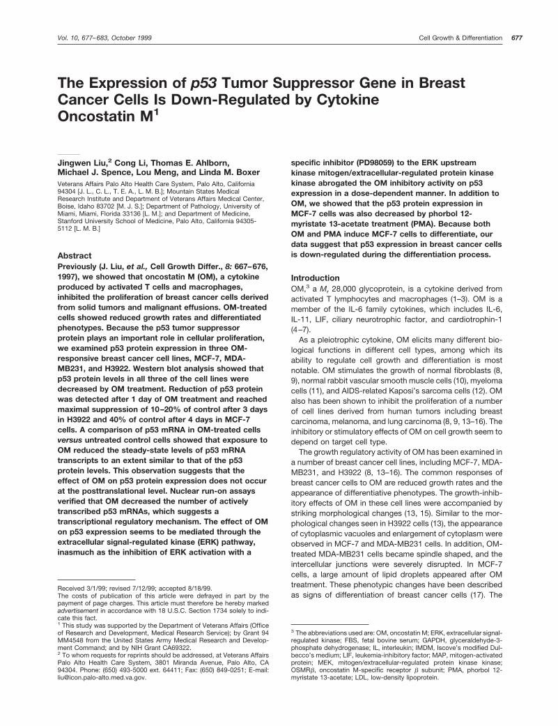
with anti-p53 monoclonal antibody. Fig. 1 shows that the p53protein level was decreased in OM-treated cells in an OMdose-dependent manner. A maximal effect of 80% suppres-sion was observed at 10 ng/ml and higher. In contrast to p53,the level of b-actin was not altered by OM.
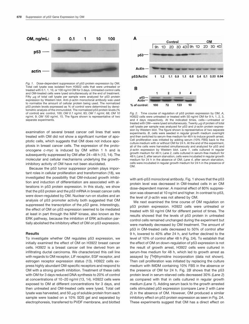
We next examined the time course of OM regulation onp53 protein expression. H3922 cells were untreated ortreated with 50 ng/ml OM for different period of times. Theresults showed that the levels of p53 protein in untreatedcontrol cells remained unchanged during the experiment butwere markedly decreased by OM treatment. The amount ofp53 in OM-treated cells decreased to 50% of control after9 h, lowered to 40% after 24 h, and further declined to thelevel of 10% of control after 48 h (Fig. 2A). To establish thatthe effect of OM on down-regulation of p53 expression is notthe result of growth arrest, H3922 cells were cultured inserum-free medium for 48 h, which led to growth arrest asassayed by [3H]thymidine incorporation (data not shown).Then cell proliferation was initiated by replacing the culturemedium with IMDM containing 10% FBS in the absence orthe presence of OM for 24 h. Fig. 2B shows that the p53protein level in serum-starved cells decreased 30% (Lane 2)as compared with that in cells cultured in regular growthmedium (Lane 1). Adding serum back to the growth arrestedcells stimulated p53 expression (compare Lane 3 with Lane2) in the absence of OM. However, OM produced a similarinhibitory effect on p53 protein expression as seen in Fig. 2A.These experiments suggest that OM has a direct effect on
Fig. 1. Dose-dependent suppression of p53 protein expression by OM.Total cell lysate was isolated from H3922 cells that were untreated ortreated with 0.1, 1, 10, or 100 ng/ml OM for 3 days. Untreated control cellsand OM-treated cells were lysed simultaneously at the end of treatment.Fifty mg of total cell lysate per sample were analyzed for p53 proteinexpression by Western blot. Anti-b-actin monoclonal antibody was usedto normalize the amount of cellular protein being used. The normalizedp53 protein levels expressed as % of control were determined by densi-tometric analysis of the immunoblot. The normalized p53 protein levels (%of control) are: control, 100; OM 0.1 ng/ml, 83; OM 1 ng/ml, 68; OM 10ng/ml, 9; OM 100 ng/ml, 15. The figure shown is representative of twoseparate experiments.
Fig. 2. Time course of regulation of p53 protein expression by OM. A,H3922 cells were untreated or treated with 50 ng/ml OM for 9 h, 1, 2, 3,and 4 days respectively. At the indicated times, cells—untreated ortreated with OM—were lysed simultaneously. Twenty mg of protein of totalcell lysate per sample was analyzed for p53 and b-actin protein expres-sion by Western blot. The figure shown is representative of two separateexperiments. B, cells were seeded in regular growth medium overnightand then switched to serum-free medium for 48 h to induce growth arrest.Cell proliferation was initiated by adding serum (10% FBS) back to theculture medium with or without OM for 24 h. At the end of the experiment,all of the cells were harvested simultaneously and analyzed for p53 andb-actin expression by Western blot. Lane 1, cells cultured in regulargrowth medium for 48 h; Lane 2, cells cultured in serum-free medium for48 h; Lane 3, after serum starvation, cells were incubated in regular growthmedium for 24 h in the absence of OM; Lane 4, after serum starvation,cells were incubated in regular growth medium for 24 h in the presence ofOM.
678 Suppression of p53 Gene Expression by OM
p53 expression independent of the growth status of the cells.However, these studies cannot rule out completely the pos-sibility that the growth arrest induced by OM contributespartially to the decreased expression of p53.
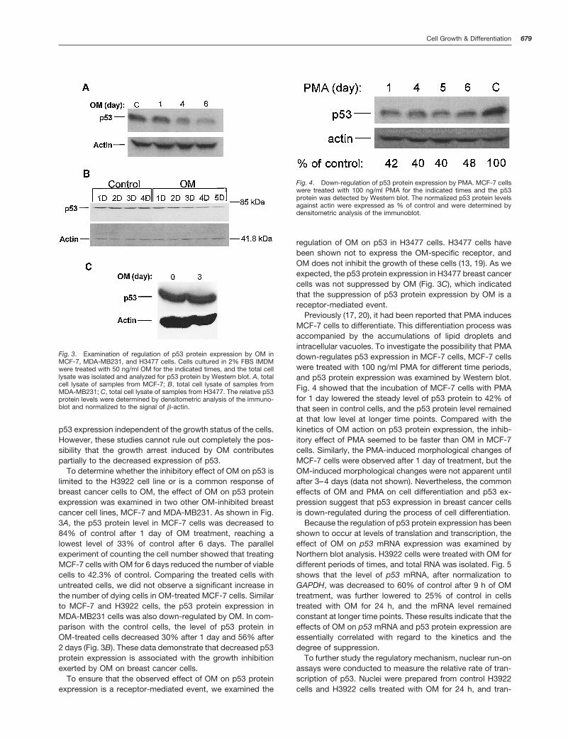
To determine whether the inhibitory effect of OM on p53 islimited to the H3922 cell line or is a common response ofbreast cancer cells to OM, the effect of OM on p53 proteinexpression was examined in two other OM-inhibited breastcancer cell lines, MCF-7 and MDA-MB231. As shown in Fig.3A, the p53 protein level in MCF-7 cells was decreased to84% of control after 1 day of OM treatment, reaching alowest level of 33% of control after 6 days. The parallelexperiment of counting the cell number showed that treatingMCF-7 cells with OM for 6 days reduced the number of viablecells to 42.3% of control. Comparing the treated cells withuntreated cells, we did not observe a significant increase inthe number of dying cells in OM-treated MCF-7 cells. Similarto MCF-7 and H3922 cells, the p53 protein expression inMDA-MB231 cells was also down-regulated by OM. In com-parison with the control cells, the level of p53 protein inOM-treated cells decreased 30% after 1 day and 56% after2 days (Fig. 3B). These data demonstrate that decreased p53protein expression is associated with the growth inhibitionexerted by OM on breast cancer cells.
To ensure that the observed effect of OM on p53 proteinexpression is a receptor-mediated event, we examined the
regulation of OM on p53 in H3477 cells. H3477 cells havebeen shown not to express the OM-specific receptor, andOM does not inhibit the growth of these cells (13, 19). As weexpected, the p53 protein expression in H3477 breast cancercells was not suppressed by OM (Fig. 3C), which indicatedthat the suppression of p53 protein expression by OM is areceptor-mediated event.
Previously (17, 20), it had been reported that PMA inducesMCF-7 cells to differentiate. This differentiation process wasaccompanied by the accumulations of lipid droplets andintracellular vacuoles. To investigate the possibility that PMAdown-regulates p53 expression in MCF-7 cells, MCF-7 cellswere treated with 100 ng/ml PMA for different time periods,and p53 protein expression was examined by Western blot.Fig. 4 showed that the incubation of MCF-7 cells with PMAfor 1 day lowered the steady level of p53 protein to 42% ofthat seen in control cells, and the p53 protein level remainedat that low level at longer time points. Compared with thekinetics of OM action on p53 protein expression, the inhib-itory effect of PMA seemed to be faster than OM in MCF-7cells. Similarly, the PMA-induced morphological changes ofMCF-7 cells were observed after 1 day of treatment, but theOM-induced morphological changes were not apparent untilafter 3–4 days (data not shown). Nevertheless, the commoneffects of OM and PMA on cell differentiation and p53 ex-pression suggest that p53 expression in breast cancer cellsis down-regulated during the process of cell differentiation.
Because the regulation of p53 protein expression has beenshown to occur at levels of translation and transcription, theeffect of OM on p53 mRNA expression was examined byNorthern blot analysis. H3922 cells were treated with OM fordifferent periods of times, and total RNA was isolated. Fig. 5shows that the level of p53 mRNA, after normalization toGAPDH, was decreased to 60% of control after 9 h of OMtreatment, was further lowered to 25% of control in cellstreated with OM for 24 h, and the mRNA level remainedconstant at longer time points. These results indicate that theeffects of OM on p53 mRNA and p53 protein expression areessentially correlated with regard to the kinetics and thedegree of suppression.
To further study the regulatory mechanism, nuclear run-onassays were conducted to measure the relative rate of tran-scription of p53. Nuclei were prepared from control H3922cells and H3922 cells treated with OM for 24 h, and tran-
Fig. 3. Examination of regulation of p53 protein expression by OM inMCF-7, MDA-MB231, and H3477 cells. Cells cultured in 2% FBS IMDMwere treated with 50 ng/ml OM for the indicated times, and the total celllysate was isolated and analyzed for p53 protein by Western blot. A, totalcell lysate of samples from MCF-7; B, total cell lysate of samples fromMDA-MB231; C, total cell lysate of samples from H3477. The relative p53protein levels were determined by densitometric analysis of the immuno-blot and normalized to the signal of b-actin.
Fig. 4. Down-regulation of p53 protein expression by PMA. MCF-7 cellswere treated with 100 ng/ml PMA for the indicated times and the p53protein was detected by Western blot. The normalized p53 protein levelsagainst actin were expressed as % of control and were determined bydensitometric analysis of the immunoblot.
679Cell Growth & Differentiation

scription was allowed to continue in the presence of[32P-]UTP for 30 min. The incorporation of 32P into p53-specific RNA was used as a measure of transcription rate.The transcription rate for GAPDH was also measured as aninternal control. OM-treated cells contained only approx-imately 20% as many active p53 transcripts as observed incontrol cells (Fig. 6A). Data were normalized by the signalsdetected in GAPDH slots. The effect of OM on p53 tran-scription was further studied by analyzing p53 promoteractivity. A human p53 promoter reporter construct, pGL3-p53LUC, and a human LDL receptor promoter reporterconstruct, pLDLR234LUC, were transiently transfectedinto H3922 cells, and the luciferase activities were meas-ured. As shown in Fig. 6B, OM treatment decreased p53promoter activity to approximately 30% of control. In con-trast, in the same experiment, the promoter activity of theLDL receptor was increased 2.5-fold, which is consistentwith the stimulatory effect of OM on this promoter re-ported in other cell lines (21). These results obtained fromthe studies of nuclear run-on and promoter activity sug-gest that transcriptional regulation is a major componentof the observed OM-mediated suppression of p53 mRNAexpression.
It has been proposed that the p53 gene is a target of theproto-oncogene c-myc. The c-Myc protein regulates thetranscription of p53 through the c-Myc-responsive elementpresent in the promoter region of the p53 gene (22). Weinvestigated the possibility that OM-mediated suppression ofp53 transcription was due to an effect on c-Myc-mediatedtranscription. Northern blot analysis was used to examine thelevels of c-myc mRNA and p53 mRNA in total RNAs isolatedfrom H3922 cells untreated or treated with OM for differentperiods of time. Fig. 7 shows that transcription of the c-mycmRNA and the p53 mRNA were not concurrently regulatedby OM. The c-myc mRNA was increased more than 3-fold byOM after 30 min and slowly decreased afterward. The sup-pression of c-myc transcription was not seen until 24 h afterOM treatment. In contrast, the p53 mRNA level was steadilydecreased during OM treatment. The biphasic effect of OMon c-myc mRNA was not seen. A similar result was obtainedfrom MDA-MB231 cells. Furthermore, Western blot to exam-ine the c-Myc protein level in untreated and OM-treated
H3922 cells showed that the level of c-Myc protein did notdecrease until after 2 days of OM treatment (data not shown).The decrease in p53 mRNA was maximal at 24 h in H3922cells. These results suggest that either c-Myc is not involvedin the regulation of p53 by OM or it is not the major tran-scriptional regulator. Other transcription factors could beinvolved in addition to c-Myc for the OM-induced down-regulation of p53.
Two major signaling pathways can be activated by OMand its related cytokines IL-6 and LIF: (a) the Janus familytyrosine kinase/signal transducer and activator of tran-scription pathway (23); and (b) the Ras-MAP kinase path-way (21, 24 –26). In OM-treated breast cancer cells, both
Fig. 5. Kinetics of p53 mRNA expression in H3922 cells treated with OM.H3922 cells cultured in IMDM containing 2% FBS were incubated with 50ng/ml OM for different times as indicated. Untreated control cells andOM-treated cells were lysed simultaneously at the end of the treatmentwith the RNA isolation solution. Total RNA was isolated and 15 mg persample was analyzed for p53 mRNA by Northern blot. The membrane wasstripped and hybridized to a human GAPDH probe. The figure shown is arepresentative of three different experiments.
Fig. 6. OM down-regulates p53 gene transcription. A, nuclear run-onanalysis of p53 transcription: two slots were blotted onto each of twonylon membrane strips. One slot received 3 mg of the 2-kb fragment of thep53 cDNA. The second slot was loaded with 5 mg of the GAPDH plasmid.One nylon strip was hybridized to a 32P-radiolabeled nuclear run-onreaction prepared from 24-h OM-treated H3922 cells. The second washybridized to a labeled nuclear run-on reaction prepared from untreatedcontrol cells. Equal amounts of radioactivity were used in each hybridiza-tion. Radioactive signals were detected by autoradiography and quanti-fied by densitometric analysis. The figure shown is representative of twodifferent experiments. B, analysis of human p53 promoter activity: H3922cells were transfected with the pGL3-p53LUC and pLDLR234LUC. Aftertransfection, cells were cultured in media minus or plus OM for 48 h.Assays of luciferase activity were conducted as described (21). The data(mean 6 SE) shown are representative of three separate experiments inwhich triplicate wells were assayed.
680 Suppression of p53 Gene Expression by OM

STAT3 and MAP kinases ERK1 and ERK2 were activated(27). To investigate whether the MAP kinase pathway isinvolved in the OM-mediated suppression of p53, H3922cells were incubated with OM for 2 days in the presence ofdifferent concentrations of PD98059, which specificallyblocks ERK activation by inhibiting the enzymatic activityof the ERK upstream kinase, MEK (28). Incubation of cellswith PD98059 alone, in up to a 30-mM concentration, didnot change the levels of p53 protein (data not shown);however, the OM-mediated suppression of p53 proteinexpression was partially (maximal 50 – 60%) inhibited byPD98059 in a dose-dependent manner (Fig. 8). Theseresults suggest that the suppression of p53 expression isa downstream event of the activation of MAP kinase path-way by OM in breast cancer cells. The fact that the OM-inhibitory activity could not be completely reversed byPD98059 at concentrations that effectively inhibited ERKactivation (21, 29) suggests that there are other signalingpathways, such as the STAT pathway, that may beinvolved.
DiscussionPrevious studies have established a differentiative role of OMin breast cancer cells (13–16). The general effects of OM onbreast cancer cells include: (a) inhibition of cellular prolifer-ation in monolayer culture and inhibition of colony formationin soft agar; (b) regulation of the cell cycle by increasing theproportion of cells in G0-G1 phase with a concomitant de-crease in the number of cells in S phase; and (c) induction ofa variety of morphological changes associated with the dif-ferentiated phenotype. These effects are believed to be me-diated through the OM-specific receptor that consists ofgp130 as a low-affinity ligand-binding subunit and OSMRb
as the signal-transducing subunit (13, 19). In this study, wedemonstrated that OM also down-regulates p53 expressionin breast cancer cells.
The effect of OM on p53 protein expression was initiallyexamined in four breast cancer cell lines, among which 3 celllines—H3922, MCF-7, and MDA-MB231—were growth-inhibited by OM. The cell line H3477 does not respond to OMtreatment because of its lack of expression of the signal-transducing subunit (OSMRb) of the OM-specific receptor.The incubation of cells with OM decreased the level of p53protein in all three of the OM-responsive cell lines, with themost dramatic effect found in H3922 cells. The expression ofp53 protein in H3477 cells was not inhibited at all by OM. Thedecrease in p53 protein was detected after 9 h of OM treat-ment and reached the lowest levels (10–20% of control inH3922 and 30–40% of control in MCF-7 and MDA-MB231)after 3–4 days. The extent of the suppression of p53 expres-sion in different cell lines seems to correlate with the expres-sion level of OSMRb, inasmuch as a previous study (19)using quantitative reverse transcription-PCR showed thatH3922 cells express the highest mRNA level of OSMRb ascompared with that detected in other cell lines.
Northern blot analysis to examine p53 mRNA expression incontrol and OM-treated cells demonstrated that OM down-regulated p53 mRNA expression to a degree similar to thatobserved in the levels of p53 protein in these cells. Thedecreased mRNA expression was due to a direct inhibition oftranscription of the p53 gene by OM as demonstrated bynuclear run-on analysis. Collectively, these data establishthat OM down-regulates the transcription of the p53 gene inbreast cancer cells, with a resulting decrease in p53 protein
Fig. 7. OM regulates the mRNA expressions of p53 and c-myc withdifferent kinetics. H3922 cells were incubated with 50 ng/ml OM fordifferent times as indicated. Total RNA was isolated and 15 mg per samplewere analyzed for p53 mRNA, c-myc mRNA, and GAPDH mRNA byNorthern blot. The radioactive signals were detected and quantitated by aPhosphorImager. The figure shown (A) is representative of two differentNorthern blots. B, the normalized c-myc and p53 mRNA levels (% ofcontrol).
Fig. 8. The dose-dependent effects of MEK inhibitor PD98059 on OMregulation of p53 protein expression. H3922 cells were treated with OM for2 days in the absence or the presence of the indicated concentrations ofPD98059. Total lysate was harvested and analyzed for p53 protein byWestern blot. The membrane was stripped and reprobed with anti-b-actinmonoclonal antibody. The normalized p53 protein levels expressed as %of control were determined by densitometric analysis of the immunoblot.
681Cell Growth & Differentiation

level. The inhibitory effect of OM on p53 promoter activitysuggests that an OM-responsive element(s) is present in thepromoter region of the p53 gene. Interactions of this putativecis-acting element with an OM-inducible transcription factormay be responsible for the suppression of p53 transcription.The nature of this interaction has not been characterized inthe present study; however, the fact that the MEK inhibitorPD98059 partially prevented the OM-inhibitory effect on p53expression suggests that a substrate of ERK may be directlyor indirectly involved in the OM-elicited signaling pathwaythat mediates this regulation.
It has been proposed that p53 is a target gene of c-Myc.The c-Myc protein was reported to transactivate the p53promoter through the c-Myc-responsive element, E box(CATGTG) (22). Because OM has been shown to regulatec-myc gene transcription in breast cancer cells and in M1leukemia cells, one potential mechanism of OM suppressionof p53 transcription would be suppression of c-myc tran-scription. However, the kinetics of the OM-induced down-regulation of p53 were different from the kinetics of theOM-induced down-regulation of the c-myc gene in thesecells. The c-myc mRNA was transiently induced by OMwithin 0.5–8 h and was subsequently suppressed at latertime points. The maximal suppression occurred after 2–3days of OM treatment (Fig. 7; Refs. 13, 14). In contrast, p53mRNA was not induced by OM at any time point examined.Instead, it was gradually decreased during the period of OMtreatment. A significant decrease in p53 mRNA level wasobserved after 8 h of OM treatment. This difference in kinet-ics between the expression of p53 mRNA and c-myc mRNAsuggests that the effect of OM on p53 transcription is not adirect effect of OM on c-myc transcription alone. Additionalstudies to identify the cis-acting element in the p53 promoterthat is responsible for the OM-mediated suppression of p53transcription will clarify the relationship between p53 tran-scription and c-myc transcription in breast cancer cells.
The p53 tumor suppressor protein is involved in severalcentral cellular processes that are critical for maintainingcellular homeostasis, including gene transcription, DNA re-pair, cell cycling, senescence, and apoptosis (18, 30). Com-pared with the vast information and knowledge availableregarding the role of p53 protein in apoptosis, the function ofp53 in cell differentiation is not well understood. This study isthe first report to show that p53 expression is down-regu-lated in growth-inhibited and -differentiated breast cancercells through a transcriptional mechanism. Additional studiesto examine the functional role of p53 in the differentiation ofbreast cancer cells will be needed for a better understandingof the complexity of p53 functions that are essential formaintaining cell homeostasis.
Materials and MethodsCells and Reagents. The human breast cancer cell line H3922 wasderived from an infiltrating ductal carcinoma, and the human breast can-cer cell line H3477 was derived from primary solid tumor (13). MDA-MB231 and MCF-7 cells were obtained from American Type CultureCollection (Manassas, VA). All of the cell lines were cultured in IMDMsupplemented with 10% heat-inactivated FBS. Purified human recombi-nant OM was provided by Bristol-Myers Squibb (Princeton, NJ). PMA andanti-b-actin monoclonal antibody were purchased from Sigma Chemical
Co. (St. Louis, MO). The MEK inhibitor PD98059 was purchased from NewEngland Biolabs (Beverly, MA). The antibodies against p53 and c-Mycwere purchased from Santa Cruz Biotechnology (Santa Cruz, CA).
Western Blot Analysis of p53 Protein. Cells were cultured in 60-mmculture plates in 2% FBS IMDM with or without OM. Cells were rinsed withcold PBS and lysed with 0.25 ml of lysis buffer [50 mM Tris (pH 7.4), 1%NP40, 0.25% sodium deoxycholate, 150 mM NaCl, 1 mM EGTA, 1 mM
phenylmethylsulfonyl fluoride, 1 mM NaF, 5 mg/ml aprotinin, 1 mg/mlleupeptin, 1.25 mg/ml pepstatin, 1 mM Na3VO4, 10 mM okadaic acid, and10 mM cypermethrin]. Concentration of soluble protein from total celllysate was determined using BCA reagent with BSA as a standard(Pierce). Approximately 10–50 mg of protein of total cell lysate per samplewas separated on 10% SDS PAGE, transferred to PVDF membrane,blotted with anti-p53 monoclonal antibody (DO-1, Santa Cruz Biotech-nology) using an enhanced chemiluminescence (ECL) detection system(Amersham). Membranes were stripped and reblotted with anti-b-actinmonoclonal antibody to ensure that an equal amount of protein is loadedon gel. The signals were quantitated with a Bio-Rad Fluro-S MultiImagersystem. Densitometric analysis of autoradiographs in these studies in-cluded various exposure times to ensure linearity of signals.
RNA Isolation and Northern Blot Analysis. Cells were lysed in Ul-traspec RNA lysis solution (Biotecx Laboratory, Houston, TX), and totalcellular RNA was isolated according to the vendor’s protocol. Approxi-mately 15 mg of each total RNA sample were used in Northern blotanalysis as described previously. The p53 mRNA was detected with a2-kb 32P-labeled human p53 cDNA probe, the c-myc mRNA was detectedwith a 1.4-kb probe containing c-myc exons 2 and 3, and the GAPDHmRNA was detected with a plasmid containing a human GAPDH cDNA.Differences in hybridization signals of Northern blots were quantitated bya PhosphorImager.
Nuclear Run-On Analysis. Reaction was performed with the methoddescribed previously (14). Briefly, the nuclei were harvested from H3922cells that were untreated or treated with 50 ng/ml OM for 24 h. Approx-imately 2 3 107 nuclei in 100 ml were mixed with 100 ml of 23 reactionbuffer containing 250 mCi [32P]rUTP. Approximately 2.0 3 106 cpm ofeach nuclear run-on reaction was used as a probe to hybridize a HybondN membrane (Amersham) slot blot. Each slot blot contained 5 mg ofGAPDH plasmid and 3 mg of the 2-kb fragment of the p53 cDNA asdescribed in the Northern blot analysis. Probing the GAPDH plasmidallowed normalization of the p53 signals measured by densitometry.
Transient Transfection Assays. The p53 promoter luciferase-reporter construct p53Ex1aLUC was generously provided by Dr. PeterGruss at the Max-Planck-Institute (Gottingen, Germany). For constructionof pGL3-p53LUC, the 550-bp insert containing the human p53 promoterregion and Exon 1 (31) was released from p53Ex1aLUC by KpnI and BglIIdigestion and subcloned into pGL3-basic digested with KpnI and BglII.The plasmid vector pLDLR234LUC has been described previously (21).H3922 cells, seeded in 12-well tissue culture plates, were transientlytransfected with plasmid DNAs pGL3-p53LUC and pLDLR234LUC by themethod of calcium phosphate coprecipitation. Fifteen h after transfection,OM (50 ng/ml) was added. After a 48-h treatment, cells were lysed. Equalamounts of cell lysate from each well were used for measuring luciferaseactivity. The data (mean 6 SE) shown are representative of three separateexperiments in which triplicate wells were assayed (Fig. 6).
AcknowledgmentsWe thank Jessy Dorn for her technical assistance in Western blot analysisof p53.
References1. Zarling, J. M., Shoyab, M., Marquardt, H., Hanson, M. B., Lionbin,M. N., and Todaro, G. J. Oncostatin M: a growth regulator produced bydifferentiated lymphoma cells. Proc. Natl. Acad. Sci. USA, 83: 9739–9743,1986.
2. Brown, T. J., Lionbin, M. N., and Marquardt, H. Purification and char-acterization of cytostatic lymphokines produced by activated human T-lymphocytes: synergistic antiproliferative activity of transforming growthfactor b1, interferon g, and oncostatin M for human melanoma cells.J. Immunol., 139: 2977–2983, 1987.
682 Suppression of p53 Gene Expression by OM

3. Grove, R. I., Mazzucco, C. E., Allegretto, N., Kiener, P. A., Spitalny, G.,Radka, S. F., Shoyab, M., Antonaccio, M., and Warr, G. A. Macrophages-derived factors increase low-density lipoprotein uptake and receptornumber in cultured human liver cells. J. Lipid Res., 32: 1889–1897, 1991.
4. Rose, T. M., and Bruce, A. G. Oncostatin M is a member of a cytokinefamily that includes leukemia inhibitory factor, granulocyte colony stimu-lating factor, and interleukin 6. Proc. Natl. Acad. Sci. USA, 88: 8641–8645,1991.
5. Bazan, F. Neuropoietic cytokines in the hematopoietic fold. Neuron, 7:197–208, 1991.
6. Pennica, D. Cardiotrophin-1: biological activities and binding to theleukemia inhibitory factor receptor/gp130 signaling complex. J. Biol.Chem., 270: 10915–10922, 1998.
7. Ip, N. Y., Nye, S., Boulton, T. G., Davis, S., Taga, T., Li, Y., Birren, S. J.,Yasukawa, K., Kishimoto, T., Anderson, D. J., Stah, N., and Yancopoulos,D. CNTF and LIF act on neuronal cells via shared signaling pathways thatinvolve the IL-6 signal transducer receptor component gp130. Cell, 69:1121–1132, 1992.
8. Horn, D., Fitzpatrick, W. C., Gompper, P. T., Ochs, V., Bolton-Hanson,M., Zarling, J. M., Malik, N., Todaro, G. J., and Linsley, P. S. Regulation ofcell growth by recombinant oncostatin M. Growth Factors, 2: 157–165,1990.
9. Liu, J., Clegg, J. C., and Shoyab, M. Regulation of EGR-1, c-jun, andc-myc gene expression by oncostatin M. Cell Growth Differ., 3: 307–313,1992.
10. Grove, R. I., Eberthardt, C., Abid, S., Mazzucco, C. E., Liu, J., Todaro,G. J., Kiener, P. A., and Shoyab, M. Oncostatin M is a mitogen for rabbitvascular smooth muscle cells. Proc. Natl. Acad. Sci. USA, 90: 823–827,1993.
11. Zhang, X. G., Gu, J. J., Lu, Z. Y., Yasukawa, K., Yancopoulos, G. D.,Turner, K., Shoyab, M., Taga, T., Kishimoto, T., Bataille, R., and Klein, B.Ciliary neurotrophic factor, interleukin 11, leukemia inhibitory factor, andoncostatin M are growth factors for human myeloma cell lines using theinterleukin 6 signal transducer gp130. J. Exp. Med., 179: 1343–1347,1994.
12. Nair, B. C., DeVico, A. L., Nakamura, S., Copeland, T. D., Chen, Y.,Patel, A., O’Neil, T., Oroszlan, S., and Gallo, R. C. S. M. G. Identificationof a major growth factor for AIDS-Kaposi’s sarcoma cell as oncostatin M.Science (Washington DC), 255: 1430–1432, 1992.
13. Liu, J., Spence, M. J., Wallace, P. M., Forcier, K., Hellstrom, I., andVestal, R. E. Oncostatin M-specific receptor mediates inhibition of breastcancer cell growth and down-regulation of the c-myc proto-oncogene.Cell Growth Differ., 8: 667–676, 1997.
14. Spence, M. J., Vestal, R. E., and Liu, J. Oncostatin M-mediatedtranscriptional suppression of the c-myc gene in breast cancer cells.Cancer Res., 57: 2223–2228, 1997.
15. Douglas, A. M., Grant, S. L., Goss, G. A., Clouston, D. R., Sutherland,R. L., and Begley, C. G. Oncostatin M induces the differentiation of breastcancer cells. Int. J. Cancer, 75: 64–73, 1998.
16. Douglas, A. M., Goss, G. A., Sutherland, R. L., Hilton, D. J., Berndt,M. C., Nicola, N. A., and Begley, C. G. Expression and function of mem-bers of the cytokine receptor superfamily on breast cancer cells. Onco-gene, 14: 661–669, 1997.
17. Guilbaud, N. F., Gas, N., DuPont, M. A., and Valette, A. Effects ofdifferentiation-inducing agents on maturation of human MCF-7 breastcancer cells. J. Cell. Physiol., 145: 162–172, 1990.
18. Vogelstein, B., and Kinzler, K. W. p53 function and dysfunction. Cell,70: 523–526, 1992.
19. Liu, J., Hadjokas, N., Mosley, B., Estrov, Z., Spence, M. J., and Vestal,R. E. Oncostatin M-specific receptor expressions and function in regulat-ing cell proliferation of normal and malignant mammary epithelial cells.Cytokine, 10: 295–302, 1998.
20. Bacus, S. S., Kiguchi, K., Chin, D., King, C. R., and Huberman, E.Differentiation of cultured human breast cancer cells (AU-565 and MCF-7)associated with loss of cell surface HER-2/neu antigen. Mol. Carcinog., 3:350–352, 1990.
21. Li, C., Kraemer, F. B., Ahlborn, T. E., and Liu, J. Induction of lowdensity lipoprotein receptor (LDLR) transcription by oncostatin M is me-diated by the extracellular signal-regulated kinase signaling pathway andthe repeat 3 element of the LDLR promoter. J. Biol. Chem., 274: 6747–6753, 1999.
22. Roy, B. B. J., Balint, E., and Reisman, D. Transactivation of the humanp53 tumor suppressor gene by c-myc/max contributes to elevated mutantp53 expression in some tumors. Mol. Cell. Biol., 14: 7805–7815, 1994.
23. Stahl, N., Boulton, T. G., Farruggella, T., Ip, N. Y., Davis, S., Witthuhn,B. A., Quelle, F. W., Silvennoinen, O., Barbieri, G., Pellegrini, S., Ihle, J. N.,and Yancopoulos, G. D. Association and activation of Jak-Tyk kinase byCNTF-LIF-OSM-IL-6 receptor components. Science (Washington DC),263: 92–95, 1994.
24. Thoma, B., Bird, T. A., Friend, D. J., Gearing, D. P., and Dower, S. K.Oncostatin M and leukemia inhibitory factor trigger overlapping and dif-ferent signals through partially shared receptor complexes. J. Biol. Chem.,269: 6215–6222, 1994.
25. Yin, T., and Yang, Y. C. Mitogen-activated protein kinases and ribo-somal S6 protein kinases are involved in signaling pathways shared byinterleukin-11, interleukin-6, leukemia inhibitory factor, and oncostatin Min mouse 3T3–L1 cells. J. Biol. Chem., 269: 3731–3738, 1994.
26. Stancato, L. F., Yu, C. R., Petricoin, E. F., III, and Larner, A. C.Activation of Raf-1 by interferon g and oncostatin M requires expressionof the Stat1 transcription factor. J. Biol. Chem., 273: 18701–18704, 1998.
27. Liu, J., Li, C., Ahlborn, T. E., and Kraemer, F. B. Oncostatin M-inducedgrowth inhibition and differentiation of breast cancer cells is mediated bythe MAP kinase ERK signaling pathway. Proc. Am. Assoc. Cancer Res.,90: 334, 1999.
28. Alessi, D. R., Cuenda, A., Cohen, P., Dudley, D. T., and Saltiel, A. R.PD 098059 is a specific inhibitor of the activation of mitogen-activatedprotein kinase kinase in vitro and in vivo. J. Biol. Chem., 270: 27489–27494, 1995.
29. Kumar, A., Chambers, T. C., Cloud-Heflin, B. A., and Mehta, K. D.Phorbol ester-induced low density lipoprotein receptor gene expression inHepG2 cells involves protein kinase C-mediated p42/44 MAP kinaseactivation. J. Lipid Res., 38: 2240–2248, 1997.
30. Levine, A. J. p53, the cellular gatekeeper for growth and division. Cell,88: 323–331, 1997.
31. Stuart, E. T., Haffner, R., Oren, M., and Gruss, P. Loss of p53 functionthrough PAX-mediated transcriptional repression. EMBO J., 14: 5638–5645, 1995.
683Cell Growth & Differentiation