synthetic polymeric vectors in gene therapy
TRANSCRIPT

8/8/2019 Synthetic Polymeric Vectors in Gene Therapy
http://slidepdf.com/reader/full/synthetic-polymeric-vectors-in-gene-therapy 1/14
Synthetic polymeric vectors in gene therapy
Patit P. Kundu *, Vinay Sharma
Department of Polymer Science & Technology, University of Calcutta, 92, A. P. C. Road, Kolkata 700009, WB, India
a r t i c l e i n f o
Article history:
Received 9 October 2008Accepted 14 January 2009
Keywords:
Polymeric vectorsGene therapyNon-viral therapypDNA
a b s t r a c t
The synthetic polymer vectors in gene therapy have opened a very prospective field for research in genetherapy and the area in the use of these synthetic polymers as vectors in targeting oncogenes is develop-ing very fast. With the completion of Human Genome Project, the list of genetic targets is growing veryfast. These diseases sparked the initiative to create such gene based therapeutics. The low levels of trans-fection and expression of the gene held non-viral methods are at a disadvantage; however, recentadvances in vector technology have yielded molecules and techniques with transfection efficiencies, sim-ilar to those of viruses. Amongst these synthetic polymers, polyethylenimine (PEI) and the poly(amido-amine) (PAMAM) dendrimers and poly(b-amino ester) are used extensively as vectors in gene therapy.
Ó 2009 Elsevier Ltd. All rights reserved.
1. Introduction
Gene therapy may be defined as the treatment of human dis-ease by the transfer of genetic material into specific cells of the
patient [1]. The advances in molecular biology and biotechnologyof the last 30 years have greatly enhanced the understanding of the genetics of pathogenesis and have led to the identification of numerous disease-causing genes. With the impending completionof the Human Genome Project, the list of genetic disease targets islikely to grow tremendously. It is not difficult to predict treatmentof genetic diseases such as hemophilia [2], muscular dystrophy[3–5], or cystic fibrosis [6] through replacement of errant geneswithin the affected cells. However, the gene therapy approachesare also being developed for treatments of virtually all forms of cardiovascular diseases [7], neurological diseases [8–10], infectiousdiseases [11], wound healing [12], and cancer [13–15]. In these in-stances, the delivered genes may be intended to augment naturallyoccurring proteins (e.g growth factors or cytokines), to alter theexpression of existing genes facilitating a desired cellular or tissueresponse, or to produce cytotoxic proteins or pro-drug-activatingenzymes, for example, to kill tumor cells in the cancer treatment[13,15] or proliferating endothelial cells to inhibit restenosisfollowing balloon angioplasty [7,16]. The expression of viral geneswithin certain tissues can also result in an efficient immuneresponse, leading to the development of DNA vaccines [17].
The gene carriers for these systems are of two kinds: one basedon recombinant viruses and another based on synthetic polymers.The primary function of a virus is to efficiently carry its own gen-ome from one host cell to another through (perhaps) hostile envi-
ronments, enter the new target cell, navigate to the cell nucleus,and initiate expression of its genome-albeit for the purpose of self-replication. Viruses can be transformed into gene deliveryvehicles by removing part of the virus genome and replacing it
with a therapeutic gene. If done carefully, the resulting recombi-nant virus will retain the functions essential for infection of spe-cific target cells, but will be rendered incapable of replicationand, hence, will be nonpathogenic. The viruses evolved essentiallyare sophisticated gene delivery vehicles and such recombinantviral vectors are incredibly efficient.
Although viruses are incredibly efficient gene delivery agents,synthetic vectors provide opportunities for improved safety, greaterflexibility, and more facile manufacturing. In general, syntheticvectors are materials that bind electrostatically to DNA or RNA,condensing the genetic material into nanometer-scale complexes(a few tens to several hundred nanometers in diameter) that pro-tect the genes and allow them to enter cells. Such materials haveincluded cationic peptides, proteins, polymers, and liposomes. Var-ious synthetic vectors, including (diethylamino)ether (DEAE)-dex-tran [18] and calcium phosphate [19], have been used extensivelyfor in vitro gene transfer studies since 1960s. However, develop-ment of non-viral vectors for in vivo gene delivery, especially clin-ical applications, has suffered from problems including toxicity,low gene transfer efficiency, and in vivo instability. Despite thehigh efficiency of viral vector in vitro, clinical trials are often lim-ited by several concerns, e.g. toxicity, immunogenicity, inflamma-tory properties, the limited size of the DNA, production andpacking problems, and the high cost. In addition, an overwhelmingimmune reaction against adenovirus occurred in a patient at Penn-sylvania University in 1999 [20,21] and a leukemia-like diseasewere reported in a French patient in 2002 [22,23] As a result,non-viral vector-mediated systems have become of interest,
1359-0286/$ - see front matter Ó 2009 Elsevier Ltd. All rights reserved.doi:10.1016/j.cossms.2009.01.005
* Corresponding author.E-mail address: [email protected] (P.P. Kundu).
Current Opinion in Solid State and Materials Science 12 (2008) 89–102
Contents lists available at ScienceDirect
Current Opinion in Solid State and Materials Science
j o u r n a l h o m e p a g e : w w w . e l s e v i e r . c o m / l o c a t e / c o s s m s

8/8/2019 Synthetic Polymeric Vectors in Gene Therapy
http://slidepdf.com/reader/full/synthetic-polymeric-vectors-in-gene-therapy 2/14
because they are much safer, more cost-effective, and easier tomanufacture than viral vector systems.
Non-viral vectors for gene delivery are receiving increasingattention for application in a broad variety of gene-mediated ther-apies for humans. Chemical vectors are attractive to the pharma-ceutical industry as alternatives to viral vectors, because of compound stability and easy chemical modification. Furthermore,the low cost and consistent standard of production (compared togrowth of viruses in bioreactors followed by purification), higherbio-safety (less immunogenic as compared to viruses such asadenoviruses), and a high flexibility (once a formulation has beendefined) make these compounds very attractive [24,25].
To design the optimal non-viral vector for particle-mediatedgene delivery, it must be able to partially fulfill a number of prede-fined biological criteria depending on its specific therapeutic aim.First, the vector must be able to transfer DNA molecules of varyingsizes. Second, the vector should be able to transducer dividing and/or non-dividing cells, depending on the target cell type. Third, thevector should target a specific cell type. Fourth, the vector shouldhave no or very low toxicity in vivo and avoid stimulating theimmune system. Fifth, depending on the therapeutic application,the vector should be able to induce a sustained expression over adefined period of time (optionally integrating into the host gen-ome) and, finally, the vector must not transform the target cell.
2. Polymer characteristics required for gene delivery
For effective polyplex-mediated gene delivery, the cationicpolymer carrier has to fulfill a series of drug delivery functions inthe extracellular and intracellular transport of the DNA vector(see Fig. 1). The polymer has to compact DNA into particles of virus-like dimensions that can migrate through the blood circula-tion into the target tissue, it has to protect the DNA from degrada-tion and against undesired interactions with the biologicalenvironment, to facilitate target cell binding and internalization[26–30], ideally in a target cell specific manner [31,32], endosomal
escape [33], trafficking the cytoplasmic environment [34], andlocalizing into the nucleus [35,36] as well as vector unpacking[37]. In addition, the polymer should be nontoxic, non-immuno-genic, and biodegradable. In reality, no polymer is able to carryout all the extracellular and intracellular delivery functions; there-fore additional functional elements have to be included into thepolyplex formulation (see Fig. 1).
Factors influencing DNA binding affinity which are inherent tothe chemical structure of the polymer (intrinsic properties) are:(i) the number of charge groups per single polymer; (ii) the typeof charge groups (e.g. primary, secondary, quaternary aminogroups, amidine groups); (iii) the spacing of charge groups withinthe polymer; (iv) the degree of branching in the polymer back-bone; and (v) hydrophobicity of the cationic carrier. In addition,
the ‘‘external / extrinsic” conditions of the microenvironment, suchas: (vi) the ionic strength of the polyplex solution; (vii) the concen-tration and (viii) positive / negative charge ratios of polymer andDNA; and (ix) the technical process of polyplex formation (i.e.kinetically vs. thermodynamically controlled process) stronglyinfluence the polyplex formation. The latter extrinsic factors pro-vide some flexibility in selecting the most appropriate conditions
for polyplex formation. However, it has to be kept in mind thatfinally upon administration the physiological environment willdictate the stability and fate of polyplexes; therefore the properintrinsic polymer characteristics in DNA binding and polyplex for-mation are the dominating issues. In regard to the required num-ber of positive polymer charges, systematic studies demonstratedthat a minimum length of six to eight cationic amino acids (lysine,arginine) is required to compact DNA into polyplex structures,active in gene delivery in vitro [38].
The number is presumably higher when in vivo application isconsidered. DNA binding can be driven by application of higherpolymer to DNA charge ratios. This can be nicely monitored by aga-rose gel retardation assay or ethidium bromide exclusion assay[39]. Depending on the size and affinity of the polycation, thismay also result in increased net positive polyplex charge, whichpromotes cellular uptake and transfection efficiency. However, thisalso results in toxicity, due to destabilization and loss of integrityof cellular membranes, and the presence of excess free polycation.In addition, the excess positive charge on the polycations may hin-der the transfection efficiency because unpacking of the complexesand release of the DNA is difficult to achieve if the binding is verystrong, which results in a low transfection efficiency [40]. Theinfluence of charge group type [41] and spacing [42] was evaluatedby Davis and colleagues in a carbohydrate-containing polycationseries. They demonstrated that both the distance between thecarbohydrate unit and the charge groups in the backbone, and alsothe types of amino group (quaternary amines vs. amidine group)are the primary factors that influence the carrier’s transfection effi-ciency in vitro.
3. Polymers vectors
Many types of polymers have been specifically designed for useas gene delivery vectors. In many cases, the polymers were de-signed to address one of the perceived gene delivery barriers; forexample, DNA packaging and stability in vivo, biocompatibility,and endosomal escape have been used as design criteria. Theresults of such studies have been mixed, with some polymers per-forming as well as, or even slightly better than, polyethylenimine(PEI) and the poly(amidoamine) (PAMAM) dendrimers. The differ-ent synthetic polymers used for non-viral delivery are discussedhereunder.
3.1. Polyethylenimine (PEI) based vectors
Synthetic polymers have been used extensively in tissue engi-neering, drug delivery, and gene therapy [43,44]. PEI is one of themost used polymer for gene delivery [45–49]. Hornsby andcoworkers [50] covalently attached PEI to polylysine based filmsusing the bifunctional reagent Bis-(sulfosuccinimidyl) suberate(BS3), which cross-links amino groups and was also attached tothe collagen-coated cell culture polystyrene. After attaching PEIto the polymer films, they measured the amount of PEI attachedto the films. Plasmid pEGFP-N1 was used to test the transfectionfrom PEI-coated films. The plasmid contained the complementaryDNA (cDNA) for enhanced green fluorescent protein (GFP) underthe control of the cytomegalovirus promoter. It was observed that
the transfection continued for several days and only the covalentlyattached PEI was capable of transfection.Fig. 1. The extracellular and intra cellular transport of DNA vector.
90 P.P. Kundu, V. Sharma / Current Opinion in Solid State and Materials Science 12 (2008) 89–102

8/8/2019 Synthetic Polymeric Vectors in Gene Therapy
http://slidepdf.com/reader/full/synthetic-polymeric-vectors-in-gene-therapy 3/14
Blessing et al. [51] synthesized a conjugate of PEI covalentlymodified with epidermal growth factor (EGF) peptides. EGF recep-tor is an attractive therapeutic target for tumor targeting due to itshigh percentage in human carcinomas. They found it very usefulfor the high transfection ability with lower DNA doses, which isvery essential for in vivo applications. These EGF containing DNAcomplexes were specifically taken by the EGF receptor-mediatedendocytosis pathway. The reversibly cross-linked PEI was usedfor non-viral gene transfer by Lee et al. [52]. They successfullyadministered in vitro gene delivery of primary amines [dithi-obis(succinimidylpropionate) (DSP) or dimethyl-3,30-dithiobispro-pionimidate.2HCl (DTBP)] cross-linked PEI to Chinese hamsterovary (CHO) cells. The transfection efficiencies were evaluated inCHO cells by using a luciferase reporter gene under a cytomegalo-virus (CMV) promoter. The proposed transfection scheme usingreversibly cross-linked low molecular weight PEI is shown inFig. 2. The scheme also shows the endocytosis and subsequentendosome release and the intracellular reduction of polymer disul-fide bonds leads to the release of DNA for the nuclear uptake andtranscription. The cross-linking of low molecular weight PEI withhomobifunctional amine reactive cross-linking agents to form highmolecular weight conjugates resulted in effective transfection of CHO cells in vitro. It is observed that the DSP–PEI conjugates out-perform the DTBP-PEI conjugates at similar cross-linking ratios.
The star-block copolymers were reported via a facile syntheticroute using hexamethylene diisocyanate as linker betweenpoly(ethylene glycol) (PEG) and PEI blocks by Kissel and coworkers[53]. They first characterized the block copolymers obtained viaNMR, FTIR, TGA, DSC, capillary viscometry, and size exclusion chro-matography coupled with multiple angle laser light scattering(SEC-MALLS). The copolymer–DNA complexes were also character-ized via atomic force spectroscopy (AFM), dynamic light scattering(DLS), laser Doppler (LDA), and DNA condensation assay tech-niques. Kissel and coworkers [54] also synthesized copolymers of PEI and N -(2-hydroxyethyl)-ehylene imine (HEEI) to study theeffect of the polymer structure on the physicochemical and biolog-
ical properties of DNA complexes. Polymers with a higher PEI con-tents and hence higher branching yielded higher reporter gene
expression than polymers with more HEEI. There was increase intransfection efficiency, which was correlated with higher abilityof DNA binding and condensation and formation of the small sizedcomplexes. It was also observed that the polymer structure alsoinfluenced the basicity and protonation of copolymers and leadto different DNA binding, complex size, cytotoxicity and transfec-tion efficiency.
The improvement in the stability of the polycationic vectors bydextran-grafted branched PEI was reported by Tseng and Jong [55].Dextrans of molecular weight 10,000 (dex-10,000) and 1500 (dex-1500) were used to produce various degrees of grafting on linearand branched PEI, and the dextran-grafted polymers were usedto prepare DNA–polymer complexes. The changes in size and inf-potential and the extent of DNA release after the exposure of the complexes to bovine serum albumin (BSA) were used to evalu-ate the stability of the complexes prepared at various ratios of DNAto polymer. Only the use of dextran-grafted branched PEI wasfound to be effective to improve the stability of the complexes inthe presence of BSA. Dex-10,000 was noted to provide a slightlybetter shielding than dex-1500 against the aggregation caused byBSA and helped to maintain the sizes within 200 nm and thef-potentials close to neutral. Fig. 3A and B showed the extent of DNA being condensed by the dex- g -PEI. When DNA was condensedafter the addition of polymer, the intercalating dye was excludedout of the DNA double helix, causing a reduction in fluorescenceintensity [56]. The percentage of uncondensed DNA was calculatedas the fluorescence intensity after the addition of polymer dividedby that before the addition of polymer. When N/P ratio exceeded 6,
Fig. 2. The transfection scheme using reversibly cross-linked low molecular weightPEI.
Fig. 3. Percentage (mean±SD; n = three independent preparations of complex) of uncondensed DNA within the complexes prepared with dextran-grafted andunmodified PEIs at different charge ratios. (A) The amount of uncondensed DNAfor using linear PEIs. The estimated degrees of grafting on each linear PEI are 4 ( 4)and 10 (h) molecules of dex-1500, and one (N) and two (j) molecules of dex-10,000, respectively. (B) The amount of uncondensed DNA for using branched PEIs.The estimated degree of grafting on each branched PEI are 5 (N), 22 (j), and 45 (Ç)molecules of dex- 10,000, and 10 (4), 42 (h), and 76 (}) molecules of dex-1500,
respectively. In both panels the solid and dashed lines represent the unmodifiedand dextran-grafted PEIs, respectively.
P.P. Kundu, V. Sharma / Current Opinion in Solid State and Materials Science 12 (2008) 89–102 91
8/8/2019 Synthetic Polymeric Vectors in Gene Therapy
http://slidepdf.com/reader/full/synthetic-polymeric-vectors-in-gene-therapy 4/14
the percentages of uncondensed DNA approached different asymp-totes, irrespective of the forms of polymer.
The extent of condensation, however, varied with differentforms of polymers. Branched PEI condensed DNA more efficientlythan linear PEI, presumably because of the strong interactions be-tween DNA and the tertiary amines on branched PEI. The graftingof dextran promoted the ability of DNA condensation of linearPEI, for example, a reduction in the percentage of uncondensedDNA from 30% to 15% at N/P ratio of 6 (Fig. 3A). For dex- g -linearPEI, the various degrees of grafting and the different molecularweights of dextran used in this study showed roughly the same ex-tent of DNA condensation. For branched PEI, the grafting of dextranslightly decreased percentage of uncondensed DNA at N/P ratiosbelow 2, but had little effect on the promotion of DNA condensa-tion at other N/P ratios (Fig. 3B). For those branched PEIs thatgrafted more than 40 molecules of either dex-1500 or dex-10,000, the extents of DNA condensation decreased in comparisonswith the use of unmodified branched PEI.
Davis et al. [57] studied the cyclodextrin modified PEI polymersfor gene delivery. Linear and branched poly(ethylenimines), lPEI,and bPEI, respectively, grafted with b-cyclodextrin are preparedto give CD-lPEI and CD-bPEI, respectively, and were investigatedas in vitro and in vivo non-viral gene delivery agents. The in vitro
toxicity and transfection efficiency were sensitive to the level of cyclodextrin grafting. The cyclodextrin-containing polycations,when combined with adamantane-poly(ethylene glycol) (AD-PEG) conjugates, form particles that were stable at physiologicalsalt concentrations. PEGylated CD-lPEI-based particles generatedan in vitro gene expression equal to or greater than lPEI as mea-sured by the percentage of Enhanced Green Fluorescent Proteins(EGFP) expressing cells. Tail vein injections into mice of 120 lgof plasmid DNA formulated with CD-lPEI and AD-PEG did notrevealed observable toxicities, and both nucleic acid accumulationand expression were observed in liver.
Pack et al. [58] studied the effect of acetylation of PEI on thegene delivery through weakened polymer–DNA interactions. The
effects of increasing the degree of acetylation elucidated the sourceof the higher gene delivery efficiency. Despite a reduced bufferingcapacity, gene delivery activity continued to increase (up to 58-foldin HEK293) with acetylation of up to 57% of primary amines butdecreased at higher degrees of acetylation. Characterization of polymer–DNA interactions showed that acetylated polymerswould bind less strongly to DNA. Further, a fluorescence resonanceenergy transfer assay showed that increasing acetylation causedpolyplexes to unpackage inside cells to a higher degree than poly-plexes formed with unmodified PEI. Overall, the data suggestedthat the increased gene delivery activity might be attributable toan appropriate balance between polymer buffering capacity andstrength of polymer/DNA interactions.
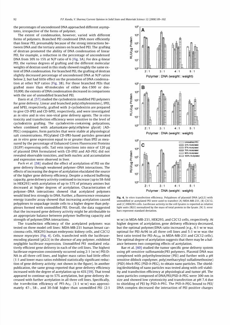
The transfection efficiency of the acetylated polymers was
tested on three model cell lines: MDA-MB-231 human breast car-cinoma cells, HEK293 human embryonic kidney cells, and C2C12mouse myocytes (Fig. 4). Cells, transfected with the luciferase-encoding plasmid (pGL3) in the absence of any polymer, exhibitednegligible luciferase expression. Unmodified PEI mediated rela-tively efficient gene delivery in each of the cell lines. The highestluciferase expression consistently occurred using 2:1 (w:w) PEI:D-NA in all three cell lines, and higher mass ratios had little effect(1:1 and lower mass ratios exhibited statistically significant reduc-tion of gene delivery activity; not shown in the figure). In anotherpublication, the same group reported that gene delivery efficiencyincreased with the degree of acetylation up to 43% [59]. That trendappeared to continue up to 57% acetylation, but gene delivery de-creased with further acetylation in all three cell lines. Specifically,
the transfection efficiency of PEI-Ac57 (3:1 w:w) was approxi-mately 47-, 58-, and 30-fold higher than unmodified PEI (2:1
w:w) in MDA-MB-231, HEK293, and C2C12 cells, respectively. Athigher degrees of acetylation, gene delivery efficiency decreased,
but the optimal polymer/DNA ratio increased (e.g., 4:1 w:w wasoptimal for PEI-Ac96 in all three cell lines and 5:1 w:w was thebest ratio tested for PEI-Ac100 in MDA-MB-231 and C2C12 cells).The optimal degree of acetylation suggests that there may be a bal-ance between two competing effects of acetylation.
Bae et al. [60] studied the tumor specific gene delivery systemusing pH sensitive sulfonamide/PEI polymers. Plasmid DNA wascomplexed with polyethyleneimine (PEI) and further with a pHsensitive diblock copolymer, poly(methacryloyl sulfadimethoxine)(PSD)-block-PEG (PSD-b-PEG), to obtain nano particles. The shield-ing/deshielding of nano particles was tested along with cell viabil-ity and transfection efficiency at physiological and tumor pH. Thenano particles composed of DNA/PEI/PSD-b-PEG were 300 nm insize and showed low cytotoxicity and transfection at pH 7.4 due
to shielding of PEI by PSD-b-PEG. The PSD-b-PEG bound to PEI/DNA complex decreased the interaction of PEI positive charges
Fig. 4. In vitro transfection efficiency. Polyplexes of plasmid DNA (pGL3) withunmodified or acetylated PEI were used to transfect (A) MDA-MB-231, (B) C2C12,and (C) HEK293 cells. Luciferase activity in the cell lysates is reported as relativelight units (RLU) normalized by the mass of total protein in the lysate. (N) 3; errorbars represent standard deviation.
92 P.P. Kundu, V. Sharma / Current Opinion in Solid State and Materials Science 12 (2008) 89–102

8/8/2019 Synthetic Polymeric Vectors in Gene Therapy
http://slidepdf.com/reader/full/synthetic-polymeric-vectors-in-gene-therapy 5/14
with cells and reduced the cytotoxicity by 60%. At pH 6.6, the nanoparticles showed high cytotoxicity and transfection, indicatingPSD-b-PEG detachment from the nano particles and permit PEI tointeract with cells. PSD-b-PEG was able to distinguish the small dif-ference in pH. The polymeric nano particle formed through electro-static attraction was designed in such a way that the final particleremained neutral. The neutral particle had the least interactionwith the body and would be more stable compared to anionicand cationic particles, during systemic circulation. The central ideaof this design was that when the particles experience a decrease inpH as they extravasated into tumor tissue due to enhanced perme-ability and retention effect, the sulfonamide groups would losetheir charge and got detached from the carrier complex. Thedetachment of sulfonamide from the complex exposed pCMV-Luc-DNA condensed with cell transfecting PEI, to act on cancer
cells. The scheme is shown in Fig. 5.Pun and Burke [61] studied the extracellular barriers to in vivo
PEI and PEGylated PEI polyplex-mediated gene delivery to theliver. The unpackaging of polyplexes by serum proteins, solubleglycosaminoglycans, and an extracellular matrix extract was dem-onstrated by a YOYO-1 fluorescence quenching assay. Additionally,exposing polyplexes to serum or proteoglycans before in vitro
transfection caused a decreased cellular uptake of DNA. Lastly,PEI polyplexes and PEGylated PEI polyplexes were injected intothe portal vein of mice, and the intrahepatic distributions of labeled DNA and polymer were assessed by confocal microscopy.PEI polyplexes delivered DNA to the liver, but extensive vectorunpackaging was observed, with PEI primarily colocalized withthe extracellular matrix. PEGylated polyplexes mediated less
DNA delivery to the liver, possibly due to premature vectorunpackaging in the blood. It was observed that both the bloodand the extracellular matrix have significant extracellular barriersto polyplex-mediated in vivo gene delivery to the liver.
3.2. Polyamidoamine (PAMAM) based vectors
Polyamidoamine (Starburst) dendrimers are spheroidal, cascadepolymers that can be synthesized from an ammonia or ethylenedi-amine core by successive addition of methyl acrylate and ethylene-diamine [62]. The size of the polymer, and importantly the surfacecharge, are controlled by varying the number of generations in thesynthesis. Haensler and Szoka [63] originally reported the use of PAMAM dendrimers for gene delivery. Uekama et al. [64,65] re-
ported the conjugates of polyamidoamine with cyclodextrin. Thestarburst polyamidoamine dendrimer conjugates with a-, b-, and
c-cyclodextrins (CDE conjugates), expecting the synergistic effectof dendrimer and cyclodextrins (CyDs) were synthesized [64].The CyDs were covaletly bound in a 1:1 molar ratio (confirmedby 1H NMR) to the dendrimer. The agarose gel electrophoreticstudies confirmed that the CDE conjugates formed the complexeswith plasmid DNA (pDNA) and protected the degradation of pDNAby DNase I in the same manner as dendrimer. CDE conjugatesshowed a potent luciferase gene expression, especially in the den-drimer conjugate with a-CyD (R-CDE conjugate), which providedthe greatest transfection activity (approximately 100 times higherthan those of dendrimer alone and of the physical mixture of den-drimer and a-CyD) in NIH3T3 and RAW264.7 cells. In addition, thegene transfer activity of a-CDE conjugate was superior to that of lipofectin. The enhancing gene transfer effect of a-CDE conjugatemight be attributable to not only increasing the cellular associa-tion, but also changing the intracellular trafficking of pDNA. Thesestudies indicated that a-CDE conjugate, had great advantages asnon-viral vectors, i.e., an easy preparation, superior transfectionefficiency, and less cytotoxicity.
Griffiths et al. [66] studied the mechanism of poly(amidoamine)as endosomolytic polymer and correlated the physicochemical andbiological properties. Bioresponsive poly(amidoamine)s (PAA)s arecurrently under development as endosomolytic polymers forintracellular delivery of proteins and genes. Small-angle neutronscattering (SANS) was used to systematically investigate the pH-dependent conformational change of an endosomolytic polymer,the PAA ISA 23. The radius of gyration of the ISA23 was determinedas a function of pH and counterion, the aim being to correlatechanges in polymer conformation with membrane activity as-sessed using a rat red blood cell hemolysis assay. With decreasingpH, the ISA23 radius of gyration increased to a maximum(Rg 80 Å) at around pH 3, before subsequently decreasing oncemore. At high pH and high ionic strengths, the polymer was nega-tively charged and adopts a rather compact structure (Rg < 20 Å),presumably with the dissociated carboxylic groups on the exteriorof the polymer coil. At low pH, the coil again collapses (Rg < 20 Å),
presumably due to the effects of the high ionic strength. It wasconcluded that the nature of the salt form had no direct bearingon the size of the polymer coil, but it did indirectly determinethe prevailing pH and, hence, polymer conformation. Pulsed-gradi-ent spin-echo NMR measurements were in good agreement withthe SANS estimates of the radius of gyration.
Szoka et al. [67] used degraded PAA dendrimers for in vitro genedelivery. They transfected cultured cells using complexes betweenDNA and spherical cationic polyamidoamine polymers (Starburstdendrimers) that consist of primary amines on the surface and ter-tiary amines in the interior. A 50-fold increase in the transfectionactivity of the dendrimers was observed by heat treatment in avariety of solvolytic solvents, e.g., water or butanol. This treatmentinduced significant degradation of the dendrimer at the amide
linkage, resulting in a heterodisperse population of compoundswith molecular weights ranging from the very low (<1500 Da) toseveral tens of kilodaltons. The compound facilitating transfectionwas the high molecular weight component of the degraded prod-uct and was denoted as a fractured dendrimer. Transfection activ-ity was related both to the initial size of the dendrimer and thedegree of degradation. Fractured dendrimers exhibited an increasein apparent volume as measured by an increase in the reducedviscosity upon protonation of the terminal amines as pH wasreduced from 10.5 to 7.2, whereas intact dendrimers did not re-duced. Dendrimers with defective branching were also synthesizedand also showed improved transfection activity compared to thatof the intact dendrimers. For a series of heat-treated dendrimers,a correlation between transfection activity and the degree of flex-
ibility was also observed. The transfection activity (relative to theuntreated dendrimer) of acetic anhydride titrated dendrimers (fifth
Fig. 5. Targeting based on difference in pH: (a) shows formation of the nanoparticlecomplex through charge–charge interaction between DNA, cationic polymer (PEI),and PSD-b-PEG; (b) shows the complex shielded at physiological pH and deshieldedat cancer pH.
P.P. Kundu, V. Sharma / Current Opinion in Solid State and Materials Science 12 (2008) 89–102 93

8/8/2019 Synthetic Polymeric Vectors in Gene Therapy
http://slidepdf.com/reader/full/synthetic-polymeric-vectors-in-gene-therapy 6/14
generation ammonia core) are shown in Fig. 6. The primary aminecontent of the fifth generation ammonia core dendrimer was var-ied by direct reaction with acetic anhydride. Ninhydrin analysisshowed nearly stoichiometric addition by acetic anhydride to ter-minal amines. Transfection by these amine-titrated dendrimersshowed a significant decrease in activity for a 25% reduction in
available terminal amines.Complete abolishment of activity was seen at 50% reduction,
even in the presence of sufficient excess positive charge to neutral-ize the DNA. Thus, if all primary amines were assumed to be pro-tonated at physiological pH, a high surface charge density wasnecessary for dendrimer transfection. However, the terminalamine contents in highly active, fractured dendrimers that hadbeen dialyzed and lyophilized to remove low molecular weightcomponents following heat treatment were significantly lowerthan their intact counterparts, which were not active in transfec-tion (Tables 1–2). Therefore, a high number of terminal amineswere required for transfection by dendrimers, other physical prop-erties of the polymer also significantly contributed to their activity.
A PAMAM-PEG-PAMAM based triblock copolymers were re-
ported as carrier for gene delivery [68]. PAMAM dendrimer is anefficient gene carrier itself, but it is associated with certain prob-lems, such as low water solubility and considerable cytotoxicity.
The introduction of PEG to engineer a nontoxic and highly transfec-tion efficient polymeric gene carrier was analyzed (because PEG isknown to convey water solubility and biocompatibility to the con-
jugated copolymer). The copolymer was composed of a PEG coreand two PAMAM substructures on both sides. It was able to self-assemble with plasmid DNA forming a compact polyplex, whichshowed greatly enhanced water solubility compared to the PA-
MAM dendrimer itself. The polyplex thus formed was found tohave a spherical shape with a nanosize appropriate to gene deliv-ery and a narrow size distribution. The copolymer had little cyto-toxicity in mammalian cells, low interaction with serum, andhigh transfection efficiency comparable to that of PEI in 293 cells.
Li et al. [69] used G5 Polyamidoamine (PAMAM) dendrimers forgene delivery (from G0 to G10 generation, the molecular weightand the number of surface active units of PAMAM dendrimers in-crease exponentially, and their diameters increase linearly). Theyisolated plasmid DNA ensuring an OD 260/280 ratio above 1.8and incubated these components (PAMAM & plasmid DNA) for1 h at room temperature in different weight ratios. The bindingassay was analyzed by agarose gel electrophoresis. These studiesindicated that these G5 PAMAM dendrimers could be used tomediate efficient, nonspecific in vitro transfer of genetic materialinto eukaryotic cells and in vivo transfer. PAMAM dendrimers havesufficient surface amine groups, which make them feasibly interactwith DNA, form complexes through their charge-based interac-tions, and transfer DNA to cells efficiently both in vitro and in vivo.
3.3. Acrylate based polymer vectors
The group of Hennink et al. [70–73] synthesized and evaluatedpoly(2-(dimethylamino) ethyl methacrylate) (pDMAEMA) for genetransfer. The polymer forms positively charged DNA polyplexes,which can be used successfully for in vitro transfection of differentcell lines, including COS-7 and OVCAR-3 cells. HMW forms of thepolymer (>300 kDa) were able to condense DNA effectively intoparticles of 150–200 nm, whereas LMW pDMAEMA forms large
complexes (size 0.5–1.0 micrometer). Like other cationic polymers,pDMAEMA is cytotoxic, therefore, copolymers of DMAEMA withmethyl methacrylate (MMA), ethoxytriethylene glycol methacry-late (triEGMA), or N -vinyl-pyrrolidone (NVP) were also evaluated.A copolymer with 20 mol% of MMA showed reduced transfectionefficiency but an increased cytotoxicity. A copolymer with triEGMA(48 mol%) showed both a reduced transfection efficiency and areduced cytotoxicity, whereas a copolymer with NVP (54 mol%)showed an increased transfection efficiency but a decreasedcytotoxicity as compared to pDMAEMA. There data indicated thataggregate formation of positively charged polyplexes with bloodcomponents followed by trapping of the polyplex aggregates inthe lung capillaries is probably responsible for a preferential lunguptake and transfection.
Henninck and colleagues [74] also synthesized a new polymer,with two tertiary amine groups in each monomeric unit, poly-(2-methyl-acrylic acid 2-[(2-(dimethylamino)-ethyl)-methyl-
Fig. 6. Transfection activity (relative to the untreated dendrimer) of aceticanhydride titrated dendrimers (fifth generation ammonia core). Complexes wereformed and added to CV-1 cells cultured in 96-well trays as described in theExperimental procedures. The pCMV-bGal plasmid DNA was held constant at 0.6 lgper well, and the ratio of all dendrimer terminal groups (modified or not) to DNAphosphates was varied from 0.5 to 24. The maximum activity level, regardless of terminal group to phosphate ratio used, is reported. Control sample is theunmodified dendrimer; the 0% sample underwent the same treatment as the othersamples, but in the absence of acetic anhydride. Data reported are the mean andrange of duplicate wells in one run which was representative of three similarexperiments.
Table 1
Polymer characteristics associated with dendrimers heated to the point of highest transfection activity.a
Dendrimer Heatingtime (h)b
Measured relativeamine contentc
Computed % amidebonds cleaved
Initialmolecular (kD)
Computed finalmolecular (kD)
Initialmolecule
Computed finalmolecule
Computed netmolecule
Computedflexibility (vol/wt)
Computed% defects
5-TAEA 8 0.93 13 21 18.3 96 77 69 6.9 56-TAEA 10 0.83 35 43 28.5 192 107 80 7.6 106-EDA 12 0.76 50 58 28.9 256 97 57 7.5 138-EDA 20 0.62 70 233 70.1 1024 190 41 7.4 17
a Dendrimers of various initial size were heat treated, dialyzed against water, then assayed for primary amine content. The amine content for each dendrimer, relative tothat of its intact counterpart, was used to determine the extent of degradation based on the fractured dendrimer model, and thus, the model parameters.
b
Heating time at which maximum activity was observed.c Amine content by weight relative to the corresponding intact dendrimer as determined by ninhydrin assay.
94 P.P. Kundu, V. Sharma / Current Opinion in Solid State and Materials Science 12 (2008) 89–102

8/8/2019 Synthetic Polymeric Vectors in Gene Therapy
http://slidepdf.com/reader/full/synthetic-polymeric-vectors-in-gene-therapy 7/14

8/8/2019 Synthetic Polymeric Vectors in Gene Therapy
http://slidepdf.com/reader/full/synthetic-polymeric-vectors-in-gene-therapy 8/14
observed that the highly versatile nanoparticles can be used forDNA vaccination via size-dependent targeting of phagocytic cells.The properties of these nanoparticles such as acid degradability,surface charges, and types of cargo (e.g., siRNA, miRNA, and oligo-deoxynucleotides) can easily be tuned for specific applications, and
the targeting and imaging modalities can also be conjugated withthese nanoparticles to achieve targeted delivery, as well as com-bined imaging and gene therapy.
3.4. Poly(amino-esters) based polymer vectors
Prof. Langer’s group at MIT developed a large library of poly-(b-amino esters) (PBAEs) based on parallel synthesis using differ-ent classes of amines and diol-diacrylates, and these were testedfor gene delivery applications [81–83]. By using high-throughputsynthesis techniques, they created libraries of over 2200 structur-ally unique poly(b-amino esters) (PBAEs). Diacrylate monomers(letters) and amine monomers (numbers) used to synthesizePBAEs. are shown in Fig. 8. PBAEs are hydrolytically degradable,
condense plasmid DNA at physiological pH, and are readily synthe-sized via the conjugate addition of primary or secondary amines to
diacrylates (Fig. 9) [82–84]. An initial screen of model polymersidentified these materials as potential gene carriers and demon-strated that structural variations could have a significant impacton DNA binding and transfection efficacies [82,85–87].
The amino-esters based polymer vector has numerous advanta-
ges over other polymer vectors such as (1) diamine and diacrylatemonomers are inexpensive, commercially available starting mate-rials, (2) polymerization in a single synthetic step reaction, and (3)no byproducts are generated during polymerization. Poly(b-aminoesters) (PBAEs) are promising compared with PEI due to theirbiodegradability via hydrolytically degradable ester groups, theirreduced cytotoxicity, their ability for triggered DNA release withinthe cell, and their potential for structural diversity [88]. Initialstudies of this class of polymer showed that by varying synthesisconditions the polymer molecular weight can be varied from2000 to 50,000 Da [89]. In physiological conditions, these polymershave a degradation half-life on the order of hours, but this is slo-wed considerably at a pH of 5 or when the polymers electrostati-cally condense DNA and form nanoparticles [81–83].
Investigative studies on the proton sponge mechanism andendosomal release by Akinc et al. [83,84] also revealed that PBAEs
Fig. 7. (a) ATRP of 2-(dimethylamino)ethyl methacrylate (DMAEMA) using N3-functionalized initiator in dichlorobenzene (DCB); (b) pHEMA with side chain of alkynes; (c)Click chemistry to form degradable brushed pHEMA–pDMAEMA.
96 P.P. Kundu, V. Sharma / Current Opinion in Solid State and Materials Science 12 (2008) 89–102

8/8/2019 Synthetic Polymeric Vectors in Gene Therapy
http://slidepdf.com/reader/full/synthetic-polymeric-vectors-in-gene-therapy 9/14
can successfully buffer the endosomal compartment, similarly toPEI. A library of 140 PBAEs composed of 7 diacrylate monomersand 20 amine monomers was synthesized in parallel and screenedfor gene delivery efficacy [82,83]. In general, the best performing
complexes were found to have effective diameters smaller than250 nm and positive f-potentials in 10 mM HEPES buffer. It was
determined that the majority of PBAE/DNA particles were limitedby poor cell uptake, two of the polymers had high uptake and med-iated gene delivery 4- to 8-fold higher than PEI, comparable to theefficacy of Lipofectamine 2000. These studies demonstrated the
utility of using parallel synthesis and screening of a polymerlibrary to identify novel gene delivery polymers with efficacy
Fig. 8. Diacrylate monomers (letters) and amine monomers (numbers) used to synthesize PBAEs.
P.P. Kundu, V. Sharma / Current Opinion in Solid State and Materials Science 12 (2008) 89–102 97

8/8/2019 Synthetic Polymeric Vectors in Gene Therapy
http://slidepdf.com/reader/full/synthetic-polymeric-vectors-in-gene-therapy 10/14
greater than PEI. PBAEs that were poor transfection agents failedfor a variety of reasons [83].
Some polymers were simply not sufficiently water soluble,while others were unable to electrostatically bind DNA tightly en-ough to prevent its movement during gel electrophoresis. Thepolymers which could bind DNA, many of them formed particleswith low cellular uptake. Some particles caused too high toxicityeven they had sufficient cellular uptake. Two leading PBAEs werefurther optimized by varying polymer molecular weight, polymerend group, and polymer to DNA weight ratio [90–92]. Interestingly,it was also discovered that polymers terminated with diacrylatemonomers were unable to deliver DNA to cells, unlike the nearlyidentical polymers that were terminated with amine monomers in-stead. Zugates et al. [93,94] used one step reaction to conjugate 12new amino monomers and three diacrylate terminated base poly-mers. These newly synthesized amino-esters were studied for theeffect of the modification on the properties of these polymers forgene delivery. In another study, the end groups were modified bythe two-step approach; the first step involved preparation of anacrylate-terminated polymer followed by the second step of postpolymerization amine-capping to generate end-functionalizedpolymers [95]. It is observed that the in vitro transfection levelscan be increased by 30% and the optimal polymer/DNA ratio low-ered 5-fold by conjugation of the appropriate end group. The most
effective modifications were made by grafting primary diaminemolecules to the chain end.
Prof. Langer’s lab also used the best performing amino-esterbased polymer (C32) for gene therapy in the treatment of cancer[96]. The best performing amino-ester polymer (C32) was testedin mice for toxicity and DNA delivery after intratumor and intra-muscular injection. C32 delivered DNA intratumorally 4-fold betterthan one of the best commercially available reagents, jetPEI (poly-ethyleneimine), and 26-fold better than naked DNA. In the samelab, poly amino-ester containing microparticles were used to in-crease the efficiency of the non-viral genetic vaccines [97]. Theseformulations generated an increase of 3–5 orders of magnitudein transfection efficiency and were potent activators of dendriticcells in vitro. When these were used as vaccines in vivo, these
microparticle formulations, unlike conventional formulations, in-duced antigen-specific rejection of transplanted syngenic tumorcells.
Park et al. [98] synthesized biodegradable cross-linked poly-(b-amino ester) (CLPAE) by Michael addition of pentaerythritol tri-acrylate and N ,N -dimethylethylenediamine and modified withaminohexanoic acid and lysine to CLPAE-Ahx and CLPAE-Lys,respectively, for a gene delivery system. The polymers showedminimal cytotoxicity on 293 cells due to their biodegradabilityand biocompatibility. Transfection efficiencies of CLPAEAhx andCLPAE-Lys were comparable to that of PEI in 293 cells and C2C12cells. Additionally, high transfection of CLPAEAhx on primary rataorta vascular smooth muscle cells (SMC) and primary mouseembryonic fibroblast (MEF) cells showed a potential for a gene
delivery system on primary cells, restenosis treatment of humanSMC, and MEF cells. Park and coworkers [99] also compared the
transfection efficiency of the network poly(amino ester), (n-PAE),with PEI. There was a marked difference in the cytotoxicity of the polymers. The majority of PEI-transfected cells were granu-lated and dead, whereas most of the cells transfected with n-PAEwere viable and healthy. In addition to this, the n-PAE-mediatedtransfection was also very efficient in the presence of serum.
3.5. Poly(ethylene glycol) based polymer vectors
PEG is very useful because of its ease of preparation, relativelylow cost, controllable molecular weight and the ability to link it tolipids or protein (including antibody) by a variety of methods. PEGisthepolymerofchoiceforproteinmodificationbecauseitpossessesseveral favorable properties such as the lack of immunogenicity,antigenicity, andtoxicity, anda high solubility in water and in manyorganic solvents. PEG is also approved by the FDA for human use.Commonreasons forthe PEGylation of a drug areto reduceits excre-tion by thekidneys, to avoid or reduceits degradation by proteolyticenzymes and / or hydrolytic media, to enhance its water solubility(forhighlyhydrophobicmolecules),to reduce its reticuloendothelial(RES) clearance, and to reduce its immunogenicity and antigenicity(mainly for peptides and proteins) [100–105]. Furthermore, forsmall drugs, polymer conjugation may yield improved and moreconvenient biodistribution, selected cellular uptake [106–108] or,
through tailor-madechemistry, a triggered drug release or targetinginto specific organs or cells [109].
Modificationof polyplexes with poly(ethylene glycol) (PEG), typ-ically grafted onto the polymer as a brush, can stabilize polyplexesagainst salt, protein, and complement-induced inactivation[110,111]. The increased stability due to PEGylation is presumed toresult from steric effects, leading to a decreased particle–particleand particle–protein interactions. The effect depends on the PEGmolecular weight, the grafting density, and the method of attach-ment of PEG to the polymer [110,112]. Langer et al. [113] synthe-sized PEG-polyhistidine conjugates to evaluate the material aspotential gene delivery vehicles. Two conjugate architectures(comb-shaped and linear A–B block copolymers) were synthesizedand formulated with plasmid DNA. The complexes were character-
ized with respect to DNA complexation capacity, hydrodynamicdiameter, f-potential, in vitro cytotoxicity and transfection capacityin a model cell line. PEG content of the conjugate significantly influ-enced the hydrodynamic diameter of theDNA: conjugate compositein aqueous solution. For comb-shaped conjugates, steric hindranceattributed to PEG led to a direct relationship between the PEG con-tent and the complex size. Both architectures could condense plas-mid DNA into complexes with hydrodynamic diameters <150 nm.Complexation of DNA with the PEG-polyhistidine conjugates re-sulted in nanocomposites with negative zeta potentials that re-tarded DNase I mediated hydrolysis, and all conjugates showed alow cytotoxicity to macrophages cultured in vitro. The transfectionefficiency was approximately equivalent to DNA-polylysinecomplexes.
Yin et al. [114] reported the synthesis of poly(ethylene glycol)-poly(n-butyl cyanoacrylate) nanocapsules with oil core via mini-
Fig. 9. Synthesis of PBAEs by conjugate addition of amines to diacrylates.
98 P.P. Kundu, V. Sharma / Current Opinion in Solid State and Materials Science 12 (2008) 89–102
8/8/2019 Synthetic Polymeric Vectors in Gene Therapy
http://slidepdf.com/reader/full/synthetic-polymeric-vectors-in-gene-therapy 11/14
emulsion polymerization. The nanocapsules were synthesizedthrough miniemulsion polymerization of butylcyanoacrylate (BCA)with PEG as initiator. The particle size and the f-potential of nano-capsules were influenced by the PEG content in the polymerizationsystem. Fourier transform infrared (FTIR) spectra and 1H NMR dem-onstrated the chemical coupling betweenPEG and poly(butylcyano-acrylate) (PBCA). Thermal characteristics of the copolymer wereinvestigated by differential scanning calorimetry (DSC). The encap-sulation efficiency increased concurrently with the increase of PEGcontent in the system. The hemolytic assay and the cytotoxicitymeasurement showed that the PEG coating could significantlyreduce the hemolyticpotentialand cytotoxicityof the nanocapsules.Nagasaki and coworkers [115] preparedanovelABCtriblockcopoly-mer for constructing a pH-responsive and targetable non-viral genevector. The copolymer, lactosylated poly(ethylene glycol)-block-poly(silamine)-block-poly[2-(N ,N -dimethylamino)ethylmethacry-late] (Lac-PEG-PSAO-PAMA),consistsof lactosylatedpoly(ethyleneglycol) (A-segment), a pH-responsive polyamine segment (B-seg-ment) and a DNA-condensing polyamine segment (C-segment).The Lac-PEG-PSAO-PAMA spontaneously associated with plasmidDNA (pDNA) to form three-layered polyplex micelles with aPAMA/pDNA polyion complex (PIC) core, an uncomplexed PSAOinner shell, and a lactosylated PEG outer shell, as confirmed by 1HNMR spectroscopy. Under physiological conditions, the Lac-PEG-PSAO-PAMA/pDNA polyplex micelles prepared at an N/P (numberof amino groups in the copolymer/number of phosphate groupsin pDNA) ratio above 3 were found to be able to condense pDNA,thus adopting a relatively smallsize (<150 nm) andan almost neu-tral surface charge (f+5 mV). The micelle underwent a pH-in-duced size variation (132.6 nm for pH 7.4 and 181.8 nm for pHpH 4.0). This is due to the conformational changes (globule-rodtransition) of the uncomplexed PSAOchain in responseto pH, lead-ing to swelling of the free PSAO inner shell at lowered pH whileretaining the condensed pDNA in the PAMA/pDNA PIC core. Fur-thermore, the micelles exhibited a specific cellular uptake intoHuH-7 cells (hepatocytes) through asialoglycoprotein (ASGP)
receptor-mediated endocytosis and achieved a far more efficienttransfection ability of a reporter gene compared to the Lac-PEG-PSAO/pDNA and Lac-PEG-PAMA/pDNA polyplex micelles com-posed of the diblock copolymers and pDNA.
Li et al. [116] synthesized acid labile block copolymers based onpoly(ethylene glycol) and poly(2-(dimethylamino)ethyl methacry-late) segments connected through a cyclic ortho ester linkage.PEG-a-PDMAEMA condensed with plasmid DNA formed polyplexnanoparticles with an acid-triggered reversible PEG shield. ThepH-dependent shielding/deshielding effect of PEG chains on thepolyplex particles were evaluated by f-potential and size measure-ments. At pH 7.4, polyplexes generated from PEG-a-PDMAEMAexhibited smaller particle size, lower surface charge, reduced inter-action with erythrocytes, and less cytotoxicity compared to PDMA-
EMA-derived polyplexes. At pH 5.0, f-potential of polyplexesformed from PEG-a-PDMAEMA increased, leveled up after 2 h of incubation and gradual aggregation occurred in the presence of bovine serum albumin (BSA). In contrast, the stably shieldedpolyplexes formed by DNA and an acid-stable block copolymer,PEG-b-PDMAEMA, did not change in size and f-potential in 6 h.In vitro transfection efficiency of the acid-labile copolymer greatlyincreased after 6 h incubation at pH 5.0, approaching the samelevel of PDMAEMA, whereas there was only slight increase in effi-ciency for the stable copolymer, PEG-b-PDMAEMA.
3.6. Other polymer vectors
There are various other synthetic polymer vectors which arecurrently being tested for gene delivery research such as poly(4-
vinylimidazole) [117], poly(vinyl alcohol) [118], polyallylamine
derivatives [119], aliphatic ionenes [120], polyvinyl derivatives[121], etc. These polymers showed promising properties for genedelivery. Poly(4-vinylimidazole) was studied as gene carrier byCho et al. [117] and they found that this polymer showed hightransfection efficiency through the proton sponge mechanism of imidazole groups. Kissel and coworkers [118] reported the bio-physical and transfection studies of an amine modified poly(vinyl
alcohol) for gene delivery. The resulting polymers were character-ized using NMR, thermogravimetric analysis (TGA), and gel perma-tion chromatography (GPC). Atomic force microscopy (AFM),dynamic light scattering photon correlation spectroscopy (PCS),and f-potential were used to investigate polyplexes of DNA withPVA copolymers. These studies suggested an influence of the poly-cation structure on the morphology of condensed DNA in polyplex-es. Significant differences were observed by changing both thedegrees of amine substitution and the structure of the PVA back-bone, demonstrating that both electrostatic and hydrophobic inter-actions affected DNA condensation. DNA condensation measuredby an ethidium bromide intercalation assay showed a higher de-gree of condensation with pDNA with increasing degrees of aminesubstitution and more hydrophobic functional groups. These find-ings were in line with transfection experiments, in which a gooduptake of these polymer–DNA complexes was noted with littleendosomal escape. Co-administration of chloroquine resulted inan increased endosomal escape and higher transfection efficien-cies, due to disruption of the endosomal membrane.
Boussif et al. [119] used polyallylamine derivatives for genedelivery. They found that its ability to mediate gene transfer intocells increased by several orders of magnitude. Transfection effi-ciency was found to be dependent on the substitution level of ami-no groups and reached a highest levels in the presence of lysosomotropic and/or fusogenic agents. At optimal conditions,glycolylated PAM was shown to be as efficient as the linear PEIof 22 kDa. Langer et al. [120] used aliphatic ionenes as gene deliv-ery agents and elucidated the structure-function relationship. Theionene fractions and their polyelectrolyte complexes (PEC) with
DNA were studied using physicochemical and biological methods.Ionene polymers were shown to possess low cytotoxicity (minimalviability of the P388D1 murine macrophage cells was 80%). Degreeof polymerization (DP) and charge density of the ionenes wereshown to be factors of effective control of PEC dissolution inwater-salt solutions. Rolland et al. [121] used polyvinyl derivativesas novel interactive polymers for controlled gene delivery to mus-cle. A pDNA, containing a cytomegalovirus (CMV) promoter and agalactosidase reporter gene (CMV-beta-gal), was injected eitherin saline or formulated in polyvinyl pyrrolidone (PVP) and polyvi-nyl alcohol (PVA) solutions. The interactions between PVP andpDNA were assessed by dynamic dialysis, isothermal titration cal-orimetry (ITC), and fourier transformed infrared (FTIR) spectros-copy. The interaction between PVP and pDNA was found to be an
endothermic process governed by hydrogen bonding and resultsin protection of pDNA from extracellular nucleases.
4. Conclusions and future prospective
The gene therapy is presenting a promising approach for thetreatment of genetic diseases. Over the past decade and at present,there has been continuous research to find better non-viral vectorsto overcome the limitations of viral vectors. There are many syn-thetic polymers, which are being used and assessed for their appli-cations in gene therapy. One of these is the poly(b-amino ester)based vectors. Prof. Langer’s group has developed the library of these newly synthesized poly(b-amino ester) based vectors. Withan increase in the understanding of different diseases, the clinical
applications will potentially expand into areas such as neurological
P.P. Kundu, V. Sharma / Current Opinion in Solid State and Materials Science 12 (2008) 89–102 99

8/8/2019 Synthetic Polymeric Vectors in Gene Therapy
http://slidepdf.com/reader/full/synthetic-polymeric-vectors-in-gene-therapy 12/14
disorders, tissue regeneration, cancer, monogeneic diseases, vascu-lar disease, and infectious diseases. The non-viral vectors representthe future of human gene therapy. From the results published inthe past decade, simple polymer vectors are difficult to help DNAcross all barriers in the gene delivery. The future polymer vectorswill be much like virus, called synthetic virus, combining the bestof current polymer and viral vectors. The synthetic multi-compo-nent and multifunction polymer vectors, mimicking some func-tions of viral vectors, will be designed and applied in the nextdecade.
References
[1] Mulligan RC. The basic science of gene therapy. Science 1993;260:926–32.[2] Lozier JN, Brinkhous KM. Gene therapy and the hemophilias. J Am Med Assoc
1994;271:47–51.[3] Inui K, Okada S, Dickson G. Gene therapy in Duchenne muscular dystrophy.
Brain Dev 1996;18:357–61.[4] Hartigan-O’Connor D, Chamberlain JS. Progress toward gene therapy of
Duchenne muscular dystrophy. Sem Neurol 1999;19:323–32.[5] Hartigan-O’Connor D, Chamberlain JS. Developments in gene therapy for
muscular dystrophy. Microsc Res Tech 2000;48:223–38.[6] Alton EWFW, Geddes DM, Gill DR, Higgins CF, Hyde SC, Innes JA, et al.
Towards gene therapy for cystic fibrosis: a clinical progress report. Gene Ther
1998;5:291–2.[7] Nabel EG. Gene therapy for cardiovascular diseases. J Nucl Cardiol
1999;6:69–75.[8] Dunnett SB, Bjorklund A. Prospects for new restorative and neuroprotective
treatments in Parkinson’s disease. Nature 1999;399:A32–9.[9] Alisky JM, Davidson BL. Gene therapy for amyotrophic lateral sclerosis and
other motor neuron diseases. Hum Gene Ther 2000;11:2315–29.[10] Barkats M, Bilang-bleuel A, Buc-caron MH, Castelbarthe MN, Corti O, Finiels F,
et al. Adenovirus in the brain: recent advances of gene therapy forneurodegenerative diseases. Prog Neuorbiol 1998;55:333–41.
[11] Chadwick DR, Lever AML. Gene therapy for HIV infection. Exp Opin TherPatents 1998;8:983–90.
[12] Bonadio J, Goldstein SA, Levy RJ. Gene therapy for tissue repair andregeneration. Adv Drug Delivery Rev 1998;33:53–69.
[13] Vile RG, Russell SJ, Lemoine NR. Cancer gene therapy: hard lessons and newcourses. Gene Ther 2000;7:2–8.
[14] Folkman J. Antiangiogenic gene therapy. Proc Natl Acad Sci USA1998;95:9064–6.
[15] Roth JA, Cristiano RJ. Gene therapy for cancer: what have we done and whereare we going? J Natl Cancer Inst 1997;89:21–39.
[16] Nabel EG. Gene therapy for vascular diseases. Atherosclerosis1995;118:S51–6.
[17] Donnelly JJ, Ulmer JB, Shiver JW, Liu MA. DNA vaccines. Annu Rev Immunol1997;15:617–48.
[18] Vaheri A, Pagano JS. Infectious poliovirus RNA a sensitive method of assay.Virology 1965;27:434–6.
[19] Graham FL, Eb AJVD. A new technique for the assay of infectivity of humanadenovirus 5 DNA. Virology 1973;52:456–67.
[20] Marshall E. Clinical trials: gene therapy death prompts review of adenovirusvector. Science 1999;286:2244–5.
[21] Marshall E. FDA halts all gene therapy trials at Penn. Science 2000;287:565–7.
[22] Marshall E. Clinical research. Gene therapy a suspect in leukemia-like disease.Science 2002;298:34–5.
[23] Kaiser J. Gene therapy. Seeking the cause of induced leukemias in X-SCID trial.Science 2003;299:495.
[24] Ledley FD. Nonviral gene therapy: the promise of genes as pharmaceutical
products. Hum Gene Ther 1995;6:1129–44.[25] Lollo CP, Banaszczyk MG, Chiou HC. Obstacles and advances in non-viral genedelivery. Curr Opin Mol Ther 2000;2:136–42.
[26] Mislick KA, Baldeschwieler JD. Evidence for the role of proteoglycans incation-mediated gene transfer. Proc Natl Acad Sci USA 1996;93:12349–54.
[27] Goncalves C, Pichon C, Guerin B, Midoux P. Intracellular processing andstability of DNA complexed with histidylated polylysine conjugates. J GeneMed 2002;4:271–81.
[28] Goncalves C, Mennesson E, Fuchs R, Gorvel JP, Midoux P, Pichon C.Macropinocytosis of polyplexes and recycling of plasmid via the clathrin-dependent pathway impair the transfection efficiency of humanhepatocarcinoma cells. Mol Ther 2004;10:373–85.
[29] Kopatz I, Remy JS, Behr JP. A model for non-viral gene delivery: throughsyndecan adhesion molecules and powered by actin. J Gene Med 2004;6:769–76.
[30] Ruponen M, Honkakoski P, Tammi M, Urtti A. Cell-surfaceglycosaminoglycans inhibit cation-mediated gene transfer. J Gene Med2004;6:405–14.
[31] Wu GY, Wu CH. Receptor-mediated in vitro gene transformation by a soluble
DNA carrier system. J Biol Chem 1987;262:4429–32.
[32] Wagner E, Zenke M, Cotten M, Beug H, Birnstiel ML. Transferrin–polycationconjugates as carriers for DNA uptake into cells. Proc Natl Acad Sci USA1990;87:3410–4.
[33] Curiel DT, Agarwal S, Wagner E, Cotten M. Adenovirus enhancement of transferrin–polylysine-mediated gene delivery. Proc Natl Acad Sci USA1991;88:8850–4.
[34] Lukacs GL, Haggie P, Seksek O, Lechardeur D, Freedman N, Verkman AS. Size-dependent DNA mobility in cytoplasm and nucleus. J Biol Chem2000;275:1625–9.
[35] Ludtke JJ, Zhang G, Sebestyén MG, Wolff JA. A nuclear localization signal can
enhance both the nuclear transport and expression of 1 kb DNA. J Cell Sci1999;112:2033–41.
[36] Brunner S, Furtbauer E, Sauer T, Kursa M, Wagner E. Overcoming the nuclearbarrier cell: cycle independent nonviral gene transfer with linearpolyethylenimine or electroporation. Mol Ther 2002;5:80–6.
[37] Schaffer DV, Fidelman NA, Dan N, Lauffenburger DA. Vector unpacking as apotential barrier for receptor-mediated polyplex gene delivery. BiotechnolBioeng 2000;67:598–606.
[38] Plank C, Tang MX, Wolfe AR, Szoka FC. Branched cationic peptides forgene delivery: role of type and number of cationic residues in formationand in vitro activity of DNA polyplexes. Hum Gene Ther 1999;10:319–32.
[39] Parker AL, Oupicky D, Dash PR, Seymour LW. Methodologies for monitoringnanoparticle formation by self-assembly of DNA with poly(L -lysine). AnalBiochem 2002;302:75–80.
[40] Hardy JG, Kostiainen MA, Smith DK, Gabrielson NP, Pack DW. Dendrons withspermine surface groups as potential building blocks for nonviral vectors ingene therapy. Bioconjug Chem 2006;17:172–8.
[41] Reineke TM, Davis ME. Structural effects of carbohydrate-containingpolycations on gene delivery. 2. Charge center type. Bioconjug Chem2003;14:255–61.
[42] Reineke TM, Davis ME. Structural effects of carbohydrate-containingpolycations on gene delivery. 1. Carbohydrate size and its distance fromcharge centers. Bioconjug Chem 2003;14:247–54.
[43] Gilding DK. Biocompatibility of clinical implant materials, Vol. 2. CRC Press;1981.
[44] Shea LD, Smiley E, Bonadio J, Mooney DJ. DNA delivery from polymer matricesfor tissue engineering. Nat Biotechnol 1999;17:551–4.
[45] Abdallah B, Hassan A, Benoist C, Goula D, Behr JP, Demeneix BA. A powerfulnonviral vector for in vivo gene transfer into the adult mammalian brain:polyethylenimine. Hum Gene Ther 1996;7:1947–54.
[46] Ferrari S, Moro E, Pettenazzo A, Behr JP, Zacchello F, Scarpa M. ExGen 500 isan efficient vector for gene delivery to lung epithelial cells in vitro and in vivo.Gene Ther 1997;4:1100–6.
[47] Chemin I, Moradpour D, Wieland S, Offensperger WB, Walter E, Behr JP, et al.Liverdirected gene transfer: a linear polyethylenimine derivative mediateshighly efficient DNA delivery to primary hepatocytes in vitro and in vivo. J
Viral Hepat 1998;5:369–75.[48] Turunen MP, Hiltunen MO, Ruponen M, Virkamaki L, Szoka FC, Urtti A, et al.Efficient adventitial gene delivery to rabbit carotid artery with cationicpolymer–plasmid complexes. Gene Ther 1999;6:6–11.
[49] Zou SM, Erbacher P, Remy JS, Behr JP. Systemic linear polyethylenimine(L-PEI)-mediated gene delivery in the mouse. J Gene Med2000;2:128–34.
[50] Ji Z, Manuel WS, Hornsby PJ. Transfection of cells mediated by biodegradablepolymer materials with surface bound polyethlenimine. Biotechnol Prog2000;16:254–7.
[51] Blessing T, Kursa M, Holzhauser R, Kircheis R, Wagner E. Different strategiesfor formation of PEGylated EGF-conjugated PEI/DNA complexes for targetedgene delivery. Bioconjugate Chem 2001;12:529–37.
[52] Gosselin MA, Guo W, Lee RJ. Efficient gene transfer using reversibly cross-linked lowmolecular weight polyethylenimine. Bioconjugate Chem2001;12:989–94.
[53] Petersen H, Kunath K, Martin AL, Stolnik S, Roberts CJ, Davies MC, et al. Star-shaped poly(ethylene glycol)-block-polyethylenimine copolymers enhanceDNA condensation of low molecular weight polyethylenimines.
Biomacromolecules 2002;3:926–36.[54] Fischer D, Harpe AV, Kunath K, Peterson H, Li Y, Kissel T. Copolymers of
ethylene imine and N -(2-hydroxyethyl)-ethylene imine as tools to studyeffects of polymer structure on physicochemical and biological properties of DNA complexes. Bioconjugate Chem 2002;13:1124–33.
[55] Tseng WC, Jong CM. Improved stability of polycationic vector bydextran-grafted branched polyethylenimine. Biomacromolecules 2003;4:1277–84.
[56] Li S, Tseng WC, Stolz DB, Wu SP, Watkins SC, Huang L. Dynamic changes in thecharacteristics of cationic lipidic vectors after exposure to mouse serum:implications for intravenous lipofection. Gene Ther 1999;6:585–94.
[57] Pun SH, Bellocq NC, Liu A, Jensen G, Machemer T, Schluep QuijanoE, et al.Cyclodextrin-modified polyethylenimine polymers for gene delivery.Bioconjugate Chem 2004;15:831–40.
[58] Gabrielson NP, Pack DW. Acetylation of polyethylenimine enhances genedelivery via weakened polymer/DNA interactions. Biomacromolecules2006;7:2427–35.
[59] Forrest ML, Meister GE, Koerber JT, Pack DW. Partial acetylation of
polyethylenimineenhances in vitrogenedelivery.PharmRes 2004;21:365–71.
100 P.P. Kundu, V. Sharma / Current Opinion in Solid State and Materials Science 12 (2008) 89–102

8/8/2019 Synthetic Polymeric Vectors in Gene Therapy
http://slidepdf.com/reader/full/synthetic-polymeric-vectors-in-gene-therapy 13/14
[60] Sethuraman VA, Na K, Bae YH. PH-responsive sulfonamide/PEI system fortumor specific gene delivery: an in vitro study. Biomacromolecules 2006;7:64–70.
[61] Burke RS, Pun SH. Extracellular barriers to in vivo PEI and PEGylated PEIpolyplex-mediated gene delivery to the liver. Bioconjugate Chem 2008;19:693–704.
[62] Tomalia DA, Naylor AM, Goddard WA. Starburst dendrimers: molecular-level control of size, shape, surface chemistry, topology, and flexibilityfrom atoms to macroscopic matter. Angew Chem Int Ed Engl1990;29:138–75.
[63] Haensler J, Szoka FC. Polyamidoamine cascade polymers mediate efficienttransfection of cells in culture. Bioconjugate Chem 1993;4:372–9.
[64] Arima H, Kihara F, Hirayama F, Uekama K. Enhancement of gene expressionby polyamidoamine dendrimer conjugates with a-, b-, and c-cyclodextrins.Bioconjugate Chem 2001;12:476–84.
[65] Arima H, Kihara F, Tsutsumi T, Hirayama F, Uekama K. Effects of structureof polyamidoamine dendrimer on gene transfer efficiency of the dendrimerconjugate with a-cyclodextrin. Bioconjugate Chem 2002;13:1211–9.
[66] Griffiths PC, Paul A, Khayat Z, Wan KW, King SM, Grillo I, et al. Understandingthe mechanism of action of poly(amidoamine)s as endosomolytic polymers:correlation of physicochemical and biological properties. Biomacromolecules2004;5:1422–7.
[67] Tang MX, Redemann C, Szoka FC. In vitro gene delivery by degradedpolyamidoamine dendrimers. Bioconjugate Chem 1996;7:703–14.
[68] Kim T, Seo HJ, Choi JS, Jang HS, Baek J, Kim K, et al. PAMAM-PEG-PAMAM:novel triblock copolymer as a biocompatible and efficient gene deliverycarrier. Biomacromolecules 2004;5:2487–92.
[69] Zhong H, He ZG, Li Z, Li GY, Shen SR, Li XL. Studies on polyamidoaminedendrimers as efficient gene delivery vector. J Biomater Appl 2007.
[70] Cherng JY, van de WP, Talsma H, Crommelin DJ, Hennink WE. Effect of size andserum proteins on transfection efficiency of poly ((2-dimethyl-amino)ethylmethacrylate)-plasmidnanoparticles. Pharm Res 1996;13:1038–42.
[71] van de WP, Cherng JY, Talsma H, Crommelin DJ, Hennink WE. 2-(Dimethylamino) ethyl methacrylate based (co)polymers as gene transferagents. J Control Release 1998;53:145–53.
[72] Van de WP, Schuurmans-Nieuwenbroek NM, Hennink WE, Storm G.Comparative transfection studies of human ovarian carcinoma cells in vitro,ex vivo and in vivo with poly(2-(dimethylamino)ethyl methacrylate)-basedpolyplexes. J Gene Med 1999;1:156–65.
[73] Verbaan FJ, Oussoren C, van Dam IM, Takakura Y, Hashida M, CrommelinDJ, et al. The fate of poly(2-dimethyl amino ethyl)methacrylate-basedpolyplexes after intravenous administration. Int J Pharm 2001;214:99–101.
[74] Funhoff AM, van Nostrum CF, Koning GA, Schuurmans-Nieuwenbroek NM,Crommelin DJ, Hennink WE. Endosomal escape of polymeric gene deliverycomplexes is not always enhanced by polymers buffering at low pH.Biomacromolecules 2004;5:32–9.
[75] Jiang X, Lok MC, Hennink WE. Degradable-brushed PHEMA–PDMAEMAsynthesized via ATRP and click chemistry for gene delivery. BioconjugateChem 2007;18:2077–84.
[76] Wakebayashi D, Nishiyama N, Itaka K, Miyata K, Yamasaki Y, Harada A, et al.Polyion complex micelles of pdna with acetal-poly(ethylene glycol)-poly(2-(dimethylamino)ethyl methacrylate) block copolymer as the gene carriersystem: physicochemical properties of micelles relevant to gene transfectionefficacy. Biomacromolecules 2004;5:2128–36.
[77] Kloeckner J, Bruzzano S, Ogris M, Wagner E. Gene carriers based onhexanediol diacrylate linked oligoethylenimine: effect of chemical structureof polymer on biological properties. Bioconjugate Chem 2006;17:1339–45.
[78] Tan JF, Hatton TA, Tam KC, Too HP. Correlating transfection barriers andbiophysical properties of cationic polymethacrylates. Biomacromolecules2007;8:448–54.
[79] Zhang L, Nguyen TLU, Bernard J, Davis TP, Kowollik CB, Stenzel MH. Shell-cross-linked micelles containing cationic polymers synthesized via the raftprocess: toward a more biocompatible gene delivery system.Biomacromolecules 2007;8:2890–901.
[80] Ko IK, Ziady A, Lu S, Kwon YJ. Acid-degradable cationic methacrylamide
polymerized in the presence of plasmid DNA as tunable non-viral genecarrier. Biomaterials 2008;29:3872–81.
[81] Lynn DM, Langer R. Degradable poly(beta-amino esters): synthesis,characterization, and self-assembly with plasmid DNA. J Am Chem Soc2000;122:10761–8.
[82] Lynn DM, Anderson DG, Putnam D, Langer R. Accelerated discovery of synthetic transfection vectors: parallel synthesis and screening of adegradable polymer library. J Am Chem Soc 2001;123:8155–6.
[83] Akinc A, Lynn DM, Anderson DG, Langer R. Parallel synthesis and biophysicalcharacterization of a degradable polymer library for gene delivery. J Am ChemSoc 2003;125:5316–23.
[84] Akinc A, Langer R. Measuring the pH environment of DNA delivered usingnonviral vectors: implications for lysosomal trafficking. Biotechnol Bioeng2002;78:503–8.
[85] Akinc A, Anderson DG, Lynn DM, Langer R. Synthesis of poly(beta-aminoester)s optimized for highly effective gene delivery. Bioconjugate Chem2003;14:979–88.
[86] Anderson DG, Lynn DM, Langer R. Semi-automated synthesis and screening of
a large library of degradable cationic polymers for gene delivery. AngewChem Int Ed 2003;42:3153–8.
[87] Anderson DG, Akinc A, Hossain N, Langer R. Structure/property studies of polymeric gene delivery using a library of poly(beta-amino esters). Mol Ther2005;11:426–34.
[88] Green JJ, Shi J, Chiu E, Leshchiner ES, Langer R, Anderson DG. Biodegradablepolymeric vectors for gene delivery to human endothelial cells. BioconjugateChem 2006;17:1162–9.
[89] Green JJ, Langer R, Anderson DG. A combinatorial polymer library approachyields insight into non-viral gene delivery. Accounts Chem Res2008;41:749–59.
[90] Zugates G, Anderson D, Little S, Lawhorn I, Langer R. Synthesis of
poly(betaamino ester)s with thiol-reactive side chains for DNA delivery. JAm Chem Soc 2006;128:12726–34.
[91] Green JJ, Chiu E, Leshchiner ES, Shi J, Langer R, Anderson DG. Electrostaticligand coatings of nanoparticles enable ligand-specific gene delivery tohuman primary cells. Nano Lett 2007;7:874–9.
[92] Putnam D, Gentry CA, Pack DW, Langer R. Polymer-based gene delivery withlow cytotoxicity by a unique balance of side-chain termini. Proc Natl Acad SciUSA 2001;98:1200–5.
[93] Green JJ, Zugates GT, Tedford NC, Huang Y, Griffith LG, Lauffenburger DA,et al. Combinatorial modification of degradable polymers enablestransfection of human cells comparable to adenovirus. Adv Mater2007;19:2836–42.
[94] Zugates GT, Peng W, Zumbuehl A, Jhunjhunwala S, Huang YH, Langer R, et al.Rapid optimization of gene delivery by parallel end modification of poly(b-amino ester)s. Mol Ther 2007;15:1306–12.
[95] Zugates GT, Tedford NC, Zumbuehl A, Jhunjhunwala S, Kang CS, Griffith LG,et al. Gene delivery properties of end-modified poly(beta-amino ester)s.Bioconjugate Chem 2007;18:1887–96.
[96] Anderson DG, Peng W, Akinc A, Hossain N, Kohn A, Padera R, et al. A polymerlibrary approach to suicide gene therapy for cancer. Proc Natl Acad Sci USA2004;101:16028–33.
[97] Little SR, Lynn DM, Ge Q, Anderson DG, Puram SV, Chen JZ, et al. Poly-betaamino ester-containing microparticles enhance the activity of nonviralgenetic vaccines. Proc Natl Acad Sci USA 2004;101:9534–9.
[98] Kim T, Seo HJ, Choi JS, Yoon JK, Baek J, Kim K, et al. Synthesis of biodegradablecross-linked poly(b-amino ester) for gene delivery and its modification,inducing enhanced transfection efficiency and stepwise degradation.Bioconjugate Chem 2005;16:1140–8.
[99] Lim Y, Kim S, Suh H, Park JS. Biodegradable, endosome disruptive and cationicnetwork-type polymer as a highly efficient and nontoxic gene deliverycarrier. Bioconjugate Chem 2002;13:952–7.
[100] Kopecek J, Kopeckova P, Minko T, Lu Z. HPMA copolymer-anticancer drugconjugates: design, activity, and mechanism of action. Eur Pharm J Biopharm2000;50:61–81.
[101] Harris JM, Chess RB. Effect of pegylation on pharmaceuticals. Nat Rev DrugDiscov 2003;2:214–21.
[102] Veronese FM, Morpurgo M. Bioconjugation in pharmaceutical chemistry.
Farmaco 1999;54:497–516.[103] Harris JM, Martin NE, Modi M. Pegylation: a novel process for modifyingpharmacokinetics. Clin Pharmacokinet 2001;40:539–51.
[104] Davis FF, Kazo GM, Nucci ML, Abuchowski A. In: Lee VHL, editor. Peptide andprotein drug delivery. New York: Dekker; 1990. p. 226.
[105] Nucci ML, Schorr R, Abuchowski A. The therapeutic value of poly(ethyleneglycol)-modified proteins. Adv Drug Deliv Rev 1991;6:133–51.
[106] Russell-Jones GJ. The potential use of receptor-mediated endocytosis for oraldrug delivery. Adv Drug Deliv Rev 1996;20:83–97.
[107] Okamoto CT. Endocytosis and transcytosis. Adv Drug Deliv Rev1998;29:215–28.
[108] Takakura Y, Mahoto RI, Hashida M. Extravasation of macromolecules. AdvDrug Deliv Rev 1998;34:93–108.
[109] Allen TM. Ligand-targeted therapeutics in anticancer therapy. Nat Rev Cancer2002;2:750–63.
[110] Ogris M, Brunner S, Schuller S, Kircheis R, Wagner E. PEGylated DNA/transferrin–PEI complexes: reduced interaction with blood components,extended circulation in blood and potential for systemic gene delivery. GeneTher 1999;6:595–605.
[111] Toncheva V, Wolfert MA, Dash PR, Oupicky D, Ulbrich K, Seymour LW, et al.Novel vectors for gene delivery formed by self-assembly of DNA with poly(L -lysine) grafted with hydrophilic polymers. Biochim Biophys Acta1998;1380:354–68.
[112] Banaszczyk MG, Lollo CP, Kwoh DY, Phillips AT, Amini A, Wu DP, et al. Poly- L -lysine-graft-PEG comb-type polycation copolymers forgene delivery. JMacromol Sci A 1999;36:1061–84.
[113] Putnam D, Zelikin AN, Izumrudov VA, Langer R. Polyhistidine–PEG: DNAnanocomposites for gene delivery. Biomaterials 2003;24:4425–33.
[114] Zhang Y, Zhu S, Yin L, Qian F, Tang C, Yin C. Preparation, characterization andbiocompatibility of poly(ethylene glycol)-poly(n-butyl cyanoacrylate)nanocapsules with oil core via miniemulsion polymerization. Eur Polym J2008;44:1654–61.
[115] Oishi M, Kataoka K, Nagasaki Y. PH-Responsive three-layered pegylatedpolyplex micelle based on a lactosylated abc triblock copolymer as atargetable and endosome-disruptive nonviral gene vector. BioconjugateChem 2006;17:677–88.
[116] Lin S, Du F, Wang Y, Ji S, Liang D, Yu L, et al. An acid-labile block copolymer of
PDMAEMA and PEG as potential carrier for intelligent gene delivery systems.Biomacromolecules 2008;9:109–15.
P.P. Kundu, V. Sharma / Current Opinion in Solid State and Materials Science 12 (2008) 89–102 101

8/8/2019 Synthetic Polymeric Vectors in Gene Therapy
http://slidepdf.com/reader/full/synthetic-polymeric-vectors-in-gene-therapy 14/14
[117] Ihm JE, Han KO, Han IK, Ahn KD, Han DK, Cho CS. High transfection efficiency of poly(4-vinylimidazole)as a newgene carrier. BioconjugateChem2003;14:707–8.
[118] Wittmar M, Ellis JS, Morell F, Unger F, Schumacher JC, Roberts CJ, et al.Biophysical and transfection studies of an amine-modified poly(vinylalcohol) for gene delivery. Bioconjugate Chem 2005;16:1390–8.
[119] Boussif O, Delair T, Brua C, Veron L, Pavirani A, Kolbe HVJ. Synthesis of polyallylamine derivatives and their use as gene transfer vectors In Vitro.Bioconjugate Chem 1999;10:877–83.
[120] Zelikin AN, Putnam D, Shastri P, Langer R, Izumrudov VA. Aliphatic ionenes asgene delivery agents: elucidation of structure-function relationship throughmodification of charge density aand polymer length. Bioconjugate Chem2002;13:548–53.
[121] Mumper RJ, Duguid JG, Anwer K, Barron MK, Nitta H, Rolland AP. Polyvinylderivatives as novel interactive polymers for controlled gene delivery tomuscle. Pharm Res 1996;13:701–9.
102 P.P. Kundu, V. Sharma / Current Opinion in Solid State and Materials Science 12 (2008) 89–102