systematic identification and functional screens of
TRANSCRIPT
Systematic identification and functionalscreens of uncharacterized proteinsassociated with eukaryotic ribosomalcomplexesTracey C. Fleischer, Connie M. Weaver, K. Jill McAfee, Jennifer L. Jennings, and Andrew J. Link1
Department of Microbiology and Immunology, Vanderbilt University School of Medicine, Nashville, Tennessee 37232, USA
Translation regulation is a critical means by which cells control growth, division, and apoptosis. To gainfurther insight into translation and related processes, we performed multifaceted mass spectrometry-basedproteomic screens of yeast ribosomal complexes and discovered an association of 77 uncharacterized yeastproteins with ribosomes. Immunoblotting revealed an EDTA-dependent cosedimentation with ribosomes insucrose gradients for 11 candidate translation-machinery-associated (TMA) proteins. Tandem affinitypurification linked one candidate, LSM12, to the RNA processing proteins PBP1 and PBP4. A secondcandidate, TMA46, interacted with RBG1, a GTPase that interacts with ribosomes. By adapting translationassays to high-throughput screening methods, we showed that null yeast strains harboring deletions forseveral of the TMA genes had alterations in protein synthesis rates (TMA7 and TMA19), susceptibility todrugs that inhibit translation (TMA7), translation fidelity (TMA20), and polyribosome profiles (TMA7, TMA19,and TMA20). TMA20 has significant sequence homology with the oncogene MCT-1. Expression of humanMCT-1 in the �tma20 yeast mutant complemented translation-related defects, strongly implying that MCT-1functions in translation-related processes. Together these findings implicate the TMA proteins and,potentially, their human homologs, in translation related processes.
[Keywords: Mass spectrometry; proteomics; ribosome; Saccharomyces cerevisiae; translation]
Supplemental material is available at http://www.genesdev.org.
Received February 17, 2006; revised version accepted March 15, 2006.
Protein translation is an essential cellular activity. Ineukaryotic cells, it is an important mechanism for con-trolling gene expression. Regulation of gene expressionat the level of translation allows for a rapid response toenvironmental stimuli, without the need for new tran-scription and nuclear export of mRNAs. The importanceof translational control in eukaryotic gene expression isbecoming more apparent, with the discovery that trans-lational control plays a role in oncogenesis, viral infec-tion, synaptic plasticity, and fragile X syndrome (for re-views, see Pe’ery and Mathews 2000; Dua et al. 2001;Calkoven et al. 2002; Schneider and Mohr 2003; Hollandet al. 2004; Klann and Dever 2004; Rajasekhar and Hol-land 2004; Vanderklish and Edelman 2005).
The translation of mRNA into polypeptides is cata-lyzed by the ribosome, a large riboprotein complex com-prised of two major subunits: a smaller 40S subunit anda larger 60S subunit. In eukaryotes, a large number ofinitiation factors are required in a multistep process toform the 80S initiation complex containing both ribo-
somal subunits plus the methionyl initiator tRNA base-paired with the AUG start codon of the mRNA (for re-views, see Kozak 1999; Hinnebusch 2000). After initia-tion, the 80S complex matches successive codons withtheir respective aminoacylated-tRNAs as the polypep-tide chain elongates. Each new amino acid forms a pep-tide bond with the previously recruited amino acid.Elongation continues until a stop codon signals releaseof the polypeptide chain and dissociation of the ribosomesubunits, thereby terminating translation.
The network of proteins regulating translation is com-plex, and many details remain to be discovered. For ex-ample, ∼10% of characterized yeast genes have beenfound to play roles in protein synthesis (Costanzo et al.2000). Because two-thirds of the yeast ORFs have notbeen functionally characterized (Costanzo et al. 2000), asignificant subset of these novel ORFs will probablyhave roles in mRNA translation. Historically, the major-ity of translation factors were purified using biochemicalmethods including sucrose gradients and ribosome saltwashes and identified using Edman sequencing (Gross-man and Moldave 1979). While largely successful, thesemethods would not be expected to identify proteins withsubtle roles in translation or proteins of low abundance.
1Corresponding author.E-MAIL [email protected]; FAX (615) 343-7392.Article and publication are at http://www.genesdev.org/cgi/doi/10.1101/gad.1422006.
1294 GENES & DEVELOPMENT 20:1294–1307 © 2006 by Cold Spring Harbor Laboratory Press ISSN 0890-9369/06; www.genesdev.org
Cold Spring Harbor Laboratory Press on March 22, 2022 - Published by genesdev.cshlp.orgDownloaded from
Additional factors were later identified in Saccharo-myces cerevisiae using genetic suppressor screens,which are not dependent on a protein’s cellular abun-dance (Donahue 2000). Suppressor screens were particu-larly effective in dissecting components of the aminoacid starvation response pathway and in elucidatingmechanisms involved in translation start site selection(for review, see Donahue 2000; Hinnebusch 2000). Be-cause proteins with subtle phenotypes may still bemissed with genetic screens, it is impossible to rule outthat all translation factors have been identified.
Numerous studies in the past several years have usedmass spectrometry to discover new components of pro-tein complexes (Pandey and Mann 2000). In one massspectrometry approach termed Direct Analysis of LargeProtein Complexes (DALPC), multidimensional micro-capillary liquid chromatography and tandem mass spec-trometry are coupled with genome-assisted data analysisto directly identify the composition of purified proteincomplexes (Link et al. 1999). DALPC allows the identi-fication of proteins at the femtomole level and bypassesthe detection, resolution, and extraction problems asso-ciated with conventional SDS-PAGE or 2D-electropho-resis protocols (Link et al. 1999). As such, the use of thissensitive technology allows the identification of transla-tion factors that were not detected in gel-based studies.This was demonstrated with the discovery of a novel 40Sribosomal subunit, ASC1 (Link et al. 1999; Gerbasi et al.2004). Similar approaches have been very successful inidentifying components of preribosomal complexes andthe RNA-processing machinery (Granneman and Baserga2003, 2004; Milkereit et al. 2003; Takahashi et al. 2003).
The identification and characterization of all transla-tion factors are critical for obtaining a better understand-ing of the biochemical mechanisms regulating proteinsynthesis. To address this issue, we have purified trans-lation complexes using a variety of conventional ap-proaches and applied state-of-the-art mass spectrometryto identify novel ribosome-associated factors. We thenadapted established biochemical assays to high-through-put analysis to test the novel proteins identified in ourproteomic screens for translation defects. Lastly, weidentified a human homolog to one novel factor that cancomplement translation defects in yeast.
Results
Purification of ribosomal complexes from S. cerevisiae
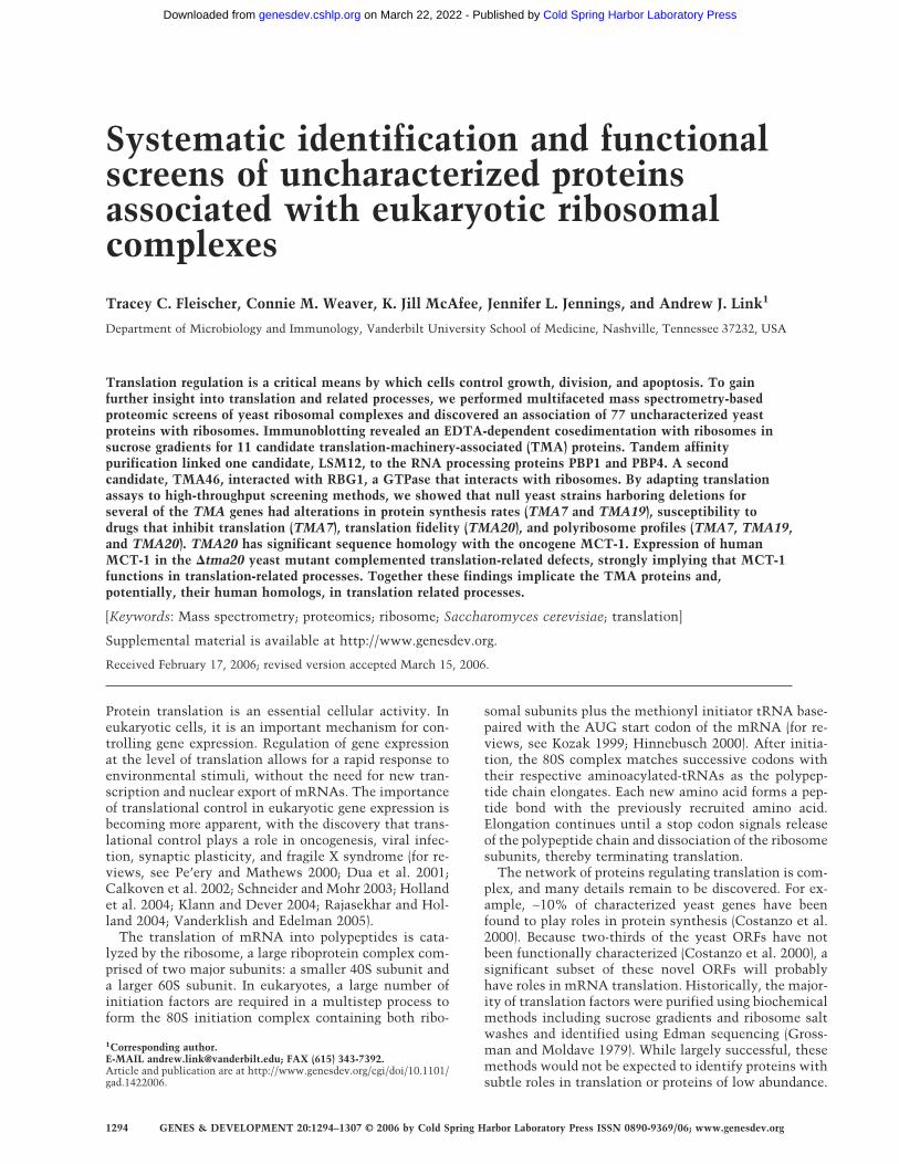
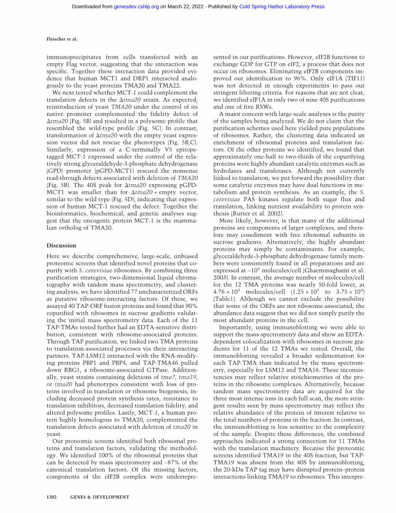
We carried out a multifaceted approach to identify com-ponents associated with the translation machinery usinglarge-scale proteomic screens. Our initial goal was to de-fine a comprehensive list of putative translation-ma-chinery-associated (TMA) proteins, ordered by their rela-tive abundance in the ribosome purifications. We com-bined several purification strategies to minimize the biasof any one approach and to increase the ability to detecttrue ribosome-interacting proteins (Fig. 1A). First, 40S,60S, 80S, and polyribosomal complexes were fractionedusing sucrose gradients (Link et al. 2005). Second, ribo-
somes were purified under increasing salt concentrationsusing discontinuous sucrose gradients. Third, ribosomesalt washes (RSW) with three different salt concentra-tions were used to dissociate potential regulatory factorsfrom core ribosomes (Link et al. 2005).
To identify purified proteins, each sample was di-gested with trypsin and analyzed by DALPC tandemmass spectrometry (Link et al. 1999, 2005). Relative pro-tein abundances in each experiment were expressed asthe total number of nonredundant tandem mass spectrathat correlated significantly to each ORF normalized tothe molecular weight of the cognate protein (×104) (Pow-ell et al. 2004). We call this value a protein abundancefactor (PAF) (see Supplemental Material). Proteins wereclustered by their average PAF across the replicate ex-periments (Lewis et al. 2004; Powell et al. 2004). Theresults are displayed in heat maps using a range of colorsto show patterns of enrichment. The most abundant pro-teins are shown as red, intermediate are yellow, and un-detected proteins are black. Since controls for identifyingnonspecific proteins interacting with the translation ma-chinery were not feasible, this clustering method wasused to identify proteins that showed a significant en-richment with ribosomes. Only proteins identified in atleast 30% of the replicates are depicted in the heat maps.
As points of reference, known ribosomal proteins andribosome-associated proteins were sorted into eightfunctional classes before clustering: 40S, 60S, translationinitiation, elongation, and release factors, translation-re-lated, ribosome biogenesis, and mitochondrial transla-tion proteins. Proteins in these categories are known toor are predicted to copurify with ribosomes, and there-fore can be used to validate our methods. Althoughplacement of proteins into these categories was some-what arbitrary, assignment was largely based upon infor-mation obtained from the Saccharomyces Genome Da-tabase (SGD) and Gene Ontology (GO) (Ashburner et al.2000; Hong et al. 2005).
For the sucrose gradient fractionation (SGF) experi-ments, a minimum of nine independent purifications of40S, 60S, and 80S and five for polysomes were analyzed(Fig. 1B). For the 40S, 60S, and 80S clusters, the data werefurther filtered to include only proteins that were iden-tified in three or more experiments. For the polysomecluster, the cutoff was two or more experiments. Theclustering clearly showed enrichment of ribosomal andtranslation-related proteins in the various purifications(Fig. 1B; Supplementary Tables S1–S4). All 33 compo-nents of the small ribosomal subunit were identified inthe 40S purification. Similarly, we were able to identify43 of 46 components of the large subunit in the 60Spurification. Overall, the 60S purification was enrichedin RPLs relative to the 40S and vice versa. There wassome overlap because the sucrose gradient peaks par-tially overlapped and likely contained comigrating pre-ribosomal complexes. As expected, the 80S and poly-some purifications contained most of the 40S and 60Sproteins. More translation factors and translation relatedproteins copurified with the 40S and 60S as comparedwith the 80S and polysomes, consistent with the large
Proteomic screens of translation complexes
GENES & DEVELOPMENT 1295
Cold Spring Harbor Laboratory Press on March 22, 2022 - Published by genesdev.cshlp.orgDownloaded from
number of proteins required for translation initiation incontrast to elongation.
For the total ribosome analysis (TRA), proteins dis-played in the cluster were identified in two or more pu-rifications (at least five purifications for each salt con-centration). Similar numbers of ribosomal componentswere identified here as compared with the SGF (Fig. 1B;Supplementary Tables S5–S7). At 0.05 M ammoniumchloride, 78 of 79 ribosomal proteins were identified.RPL41 was the only exception. However, RPL41 is a 3.3-kDa protein that produces tryptic peptides two aminoacids long, and therefore cannot generate fragmentationdata that significantly match a peptide. Only two addi-tional subunits (RPL29 and RPL37) were not identifiedwhen the salt concentration was increased to 1 M, dem-onstrating the stability of the ribosome. For each saltconcentration, a subset of known translation-related pro-teins was also identified.
For the ribosome salt washes (RSW), ribosomes werepelleted by centrifugation, and the supernatants (RSWs)were saved for analysis. Proteins identified in two ormore of at least five replicates were included in the clus-ter (Fig. 1B; Supplementary Tables S8–S10). Consistent
with the ribosome stability observed in the TRA, wedetected only a limited number of ribosomal proteinsreleased by the 0.05 M salt wash. The number of releasedribosomal proteins increased with higher salt concentra-tions, but the PAFs were lower as compared with thosein the TRA, indicating that even the most stringent saltwash was not entirely effective at dissociating ribosomalproteins. Together, the RSWs contained a large subset ofknown ribosome-associated proteins, with PAFs similarto those seen in the 40S and 60S SGF experiments. In-terestingly, the 0.05 M salt washes contained similarnumbers of characterized translation-related proteins ascompared with the 1 M salt wash, although they wererelatively more abundant in the 1 M wash. This suggeststhat translation factors are not tightly associated or aretransiently associated with the ribosomes.
Overall, ribosomal proteins were among the mosthighly abundant proteins in the purifications. Impor-tantly, we also identified 23 of 28 canonical translationinitiation factors. All of the elongation factors and one oftwo release factors were also identified. Interestingly,translation factors had a more variable relative abun-dance with lower PAFs in general than the ribosomal
Figure 1. Cluster analysis of ribosomefractions. (A) Purification schemes used topurify ribosomes and ribosome-associatedproteins. (B) Clustering of proteins identi-fied in ribosome purifications. Heat mapclustering proteins by their PAF withinfunctional categories (40S, 60S, translationrelated). The most abundant proteins arebright red and cluster at the top, yellow isintermediate, and black indicates absenceof a particular protein. Each column rep-resents an independent purification, andeach row represents an individual protein.
Fleischer et al.
1296 GENES & DEVELOPMENT
Cold Spring Harbor Laboratory Press on March 22, 2022 - Published by genesdev.cshlp.orgDownloaded from
proteins, suggesting that only a subset of ribosomes isassociated with translation factors. These combinedanalyses using PAFs clearly indicated that both ribo-somes and known translation-related proteins were sub-stantially represented in our purifications.
Identification of novel proteins that copurify withribosomal complexes
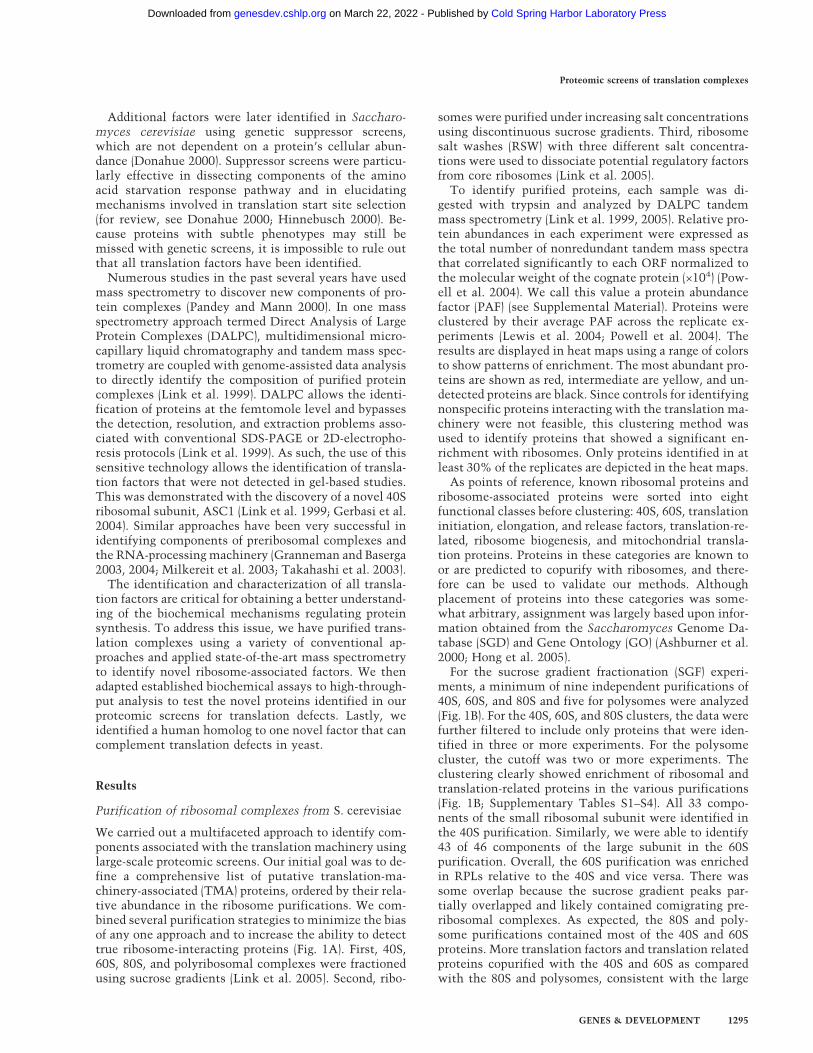
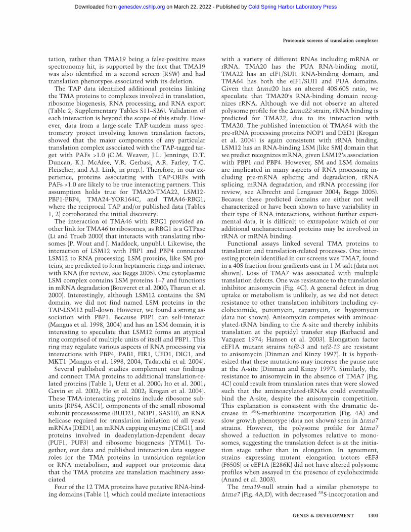
We next used PAFs to identify additional proteins en-riched in our purifications. We chose to focus our effortson uncharacterized ORFs. We applied the same filteringcriteria to each data set and generated a list of expressedORFs found in each purification approach (Fig. 2A–C).The majority of ORFs (76%) were identified in just asingle purification scheme (Fig. 2D). Approximately 23%were identified in two types of purifications, and <1%were identified in all three purification strategies (Fig.2D). The distribution of the ORFs was similar to that forthe known translation proteins, but with more ORFsuniquely identified in the TRA and RSW and fewer iden-tified in all three methods (Fig. 2D). Together the distri-bution data suggested that all three purification methodscontributed to the identification of both known andnovel translation factors.
To see whether the ORFs we identified were biased toa particular molecular weight or pI range, we plotted the
values for each ORF and compared the distribution tothat of the entire yeast proteome (Supplementary Fig.S1). The ORF distribution for both molecular weight andpI mirrored that of the entire proteome, confirming thatour screens were not biased in this manner.
Of the novel ORFs identified in these screens, wechose 12 for further evaluation (Fig. 2, highlighted pro-teins; Table 1). The candidates represented different pu-rification profiles, and therefore might represent proteinswith different functions. Ten of the candidates wereidentified in the SGF. Among these 10 proteins, all butone were also identified in the RSWs. Because ∼10% ofknown translation-related proteins were identified solelyby RSW purification (Fig. 2D), we also chose two ORFsfrom this category. Lastly, because we chose the candi-dates as the screens progressed, we did not always selectthe most enriched proteins in Figure 2. Screening of theremaining proteins is currently under way.
Cosedimentation of expressed ORFs with ribosomalcomplexes is EDTA-sensitive
To confirm the mass spectrometry data identifying novelproteins comigrating with ribosomes in sucrose gradi-ents, polysome profiles were generated for each strainexpressing the corresponding ORF as a tandem affinitypurification (TAP) fusion (Ghaemmaghami et al. 2003).
Figure 2. Identification of novel TMA proteins.Cluster analysis showing the relative abundances(PAFs) of uncharacterized ORFs identified in sucrosegradient fractions (A), total ribosome analysis (B), orribosome salt washes (C). Proteins highlighted inyellow were selected for further analysis. (D) Venndiagram showing the numbers of ORFs (bold) orknown translation factors (parentheses) identified inthe three purification schemes.
Proteomic screens of translation complexes
GENES & DEVELOPMENT 1297
Cold Spring Harbor Laboratory Press on March 22, 2022 - Published by genesdev.cshlp.orgDownloaded from
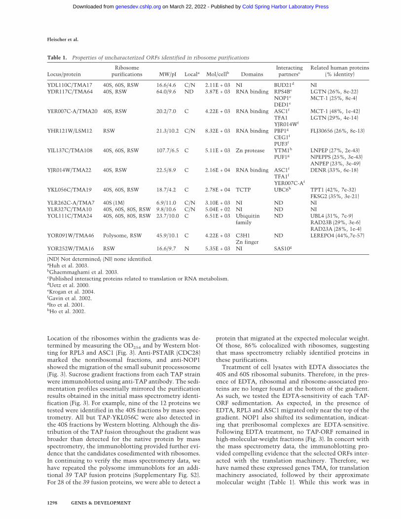
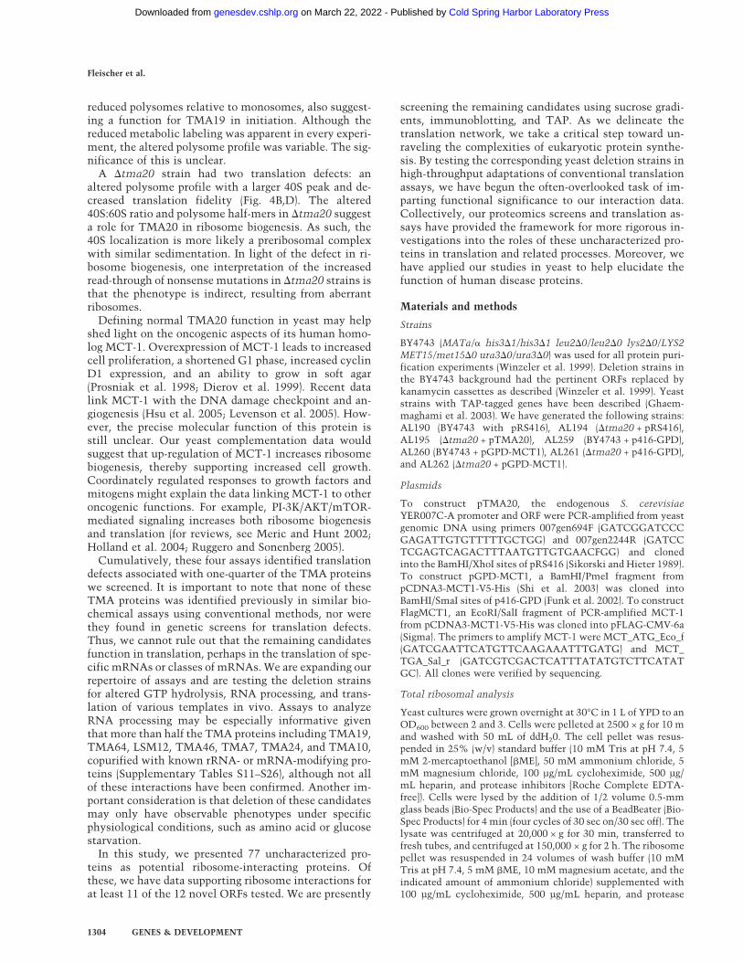
Location of the ribosomes within the gradients was de-termined by measuring the OD254 and by Western blot-ting for RPL3 and ASC1 (Fig. 3). Anti-PSTAIR (CDC28)marked the nonribosomal fractions, and anti-NOP1showed the migration of the small subunit processosome(Fig. 3). Sucrose gradient fractions from each TAP strainwere immunoblotted using anti-TAP antibody. The sedi-mentation profiles essentially mirrored the purificationresults obtained in the initial mass spectrometry identi-fication (Fig. 3). For example, nine of the 12 proteins wetested were identified in the 40S fractions by mass spec-trometry. All but TAP-YKL056C were also detected inthe 40S fractions by Western blotting. Although the dis-tribution of the TAP fusion throughout the gradient wasbroader than detected for the native protein by massspectrometry, the immunoblotting provided further evi-dence that the candidates cosedimented with ribosomes.In continuing to verify the mass spectrometry data, wehave repeated the polysome immunoblots for an addi-tional 39 TAP fusion proteins (Supplementary Fig. S2).For 28 of the 39 fusion proteins, we were able to detect a
protein that migrated at the expected molecular weight.Of those, 86% colocalized with ribosomes, suggestingthat mass spectrometry reliably identified proteins inthese purifications.
Treatment of cell lysates with EDTA dissociates the40S and 60S ribosomal subunits. Therefore, in the pres-ence of EDTA, ribosomal and ribosome-associated pro-teins are no longer found at the bottom of the gradient.As such, we tested the EDTA-sensitivity of each TAP-ORF sedimentation. As expected, in the presence ofEDTA, RPL3 and ASC1 migrated only near the top of thegradient. NOP1 also shifted its sedimentation, indicat-ing that preribosomal complexes are EDTA-sensitive.Following EDTA treatment, no TAP-ORF remained inhigh-molecular-weight fractions (Fig. 3). In concert withthe mass spectrometry data, the immunoblotting pro-vided compelling evidence that the selected ORFs inter-acted with the translation machinery. Therefore, wehave named these expressed genes TMA, for translationmachinery associated, followed by their approximatemolecular weight (Table 1). While this work was in
Table 1. Properties of uncharacterized ORFs identified in ribosome purifications
Locus/proteinRibosome
purifications MW/pI Locala Mol/cellb DomainsInteractingpartnersc
Related human proteins(% identity)
YDL110C/TMA17 40S, 60S, RSW 16.6/4.6 C/N 2.11E + 03 NI BUD21d NIYDR117C/TMA64 40S, RSW 64.0/9.6 ND 3.87E + 03 RNA binding RPS4Be
NOP1e
DED1e
LGTN (26%, 8e-22)MCT-1 (25%, 8e-4)
YER007C-A/TMA20 40S, RSW 20.2/7.0 C 4.22E + 03 RNA binding ASC1f
TFA1YJR014Wf
MCT-1 (48%, 1e-42)LGTN (29%, 4e-14)
YHR121W/LSM12 RSW 21.3/10.2 C/N 8.32E + 03 RNA binding PBP1g
CEG1f
PUF3f
FLJ30656 (26%, 8e-13)
YIL137C/TMA108 40S, 60S, RSW 107.7/6.5 C 5.11E + 03 Zn protease YTM1h
PUF1gLNPEP (27%, 2e-43)NPEPPS (25%, 3e-43)ANPEP (23%, 3e-49)
YJR014W/TMA22 40S, RSW 22.5/8.9 C 2.16E + 04 RNA binding ASC1f
TFA1f
YER007C-Af
DENR (33%, 6e-18)
YKL056C/TMA19 40S, 60S, RSW 18.7/4.2 C 2.78E + 04 TCTP UBC6h TPT1 (42%, 7e-32)FKSG2 (35%, 3e-21)
YLR262C-A/TMA7 40S (1M) 6.9/11.0 C/N 3.10E + 03 NI ND NIYLR327C/TMA10 40S, 60S, 80S, RSW 9.8/10.6 C/N 5.04E + 02 NI ND NIYOL111C/TMA24 40S, 60S, 80S, RSW 23.7/10.0 C 6.51E + 03 Ubiquitin
familyND UBL4 (31%, 7e-9)
RAD23B (29%, 3e-6)RAD23A (28%, 1e-4)
YOR091W/TMA46 Polysome, RSW 45.9/10.1 C 4.22E + 03 C3H1Zn finger
ND LEREPO4 (44%,7e-57)
YOR252W/TMA16 RSW 16.6/9.7 N 5.35E + 03 NI SAS10g
(ND) Not determined; (NI) none identified.aHuh et al. 2003.bGhaemmaghami et al. 2003.cPublished interacting proteins related to translation or RNA metabolism.dUetz et al. 2000.eKrogan et al. 2004.fGavin et al. 2002.gIto et al. 2001.hHo et al. 2002.
Fleischer et al.
1298 GENES & DEVELOPMENT
Cold Spring Harbor Laboratory Press on March 22, 2022 - Published by genesdev.cshlp.orgDownloaded from
progress, the standard name LSM12 was issued forYHR121W, and we therefore have adopted this nomen-clature (Albrecht and Lengauer 2004).
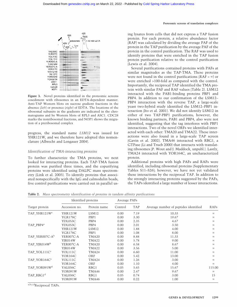
Identification of TMA-interacting proteins
To further characterize the TMA proteins, we nextlooked for interacting proteins. Each TAP-TMA fusionprotein was purified three times, and the copurifyingproteins were identified using DALPC mass spectrom-etry (Link et al. 2005). To identify proteins that associ-ated nonspecifically with the IgG and calmodulin beads,five control purifications were carried out in parallel us-
ing lysates from cells that did not express a TAP fusionprotein. For each protein, a relative abundance factor(RAF) was calculated by dividing the average PAF of theprotein in the TAP purification by the average PAF of theprotein in the control purification. The RAF was used toidentify proteins that were enriched in the TAP fusionprotein purification relative to the control purification(Lewis et al. 2004).
Several purifications contained proteins with PAFs atsimilar magnitudes as the TAP-TMA. These proteinswere not found in the control purifications (RAF = �) orwere enriched >100-fold as compared with the control.Importantly, the reciprocal TAP identified the TMA pro-tein with similar PAF and RAF values (Table 2). LSM12interacted with the PAB1-binding proteins PBP1 andPBP4. In addition to our confirmation of the LSM12–PBP4 interaction with the reverse TAP, a large-scaleyeast two-hybrid study identified the LSM12–PBP1 in-teraction (Ito et al. 2001). We did not identify LSM12 ineither of two TAP-PBP1 purifications; however, theknown binding partners, PAB1 and PBP4, also were notidentified, suggesting that the tag interferes with PBP1interactions. Two of the novel ORFs we identified inter-acted with each other: TMA20 and TMA22. These inter-actions were also found in a large-scale TAP screen(Gavin et al. 2002). TMA46 interacted with RBG1, aGTPase (Li and Trueb 2000) that interacts with translat-ing ribosomes (P. Wout and J. Maddock, unpubl.). Lastly,TMA24 interacted with YOR164C, an uncharacterizedprotein.
Additional proteins with high PAFs and RAFs wereidentified, including ribosomal proteins (SupplementaryTables S11–S26); however, we have not yet validatedthese interactions by the reciprocal TAP. In addition tothe strongly interacting proteins suggested by the PAFs,the TAPs identified a large number of lesser interactions.
Table 2. Mass spectrometry identification of proteins in tandem affinity purifications
Identified proteins Average PAFs
Target protein Accession no. Protein name Control TAP Average number of peptides identified RAFs
TAP_YHR121Wa YHR121W LSM12 0.00 7.19 10.33 �
YGR178C PBP1 0.00 3.30 19.67 �
YDL053C PBP4 0.00 2.35 4.67 �
TAP_PBP4a YDL053C PBP4 0.00 2.01 3.50 �
YHR121W LSM12 0.00 1.88 4.00 �
YGR178C PBP1 0.00 1.08 8.00 �
TAP_YER007C-Ab YER007C-A TMA20 0.00 8.88 11.33 �
YJR014W TMA22 0.00 5.78 9.00 �
TAP_YJR014Wb YER007C-A TMA20 0.00 6.58 8.67 �
YJR014W TMA22 0.00 3.56 5.00 �
TAP_YOL111Cc YOL111C TMA24 0.00 6.60 11.00 �
YOR164C ORF 0.00 5.42 13.00 �
TAP_YOR164Cc YOL111C TMA24 0.00 1.26 3.00 �
YOR164C ORF 0.00 1.10 4.00 �
TAP_YOR091Wd YAL036C RBG1 0.05 5.65 15.67 115.00YOR091W TMA46 0.00 2.47 9.67 �
TAP_RBG1d YAL036C RBG1 0.05 0.74 3.00 15YOR091W TMA46 0.00 0.22 1.00 �
a,b,c,dReciprocal TAPs.
Figure 3. Novel proteins identified in the proteomic screenscosediment with ribosomes in an EDTA-dependent manner.Anti-TAP Western blots on sucrose gradient fractions in theabsence (left) or presence (right) of EDTA. The locations of theribosomal subunits in the gradients are indicated in the chro-matograms and by Western blots of RPL3 and ASC1. CDC28marks the nonribosomal fractions, and NOP1 shows the migra-tion of a preribosomal complex
Proteomic screens of translation complexes
GENES & DEVELOPMENT 1299
Cold Spring Harbor Laboratory Press on March 22, 2022 - Published by genesdev.cshlp.orgDownloaded from
These proteins had PAFs that were 10-fold lower thanthe target protein and variable enrichment relative to thecontrols (Supplementary Tables S11–S26). Many of theseproteins are known to be involved in translation or RNAprocessing. Again, additional experiments are required todetermine whether they are true interacting proteins ornonspecific contaminants.
High-throughput translation screens identify defectsin yeast mutants lacking TMA proteins
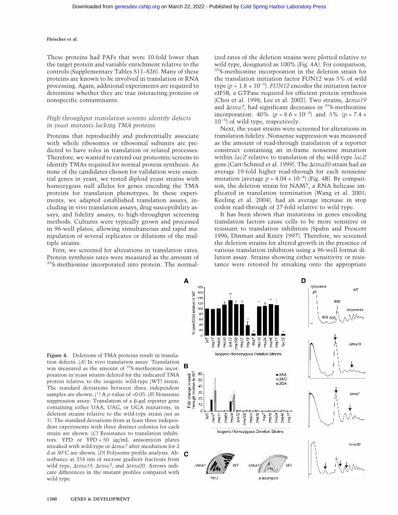
Proteins that reproducibly and preferentially associatewith whole ribosomes or ribosomal subunits are pre-dicted to have roles in translation or related processes.Therefore, we wanted to extend our proteomic screens toidentify TMAs required for normal protein synthesis. Asnone of the candidates chosen for validation were essen-tial genes in yeast, we tested diploid yeast strains withhomozygous null alleles for genes encoding the TMAproteins for translation phenotypes. In these experi-ments, we adapted established translation assays, in-cluding in vivo translation assays, drug susceptibility as-says, and fidelity assays, to high-throughput screeningmethods. Cultures were typically grown and processedin 96-well plates, allowing simultaneous and rapid ma-nipulation of several replicates or dilutions of the mul-tiple strains.
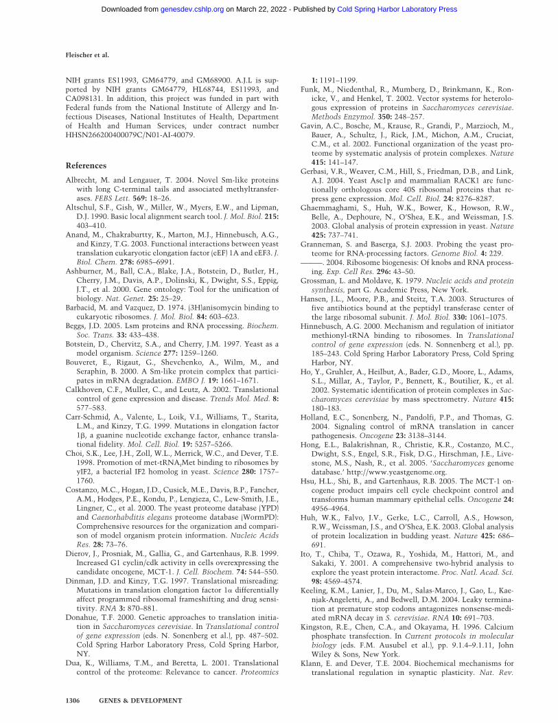
First, we screened for alterations in translation rates.Protein synthesis rates were measured as the amount of35S-methionine incorporated into protein. The normal-
ized rates of the deletion strains were plotted relative towild type, designated as 100% (Fig. 4A). For comparison,35S-methionine incorporation in the deletion strain forthe translation initiation factor FUN12 was 5% of wildtype (p = 1.8 × 10⁻⁵). FUN12 encodes the initiation factoreIF5B, a GTPase required for efficient protein synthesis(Choi et al. 1998; Lee et al. 2002). Two strains, �tma19and �tma7, had significant decreases in 35S-methionineincorporation: 40% (p = 8.6 × 10⁻³) and 5% (p = 7.4 ×10⁻⁵) of wild type, respectively.
Next, the yeast strains were screened for alterations intranslation fidelity. Nonsense suppression was measuredas the amount of read-through translation of a reporterconstruct containing an in-frame nonsense mutationwithin lacZ relative to translation of the wild-type lacZgene (Carr-Schmid et al. 1999). The �tma20 strain had anaverage 19-fold higher read-through for each nonsensemutation (average p = 4.04 × 10⁻⁴) (Fig. 4B). By compari-son, the deletion strain for NAM7, a RNA helicase im-plicated in translation termination (Wang et al. 2001;Keeling et al. 2004), had an average increase in stopcodon read-through of 27-fold relative to wild type.
It has been shown that mutations in genes encodingtranslation factors cause cells to be more sensitive orresistant to translation inhibitors (Spahn and Prescott1996; Dinman and Kinzy 1997). Therefore, we screenedthe deletion strains for altered growth in the presence ofvarious translation inhibitors using a 96-well format di-lution assay. Strains showing either sensitivity or resis-tance were retested by streaking onto the appropriate
Figure 4. Deletions of TMA proteins result in transla-tion defects. (A) In vivo translation assay. Translationwas measured as the amount of 35S-methionine incor-poration in yeast strains deleted for the indicated TMAprotein relative to the isogenic wild-type (WT) strain.The standard deviations between three independentsamples are shown. (*) A p-value of <0.05. (B) Nonsensesuppression assay. Translation of a �-gal reporter genecontaining either UAA, UAG, or UGA mutations, indeletion strains relative to the wild-type strain (set as1). The standard deviations from at least three indepen-dent experiments with three distinct colonies for eachstrain are shown. (C) Resistance to translation inhibi-tors. YPD or YPD + 50 µg/mL anisomycin platesstreaked with wild-type or �tma7 after incubation for 2d at 30°C are shown. (D) Polysome profile analysis. Ab-sorbance at 254 nm of sucrose gradient fractions fromwild type, �tma19, �tma7, and �tma20. Arrows indi-cate differences in the mutant profiles compared withwild type.
Fleischer et al.
1300 GENES & DEVELOPMENT
Cold Spring Harbor Laboratory Press on March 22, 2022 - Published by genesdev.cshlp.orgDownloaded from
drug-containing medium. Resistance to anisomycin wasdetected for �tma7 (Fig. 4C). The �tma7 strain was notresistant to the other translational inhibitors we tested(data not shown), suggesting that �tma7 is not deficientin drug transport or metabolism.
Lastly, the deletion strains were analyzed by polyribo-some profiling. Consistent with decreased protein syn-thesis (Fig. 4A), both �tma7 and �tma19 strains had par-tial polysome runoff (Fig. 4D). While the profile was con-sistent for �tma7, the magnitude of the runoff for�tma19 was more variable. The variation might corre-late with the magnitude of the decreased synthesis ob-served in the labeling experiment.
Additionally, �tma20 consistently varied from thewild-type strain, with a larger 40S peak relative to the60S peak, and half-mers on the 80S and polysome peaks(Fig. 4D). Because the phenotype was subtle, to deter-mine whether the observed difference was significant,the 40S:60S ratio was calculated from seven independentprofiles of wild-type and �tma20 strains. The average40S:60S was 1.5-fold higher in the �tma20 strain, withvalues of 1.41 ± 0.20 and 2.08 ± 0.26 for wild type and�tma20, respectively. A single-tailed nonpaired unequal-variance T-test indicated that the difference was signifi-cant, with a p-value of 8.19 × 10⁻⁵.
TMA20 and MCT-1 are orthologous proteins
Because the translation machinery is highly conservedfrom yeast to humans, we next sought to identify ho-mologs of TMA proteins. Protein or nucleotide BLASTsearches identified highly related sequences in other
fungi for TMA7, TMA10, TMA16, and TMA17 (Altschulet al. 1990). Proteins similar to TMA16 were identifiedin insects, worms, plants, rodents, and humans, al-though the expect values did not fall below 0.4 (Altschulet al. 1990). For the remaining eight yeast proteins, pro-tein BLAST queries identified mammalian proteins withsignificant sequence conservation (Expect � 7e-09)(Altschul et al. 1990), suggesting conserved functions inhigher organisms (Table 1). At least 67% of our con-firmed TMA proteins had mammalian homologs, sup-porting the idea that data from proteomic screens inyeast are applicable to studies of higher organisms. Thisnumber is higher than for yeast proteins in general(30%–40%) (Botstein et al. 1997), which might be ex-pected given that translation is a highly conserved pro-cess.
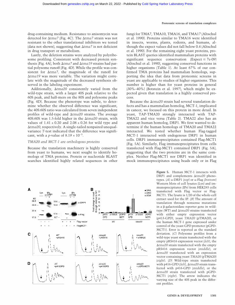
Because the �tma20 strain had several translation de-fects and has a mammalian homolog, MCT-1, implicatedin cancer, we focused on this protein in more detail. Inyeast, TAP-TMA20 strongly interacted with TAP-TMA22 and vice versa (Table 2). TMA22 also has anapparent human homolog, DRP1. We first wanted to de-termine if the human homologs of TMA20 and TMA22interacted. We tested whether human Flag-taggedMCT-1 interacted with endogenous DRP1 in humancells. DRP1 immunoprecipitates contained Flag-MCT1(Fig. 5A). Similarly, Flag immunoprecipitates from cellstransfected with Flag-MCT1 contained DRP1 (Fig. 5A),suggesting that the two proteins are in the same com-plex. Neither Flag-MCT1 nor DRP1 was identified inmock immunoprecipitates using beads only or in Flag
Figure 5. Human MCT-1 interacts withDRP1 and complements �tma20 pheno-types. (A) �-DRP1 (top) or �-Flag (bottom)Western blots of cell lysates (Lys) and im-munoprecipitates (IPs) from HEK293 cellstransfected with Flag vector or Flag-MCT1. The lysate is 1/20 of the whole-cellextract used for the IP. (B) The amount oftranslation through nonsense mutationsin a �-galactosidase reporter gene in wild-type (WT) and �tma20 strains transfectedwith either empty expression vector(p413-GPD), yeast TMA20 (pTMA20), orthe human MCT-1 gene expressed undercontrol of the yeast GPD promoter (pGPD-MCT1). Error is reported as the standarddeviation. (C) Polysome profiles from awild-type yeast strain transfected with theempty pRS416 expression vector (left), the�tma20 strain transfected with the emptypRS416 expression vector (middle), or�tma20 transfected with an expressionvector containing yeast TMA20 (pTMA20)(right). (D) Wild-type strain transfectedwith p416-GPD (left), �tma20 strain trans-fected with p416-GPD (middle), or the�tma20 strain transfected with pGPD-MCT1 (right). The arrow indicates thevarying size of the 40S peak in the differ-ent profiles.
Proteomic screens of translation complexes
GENES & DEVELOPMENT 1301
Cold Spring Harbor Laboratory Press on March 22, 2022 - Published by genesdev.cshlp.orgDownloaded from
immunoprecipitates from cells transfected with anempty Flag vector, suggesting that the interaction wasspecific. Together these interaction data provided evi-dence that human MCT1 and DRP1 interacted analo-gously to the yeast proteins TMA20 and TMA22.
We next tested whether MCT-1 could complement thetranslation defects in the �tma20 strain. As expected,reintroduction of yeast TMA20 under the control of itsnative promoter complemented the fidelity defect of�tma20 (Fig. 5B) and resulted in a polysome profile thatresembled the wild-type profile (Fig. 5C). In contrast,transformation of �tma20 with the empty yeast expres-sion vector did not rescue the phenotypes (Fig. 5B,C).Similarly, expression of a C-terminally V5 epitope-tagged MCT-1 expressed under the control of the rela-tively strong glyceraldehyde-3-phosphate dehydrogenase(GPD) promoter (pGPD-MCT1) rescued the nonsenseread-through defects associated with deletion of TMA20(Fig. 5B). The 40S peak for �tma20 expressing pGPD-MCT1 was smaller than for �tma20 + empty vector,similar to the wild type (Fig. 5D), indicating that expres-sion of human MCT-1 rescued the defect. Together thebioinformatics, biochemical, and genetic analyses sug-gest that the oncogenic protein MCT-1 is the mamma-lian ortholog of TMA20.
Discussion
Here we describe comprehensive, large-scale, unbiasedproteomic screens that identified novel proteins that co-purify with S. cerevisiae ribosomes. By combining threepurification strategies, two-dimensional liquid chroma-tography with tandem mass spectrometry, and cluster-ing analysis, we have identified 77 uncharacterized ORFsas putative ribosome-interacting factors. Of these, weassayed 40 TAP-ORF fusion proteins and found that 90%copurified with ribosomes in sucrose gradients validat-ing the initial mass spectrometry data. Each of the 11TAP-TMAs tested further had an EDTA-sensitive distri-bution, consistent with ribosome-associated proteins.Through TAP purification, we linked two TMA proteinsto translation-associated processes via their interactingpartners. TAP-LSM12 interacted with the RNA-modify-ing proteins PBP1 and PBP4, and TAP-TMA46 pulleddown RBG1, a ribosome-associated GTPase. Addition-ally, yeast strains containing deletions of tma7, tma19,or tma20 had phenotypes consistent with loss of pro-teins involved in translation or ribosome biogenesis, in-cluding decreased protein synthesis rates, resistance totranslation inhibitors, decreased translation fidelity, andaltered polysome profiles. Lastly, MCT-1, a human pro-tein highly homologous to TMA20, complemented thetranslation defects associated with deletion of tma20 inyeast.
Our proteomic screens identified both ribosomal pro-teins and translation factors, validating the methodol-ogy. We identified 100% of the ribosomal proteins thatcan be detected by mass spectrometry and ∼87% of thecanonical translation factors. Of the missing factors,components of the eIF2B complex were underrepre-
sented in our purifications. However, eIF2B functions toexchange GDP for GTP on eIF2, a process that does notoccur on ribosomes. Eliminating eIF2B components im-proved our identification to 96%. Only eIF1A (TIF11)was not detected in enough experiments to pass ourstringent filtering criteria. For reasons that are not clear,we identified eIF1A in only two of nine 40S purificationsand one of five RSWs.
A major concern with large-scale analyses is the purityof the samples being analyzed. We do not claim that thepurification schemes used here yielded pure populationsof ribosomes. Rather, the clustering data indicated anenrichment of ribosomal proteins and translation fac-tors. Of the other proteins we identified, we found thatapproximately one-half to two-thirds of the copurifyingproteins were highly abundant catalytic enzymes such ashydrolases and transferases. Although not currentlylinked to translation, we put forward the possibility thatsome catalytic enzymes may have dual functions in me-tabolism and protein synthesis. As an example, the S.cerevisiae PAS kinases regulate both sugar flux andtranslation, linking nutrient availability to protein syn-thesis (Rutter et al. 2002).
More likely, however, is that many of the additionalproteins are components of larger complexes, and there-fore may cosediment with free ribosomal subunits insucrose gradients. Alternatively, the highly abundantproteins may simply be contaminants. For example,glyceraldehyde-3-phosphate dehydrogenase family mem-bers were consistently found in all preparations and areexpressed at ∼105 molecules/cell (Ghaemmaghami et al.2003). In contrast, the average number of molecules/cellfor the 12 TMA proteins was nearly 50-fold lower, at4.76 × 103 molecules/cell (1.25 × 10² to 3.75 × 10⁴)(Table1). Although we cannot exclude the possibilitythat some of the ORFs are not ribosome-associated, theabundance data suggest that we did not simply purify themost abundant proteins in the cell.
Importantly, using immunoblotting we were able tosupport the mass spectrometry data and show an EDTA-dependent colocalization with ribosomes in sucrose gra-dients for 11 of the 12 TMAs we tested. Overall, theimmunoblotting revealed a broader sedimentation foreach TAP-TMA than indicated by the mass spectrom-etry, especially for LSM12 and TMA16. These inconsis-tencies may reflect relative stoichiometries of the pro-teins in the ribosome complexes. Alternatively, becausetandem mass spectrometry data are acquired for thethree most intense ions in each full scan, the more strin-gent results seen by mass spectrometry may reflect therelative abundance of the protein of interest relative tothe total numbers of proteins in the fraction. In contrast,the immunoblotting is less sensitive to the complexityof the sample. Despite these differences, the combinedapproaches indicated a strong connection for 11 TMAswith the translation machinery. Because the proteomicscreens identified TMA19 in the 40S fraction, but TAP-TMA19 was absent from the 40S by immunoblotting,the 20-kDa TAP tag may have disrupted protein–proteininteractions linking TMA19 to ribosomes. This interpre-
Fleischer et al.
1302 GENES & DEVELOPMENT
Cold Spring Harbor Laboratory Press on March 22, 2022 - Published by genesdev.cshlp.orgDownloaded from
tation, rather than TMA19 being a false-positive massspectronomy hit, is supported by the fact that TMA19was also identified in a second screen (RSW) and hadtranslation phenotypes associated with its deletion.
The TAP data identified additional proteins linkingthe TMA proteins to complexes involved in translation,ribosome biogenesis, RNA processing, and RNA export(Table 2; Supplementary Tables S11–S26). Validation ofeach interaction is beyond the scope of this study. How-ever, data from a large-scale TAP-tandem mass spec-trometry project involving known translation factors,showed that the major components of any particulartranslation complex associated with the TAP-tagged tar-get with PAFs >1.0 (C.M. Weaver, J.L. Jennings, D.T.Duncan, K.J. McAfee, V.R. Gerbasi, A.R. Farley, T.C.Fleischer, and A.J. Link, in prep.). Therefore, in our ex-perience, proteins associating with TAP-ORFs withPAFs >1.0 are likely to be true interacting partners. Thisassumption holds true for TMA20-TMA22, LSM12-PBP1-PBP4, TMA24-YOR164C, and TMA46-RBG1,where the reciprocal TAP and/or published data (Tables1, 2) corroborated the initial discovery.
The interaction of TMA46 with RBG1 provided an-other link for TMA46 to ribosomes, as RBG1 is a GTPase(Li and Trueb 2000) that interacts with translating ribo-somes (P. Wout and J. Maddock, unpubl.). Likewise, theinteraction of LSM12 with PBP1 and PBP4 connectedLSM12 to RNA processing. LSM proteins, like SM pro-teins, are predicted to form heptameric rings and interactwith RNA (for review, see Beggs 2005). One cytoplasmicLSM complex contains LSM proteins 1–7 and functionsin mRNA degradation (Bouveret et al. 2000; Tharun et al.2000). Interestingly, although LSM12 contains the SMdomain, we did not find named LSM proteins in theTAP-LSM12 pull-down. However, we found a strong as-sociation with PBP1. Because PBP1 can self-interact(Mangus et al. 1998, 2004) and has an LSM domain, it isinteresting to speculate that LSM12 forms an atypicalring comprised of multiple units of itself and PBP1. Thisring may regulate various aspects of RNA processing viainteractions with PBP4, PAB1, FIR1, UFD1, DIG1, andMKT1 (Mangus et al. 1998, 2004; Tadauchi et al. 2004).
Several published studies complement our findingsand connect TMA proteins to additional translation-re-lated proteins (Table 1; Uetz et al. 2000; Ito et al. 2001;Gavin et al. 2002; Ho et al. 2002; Krogan et al. 2004).These TMA-interacting proteins include ribosome sub-units (RPS4, ASC1), components of the small ribosomalsubunit processosome (BUD21, NOP1, SAS10), an RNAhelicase required for translation initiation of all yeastmRNAs (DED1), an mRNA capping enzyme (CEG1), andproteins involved in deadenylation-dependent decay(PUF1, PUF3) and ribosome biogenesis (YTM1). To-gether, our data and published interaction data suggestroles for the TMA proteins in translation regulationor RNA metabolism, and support our proteomic datathat the TMA proteins are translation machinery asso-ciated.
Four of the 12 TMA proteins have putative RNA-bind-ing domains (Table 1), which could mediate interactions
with a variety of different RNAs including mRNA orrRNA. TMA20 has the PUA RNA-binding motif,TMA22 has an eIF1/SUI1 RNA-binding domain, andTMA64 has both the eIF1/SUI1 and PUA domains.Given that �tma20 has an altered 40S:60S ratio, wespeculate that TMA20’s RNA-binding domain recog-nizes rRNA. Although we did not observe an alteredpolysome profile for the �tma22 strain, rRNA binding ispredicted for TMA22, due to its interaction withTMA20. The published interaction of TMA64 with thepre-rRNA processing proteins NOP1 and DED1 (Kroganet al. 2004) is again consistent with rRNA binding.LSM12 has an RNA-binding LSM (like SM) domain thatwe predict recognizes mRNA, given LSM12’s associationwith PBP1 and PBP4. However, SM and LSM domainsare implicated in many aspects of RNA processing in-cluding pre-mRNA splicing and degradation, tRNAsplicing, mRNA degradation, and rRNA processing (forreview, see Albrecht and Lengauer 2004; Beggs 2005).Because these predicted domains are either not wellcharacterized or have been shown to have variability intheir type of RNA interactions, without further experi-mental data, it is difficult to extrapolate which of ouradditional uncharacterized proteins may be involved inrRNA or mRNA binding.
Functional assays linked several TMA proteins totranslation and translation-related processes. One inter-esting protein identified in our screens was TMA7, foundin a 40S fraction from gradients cast in 1 M salt (data notshown). Loss of TMA7 was associated with multipletranslation defects. One was resistance to the translationinhibitor anisomycin (Fig. 4C). A general defect in druguptake or metabolism is unlikely, as we did not detectresistance to other translation inhibitors including cy-cloheximide, puromycin, rapamycin, or hygromycin(data not shown). Anisomycin competes with aminoac-ylated-tRNA binding to the A-site and thereby inhibitstranslation at the peptidyl transfer step (Barbacid andVazquez 1974; Hansen et al. 2003). Elongation factoreEF1A mutant strains tef2-3 and tef2-13 are resistantto anisomycin (Dinman and Kinzy 1997). It is hypoth-esized that these mutations may increase the pause rateat the A-site (Dinman and Kinzy 1997). Similarly, theresistance to anisomycin in the absence of TMA7 (Fig.4C) could result from translation rates that were slowedsuch that the aminoacylated-tRNAs could eventuallybind the A-site, despite the anisomycin competition.This explanation is consistent with the dramatic de-crease in 35S-methionine incorporation (Fig. 4A) andslow growth phenotype (data not shown) seen in �tma7strains. However, the polysome profile for �tma7showed a reduction in polysomes relative to mono-somes, suggesting the translation defect is at the initia-tion stage rather than in elongation. In agreement,strains expressing mutant elongation factors eEF3(F650S) or eEF1A (E286K) did not have altered polysomeprofiles when assayed in the presence of cycloheximide(Anand et al. 2003).
The tma19-null strain had a similar phenotype to�tma7 (Fig. 4A,D), with decreased 35S-incorporation and
Proteomic screens of translation complexes
GENES & DEVELOPMENT 1303
Cold Spring Harbor Laboratory Press on March 22, 2022 - Published by genesdev.cshlp.orgDownloaded from
reduced polysomes relative to monosomes, also suggest-ing a function for TMA19 in initiation. Although thereduced metabolic labeling was apparent in every experi-ment, the altered polysome profile was variable. The sig-nificance of this is unclear.
A �tma20 strain had two translation defects: analtered polysome profile with a larger 40S peak and de-creased translation fidelity (Fig. 4B,D). The altered40S:60S ratio and polysome half-mers in �tma20 suggesta role for TMA20 in ribosome biogenesis. As such, the40S localization is more likely a preribosomal complexwith similar sedimentation. In light of the defect in ri-bosome biogenesis, one interpretation of the increasedread-through of nonsense mutations in �tma20 strains isthat the phenotype is indirect, resulting from aberrantribosomes.
Defining normal TMA20 function in yeast may helpshed light on the oncogenic aspects of its human homo-log MCT-1. Overexpression of MCT-1 leads to increasedcell proliferation, a shortened G1 phase, increased cyclinD1 expression, and an ability to grow in soft agar(Prosniak et al. 1998; Dierov et al. 1999). Recent datalink MCT-1 with the DNA damage checkpoint and an-giogenesis (Hsu et al. 2005; Levenson et al. 2005). How-ever, the precise molecular function of this protein isstill unclear. Our yeast complementation data wouldsuggest that up-regulation of MCT-1 increases ribosomebiogenesis, thereby supporting increased cell growth.Coordinately regulated responses to growth factors andmitogens might explain the data linking MCT-1 to otheroncogenic functions. For example, PI-3K/AKT/mTOR-mediated signaling increases both ribosome biogenesisand translation (for reviews, see Meric and Hunt 2002;Holland et al. 2004; Ruggero and Sonenberg 2005).
Cumulatively, these four assays identified translationdefects associated with one-quarter of the TMA proteinswe screened. It is important to note that none of theseTMA proteins was identified previously in similar bio-chemical assays using conventional methods, nor werethey found in genetic screens for translation defects.Thus, we cannot rule out that the remaining candidatesfunction in translation, perhaps in the translation of spe-cific mRNAs or classes of mRNAs. We are expanding ourrepertoire of assays and are testing the deletion strainsfor altered GTP hydrolysis, RNA processing, and trans-lation of various templates in vivo. Assays to analyzeRNA processing may be especially informative giventhat more than half the TMA proteins including TMA19,TMA64, LSM12, TMA46, TMA7, TMA24, and TMA10,copurified with known rRNA- or mRNA-modifying pro-teins (Supplementary Tables S11–S26), although not allof these interactions have been confirmed. Another im-portant consideration is that deletion of these candidatesmay only have observable phenotypes under specificphysiological conditions, such as amino acid or glucosestarvation.
In this study, we presented 77 uncharacterized pro-teins as potential ribosome-interacting proteins. Ofthese, we have data supporting ribosome interactions forat least 11 of the 12 novel ORFs tested. We are presently
screening the remaining candidates using sucrose gradi-ents, immunoblotting, and TAP. As we delineate thetranslation network, we take a critical step toward un-raveling the complexities of eukaryotic protein synthe-sis. By testing the corresponding yeast deletion strains inhigh-throughput adaptations of conventional translationassays, we have begun the often-overlooked task of im-parting functional significance to our interaction data.Collectively, our proteomics screens and translation as-says have provided the framework for more rigorous in-vestigations into the roles of these uncharacterized pro-teins in translation and related processes. Moreover, wehave applied our studies in yeast to help elucidate thefunction of human disease proteins.
Materials and methods
Strains
BY4743 (MATa/� his3�1/his3�1 leu2�0/leu2�0 lys2�0/LYS2MET15/met15�0 ura3�0/ura3�0) was used for all protein puri-fication experiments (Winzeler et al. 1999). Deletion strains inthe BY4743 background had the pertinent ORFs replaced bykanamycin cassettes as described (Winzeler et al. 1999). Yeaststrains with TAP-tagged genes have been described (Ghaem-maghami et al. 2003). We have generated the following strains:AL190 (BY4743 with pRS416), AL194 (�tma20 + pRS416),AL195 (�tma20 + pTMA20), AL259 (BY4743 + p416-GPD),AL260 (BY4743 + pGPD-MCT1), AL261 (�tma20 + p416-GPD),and AL262 (�tma20 + pGPD-MCT1).
Plasmids
To construct pTMA20, the endogenous S. cerevisiaeYER007C-A promoter and ORF were PCR-amplified from yeastgenomic DNA using primers 007gen694F (GATCGGATCCCGAGATTGTGTTTTTGCTGG) and 007gen2244R (GATCCTCGAGTCAGACTTTAATGTTGTGAACFGG) and clonedinto the BamHI/XhoI sites of pRS416 (Sikorski and Hieter 1989).To construct pGPD-MCT1, a BamHI/PmeI fragment frompCDNA3-MCT1-V5-His (Shi et al. 2003) was cloned intoBamHI/SmaI sites of p416-GPD (Funk et al. 2002). To constructFlagMCT1, an EcoRI/SalI fragment of PCR-amplified MCT-1from pCDNA3-MCT1-V5-His was cloned into pFLAG-CMV-6a(Sigma). The primers to amplify MCT-1 were MCT_ATG_Eco_f(GATCGAATTCATGTTCAAGAAATTTGATG) and MCT_TGA_Sal_r (GATCGTCGACTCATTTATATGTCTTCATATGC). All clones were verified by sequencing.
Total ribosomal analysis
Yeast cultures were grown overnight at 30°C in 1 L of YPD to anOD600 between 2 and 3. Cells were pelleted at 2500 × g for 10 mand washed with 50 mL of ddH20. The cell pellet was resus-pended in 25% (w/v) standard buffer (10 mM Tris at pH 7.4, 5mM 2-mercaptoethanol [�ME], 50 mM ammonium chloride, 5mM magnesium chloride, 100 µg/mL cycloheximide, 500 µg/mL heparin, and protease inhibitors [Roche Complete EDTA-free]). Cells were lysed by the addition of 1/2 volume 0.5-mmglass beads (Bio-Spec Products) and the use of a BeadBeater (Bio-Spec Products) for 4 min (four cycles of 30 sec on/30 sec off). Thelysate was centrifuged at 20,000 × g for 30 min, transferred tofresh tubes, and centrifuged at 150,000 × g for 2 h. The ribosomepellet was resuspended in 24 volumes of wash buffer (10 mMTris at pH 7.4, 5 mM �ME, 10 mM magnesium acetate, and theindicated amount of ammonium chloride) supplemented with100 µg/mL cycloheximide, 500 µg/mL heparin, and protease
Fleischer et al.
1304 GENES & DEVELOPMENT
Cold Spring Harbor Laboratory Press on March 22, 2022 - Published by genesdev.cshlp.orgDownloaded from

inhibitors (Roche Complete EDTA-free). A 6-mL aliquot waslayered on a 6-mL 5%:20% discontinuous sucrose gradient andcentrifuged at 40,000 × g for 18 h. The ribosome pellet was re-suspended in 25 volumes of standard buffer and centrifuged at7100 × g for 10 min.
Ribosome salt wash
Ribosome salt washes were carried out as described (Link et al.2005). Briefly, yeast whole-cell lysates were centrifuged to re-move cellular debris. Ribosomes were then pelleted by ultra-centrifugation and resuspended by sonicating in buffer supple-mented to 0.05 M, 0.5 M, or 1 M potassium acetate. Ribosomalproteins were again pelleted by centrifugation, and the super-natant (ribosome salt wash) was saved for analysis.
Sucrose gradient fractionation
A high-resolution purification of ribosome complexes was em-ployed using sucrose gradient fractionation (Link et al. 2005).The method was used to analyze the polysome profiles of theyeast deletion and complemented strains with the followingmodifications. Strains were grown to log phase in 100 mL ofSC-URA medium with the final OD600 ∼ 0.8. Briefly, cyclohexi-mide (50 µg/mL) was added directly to each culture, and thenthe culture was chilled on ice for 10 min. Cells were lysed inchilled Breaking Buffer (10 mM Tris-HCl at pH 7.5, 100 mMNaCl, 30 mM MgCl2, 50 µg/mL cycloheximide, 200 µg/mLheparin, and protease inhibitors [Roche Complete EDTA-free]),and 20 OD260 of the cleared extract was loaded onto 12-mL7%–47% sucrose gradients. The gradients were centrifuged inan SW-41 rotor for 18 h at 15,000 rpm at 4°C.
Immunoprecipitation and immunoblotting
Aliquots (30 µL) of 0.5-mL sucrose gradient fractions were runon Tris-glycine gels, transferred to Immobilon-P (Millipore), andimmunoblotted with indicated antibodies. For immunoprecipi-tations, HEK293 cells were seeded at 1.0 × 10⁶ and 16 h laterwere transfected with 5 µg of either pFLAG-CMV-6a (Sigma) orFlag-MCT1 using calcium phosphate (Kingston et al. 1996). Af-ter 24 h, cells were lysed in 0.5 mL of L-buffer (PBS, 0.1% NP-40)containing EDTA-free complete mini-protease inhibitors(Roche), 50 µg/mL cycloheximide, 1 mM dithiotreitol, and 40U/mL RNAsin (Promega), by freezing on dry ice and thawing at37°C. Nuclei and debris were pelleted by centrifugation for 5min at 13,000g. Cytoplasmic extracts were immunoprecipitatedfor 1 h at 4°C with 20 µL of Flag-M2 agarose (Sigma) or 20 µL ofImmunoPure Immobilized Protein A (Pierce) with or without 5µL of �-DRP1 antibody. Lysates and immunoprecipitates wereresolved on 12% Tris-glycine gels, transferred to Immobilon-P(Millipore), and immunoblotted with indicated antibodies. In-teractions were detected using chemiluminescence (ECL Plus;Amersham Biosciences).
Antibodies
Anti-TAP (Open Biosystems), anti-Flag M2-peroxidase (HRP;Sigma), anti-DRP1 (BD Transduction Laboratories), anti-NOP1(EnCor Biotechnology, Inc.), and anti-PSTAIR (Sigma) antibod-ies are commercially available. Anti-RPL3 (Vilardell and Warner1997) and anti-ASC1 (Gerbasi et al. 2004) have been described.Anti-rabbit HRP and anti-mouse HRP were from Promega.
Tandem affinity purification
TAP fusion proteins were purified using IgG and calmodulinaffinity resins essentially as described (Link et al. 2005).
DALPC
To identify the proteins found in purified ribosomes or TAPpurifications, samples were trypsin-digested and analyzed byoff-line SCX fractionation coupled to RP-LC-ESI-MS/MS asdescribed (Link et al. 2003, 2005). Data processing of theSE-QUEST output files into a list of proteins has been previouslydescribed (Link et al. 1999).
[35S]-methionine incorporation
Overnight cultures grown in YPD were diluted 1:10 into 1 mLof SC-MET (OD595 ∼ 0.5) in triplicate in a 96-well 2-mL culturedish and grown for 3 h at 30°C. The OD595 of one replicate wasmeasured to determine relative cell numbers. The remainingtwo samples were labeled with 6 µCi/mL [35S]-methionine/cys-teine (EXPRE35S35S Protein Labeling Mix; >1000 Ci/mmol;NEN Research Products) for 15 min at 30°C. Labeling wasstopped by the addition of 1/10 volume of 100% TCA to eachculture and heating the entire 96-well dish at 100°C for 30 min.TCA precipitates were collected on GFC filters (Whatman),washed sequentially with 2 mL of each 10% TCA and 95%ethanol, and counted in 5 mL of UniverSol (ICN). For eachsample, the counts per minute/OD595 was normalized to wildtype, set as 100%. Three independent experiments weregraphed with the error bars representing the standard deviation.The P-values shown were determined using a single-tailed non-paired unequal-variance T-test.
Drug dilution assay
Cells were grown overnight in 1 mL of YPD. The OD595 wasmeasured, and an equal number of cells for each strain (up to 24different strains/plate) was aliquoted into a 96-well plate, andthen serially diluted, fivefold each, into YPD. Using a 96-pinmultiblot replicator (V&P Scientific, Inc.), the master dilutionplate was replica-plated in duplicate onto YPD plates containingno drug, 5 ng/mL rapamycin, 50 ng/mL rapamycin, 0.2 µg/mLcycloheximide, 2.0 µg/mL cycloheximide, 20 µg/mL anisomy-cin, 50 µg/mL anisomycin, 10 µg/mL hygromycin, or 100 µg/mLhygromycin. Plates were incubated at 30°C for up to 4 d. Allstrains were tested for sensitivity or resistance to each drugconcentration in at least two independent experiments. Perti-nent strain/drug combinations were verified in at least threeexperiments by streaking overnight cultures onto duplicatedrug plates and growing at 30°C.
Nonsense suppression assay
Nonsense suppression was assayed as described (Carr-Schmid etal. 1999). Production of �-galactosidase (�-gal) was measuredusing the Tropix Gal-Screen chemiluminescent reporter genesystem (Applied Biosystems) with 50 µL of cells and 50 µL ofReaction Buffer B. Luminescence was measured for 20 sec in aTurner Designs TD-20/20. Assays were repeated at least twicewith three independent colonies for each strain.
Acknowledgments
We thank Amar Kar for assistance with the translation fidelityexperiments, R.B. Gartenhaus for plasmid MCT-V5, T.G. Kinzyfor the nonsense suppression plasmids, and J.R. Warner for an-tibodies to RPL3. We thank Elizabeth M. Link, David W. Pow-ell, R. Vincent Gerbasi, and Adam Farley for critical commentsin the preparation of this manuscript. This project was sup-ported by NIH grant GM64779. T.C.F. is supported by NIHpost-doctoral training grant T32 AI49824 and ACS post-doctoralfellowship PF-05-148-01-MBC. C.M.W. and J.L.J are supportedby NIH grants GM64779 and HL68744. K.J.M. is supported by
Proteomic screens of translation complexes
GENES & DEVELOPMENT 1305
Cold Spring Harbor Laboratory Press on March 22, 2022 - Published by genesdev.cshlp.orgDownloaded from
NIH grants ES11993, GM64779, and GM68900. A.J.L is sup-ported by NIH grants GM64779, HL68744, ES11993, andCA098131. In addition, this project was funded in part withFederal funds from the National Institute of Allergy and In-fectious Diseases, National Institutes of Health, Departmentof Health and Human Services, under contract numberHHSN266200400079C/N01-AI-40079.
References
Albrecht, M. and Lengauer, T. 2004. Novel Sm-like proteinswith long C-terminal tails and associated methyltransfer-ases. FEBS Lett. 569: 18–26.
Altschul, S.F., Gish, W., Miller, W., Myers, E.W., and Lipman,D.J. 1990. Basic local alignment search tool. J. Mol. Biol. 215:403–410.
Anand, M., Chakraburtty, K., Marton, M.J., Hinnebusch, A.G.,and Kinzy, T.G. 2003. Functional interactions between yeasttranslation eukaryotic elongation factor (eEF) 1A and eEF3. J.Biol. Chem. 278: 6985–6991.
Ashburner, M., Ball, C.A., Blake, J.A., Botstein, D., Butler, H.,Cherry, J.M., Davis, A.P., Dolinski, K., Dwight, S.S., Eppig,J.T., et al. 2000. Gene ontology: Tool for the unification ofbiology. Nat. Genet. 25: 25–29.
Barbacid, M. and Vazquez, D. 1974. (3H)anisomycin binding toeukaryotic ribosomes. J. Mol. Biol. 84: 603–623.
Beggs, J.D. 2005. Lsm proteins and RNA processing. Biochem.Soc. Trans. 33: 433–438.
Botstein, D., Chervitz, S.A., and Cherry, J.M. 1997. Yeast as amodel organism. Science 277: 1259–1260.
Bouveret, E., Rigaut, G., Shevchenko, A., Wilm, M., andSeraphin, B. 2000. A Sm-like protein complex that partici-pates in mRNA degradation. EMBO J. 19: 1661–1671.
Calkhoven, C.F., Muller, C., and Leutz, A. 2002. Translationalcontrol of gene expression and disease. Trends Mol. Med. 8:577–583.
Carr-Schmid, A., Valente, L., Loik, V.I., Williams, T., Starita,L.M., and Kinzy, T.G. 1999. Mutations in elongation factor1�, a guanine nucleotide exchange factor, enhance transla-tional fidelity. Mol. Cell. Biol. 19: 5257–5266.
Choi, S.K., Lee, J.H., Zoll, W.L., Merrick, W.C., and Dever, T.E.1998. Promotion of met-tRNAiMet binding to ribosomes byyIF2, a bacterial IF2 homolog in yeast. Science 280: 1757–1760.
Costanzo, M.C., Hogan, J.D., Cusick, M.E., Davis, B.P., Fancher,A.M., Hodges, P.E., Kondu, P., Lengieza, C., Lew-Smith, J.E.,Lingner, C., et al. 2000. The yeast proteome database (YPD)and Caenorhabditis elegans proteome database (WormPD):Comprehensive resources for the organization and compari-son of model organism protein information. Nucleic AcidsRes. 28: 73–76.
Dierov, J., Prosniak, M., Gallia, G., and Gartenhaus, R.B. 1999.Increased G1 cyclin/cdk activity in cells overexpressing thecandidate oncogene, MCT-1. J. Cell. Biochem. 74: 544–550.
Dinman, J.D. and Kinzy, T.G. 1997. Translational misreading:Mutations in translation elongation factor 1� differentiallyaffect programmed ribosomal frameshifting and drug sensi-tivity. RNA 3: 870–881.
Donahue, T.F. 2000. Genetic approaches to translation initia-tion in Saccharomyces cerevisiae. In Translational controlof gene expression (eds. N. Sonenberg et al.), pp. 487–502.Cold Spring Harbor Laboratory Press, Cold Spring Harbor,NY.
Dua, K., Williams, T.M., and Beretta, L. 2001. Translationalcontrol of the proteome: Relevance to cancer. Proteomics
1: 1191–1199.Funk, M., Niedenthal, R., Mumberg, D., Brinkmann, K., Ron-
icke, V., and Henkel, T. 2002. Vector systems for heterolo-gous expression of proteins in Saccharomyces cerevisiae.Methods Enzymol. 350: 248–257.
Gavin, A.C., Bosche, M., Krause, R., Grandi, P., Marzioch, M.,Bauer, A., Schultz, J., Rick, J.M., Michon, A.M., Cruciat,C.M., et al. 2002. Functional organization of the yeast pro-teome by systematic analysis of protein complexes. Nature415: 141–147.
Gerbasi, V.R., Weaver, C.M., Hill, S., Friedman, D.B., and Link,A.J. 2004. Yeast Asc1p and mammalian RACK1 are func-tionally orthologous core 40S ribosomal proteins that re-press gene expression. Mol. Cell. Biol. 24: 8276–8287.
Ghaemmaghami, S., Huh, W.K., Bower, K., Howson, R.W.,Belle, A., Dephoure, N., O’Shea, E.K., and Weissman, J.S.2003. Global analysis of protein expression in yeast. Nature425: 737–741.
Granneman, S. and Baserga, S.J. 2003. Probing the yeast pro-teome for RNA-processing factors. Genome Biol. 4: 229.
———. 2004. Ribosome biogenesis: Of knobs and RNA process-ing. Exp. Cell Res. 296: 43–50.
Grossman, L. and Moldave, K. 1979. Nucleic acids and proteinsynthesis, part G. Academic Press, New York.
Hansen, J.L., Moore, P.B., and Steitz, T.A. 2003. Structures offive antibiotics bound at the peptidyl transferase center ofthe large ribosomal subunit. J. Mol. Biol. 330: 1061–1075.
Hinnebusch, A.G. 2000. Mechanism and regulation of initiatormethionyl-tRNA binding to ribosomes. In Translationalcontrol of gene expression (eds. N. Sonnenberg et al.), pp.185–243. Cold Spring Harbor Laboratory Press, Cold SpringHarbor, NY.
Ho, Y., Gruhler, A., Heilbut, A., Bader, G.D., Moore, L., Adams,S.L., Millar, A., Taylor, P., Bennett, K., Boutilier, K., et al.2002. Systematic identification of protein complexes in Sac-charomyces cerevisiae by mass spectrometry. Nature 415:180–183.
Holland, E.C., Sonenberg, N., Pandolfi, P.P., and Thomas, G.2004. Signaling control of mRNA translation in cancerpathogenesis. Oncogene 23: 3138–3144.
Hong, E.L., Balakrishnan, R., Christie, K.R., Costanzo, M.C.,Dwight, S.S., Engel, S.R., Fisk, D.G., Hirschman, J.E., Live-stone, M.S., Nash, R., et al. 2005. ‘Saccharomyces genomedatabase.’ http://www.yeastgenome.org.
Hsu, H.L., Shi, B., and Gartenhaus, R.B. 2005. The MCT-1 on-cogene product impairs cell cycle checkpoint control andtransforms human mammary epithelial cells. Oncogene 24:4956–4964.
Huh, W.K., Falvo, J.V., Gerke, L.C., Carroll, A.S., Howson,R.W., Weissman, J.S., and O’Shea, E.K. 2003. Global analysisof protein localization in budding yeast. Nature 425: 686–691.
Ito, T., Chiba, T., Ozawa, R., Yoshida, M., Hattori, M., andSakaki, Y. 2001. A comprehensive two-hybrid analysis toexplore the yeast protein interactome. Proc. Natl. Acad. Sci.98: 4569–4574.
Keeling, K.M., Lanier, J., Du, M., Salas-Marco, J., Gao, L., Kae-njak-Angeletti, A., and Bedwell, D.M. 2004. Leaky termina-tion at premature stop codons antagonizes nonsense-medi-ated mRNA decay in S. cerevisiae. RNA 10: 691–703.
Kingston, R.E., Chen, C.A., and Okayama, H. 1996. Calciumphosphate transfection. In Current protocols in molecularbiology (eds. F.M. Ausubel et al.), pp. 9.1.4–9.1.11, JohnWiley & Sons, New York.
Klann, E. and Dever, T.E. 2004. Biochemical mechanisms fortranslational regulation in synaptic plasticity. Nat. Rev.
Fleischer et al.
1306 GENES & DEVELOPMENT
Cold Spring Harbor Laboratory Press on March 22, 2022 - Published by genesdev.cshlp.orgDownloaded from
Neurosci. 5: 931–942.Kozak, M. 1999. Initiation of translation in prokaryotes and
eukaryotes. Gene 234: 187–208.Krogan, N.J., Peng, W.T., Cagney, G., Robinson, M.D., Haw, R.,
Zhong, G., Guo, X., Zhang, X., Canadien, V., Richards, D.P.,et al. 2004. High-definition macromolecular composition ofyeast RNA-processing complexes. Mol. Cell 13: 225–239.
Lee, J.H., Pestova, T.V., Shin, B.S., Cao, C., Choi, S.K., and De-ver, T.E. 2002. Initiation factor eIF5B catalyzes second GTP-dependent step in eukaryotic translation initiation. Proc.Natl. Acad. Sci. 99: 16689–16694.
Levenson, A.S., Thurn, K.E., Simons, L.A., Veliceasa, D., Jarrett,J., Osipo, C., Jordan, V.C., Volpert, O.V., Satcher Jr., R.L., andGartenhaus, R.B. 2005. MCT-1 oncogene contributes to in-creased in vivo tumorigenicity of MCF7 cells by promotionof angiogenesis and inhibition of apoptosis. Cancer Res. 65:10651–10656.
Lewis, P.W., Beall, E.L., Fleischer, T.C., Georlette, D., Link, A.J.,and Botchan, M.R. 2004. Identification of a Drosophila Myb-E2F2/RBF transcriptional repressor complex. Genes & Dev.18: 2929–2940.
Li, B. and Trueb, B. 2000. DRG represents a family of twoclosely related GTP-binding proteins. Biochim. Biophys.Acta 1491: 196–204.
Link, A.J., Eng, J., Schieltz, D.M., Carmack, E., Mize, G.J., Mor-ris, D.R., Garvik, B.M., and Yates III, J.R. 1999. Direct analy-sis of protein complexes using mass spectrometry. Nat. Bio-technol. 17: 676–682.
Link, A.J., Jennings, J.L., and Washburn, M.P. 2003. Analysis ofprotein composition using multidimensional chromatogra-phy and mass spectrometry. In Current protocols in proteinscience (eds. J.E. Coligan et al.), pp. 23.21.21–23.21.25. JohnWiley and Sons, New York.
Link, A.J., Fleischer, T.C., Weaver, C.M., Gerbasi, V.R., andJennings, J.L. 2005. Purifying protein complexes for massspectrometry: Applications to protein translation. Methods35: 274–290.
Mangus, D.A., Amrani, N., and Jacobson, A. 1998. Pbp1p, afactor interacting with Saccharomyces cerevisiae poly(A)-binding protein, regulates polyadenylation. Mol. Cell. Biol.18: 7383–7396.
Mangus, D.A., Smith, M.M., McSweeney, J.M., and Jacobson, A.2004. Identification of factors regulating poly(A) tail synthe-sis and maturation. Mol. Cell. Biol. 24: 4196–4206.
Meric, F. and Hunt, K.K. 2002. Translation initiation in cancer:A novel target for therapy. Mol. Cancer Ther. 1: 971–979.
Milkereit, P., Kuhn, H., Gas, N., and Tschochner, H. 2003. Thepre-ribosomal network. Nucleic Acids Res. 31: 799–804.
Pandey, A. and Mann, M. 2000. Proteomics to study genes andgenomes. Nature 405: 837–846.
Pe’ery, T. and Mathews, M.B. 2000. Viral translation strategiesand host defense mechanisms. In Translational control ofgene expression (eds. N. Sonnenberg et al.), pp. 371–424.Cold Spring Harbor Laboratory Press, Cold Spring Harbor,NY.
Powell, D.W., Weaver, C.M., Jennings, J.L., McAfee, K.J., He, Y.,Weil, P.A., and Link, A.J. 2004. Cluster analysis of massspectrometry data reveals a novel component of SAGA. Mol.Cell. Biol. 24: 7249–7259.
Prosniak, M., Dierov, J., Okami, K., Tilton, B., Jameson, B.,Sawaya, B.E., and Gartenhaus, R.B. 1998. A novel candidateoncogene, MCT-1, is involved in cell cycle progression. Can-cer Res. 58: 4233–4237.
Rajesekhar, V.K. and Holland, E.C. 2004. Postgenomic globalanalysis of translational control induced by oncogenic sig-naling. Oncogene 23: 3248–3264.
Ruggero, D. and Sonenberg, N. 2005. The Akt of translationalcontrol. Oncogene 24: 7426–7434.
Rutter, J., Probst, B.L., and McKnight, S.L. 2002. Coordinateregulation of sugar flux and translation by PAS kinase. Cell111: 17–28.
Schneider, R.J. and Mohr, I. 2003. Translation initiation andviral tricks. Trends Biochem. Sci. 28: 130–136.
Shi, B., Hsu, H.L., Evens, A.M., Gordon, L.I., and Gartenhaus,R.B. 2003. Expression of the candidate MCT-1 oncogene inB- and T-cell lymphoid malignancies. Blood 102: 297–302.
Sikorski, R.S. and Hieter, P. 1989. A system of shuttle vectorsand yeast host strains designed for efficient manipulation ofDNA in Saccharomyces cerevisiae. Genetics 122: 19–27.
Spahn, C.M. and Prescott, C.D. 1996. Throwing a spanner in theworks: Antibiotics and the translation apparatus. J. Mol.Med. 74: 423–439.
Tadauchi, T., Inada, T., Matsumoto, K., and Irie, K. 2004. Post-transcriptional regulation of HO expression by the Mkt1–Pbp1 complex. Mol. Cell. Biol. 24: 3670–3681.
Takahashi, N., Yanagida, M., Fujiyama, S., Hayano, T., and Is-obe, T. 2003. Proteomic snapshot analyses of preribosomalribonucleoprotein complexes formed at various stages of ri-bosome biogenesis in yeast and mammalian cells. MassSpectrom. Rev. 22: 287–317.
Tharun, S., He, W., Mayes, A.E., Lennertz, P., Beggs, J.D., andParker, R. 2000. Yeast Sm-like proteins function in mRNAdecapping and decay. Nature 404: 515–518.
Uetz, P., Giot, L., Cagney, G., Mansfield, T.A., Judson, R.S.,Knight, J.R., Lockshon, D., Narayan, V., Srinivasan, M., Po-chart, P., et al. 2000. A comprehensive analysis of protein–protein interactions in Saccharomyces cerevisiae. Nature403: 623–627.
Vanderklish, P.W. and Edelman, G.M. 2005. Differential trans-lation and fragile X syndrome. Genes Brain Behav. 4: 360–384.
Vilardell, J. and Warner, J.R. 1997. Ribosomal protein L32 ofSaccharomyces cerevisiae influences both the splicing of itsown transcript and the processing of rRNA. Mol. Cell. Biol.17: 1959–1965.
Wang, W., Czaplinski, K., Rao, Y., and Peltz, S.W. 2001. The roleof Upf proteins in modulating the translation read-throughof nonsense-containing transcripts. EMBO J. 20: 880–890.
Winzeler, E.A., Shoemaker, D.D., Astromoff, A., Liang, H.,Anderson, K., Andre, B., Bangham, R., Benito, R., Boeke, J.D.,Bussey, H., et al. 1999. Functional characterization of the S.cerevisiae genome by gene deletion and parallel analysis.Science 285: 901–906.
Proteomic screens of translation complexes
GENES & DEVELOPMENT 1307
Cold Spring Harbor Laboratory Press on March 22, 2022 - Published by genesdev.cshlp.orgDownloaded from
10.1101/gad.1422006Access the most recent version at doi: 20:2006, Genes Dev.
Tracey C. Fleischer, Connie M. Weaver, K. Jill McAfee, et al. proteins associated with eukaryotic ribosomal complexesSystematic identification and functional screens of uncharacterized
Material
Supplemental
http://genesdev.cshlp.org/content/suppl/2006/05/15/20.10.1294.DC1
References
http://genesdev.cshlp.org/content/20/10/1294.full.html#ref-list-1
This article cites 59 articles, 23 of which can be accessed free at:
License
ServiceEmail Alerting
click here.right corner of the article or
Receive free email alerts when new articles cite this article - sign up in the box at the top
Copyright © 2006, Cold Spring Harbor Laboratory Press
Cold Spring Harbor Laboratory Press on March 22, 2022 - Published by genesdev.cshlp.orgDownloaded from