systems pharmacology approaches decoction on liver brosis
TRANSCRIPT
Page 1/24
Insights into the molecular mechanisms of Huangqidecoction on liver �brosis via computationalsystems pharmacology approachesBiting Wang
East China University of Science and TechnologyZengrui Wu
East China University of Science and TechnologyWeihua Li
East China University of Science and TechnologyGuixia Liu
East China University of Science and TechnologyYun Tang ( [email protected] )
East China University of Science and Technology https://orcid.org/0000-0003-2340-1109
Research
Keywords: Huangqi decoction, Liver �brosis, Mechanism of action, Metabolomics, Molecular docking,Network pharmacology
Posted Date: April 6th, 2021
DOI: https://doi.org/10.21203/rs.3.rs-389363/v1
License: This work is licensed under a Creative Commons Attribution 4.0 International License. Read Full License
Version of Record: A version of this preprint was published at Chinese Medicine on July 23rd, 2021. Seethe published version at https://doi.org/10.1186/s13020-021-00473-8.
Page 2/24
AbstractBackground: The traditional Chinese medicine Huangqi decoction (HQD) consists of Radix Astragali andRadix Glycyrrhizae in a ratio of 6 : 1, which has been used for the treatment of liver �brosis. In this study,we tried to elucidate its action of mechanism (MoA) via a combination of metabolomics data, networkpharmacology and molecular docking methods.
Methods: Firstly, we collected prototype components and metabolic products after administration of HQDfrom a publication. With known and predicted targets, compound-target interactions were obtained. Then,the global compound-liver �brosis target bipartite network and the HQD-liver �brosis protein-proteininteraction network were constructed, separately. KEGG pathway analysis was applied to furtherunderstand the mechanisms related to the target proteins of HQD. Additionally, molecular dockingsimulation was performed to determine the binding e�ciency of compounds with targets. Finally, aftertaking concentration of prototype compounds and metabolites of HQD after administration intoconsideration, the critical compound-liver �brosis target bipartite network was constructed.
Results: We collected 68 components, including 17 prototype components and 51 metabolic productsafter administration of HQD, and 540 compound-target interactions were obtained between the 68components and 95 targets.
Combining network analysis and molecular docking, as well as concentration of prototype compoundsand metabolites of HQD, our �nal results demonstrated that eight compounds (three prototypecompounds and �ve metabolites) and eight targets (CDK1, MMP9, PPARD, PPARG, PTGS2, SERPINE1,TP53, and HIF1A) might contribute to the effects of HQD on liver �brosis by reducing �brogenesis andstimulate degradation, which through p53 signaling pathway, PPAR signaling pathway, HIF-1 signalingpathway, IL-17 signaling pathway, and TNF signaling pathway.
Conclusions: Our results would shed light on the complicated MoA of traditional Chinese medicine andhelp to attract attention to the therapeutic effects of metabolites of original components in Chinese herbsthrough computational methods.
BackgroundLiver �brosis is a pathological condition that occurs as a response to chronic liver injury. Variouspathological factors, such as hepatitis B and C viruses (HBV and HCV), alcoholic liver disease (ALD), non-alcoholic fatty liver disease (NAFLD) especially non-alcoholic steatohepatitis (NASH), as well as primarybiliary cholangitis, primary sclerosing cholangitis, and other autoimmune liver diseases, all contribute tothe development of liver �brosis [1]. After the liver injury, the repair process of damaged liver involves twodistinct paths: one is the regenerative path, in which injured cells are replaced by the same type of cells;the other is �brosis, in which normal parenchymal tissue is replaced by connective tissue in anuncontrolled manner [2]. In the context of chronic liver injury, hepatic stellate cells (HSCs) are over-activated, which triggers the excessive deposition of extracellular matrix (ECM) proteins and tissue
Page 3/24
structural remodelling [3]. Liver �brosis can further progress to liver cirrhosis, hepatocellular carcinoma, oreven death. However, there is currently no validated anti-�brogenic therapy.
Huangqi decoction (HQD) is a classical formula of traditional Chinese medicine (TCM) to improve liverfunction and life quality in patients with chronic liver disease. It consists of Radix Astragali (R. Astragali)and Radix Glycyrrhizae (R. Glycyrrhizae) in a ratio of 6 : 1, so it is also known as Huangqi Liuyi decoction.
R. Astragali, known as Huangqi in China, is one of the most commonly used herbal medicines in TCM. Itis the dried root of Astragalus membranaceus (Fisch.) Bge. or Astragalus membranaceus (Fisch.) Bge.var. mongholicus (Bge.) Hsiao. As a “Qi toni�er”, R. Astragali could invigorate Qi to improve bloodcirculation and promote the discharge of pus and the growth of new tissue according to TCM theory.Modern pharmacological studies have indicated that R. Astragali possesses many biological functionsincluding immunomodulatory, hepatoprotective, anti-hyperglycemic, anti-in�ammatory, antioxidant, andantiviral activities, among others [4].
R. Glycyrrhizae, Chinese name Gancao, is also one of the most commonly used and oldest herbs in TCM.It is the dried roots and rhizomes of three Glycyrrhiza species—Glycyrrhiza uralensis Fisch., Glycyrrhizain�ata Bat., and Glycyrrhiza glabra L., recorded in Chinese Pharmacopoeia. R. Glycyrrhizae has thereputation of “national elders”, which is attributed to its detoxi�cation function to reconcile the toxicity ofvarious herbs. In clinical practice, R. Glycyrrhizae has been used to manage various symptoms indifferent organ systems, such as cough, sore throat, in�uenza, and liver damage. R. Glycyrrhizae hasbeen shown to have antioxidant, anti-in�ammatory, anti-viral, anti-diabetic, cytotoxic and cholinergicactivities [5].
Researches indicate that HQD exerts signi�cant therapeutic effects on liver �brosis or cirrhosis inducedby dimethyl nitrosamine [6–8]. Modern pharmacological studies have shown that the key mechanisms ofHQD in the treatment of liver �brosis include anti-oxidative stress, inhibition of HSC activation, inhibitionof hepatocyte apoptosis and trans-differentiation, inhibiting in�ammatory regulation of immunity,inducing hepatic oval cells to differentiate into bile duct epithelial cells, and so on [9].
Most herbal medicines are administered orally. Afterwards, the herb ingredients are directly absorbed intothe blood through the digestive tract, or decomposed into secondary metabolites by the action of theintestinal �ora to enter the blood, or metabolized into active metabolites by the liver microsomalenzymes. Either way, it can only work if it is transported through the blood to various organs, tissues, ortargets and reaches a certain blood concentration. No matter how many ingredients are contained in anherb, only those entering into the blood can become effective ingredients (except external medication andherbs that directly stimulate the gastrointestinal tract). The constituents include prototype componentscontained in an herb, metabolic products of prototype components, and physiologically activesubstances.
Traditional research modes of TCM only focus on the effect of herbs on the human body, to blindly studywhich ingredients are contained in TCM, and which ones show activities in pharmacological experiments
Page 4/24
in vivo or in vitro. However, the impact of the human body on TCM has always been ignored, which oftenleads to misunderstanding results. The determining active ingredients may be general constituents, orprodrugs of the active ingredients, and cannot clarify the pharmacodynamics material basis of TCM.
In 2018, Xie et al. published a research work in Clinical Pharmacology & Therapeutics, which studied theprocess of absorption and metabolism of drug components in healthy volunteers after taking HQD [10].That work reported the pharmacokinetics of multi-component drugs in vivo for the �rst time bymetabolomics method. It is more scienti�c and practical than traditional study which only focuses on theoriginal components of herbal medicines and ignores the in�uence of metabolism. However, the authorsonly discussed the possible effects of several active components in R. Astragali and R. Glycyrrhizae,which have been studied and reported many times. They did not give the speci�c molecular mechanismof HQD in the treatment of liver �brosis from the target level.
In this study, by taking advantage of Xie’s metabolomics data, we applied network pharmacology andmolecular docking methods to reveal new candidate active components of HQD, the MoA of thecomponents and their metabolites in vivo of HQD against liver �brosis.
MethodsData collection and preparation
At �rst, we collected prototype compounds of HQD and their metabolites from the supporting informationof Xie’s paper [10]. The screening rules for compounds were listed as follows: (1) Compounds whosestructural information cannot be obtained by chemical name were deleted. (2) Compounds whose 16 Fc(fold change) values were less than 2 were deleted (for each compound, Xie’s paper provided the Fc valuefor 16 time periods). (3) We also deleted those with short aliphatic chains, and those usually notconsidered as the major active compounds according to literature, such as various amino acids and theirderivatives. In addition, we deleted such simple compounds as hydroquinone and 1,4-Dithiothreitol.Compounds that meet the above screening criteria were retained, and their structures were downloadedfrom the NCBI PubChem (http://www.ncbi.nlm.nih.gov/pccompound/), which were saved as sdf format.
To determine which herb the prototype compounds came from, R. Astragali or R. Glycyrrhizae, we did aliterature search using PubMed (https://www.ncbi.nlm.nih.gov/pubmed/), Web of Science(http://apps.webofknowledge.com) and CNKI (http://www.cnki.net/). The compounds were divided intothree classes: (1) compounds from R. Astragali, (2) compounds from R. Glycyrrhizae, and (3) commoncompounds from both. It should be noted that for those present in both herbs, if there are concentrationdata of the compounds in one herb rather than the other, the compounds are considered to belong to thatherb with concentration data.
The known CTIs were collected from PubChem [11], IUPHAR/BPS Guide to PHARMACOLOGY [12],PharmGKB [13], BindingDB [14], and DrugBank [15]. An interaction between a compound and a proteintarget was de�ned by Ki, Kd, IC50, or EC50 ≤ 10 μM. Duplicates were removed.
Page 5/24
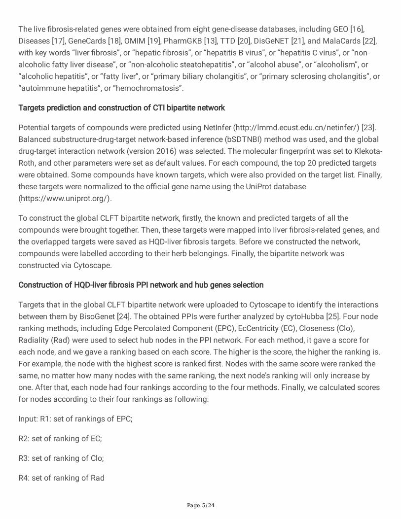
The live �brosis-related genes were obtained from eight gene-disease databases, including GEO [16],Diseases [17], GeneCards [18], OMIM [19], PharmGKB [13], TTD [20], DisGeNET [21], and MalaCards [22],with key words “liver �brosis”, or “hepatic �brosis”, or “hepatitis B virus”, or “hepatitis C virus”, or “non-alcoholic fatty liver disease”, or “non-alcoholic steatohepatitis”, or “alcohol abuse”, or “alcoholism”, or“alcoholic hepatitis”, or “fatty liver”, or “primary biliary cholangitis”, or “primary sclerosing cholangitis”, or“autoimmune hepatitis”, or “hemochromatosis”.
Targets prediction and construction of CTI bipartite network
Potential targets of compounds were predicted using NetInfer (http://lmmd.ecust.edu.cn/netinfer/) [23].Balanced substructure-drug-target network-based inference (bSDTNBI) method was used, and the globaldrug-target interaction network (version 2016) was selected. The molecular �ngerprint was set to Klekota-Roth, and other parameters were set as default values. For each compound, the top 20 predicted targetswere obtained. Some compounds have known targets, which were also provided on the target list. Finally,these targets were normalized to the o�cial gene name using the UniProt database(https://www.uniprot.org/).
To construct the global CLFT bipartite network, �rstly, the known and predicted targets of all thecompounds were brought together. Then, these targets were mapped into liver �brosis-related genes, andthe overlapped targets were saved as HQD-liver �brosis targets. Before we constructed the network,compounds were labelled according to their herb belongings. Finally, the bipartite network wasconstructed via Cytoscape.
Construction of HQD-liver �brosis PPI network and hub genes selection
Targets that in the global CLFT bipartite network were uploaded to Cytoscape to identify the interactionsbetween them by BisoGenet [24]. The obtained PPIs were further analyzed by cytoHubba [25]. Four noderanking methods, including Edge Percolated Component (EPC), EcCentricity (EC), Closeness (Clo),Radiality (Rad) were used to select hub nodes in the PPI network. For each method, it gave a score foreach node, and we gave a ranking based on each score. The higher is the score, the higher the ranking is.For example, the node with the highest score is ranked �rst. Nodes with the same score were ranked thesame, no matter how many nodes with the same ranking, the next node's ranking will only increase byone. After that, each node had four rankings according to the four methods. Finally, we calculated scoresfor nodes according to their four rankings as following:
Input: R1: set of rankings of EPC;
R2: set of ranking of EC;
R3: set of ranking of Clo;
R4: set of ranking of Rad
Page 6/24
Output: the score of each node
S = 0
for i from 1 to Nn: #Nn: the number of nodes in PPI network
if R1(i) < median R1: #R1(i): the ranking of the i-th node in R1
S(i) +=1 #S(i): the S value of the i-th node
if R2(i) < median R2: #R2(i): the ranking of the i-th node in R2
S(i) +=1
If R3(i) < median R3: #R3(i): the ranking of the i-th node in R3
S(i) +=1
If R4(i) < median R4: #R4(i): the ranking of the i-th node in R4
S(i) +=1
return S(i)
end
For these targets with a �nal score greater than 1, if there was more than one compound interacting withthem, then they were selected as hub genes.
Enrichment of KEGG pathways
Hub genes were imported to STRING (https://string-db.org/) [26] database to perform KEGG pathwayenrichment analysis. Only KEGG pathways with the false discovery rate < 0.05 were regarded assigni�cant and were retained. Based on the knowledge accumulated in previous literature research, wedeleted pathways that were less related to liver �brosis, such as cancer-related pathways, and onlypathways that were closely related to liver �brosis were retained.
Molecular docking and construction of the critical compound-liver �brosis target bipartite network
Molecular docking software Glide was used to evaluate the potential interactions between criticalcompounds and liver �brosis targets. We collected all the crystal structures of hub gene-encoded proteinsthat appeared in retained pathways closely related to liver �brosis from the RCSB Protein Data Bank(PDB, https://www.rcsb.org/) [27]. Only those with relatively higher resolution were reserved for moleculardocking.
Page 7/24
The protein preparation module of Schrodinger’s Maestro molecular modeling suite (Schrödinger Release2015-2) was used to prepare the protein crystallographic structures. Water molecules were deleted fromthe structures, and the amide moieties in the side chain were adjusted to optimize their interactions withsurrounding residues and groups of atoms. Force �eld OPLS_2005 was added. LigPrep module inMaestro was utilized to obtain the three-dimensional structures and energy minimization of compounds.A ligand grid generation was based on the ligand in the co-crystallographic structure. After the ligand gridwas generated, the prepared compounds were imported into Maestro and docked in the generated gridusing the standard Glide docking mode. The Glide Gscore greater than the median value of all the GlideGscores indicated a potential strong binding ability of candidate targets to their correspondingcompounds. The docking results were visualized by PyMOL software, and the hydrogen bonds and theirbinding sites were observed and analyzed.
Before we constructed the critical CLFT bipartite network, the critical CTIs need to be chosen from knownCTIs and predicted CTIs that have been docked, and the selection criteria were as follows: (1) For theknown CTIs of each target, the known CTI with the larger FC_MAX value of the compound was reserved.(2) For the predicted CTIs of each target, the predicted CTI with the lower Gscore value, and the greater theFC_MAX value of the compound was retained. After that, the obtained predicted CTIs as well as knownCTIs were imported to Cytoscape to construct the critical CLFT bipartite network.
Results
Data collectionThe original prototype components of HQD and their metabolites were collected from the supportinginformation of Xie’s paper [10]. After re�nement via a few screening criteria, a total of 68 compounds,including 17 prototype components and 51 metabolites, were �nally obtained.
To further clarify the relationships between compounds and genes, according to the division rules, werealized that four of the 17 prototype compounds were derived from R. Astragali, eight from R.Glycyrrhizae, and �ve from both herbs. As for most of the metabolites that cannot be identi�ed as herbbelongings from literature, we did not classify them. Details of the 68 compounds were listed inSupplementary Table S1.
We also tried to search for the known targets for the 68 compounds from four databases, includingPubChem [11], IUPHAR/BPS Guide to PHARMACOLOGY [12], PharmGKB [13], BindingDB [14], andDrugBank [15]. After removing duplicates, 186 known compound-target interactions (CTIs) were obtainedfor only 23 of the 68 compounds.
Liver �brosis is not a single disease but a pathological concept for characterizing a variety of chronic liverdiseases, so we regarded those genes related to several types of chronic liver diseases to cause liver�brosis as liver �brosis-related genes. Then eight gene-disease databases, including GEO [16], Diseases[17], GeneCards [18], OMIM [19], PharmGKB [13], TTD [20], DisGeNET [21], and MalaCards [22], were
Page 8/24
searched with a lot of key words. After removing the duplicates, 1192 liver �brosis-related genes wereobtained.
Compound-liver �brosis target bipartite networkA compound-target bipartite network is helpful for understanding the molecular mechanism of chemicalaction. However, only 23 of the 68 components in HQD were found to have known targets, and most ofthem do not have. Therefore, potential targets were predicted for the all 68 compounds by our webserverNetInfer [23], and 1360 CTIs were acquired. Combining with the above 186 known CTIs, totally 1520 CTIswere obtained after deleting duplicates, which were involved in 310 targets (see Supplementary TableS2).
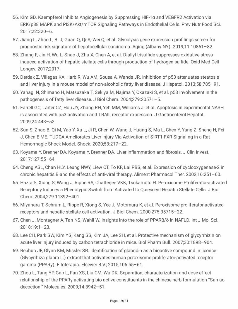
Among the 310 targets, 95 ones were present in the collected 1192 liver �brosis-related genes, which ledto 540 CTIs between the 95 targets and the 68 components. A global compound-liver �brosis target(CLFT) bipartite network was then constructed via Cytoscape 3.8.0, as shown in Fig. 1. In total, thisbipartite network consisted of 163 nodes and 540 edges, with 68 compounds as triangle nodes and 95targets as circle nodes.
To more intuitively represent the relationships between compounds and targets, 68 compounds weredivided into four groups according to the division rules: four from R. Astragali (colored with purple), eightfrom R. Glycyrrhizae (colored with green), �ve from both herbs (orange), and the other 51 metabolites(blue). It is obvious that the number of compounds in R. Astragali is less than that in R. Glycyrrhizae.Among these compounds, Genistein (MOL14), Kaempferol (MOL5), Glycyrrhetinic acid (MOL11),Apocholic acid (MOL48), Daidzein (MOL52), Hyocholic acid (MOL55), and Lucidenic acid G (MOL59)have the highest number of targets. The 540 CTIs include 53 known CTIs and 487 predicted CTIs. Amongthe 53 known CTIs, Genistein has interactions with 14 targets, Kaempferol has associations with 12targets, both compounds are well-studied in herbal medicine.
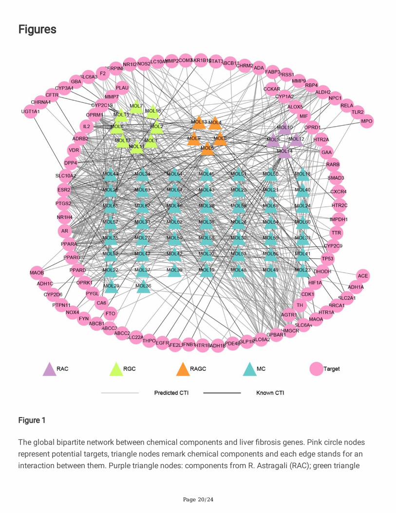
Protein-protein interaction network and KEGG pathwaysBesides the direct interactions with target proteins, the compounds might also affect the other proteinsindirectly, for example, via protein-protein interactions (PPIs). Therefore, PPIs were searched for the 95targets in the global CLFT bipartite network via BisoGenet, a Cytoscape plugin [24], which resulted in atotal of 71 PPIs for 48 of the 95 targets. Then, the HQD-liver �brosis PPI network was constructed, asshown in Fig. 2.
In order to identify important targets from the PPI network, four values including Edge PercolatedComponent (EPC), EcCentricity (EC), Closeness (Clo), and Radiality (Rad) were calculated by cytoHubba,another Cytoscape plugin [25] for each node in the PPI network. After calculation, there were seventargets, including AR, PPARG, CDK1, TP53, HIF1A, VDR, and PPARD with a �nal score greater than 1, andmore than one compound has interactions with them. Although PTGS2, SERPINE1 and MMP9 did notmeet the requirements, they have also opted for the next step based on the knowledge of previousliterature research.
Page 9/24
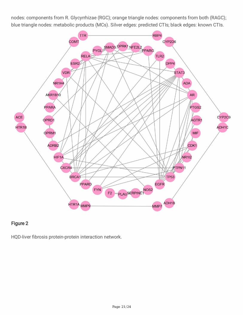
These 10 hub genes were enriched into 30 KEGG pathways with signi�cance. Except for cancer-relatedpathways (e.g. Pathways in cancer (hsa05200), Prostate cancer (hsa05215), etc.) and other pathways(for instance, Longevity regulating pathway (hsa04211), etc.) were not directly related to liver �brosis thatbased on knowledge from literature research, eight liver �brosis-related pathways were selected forfurther analysis, as shown in Fig. 3a.
These eight pathways include p53 signaling pathway (hsa04115), Cellular senescence (hsa04218), PPARsignaling pathway (hsa03320), HIF-1 signaling pathway (hsa04066), IL-17 signaling pathway(hsa04657), TNF signaling pathway (hsa04668), Cell cycle (hsa04110), and Hepatitis B (hsa05161).CDK1, SERPINE1, and TP53 were enriched in p53 signaling pathway and Cellular senescence, which wererelated to cell cycle arrest according to the KEGG Pathway database(https://www.kegg.jp/kegg/pathway.html). Furthermore, peroxisome proliferator-activated receptor(PPAR) family genes, including PPARD and PPARG were enriched into one pathway - PPAR signalingpathway, which is mainly related to liver lipid metabolism. The HIF-1 signaling pathway contains HIF1Aand SERPINE1 and is related to angiogenesis. MMP9 and PTGS2 were enriched into IL-17 signalingpathway and TNF signaling pathway, two in�ammation-related pathways. Besides, Hepatitis B alsoappeared, which was pathogenic factor of liver �brosis. The pathway-gene bipartite network was shownin Fig. 3b, consisting of eight pathways and eight genes, and each pathway contains two or three genes.
Molecular docking to evaluate critical compound-liver�brosis target interactionsThere were eight genes in the above eight pathways, and 43 compounds have interactions with theseeight targets in the global CLFT bipartite network, of which 23 compounds interacted with PPARG, 17compounds had links with PTGS2, 10 compounds could act on PPARD, and 7 compounds wereassociated with SERPINE1. For the convenience of analyzing the relationships between criticalcompounds and targets from the compounds that could act on PPARG, PTGS2, PPARD, or SERPINE1, weselected some compounds that have a higher concentration in vivo after administration of HQD.Concretely speaking, for metabolites that have interactions with PPARG or PTGS2, only the maximum offold change (FC_MAX) values greater than 10 were reserved; while for metabolites that could act onPPARD or SERPINE1, compounds with a maximum value of FC greater than 5 remained. In the end, 34pairs of CTIs (including 5 known CTIs and 29 predicted CTIs) were reserved, including eight targets and27 compounds.
We performed molecular docking on these 29 pairs of predicted CTIs, including six targets (CDK1, MMP9,PPARD, PPARG, PTGS2, and SERPINE1) and 23 compounds (see Supplementary Table S3). Notably, thelower of Glide Gscore meant the binding between the compounds and the targets were stronger. Then, weselected critical CTIs from predicted CTIs and known CTIs with the following selection criteria: (1) For thepredicted CTIs of each target, the predicted CTI with the lower Gscore value, and the greater the FC_MAXvalue of the compound was retained. (2) For the known CTIs of each target, the known CTI with the larger
Page 10/24
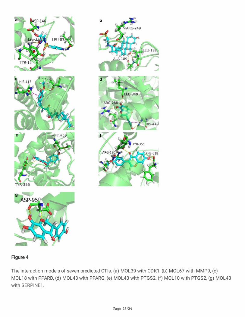
FC_MAX value of the compound was reserved. Finally, seven predicted CTIs and three known CTIs wereregarded as critical CTIs, which include eight compounds and eight targets.
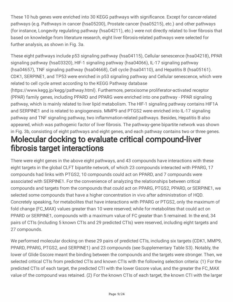
The Gscores of seven predicted CTIs were listed in Table 1. As shown, 4',7,8-Trihydroxyiso�avanone(MOL43) had strong interactions with three targets, including PTGS2, PPARG and SERPINE1, with highscores of -8.111, -7.534 and − 5.377, respectively. The binding sites of the seven predicted CTIs wereshown in Fig. 4. According to Fig. 4 and Table 1, all the �ve compounds in the seven interaction modelsdemonstrated good binding with the six hub genes, suggesting that the HQD had a strong tendency as atherapeutic strategy for liver �brosis via these hub genes and compounds. As shown, 5-Hydroxysulfamethoxazole (MOL39) had a strong binding ability with CDK1 (Gscore = -6.125),Tauroursodeoxycholic acid (MOL67) with MMP9 (Gscore = -6.906), (S)- [8]-Gingerol (MOL18) with PPARD(Gscore = -7.786), 4',7,8-Trihydroxyiso�avanone (MOL43) with PPARG (Gscore = -7.534), 4',7,8-Trihydroxyiso�avanone (MOL43) with PTGS2 (Gscore = -8.111), Calycosin (MOL10) with PTGS2 (Gscore= -7.502), 4',7,8-Trihydroxyiso�avanone (MOL43) with SERPINE1 (Gscore = -5.377).
Table 1The Glide Gscores of seven PCTIs.
Compound ID Compound Name Target Glide Gscore
MOL39 5-Hydroxysulfamethoxazole CDK1 -6.125
MOL67 Tauroursodeoxycholic acid MMP9 -6.906
MOL18 (S)-[8]-Gingerol PPARD -7.786
MOL43 4',7,8-Trihydroxyiso�avanone PPARG -7.534
MOL43 4',7,8-Trihydroxyiso�avanone PTGS2 -8.111
MOL10 Calycosin PTGS2 -7.502
The interaction model of MOL39 in the active site of CDK1 (Fig. 4a) showed the presence of a pi-pistacking with the key residue Tyr15 in addition to the formation of three hydrogen bonds with the residueAsp146, Leu83, Lys33, which may help the stabilization of the ligand in the active site of the targetprotein. Four hydrogen bonds were observed between MOL67 and the residue Leu188, Ala189 andArg249 in the active site of MMP9 (Fig. 4b). MOL18 showed favorable binding with PPARD, where itsinteraction diagram in the binding site of PPARD (Fig. 4c) showed the formation of a stacking pi-piinteraction between the aromatic ring of MOL18 and the residue His413, a hydrogen bond was observedbetween MOL18 and Thr253. The interaction diagram between MOL43 and PPARG (Fig. 4d) showed theformation of a pi-pi stacking with the key residue His449, and the formation of three hydrogen bonds withthe residue His449, Leu340, Arg288. Likewise, MOL43 showed a high a�nity towards PTGS2 (Fig. 4e).Studying the interaction diagrams of MOL43 in the active site of PTGS2 showed the formation of astacking pi-pi interaction between MOL43 and the residue Tyr355. A hydrogen bond was also observedbetween MOL43 and Met522. Besides, among all the predicted CTIs, MOL43 showed the lowest Gscoreagainst SERPINE1 (Fig. 4g), it only formatted two hydrogen bonds with the residue Asp95. The interaction
Page 11/24
diagram between MOL10 and PTGS2 (Fig. 4f) showed the formation of four hydrogen bonds with theresidue Tyr355, Arg120 and Phe518.
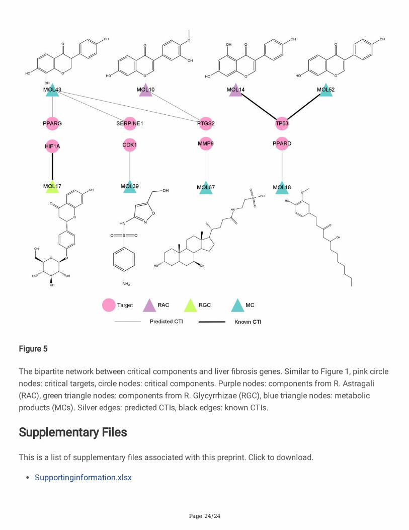
Critical compound-liver �brosis target bipartite network andmechanism analysisSeven predicted CTIs according to the results of molecular docking and chemical concentration, as wellas three known CTIs constructed the critical CLFT bipartite network, as shown in Fig. 5. In the graph, therewere two compounds from R. Astragali (colored with purple), one from R. Glycyrrhizae (green) and �vemetabolites (blue). Among them, MOL10, MOL14, MOL17, MOL43 and MOL52 were �avonoids. Inaddition, three known CTIs (black edge) were MOL14 and MOL52 targeting TP53, MOL17 acting onHIF1A. Seven predicted CTIs (silver edge) include MOL43 to PTGS2, PPARG and SERPINE1, respectively;MOL10 to PTGS2; MOL39 to CDK1; MOL67 to MMP9; MOL18 to PPARD. It can be found that except forMOL43 which interacted with three targets, all other compounds were directed against a single target.Among the eight critical compounds, MOL43, a metabolite compound, has the highest concentration invivo after administration of HQD with FC_MAX = 2390.64, followed by MOL10 and MOL17, with FC_MAX = 772.69 and 14.35, respectively. It is worth noting that MOL10 is a prototype compound in R. Astragaliand MOL17 is an original component of R. Glycyrrhizae. Furthermore, from the structures of these eightcompounds, it is easy to see that MOL10 and MOL14, two prototype compounds in R. Astragali, havesimilar structures, in addition, MOL43 and MOL52, two metabolites, have similar structures with MOL10and MOL14. For this reason, we can reasonably speculate that MOL43 and MOL52 may be metabolitesof original components in R. Astragali.
DiscussionAs a classical herb pair, HQD has been used to improve liver function and quality of life in patients withchronic liver disease, such as liver �brosis [28]. Though there have been some studies on HQD [6, 7, 29,30], there is a lack of target-level study on the mechanisms of its prototype compounds and theirmetabolites in the treatment of liver �brosis. Here, we tried to understand the MoA of HQD from asystematic perspective by combining metabolomics data with network pharmacology and moleculardocking methods. From the results, we analyzed the potential MoA for HQD to treat liver �brosis througheight critical targets.
MMP9, HIF1A and SERPINE1, three targets that in the critical CLFT bipartite network, in addition toMMP2, a target not appeared in the critical CLFT network but in the global CLFT network, which werecorrelated with �brogenesis and degradation.
Liver �brosis is a dynamic pathologic process characterized by an accumulation of the ECM, which is aconsequence of an imbalance between ECM deposition and degradation, re�ecting dysregulation ofmatrix metalloproteinases (MMPs) and their speci�c inhibitors (tissue inhibitors of metalloproteinases,TIMP) [31]. Upon chronic damage of liver tissue, HSCs become activated and differentiate into a�broblast-like phenotype, and upregulated the expression of TIMP1, which leading to the inhibition of
Page 12/24
MMP activity and subsequent accumulation of ECM [31]. In the family of MMPs, MMP2 and MMP9 areparticularly important for the development of liver �brosis since they degrade type IV collagen (basalmembrane) [32]. However, some studies also demonstrated MMPs, especially MMP2 and MMP9,promoted HSCs proliferation and migration [33, 34]. MMP9 was up-expressed in HCV patients withdifferent stages of �brosis [35]. The activity of MMP2 and MMP9 in patients with liver cirrhosis wereincreased [36]. There was also a trend for higher serum MMP9 in patients with HCC [37]. In contrast, italso has been reported that MMP2 and MMP9 levels showed a signi�cant elevation in chronic HCVpatients [38].
Accumulating evidence suggested that hypoxia may be a key driving force for the production of pro-�brotic mediators during �brosis through activation of hypoxia-inducible factor-1α (HIF1A) [39].Furthermore, activation of HIF1A can affect HSCs function, angiogenesis, matrix deposition and removal,and carcinogenesis [39]. Several studies have shown that HIF1A is critical for upregulation of pro-�broticmediators, such as platelet-derived growth factor A/B, and plasminogen activator inhibitor-1 (SERPINE1),and mice de�cient in HIF1A had reduced liver �brosis [40–42]. Moreover, MMP2 has been proved to bepositively correlated with HIF1A protein levels in HCC tissues, the expression levels of MMP2 and HIF1Ain the HCC tissues were higher than those in the adjacent normal tissues [43].
SERPINE1 is a major inhibitor of both tissue-type plasminogen activator and urokinase-type plasminogenactivator, it is a �brosis-promoting molecule and is a promising therapeutic target for �brotic diseases[44]. It has been reported that the urokinase-type plasminogen activator gene delivered into HSCsdecreased the amount of collagen types I and III accompanied by the increased expression of MMP2 anddecreased area of ECM in the �brotic liver [45]. Plasma SERPINE1 level was signi�cantly increased inchildren with increased severity of steatosis, and �brosis [46]. Higher expression of SERPINE1 was alsofound to be present in adults with NAFLD and children with NASH [47, 48]. SERPINE1 de�ciency reducedcholestatic liver injury and �brosis [49, 50]. Wang et al. also found that SERPINE1 de�ciency reducedhepatic �brosis after bile duct obstruction [51].
Liang et al. reported that expression of MMP2 and MMP9 proteins were up-regulated in carbontetrachloride-induced liver injury, while treatment with MOL2 signi�cantly reduced the expression levels ofMMP2 and MMP9 proteins [52]. MOL10 has also been reported to be able to inhibit the expression ofMMP9 [53]. MOL10, MOL13, and MOL5 were reported to decrease the expression of HIF1A [54–56].Based on the above analysis, we speculate that three compounds of HQD maintain the balance of ECMand reduces liver damage by regulating the expression of MMP2, MMP9 and SERPINE1.
Among the eight critical targets obtained in this study, CDK1 and TP53 were enriched in the p53signalling pathway and cellular senescence signalling pathway, which were related to apoptosis and cellcycle arrest. It has been reported that CDK1 was signi�cantly up-regulated in 309 HCC tissues comparedwith adjacent tissues [57]. Zhang et al. reported that downregulated cyclin B1 and CDK1, inducedcaspase-dependent apoptosis, and reduced migration in HSCs [58]. A growing amount of evidencesuggests that TP53 performs a central function in the development of chronic liver diseases. For
Page 13/24
example, Derdak et al. found that inhibition of TP53 attenuated steatosis and liver injury in a NAFLDmodel [59]. Yahagi et al. demonstrated that TP53 was activated in hepatic steatosis models and the p53pathway was involved in the pathogenesis of the fatty liver disease [60]. Moreover, hepatocyte apoptosiswas linked to TP53 activation in experimental NASH [61]. Based on these �ndings, overexpression ofCDK1 and TP53 may exacerbate liver �brogenesis. There were four compounds in the global CLFTbipartite network linking to CDK1, and three compounds interacting with TP53. Among them, MOL67 wasreported to inhibit expression and acetylation of NF-κB and TP53, and attenuated hemorrhagic shock-induced liver injury [62].
PTGS2, PPARD, and PPARG were three of eight targets in the critical CLFT network. After hepatocyteinjury, in�ammation and the activation of the innate immune system lead to HSCs activation and ECMsecretion and deposition, which cause liver �brogenesis [63]. Patients with chronic hepatitis B hadsigni�cantly higher PTGS2 expression compared with controls [64]. PPARG plays an important role in theinhibition of HSC activation and has been proposed as a potential molecular target for liver �brosis [65].There has been clear evidence that PPARG level and activity are reduced in activated HSCs [66].Activation of PPARG modulates pro�brogenic and pro-in�ammatory actions in HSCs [62]. Moreover, liverin�ammatory responses were also suppressed by PPARA, PPARD and PPAG by inhibition of NF-κB [67].MOL2 alleviated carbon tetrachloride-induced liver injury partly due to downregulate the expression ofpro-in�ammatory mediators, including PTGS2 [68]. MOL1 was found to attenuate pro-in�ammatorycytokines through activating PPARG [69]. MOL16, MOL7, MOL15, MOL13, showed an effect on PPARGactivation [70].
In this study, we visualized an intricate network among prototype compounds and metabolites of HQDand their potential targets of liver �brosis. Based on our topology analysis and molecular dockingsimulation, eight compounds (MOL39, MOL67, MOL18, MOL43, MOL10, MOL14, MOL52, and MOL17)and eight targets (CDK1, MMP9, PPARD, PPARG, PTGS2, SERPINE1, TP53, and HIF1A) were regarded ascritical compounds and targets for the mechanism of HQD in the treatment of liver �brosis. In ourresearch results, not only three prototype compounds such as MOL10, MOL14, and MOL17 that havebeen well-studied in R. Astragali and R. Glycyrrhizae were found to be closely related to the therapeuticeffects of HQD, which is consistent with the results of existing experimental studies, but also somemetabolites such as MOL39, MOL43 and so on have been found to be the key to MoA of HQD on liver�brosis. Though more biological validation is needed to further validate the current results, for the �rsttime, the MoA of HQD in the treatment of liver �brosis has been explored from the target level in asystemic approach by combing the network pharmacology approach, metabolomics data and moleculardocking simulation. The combination of TCM and modern analytical methods may provide new ideas forthe study of TCM, and provide new therapeutic strategies and targets for liver �brosis.
ConclusionsThe classical herb pair HQD is widely used in clinic for the treatment of liver �brosis. In this study, we triedto understand the MoA of HQD on liver �brosis for the purpose of utilizing it more safely and effectively.

Page 14/24
By combining metabolomics data, network pharmacology and molecular docking methods, we tookprototype compounds and metabolites of HQD after administration together with their concentration intoconsideration, and found that eight compounds (5-Hydroxysulfamethoxazole, Tauroursodeoxycholic acid,(S)-[8]-Gingerol, 4',7,8-Trihydroxyiso�avanone, Calycosin, Genistein, Daidzein, and Liquiritin) and eighttargets (CDK1, MMP9, PPARD, PPARG, PTGS2, SERPINE1, TP53, and HIF1A) might contribute to the effectof HQD on liver �brosis by reducing �brogenesis and stimulate degradation.
AbbreviationsALD: Alcoholic liver disease; AR: Androgen receptor; bSDTNBI: Balanced substructure-drug-target network-based inference; CDK1: Cyclin-dependent kinase 1; CLFT: Compound-liver �brosis target; Clo: Closeness;CTI: Compound-target interaction; EC: EcCentricity; ECM: Extracellular matrix; EPC: Edge PercolatedComponent; Fc: Fold change; FC_MAX: Maximum of fold change; HBV: Hepatitis B virus; HCC:Hepatocellular carcinoma; HCV: Hepatitis C virus; HIF1A: Hypoxia-inducible factor-1α; HQD: Huangqidecoction; HSC: Hepatic stellate cell; IL1B: Interleukin-1 beta; IL6: Interleukin-6; MMP: Matrixmetalloproteinase; MMP2: Matrix metalloproteinase-2; MMP9: Matrix metalloproteinase-9; MoA: Action ofmechanism; NAFLD: Nonalcoholic fatty liver disease; NASH: Nonalcoholic steatohepatitis; NF-κB: Nuclearfactor-kappa B; PPAR: Peroxisome proliferator-activated receptor; PPARA: Peroxisome proliferator-activated receptor alpha; PPARD: Peroxisome proliferator-activated receptor delta; PPARG: Peroxisomeproliferator-activated receptor gamma; PPI: Protein-protein interaction; PTGS2: Prostaglandin G/Hsynthase 2; R. Astragali: Radix Astragali; Rad: Radiality; R. Glycyrrhizae: Radix Glycyrrhizae; SERPINE1:Plasminogen activator inhibitor-1; TCM: Traditional Chinese Medicine; TIMP: Tissue inhibitors ofmetalloproteinases; TNF: Tumor necrosis factor; TP53: Cellular tumor antigen p53; VDR: Vitamin D3receptor.
DeclarationsAcknowledgements
The authors would like to thank Guoxiang Xie and others for their published research paper, whichprovided metabolomics data for us.
Funding
This work was supported by the National Key Research and Development Program of China (Grant2019YFA0904800) and the National Natural Science Foundation of China (Grant 81872800).
Availability of data and materials
The data can be requested from the author upon reasonable request.
Ethics approval and consent to participate

Page 15/24
Not applicable.
Competing interests
The authors declare that they have no competing interests.
Consent for publication
Not applicable.
Authors’ Contributions
YT and BW contributed to conception and design of the study. BW performed the experiments and wrotethe manuscript. ZW provided the method of target prediction. WL and GL contributed to the writing of thisarticle. YT implemented the study and modi�ed the manuscript. All authors have discussed the resultsand approved the �nal manuscript.
References1. Bataller R, North KE, Brenner DA. Genetic polymorphisms and the progression of liver �brosis: A
critical appraisal. Hepatology. 2003;37:493–503.
2. Liu T, Wang X, Karsdal MA, Leeming DJ, Genovese F. Molecular serum markers of liver �brosis.Biomark Insights. 2012;7:105–17.
3. Bataller R, Brenner DA. Liver �brosis. J Clin Invest. 2005;115:209–18.
4. Fu J, Wang Z, Huang L, Zheng S, Wang D, Chen S, et al. Review of the botanical characteristics,phytochemistry, and pharmacology of Astragalus membranaceus (Huangqi). Phyther Res.2014;28:1275–83.
5. Li X, Sun R, Liu R. Natural products in licorice for the therapy of liver diseases: Progress and futureopportunities. Pharmacol Res. Elsevier; 2019;144:210–26.
�. Liu C, Wang G, Chen G, Mu Y, Zhang L, Hu X, et al. Huangqi decoction inhibits apoptosis and �brosis,but promotes Kupffer cell activation in dimethylnitrosamine-induced rat liver �brosis. BMCComplement Altern Med. BMC Complementary and Alternative Medicine; 2012;12:1.
7. Du JX, Sun MY, Du GL, Li FH, Liu C, Mu YP, et al. Ingredients of Huangqi decoction slow biliary�brosis progression by inhibiting the activation of the transforming growth factor-beta signalingpathway. BMC Complement Altern Med. BioMed Central Ltd; 2012;12:33.
�. Zhang GB, Song YN, Chen QL, Dong S, Lu YY, Su MY, et al. Actions of Huangqi decoction against ratliver �brosis: A gene expression pro�ling analysis. Chinese Med (United Kingdom). 2015;10:1–11.
9. Liu C, Liu P, Mu Y, Zhang H. Research Development on Treatment of HuangQi Decoction for ChronicLiver Disease. World Chinese Med. 2015;10:157–61.
10. Xie G, Wang S, Zhang H, Zhao A, Liu J, Ma Y, et al. Poly-pharmacokinetic Study of a MulticomponentHerbal Medicine in Healthy Chinese Volunteers. Clin Pharmacol Ther. 2018;103:692–702.
Page 16/24
11. Wang Y, Xiao J, Suzek TO, Zhang J, Wang J, Bryant SH. PubChem: A public information system foranalyzing bioactivities of small molecules. Nucleic Acids Res. 2009;37:623–33.
12. Harding SD, Sharman JL, Faccenda E, Southan C, Pawson AJ, Ireland S, et al. The IUPHAR/BPSGuide to PHARMACOLOGY in 2018: Updates and expansion to encompass the new guide toIMMUNOPHARMACOLOGY. Nucleic Acids Res. Oxford University Press; 2018;46:D1091–106.
13. Lamp J. Pharmacogenomics knowledge for personalized medicine. Semikron Appl Note AN-7006.2008;92:1–8.
14. Gilson MK, Liu T, Baitaluk M, Nicola G, Hwang L, Chong J. BindingDB in 2015: A public database formedicinal chemistry, computational chemistry and systems pharmacology. Nucleic Acids Res.2016;44:D1045–53.
15. Wishart DS, Knox C, Guo AC, Shrivastava S, Hassanali M, Stothard P, et al. DrugBank: acomprehensive resource for in silico drug discovery and exploration. Nucleic Acids Res.2006;34:D668-72.
1�. Barrett T, Wilhite SE, Ledoux P, Evangelista C, Kim IF, Tomashevsky M, et al. NCBI GEO: Archive forfunctional genomics data sets - Update. Nucleic Acids Res. 2013;41:991–5.
17. Pletscher-Frankild S, Pallejà A, Tsafou K, Binder JX, Jensen LJ. DISEASES: Text mining and dataintegration of disease-gene associations. Methods. Elsevier Inc.; 2015;74:83–9.
1�. Fishilevich S, Nudel R, Rappaport N, Hadar R, Plaschkes I, Iny Stein T, et al. GeneHancer: genome-wide integration of enhancers and target genes in GeneCards. Database (Oxford). 2017;2017:1–17.
19. Amberger J, Bocchini CA, Scott AF, Hamosh A. McKusick’s Online Mendelian Inheritance in Man(OMIM). Nucleic Acids Res. 2009;37:D793–6.
20. Li YH, Yu CY, Li XX, Zhang P, Tang J, Yang Q, et al. Therapeutic target database update 2018:Enriched resource for facilitating bench-to-clinic research of targeted therapeutics. Nucleic Acids Res.Oxford University Press; 2018;46:D1121–7.
21. Piñero J, Bravo Á, Queralt-Rosinach N, Gutiérrez-Sacristán A, Deu-Pons J, Centeno E, et al. DisGeNET:A comprehensive platform integrating information on human disease-associated genes and variants.Nucleic Acids Res. 2017;45:D833–9.
22. Rappaport N, Twik M, Plaschkes I, Nudel R, Stein TI, Levitt J, et al. MalaCards: An amalgamatedhuman disease compendium with diverse clinical and genetic annotation and structured search.Nucleic Acids Res. 2017;45:D877–87.
23. Wu Z, Peng Y, Yu Z, Li W, Liu G, Tang Y. NetInfer: A Web Server for Prediction of Targets andTherapeutic and Adverse Effects via Network-Based Inference Methods. J Chem Inf Model.2020;60:3687–91.
24. Martin A, Ochagavia ME, Rabasa LC, Miranda J, Fernandez-de-Cossio J, Bringas R. BisoGenet: A newtool for gene network building, visualization and analysis. BMC Bioinformatics. 2010;11.
25. Chin CH, Chen SH, Wu HH, Ho CW, Ko MT, Lin CY. cytoHubba: Identifying hub objects and sub-networks from complex interactome. BMC Syst Biol. BioMed Central Ltd; 2014;8:S11.
Page 17/24
2�. Szklarczyk D, Franceschini A, Wyder S, Forslund K, Heller D, Huerta-Cepas J, et al. STRING v10:Protein-protein interaction networks, integrated over the tree of life. Nucleic Acids Res.2015;43:D447–52.
27. Berman HM, Battistuz T, Bhat TN, Bluhm WF, Bourne PE, Burkhardt K, et al. The protein data bank.Acta Crystallogr Sect D Biol Crystallogr. 2002;58:899–907.
2�. Li H. Advances in anti hepatic �brotic therapy with Traditional Chinese Medicine herbal formula. JEthnopharmacol. Elsevier Ireland Ltd; 2020;251:112442.
29. Zhang X, Xu Y, Chen JM, Liu C, Du GL, Zhang H, et al. Huang Qi Decoction Prevents BDL-InducedLiver Fibrosis Through Inhibition of Notch Signaling Activation. Am J Chin Med. 2017;45:85–104.
30. Li WK, Wang GF, Wang TM, Li YY, Li YF, Lu XY, et al. Protective effect of herbal medicine Huangqidecoction against chronic cholestatic liver injury by inhibiting bile acid-stimulated in�ammation inDDC-induced mice. Phytomedicine. 2019;62.
31. Hemmann S, Graf J, Roderfeld M, Roeb E. Expression of MMPs and TIMPs in liver �brosis - asystematic review with special emphasis on anti-�brotic strategies. J Hepatol. 2007;46:955–75.
32. Kurzepa J, M A, Czechowska G, Kurzepa J, Celiński K, Kazmierak W, et al. Role of MMP-2 and MMP-9and their natural inhibitors in liver �brosis, chronic pancreatitis and non-speci�c in�ammatory boweldiseases. Hepatobiliary Pancreat Dis Int. 2014;13:570–9.
33. Yang C, Zeisberg M, Mosterman B, Sudhakar A, Yerramalla U, Holthaus K, et al. Liver �brosis: Insightsinto migration of hepatic stellate cells in response to extracellular matrix and growth factors.Gastroenterology. 2003;124:147–59.
34. Galli A, Svegliati-Baroni G, Ceni E, Milani S, Ridol� F, Salzano R, et al. Oxidative stress stimulatesproliferation and invasiveness of hepatic stellate cells via a MMP2-mediated mechanism.Hepatology. 2005;41:1074–84.
35. Capone F, Guerriero E, Sorice A, Maio P, Colonna G, Castello G, et al. Characterization ofmetalloproteinases, oxidative status and in�ammation levels in the different stages of �brosis inHCV patients. Clin Biochem. The Canadian Society of Clinical Chemists; 2012;45:525–9.
3�. Prystupa A, Boguszewska-Czubara A, Bojarska-Junak A, Toruń-Jurkowska A, Roliński J, Załuska W.Activity of MMP-2, MMP-8 and MMP-9 in serum as a marker of progression of alcoholic liver diseasein people from Lublin region, eastern Poland. Ann Agric Environ Med. 2015;22:325–8.
37. Kozłowska J, Mikuła T, Suchacz M, Jabłońska J, Stańczak W, Cianciara J, et al. Pigment epithelium-derived factor and matrix metalloproteinase-9 in liver cirrhosis. Saudi J Gastroenterol. 2016;22:375–9.
3�. Abdel-Latif MS. Plasma Levels of Matrix Metalloproteinase (MMP)-2, MMP-9 and Tumor NecrosisFactor-α in Chronic Hepatitis C Virus Patients. Open Microbiol J. 2015;9:136–40.
39. Copple BL. Hypoxia stimulates hepatocyte epithelial to mesenchymal transition by hypoxia-induciblefactor and transforming growth factor-β-dependent mechanisms. Liver Int. 2010;30:669–82.
40. Copple BL, Kaska S, Wentling C. Hypoxia-inducible factor activation in myeloid cells contributes tothe development of liver �brosis in cholestatic mice. J Pharmacol Exp Ther. 2012;341:307–16.
Page 18/24
41. Bryan L. Copple, Shan Bai J-OM. Hypoxia-inducible Factor-dependent Production of Pro�broticMediators by Hypoxic Kupffer Cells. Hepatol Res. 2010;40:530–9.
42. Moon JOK, Welch TP, Gonzalez FJ, Copple BL. Reduced liver �brosis in hypoxia-inducible factor-1α-de�cient mice. Am J Physiol - Gastrointest Liver Physiol. 2009;296:582–92.
43. Wang B, Ding YM, Fan P, Wang B, Xu JH, Wang WX. Expression and signi�cance of MMP2 and HIF-1α in hepatocellular carcinoma. Oncol Lett. 2014;8:539–46.
44. Hu PF, Chen H, Zhong W, Lin Y, Zhang X, Chen YX, et al. Adenovirus-mediated transfer of siRNAagainst PAI-1 mRNA ameliorates hepatic �brosis in rats. J Hepatol. European Association for theStudy of the Liver; 2009;51:102–13.
45. Lin Y, Xie WF, Chen YX, Zhang X, Zeng X, Qiang H, et al. Treatment of experimental hepatic �brosis bycombinational delivery of urokinase-type plasminogen activator and hepatocyte growth factor genes.Liver Int. 2005;25:796–807.
4�. Jin R, Krasinskas A, Le NA, Konomi J V., Holzberg J, Romero R, et al. Association betweenplasminogen activator inhibitor-1 and severity of liver injury and cardiovascular risk in children withnon-alcoholic fatty liver disease. Pediatr Obes. 2018;13:23–9.
47. Alisi A, Manco M, Devito R, Piemonte F, Nobili V. Endotoxin and plasminogen activator inhibitor-1serum levels associated with nonalcoholic steatohepatitis in children. J Pediatr Gastroenterol Nutr.2010;50:645–9.
4�. Thuy S, Ladurner R, Volynets V, Wagner S, Strahl S, Königsrainer A, et al. Nonalcoholic fatty liverdisease in humans is associated with increased plasma endotoxin and plasminogen activatorinhibitor 1 concentrations and with fructose intake. J Nutr. 2008;138:1452–5.
49. Bergheim I, Guo L, Davis MA, Duveau I, Arteel GE. Critical role of plasminogen activator inhibitor-1 incholestatic liver injury and �brosis. J Pharmacol Exp Ther. 2006;316:592–600.
50. Wang H, Vohra BPS, Zhang Y, Heuckeroth RO. Transcriptional pro�ling after bile duct ligationidenti�es PAI-1 as a contributor to cholestatic injury in mice. Hepatology. 2005;42:1099–108.
51. Wang H, Zhang Y, Heuckeroth RO. PAI-1 de�ciency reduces liver �brosis after bile duct ligation inmice through activation of tPA. FEBS Lett. 2007;581:3098–104.
52. Liang B, Guo XL, Jin J, Ma YC, Feng ZQ. Glycyrrhizic acid inhibits apoptosis and �brosis incarbontetrachloride-induced rat liver injury. World J Gastroenterol. 2015;21:5271–80.
53. Quan GH, Wang H, Cao J, Zhang Y, Wu D, Peng Q, et al. Calycosin suppresses RANKL-mediatedosteoclastogenesis through inhibition of MAPKs and NF-κB. Int J Mol Sci. 2015;16:29496–507.
54. Jia Z, Wang X, Wang X, Wei P, Li L, Wu P, et al. Calycosin alleviates allergic contact dermatitis byrepairing epithelial tight junctions via down-regulating HIF-1α. J Cell Mol Med. 2018;22:4507–21.
55. Wu J, Ke X, Ma N, Wang W, Fu W, Zhang H, et al. Formononetin, an active compound of Astragalusmembranaceus (Fisch) Bunge, inhibits hypoxia-induced retinal neovascularization via the HIF-1α/VEGF signaling pathway. Drug Des Devel Ther. 2016;10:3071–81.
Page 19/24
5�. Kim GD. Kaempferol Inhibits Angiogenesis by Suppressing HIF-1α and VEGFR2 Activation viaERK/p38 MAPK and PI3K/Akt/mTOR Signaling Pathways in Endothelial Cells. Prev Nutr Food Sci.2017;22:320–6.
57. Jiang L, Zhao L, Bi J, Guan Q, Qi A, Wei Q, et al. Glycolysis gene expression pro�lings screen forprognostic risk signature of hepatocellular carcinoma. Aging (Albany NY). 2019;11:10861–82.
5�. Zhang F, Jin H, Wu L, Shao J, Zhu X, Chen A, et al. Diallyl trisul�de suppresses oxidative stress-induced activation of hepatic stellate cells through production of hydrogen sul�de. Oxid Med CellLongev. 2017;2017.
59. Derdak Z, Villegas KA, Harb R, Wu AM, Sousa A, Wands JR. Inhibition of p53 attenuates steatosisand liver injury in a mouse model of non-alcoholic fatty liver disease. J Hepatol. 2013;58:785–91.
�0. Yahagi N, Shimano H, Matsuzaka T, Sekiya M, Najima Y, Okazaki S, et al. p53 Involvement in thepathogenesis of fatty liver disease. J Biol Chem. 2004;279:20571–5.
�1. Farrell GC, Larter CZ, Hou JY, Zhang RH, Yeh MM, Williams J, et al. Apoptosis in experimental NASHis associated with p53 activation and TRAIL receptor expression. J Gastroenterol Hepatol.2009;24:443–52.
�2. Sun S, Zhao B, Qi M, Yao Y, Xu L, Ji R, Chen W, Wang J, Huang S, Ma L, Chen Y, Yang Z, Sheng H, FeiJ, Chen E ME. TUDCA Ameliorates Liver Injury Via Activation of SIRT1-FXR Signaling in a RatHemorrhagic Shock Model. Shock. 2020;53:217–22.
�3. Koyama Y, Brenner DA, Koyama Y, Brenner DA. Liver in�ammation and �brosis. J Clin Invest.2017;127:55–64.
�4. Cheng ASL, Chan HLY, Leung NWY, Liew CT, To KF, Lai PBS, et al. Expression of cyclooxygenase-2 inchronic hepatitis B and the effects of anti-viral therapy. Aliment Pharmacol Ther. 2002;16:251–60.
�5. Hazra S, Xiong S, Wang J, Rippe RA, Chatterjee VKK, Tsukamoto H. Peroxisome Proliferator-activatedReceptor γ Induces a Phenotypic Switch from Activated to Quiescent Hepatic Stellate Cells. J BiolChem. 2004;279:11392–401.
��. Miyahara T, Schrum L, Rippe R, Xiong S, Yee J, Motomura K, et al. Peroxisome proliferator-activatedreceptors and hepatic stellate cell activation. J Biol Chem. 2000;275:35715–22.
�7. Chen J, Montagner A, Tan NS, Wahli W. Insights into the role of PPARβ/δ in NAFLD. Int J Mol Sci.2018;19:1–23.
��. Lee CH, Park SW, Kim YS, Kang SS, Kim JA, Lee SH, et al. Protective mechanism of glycyrrhizin onacute liver injury induced by carbon tetrachloride in mice. Biol Pharm Bull. 2007;30:1898–904.
�9. Rebhun JF, Glynn KM, Missler SR. Identi�cation of glabridin as a bioactive compound in licorice(Glycyrrhiza glabra L.) extract that activates human peroxisome proliferator-activated receptorgamma (PPARγ). Fitoterapia. Elsevier B.V.; 2015;106:55–61.
70. Zhou L, Tang YP, Gao L, Fan XS, Liu CM, Wu DK. Separation, characterization and dose-effectrelationship of the PPARγ-activating bio-active constituents in the chinese herb formulation “San-aodecoction.” Molecules. 2009;14:3942–51.
Page 20/24
Figures
Figure 1
The global bipartite network between chemical components and liver �brosis genes. Pink circle nodesrepresent potential targets, triangle nodes remark chemical components and each edge stands for aninteraction between them. Purple triangle nodes: components from R. Astragali (RAC); green triangle
Page 21/24
nodes: components from R. Glycyrrhizae (RGC); orange triangle nodes: components from both (RAGC);blue triangle nodes: metabolic products (MCs). Silver edges: predicted CTIs; black edges: known CTIs.
Figure 2
HQD-liver �brosis protein-protein interaction network.
Page 22/24
Figure 3
KEGG pathways. (a). Dot plot of the eight KEGG pathways. (b). The pathway-gene bipartite network.Green diamond nodes remark KEGG pathway, pink circle nodes remark target.
Page 23/24
Figure 4
The interaction models of seven predicted CTIs. (a) MOL39 with CDK1, (b) MOL67 with MMP9, (c)MOL18 with PPARD, (d) MOL43 with PPARG, (e) MOL43 with PTGS2, (f) MOL10 with PTGS2, (g) MOL43with SERPINE1.
Page 24/24
Figure 5
The bipartite network between critical components and liver �brosis genes. Similar to Figure 1, pink circlenodes: critical targets, circle nodes: critical components. Purple nodes: components from R. Astragali(RAC), green triangle nodes: components from R. Glycyrrhizae (RGC), blue triangle nodes: metabolicproducts (MCs). Silver edges: predicted CTIs, black edges: known CTIs.
Supplementary Files
This is a list of supplementary �les associated with this preprint. Click to download.
Supportinginformation.xlsx