anti-inflammatory effects of huang-lian-jie-du decoction
TRANSCRIPT
Aa
JD
a
ARRAA
KHARLC
1
iRpgonudctito(
0d
Journal of Ethnopharmacology 134 (2011) 911–918
Contents lists available at ScienceDirect
Journal of Ethnopharmacology
journa l homepage: www.e lsev ier .com/ locate / je thpharm
nti-inflammatory effects of Huang-Lian-Jie-Du decoction, its two fractionsnd four typical compounds
uan Lu, Jun-Song Wang, Ling-Yi Kong ∗
epartment of Natural Medicinal Chemistry, China Pharmaceutical University, 24 Tong Jia Xiang, Nanjing 210009, People’s Republic of China
r t i c l e i n f o
rticle history:eceived 17 July 2010eceived in revised form 16 January 2011ccepted 28 January 2011vailable online 4 February 2011
eywords:uang-Lian-Jie-Du decoctionnti-inflammationAW264.7 cellsipopolysaccharidearrageenan edema
a b s t r a c t
Ethnopharmacological relevance: Huang-Lian-Jie-Du decoction (HLJDD) (Oren-gedoku-to in Japanese) as afamous traditional Chinese recipe is composed of Rhizoma coptidis, Radix scutellariae, Cortex phellodendriand Fructus gardeniae. It has been used to treat inflammation for nearly two thousand years.Aim of the study: To explore the material base for the anti-inflammatory activity of formula HLJDD, itsextract was fractionated on D101 macroporous resin to afford two fractions, HLJDD-1 and HLJDD-2. Thewhole formula, HLJDD-1 and HLJDD-2, and four typical component compounds were then evaluatedfor their effects on inflammation-related parameters using lipopolysaccharide (LPS)-induced RAW264.7cells as a model system.Materials and methods: The effect of HLJDD on carrageenan-induced mice paw edema was first evaluated.A series of inflammation-related parameters including malondialdehyde (MDA), nitric oxide (NO), super-oxide dismutase (SOD), prostaglandin E2 (PGE2), tumor necrosis factor-� (TNF-�), interleukin-6 (IL-6)and interleukin-10 (IL-10) were then measured in LPS-induced RAW264.7 cells treated with HLJDD, its
two fractions, and four typical component compounds (geniposide, baicalin, berberine and baicalein).Results: With the help of principal component analysis (PCA) technique, the data obtained revealedthat the two fractions and the major group of compounds in HLJDD (iridoids, flavonoids and alkaloids)complement each other with particular emphasis to synergistically exert anti-inflammatory effects.Conclusions: This study demonstrated that HLJDD exhibited anti-inflammatory effect as a “whole”, whichjustified the combined use of the four component herbs forming the compound prescription and sug-HLJDDCrow
gested quality control of
. Introduction
Huang-Lian-Jie-Du decoction (HLJDD) is a preparation consist-ng of Rhizoma coptidis (Coptis chinensis Franch, Ranunculaceae),adix scutellariae (Scutellaria baicalensis Georgi, Labiatae), Cortexhellodendri (Phellodendron amurense Rupr. Rutaceae) and Fructusardeniae (Gardenia jasminoide Ellis, Rubiaceae) in a weight ratiof 3:2:2:3. This formula was described by Wang Tao (in the Chi-ese Tang Dynasty) in his treatise “Wai Tai Mi Yao”. It has beensed to treat inflammation, hypertension, gastrointestinal disor-ers, and liver and cerebrovascular diseases (Cao et al., 1996) in thelinical practice of Traditional Chinese Medicine. Oral administra-ion of HLJDD significantly inhibited the inflammatory responses
n carrageenan injected rat air pouches, and also greatly reducedhe production of leukotriene (LTB4) in vivo without any influencen the biosynthesis of cyclooxygenase (COX)-derived eicosanoidsZeng et al., 2009). Though the main components of the whole∗ Corresponding author. Tel.: +86 25 83271405; fax: +86 25 83271405.E-mail address: cpu [email protected] (L.-Y. Kong).
378-8741/$ – see front matter. Crown Copyright © 2011 Published by Elsevier Ireland Ltoi:10.1016/j.jep.2011.01.049
based on its three types of components.n Copyright © 2011 Published by Elsevier Ireland Ltd. All rights reserved.
formula and individual herb of HLJDD have been reported (Sunet al., 2006), principles in HLJDD responsible for anti-inflammatoryeffects remain unclear, hindering the rational use of this formula.
Inflammation involves a complex web of intercellular cytokinesignals (Han and Ulevitch, 2005) and is implicated in the pathogen-esis of many diseases, including cancer, diabetes, cardiovascular,neurodegenerative and other life-threatning and debilitating dis-eases (Lawrence et al., 2002). Macrophages play a central role in theinflammatory response, and serve as an essential interface betweeninnate and adaptive immunity (Adams and Hamilton, 1984). Inthe process of inflammatory response, macrophages release nitricoxide (NO), a reactive molecule originated from the guanidinonitrogen of l-arginine, catalyzed by nitric oxide synthases enzymes(NOS) and other cytokines, e.g. interleukin-6 (IL-6) (Kock et al.,1990). Excessive NO production leads to the development of manyinflammatory related diseases (Skidgel et al., 2002). Prostaglandin
E2 (PGE2), generated by specific COX-2 function, was another anti-inflammatory parameter (Surh et al., 2001). Moreover, MDA andSOD, due to their contributions to the alleviation of inflammatoryresponses (Cherubini et al., 2005; Baskol et al., 2007), were bothdetected for their levels to assess the radical scavenge abilitiesd. All rights reserved.
912 J. Lu et al. / Journal of Ethnopharmacology 134 (2011) 911–918
nstitu
omctdaePo
ma2Htpi
2
2
MQojf
2
iKMtwmbRr
2
twior(r
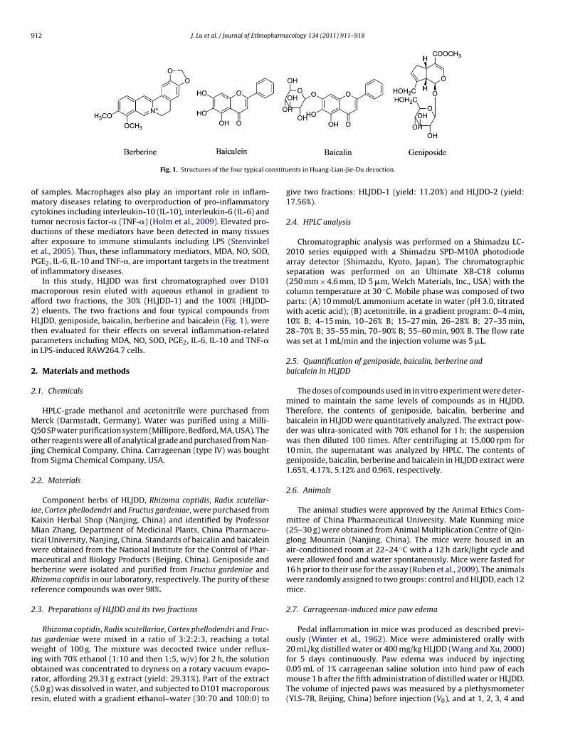
Fig. 1. Structures of the four typical co
f samples. Macrophages also play an important role in inflam-atory diseases relating to overproduction of pro-inflammatory
ytokines including interleukin-10 (IL-10), interleukin-6 (IL-6) andumor necrosis factor-� (TNF-�) (Holm et al., 2009). Elevated pro-uctions of these mediators have been detected in many tissuesfter exposure to immune stimulants including LPS (Stenvinkelt al., 2005). Thus, these inflammatory mediators, MDA, NO, SOD,GE2, IL-6, IL-10 and TNF-�, are important targets in the treatmentf inflammatory diseases.
In this study, HLJDD was first chromatographed over D101acroporous resin eluted with aqueous ethanol in gradient to
fford two fractions, the 30% (HLJDD-1) and the 100% (HLJDD-) eluents. The two fractions and four typical compounds fromLJDD, geniposide, baicalin, berberine and baicalein (Fig. 1), were
hen evaluated for their effects on several inflammation-relatedarameters including MDA, NO, SOD, PGE2, IL-6, IL-10 and TNF-�
n LPS-induced RAW264.7 cells.
. Materials and methods
.1. Chemicals
HPLC-grade methanol and acetonitrile were purchased fromerck (Darmstadt, Germany). Water was purified using a Milli-50 SP water purification system (Millipore, Bedford, MA, USA). Thether reagents were all of analytical grade and purchased from Nan-ing Chemical Company, China. Carrageenan (type IV) was boughtrom Sigma Chemical Company, USA.
.2. Materials
Component herbs of HLJDD, Rhizoma coptidis, Radix scutellar-ae, Cortex phellodendri and Fructus gardeniae, were purchased fromaixin Herbal Shop (Nanjing, China) and identified by Professorian Zhang, Department of Medicinal Plants, China Pharmaceu-
ical University, Nanjing, China. Standards of baicalin and baicaleinere obtained from the National Institute for the Control of Phar-aceutical and Biology Products (Beijing, China). Geniposide and
erberine were isolated and purified from Fructus gardeniae andhizoma coptidis in our laboratory, respectively. The purity of theseeference compounds was over 98%.
.3. Preparations of HLJDD and its two fractions
Rhizoma coptidis, Radix scutellariae, Cortex phellodendri and Fruc-us gardeniae were mixed in a ratio of 3:2:2:3, reaching a totaleight of 100 g. The mixture was decocted twice under reflux-
ng with 70% ethanol (1:10 and then 1:5, w/v) for 2 h, the solutionbtained was concentrated to dryness on a rotary vacuum evapo-ator, affording 29.31 g extract (yield: 29.31%). Part of the extract5.0 g) was dissolved in water, and subjected to D101 macroporousesin, eluted with a gradient ethanol–water (30:70 and 100:0) to
ents in Huang-Lian-Jie-Du decoction.
give two fractions: HLJDD-1 (yield: 11.20%) and HLJDD-2 (yield:17.56%).
2.4. HPLC analysis
Chromatographic analysis was performed on a Shimadzu LC-2010 series equipped with a Shimadzu SPD-M10A photodiodearray detector (Shimazdu, Kyoto, Japan). The chromatographicseparation was performed on an Ultimate XB-C18 column(250 mm × 4.6 mm, ID 5 �m, Welch Materials, Inc., USA) with thecolumn temperature at 30 ◦C. Mobile phase was composed of twoparts: (A) 10 mmol/L ammonium acetate in water (pH 3.0, titratedwith acetic acid); (B) acetonitrile, in a gradient program: 0–4 min,10% B; 4–15 min, 10–26% B; 15–27 min, 26–28% B; 27–35 min,28–70% B; 35–55 min, 70–90% B; 55–60 min, 90% B. The flow ratewas set at 1 mL/min and the injection volume was 5 �L.
2.5. Quantification of geniposide, baicalin, berberine andbaicalein in HLJDD
The doses of compounds used in in vitro experiment were deter-mined to maintain the same levels of compounds as in HLJDD.Therefore, the contents of geniposide, baicalin, berberine andbaicalein in HLJDD were quantitatively analyzed. The extract pow-der was ultra-sonicated with 70% ethanol for 1 h; the suspensionwas then diluted 100 times. After centrifuging at 15,000 rpm for10 min, the supernatant was analyzed by HPLC. The contents ofgeniposide, baicalin, berberine and baicalein in HLJDD extract were1.65%, 4.17%, 5.12% and 0.96%, respectively.
2.6. Animals
The animal studies were approved by the Animal Ethics Com-mittee of China Pharmaceutical University. Male Kunming mice(25–30 g) were obtained from Animal Multiplication Centre of Qin-glong Mountain (Nanjing, China). The mice were housed in anair-conditioned room at 22–24 ◦C with a 12 h dark/light cycle andwere allowed food and water spontaneously. Mice were fasted for16 h prior to their use for the assay (Ruben et al., 2009). The animalswere randomly assigned to two groups: control and HLJDD, each 12mice.
2.7. Carrageenan-induced mice paw edema
Pedal inflammation in mice was produced as described previ-ously (Winter et al., 1962). Mice were administered orally with20 mL/kg distilled water or 400 mg/kg HLJDD (Wang and Xu, 2000)
for 5 days continuously. Paw edema was induced by injecting0.05 mL of 1% carrageenan saline solution into hind paw of eachmouse 1 h after the fifth administration of distilled water or HLJDD.The volume of injected paws was measured by a plethysmometer(YLS-7B, Beijing, China) before injection (V0), and at 1, 2, 3, 4 and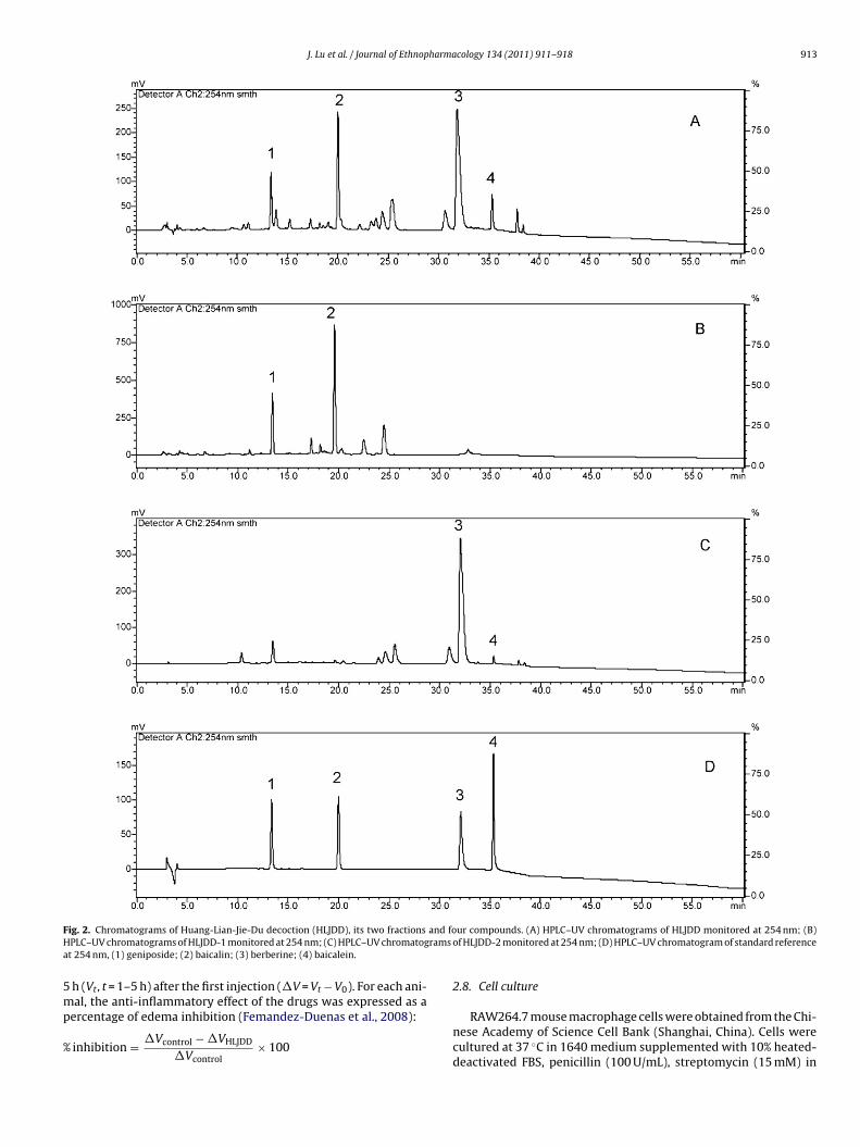
J. Lu et al. / Journal of Ethnopharmacology 134 (2011) 911–918 913
F and foH ramsa
5mp
%
ig. 2. Chromatograms of Huang-Lian-Jie-Du decoction (HLJDD), its two fractionsPLC–UV chromatograms of HLJDD-1 monitored at 254 nm; (C) HPLC–UV chromatogt 254 nm, (1) geniposide; (2) baicalin; (3) berberine; (4) baicalein.
h (Vt, t = 1–5 h) after the first injection (�V = Vt − V0). For each ani-
al, the anti-inflammatory effect of the drugs was expressed as aercentage of edema inhibition (Femandez-Duenas et al., 2008):
inhibition = �Vcontrol − �VHLJDD
�Vcontrol× 100
ur compounds. (A) HPLC–UV chromatograms of HLJDD monitored at 254 nm; (B)of HLJDD-2 monitored at 254 nm; (D) HPLC–UV chromatogram of standard reference
2.8. Cell culture
RAW264.7 mouse macrophage cells were obtained from the Chi-nese Academy of Science Cell Bank (Shanghai, China). Cells werecultured at 37 ◦C in 1640 medium supplemented with 10% heated-deactivated FBS, penicillin (100 U/mL), streptomycin (15 mM) in
914 J. Lu et al. / Journal of Ethnopharmacology 134 (2011) 911–918
Table 1Dose levels for all samples in cell assay.
Concentration I Concentration II Concentration III Concentration IV
Dexamethasonea 10 1HLJDDb 10−4 5 × 10⁻⁵ 10−5 5 × 10⁻⁶
HLLJDD-1b 0.3821 × 10⁻⁴ 1.9105 × 10⁻⁵ 0.3821 × 10⁻⁵ 1.9105 × 10⁻⁶
HLJDD-2b 0.5991 × 10⁻⁴ 2.9955 × 10⁻⁵ 0.5991 × 10⁻⁵ 2.9955 × 10⁻⁶
Geniposideb 0.0165 × 10⁻⁴ 0.0825 × 10⁻⁵ 0.0165 × 10⁻⁵ 0.0825 × 10⁻⁶
Bacalinb 0.0417 × 10⁻⁴ 0.2085 × 10⁻⁵ 0.0417 × 10⁻⁵ 0.2085 × 10⁻⁶
Berberineb 0.0512 × 10⁻⁴ 0.2560 × 10⁻⁵ 0.0512 × 10⁻⁵ 0.2560 × 10−6
× 10
d in g
a8s
2
1sts
2
cwdtawumsI
2T
wfupl
Niw1a(tt
2
pb
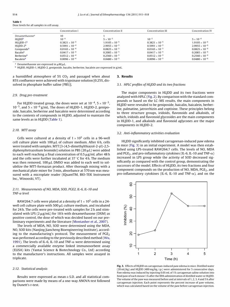
increased in LPS group while the activity of SOD decreased sig-nificantly as compared with the control group, demonstrating thesuccesses of the model. Effects of HLJDD, its two fractions and fourcomponent compounds on the production of NO, MDA, PGE2, andpro-inflammatory cytokines (IL-6, IL-10 and TNF-�), and on the
Fig. 3. Effects of HLJDD on carrageenan-induced paw edema in mice. Distilled water
Bacaleinb 0.0096 × 10⁻⁴ 0.0480
a Dexamethasone are expressed in �M/�L.b HLJDD, HLJDD-1, HLJDD-2, geniposide, bacalin, berberine, bacalein are expresse
humidified atmosphere of 5% CO2 and passaged when about5% confluence were achieved with trypsinase solution [0.25%, dis-olved in phosphate buffer saline (PBS)].
.9. Drug pre-treatment
For HLJDD treated group, the doses were set at 10−4, 5 × 10−5,0−5, and 5 × 10⁻⁶ g/mL. The doses of HLJDD-1, HLJDD-2, genipo-ide, baicalin, berberine and baicalein were determined accordingo the contents of compounds in HLJDD, adjusted to maintain theame levels as in HLJDD (Table 1).
.10. MTT assay
Cells were cultured at a density of 1 × 10⁵ cells in a 96-wellell culture plate with 100 �L of culture medium. After 6 h, cellsere treated with samples. MTT [3-(4,5-dimethylthiazol-2-yl)-2,5-iphenyltetrazolium bromide] solution in PBS (20 �L) were addedo each well reaching a final concentration of 0.5 �g/mL after 48 hnd the cells were further incubated at 37 ◦C for 4 h. The mediumas then removed, 100 �L DMSO was added to each well to sol-bilize the MTT-formazan product. After thorough mixing with aechanical plate mixer for 3 min, absorbance at 570 nm was mea-
ured with a microplate reader (IQuantTM, BIO-TEK Instrumentnc., Winooski, VT).
.11. Measurements of NO, MDA, SOD, PGE2, IL-6, IL-10 andNF-˛ level
RAW264.7 cells were plated at a density of 1 × 10⁵ cells in a 24-ell cell culture plate with 500 �L culture medium, and incubated
or 24 h. The cells were pre-treated with samples for 2 h and stim-lated with LPS (2 �g/mL) for 18 h with dexamethasone (DXM) asositive control, the dose of which was decided based on our pre-
iminary experiments and the literature (Montanher et al., 2007).The levels of MDA, NO, SOD were determined using the MDA,
O, SOD kits (Nanjing Jiancheng Bioengineering Institute), accord-ng to the manufacturing’s protocol. The measurement of PGE2
as performed according to the previously described method (Wu,991). The levels of IL-6, IL-10 and TNF-� were determined usingcommercially available enzyme linked immunosorbent assay
ELISA) kits (Yantai Science & Biotechnology Co., Ltd) accordingo the manufacturer’s instructions. All samples were assayed inriplicate.
.12. Statistical analysis
Results were expressed as mean ± S.D. and all statistical com-arisons were made by means of a one-way ANOVA test followedy Dunett’s t-test.
−5 0.0096 × 10⁻⁵ 0.0480 × 10⁻⁶
/mL.
3. Results
3.1. HPLC profiles of HLJDD and its two fractions
The major components in HLJDD and its two fractions wereanalyzed with HPLC (Fig. 2). By comparison with the standard com-pounds or based on the LC–MS results, the main components inHLJDD were revealed to be geniposide, baicalin, baicalein, berber-ine, palmatine, jatrorrhizin and coptisine. These principles belongto three structure groups, iridoids, flavonoids and alkaloids, ofwhich, iridoids and flavonoid glycosides are the main componentsin HLJDD-1, and alkaloids and flavonoid aglycones are the majorcomponents in HLJDD-2.
3.2. Anti-inflammatory activities evaluation
HLJDD significantly inhibited carrageenan-induced paw edemain mice (Fig. 3) in an initial experiment. A model was then estab-lished using LPS-treated RAW264.7 cells. The levels of NO, MDAand PGE2, and pro-inflammatory cytokines (IL-6, IL-10 and TNF-�)
(20 mL/kg) and HLJDD (400 mg/kg, i.p.) were administered for 5 consecutive days.Paw edema was induced by injecting 0.05 mL of 1% carrageenan saline solution intohind paw of each mouse 1 h after the fifth administration of distilled water or HLJDD.The volume of the paw was measured before and at intervals of 1, 2, 3, 4 and 5 h aftercarrageenan injection. Each point represents the percent increase of paw volume,which was calculated based on the volume of the paw before carrageenan injection.
J. Lu et al. / Journal of Ethnopharmacology 134 (2011) 911–918 915
F undst ata we
a(
tpas1ezebf
4
ii
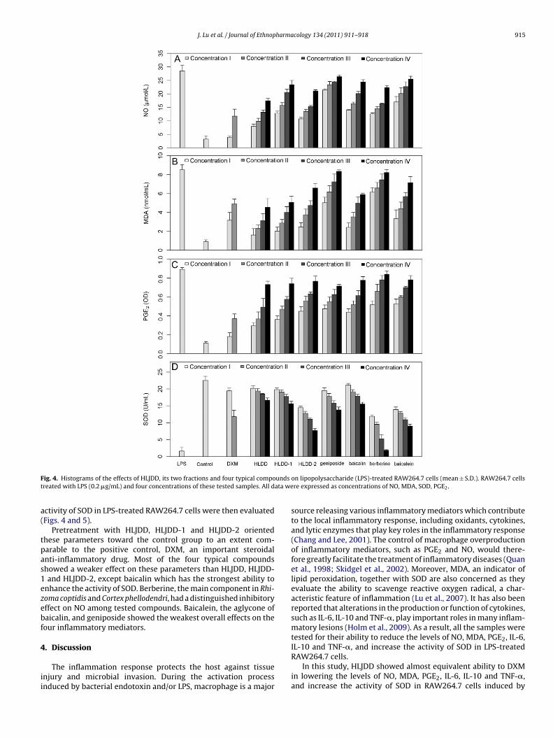
ig. 4. Histograms of the effects of HLJDD, its two fractions and four typical comporeated with LPS (0.2 �g/mL) and four concentrations of these tested samples. All d
ctivity of SOD in LPS-treated RAW264.7 cells were then evaluatedFigs. 4 and 5).
Pretreatment with HLJDD, HLJDD-1 and HLJDD-2 orientedhese parameters toward the control group to an extent com-arable to the positive control, DXM, an important steroidalnti-inflammatory drug. Most of the four typical compoundshowed a weaker effect on these parameters than HLJDD, HLJDD-and HLJDD-2, except baicalin which has the strongest ability to
nhance the activity of SOD. Berberine, the main component in Rhi-oma coptidis and Cortex phellodendri, had a distinguished inhibitoryffect on NO among tested compounds. Baicalein, the aglycone ofaicalin, and geniposide showed the weakest overall effects on theour inflammatory mediators.
. Discussion
The inflammation response protects the host against tissuenjury and microbial invasion. During the activation processnduced by bacterial endotoxin and/or LPS, macrophage is a major
on lipopolysaccharide (LPS)-treated RAW264.7 cells (mean ± S.D.). RAW264.7 cellsre expressed as concentrations of NO, MDA, SOD, PGE2.
source releasing various inflammatory mediators which contributeto the local inflammatory response, including oxidants, cytokines,and lytic enzymes that play key roles in the inflammatory response(Chang and Lee, 2001). The control of macrophage overproductionof inflammatory mediators, such as PGE2 and NO, would there-fore greatly facilitate the treatment of inflammatory diseases (Quanet al., 1998; Skidgel et al., 2002). Moreover, MDA, an indicator oflipid peroxidation, together with SOD are also concerned as theyevaluate the ability to scavenge reactive oxygen radical, a char-acteristic feature of inflammation (Lu et al., 2007). It has also beenreported that alterations in the production or function of cytokines,such as IL-6, IL-10 and TNF-�, play important roles in many inflam-matory lesions (Holm et al., 2009). As a result, all the samples weretested for their ability to reduce the levels of NO, MDA, PGE2, IL-6,
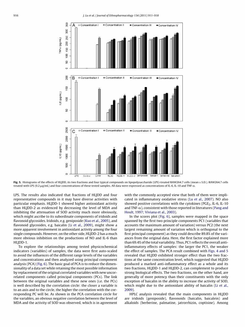
IL-10 and TNF-�, and increase the activity of SOD in LPS-treatedRAW264.7 cells.In this study, HLJDD showed almost equivalent ability to DXMin lowering the levels of NO, MDA, PGE2, IL-6, IL-10 and TNF-�,and increase the activity of SOD in RAW264.7 cells induced by
916 J. Lu et al. / Journal of Ethnopharmacology 134 (2011) 911–918
F undst ata we
LrptiwflflmsmH
itaasbrbitrtM
ig. 5. Histograms of the effects of HLJDD, its two fractions and four typical comporeated with LPS (0.2 �g/mL) and four concentrations of these tested samples. All d
PS. The results also indicated that fractions of HLJDD and fourepresentative compounds in it may have diverse activities witharticular emphasis. HLJDD-1 showed higher antioxidant activityhan HLJDD-2 as evidenced by decreasing the level of MDA andnhibiting the attenuation of SOD activity much more obviously,
hich might ascribe to its subordinate components of iridoids andavonoid glycosides. Iridoids, e.g. geniposide (Kuo et al., 2005), andavonoid glycosides, e.g. baicalin (Li et al., 2009), might show aore apparent involvement in antioxidant activity among the four
ingle compounds. However, on the other side, HLJDD-2 has a muchore obvious inhibition on the productions of NO and IL-6 thanLJDD-1.
To explore the relationships among tested physicochemicalndicators (variables) of samples, the data were first auto-scaledo avoid the influences of the different range levels of the variablesnd concentrations and then analyzed using principal componentnalysis (PCA) (Fig. 6). The basic goal of PCA is to reduce the dimen-ionality of a data set while retaining the most possible informationy replacement of the original correlated variables with new uncor-elated components called principal components (PCs). The linketween the original variables and these new ones (i.e. the PCs)
s well described by the correlation circle: the closer a variable iso an axis and to the circle, the higher the correlation with the cor-esponding PC will be. As shown in the PCA correlation circle forhe variables, an obvious negative correlation between the level of
DA and the activity of SOD was observed, which is in agreement
on lipopolysaccharide (LPS)-treated RAW264.7 cells (mean ± S.D.). RAW264.7 cellsre expressed as concentrations of IL-6, IL-10 and TNF-�.
with the commonly accepted view that both of them were impli-cated in inflammatory oxidative stress (Lu et al., 2007). NO alsoshowed positive correlations with the cytokines (PGE2, IL-6, IL-10and TNF-�), consistent with those reported in literatures (Pang andHoult, 1997; Viviana et al., 2003).
In the scores plot (Fig. 6), samples were mapped in the spacespanned by the first two principle components PC1 (variables thataccounts the maximum amount of variation) versus PC2 (the nextlargest remaining amount of variation which is orthogonal to thefirst principal component) as they could describe 89.8% of the vari-ances from the original data. Here, the first factor explained morethan 69.4% of the total variability. Thus, PC1 reflects the overall anti-inflammatory effects of samples: the larger the PC1, the weakerthe effect of samples. The PCA result combined with Figs. 4 and 5revealed that HLJDD exhibited stronger effect than the two frac-tions at the same concentration level, which suggested that HLJDDproduced a marked anti-inflammatory effect as a whole and itstwo fractions, HLJDD-1 and HLJDD-2, can complement to producestrong biological effects. The two fractions, on the other hand, aregenerally of more potency than their constituents with the onlyexception of baicalin in the ability to increase the activity of SOD,
which might due to the antioxidant ability of baicalin (Li et al.,2009).HPLC analysis revealed that the main components in HLJDDare iridoids (geniposide), flavonoids (baicalin, baicalein) andalkaloids (berberine, palmatine, jatrorrhizin, coptisine). Among

J. Lu et al. / Journal of Ethnopharma
Fig. 6. Score (PC1 vs PC2) plot of principal component analysis (PCA) on theanti-inflammatory results obtained from seven measured parameters, NO, MDA,PGE2, SOD, IL-6, IL-10 and TNF-� in samples of HLJDD, its two fractions (HLJDD-1 and HLJDD-2) and four typical compounds (geniposide, baicalin, berberine andbaicalein): correlation circle of parameters measured (represented by arrows) andpF(
tpmtgggilciiH
5
iectpptsifeRvHlrpie
rojection of the samples onto the first two factors of the PCA analysis are presented.irst principle component (PC1): contribution 69.4%; second principle componentPC2): contribution 20.4%.
hem, iridoids and flavonoid glycosides are the main princi-les in HLJDD-1, and flavonoid aglycones and alkaloids are theain constituents in HLJDD-2. The PCA could also classify the
wo fractions and their components into two groups. The firstroup including HLJDD-1, and its two constituents, baicalin andeniposide, located in the upper-left region, in general, hasreater anti-inflammatory potential than the second group includ-ng HLJDD-2, and its two constituents, berberine and baicalein,ocated in the lower-right region. It may postulate that theseompounds in the fractions had synergistic anti-inflammatorynteractions against the LPS-treated RAW264.7 cells and that reduc-ng oxidative injury may be an important therapeutic pathway forLJDD.
. Conclusion
The present study suggested that HLJDD has a potent anti-nflammatory activity against carrageenan-induced mice pawdema and LPS-induced RAW264.7 cells. HLJDD contains multi-omponents that produce a marked anti-inflammatory effecthrough multi-target and multi-channel actions. Moreover, HLJDDroduced a much more obvious anti-inflammatory effect as a wholerescription compared with the anti-inflammatory effects of itswo fractions and four typical compounds. The chemical analy-is has shown that the main active compounds in HLJDD includedridoids, flavonoids and alkaloids derived from four herbs. There-ore, the combinational use of these herbal drugs is necessary toxert the anti-inflammatory effect of HLJDD against LPS-inducedAW264.7 cells. On the basis of anti-inflammatory results, it pro-ided convincing data supporting the potential clinical use of
LJDD, and it also revealed that iridoids, flavonoids and alka-oids should be used for the quality control of HLJDD. Furtheresearch elucidating the mode of action of these effects using com-onent herbs and combinations of them would give an insight
nto the usefulness of this prescription for its anti-inflammatoryffects.
cology 134 (2011) 911–918 917
Acknowledgement
This project was supported by the National Key Scientific andTechnological Special Projects (2009ZX09502-011).
References
Adams, D.O., Hamilton, T.A., 1984. The cell biology of macrophage activation. AnnualReview of Immunology 2, 283–318.
Baskol, G., Atmaca, H., Tanriverdi, F., Baskol, M., Kocer, D., Bayram, F., 2007. Oxidativestress and enzymatic antioxidant status in patients with hypothyroidism beforeand after treatment. Experimental and Clinical Endocrinology and Diabetes 115,522–526.
Cao, Y.P., Gao, C., Sun, J.H., Wang, J.Z., Zhou, Q., Liu, G.Q., 1996. Study on pharmaco-logical effects of Huanglianjiedu Tang extract. Journal of China PharmaceuticalUniversity 27, 605–608.
Chang, Y.H., Lee, S.T., 2001. Effects of cannabinoids on LPS-stimulated inflammatorymediator release from macrophages: involvement of eicosanoids. Journal of CellBiochemistry 81, 715–723.
Cherubini, A., Ruggiero, C., Polidori, M.C., Mecocci, P., 2005. Potential markers ofoxidative stress in stroke. Free Radical Biology and Medicine 39, 841–852.
Femandez-Duenas, V., Sanchez, S., Planas, E., Poveda, R., 2008. Adjuvant effect ofcaffeine on acetylsalicylic acid anti-nociception: prostaglandin E2 synthesisdetermination in carrageenan-induced peripheral inflammation in rat. Euro-pean Journal of Pain 12, 157–163.
Han, J., Ulevitch, R.J., 2005. Limiting inflammatory responses during activation ofinnate immunity. Nature Immunology 6, 1198–1205.
Holm, S., Mackiewicz, Z., Holm, A.K., Kontinen, Y.T., Kouri, V.P., Indahl, A., Salo,J., 2009. Pro-inflammatory, pleiotropic, and anti-inflammatory TNF-�, IL-6,and IL-10 in experimental porcin intervertebral disk degeneration. VeterinaryPathology 46, 1292–1300.
Kock, A., Schwarz, T., Kirnbauer, R., Urbanski, A., Perry, P., Ansel, J.C., 1990. Humankeratinocytes are a source for tumor necrosis factor-�: evidence for synthe-sis and release upon stimulation with endotoxin or ultraviolet light. Journal ofExperimental Medicine 172, 1609–1614.
Kuo, W.H., Chou, F.P., Young, S.C., Chang, Y.C., Wang, C.J., 2005. Geniposide activatesGSH S-transferase by he induction of GSH M1 and GSH M2 subunits involvingthe transcription and phosphorylation of MEK-1 signaling in rat heapatocytes.Toxicology and Applied Pharmacology 208, 155–162.
Lawrence, T., Willoughby, D.A., Gilroy, D.W., 2002. Anti-inflammatory lipid medi-ators and insights into the resolution of inflammation. Nature ReviewsImmunology 2, 787–795.
Li, J.X., Zhang, M., Chao, J.B., Shuang, S.M., 2009. Preparation and characterizationof the inclusion complex of Baicalin (BG) with �-CD and HP-�-CD in solu-tion: an antioxidant ability study. Spectrochimica Acta Part A: Molecular andBiomolecular Spectroscopy 73, 752–756.
Lu, T.C., Ko, Y.Z., Huang, H.W., Hung, Y.C., Lin, Y.C., Peng, W.H., 2007. Analgesic andanti-inflammatory activities of aqueous extract from Glycine tomentella root inmice. Journal of Ethnopharmacology 113, 142–148.
Montanher, A.B., Zucolotto, S.M., Schenkel, E.P., Frode, T.S., 2007. Evidence of anti-inflammatory effects of Passiflora edulis in an inflammation model. Journal ofEthnopharmacology 109, 281–288.
Pang, L., Hoult, J.R., 1997. Repression of inducible nitric oxide synthase andcyclooxygenase 2 by prostaglandin E2 and other cyclic AMP stimulants in J774macrophages. Biochemical Pharmacology 53, 493–500.
Quan, N., Whiteside, M., Herkenham, M., 1998. Cyclooxygenase 2 mRNA expressionin rat brain after peripheral injection of lipopolysaccharide. Brain Research 802,189–197.
Ruben, M.S., Maurilio, F.P., Rodolfo, C.S., Martha, M., Jose, G.B., Luis Alfredo, M.G.,Abigail, A.C., Vicente Jesus, H.A., 2009. The effect of the aqueous extract of Heliettaparvifolia A. Gray (Rutaceae) stem bark on carrageenan-induced paw oedemaand granuloma tissue formation in mice. Journal of Ethnopharmacology 124,639–641.
Skidgel, R.A., Gao, X.P., Brovkovych, V., Rahman, A., Jho, D., Predescu, S., Standiford,T.J., Malik, A.B., 2002. Nitric oxide stimulates macrophage inflammatory protein-2 expression in sepsis. Journal of Immunology 169, 2093–2101.
Stenvinkel, P., Ketteler, M., Johnson, R.J., Lindholm, B., Pecoits-Filho, R., Riella, M.,2005. IL-10, IL-6, and TNF-alpha: central factors in the altered cytokine networkof uremia—the good, the bad, and the ugly. Kidney International 67, 1216–1233.
Sun, J., Ma, J.S., Jin, J., Wang, H.S., Wen, Q.H., Zhang, H.G., Zhou, L., 2006. Qualita-tive and quantitative determination of the main components of Huanglianjiedudecoction by HPLC-UV/MS. Acta Pharmacological Sinica 41, 380–384.
Surh, Y.J., Chun, K.S., Cha, H.H., Han, S.S., Keum, Y.S., Park, K.K., Lee, S.S., 2001. Molec-ular mechanisms underlying chemopreventive activities of anti-inflammatoryphytochemicals: down-regulation of COX-2 and iNOS through suppression ofNF-�B activation. Mutation Research 480–481, 243–268.
Viviana, F., Pietro, D.A., Salvatore, M., Rosalba, C., Salvatore, F., Enrico, C., Luca,
P., 2003. Anti-inflammatory effects of annexin-1: stimulation of IL-10 releaseand inhibition of nitric oxide synthesis. International Immunopharmacology 3,1363–1369.Wang, L.J., Xu, Q., 2000. Mechanism of anti-inflammatory action of Huanglian Jiedudecoction a traditional Chinese prescription. China Journal of Chinese MateriaMedica 25, 493–496.
9 harmacology 134 (2011) 911–918
W
W
18 J. Lu et al. / Journal of Ethnop
inter, C.A., Risley, E.A., Nuss, G.W., 1962. Carrageenan-induced edema in hind pawof the rat as an assay for anti-inflammatory drugs. Proceedings of Society forExperimental Biology and Medicine 111, 544–547.
u, Y.J., 1991. Anti-inflammatory mechanism of sodium glycyrrhetinate. ChinesePharmacological Bulletin 7, 46–49.
Zeng, H.W., Liu, X.H., Dou, S.S., Xu, W., Li, N., Zhang, W.D., 2009. Huang-Lian-Jie-Du-Tang exerts anti-inflammatory effects in rats through inhibition of nitric oxideproduction and eicosanoid biosynthesis via the lipoxygenase pathway. Journalof Pharmacy and Pharmacology 61, 1699–1707.
本文献由“学霸图书馆-文献云下载”收集自网络,仅供学习交流使用。
学霸图书馆(www.xuebalib.com)是一个“整合众多图书馆数据库资源,
提供一站式文献检索和下载服务”的24 小时在线不限IP
图书馆。
图书馆致力于便利、促进学习与科研,提供最强文献下载服务。
图书馆导航:
图书馆首页 文献云下载 图书馆入口 外文数据库大全 疑难文献辅助工具