szilvia benk ő, phd - university of debrecen...
TRANSCRIPT
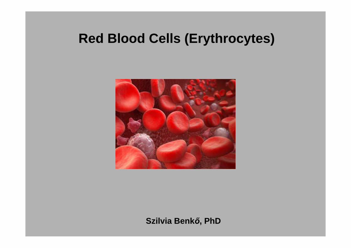
Red Blood Cells (Erythrocytes)
Szilvia Benkő, PhD

Red Blood Cells (Erythrocytes)
� Main characteristics of erythrocytes
� Differentiation and maturation of erythrocytes
� Regulation of erythrocyte development
� Clinical aspects
� Blood groups (ABO, RhD)
Properties of Red Blood Cells (Erythrocytes)
1. Concentration in blood is 4-6 million/cubic mm (4-6 T/l)1. Gender differences2. High individual variability
2. Morphoplogy: biconcave discs1. Large surface area2. Enables cells to bend in small capillaries
3. Main characteristics: reduced cell1. No nucleus → cannot reproduce (average life span: 120 days)2. No mitochondria → no metabolism3. No ribosomes → no protein synthesis
4. Function: transport hemoglobin (280 million hemoglobin molecules/cell)1. Contain high concentration of carbonic anhydrase2. Contain high concentration of HCO3
-/Cl- pump

No mitochondria GLUT1
Red blood cells
Glycolysis: in the cytosol
Tricarboxylic acid (TCA) cycle : in the mitochondri a
C6H12O6 + 6O2 + 32 ADP3- + 32 Pi2- 6CO2 + 6H2O + 32 ATP4- + 32 OH-
Blood sugar level!!

Properties of Hemoglobin
1. Stucture
1. Quaternary structure: (α2ß2) (Foetal: α2γ2)2. Each subunit is: 1 Heme + 1 globin3. Each heme contains 1 iron (2+ ↔ 3+)
2. Function1. Oxygen binding and transport2. CO2 binding and transport
3. Hemoglobin levels1. infants: 140-200 g/l2. Adult males: 140-180 g/l3. Adult females: 120-160 g/l
Heme: porfirine derivative, contains an iron (Fe2+ - ferro!)
1. Stucture1. Quaternary structure: (α2ß2)2. Each subunit is: 1 Heme + 1 globin3. Each heme contains 1 iron (2+ ↔ 3+)
2. Function1. Oxygen binding and transport2. CO2 binding and transport
3. Hemoglobin levels1. infants: 140-200 g/l2. Adult males: 140-180 g/l3. Adult females: 120-160 g/l
Heme: porfirine derivative, contains an iron (Fe2+ - ferro!)
Properties of Hemoglobin

Adults: mainly in the flat bones such as hip bone, breast bone, skull, ribs, vertebrae and shoulder blades, and in the "spongy” material at the proximal ends of the long bones femur and humerus.
Originatefrom red bone marrow
Emryo: yolk sac, liver

Development of Red Blood Cells (Erythrocytes)
(HSC)
(CMP) (CLP)
- proliferate- self-renewal

ErythropoesisFormation of erythrocytes
1. Location of erythropoiesis: Bone marrow 2. Takes 7 days3. Rate of Erythropoiesis: 2.5 million RBC/second
MultipotentStem cell
Phase 1: ribosome synthesis
Phase 2: hemoglobin accumulation
Phase 3: ejection of nucleus
Proerythroblast
Early erythroblast
Late erythroblast
Normoblast
Reticulocyte
Erythrocyte
� Adenylate cyclase activity drops
� Metabolism turns to anaerobic
� ATPase activity stops
� Passive transportinstead of active transport
� Decreased transferrin receptor expression
� No iron uptake
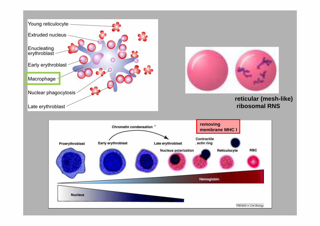
removingmembrane MHC I
reticular (mesh-like)ribosomal RNS
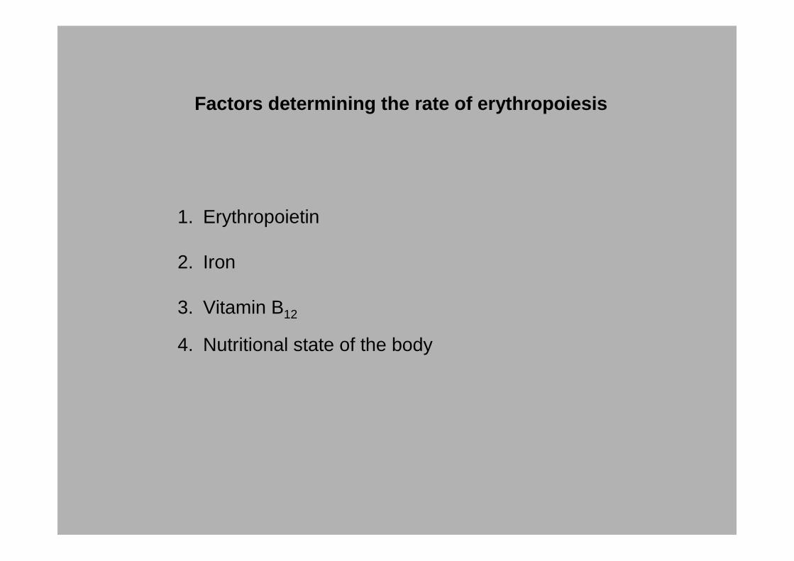
Factors determining the rate of erythropoiesis
1. Erythropoietin
2. Iron
3. Vitamin B12
4. Nutritional state of the body
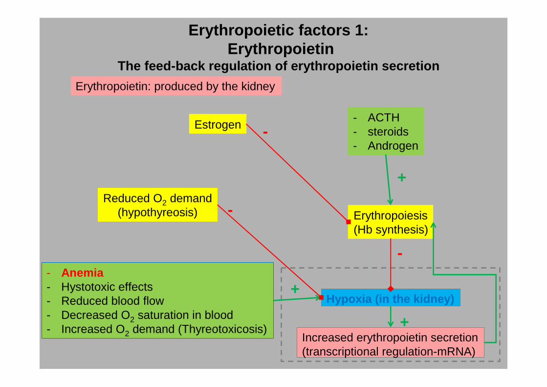
Erythropoietic factors 1:Erythropoietin
The feed-back regulation of erythropoietin secretion
Increased erythropoietin secretion(transcriptional regulation-mRNA)
Hypoxia (in the kidney)
Reduced O2 demand(hypothyreosis)
- Anemia- Hystotoxic effects- Reduced blood flow- Decreased O2 saturation in blood- Increased O2 demand (Thyreotoxicosis)
Erythropoiesis(Hb synthesis)
+
-
+
+
-
- ACTH- steroids- Androgen
Estrogen -
Erythropoietin: produced by the kidney
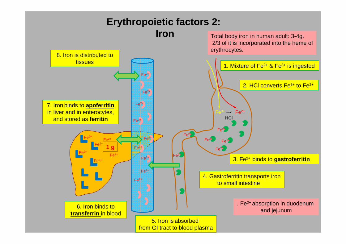
Erythropoietic factors 2:Iron
Fe3+→ Fe2+
Fe2+
Fe2+Fe2+
Fe2+
Fe2+
1. Mixture of Fe2+ & Fe3+ is ingested
2. HCl converts Fe3+ to Fe2+
3. Fe2+ binds to gastroferritinFe2+
4. Gastroferritin transports ironto small intestine
5. Iron is absorbed from GI tract to blood plasma
6. Iron binds totransferrin in blood
8. Iron is distributed to tissues
7. Iron binds to apoferritinin liver and in enterocytes,
and stored as ferritin
Fe2+
Fe2+
Fe2+
Fe2+
Fe2+
Fe2+
Fe2+
Fe2+
Fe2+
Fe2+
Fe2+
Fe2+
Fe2+
Fe2+
Fe2+
Total body iron in human adult: 3-4g.2/3 of it is incorporated into the heme of
erythrocytes.
1 g
. Fe2+ absorption in duodenum and jejunum
HCl

Erythropoietic factors 3:Vitamin B 12 (cobalamine)
• water soluble• corrinoid ring• similar to porfirin• only some bacteria and protozoa
are able to synthesize(importance of colon bacteria!)
- plants do not contain (VEGETARIANS!)- meat, LIVER, egg, milk- nicotine reduces the absorpion (smoking!)
- daily requirement:1-2ug- stored in liver (3-6 years)
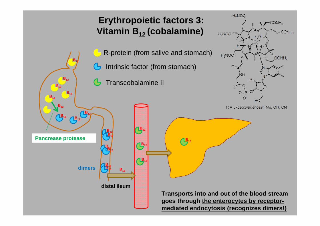
Erythropoietic factors 3:Vitamin B 12 (cobalamine)
B12
B12B12
B12
B12
B12
B12 B12
B12
B12
Pancrease proteaseB12
B12
B12
B12
B12
R-protein (from salive and stomach)
Intrinsic factor (from stomach)
Transcobalamine II
B12
B12
distal ileumTransports into and out of the blood stream goes through the enterocytes by receptor-mediated endocytosis (recognizes dimers!)
B12
B12
B12dimers
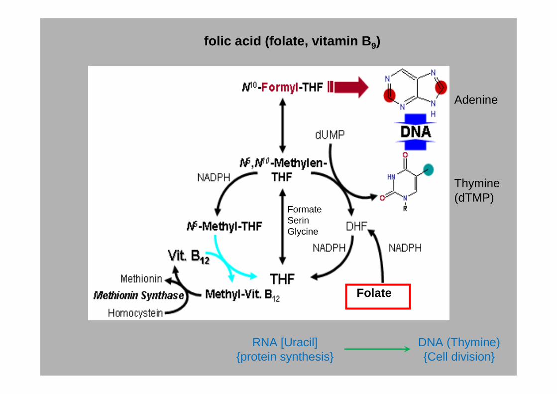
Folate
FormateSerinGlycine
Thymine(dTMP)
Adenine
folic acid (folate, vitamin B 9)
RNA [Uracil]{protein synthesis}
DNA (Thymine){Cell division}

Complete Blood Count (CBC)
- Determination of the number of red blood cell count
- One of the most routinely performed clinical tests. Different disorders can have dramatic effect on the total number or relative proportions of blood cells
- Can be determined only from blood samples taken from big veins
- Can be performed manually (hemocytometer) or with electric counter
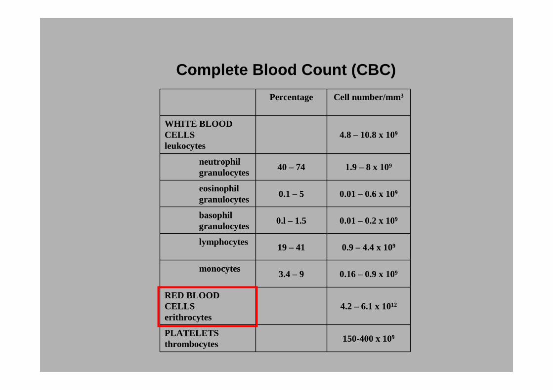
Complete Blood Count (CBC)
Percentage Cell number/mm3
WHITE BLOOD CELLSleukocytes
4.8 – 10.8 x 10⁹
neutrophilgranulocytes
40 – 74 1.9 – 8 x 10⁹
eosinophil granulocytes
0.1 – 5 0.01 – 0.6 x 10⁹
basophil granulocytes
0.l – 1.5 0.01 – 0.2 x 10⁹
lymphocytes19 – 41 0.9 – 4.4 x 10⁹
monocytes3.4 – 9 0.16 – 0.9 x 10⁹
RED BLOOD CELLSerithrocytes
4.2 – 6.1 x 10¹²
PLATELETSthrombocytes
150-400 x 109

Hematocrit
Definition:Percentage of whole blood occupied by packed red blood cells
Normal values:- Males: 46% (40-54)- Females: 42% (37-47)
Determination: centrifuging a blood sample, so that formed elements come out of suspension
Indicates:- Anemia (low Hct)- Polycythemia / dehydration (high Hct)
throbocytesthrobocytes80 mm
36 mm36 mm80 mm
x 100 = 45%

DEFINITIONS
Haematocrit: volume of cells / total volume ~0,37 - 0,52
MCV (mean corpuscular volume): haematokrit / RBC count 80 – 100 fl (10 -15)
MCH (mean corpuscular hemoglobine): Hb concentration / RBC count27 – 31pg /RBC
Stain index: Hb% / RBC%Hb%= Hb concentration actual / Hb concentration standard RBCt %= RBC count actual / RBC count standard
FI= 1 normochromFI<1 hypochrom (pl. iron deficiency anemia)FI>1 hyperchrom (pl. B12 vitamine deficiency anémia)
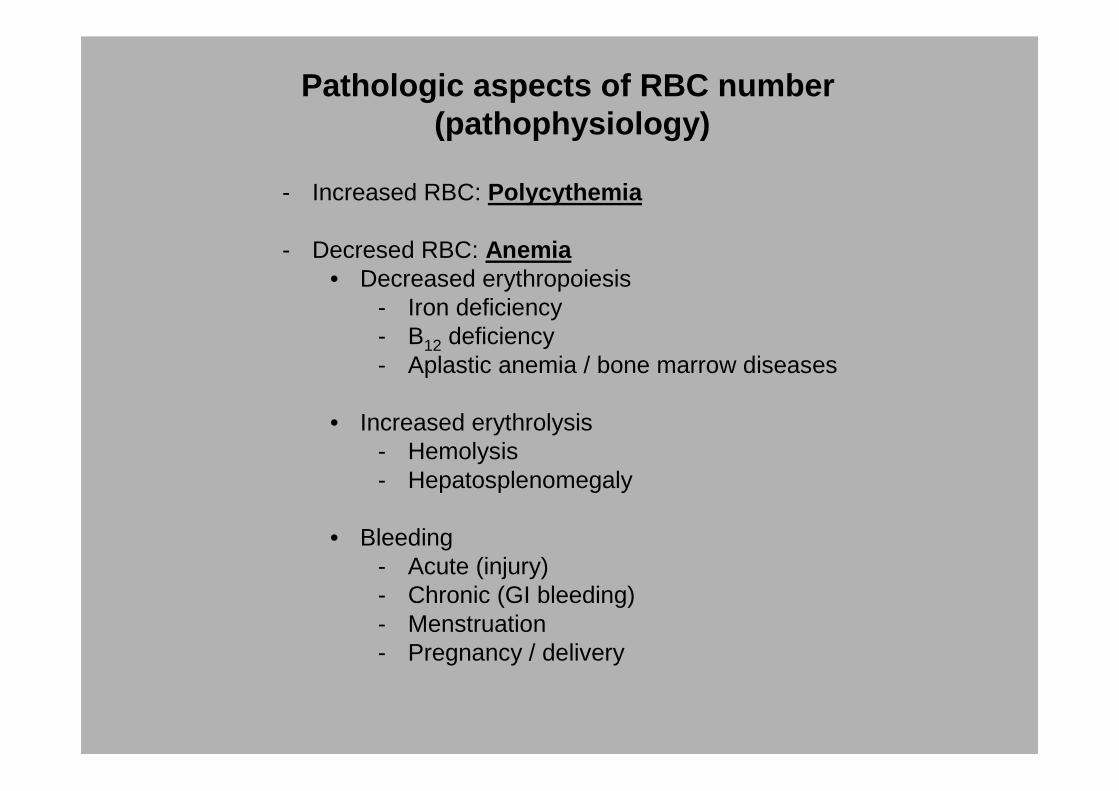
Pathologic aspects of RBC number(pathophysiology)
- Increased RBC: Polycythemia
- Decresed RBC: Anemia• Decreased erythropoiesis
- Iron deficiency- B12 deficiency- Aplastic anemia / bone marrow diseases
• Increased erythrolysis- Hemolysis- Hepatosplenomegaly
• Bleeding- Acute (injury)- Chronic (GI bleeding)- Menstruation- Pregnancy / delivery
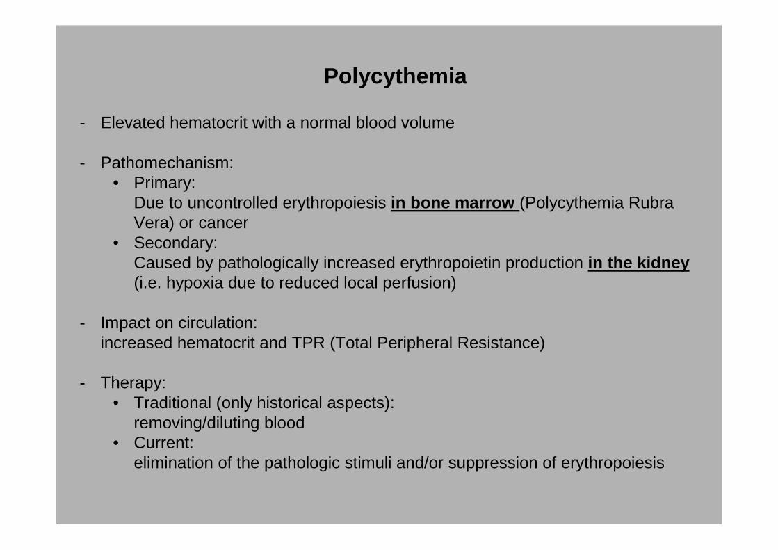
Polycythemia
- Elevated hematocrit with a normal blood volume
- Pathomechanism:• Primary:
Due to uncontrolled erythropoiesis in bone marrow (Polycythemia Rubra Vera) or cancer
• Secondary: Caused by pathologically increased erythropoietin production in the kidney (i.e. hypoxia due to reduced local perfusion)
- Impact on circulation: increased hematocrit and TPR (Total Peripheral Resistance)
- Therapy: • Traditional (only historical aspects):
removing/diluting blood• Current:
elimination of the pathologic stimuli and/or suppression of erythropoiesis

Clinical classification of anemias(based on chromic index)
1. Hypochromic (iron dependent or hypochromic microcyter anemia)
2. Hyperchromic (pernicious anemia)
3. Normochromic (aplastic anemia)
4. Other types

Anemia 1: Hypochromic microcyter anemia
- Pathomechanism: Iron deficiency • Iron deficit or malabsorbtion• Increased iron requirement (pregnancy, lactation, rapid growth phase in
children)
- Symptoms: unspecific
- Lab. Findings: • Decreased HCT, Hgb, MCV (mean corpuscular volume) – cell size,
MCH (mean corpuscular hemoglobin) – amount of Hgb,• Low ferritin and iron level in serum• Increased Total Iron Binding Capacity (TIBC) in the blood - transferrin
- Therapy: Oral iron supplement(food: Total body iron in human adult: 3-4g.2/3 of it is incorporated into the heme of erythrocytes.
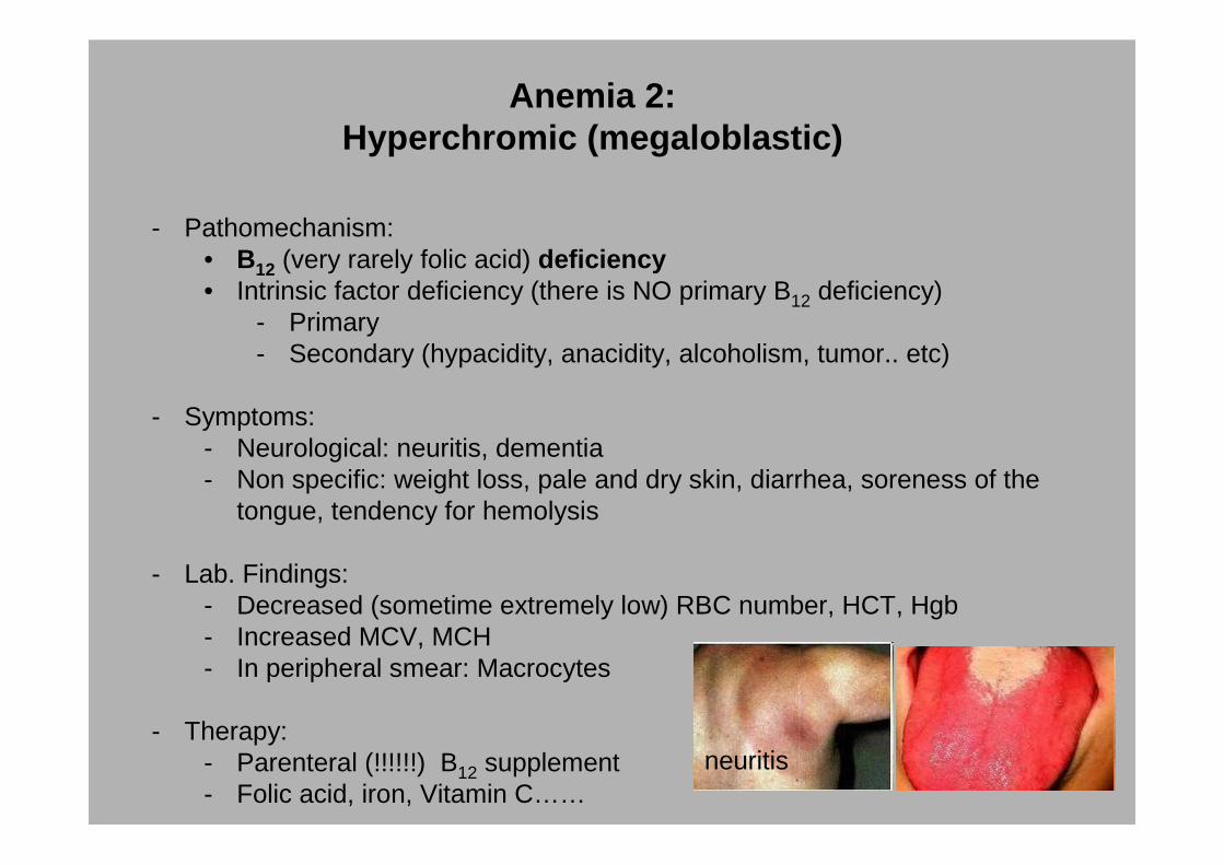
Anemia 2:Hyperchromic (megaloblastic)
- Pathomechanism: • B12 (very rarely folic acid) deficiency • Intrinsic factor deficiency (there is NO primary B12 deficiency)
- Primary- Secondary (hypacidity, anacidity, alcoholism, tumor.. etc)
- Symptoms:- Neurological: neuritis, dementia- Non specific: weight loss, pale and dry skin, diarrhea, soreness of the
tongue, tendency for hemolysis
- Lab. Findings: - Decreased (sometime extremely low) RBC number, HCT, Hgb- Increased MCV, MCH- In peripheral smear: Macrocytes
- Therapy: - Parenteral (!!!!!!) B12 supplement- Folic acid, iron, Vitamin C……
neuritis

Anemia 3-4: Normochromic and other types
- Hemorrhagic: Acute or chronic blood loss (GI/urinary tract ..etc)
- Hemolytic: Life span of RBCs is reduced- Hemoglobin abnormalities- Bacterial/viral infections, autoimmune disease- Toxins
- Aplastic: Reduced hemopoiesis due to bone marrow disease- Tumor/immune disease- Destruction of bone marrow by infection, radiation, drugs or toxins
- Sickle cell anemia: Mutation in the gene for the ß chain of hemoglobin- Due to the mutation Hgb link together forming stiff rods at low O2 of pH →
sickle cell formation- increased malaria resistance
- Thalassemia- reduced rate of synthesis or no synthesis of one of the globin chains due to
mutation (deletion) in regulatory region of Hgb genes - High incidence in the Mediterranean region- increased malaria resistance

Blood groups
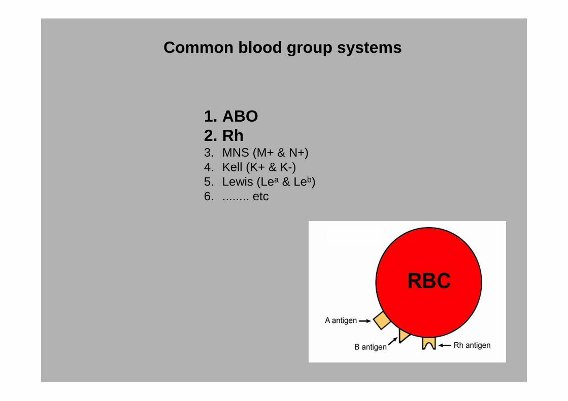
Common blood group systems
1. ABO 2. Rh3. MNS (M+ & N+)4. Kell (K+ & K-)5. Lewis (Lea & Leb)6. ........ etc

Terminology
- Blood type (blood group): A classification of blood based on the presence or absence of inherited antigenic substances (proteins, carbohydrates, glycoproteins, or glycolipids) on the surface of red blood cells.
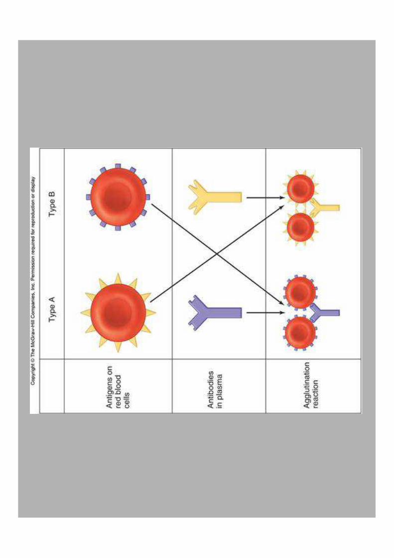
- Agglutination: clumping red blood cells as a result of mixing of samples from incompatible blood groups (precipitation, coagulation)
- Agglutinin: a substance that causes particles to coagulate to form a thickened mass (antibody )
- Agglutinogen: a substance that, acting as an antigen , stimulates the production of agglutinin
- Transfusion: It is the most frequent type of organ transplantation

AB0 system
- H-gene :Codes for H-transferase, puts fucose on galactoseit is common in each blood typefucose is a requirement
- A-gene:Codes for a specific transferase to put N-acetyl-galactosamine to galactose
- B-gene:Codes for a specific transferase to put galactose to galactose
- 0-geneCodes for an inactive „enzyme”, no additional carbohydrate on galactose
Phenotype (genotype)• A (AA or A0)• B (BB or B0)• AB (AB)• 0 (0)
Codominant inheritance

Structures of the ABO blood group antigens
Defined by specific enzymes inherited co-dominant genes (Mendelian rules)
Fuc - Fucose GalNAc - N acetyl-galactosamine Gal - Galactose
RBC
Glu - Glucose
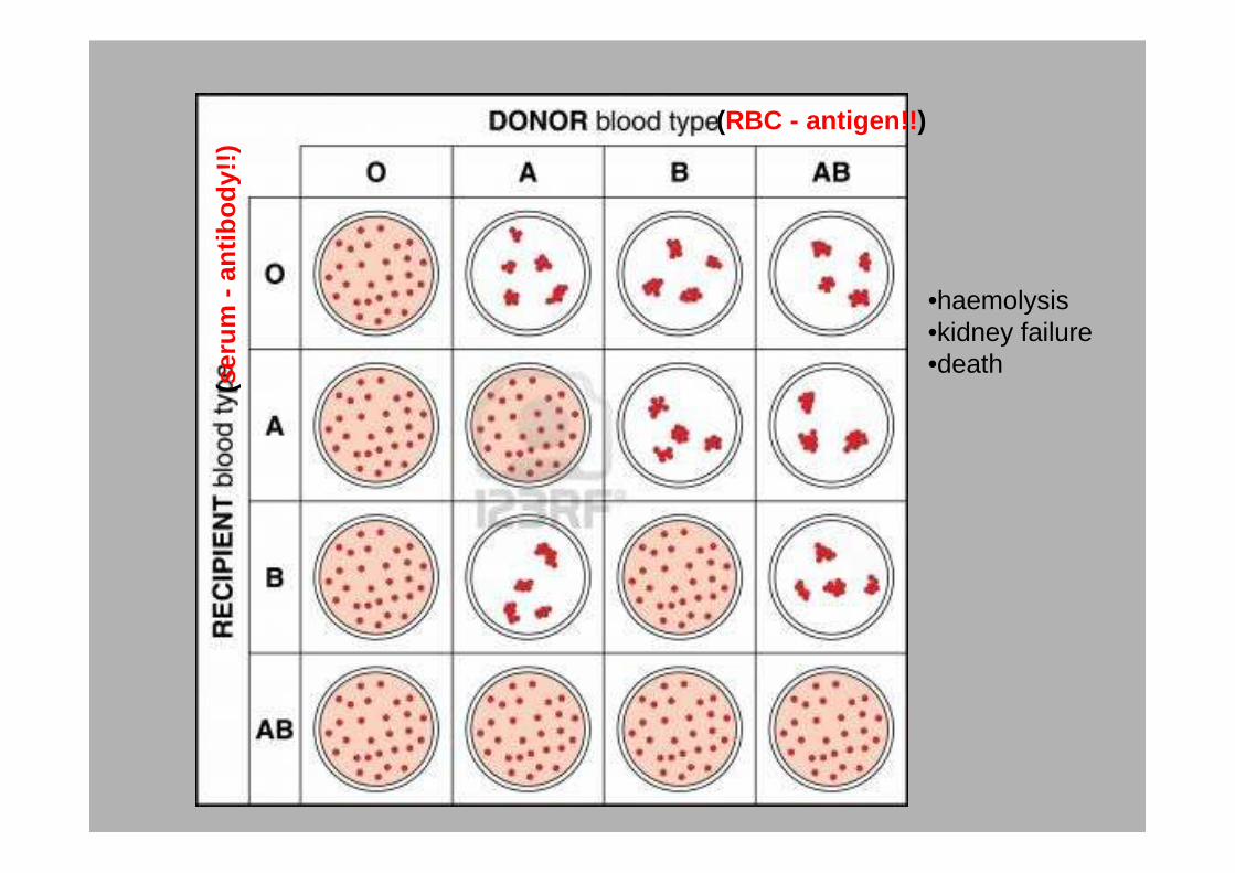
(RBC - antigen!! )
(ser
um -
antib
ody!
!)
•haemolysis•kidney failure•death

Rh system
Antibody production is induced only when an RhD-RBC enters the blood stream!!!(difference from AB0-system)
PROBLEM : - Rh- gets Rh+ blood- pregnancy
Two pair of genes with multiple allel combination

Rh phenotypes
1. Rh+: characterized serologically by their strong reactivity with monoclonal anti-D antibody.
2. Rh-: absence of reaction with anti-D antibody.- C or E antigens in Rh- negative person can still cause mild transfusion
reaction
3. Many transitional forms between Rh+ and Rh- were reported:- Weak D - Partial D (D mosaic)- D epitope expressed on RhCE- Etc.....
Agglutinins of the Rh system
1. No anti-D antibodies are found in the plasma of a Rh- person’s plasma prior to transfusion of Rh+ RBCs
2. Production of anti-D antibody is proportional with the number of transfusions with Rh+ blood/RBCs
3. Anti-D antobody (IgG) can diffuse trough placenta causing RBC agglutination/hemolysis (Erythroblastosis fetalis)
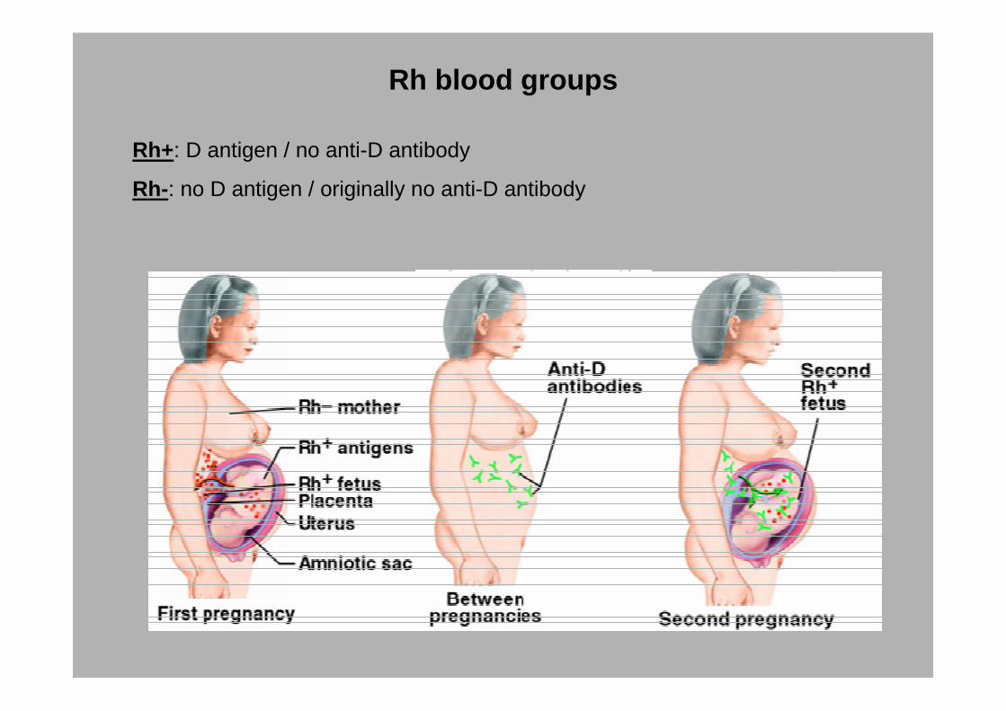
Rh blood groups
Rh+: D antigen / no anti-D antibody
Rh-: no D antigen / originally no anti-D antibody
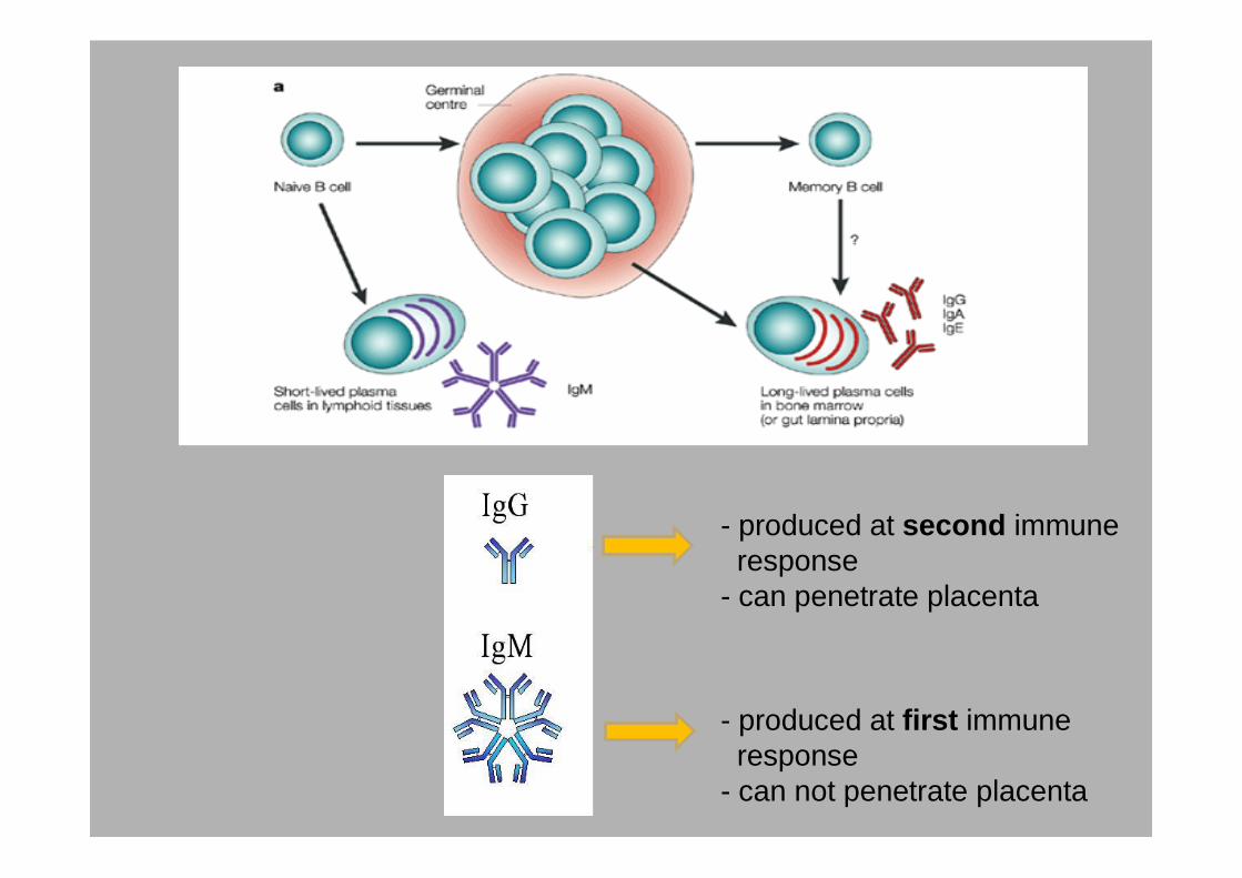
- produced at first immuneresponse
- can not penetrate placenta
- produced at second immune response
- can penetrate placenta
Transfusion rules
- Whole blood is very rarely used for transfusion
- Only the same AB0/Rh type blood can be used for transfusion
- No Universal donor or Universal acceptor