tctap c-088 repetitive subacute re-occlusion of rca cto lesion via retrograde pci
TRANSCRIPT
CASES
19th CardioVascular Summit: TCTAP 2014
Case Summary:In summary, we experienced the huge coronary artery hematoma by the rupturedballoon inflation in the subintimal space after the Reverse CART technique. Theimplanted EES was fractured because of the stent implantation into the huge coronaryhematoma.In conclusion, the coronary aneurysm formation after EES implantation is very rarecompared with Sirolimus-Eluting Stent, so the stent fracture with aneurysm maybeunlikely with EES, but the stent implantation even EES into the coronary aneurysm isone of the risk of stent fracture.
TCTAP C-087
Strategies of Limiting Contrast Use in Uremic Patient with Killip III MyocardialInfarction Receiving Ad-hoc Intervention to Chronic Total Occlusion
Shu-Kai Hsueh, Chiung-Jen WuKaohsiung Chang Gung Memorial Hospital, Taiwan
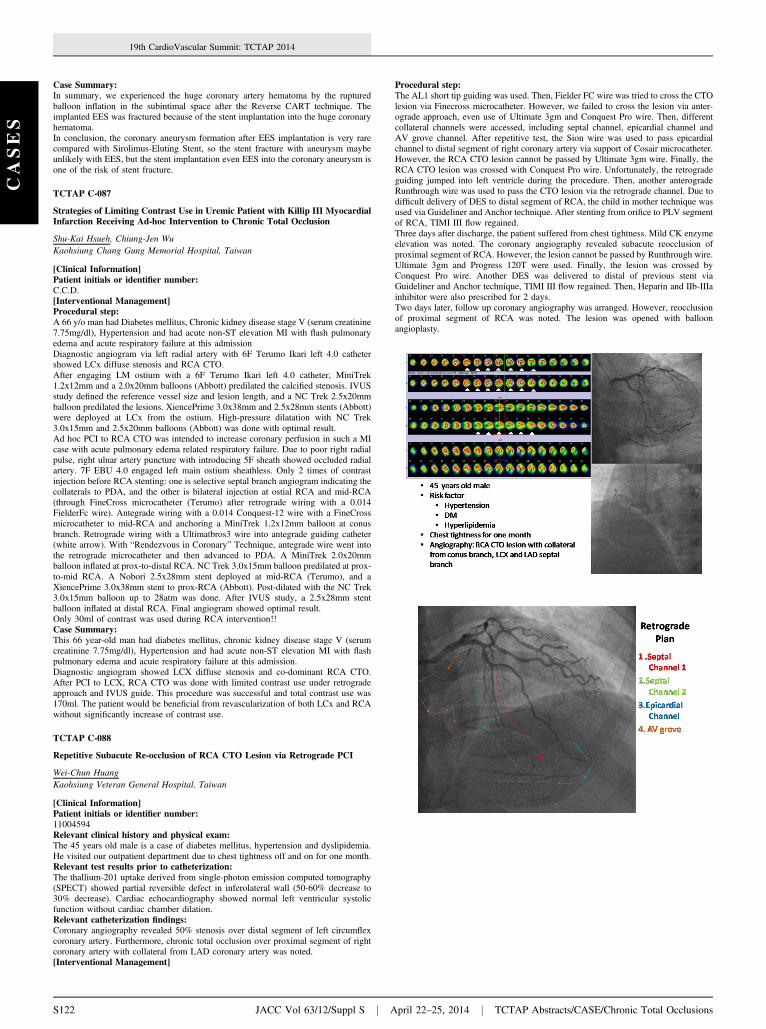
[Clinical Information]Patient initials or identifier number:C.C.D.[Interventional Management]Procedural step:A 66 y/o man had Diabetes mellitus, Chronic kidney disease stage V (serum creatinine7.75mg/dl), Hypertension and had acute non-ST elevation MI with flash pulmonaryedema and acute respiratory failure at this admissionDiagnostic angiogram via left radial artery with 6F Terumo Ikari left 4.0 cathetershowed LCx diffuse stenosis and RCA CTO.After engaging LM ostium with a 6F Terumo Ikari left 4.0 catheter, MiniTrek1.2x12mm and a 2.0x20mm balloons (Abbott) predilated the calcified stenosis. IVUSstudy defined the reference vessel size and lesion length, and a NC Trek 2.5x20mmballoon predilated the lesions. XiencePrime 3.0x38mm and 2.5x28mm stents (Abbott)were deployed at LCx from the ostium. High-pressure dilatation with NC Trek3.0x15mm and 2.5x20mm balloons (Abbott) was done with optimal result.Ad hoc PCI to RCA CTO was intended to increase coronary perfusion in such a MIcase with acute pulmonary edema related respiratory failure. Due to poor right radialpulse, right ulnar artery puncture with introducing 5F sheath showed occluded radialartery. 7F EBU 4.0 engaged left main ostium sheathless. Only 2 times of contrastinjection before RCA stenting: one is selective septal branch angiogram indicating thecollaterals to PDA, and the other is bilateral injection at ostial RCA and mid-RCA(through FineCross microcatheter (Terumo) after retrograde wiring with a 0.014FielderFc wire). Antegrade wiring with a 0.014 Conquest-12 wire with a FineCrossmicrocatheter to mid-RCA and anchoring a MiniTrek 1.2x12mm balloon at conusbranch. Retrograde wiring with a Ultimatbros3 wire into antegrade guiding catheter(white arrow). With “Rendezvous in Coronary” Technique, antegrade wire went intothe retrograde microcatheter and then advanced to PDA. A MiniTrek 2.0x20mmballoon inflated at prox-to-distal RCA. NC Trek 3.0x15mm balloon predilated at prox-to-mid RCA. A Nobori 2.5x28mm stent deployed at mid-RCA (Terumo), and aXiencePrime 3.0x38mm stent to prox-RCA (Abbott). Post-dilated with the NC Trek3.0x15mm balloon up to 28atm was done. After IVUS study, a 2.5x28mm stentballoon inflated at distal RCA. Final angiogram showed optimal result.Only 30ml of contrast was used during RCA intervention!!Case Summary:This 66 year-old man had diabetes mellitus, chronic kidney disease stage V (serumcreatinine 7.75mg/dl), Hypertension and had acute non-ST elevation MI with flashpulmonary edema and acute respiratory failure at this admission.Diagnostic angiogram showed LCX diffuse stenosis and co-dominant RCA CTO.After PCI to LCX, RCA CTO was done with limited contrast use under retrogradeapproach and IVUS guide. This procedure was successful and total contrast use was170ml. The patient would be beneficial from revascularization of both LCx and RCAwithout significantly increase of contrast use.
TCTAP C-088
Repetitive Subacute Re-occlusion of RCA CTO Lesion via Retrograde PCI
Wei-Chun HuangKaohsiung Veteran General Hospital, Taiwan
[Clinical Information]Patient initials or identifier number:11004594Relevant clinical history and physical exam:The 45 years old male is a case of diabetes mellitus, hypertension and dyslipidemia.He visited our outpatient department due to chest tightness off and on for one month.Relevant test results prior to catheterization:The thallium-201 uptake derived from single-photon emission computed tomography(SPECT) showed partial reversible defect in inferolateral wall (50-60% decrease to30% decrease). Cardiac echocardiography showed normal left ventricular systolicfunction without cardiac chamber dilation.Relevant catheterization findings:Coronary angiography revealed 50% stenosis over distal segment of left circumflexcoronary artery. Furthermore, chronic total occlusion over proximal segment of rightcoronary artery with collateral from LAD coronary artery was noted.[Interventional Management]
S122 JACC Vol 63/12/Suppl S j A
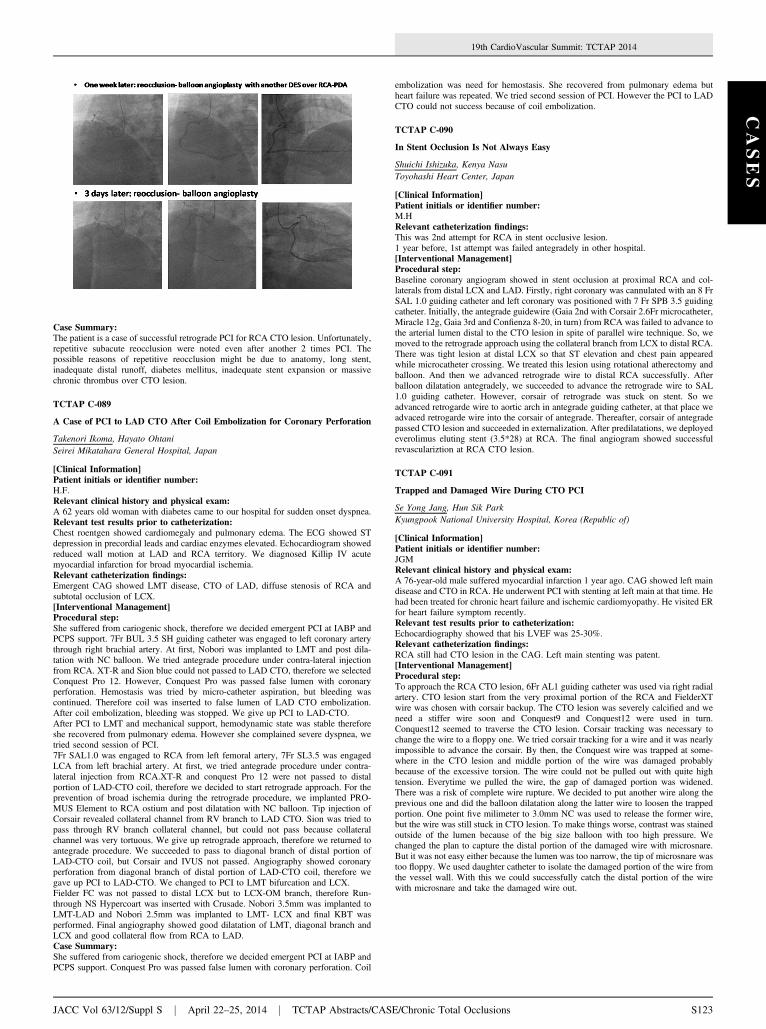
Procedural step:The AL1 short tip guiding was used. Then, Fielder FC wire was tried to cross the CTOlesion via Finecross microcatheter. However, we failed to cross the lesion via anter-ograde approach, even use of Ultimate 3gm and Conquest Pro wire. Then, differentcollateral channels were accessed, including septal channel, epicardial channel andAV grove channel. After repetitive test, the Sion wire was used to pass epicardialchannel to distal segment of right coronary artery via support of Cosair microcatheter.However, the RCA CTO lesion cannot be passed by Ultimate 3gm wire. Finally, theRCA CTO lesion was crossed with Conquest Pro wire. Unfortunately, the retrogradeguiding jumped into left ventricle during the procedure. Then, another anterogradeRunthrough wire was used to pass the CTO lesion via the retrograde channel. Due todifficult delivery of DES to distal segment of RCA, the child in mother technique wasused via Guideliner and Anchor technique. After stenting from orifice to PLV segmentof RCA, TIMI III flow regained.Three days after discharge, the patient suffered from chest tightness. Mild CK enzymeelevation was noted. The coronary angiography revealed subacute reocclusion ofproximal segment of RCA. However, the lesion cannot be passed by Runthrough wire.Ultimate 3gm and Progress 120T were used. Finally, the lesion was crossed byConquest Pro wire. Another DES was delivered to distal of previous stent viaGuideliner and Anchor technique, TIMI III flow regained. Then, Heparin and IIb-IIIainhibitor were also prescribed for 2 days.Two days later, follow up coronary angiography was arranged. However, reocclusionof proximal segment of RCA was noted. The lesion was opened with balloonangioplasty.
pril 22–25, 2014 j TCTAP Abstracts/CASE/Chronic Total Occlusions
CASES
19th CardioVascular Summit: TCTAP 2014
Case Summary:The patient is a case of successful retrograde PCI for RCA CTO lesion. Unfortunately,repetitive subacute reocclusion were noted even after another 2 times PCI. Thepossible reasons of repetitive reocclusion might be due to anatomy, long stent,inadequate distal runoff, diabetes mellitus, inadequate stent expansion or massivechronic thrombus over CTO lesion.
TCTAP C-089
A Case of PCI to LAD CTO After Coil Embolization for Coronary Perforation
Takenori Ikoma, Hayato OhtaniSeirei Mikatahara General Hospital, Japan
[Clinical Information]Patient initials or identifier number:H.F.Relevant clinical history and physical exam:A 62 years old woman with diabetes came to our hospital for sudden onset dyspnea.Relevant test results prior to catheterization:Chest roentgen showed cardiomegaly and pulmonary edema. The ECG showed STdepression in precordial leads and cardiac enzymes elevated. Echocardiogram showedreduced wall motion at LAD and RCA territory. We diagnosed Killip IV acutemyocardial infarction for broad myocardial ischemia.Relevant catheterization findings:Emergent CAG showed LMT disease, CTO of LAD, diffuse stenosis of RCA andsubtotal occlusion of LCX.[Interventional Management]Procedural step:She suffered from cariogenic shock, therefore we decided emergent PCI at IABP andPCPS support. 7Fr BUL 3.5 SH guiding catheter was engaged to left coronary arterythrough right brachial artery. At first, Nobori was implanted to LMT and post dila-tation with NC balloon. We tried antegrade procedure under contra-lateral injectionfrom RCA. XT-R and Sion blue could not passed to LAD CTO, therefore we selectedConquest Pro 12. However, Conquest Pro was passed false lumen with coronaryperforation. Hemostasis was tried by micro-catheter aspiration, but bleeding wascontinued. Therefore coil was inserted to false lumen of LAD CTO embolization.After coil embolization, bleeding was stopped. We give up PCI to LAD-CTO.After PCI to LMT and mechanical support, hemodynamic state was stable thereforeshe recovered from pulmonary edema. However she complained severe dyspnea, wetried second session of PCI.7Fr SAL1.0 was engaged to RCA from left femoral artery, 7Fr SL3.5 was engagedLCA from left brachial artery. At first, we tried antegrade procedure under contra-lateral injection from RCA.XT-R and conquest Pro 12 were not passed to distalportion of LAD-CTO coil, therefore we decided to start retrograde approach. For theprevention of broad ischemia during the retrograde procedure, we implanted PRO-MUS Element to RCA ostium and post dilatation with NC balloon. Tip injection ofCorsair revealed collateral channel from RV branch to LAD CTO. Sion was tried topass through RV branch collateral channel, but could not pass because collateralchannel was very tortuous. We give up retrograde approach, therefore we returned toantegrade procedure. We succeeded to pass to diagonal branch of distal portion ofLAD-CTO coil, but Corsair and IVUS not passed. Angiography showed coronaryperforation from diagonal branch of distal portion of LAD-CTO coil, therefore wegave up PCI to LAD-CTO. We changed to PCI to LMT bifurcation and LCX.Fielder FC was not passed to distal LCX but to LCX-OM branch, therefore Run-through NS Hypercoart was inserted with Crusade. Nobori 3.5mm was implanted toLMT-LAD and Nobori 2.5mm was implanted to LMT- LCX and final KBT wasperformed. Final angiography showed good dilatation of LMT, diagonal branch andLCX and good collateral flow from RCA to LAD.Case Summary:She suffered from cariogenic shock, therefore we decided emergent PCI at IABP andPCPS support. Conquest Pro was passed false lumen with coronary perforation. Coil
JACC Vol 63/12/Suppl S j April 22–25, 2014 j TCTAP Abstracts/CAS
embolization was need for hemostasis. She recovered from pulmonary edema butheart failure was repeated. We tried second session of PCI. However the PCI to LADCTO could not success because of coil embolization.
TCTAP C-090
In Stent Occlusion Is Not Always Easy
Shuichi Ishizuka, Kenya NasuToyohashi Heart Center, Japan
[Clinical Information]Patient initials or identifier number:M.HRelevant catheterization findings:This was 2nd attempt for RCA in stent occlusive lesion.1 year before, 1st attempt was failed antegradely in other hospital.[Interventional Management]Procedural step:Baseline coronary angiogram showed in stent occlusion at proximal RCA and col-laterals from distal LCX and LAD. Firstly, right coronary was cannulated with an 8 FrSAL 1.0 guiding catheter and left coronary was positioned with 7 Fr SPB 3.5 guidingcatheter. Initially, the antegrade guidewire (Gaia 2nd with Corsair 2.6Fr microcatheter,Miracle 12g, Gaia 3rd and Confienza 8-20, in turn) from RCA was failed to advance tothe arterial lumen distal to the CTO lesion in spite of parallel wire technique. So, wemoved to the retrograde approach using the collateral branch from LCX to distal RCA.There was tight lesion at distal LCX so that ST elevation and chest pain appearedwhile microcatheter crossing. We treated this lesion using rotational atherectomy andballoon. And then we advanced retrograde wire to distal RCA successfully. Afterballoon dilatation antegradely, we succeeded to advance the retrograde wire to SAL1.0 guiding catheter. However, corsair of retrograde was stuck on stent. So weadvanced retrogarde wire to aortic arch in antegrade guiding catheter, at that place weadvaced retrogarde wire into the corsair of antegrade. Thereafter, corsair of antegradepassed CTO lesion and succeeded in externalization. After predilatations, we deployedeverolimus eluting stent (3.5*28) at RCA. The final angiogram showed successfulrevasculariztion at RCA CTO lesion.
TCTAP C-091
Trapped and Damaged Wire During CTO PCI
Se Yong Jang, Hun Sik ParkKyungpook National University Hospital, Korea (Republic of)
[Clinical Information]Patient initials or identifier number:JGMRelevant clinical history and physical exam:A 76-year-old male suffered myocardial infarction 1 year ago. CAG showed left maindisease and CTO in RCA. He underwent PCI with stenting at left main at that time. Hehad been treated for chronic heart failure and ischemic cardiomyopathy. He visited ERfor heart failure symptom recently.Relevant test results prior to catheterization:Echocardiography showed that his LVEF was 25-30%.Relevant catheterization findings:RCA still had CTO lesion in the CAG. Left main stenting was patent.[Interventional Management]Procedural step:To approach the RCA CTO lesion, 6Fr AL1 guiding catheter was used via right radialartery. CTO lesion start from the very proximal portion of the RCA and FielderXTwire was chosen with corsair backup. The CTO lesion was severely calcified and weneed a stiffer wire soon and Conquest9 and Conquest12 were used in turn.Conquest12 seemed to traverse the CTO lesion. Corsair tracking was necessary tochange the wire to a floppy one. We tried corsair tracking for a wire and it was nearlyimpossible to advance the corsair. By then, the Conquest wire was trapped at some-where in the CTO lesion and middle portion of the wire was damaged probablybecause of the excessive torsion. The wire could not be pulled out with quite hightension. Everytime we pulled the wire, the gap of damaged portion was widened.There was a risk of complete wire rupture. We decided to put another wire along theprevious one and did the balloon dilatation along the latter wire to loosen the trappedportion. One point five milimeter to 3.0mm NC was used to release the former wire,but the wire was still stuck in CTO lesion. To make things worse, contrast was stainedoutside of the lumen because of the big size balloon with too high pressure. Wechanged the plan to capture the distal portion of the damaged wire with microsnare.But it was not easy either because the lumen was too narrow, the tip of microsnare wastoo floppy. We used daughter catheter to isolate the damaged portion of the wire fromthe vessel wall. With this we could successfully catch the distal portion of the wirewith microsnare and take the damaged wire out.
E/Chronic Total Occlusions S123