tdc 5th sem major
TRANSCRIPT

Classification and Biological
significance of Lipids
TDC 5TH SEM MAJOR
PAPER 5.2
By-Dr. Luna Phukan

Definition
A lipid is a macrobiomolecule that is soluble in nonpolar solvents. Non-polarsolvents are typically hydrocarbons used to dissolve other naturally occurringhydrocarbon lipid molecules that do not (or do not easily) dissolve in water,including fatty acids, waxes, sterols, fat-soluble vitamins (such as vitamins A, D,E, and K), monoglycerides, diglycerides, triglycerides, and phospholipids.
Although the term "lipid" is sometimes used as a synonym for fats, fats are asubgroup of lipids called triglycerides. Lipids also encompass molecules suchas fatty acids and their derivatives (including tri-, di-, monoglycerides, andphospholipids), as well as other sterol-containing metabolites such ascholesterol. Although humans and other mammals use various biosyntheticpathways both to break down and to synthesize lipids, some essential lipidscan't be made this way and must be obtained from the diet.

Scientists sometimes define lipids as hydrophobic or amphiphilic small molecules; the amphiphilic nature of some lipids allows them to form structures such as vesicles, multilamellar/unilamellar liposomes, or membranes in an aqueous environment.
Biological lipids originate entirely or in part from two distinct types of biochemical subunits or "building-blocks": ketoacyl and isoprene groups.[4] Using this approach, lipids may be divided into eight categories: fatty acids, glycerolipids, glycerophospholipids, sphingolipids, saccharolipids, and polyketides (derived from condensation of ketoacyl subunits); and sterol lipids and prenol lipids (derived from condensation of isoprene subunits).

Classification & Structure of Lipids
Fats are classified into 4 categories as follows:
1. On the basis of chemical composition2. On the basis of fatty acids3. On the basis of requirement4. On the basis of sources

Classification of Lipids
I. On the basis of chemical composition
Fats can be classified into 3 main groups as follows:

1. Simple lipids:
These are esters of fatty acids and glycerol. They are also called as neutral fats or triglycerides. These neutral fats make up 98 -99% of food and body fats.(e.g) fats and oils.
Waxes: A wax is a simple lipid which is an ester of fatty acids and long chain aliphatic alcohols. The alcohol may contain 12-32 carbon atoms. Waxes are found in nature as coatings on leaves and stems. The wax prevents the plant from losing excessive amounts of water.
2. Compound lipids:
The compound lipids contain, in addition to fatty acids and glycerol,some other organic compounds.
1 Phospholipids: These contain phosphoric acid and a nitrogenous base in addition to fatty acids and glycerol(e.g.)Lecithin and cephalin.
2. Glycolipids: Complex lipids containing carbohydrates in combination with fatty acids and glycerol(e.g) Cerebrosides
3. Lipoproteins: Lipoproteins are the most important as they are the carriers of lipids in the blood and form cell membranes.

3. Derived lipids:
These are substances liberated during hydrolysis of simple and compound lipids which still retain the properties of lipids. The important members of this group are sterols, fatty acids and alcohol.
i. Sterols : Sterols are solid alcohols and form esters with fatty acids. In nature they occur in the free state in the form of esters. Based on their origin sterols are classified as cholesterol (animal origin) and phytosterol(in plants).
Cholesterol is a waxy, fat-like substance found in all cells of the body and has several important functions in the body. It is synthesized in the body by the liver independent of the dietary intake. The body normally synthesizes about 2 grams of cholesterol. The dietary sources of cholesterol includes animal foods. It is used in the body for synthesizing hormones, Vitamine D and substances which help digest foods. High blood cholesterol is a risk factor for heart disease. Rich sources of dietary cholesterol include meat, poultry(with skin), organ meats like brain, kidney, liver and full fat dairy products.

ii. Fatty acids: They are the key, refined fuel form of fat that the cell burns for energy.They are the basic structural unit of fats and they may be saturated or unsaturated. (e.g)Oleic acid, linoleic acid, linolenic acid, palmitic acid and myristic acid.
II. On the basis of fatty acids
Fats can be classified based on the fatty acids present in them as follows:

1. Saturated fatty acids:
A saturated fat is a type of fat in which the fatty acid chains have all or predominantly single bonds. Various fats contain different proportions of saturated fat. aturated fatty acids, especially palmitic and stearic acids are found in animal products such as cream, cheese, butter, other whole milk dairy products and fatty meats which also contain dietary cholesterol. Certain vegetable products have high saturated fat content, such as coconut oil and palm kernel oil. Many prepared foods are high in saturated fat content, such as pizza, dairy desserts and sausage.

2. Unsaturated fatty acids
An unsaturated fat is a fat or fatty acid in which there is at least one doublebond within the fatty acid chain.i. Monounsaturated fatty acid (MUFA): A fatty acid chain ismonounsaturated if it contains one double bond. Monounsaturated fats aregood fats. A diet high in MUFA can reduce blood cholesterol levels, lowers riskof heart disease, stroke and breast cancer, reduces pain in rheumatoidarthritis and helps in weight loss. Foods which contain MUFA (Oleic acid) areavocados, olives, olive oil, peanut butter and peanut oil. It is also known asomega-9 fatty acid.(ii) Polyunsaturated fatty acid (PUFA): A fatty acid is polyunsaturated if itcontains more than one double bond. They are of 2 types, namely Omega-3and omega-6 fatty acids.

a. Omega-3: It is also called ω−3 fatty acids or n−3 fatty acids with a double bond
(C=C) at the third carbon atom from the end of the carbon chain. The three types of
omega−3 fatty acids involved in human physiology are α-linolenic acid (ALA) [found in
plant oils], eicosapentaenoic acid (EPA), and docosahexaenoic acid (DHA) [both
commonly found in marine oils]. Common sources of plant oils containing the omega−3
ALA fatty acid include walnut, flaxseed, flaxseed oil, soybeans and chia seeds. The
sources of animal omega−3 EPA and DHA fatty acids include fish and fish oils.
b. Omega-6: Omega-6 fatty acids (also referred to as ω-6 fatty acids or n-6
fatty acids) are a family of pro- inflammatory and anti-inflammatory
polyunsaturated fatty acids that have in common a final carbon-carbon
double bond in the n-6 position, that is the sixth bond, counting from the
methyl end.

III. On the basis of requirement
Fatty acids are of 2 types:
1. Essential fatty acids
Fatty acids which are essential to be taken in our diet because they cannot be synthesized in our body are known as essential fatty acids.(eg.) Linoleic, linolenic and arachidonic acids.
2. Non-essential fatty acids
Non-essential fatty acids are those which can be synthesized by the body and which need not be supplied through the diet. Palmitic acid, oleic acid and butyric acid are examples of non– essential fatty acids.

IV. On the Basis of Sources
Fats are divided into 2 types based on their source, namely: visible and invisible fats. Some fats and oils added to food or used for frying are visible fats.

Structure of Lipids

Mechanisms of lipid
biosynthesis. Biosynthesis of
ketoacyl- and isoprene-
containing lipids proceeds
by carbanion and
carbocation-mediated chain
extension, respectively.
Lipids display remarkable structural diversity, driven by factors such as variable
chain length, a multitude of oxidative, reductive, substitutional and ring-forming
biochemical transformations as well as modification with sugar residues and
other functional groups of different biosynthetic origin. There are no reliable
estimates of the number of discrete lipid structures in nature, due to the
technical challenges of elucidating chemical structures. Estimates in the order
of 200,000, based on acyl/alkyl chain and glycan permutations for glycerolipids,
glycerophospholipids and sphingolipids are almost certainly on the
conservative end . This number is actually exceeded by the list of known natural
products, most of which are of either prenol or polyketide origin . Given the
importance of these molecules in cellular function and pathology, it is essential
to have well-organized databases of lipids with relevant structural information
and related features.

In general, the acid/acyl group or its equivalent is drawn on the right side and hydrophobic chain is on the left.
Thus for glycerolipids and glycerophospholipids, the radylhydrocarbon chains are drawn to the left; the glycerol group is drawn horizontally with stereochemistry defined at sn carbons and the headgroup for glycerophospholipids is depicted on the right. For sphingolipids, the C1 hydroxyl group of the long-chain base is placed on the right and the alkyl portion on the left; the headgroup of sphingolipids ends up on the right.
The linear prenols and isoprenoids are drawn like fatty acids with the terminal functional groups on the right.

A number of structurally complex lipids – polycyclic isoprenoids, and polyketides – cannot be drawn using these simple rules; these structures are drawn using commonly accepted representations. Structures of all lipids in LMSD adhere to the structure drawing rules proposed by the LIPID MAPS consortium.
Fig. 4 shows representative structures for several lipid categories. This approach has the advantage of making it easier to recognize lipids within a category or class and also to highlight structural similarities between classes. For example the glycerophosphocholines (PC) and sphingomyelins (SM) have distinctly different biosynthetic origins but their structural similarities as amphipathic membrane lipids are evident when displayed as shown in Fig 5.

Fig. 4
Lipid structure representation.
Examples of structures from a
number lipid categories in
which acyl or prenyl chains
are oriented horizontally with
the terminal functional group
(in green) on the right and the
unsubstituted “tail” on the left.

Fig. 5
Structural similarity of PC and SM. Orientation of examples of phosphatidylcholine (PC) and
sphingomeyelin (SM) structures highlights their similarity. The sphingosine “backbone” of
SM is biosynthetically derived from palmitoyl-CoA (green) and serine (red) whereas PC
contains a glycerol core (red) with an additional acyl chain (green). Both molecules contain a
phosphocholine (pink) headgroup.

Significance of Lipids

I. Biological Importance of Lipids
The fat serves as efficient sources of energy when stored in an adipose tissue. It serves as an insulating material in the subcutaneous tissues and around certain organs. It provided building blocks for different high molecular weight substance, e.g., acetic acid and can be used for the synthesis of cholesterol and certain hormones. They produce metabolites through oxidation in the tissues which are used in the introversion of substances.

Chemical Properties of Lipids
Saponification
Saponification is a process that involves conversion of fat, oil or lipid into soap and alcohol by the action of heat in the presence of aqueous alkali (e.g. NaOH). Soaps are salts of fatty acids and fatty acids are mono that have long carbon chains (at least 10) e.g. sodium palmitate.
The number of milligrams of KOH required to saponify 1 gm. of fat or oil.
Rancidity:
Nearly all natural fats are oxidized when exposed to air, light, moisture, particularly, if warm. It develops an unpleasant odor and taste.
This happens so due to the formation of peroxides at the double bonds of unsaturated fatty acids.
It is widely recognized that malnutrition is the commonest cause of immunodeficiency. In recent years, a large number of studies have been conducted to investigate the relevance of certain fatty acids in the alteration of immune system functions in both humans and animals. At the beginning, the epidemiological findings contributed to demonstrating that certain fatty acids supplied in their diets (particularly long-chain n-3 polyunsaturated fatty acids contained in fish oil) affect the immune responses of Greenland Eskimos, as indicated by the low prevalence of inflammatory disorders in this population
Subsequently, on the basis of these epidemiological studies, experimental investigations determined the main processes by which several polyunsaturated or monounsaturated fatty acids are capable of altering the immune system, as well as the mechanisms of action involved in the regulation of immune functions.

At least four modes of action have been proposed to explain thepotential action of fatty acids on the modulation of immune system inboth animals and humans.
Accordingly, immune system modulation by dietary lipids may beattributed to changes in the composition of membrane phospholipids,lipid peroxidation, alteration of gene expression, or eicosanoidproduction. In addition, recent in vitro and ex vivo studies havedemonstrated the involvement of several fatty acids (such aseicosapentaenoic acid [EPA], docosahexaenoic acid [DHA], arachidonicacid [AA], or palmitic acid [PA]) in the induction or inhibition ofprogrammed cell death or apoptosis
. The participation of different fatty acids in apoptosis opens newapproaches for the study of the properties that different fatty acidsexert as modulators of immune system functions.

Schematic representation of
potential role of dietary lipids
and biological and clinical
consequences of the
administration of several dietary
lipids. PUFAs, polyunsaturated
fatty acids; MUFAs,
monounsaturated fatty acids;
SFAs, saturated fatty acids.
The adverse effects that several dietary lipids may exhibit as a direct consequence of an immunosuppressive process (Fig. 1).

BIOLOGICAL CONSEQUENCES ON IMMUNE FUNCTIONS
OR PHYSIOLOGICAL IMPORTANCE ( ASSIGNED TO FATTY
ACID ADMINISTRATION)
Differential properties of fatty acids and their role in themodulation of immune system functions.
The changes attributed to fatty acid administration include thereduction of lymphocyte proliferation, which is modified bypolyunsaturated fatty acids (n-3 or n-6) or monounsaturated fattyacids (n-9).
Studies carried out in both humans and animals have revealedthat the administration of high levels of dietary n-3polyunsaturated fatty acids or the inclusion in parenteralregimens of lipid emulsions rich in n-3 or n-9 fatty acids arerelated to the reduction of lymphocyte proliferation during thesupplementation

Thus, concanavalin-A or lipopolysaccharide-stimulatedlymphocytes have reduced the cellular proliferation in assayscarried out in vitro or ex vivo in the presence of free fattyacids or in cell cultures from both animals and humans feddietary lipids, respectively
Cytokine production is reduced by the action of n-3polyunsaturated fatty acids or n-9 monounsaturated fattyacids , cytokine receptor expression is also affected , naturalkiller (NK) activity is significantly suppressed , phagocyticactivity of macrophages is modified , and the antigen-presenting function of human monocytes is inhibited .

Mechanisim of action of lipid
Mechanisms of action . As mentioned previously, for several years numerous studies have attempted to elucidate the mechanisms by which some dietary lipids produce a potential effect on immune system functions .
Several hypotheses have been suggested as possible mechanisms. Experimental investigations have confirmed that several fatty acids exert changes in the phospholipids of plasma membrane which affect the membrane fluidity ; they also alter eicosanoid production , produce lipid peroxides , or regulate the transcription factors .
Overall, long-chain n-3 polyunsaturated fatty acids, such as EPA, are incorporated into cell membranes, replacing AA (the most important of the eicosanoid precursors). This process reduces the production of a biological mediator as prostaglandin E2 responsible for inhibiting IL-1 and tumor necrosis factor (TNF) production

A crucial role of lymphocyte subsets has been suggested to explain the effectsof different dietary polyunsaturated fatty acids on the immune system,because they alter the number of lymphocyte subsets as well as theproliferation of these cells .
Thus, antigen presentation and the proportion of T-cell subsets are modifiedafter dietary lipid manipulation. In fact, recent studies have determined thatpolyunsaturated fatty acids inhibit the surface expression of majorhistocompatibility complex (MHC) class II molecules, as well as someadhesion molecules on human monocytes .
Similarly, consumption of a monounsaturated fatty acid-rich diet by humansis also involved in the reduction of adhesion molecules from peripheral bloodmononuclear cells .
Overall, these actions may explain the different expression of CD4 or CD8 onthe T-cell surfaces of peripheral blood mononuclear cells of mice that werefed diets supplemented with DHA .

Crucial role of fatty acids in the incidence of cancer. Many lines of evidence have indicated the essential role of fish oil in the incidence of cancer.
Numerous reports have suggested that high intakes of long-chain n-3 fatty acids or n-9 fatty acids (the most important fatty acid contained in olive oil) may reduce the risk of breast cancer in both humans and animals.
Thus, n-3 polyunsaturated fatty acids may inhibit colon cancer in rats as well as reduce the risk of colorectal cancer development .
In contrast, n-6 fatty acids or saturated fatty acids may be involved in the increase of mammary or colon tumorigenesis by altering membrane phospholipid turnover. Accordingly, AA is released from plasma membrane that produces an alteration in the synthesis of prostaglandins via cyclooxygenase enzyme .

CONCLUDING REMARKS:
Nutritional status is generally recognized as an essential factor involved in the modulation of immune response, which may be determinant in the development of the clinical effects derived from a malnutrition process .
Thus, in recent years the effects that different dietary lipids exert upon immune functions have received considerable attention, because different functions of immune system are altered after dietary lipid administration.
Hence, lymphocyte proliferation, cytokine production, phagocytic activity, adhesion molecule expression, and NK cell activity are susceptible to modification by the action of certain lipids in both animals and humans. Different mechanisms of action have been proposed to be involved in these processes, such as lipid peroxidation, changes in the plasma membrane, eicosanoid production, or alteration of gene expression. Both epidemiological and experimental studies have applied the beneficial properties associated to a modulation of the immune functions.

Some important Terms & Concepts

TRIACYLGLYCEROLS
A triglyceride (TG, triacylglycerol, TAG, or triacylglyceride) is an ester derived from glycerol and three fatty acids (from tri- and glyceride). Triglycerides are the main constituents of body fat in humans and other vertebrates, as well as vegetable fat. They are also present in the blood to enable the bidirectional transference of adipose fat and blood glucose from the liver, and are a major component of human skin oils.
Many types of triglycerides exist. One classification focuses on saturated and unsaturated types. Saturated fats lack C=C groups. Unsaturated fats feature one or more C=C groups. Unsaturated fats tend to have a lower melting point than saturated analogues. Unsaturated fats often are liquid at room temperature.

Triglycerides
𝐻𝑂𝐶𝐻2𝐶𝐻(𝑂𝐻)𝐶𝐻2𝑂𝐻 + 𝑅𝐶𝑂2𝐻 + 𝑅′𝐶𝑂2𝐻 + 𝑅″𝐶𝑂2𝐻 → 𝑅𝐶𝑂2𝐶𝐻2𝐶𝐻(𝑂2𝐶𝑅′)𝐶𝐻2𝐶𝑂2𝑅″ + 3𝐻2𝑂
Triglycerides are tri-esters consisting of a glycerol bound to three fatty acid molecules. Alcohols have a hydroxyl (HO–) group. Organic acids have a carboxyl (–COOH) group. Alcohols and organic acids join to form esters. The glycerol molecule has three hydroxyl (HO–) groups and each fatty acid has a carboxyl group (–COOH). In triglycerides, the hydroxyl groups of the glycerol join the carboxyl groups of the fatty acid to form ester bonds:
The three fatty acids (RCO2H, R′CO2H, R″CO2H in the above equation) are usually different, as many kinds of triglycerides are known.
The chain lengths of the fatty acids in naturally occurring triglycerides vary, but most contain 16, 18, or 20 carbon atoms. Natural fatty acids found in plants and animals are typically composed of only even numbers of carbon atoms, reflecting the pathway for their biosynthesis from the two-carbon building-block acetyl CoA. Bacteria, however, possess the ability to synthesise odd- and branched-chain fatty acids. As a result, ruminant animal fat contains odd-numbered fatty acids, such as 15, due to the action of bacteria in the rumen. Many fatty acids are unsaturated; some are polyunsaturated (e.g., those derived from linoleic acid).
Most natural fats contain a complex mixture of individual triglycerides. Because of this, they melt over a broad range of temperatures. Cocoa butter is unusual in that it is composed of only a few triglycerides, derived from palmitic, oleic, and stearic acids in the 1-, 2-, and 3-positions of glycerol, respectively.

Homotriglycerides:
The simplest triglycerides are those where the three fatty acids are identical. Their names indicate the fatty acid: stearin derived from stearic acid, palmitin derived from palmitic acid, etc. These compounds can be obtained in three crystalline forms (polymorphs): α, β, and β′, the three forms differing in their melting points.[4][5]
Metabolism Fatty acid metabolism:
The pancreatic lipase acts at the ester bond, hydrolyzing the bond and "releasing" the fatty acid. In triglyceride form, lipids cannot be absorbed by the duodenum. Fatty acids, monoglycerides (one glycerol, one fatty acid), and some diglycerides are absorbed by the duodenum, once the triglycerides have been broken down.
In the intestine, following the secretion of lipases and bile, triglycerides are split into monoacylglycerol and free fatty acids in a process called lipolysis. They are subsequently moved to absorptive enterocyte cells lining the intestines. The triglycerides are rebuilt in the enterocytes from their fragments and packaged together with cholesterol and proteins to form chylomicrons. These are excreted from the cells and collected by the lymph system and transported to the large vessels near the heart before being mixed into the blood.

Various tissues can capture the chylomicrons, releasing the triglycerides to be used as a source of energy. Liver cells can synthesize and store triglycerides. When the body requires fatty acids as an energy source, the hormone glucagon signals the breakdown of the triglycerides by hormone-sensitive lipase to release free fatty acids. As the brain cannot utilize fatty acids as an energy source (unless converted to a ketone), the glycerol component of triglycerides can be converted into glucose, via gluconeogenesis by conversion into dihydroxyacetone phosphate and then into glyceraldehyde 3-phosphate, for brain fuel when it is broken down. Fat cells may also be broken down for that reason if the brain's needs ever outweigh the body's.

Triglycerides cannot pass through cell membranes freely. Special enzymes on the walls of blood vessels called lipoprotein lipases must break down triglycerides into free fatty acids and glycerol. Fatty acids can then be taken up by cells via the fatty acid transporter (FAT).
Triglycerides, as major components of very-low-density lipoprotein (VLDL) and chylomicrons, play an important role in metabolism as energy sources and transporters of dietary fat. They contain more than twice as much energy (approximately 9 kcal/g or 38 kJ/g) as carbohydrates (approximately 4 kcal/g or 17 kJ/g).

Role in disease Hypertriglyceridemia
In the human body, high levels of triglycerides in thebloodstream have been linked to atherosclerosis, heartdisease and stroke.
However, the relative negative impact of raised levels oftriglycerides compared to that of LDL:HDL ratios is as yetunknown. The risk can be partly accounted for by a stronginverse relationship between triglyceride level and HDL-cholesterol level. But the risk is also due to high triglyceridelevels increasing the quantity of small, dense LDL particles.

The first phospholipid identified in 1847 as such in biological tissues was lecithin, or phosphatidylcholine, in the egg yolk of chickens by the French chemist and pharmacist Theodore Nicolas Gobley.
PHOSPHOLIPIDS

PHOSPHOLIPIDS
Phospholipids (PL) are a class of lipids whose molecule has a hydrophilic "head" containing a phosphate group, and two hydrophobic "tails" derived from fatty acids, joined by an alcohol residue. The phosphate group can be modified with simple organic molecules such as choline, ethanolamine or serine.
Phospholipids are a key component of all cell membranes. They can form lipid bilayers because of their amphiphilic characteristic. In eukaryotes, cell membranes also contain another class of lipid, sterol, interspersed among the phospholipids. The combination provides fluidity in two dimensions combined with mechanical strength against rupture. Purified phospholipids are produced commercially and have found applications in nanotechnology and materials science.

Phosphatidylcholine is the major component of lecithin. It is also a source for choline in the synthesis of acetylcholine in cholinergic neurons
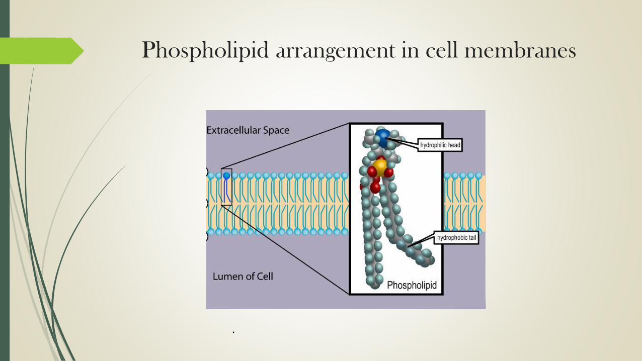
Phospholipid arrangement in cell membranes
.

Phospholipids in biological membranes
The phospholipids are amphiphilic. The hydrophilic end usually contains a negatively charged phosphate group, and the hydrophobic end usually consists of two "tails" that are long fatty acid residues.
In aqueous solutions, phospholipids are driven by hydrophobic interactions that result in the fatty acid tails aggregating to minimize interactions with water molecules. The result is often a phospholipid bilayer: a membrane that consists of two layers of oppositely oriented phospholipid molecules, with their heads exposed to the liquid on both sides, and with the tails directed into the membrane. That is the dominant structural motif of the membranes of all cells and of some other biological structures, such as vescicles or virus coatings.

Diacylglyceride structures:
Phosphatidic acid (phosphatidate) (PA)
Phosphatidylethanolamine (cephalin) (PE)
Phosphatidylcholine (lecithin) (PC)
Phosphatidylserine (PS)
Phosphoinositides:
Phosphatidylinositol (PI)
Phosphatidylinositol phosphate (PIP)
Phosphatidylinositol bisphosphate (PIP2) and
Phosphatidylinositol trisphosphate (PIP3)
In biological membranes, the phospholipids often occur with other molecules (e.g., proteins, glycolipids, sterols) in a bilayer such as a cell membrane. Lipid bilayers occur when hydrophobic tails line up against one another, forming a membrane of hydrophilic heads on both sides facing the water.

Dynamics
These specific properties allow phospholipids to play animportant role in the cell membrane.
Their movement can be described by the fluid mosaic model, thatdescribes the membrane as a mosaic of lipid molecules that act asa solvent for all the substances and proteins within it, so proteinsand lipid molecules are then free to diffuse laterally through thelipid matrix and migrate over the membrane.
Sterols contribute to membrane fluidity by hindering the packingtogether of phospholipids. However, this model has now beensuperseded, as through the study of lipid polymorphism it is nowknown that the behaviour of lipids under physiological (andother) conditions is not simple

GLYCOLIPIDS
Glycolipids are lipids with a carbohydrate attached by a glycosidic (covalent) bond. Their role is to maintain the stability of the cell membrane and to facilitate cellular recognition, which is crucial to the immune response and in the connections that allow cells to connect to one another to form tissues.
Glycolipids are found on the surface of all eukaryotic cell membranes, where they extend from the phospholipid bilayer into the extracellular environment.

StructureThe essential feature of a glycolipid is the presence of a
monosaccharide or oligosaccharide bound to a lipid moiety. The most common lipids in cellular membranes are glycerolipids and sphingolipids, which have glycerol or a sphingosine backbones, respectively.
Fatty acids are connected to this backbone, so that the lipid as a whole has a polar head and a non-polar tail. The lipid bilayer of the cell membrane consists of two layers of lipids, with the inner and outer surfaces of the membrane made up of the polar head groups, and the inner part of the membrane made up of the non-polar fatty acid tails.

The saccharides that are attached to the polar head groups on the outside of the cell are the ligand components of glycolipids, and are likewise polar, allowing them to be soluble in the aqueous environment surrounding the cell. The lipid and the saccharide form a glycoconjugate through a glycosidic bond, which is a covalent bond.
The anomeric carbon of the sugar binds to a free hydroxyl group on the lipid backbone. The structure of these saccharides varies depending on the structure of the molecules to which they bind.

Chemical structure of glycolipids
Types of glycolipidsGlyceroglycolipids: a sub-group of glycolipids characterized by an acetylated or non-acetylated glycerol with at least one fatty acid as the lipid complex. Glyceroglycolipids are often associated with photosynthetic membranes and their functions.
The subcategories of glyceroglycolipids depend on the carbohydrate attached.
1. Galactolipids: defined by a galactose sugar attached to a glycerol lipid molecule. They are found in chloroplast membranes and are associated with photosynthetic properties.
2. Sulfolipids: have a sulfur-containing functional group in the sugar moiety attached to a lipid. An important group is the sulfoquinovosyl diacylglycerolswhich are associated with the sulfur cycle in plants.

3. Glycosphingolipids: a sub-group of glycolipids based on sphingolipids. Glycosphingolipids are mostly located in nervous tissue and are responsible for cell signaling.
4. Cerebrosides: a group glycosphingolipids involved in nerve cell membranes.
5. Galactocerebrosides: a type of cerebroseide with galactose as the saccharide moiety
6. Glucocerebrosides: a type of cerebroside with glucose as the saccharide moiety; often found in non-neural tissue.
7. Sulfatides: a class of glycolipids containing a sulfate group in the carbohydrate with a ceramide lipid backbone. They are involved in numerous biological functions. They are involved in numerous biological functions ranging from immune response to nervous system signaling.

8. Gangliosides: the most complex animal glycolipids. They contain negatively charged oligosacchrides with one or more sialic acid residues; more than 200different gangliosides have been identified. They are most abundant in nerve cells.
9. Globosides: glycosphingolipids with more than one sugar as part of the carbohydrate complex. They have a variety of functions; failure to degrade these molecules leads to Fabry disease.
10. Glycophosphosphingolipids: complex glycophospholipids from fungi, yeasts, and plants, where they were originally called "phytoglycolipids". They may be as complicated a set of compounds as the negatively charged gangliosides in animals.
11. Glycophosphatidylinositols: a sub-group of glycolipids defined by a phosphatidylinositol lipid moiety bound to a carbohydrate complex. They can be bound to the C-terminus of a protein and have various functions associated with the different proteins they can be bound to.

Function of Glycolipids
Cell–cell interactions
The main function of glycolipids in the body is to serve as recognition sites for cell–cell interactions. The saccharide of the glycolipid will bind to a specific complementary carbohydrate or to a lectin (carbohydrate-binding protein), of a neighboring cell. The interaction of these cell surface markers is the basis of cell recognitions, and initiates cellular responses that contribute to activities such as regulation, growth, and apoptosis.

Immune responses
An example of how glycolipids function within the body is the interaction between leukocytes and endothelial cells during inflammation.
Selectins, a class of lectins found on the surface of leukocytes and endothelial cells bind to the carbohydrates attached to glycolipids to initiate the immune response.
This binding causes leukocytes to leave circulation and congregate near the site of inflammation. This is the initial binding mechanism, which is followed by the expression of integrin which form stronger bonds and allow leukocytes to migrate toward the site of inflammation.
Glycolipids are also responsible for other responses, notably the recognition of host cells by viruses.

Blood types
Blood types are an example of how glycolipids on cell membranes mediate cell interactions with the surrounding environment. The four main human blood types (A, B, AB, O) are determined by the oligosaccharide attached to a specific glycolipid on the surface of red blood cells, which acts as an antigen.
The unmodified antigen, called the H antigen, is the characteristic of type O, and is present on red blood cells of all blood types. Blood type A has an N-acetylgalactosamine added as the main determining structure, type B has a galactose, and type AB has all three of these antigens. Antigens which are not present in an individual's blood will cause antibodies to be produced, which will bind to the foreign glycolipids. For this reason, people with blood type AB can receive transfusions from all blood types (the universal acceptor), and people with blood type O can act as donors to all blood types (the universal donor).

METABOLISM OF GLYCOLIPIDS
Glycosyltransferases
Enzymes called glycosyltransferases link the saccharide to the lipid molecule, and also play a role in assembling the correct oligosaccharide so that the right receptor can be activated on the cell which responds to the presence of the glycolipid on the surface of the cell.
The glycolipid is assembled in the Golgi apparatus and embedded in the surface of a vesicle which is then transported to the cell membrane. The vesicle merges with the cell membrane so that the glycolipid can be presented on the cell's outside surface.

SteroidsA steroid is a biologically active organic compound
with four rings arranged in a specific molecular configuration.
Steroids have two principal biological functions: as important components of cell membranes which alter membrane fluidity; and as signaling molecules. Hundreds of steroids are found in plants, animals and fungi. All steroids are manufactured in cells from the sterols lanosterol (opisthokonts) or cycloartenol(plants). Lanosterol and cycloartenol are derived from the cyclization of the triterpene squalene.

The steroid core structure is typically composed of seventeen carbon atoms, bonded in four "fused" rings: three six-member cyclohexane rings (rings A, B and C in the first illustration) and one five-member cyclopentanering (the D ring). Steroids vary by the functional groups attached to this four-ring core and by the oxidation state of the rings. Sterols are forms of steroids with a hydroxyl group at position three and a skeleton derived from cholestane. Steroids can also be more radically modified, such as by changes to the ring structure, for example, cutting one of the rings. Cutting Ring B produces secosteroids one of which is vitamin D3.

CHEMICAL STRUCTURE OF STEROIDS
Structure of cholestane, a steroid with 27 carbon atoms. Its core ring system (ABCD), composed of 17 carbon atoms, is shown with IUPAC-approved ring lettering and atom numbering.[1]

Examples include the lipid cholesterol, the sex hormones estradioland testosterone, and the anti-inflammatory drug dexamethasone.
The major classes of steroid hormones, with prominent members and examples of related functions, are: Corticosteroids, Glucocorticoids, Cortisol, a glucocorticoid whose functions include immunosuppression
Mineralocorticoids:
Aldosterone, a mineralocorticoid which helps regulate blood pressure through water and electrolyte balance

Progestogens:
Progesterone, which regulates cyclical changes in the endometrium of the uterus and maintains a pregnancy
Androgens:
Testosterone, which contributes to the development and maintenance of male secondary sex characteristics
Estrogens:
Estradiol, which contributes to the development and maintenance of female secondary sex characteristics
Additional classes of steroids include:
Neurosteroids such as DHEA and allopregnanolone
Aminosteroid neuromuscular blocking agents such as pancuronium bromide
As well as the following class of secosteroids (open-ring steroids)
Vitamin D forms such as ergocalciferol, cholecalciferol, and calcitriol

Chemical synthesis
Microbial catabolism of phytosterol side chains yields C-19 steroids, C-22 steroids, and 17-ketosteroids (i.e. precursors to adrenocortical hormones and contraceptives).
The addition and modification of functional groups is key when producing the wide variety of medications available within this chemical classification. These modifications are performed using conventional organic synthesis and/or biotransformation techniques.
Biological significance
Steroids and their metabolites often function as signallingmolecules (the most notable examples are steroid hormones), and steroids and phospholipids are components of cell membranes. Steroids such as cholesterol decrease membrane fluidity.
Similar to lipids, steroids are highly concentrated energy stores. However, they are not typically sources of energy; in mammals, they are normally metabolized and excreted.
Steroids play critical roles in a number of disorders, including malignancies like prostate cancer, where steroid production inside and outside the tumour promotes cancer cell aggressiveness.

Thank You