technical and clinical aspects isola tie van foetale cellen … maria...isolation of fetal cells...
TRANSCRIPT

Isolation of fetal cells from maternal blood
Technical and clinical aspects
Isola tie van foetale cellen uit moederlijk bloed
Technische en klinische aspecten
Maria Wilhelmina Johanna Christina Jansen

The studies described in this thesis were performed in the Departments of Clinical Genetics, Obstetrics and Gynecology and Hematology at the Erasmus University Rotterdam. Part of the project was financially supported by The Foundation for Clinical Genetics, Rotterdam.
Financial sUppOl1 frolll Sal1bio b.v., the J.E. Jurriaanse Stichting and the Dr. Ir. van del' Lam Stichting towards the printing of this thesis is gratefully acknowledged.
Cover illustration: Wilh Hnd Xris Jansen; Lay-out: Armando Braat
Mieke Jansen Isolation of fetal cells from maternal blood. Technical and clinical aspects. Thesis, Erasmus University Rotterdam, The Netherlands - With summary in Dutch. ISBN 90-5677044-6
©2000, r-vI.W.J.c. Jansen All rights reserved
Isolation of fetal cells from maternal blood
Technical and clinical aspects
Isolalie van foelale cellen uil moederlijk bloed
Technische en klinische aspeclen
Proefschrift
TER VERKRJJGlNG VAN DEGRAAD VAN DOCTOR
AAN DE ERASMUS UNIVERSITEIT ROll'ERDt\~'1
0l'GE2AO VAN DE RECTOR MAGNIFICUS
PROF. DR. IR . .I.H. VAN BE1\·II\IEL
EN VOLGENS BESUJIT VAN HET COLLEGE VOOR PROMOTIES.
OF. OPEN HARE VERDEDIGING ZAL PLAATSVINDEN OP
WOENSDAG 13 SFPTEi>.H3ER 2000 01\1 13.45 UUR
DOOR
MARIA \VILHEUI-lINA JOHANNA CHRISTINA JANSEN
GEBOREN TE SPIJKENISSE

Promotiecommissie
Promotoren:
Ovcrigc lcden:
Prof. Jhr dr. J.W. Wladimiroff
Prot: dr. H. Galjaard
Prof. dr. R. Benner
Prof. dr. H.A. van Geijn
Dr. R.E. Plocmacher

Vooruitgang
Soms gaat vooruitgang zo snel
oat we niet niles wat mooi is en goed
met ons I11CC kunnen dragcn.
Daarom Illoeten we van lijd tot lijd even pauzeren
en achtcrom zien om de nagestuurdc bagagc in ontvangst Ie nemen.
Kade Bmin flit "Uitsmijlers WlIl scharreleieren"
Vaal' Xris & \Vilh
Vool'lngmar

Contents
PREFACE 8
CHAPTER 1 GENERAL INTRODUCTION 11
1.1. Biological basis of passage of fetal cells into the maternal circulation 13 1.1.1. Embryonic developm~llt and placentation 13 1.1.2.lIcmatopoiesis \4
l.2. Fctnl cell types 17 1.2.1. Fetal leukocytes 17 1.2.2. Trophoblast cells 18 1.2.3. Fetal nuclc,1tcd red blood cells 19
1.3. Isolation and idcntilication strategies for fetal cells ill maternal blood 19 1.3.1. Isolation strategies 19 1.3.2.ldentificatiOIl artetal cells in maternal blood 22
I A. Feta-maternal cell trafficking 26 104.1. Timing and proportion 26 1.4.2. Clennlllce versus persistence offclal cells from the maternal circulation 27
1.5. New research areas 28 1.5.1. Fetal DNA in maternal plasma and serum 28 1.5.2. III vitro expansion of fetal cells 29 1.5.3, Fetal cell mierochirncrism 30
1.6, Conclusions and o~iecti\'cs of the thesis. 31
1. 7, Rclcrences 32
CHAPTER 2 49
The use of in vitro expanded ef)1hroid cells in a model system ror the isolation of letal cells from maternal blood. ivlie"c W.J.c. J<HlS'cn, Marieke \'on lindem, I-Imtmut [1cug, Helen Bwndenburg, Hajo 1.1. Wild schut, Juriy W. Wladimiroft~ Peter A in 'I Veld. Prellatal Diagllmi.\" (1999), mI. /9, p3]J~329
CHAPTER 3
How lIselhl is the ill \'ilro e~pansioll of Ictal CD34+ progenitor cells from mllten1.11 blood samples for diagnostic purposes? t\licke W.J.c. Jansen, Karin Kot"ver-Hakkennes. Dik van Leenen, Helen Brandenburg, Hajo U. Wildschul, Juriy W. \vladimiroft~ Rob E. Ploemachcr. ,-keep/edjt)!, publication ill Prenuta! Diagl1(1.1is
65
CHAPTER 4
The effect of chorionic villus sampling on the number of fetal cells isolated from maternal blood and on maternal serum alpha~letoprotein levels. i\tieke \V.J.e. Jansen, Helen Brandenhurg, lIajo I. J. Wildschut. Anton C, i\t i\'tartens, Adriana i\.I, Hagenaars, Juriy W. \vladimiroft~ Peter A. in 't Veld, Prel/alal Diagnosis (/997), 1'01.17, p1J53-951)
CHAPTER 5
Significantly higher number of Ictal nucleated red blood cells in the maternal circulation of women with preeclampsia. Miel\c W,J.c. Jansen, Karin Korver-Hakkennes, Dik van Lecncn, Willy Visser, Peter A. in '( Veld, Chris(iannc H .. 1. de Groot, Juriy \V. WladimirolT. submit/cd
83
95
CHAPTER 6 GENERAL DISCUSSION AND CONCLUSIONS 113
6.1. floilodel systems used for the isolation of fctal cells Irom maternal blood 109
6.2.111 vitro expansion offetal cells III
6.3. Clinical implications of fetal cells in the maternal circulation 112 6.3.1. Aneuploidy 113 6.3.2. The effect of chorionic villus sampling 113 6.3.3. Felo-l11alernal cdllrafticking in preeclampsia 114
6.4. Fetal-maternal immunology: tolerance versus autoimmune disease 114
6.5. Conclusions and future research 117
6.6. Relerences 118
SUMMARY 127
SAMENVATTING 133
LIST OF ABBREVIATIONS 139
CURRICULUM VITAE 141
DANKWOORD 143

Preface
Traditional approaches towards prenatal diagnosis of fetal abnormalities are ultrasonography
and the invasive techniques of amniocentesis, chorionic villus sampling (CVS) and
cordoccntesis. Although the invasive methods reach virtually 100 % accuracy, there is a
small procedure-related risk for the felus. fetal loss rale following all invasive procedure is
estimated at 0.5 - 1 %, depending on the technique employed. In the Netherlands, invasive
prenatal testing is offered to women for various reasons: (1) advanced maternal age
(~36 years); (2) a parental carrier status of a balanced chromosomal anomaly; (3) a previolls
child wilh a chromosomal abnormality £IndIoI' multiple congenital malformations;
(4) increased risk of a neural tube detect; (5) increased risk of a monogenic disease
demonstrated by biochemical or DNA analysis; and (6) tetal congenital defects seen on
ultrasound examination.
During the last 15 years, high resolution ultrasound equipment has made possible the
identification of a host of tetal congenital anomalies as c<uly as the early second trimester of
pregnancy (\Vladimiroft~ 1994; den Hollander et al., 1998). Late first trimester and early
second trimester sonographic markers for aneuploidy have been developed of which nuchal
translucency has been shown to be the most effective onc (Pajkl1 e( a/., 1998: Snijders et al.,
1998). Biochcmical testing for chromosomc anomalies includes first trimester and early
second trimester maternal serum screening (Wald eJ al., 1997; de Graaf, 1999; van Rijn,
1999). Both sonographic markers and biochem ical tcsts represent a risk assessment with
emphasis on Down syndrome. Depending on the nature of these tests, dctection rates of
70-80 % fOJ' DOWI1 syndrome at a 5 % false positive rate have been claimed. Risk assessmcnt
by means of maternal serulll screening andlor fetal nuchal transluccncy screening is currently
subject to debate in the Netherlands (Discussion Oocur,lcnt Dutch Society of Obstetrics and
Gynecology, 1997),
An alternative non-invasive approach of potential diagnostic significancc is the isolation of
fetal cells from maternal blood, whieh is receiving increasing attention during the last two
decades, This may lead to the elimination of fetal cell sampling by invasive techniqucs sllch
as CVS or [Hl1nioeentesis. To date, it is not yet clear whether the technique of fetal cell
isolation from maternal blood will be accurate enough for fetal diagnosis. This thesis will
give an overview of the current state of the ali offetal cell isolation.
8

References
de Graaf I. (1999). On tirst trimestcr Down syndrome screcning. Dept. Obstetrics & Gynccology, Univcrsity of Amsterdam, Amsterdam (thesis).
den Hollander N.S., van del' Harten H.J., Laudy 1.A., van de Wcg P., Wladimiroff .1.11'. (1998). Early transvaginal ultrasollographic diagnosis of Becmer~Langcr dysplasia: a repOli of two cases. Ultrasound Obstet Gynecolll: 298-302.
Pajkl1 E., de Graaf I.M., Mol B.II'., van Lith 1.M., Bicker a.p., Bilardo C.M. (1998). Weekly nuchal translucency measurements in nOl1nal fetuses. Ohste{ GYllecol 91: 208~211.
Snijders R.l., Noble P., Sebire N., Souka A., Nicolaides K.H. (1998). UK muiticentre project all assessment of risk of trisomy 21 by matelllal age and fetal nuchal-transluccncy thickncss at 10-14 weeks of gestation. Fctal Medicine Foundation First Trimester Screening Group. Lallcet 352: 343-346.
van Rijn M. (1999). Maternal serum screcning in Dutch Obstetrics practice. Dept. Obstetrics, Neonatology & Gynccology, lJniverity of Utrecht, Utrecht (thesis).
\Vald N.J., Kennard A., Hackshaw A., McGuire A. (1997). Antenatal screening tor Down's syndrome. J ,lIed Screen"': 181-246.
\VladimirotT J. \V. (1994). Ultrasonic diagnosis of congenital abnol111alitics following the 16th week of pregnancy. Ned T{idsr:hr Gel1eeskd 138: 180-183.
9


General introduction

Gcneral introduction
Feto-maternal cell traffic in pathological conditions was tirst recognized in 1893, when
Schmorl idcntified trophoblast cells in lung capillaries of women dying of eclampsia
(Schmorl, 1893). In 1969, \Valknowska ef a/. identified male meta phases in cultured
lymphocytes isolated from blood of healthy pregnant women, who subsequently gave bil1h to
a boy. They were thc first to demonstrate that fetal cells enter the matel1lal circulation in
normal pregnancy and they suggested that these cells might be llsed for chromosome
analysis. Since then, many investigators have focussed on the development of a safe and
reliable test to perform non-invasive prenatal diagnosis, as an alternative for chorionic villus
sampling and amniocentesis. Before a non-invasive prenatal diagnostic test using fetal cells
in maternal blood will be available, a Humber of questions need to be addressed, such as:
(1) at what period of gestation does feto-matcrnal transfer of cells take place; (2) what is the
best fetal cell type to isolate; (3) arc fetal cells detectablc in thc ~Iood orall pregnant women;
(4) do chromosomally abnormal pregnancies result in increased or decreased transfer of fetal
cells into the maternal circulation; and (5) may fetal cells from prior pregnancies persist in
the maternal circulation, and hence interfere with a reliable prenatal diagnosis.
1.1. Biological basis of passage of fetal cells into the maternal circulation
Passage of fetal cells into the maternal circulation will occur at the feto-maternal interface.
Knowledge of embl)'onic development and the fonnation of the feto-maternal interface
(placentation) will gain insight into the process of feto-maternal cell trafficking. Furthcrl11ore~
knowledge of fetal hematopoiesis will lead to more information about the fetal cell types that
circulate in maternal blood during pregnancy.
1.1.1. Embryonic development and placentation
After fertilization of an oocyte by a sperm cell, the formed zygote undergoes a series of rapid
mitotic cell divisions known as cleavage. The zygote is cleaved into a number of blastomeres
and subsequent cell divisions of these blastomcrcs result in the formation of the morula
(32-cell stage; day 3). Subsequently~ the morula develops into the blastocyst (64-cell stage:
day 4-5) which contains an outer cell layer (trophoblast) which gives rise to p1ll1 of the
placenta, and a group of centrally located cells, the inner cell mass (embryoblast) which gives
rise to the embl)'o. After the blastocyst has attached to the endometrial epithelium (day 6),
the trophoblast ditferentiates into two eelJ types: (1) the cytotrophoblast, which is mitotically
active and forms new cells that migrate into the increasing mass of syncytiotrophoblast; and
(2) the syncytiotrophoblast. The latter rapidly becomes a large, thick, multinucleated
13
Chapter 1
protoplasmic Illass in which no cell boundaries are distinguishable (syncytium).
Subsequcntly, a lacunar network develops in thc syncytiotrophoblast, and maternal capillaries
near the syncytiotrophoblast expand to form maternal sinusoids establishing a primitive
uteroplaccntal circulation, in which the primm)' chorionic villi develop (day 13). In the
fmiher maturation of the villous tree, fetal blood vessels invadc this villolls connectivc tissue
and connect the vessels to the cmbl),onic circulation. \Vhcn the fetal hemi begins contracting
on day 2201' 23, a primitive fetal placcntal circulation establishes (Moore, 1982).
One of the imp01iant placental structures t hat assures the transfer of nutrients to enhance fetal
growth is the so-called 'placenta membrane', a thin layer of fetal tissues scparating the
matelllal and fetal circulation. This membrane consists of syncytiotrophoblast,
cytotrophoblast, thc connective tissue core of the villus, and the endothelium of the fetal
capillaries. As pregnancy advances, the placental membrane bccomes progressively thinner
while simultaneollsly fetal blood tlow and blood pressure increase as the villous tree cnlarges
(Sullon ('{ (/1., 1990).
Until now, it is not known at what period during placentation a feto-matcrnaltransfusion may
OCClll". One can readily imagine that a disruption of the relatively thin placental membrane
would lead to fetal bleeding into the intervillous space at the timc when this membranc thins
with advancing pregnancy. Another theoretical possibility may be that feto-maternal cell
trafficking takes place during the formation of villi, at the time when fetal capillaries are
formed and the pumping action of the fetal heart begins (Benirschke, 1994).
1.1.2. Hematopoiesis
Fetal hematopoiesis is one of the first processes established following implantation of the
blastocyst, and can be divided into three main overlapping periods: mesoblastic, hepatic, and
myeloid (tigure 1). It was originally assumed that the mesoblastic hematopoiesis stmis in the
yolk sac between days 16 and 19 (Huyhn el al., 1995), followed by hepatic hematopoiesis at
approximately 5 wccks aner fertilization (Migliaccio el al., 1986). The final phase of
hematopoiesis takes place in the bone marrow, starting at 10 wceks atter fertilization in the
long bones (Metcalf and Moore, 1971; Kollmann el al., 1994; Charbord ('I (/1 .. 1996).
Recently, it has been showll that in developing mamnwis stem cells can be derived from an
intraembryonic site called the amta~gol1ad-lllesonephros (AGM) region Uvledvinsky ef a/.,
1993: Medvinsky and Dzierzak, 1996; Tavian el u/., 1996). Howevcr, the site of origin of
definitive hematopoietic stem cells in the developing fetus remains controversial. Evidence
n'om some studies indicates that hematopoietic stem cells from the yolk sac arc responsible
for transient primitive hematopoiesis, but they appear to lack the ability to reconstitute the
hematopoietic system in adult animals (Cumano cl al., 1996; Medvinsky and Dzierzak,
14
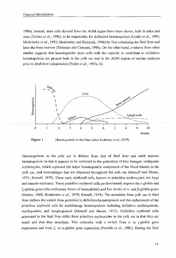
General introduclion
1996). Instead, stem cells derived from the AGM region have been shown, both in mice and
man (Tavian et al., 1996), to be responsible tor definitive hematopoiesis (Godin ct al., 1993;
Medvinskyet al., 1993; Medvinskyand Dzierzak, 1996) by first colonizing the fetal liver and
latcl' the bone marrow (Delasslls and Cumano, 1996). On the other hand, evidence from other
studies suggests that hematopoietic stem cells with the capacity to contribute to definitive
hematopoiesis are present both in the yolk sac and in the AGM region of murine embtyos
prior to fctallivcr colonization (Yoder et al., 1997n, b).
Liver ............
'.
o 2 3 4 5 6 7 8 9 10
Months
Figure 1 Hematopoiesis in the fetus (after Kelemen el al., 1979)
Hematopoiesis in the yolk saC is distinct from that of fetal liver and adult marrow
hematopoiesis in that it nppears to be restricted to the generation of two lineages: embryonic
erythrocytes, whit.:h represent the major hematopoietic component of the blood islands in the
yolk sac, and macrophagcs that are dispersed throughout the yolk sac (Metcalf and rvloore,
1971; Russell, 1979). These early el)1hroid cells, known as primitive erythrocytes, arc large
and remain nucleated. These primitive el)1hroid cells predominantly express the c-glohin and
S-globin genes (the embryonic forms of hemoglobin) and low levels of (1.- and p-globin genes
(Barker, 1968; Brotherton et al., '1979; Russell, 1979). The transition from yolk sac to fetal
liver defines the switch from primitive to definitive hematopoiesis and the replacement of the
primitive el)1hroid cells by multilineagc hematopoiesis including definitive el)1hropoiesis,
myelopoiesis, and lymphopoiesis (Metcalf and Moore, 1971). Detlnitive el)1hroid cclls
generated in the fetal liver differ from primitive cl)1hroc)1es in the yolk sac in that they arc
small and that they enucleate. This coincides with a switch frol11 c- to y-globin gene
expression and from S- to a-globin gene expression (Peschle el af., 1985). During the fetal
15

Chapter 1
period, the site of el)1hropoiesis gradually switches from the liver to the spleen and finaily
the bone marrow. Gamma-globin gene expression decreases during this period with a
reciprocal increase in the expression of the adult p-globin gene and the appearance of low
levels ofadull ii-globin gene expression (tigure 2) (Barker, 1968; BrothClton of a/., 1979).
o
Figure 2
y
3 6 9 12 t5 months
Expression of globins during fetal development (after Barker, 1968; Brotherton <'I al., 1979; Peschle ('I al., J 985)
\Vith the development of cordocentesis, access to the fetal circulation has provided an
opportunity to study fetal hematological profiles. However, it is technically difficult to obtain
Ictal blood before 18-20 weeks of gestation. In the developing fetus, the number of
erythroc)1es increases linearly, whereas the number of el)1hroblast cells decreases with
advancing gestation. In contrast, the number of fetal white blood cells increases with
gestation (table 1) (Millar of a/., 1985; De Waele ef a/., 1988; Nicolaides ef a/., 1989;
Forestier ef a/., 1991; Thilaganathan of 0/., 1992, 1994).
For the development of a non-invasive prenatal diagnostic test, fetal el)1hroblast cells appear
to be the lelal cell type to target, since these cells are the first cells that are formed during
fctal hematopoiesis in the yolk sac. In addition, erythroblast cells are abundantly present in
the fetus during the first trimester of pregnancy, when compared with lymphoid and myeloid
cells.
16

General introduction
Table 1 rvfenn values for the number of white blood cells, red blood cells !lnd erythroblast cells in fClal, neonatal and adult blood
Gestational age
16-17 weeks
18-21 weeks
22 - 25 weeks
26 - 29 weeks
neonate
adult
White blood cells
(xl0'/I)
2.00
2.98
4.51
5.16
14.1
6.00
Red blood cells EI)1hroblast cells
(x I 0"/1) (x I 0'/1)
2.62 2.70
2.82 1.75
3.00 0.95
3.46 0.99
4.60 0.50
4.70 <0.01
Dala arc lIdapkd from t>.lilIar eI al., 1985; Dc Wnelc {'I al., 1988; Foresticr ('/ 01., 1991.
1.2. Fetal cell types
The next issue conccrns the types of fetal cells that have been found in the maternal
circulation. The fetal cell types that have been studied by numerous investigators worldwide
include fetal leukocytes, i.e. fctal lymphocytes and gr<lnuloc)1es, fetal nucleated red blood
cells (NRBCs) and trophoblast cells.
1.2.1. Fetal leukocytes
L}'f11p/tocyles
The presence of fetal lymphoc)1es in the maternal circulation was first described in 1969 by
\Valknowska et a/ .. These investigators demonstrated the presence of a Y chromosome in
mitogen-stimulated lymphoc)1es obtained from pregnant women c1llTying a male fetlls. These
results were confirmed by others using similar techniques or by investigating
quinacrine-stained interphase nuclei for the presence of fluorescent Y chrolllosome signals
(Schindler and Martin-du-Pan. 1972; Schroder alld de la Chapellc, 1972).
Another breakthrough was the isolation of fetal lymphocytes by tluorescence-<lctivated cell
sorting (FACS) lIsing a monoclonal antibody against the human leukocyte antigen HLA-A2,
which was only expressed on fetal lymphocytes and not on maternal cells (Hcrzenberg ct a/.,
1979; Iverson ct al., 1981). Unfortunately, until now the isolation of fetal lymphocytes is
considered impractical due to the necessity ofpcrforming HLA typing of both parents and the
lack of other specific markers ,that distinguish fetal from maternal lymphoc)1es. !'vloreover,
17

Chapter 1
the fact that fetal lymphocytes might persist from an earlier pregnancy makes it difficult to
establish whether isolated lymphoc)1es derive from the current pregnancy (Schroder el al.,
1974). This is especially problematic for the detection or trisomic cells in women with prior
spontaneolls abortions, given the high likelihood (50%) thal these abortions were associated
with a chromosomally abnormal fetus (Sargent et (fl., 1994).
Fetal gruuulocytes
This fetal cell type has received little .1ttention. In 1975, Zilliaclls el ai, detected fetal
granulocytes in the circulation of pregnant women. Several years later, \Vessman ef al. (1992)
isolated granuioc)1es from maternal peripheral blood samples lIsing density gradient
centrifugation and they corrcctly identified the Y chromosomc in these granulocytes by ill
situ hybridization.
1.2.2. Trophoblast cells
Trophoblast cells are pm1icularly attractive for the development of a non-invasive prenatal
test because of their unique morphology, which permits microscopic identification.
Goodtellow and Taylor (1982) were the first to demonstrate trophoblast cells in peripheral
blood samples fi'olll pregnant women lIsing differential centrifugation and indirect
immunofluorescence detection. Covone el al. (l984) identified feta I trophoblast cells in the
peripheral blood from pregnant women using FACS and a monoclonal antibody against a
syncytiotrophoblast-specific antigen, H315. However, subsequent work showed that thc
H315-positive cells were of maternal origin (Covone ef al., J988). 1t was suggested that the
results were due to adsorption of the H315 onto maternal cells and no fetal cells had been
isolated either in this study or the previous study. These problcms may be overcome by the
use of new markers, snch as HASH-2, human placental lactogcn hormone (hPL) (Latham ef
al., 1996) or HLA-G (Moreau el al., 1994; vall Wijk el al., 1996).
Another difficulty is the fact that trophoblast cells appear to be rarc in the maternal
circulation, probably because they are able to form large multi-nucleated giant cells which are
filtered out by the maternal pulmonmy circulation so that they do not reach the peripheral
circulation (Attwood and Park, 1960). Especially, when the prcgnancy develops normally,
trophoblast cells do not appear to be present in grcat numbers in maternal peripheral blood
(Sargent ef al., 1994). However, trophoblast cells arc detectable in increased numbers in
cascs of preeclampsia, although it is not yet known whether these increased numbers arc a
calise or an effect of preeclampsia (Chua el al., 1991; Sargent et al., 1994).
A further concern using trophoblast cells is the fact that they are part of the placenta and
potential problems may arise when chromosome abnormalities arc prcscnt in the placenta but
18
General introduction
not in the letus (Henderson el 01., 1996; Goldberg and \Vohlferd, 1997; van Opstal, 1998).
This confincd placental mosaicism has been documented to occlir in 1 % of all cases of
chorionic villus sampling. Therefore, genetic analysis of a placental trophoblast cell might
not be representative of the fetal kmyotypc.
1.2.3. Fetal nucleated red blood cells
The last H:w years, most attention has been focussed on the isolation of fetal NRBCs from
maternal blood. Fetal NRBCs are the prcdomimlllt nucleated cell type in the fetal circulation
in the first trimester of pregnancy, during the yolk sac and liver phases of hematopoiesis
(Metcalf and Moore, 1971). In blood of 10- to 20-week fetuses, NRBCs make up
approximately 10 % oflhe total popUlation, whereas in adults they are quite rare «0.1 %). In
addition, red cell development in the fetlls is morc advanced than white cell development
during the tirst trimester (see paragraph 1.1.2., table I). If fetal cell trafficking occurs, they
are likely to be the major cell type in the maternal circulation.
NRBCs have been isolated llsing antibodies against membrane-bound markers like the
transferrin reccptor (CD7I) and glycophorin A (GPA) or intracellular antigens like fetal and
embl),onie hemoglobin (Lokcn el al., 1987; Bianchi e/ al., 1990; Zheng el 01.,1995; Mesker
el al., 1998) (see paragraph 1.3.1.).
Another reason why the isolation of fetal NRBCs is attractive is that fetal NRBCs have a
limited life span of about 120 days (Pearson, 1967), and are therefore unlikely to persist
between pregnancies, unlike fetal lymphocytes (Simpson and Elias, 1994).
1.3. Isolation and identification strategies for fetal cells in maternal blood
1.3.1. Isolation strategies
The number of fetal cells in the maternal circulation is limited, and therefore, 1110st efforts
have been concentrated all the development of a highly efficient enrichment procedure.
Enrichment call be achieved by either positive selection of target cells using uniquc fetal
characteristics, or by depletion of contaminating maternal cells. for the isolation of rare cell
populations two considerations should be taken into account: yield and purity. Yield is very
impoliant because the number of fetal cells in maternal blood is vcry low and loss of fetal
cells should be avoided. Purity will be determined by the relative number of fetal and
maternal cells remaining after enrichment. A relative increase in the absolute number of fetal
cells after enrichment allows the reduction of the amount of maternal background cells, and
19

CI,"pler I
thereby, fetal cell idcntificntion by nuorescencc il1 sill1 hybdtii:latioll (FISH) or polymerase
chain reaction (peR) analysis is facilitated.
To distinguish ictal cells from the vast majority of their maternal cOlintcll}[lrts a specific
marker is needed. Until now, no single marker antigen is known that is specific tor fetal cells.
An overview of isolation approaches that have been designed to recover fctal cells lI'olll the
maternal circulation is given in tublc 2.
Table 2 Isolation strategies used for the isolation of fetal cells from the maternal circulation
-cls~o~la~';~o~n~s~',~a'~,~gLy __________ F~,~ta~I~,c~ILI~')~'p~e ______ ~R~c~~~r~cl~lc~,~s ______________________ . ______ ___
Fluorescence activated cell sorting (FACS)
Magnetic activated cell sorting (~'IACS)
Charge now separation (CFS)
Densit)' gradient centrifugation
ImIllunomngnctic bends separation
Illlmunomagnetic colloid system
lymphocytes NRBCs trophoblast cells
NRBCs trophoblast cells
NRl3Cs
lymphocytes granulocytes trophoblast cells NRDCs
NRI1Cs trophoblast cells
NRBCs trophohlast cells
Avidin-conjugated columns NRBCs wilh biotinylated antibodies
i\licromElnipuiation of NRBCs individual cells
Carbonic anhydrase NRBCs inhibition
Fluorescence actiwtfed cell sorting (FACS,)
HerLcnbcrg c/ aI., 1979; Iverson e( af., 1981; Bianchi ('I al., 1990; Price ('/ a/., 1991; Wachtel el (1/,,1991; Tse d al., 1994; Johansen ('( al., 1995; Lewis el 01.,1996; Sollda ('I al., 1997
Ganshil1-Ahlert et al., 1992, 1993; Zheng ellll., 1993; Busch e! al., 1994; Durrant ('Ial., 1996; Ganshirt ('I III., 1998
Wachtel ('( al., 1996, 1998; Shulman ('( al., 1998
Bh<lt ct al .. 1993; Ooslt'rwijk Cf a/., 1996; Sitar dill" 1997; Ganshirt ('( al" 1998; Sekiz(l\\-a ct af., 1999
WCSSIllOIl ('I af., 1992; Johmls~n ('/ af., 1995; Bianchi c/ al., 1996b
Steele d al., 1996; i\'tartin cl al., 1997; Lim c/ al., 1999
lIall e/ al., 1994
Takabayashi ct al., 1995; Sckizawa ('/ al., 1996a, b, 1998; Watonabe ('I al., 1998
de Uraafct al., 19990
FACS or tlowcytometry is used for positive selection of target cells as well as for depletion
of contaminating maternal cells (Herzenberg el al., 1979; Iverson et al., 1981; Bianchi el al.,
1990; Price el a/., 1991; Woehlel el al., 1991; Tse el al., 1994; .Iolml1sel1 el al., 1995; Lewis
elal., 1996; Sohda et al., 1997). The sample for s0l1ing is incubated with a tluorescent-
20

(Jeneral introduction
labeled !lntibody specific for the target cell. The FACSc<ln identifies the cells labeled with the
tllltibody which arc thell collected into a tube or onto a slide for furthcr analysis, Although it
is possible to achieve a high purity of target cells making microscopic analysis easier and
more accurate, this method of fetal cell isolation requires considerable operator expcliisc, is
time-consuming and the expense of the equipmcnt also limits its application on a wide scale.
Afagnetic ((ctim/ed cell sorting (XfACS)
MACS is the most widely uscd method of fetal cell isolation and can be used for positivc
selection of fetal cells as well as depletion of maternal cells (Ganshirt-Ahlert e/ al., 1992,
1993; Zhcng ef (II., 1993; Busch ef al., 1994; D'lll'ant ef (II., 1996; Ganshirt ef al., 1998).
Tmget cells are labeled with an antibody attached to magnetic beads, The cell suspension is
passed over a separation column with a magnetizable matrix that is placed into a magnetic
field of extreme strength. Unlabeled cells flow through the matrix, and labeled cells stick to
the column and can be eluted after being taken away from the magnetic field, MACS
isolation is less expensive and time-consuming than FACS, and requires less expertise to
perform, The major disadvantage of MACS is that target cells are contaminated hy maternal
celis, resulting in low purity, and hence, complicating fetal cell identification.
Charge .110\1' separation (ep.);
Charge !low separation, an alternative approach for fetal cell enrichment reqUlnng no
antibody for cell selection has recently bcen described (\Vachtcl ef aI" 1996, 1998; Shulman
e/ aI" 1998), The method is based on the migration of cells in an electric field. It permits
differentiation of cell types according to their characteristic surface charge dcnsities lIsing a
cross-flow fluid gradient without the need of an antibody. This technology results in a
signil1cantly higher recovel)' of NRBCs than observed by other groups using conventional
methods for fetal cell isolation, like FACS and MACS. However, it is presently unclear as to
whether all NRBCs isolateu by this technique arc offetal origin.
Antibodies used/or the i.mlation (?!/etal NRBCsji"r)J1111lalernal blood
A variely of monoclonal antibodies have been used for the isolation of fetal NR13Cs from
maternal blood samples. The majority of researchers have utilized a 1110noclonal antibody to
the transferrin receptor (COli) for the isolation of fetal NRBCs (Bianchi et al., 1990;
Ganshil1-Ahlcli ef al., 1992; Lewis ef al., 1996; Sohda ef al., 1997). CD71 is expressed on all
cells actively incorporating iron, and 011 nearly all first-trimestcr fetal nucleated blood cells
(Price ef al., 1991; Wachtel ef a/., 1991; Bianchi ef al., 1992; Ganshili-Ahleli ef al., 1992;
Durrant e/ al., 1994; Zhcng et aI" 1997). Its expression declines with gestational age but is
increased in fctuses with an abnormal karyotype (Thilaganathan et aI" 1995; Zheng et aI"
1999). The disadvantage of C071 is that it is also expressed 011 a sUbpopulation of maternal
21
Chapler 1
cells, such as activated lymphocytes, which results in low purity (Bianchi et al., 1994; Zheng
e!al., 1997).
Monoclonal antibodies against GPA, present all maturing clythrocyles but not on
lymphocytes, have been used in combination with anti-CD?I in an altempt to increase the
specificity of recovery (Price el al., 1991). Unfortunately, anti-GPA causes agglutination of
the target red cells, preventing efficient sorting (Simpson et al., 1995).
Other monoclonal antibodies used for fetal NRBCs isolation are those that recognize the
embryonic (HbE) and lelal (HbF) hcmoglobin (Zheng ef al., 1993, 1995; Reading e! al.,
1995; Cheung el 0/., 1996; Demaria et al., 1996; Lewis el al., 1996; Oosterwijk et al., 1996,
1998b, c; Mesker ef al., 1998). HbE ("II;-chain), although unique 10 fclal cells, is expressed
only during a narrow window of time during gestation and ceases during the first trimester of
pregnancy (Zheng el al., 1999). HbF (y~chaill) is expressed in most fetal cells over a wide
range of gestational ages. Unf0l1unately, HbF is not fetal-specific since about 1% of adult
erythroid cells also contain HbF (Turpcincn and Stellman, 1992). Moreover, increased
cl)'thropoiesis during pregnancy will stimulate the synthesis of fetal hemoglobin in the
female adult, thereby limiting the usefulness of the HbF antibody (Pembrey et al., 1973;
Siunga-Tallberg ef al., 1995).
Additional monoclonal antibodies used for the isolation of fetal NRBCs are presented in
lable3.
1.3.2. Identification of fetal celis in maternal blood
Genetic analysis of fetal cells in maternal blood has relied primarily 011 two techniques: FISH
using chromosome-specific probes and peR to amplify unique fetal gene sequences enabling
subsequent DNA analysis of gene mutations.
Fluorescence in situ hybridization
The major fetal cell conditions associated with an abnormality in chromosome Ilumber can be
easily detected by FISH. Since sorted samples arc in interphase, cOllnting the chromosomcs
by direct visualization is impossible using standard c)1ogcnctic methods. FISH offers this
possibility by using chromosome-specific labeled probes, which bind to regions of the target
chromosome. Betore FISH can be applied tor fetal cell identitication, cnrichment of fetal
cells is necessary to avoid fetal signals being overruled by signals from maternal cells and to
decrease thc time of microscopic examination to analyse a sufticicnt number of fetal cells. In
cases of low purity. identification of fetal cells can be improved through bettcr and faster
22

General introduction
Tabid Cell markers used lor the enrichment of fetal NRBCs and depletion of maternal cells
Cell marker Expressed 011
CD7] (transferrin receptor) erythroid cells activatcd lymphocytes
(iPA (glycophorin A) erythroid cells
HbElHbF erythroid cells (embryonic/fetal hemoglobin)
Blood group antigcns erythroid cells
Erythropoietin receptor el)1hroid cells
CD36 (thrombospondin monoeytes receptor) platelets
cl)1hroid cells
HAE9, FBJ-2, 2-68/6, 113-3 erythroid cells (fetlllliver surface antigens)
CD45 leukoc)1es
CD32 granu1OC)1CS
CDI4 monocytes
Used tor
enrichmcnt
cllriehment
enrichmcnt
enrichmcnt
enrichment
enrichment
enrichment
depletion
depletion
dcplction
References
Bianchi {'( al., 1990; GanshirtAhler! et al., 1992; Lewis e/ (/1., 1996; Sohda c/ al., 1997; Ganshirt e/ (/1., ]998
Pric-c d al., 1991; Wachtel ,,/ al.. 1991; Simpson e/ al., 1995; Lewis el (It., 1996
Zhcng e/ al., 1993, 1995; Reading e/ al., 1995; Cheung ,,/ (/1., 1996; Demaria t'/ al., 1996; Lewis ('I al., 1996; Oosterwijk el al., 1996, 1998b, c; il.lcsker ef al., 1998
SavioTl d al., 1997; Troeger cl al .. 1999
Valeriocfal., 1996, 1997a,b
Bianchi ('f al., 1993; Troeger "f (//., 1999
Savion c/ al., 1997; Zheng ('/ al., 1997, 1999; Troeger ef al., 1999
Bianchi el al., 1991; Buseh r!I al., 1994; Jansen ef al., 1997, 1999; Lim.elal., 1999
Zheng ef al., 1993; Ferguson-Smith etal., 199t.J
Lewis el al., 1996; Steele ('/ al., 1996; )nnsen ef al., 1997, 1999
recognition of fetal cells through automated scanning (Oosterwijk el al., 1996, 1998a; Tanke
et al., 1996; de Graaf ct al., 1999a). By combining FISH and immunocytochemical staining
for HbF, fetal cells can be identified by automated image analysis consisting of computerized
microscopy, combining bright Held and fluorescence microscopy, and subsequent visual
evaluation of image memories. Automated image analysis appeared to be more sensitive than
manual identification of fetal cells (Oosterwijk ct al., 1998a).
It has now been possible to detect almost all of the significant fetal aneuploidies using fetal
cells isolated from maternal blood (Price et al., 1991; Bianchi el al., 1992; Cacheux el al.,
23

Chapter I
1992; Elias el al., 1992; Ganshil1-Ahlcrt el al., 1993; Simpson and Elias, 1993; Zheng el al.,
1995; Pezzolo el al., 1997; Oostelwijk el al., 1998d; AI-Mufti el a/., 1999; de Graaf el al.,
1999b; Rodriguez de Alba ef al., 1999). These comprise all of the major autosomal trisomies
including trisomy 13, trisomy 18, trisomy 21, some of the sex chromosome abnormalities like
47, XXV and 47, XYY, and triploidy. Recent technical advances in FISH analysis of fetal
cells include the capability to oeteet aneuploidies using multicolor FISH (Bischoff et al.,
1998) and repeated hybridization of cells that permit analysis of all chromosome pairs
(poly-FISH) (Zhen el al., 1998). Poly-FISH is a technique of sequential FISH analysis that
involves removal of the previolls hybridized probe and rehybridization with different probes
to improve FISH efficiency. This technique t:1cilitates the analysis of multiple
chromosome-specific probes on the Silme nuclei) and thereby) permits analysis of all
chromosome pairs.
Interestingly) fetal NRBCs isolated from maternal blood may be more representative of the
fetal karyotype than chorionic villi obtained through the traditional invasive technique. In a
case repOli, Bischoff el al. (1995) identified a 46, XY/47, XXV mosaicism in fetal cells
tlow-sorted from maternal blood. In cultured chorionic villi obtained from the same WOIlUHl)
only four 47, XXV nuclei were ideillilied out of 500 nuclei analyzed.
Po~wnerase elwin reaction
The development of PCR (Saiki et al., 1985, 1988) provides a sensitive method for DNA
analysis of fetal gene sequences in maternal peripheral blood silmples. peR has been
successfully applied for the detection of rarc eells such as in cases of minimal residual
disease in cancer by picking up few malignant eells expressing a genetic markcr susceptible
of amplification among a large multitude of negative normal cells (Lee el al., 1987, 1989; van
Dongen et al., 1998). The presence of fetal cells in maternal blood can be investigated by a
similar approach provided that a suitable genetic marker is available in the fetus and absent in
the Illothel\ i.e. the Y chromosome and paternally inherited markers.
Lo et al. (1989) were the first to identify the Y chromosome in the pcripheral blood of
pregnant women lIsing nested peR for a Y chromosome-specific sequence. They correctly
identified fetal sex on all 19 cases tested, of which 12 women were calTying a male fetus and
7 women a female fetus. Thereafter, several groups identil1ed the existence of fetal cells in
maternal blood by nested PCR analysis (Bianchi el ill., 1990; Lo "I al., 1990, 1993; Kao "I
al., 1992; Suzumori et al., 1992; Adkison el al.) 1994). However, almost all groups repOlied
false-positive as well as false-negative results. The main drawback of nested PCR is its
susceptibility to exogenous contamination necessitating 1110re stringent precautions during the
tcchnical process to minimize false-positive results. On the other hand, tblse-positive results
may be ohtained after cross-reactivity with maternal scqllcnces or residual fetal cells from
previolls conceptuses (see paragraph 1.5.3.). False-negative results can be explained by
24
General introduction
absence of fetal cells in the maternal circulation or by removal of fetal cells by the matenml
immune system due to feto-maternal blood group incompatibility. Alternatively,
false-negative results could simply retleet technical failure because the number of fetal cells
was below the limit of sensitivity to detect fetal DNA.
Other uniquely fetal gene sequences that have been detected by peR include variolls
mutations in bcta globin genes, such as hemoglobin Boston-Lcpore (Camasehella el al.,
1990) or mutations associated with p-thalasscmia (Hawes et al., 1994),lhe HLA-DR and DQ
alpha genes (Yeoh et (.d., 1991; Geifman-Holtzman et al., 1995), and Rheslls D and Rhesus C
(Lo e/ al., 1994b; Geilinan-Holtzman e/ al., 1996, 1998; TOtll e/ ,iI., 1998).
Micromanipulation ([11(/ single cell peR
Using peR for amplification of fetal sequences that arc contaminated with maternal cells,
only sex determination and paternally derived disease marker analysis are feasible. In
approximately 50% of pregnancies, i.e. those with a female fetus, the cell samples can not be
confirmed to be of tetal origin. Therefore, the development of a ncw method that can
distinguish between fetal and maternal cells is neeessm)'. By the development of
micromanipulation of single cells, Ictal cells can be distinguishcd from maternal cells by peR
amplitication of unique fetal sequences, and thcreby, the identification of other inherited
diseascs has become available in a laboratol)' setting. Takabayashi et al. (1995) were the first
to report the lise of micromanipulation to remove single fetal cells isolated from maternal
blood. They correctly identified fetal sex in ten of clcven cases of which five of six were
male, with no Hllse positives. The same technique was applied to the diagnosis of Duchenne
muscular dystrophy, Rhesus D (RhD), HLA-DQ alpha genotype by Sekizawa e/ (/1. (1996a, b,
1998), diagnosis of spinal muscular atrophy by Chan et al. (1998), and ornithine
transcarbamylase deficiency by Watanabc el al. (1998). Chcung et al. (1996) lIsed antibodies
to fetal and/or embryonic hemoglobin to label fetal cells for isolation by miero·dissection
from slides. They studied 10 pregnancies including one at risk of p-thalassemia and another
of sickle cell disease. In both cases, they correctly predicted the fetal genotype as confirmed
by chorionic villus sampling.
The lise of single cell peR requires a high number of amplification cycles with a risk of
exogenous DNA contamination resulting in false-positive results. Before single cell PCR can
be applied for fetal diagnosis, fetal cells have to be identified by e)10iogical/immul1o"
cytochemical staining requiring cell fixation on slides. This staining procedure and
subsequcnt manipulation of fetal cells may callse breakage of DNA leading to false-negative
results. Therefore, amplification of multiple fetal cells should be perfonned in order to obtain
a reliable diagnosis. So far, micromanipulation and single cell peR of fetal cells has only
been dcscribed in a laboratory setting.
25
Chapter I
1.4. Feto-maternal cell trafficking
1.4.1. Timing and proportion
Another issue involves the timing of feta-maternal transfer. Duc to the vel)' small volume of
blood in the fetus and placenta, it was originally assumed that too fc\v cells were transferred
from the fetus to the mother during the first trimester of pregnancy. However, it was
demonstrated by several investigators that Y chromosomal DNA was present in matenwl
blood smnples as early as 5 weeks ofgestatioll (La el al., 1990; Hamada el 01.,1993; Uou et
aI" 1993; Thomas ef a/" 1994), It is most likely that early in gestation trophoblast cells will
be the first to enter the maternal circulation due to the ongoing process of placentation.
During this stage of embryonic development the numbers of fetal NRBCs and leukoc)1Cs arc
expected to be very low.
In several studies, it has been investigated at what time in pregnancy the number of fetal cells
in the maternal circulation rcaches its maximum. Rclevant information regarding the
frequency of rctal NRBCs in maternal blood is contradictOl)" and the frequency of ictal
NRBCs was reported to Vat)' signiticantly among individuals and throughout the three
trimcsters of pregnancy ranging from 10.5 to 10-8 (Price et al_, 1991; Hamada et al_, 1993;
Siunga-Tallberg ef a/., 1995; Smid ef a/., 1997; Ganshh1 ef a/ .• 1998; Kuo, 1998). Using both
FISH and PCR on llilSOlicd maternal blood, Hamada et al. (1993) reported frequencies of
fetal NRBCs in the maternal blood ranging from 104 to 10-'. Sohda ef a/. (1997) estimated
fetal NRBCs frequencies at 8.1 x I 0-5 and 1.6x I 0.5 in the first and secolld trimester.
respectivcly. Kuo et al. (l998) recently demonstrated all increase in the total number of
NRBCs in maternal blood as gestation advanced. The frequency increased from 2.4x 10⁻⁷ in
early gestation (6-10 weeks) to 4.2x 10⁻⁶ near tenn. Howcvcr, the variations in the frcqueney
of male DNA equivalents measured by PCR were different, increasing tl-om 2.7:-.:10-7
(6-10 wecks) to a peak of 1.48x10·6 (15-20 weeks) and then slightly decreased to I.3lx10·6
(33-39 weeks). This implies that before 24 weeks of gestation a significant proportion of
NRBCs in matcrnal blood is of tetal origin whilst in late gestation the majority of NRBCs
llIay be of maternal origin.
The fetal cell frequency in maternal blood is inlluenced by a number of biological
parameters_ Factors that may intluence these frequencies include the type of fetal cell
analyzed, gestational age at the time of sampling and the accuracy of methods to enrich,
identify and quantify the felal target population_ The occurrence of fetnl cells in matcrnal
blood has been reported to be increased after chorionic villus smnpling (Jansen ct al., 1997),
in women with preeclampsia (Chua et a/., 1991; Ganshirt ('I al., 1994; Holzgrcvc el al., 1998;
La et al., 1999b, Chapter 5), and in pregnancies in which the retal and placental karyotypes
26

General introduction
were abnormal (Elias et 01., 1992; Ganshirt-Ahlel1 et 01., 1993; Simpson and Elias, 1993;
Bianchi et al., 1997). For chromosomal abnormal pregnancies, it has previollsly been
suggested that feto-Illaternai cell trafficking may be the result of altered placental structures
(Kuhlmann e/ aI" 1990; Simpson and Elias, 1994; Genest el (fl" 1995; Jauniaux and Hustin,
1998),
1.4,2, Clearance versus persistence of fetal cells from the maternal circulation
Disappearance of fetal cells from the maternal circulation atter delivery is an impoliant
consideration because of the implications for prenatal diagnosis of subsequent pregnancies.
Clearance of fetal cells from the maternal circulation requires that there arc mechanisms by
which fetal cells are continuously removed from the circulation. There are virtually no data
un the fate of fetal cells in maternal blood, at least not in humans. Such data could lead to a
better understanding of the fetD-maternal immune relationship, and of the purpose of feto
maternal cell traffic in general. The main mechanisms of fetal cell clearance are removal by
the maternal immune system, apoptotic cell death due to an inappropriate environment, and
retention in maternal tissues,
The fellls is a semi-allograft and it is well established that the paternal antigens elicit a
response from the maternal immune system (\Vood, 1994). The fetus is protected by an
unknown mechanism and it is not known whether this protection is also applied to individual
fetal cells in thc maternal circulation. In mice, a rapid clearance of fetal cells by the maternal
immune response has been demonstrated and this clearance mechanism would likely affect
IllOSt types of fetal cells (Bonney and Matzingcr, 1997). Another mechanism may bc
apoptosis of fetal cells. Proliferating progenitor cells in their variolls stages of differentiation
nced specific eytokines for survival, otherwise they will apoptosc rapidly (\Villiams et al.)
1990). These c)10kines are available in the hemopoietic tissues and probably in fetal blood,
but Inay not be sufficiently supplied in the maternal peripheral blood, leading to fetal ccll
death. On the other hand, fetal cells can leave the matcrnal circulation to settle in maternal
tissllcs. These cells are most likely progeny from fetal stem ceUs that have lodged in the
maternal hemopoietic tissues or trophoblast cells being trapped in the matcrnallungs.
The possible persistence of fetal cells in maternal blood after delivel), is of concern because
of the chance that diagnostic error might occur from genctic analysis of circulating cells that
originated from a previous pregnancy. Long telln persistence of malc fetal cells in maternal
blood has been described by several investigators (Schroder et aI" 1974; Ciaranti e/ 01., 1977;
Hsieh et al., 1993; Hamada el 01., 1994; Liou et aI" 1994; Bianchi el al., 1996a). Schroder et
al. (l974) originally described the persistencc of tetalleukoc)1eS in the maternal circulation
aner delivel)" In this study, interphase Y body tluorescence was used to determine the
27

Chapter 1
frequency of male fetal cells in maternal blood and the kinetics of their subsequent
disappearance. In a group of 20 primigravidae sampled postpartulll, quinacrine fluorescent
signals were detected up to I year after delivery. In a related study, Ciaranfi et (II. (1977)
analyzed samples t1'om wOl11en 5~ 7 years nfter delivcl)' and detected male lymphoc)1Cs
2 years aftcr bhth in more than half of the 62 samples <lllalyzed, However, these studies were
performed in the 19705 using techniques that were less sensitive and less accurate than those
available today. In 1994, Liou et al. investignted the presence of Y chromosome containing
cells using peR in maternal blood samples from 28 pregnant WOIll.en, These samples were
obtained lip to 10 months after delivel),. In 11 women, fetal cells were detected lip to four
months after delivery but in one woman, the Y sequence was still detectable 10 months after
delivery. Bianchi et al. (1996a) isolated 1110nonucleated cells by F ACS using antibodies to
CD antigcns 3, 4, 5, 19, 23, 34 and 38, from 32 pregnant women and 8 nonpregnant womell
who had given birth to malcs 6 months to 27 years earlier. In 4 out of 13 pregnancies with a
female fetus, male DNA was detected by PCR, whereas in 6 out of 8 nonpregnant women the
presence of male DNA was demonstrated in isolated CD34+CD38+ cells, even in a woman
who had her last son 27 years prior to blood sampling. These isolated cells that contained
male DNA may either have been lymphocytes from that time of pregnancy, or Illay be ,1
false-positive rcsult since no blood samples were included in this -study that were derived
from women who never had been pregnant at all. The occurrence of false-positive results is
not unlikely with the currently used sensitive peR methods used for Y chromosome
determination (see paragraph 1.3.2.).
1.5. New research areas
1.5.1. Fetal DNA in maternal plasma and serum
Almost all prior studies ill the prist have focused on complete and intact fetal cells in the
maternal circulation, suitable for either cell culture 01' f-ISH or DNA analysis. Recently,
however, Lo et 01. (1997, J998a) demonstrated the presence of fetal DNA in maternal plasma
and serum by llsing a quantitative peR assay tor the sex-determining region Y (SRY) gene
011 the Y chromosome, as a marker for male fctuses. These Ictal DNA levels gradually
increased in the course of pregnancy, especially towards the end of pregnancy. They
demonstrated that significantly more fetal DNA was present in the Sel1l111 and plasma than
prior studies using intact fetal cells would indicate. A mechanism that could explain these
t1ndings is continuous leakage of fetal cells across the placcnta which are rapidly destroyed
by the maternal immune system, leaving DNA remaining in the plasma. This would imply
that invcstigators who isolated fetal cells fi'ol11 maternal blood only detected a limited fraction
28

General introduction
of what had entered the maternal circulation. An alternative explanation is that there is active
remodeling of the placenta at the feto-maternal interi11ce, with continuous ccll lysis and
release of fetal DNA into the maternal circulation.
High concentrations of fetal DNA have also been detected in maternal plasma before
spontaneous pretenn deJivel)l, which may be lIsed as a marker for preterm lahour (Leung ef
al., 1998). Another clinical application of fetal DNA in maternal plasma/serulll is the
detection of RhD-speeilic sequences. Knowledge of fetal RhD genotype is imp0l1ant in the
management of rhesus allo-immunisation during pregnancy. Fetal RhD has been successtlilly
demonstrated illl11aternal plasma and serum by several investigators (Faas et al., 1998; Lo et
al., 1998b; Bischoff e/ al., 1999).
Reccntly, Lo el al. (1999c) analyzed plasma samples of women 1 to 42 days after delivery of
a male baby and found that circulating fetal DNA was undetectable by day I after delivel)"
whereas most maternal plasma samples showed undetectable levels of circulating fetal DNA
by 2 hours postpartulll. Moreover, they demonstrated a rise in plasma fetal DNA
concentrations shortly after delivel)', i.e. 5 minutes, compared with the predcIivel), fetal DNA
levels, indicating that a felo-maternal transfusion may OCClll' at time of deli vel)'. The
observation of rapid clearance of Ictal DNA from maternal plasma suggests that analysis of
circulating Ictal DNA is morc powerful than analysis of intact fetal cells, because of the
lower risk to detect Ictal DNA from previous pregnancies. However, technical limitations to
distinguish fetal DNA from maternal DNA might limit application of this technique.
The same group (Lo ef al., 1999a) recently demonstrated high concentrations of cell-free fetal
DNA in plasma samples in a proportion of women carrying a fetus with trisomy 21.
However, peR amplification of Y chromosomal sequences was lIsed and DNA
concentrations of normal pregnancies overlapped these of trisomic pregnancies. In order to
quantitatively analyse the DNA concentration in trisomy 2l pregnancies, additional markers
on chromosome 21 will be necessary. For a definitive kal)'otypic diagnosis, the isolation of
circulating nucleated fetal cells still remains the best candidate technology for the
development of non -invasive prenatal diagnosis of fetal aneuploidies.
1.5.2. /11 vitro expansion of fetal cells
If fetal cells could be stimulated to proliferate in culture, the technical limitations of working
with vel)' small numbers of cells could be overcome. The idea to increase the numbers of
fetal cells fi'olll maternal blood by amplification of progenitor cells has been discussed for a
long time. A hemopoietic clonogenic cell can produce hundreds to thousands of progeny, so
that a culture with even jllst one fetal clonogenic cell could yield a sufficient number of fetal
cells for the diagnosis of genetic abnormalities.
29

Chapter 1
The tirst attempt to culturc fetal cells was reported in 1994. Alter (1994) cultured red cells by
exploiting the difference in sensitivity of fctal and maternal red cells to cl)1hropoietill in
culture. The technique was successfully tested in a model system, confinning that there is a
growth differential in favor of neonatal cells of lip to tenfold. La et aI, (1994a) used a similar
method in a study on five maternal blood samples where the fetus was known to be male.
Using peR, they identified the Y chromosome in all five cases after seven days of culture.
Valerio el al. (1996) utilized a magnetic cell s0I1ing method to separate fetal CI)lthroid
progenitor cells from maternal blood and slIccessfully cultured them for 10-12 days, followed
by the detection ofY chromosomal sequences lIsing peR and FISH.
rvlore recently, Bohmer el al. (I998, 1999) described a novel method to distinguish fetal from
maternal cells in culture based on differences in fetal hemoglobin production. During the tlrst
week of culture, fetal el)1hroid cells exclusively expressed HbF, whereas the majority of
matel1lal cells contained high levels of adult hemoglobin (HbA) alone or a combination of
HbF and HbA. However, this preferential growth was not observed by others. Two recent
reports demonstrated that culturing of fetal el)ithroid cells derived from contaminating
maternal blood mainly produced el)1hroid colonies derived from maternal CI)1hroid
progenitors (Chen e/ a1., 1998; Han e/ a1., 1999).
So far, 1110st attcntion has been focussed on thc amplification of fetal cl)1hroid progenitors. In
1997, Little el al. cultured FACS-s0I1ed CD34+ hemopoietic progenitor cells derived from
10-13 week maternal blood samples. They showed a slight expansion of fetal CD34+ cells
after 5 days of culture, but in most cases (10 out of 18 (55 %» 110 male fetal cells could be
detected.
These studies suggest that in vitro expansion of fetal cclls is not yet suitable for clinical
application since the extent of expansion of the different fetal cell types is eontradictOl)' and
becausc of the small numbers of analyzed cases in the different studies.
1.5.3. Fetal cell microchimerism
As described abovc, the possible pcrsistenee of fetal cells in maternal blood after delivel), is a
concern because of the chance that diagnostic error might occur from genetic analysis of
circulating cells that originated from a previolls pregnancy. Long term persistence of male
fetal cells in maternal blood has been describcd by several investigators (Schroder el al.,
1974; Ciaranli el al., 1977; Hsieh et aI" 1993; Hamada el al., 1994; Bianchi el al., 1996a).
This led to the speculation that normal pregnancy can lead to a physiological state of
low-grade microchimerislll in a woman. Jt has been suggested that persistence of fetal cells
atter birth may be related to the etiology of autoimmune disorders that have a higher
incidence in women and have an onset after the child-bearing years. Evidence for this
30
General intrmluctioll
hypothesis came in 1998, when Nelson demonstrated significantly increased amounts of male
fetal DNA in peripheral blood of women who suffered from the disease scleroderma, as
compared to their heahhy sisters and normal controls. In addition, Arlelt et al. (1998)
demonstrated male lymphoc)1CS in skin biopsies of women with scleroderma.
At the time of delivel), a feto-maternal transfusion might OCClll" including some fetal cells
with proliferative potential. These fetal cells can migrate to lymphopoietic organs and
proliferate. Subsequently, a graft-versus-host response Illay occur, which may result in the
development of an autoimmune disease.
1.6. Conclusions and objectives of the thesis
The last few decades, many investigators have focllssed on the isotation of fetal cells fromlhe
matel1lal circulation in order to develop a non-invasive prenatal diagnostic test. The optimal
fetal cell type to target appears to be the fetal NRBCs since they are present most abundantly
in the fetlls during the first trimester of pregnancy, they have a limited life-span and may not
persist from prior pregnancies. However, the number of fetal NRBCs in the maternal
circulation remains velY low and extensive enrichment and purification strategies arc
necessaty to increase the detectability of these cells. If fetal cells can eventually be isolated,
possible clinical applications include screening for fetal chromosome abnormalities by FISH
and for gene abnormalities by PCR.
Before the isolation of fetal cells can be used for diagnostic purposes! several biological
questions have to be answered and technical obstaclcs have to be overcome. Froll1
biological point of view, we need to know more about the number of fetal cells, fctal cell
types and their propcliies to dit:.tinguish these cells from maternal celis, as well as the
biological consequences of their presence in the maternal circulation. Data on the frequency
of fctal cells in maternal blood are contradictOlY as the frequency of fetal NRBCs appears to
vary among individuals and throughout the three trimesters of pregnancy. Under some
circumstances, the number of fctal cells in maternal blood is increased, i.e. in cases with a
chromosomally abnormal fctus, in pregnancies complicated with preeclampsia and anel"
chorionic villus sampling. From a technical point of view, we need to maximize both yield
and purity of the isolation procedure to improve the identification of fetal cells. In order to
optimize isolation strategies, different model systems have been described using m1ifieial
mixtures of male neonatal cord blood cells 01" male fetal liver ceils, and adult fcmale
peripheral blood cells. In these model systems, different isolation protocols were evaluated.
A new research area concerns the ifll'itro expansion of fetal cells. The question as to whether
fetal cells can he elonally expanded in order to increase their deteetability has partly been
31
Chapter I
resolved by several recent studies. EI)1hroid as well as other hemopoietic progenitor cells
have been sllccessfully expanded.
According to these biological and technical questions the following objectives of this thesis
were detined:
t. The development of a model system using ill vitro expanded CI)1hroid cells derived from
umbilical cord blood samples for the evaluation of different isolation strategies for the
enrichment of fl'lal NRBCs in maternal blood. This part of the study is presented in
Chapter 2.
2. Examination of the preferential expansion of hemopoietic progenitor cells derived from
male umbilical cord blood samples diluted into female progenitor cells, According to this
expansion protocol, the usefulness of in vitfo expansion of fetal hemopoietic progenitor
cells isolated from maternal blood for diagnostic purposes was evaluated. Results of this
part of the study are prcscnted in Chapter 3.
3. Determination of the effect of chorionic villus sampling on the number of fetal cells
isolated from maternal blood and 011 maternal serum alpha-fetoprotein levels. Data of this
study are shown in Chapter 4.
4. Thc impact of maternal preeclampsia on the incidence of fetal cells in the maternal
circulation. These results are presented in Chapter 5.
1.7. References
Adkison L.R., Andrews R.B" Vowell N.L., Koontz \V.L. (1994). Improved detection of fetal cells from maternal blood with polymerase chain reaction. Am J Obslel Gynecol 170: 952-955.
AI-rvlufti R., Hambley H., Farzaneh F., Nicolaides K.H. (1999). Investigation of maternal blood enriched for fetal cells: role in scn..'l'l1ing and diagnosis of fetal trisomies. Am J J\Ied Gellel 85: 66-75.
Alter B.P. (1994). Biology of elythropoiesis. AIIII N Y Acac! Sci 731: 3(,--17.
AI11ett C.M., Smith J.B., Jimenez S.A. (1998). Identilication of feial DNA and cells in skin lesions fi'ol11 women with systemic sclerosis. N l!.'ngl.l Med 338: 1186-1191.
Attwood B.D., Park W.W. (1960). Embolism to the lungs by trophoblast. .I Obslel GYII BI' CommonU' 68: 611-617.
8arkerJ.E. (1968). Dcvelopment of the mOllse hematopoietic system. I. Types of hemoglobin produced in embl)'onic yolk sac and liver. DcI' BioI J8: 14-29.
32

General introduction
Benirschkc K. (1994). Anatomical relationship between fetus and mother. Ann N r ;lead 5'd 731: 9-20.
Bhat N.r'll., Bieber i'vU'l1., Teng N.N. (l993). One-step cnrichment of nucleated red blood cells. A potential application in perinatal diagnosis . .J Im1UwlOl,Hef/lOds 158: 277-280.
Bianchi D.W., Flint A.F., Pizzimcnti M.F., Knoll J.H., Latt S.A. (1990). Isolation of fetal DNA from nucleated el)1.hrocytes in maternal blood. Proe Natl Acad 5;ci US'A 87: 3279-3283.
Bianchi D.W., Stewm1 J.E., Garber M.F., Lucotte G., Flint A.F. (1991). Possible effect of gestational age on the detection of fetal nucleated el)1.hrocytes in maternal blood. Prenal Diagll II: 523-528.
Bianchi D.W., Mahr A., Zickwolf G.K., Honseal T.W., Flint A.F., Kli;]ger K.W. (1992). Detection of fetal cells with 47,XY,+21 km)rotype in maternal peripheral blood. Hum Genet 9U: 368-370.
Bianchi D.\V., Zickwolf G.K., Yih fI,'I.C., Flint A.F., Geifman D.H., Erikson iVI.S., \Villiams .I.M. (1993). EI)'throid-specitic antibodies cnhance detection of fetal nucleated el)1.hroc)1.es in maternal blood. Prenat Diagnl3: 293-300.
Bianchi D.W., Yih M.C., Zickwolf G.K., Flint A.F. (1994). Transferrin receptor (CD71) expression on circulating mononuclear cells during pregnancy. A1II.f Obslel Gyucco/ 170: 202-206.
Bianchi D.W., Zickwolf G.K., Wcil GJ., Sylvester S., Demaria M.A. (1996a). Malc fetal progenitor cells persist in maternal blood for as long as 27 years postpartuill. PI'OC Nal/ Acad Sci USA 93: 705-708.
Bianchi D.W., Klinger K.\V., Vadnais T.J., Dcmaria M.A., Shuber A.P., Skoletsky.l., Midura P., Diriso ]'\'1., Pelletier C, Genova ~'l1:, Erikson ivI.S., \Villiams .I .Iv!. (1996b). Developmcnt of a model system to compare cell separation methods for the isoJation of fetal cells from maternal blood. Prenal Diagn 16: 289-298.
Bianchi D.W., Williams J.i\rl., Sullivan L.i'vl., Hanson F.\V., Klingel' K.\V., Shuber A.P. (1997). peR quantitatioll of fctal cells in maternal blood in normal and aneuploid pregnancies. Am J H/{m Genet 61: 822-829.
Bischoff F.Z., Lewis D.E., Simpson .I.L., Nguyen D.O., Scott .I., Schober W., Murrell S" Elias S. (1995). Detection of low-grade mosaicism in fetal cells isolated from maternal blood. PrCllal Diagll15: 1182-1184.
Bischoff F.Z., Lewis D.E., Nguyen D.O., j'vlurrell S., Schober \V., Scott .I., Simpson .I.L., Elias S. (1998). Prenatal diagnosis with use of fetal cells isolated from maternal blood: t1ve-color fluorescent ill situ hybridization analysis 011 flo\\,- sorted cells for chromosomes X, Y, 13, 18, and 21. Alii J Obslel GYlle('oI179: 203-209.
33
Chapter I
Bischoff F.Z., Nguyen D.D., Marqucz-Do D., Moise K.J., Jr., Simpson .I.L., Elias S. (1999). Noninvasive determination orklal RhO status using lctal DNA in maternal serum and peR. J ,Soc Gynecolll1l'€!Slig 6: 64-69,
Bohmer R.M., Zhen D., Bianchi D.W. (1998). Differential development of fetal and adult haemoglobin profiles in colony cuhure: isolation of fetal nucleated red cells by two-colour tluarcscence labclting. 81'.1 Haellialo/l03: 351-360.
Bohmer R.M., Zhcn D., Bianchi D.W. (1999), Identification of fetal nucleated red cells in co-cultures from fetal and adult peripheral blood: differential effects of serum on fetal and adult elythropoiesis. Prc/wl Diagll 19: 628-636.
Bonney LA., iVlatzinger p, (1997). The maternal imlllune system's interaction with circulating fetal cells. J 111111111110/158: 40-47.
Brothclion T.\V., Chui D,H., Gauldie J., Patterson ivl. (1979). Hemoglobin ontogeny during Ilormalmollse fetal development. Proe NaIl ;-lead Sci USA 76: 2853-2857.
Busch .I., Huber P., rnuger E., Miltenyi S., Holtz .I., Radbruch A. (1994). Enrichment offetal cells from malemal blood by high gradient magnetic cell s0l1ing (double MAeS) for peR-based genetic analysis. PreHal Dhfgn 14: 1129-1140.
Cacheux V., Milesi-Fluet c., Tachdjian G., Omart L., Bruch J.F., Hsi B.L., Uzan S., Nessmann C. (1992). Detection of 47,XYY trophoblast fetal cells in maternal blood by fluorescence hi sUII hybridization after using imIHullomagnetic lymphocyte depletion and flow c)1ometry sorting. Fetal Diagl1 Ther 7: 190-194.
Camaschella c., Alfarano A., Gottardi E., Travi M., Primignani P., Caligaris Cappio F., Saglio G. (1990). Prenatal diagnosis of fetal hemoglobin Lepore-Boston disease on maternal peripheral blood. Blood 75: 2102-2106.
Chan V., Lau K., Yip B., Sin S. Y., Cheung M.e., Kan Y. \V. (1998). Diagnosis of spinal Illuscular atrophy from fetal normoblasts in maternal blood. Lallcet 352: 1196-1 198.
Charbonl P., Tavian iVI., HUllleau L., Peault B. (1996). Early ontogeny of the human Illarrow t!'om long bones: an immunohistochemical study of hematopoiesis and its microenvironment. Blood 87: 4109-4119.
Chen H., Griffin O.K., .iestice K., Hackett G., Cooper j., Ferguson-Smith iVI.A. (1998). Evaluating the culture of fetal el,)1hroblasts from maternal blood tor non-invasive prenatal diagnosis. Pre/un Diagll 18: 883-892.
Cheung l'v1.e., Goldbcrg J.D., Kan Y.\V. (1996). Prenatal diagnosis of sickle ccll tlllaemia and thalassaelllia by analysis oftetal cells in maternal blood. Nal Gellc/14: 264-268.
Chua S., \Vilkins T., Sargent 1., Redman C. (1991). Trophoblast dep0l1atiol1 in pre-eclamptic pregnancy. fJl'.1 Obslel GYllaeco/ 98: 973-979.
34

Geneml introduction
Ciaranfi A.~ Curchod A., Odartchenko N. (1977), Post-partum survival of fetal lymphocytes in the maternal blood, Schweiz Med /f'ocheJlsc/1I' 107: 134-138.
Covone A.E .• Mutton D., Johnson P.M., Adinol!i M. (1984). Trophoblast cells in peripheral blood from pregnant women, Laflcet 2: 841-843.
Covonc A.E., Kozma R., Johnson P.lvl., Latt S.;\" Adinolfi M. (1988). Analysis of peripheral maternal blood samples for the presencc of placenta-derived cells using Y-specific probes and McAb H315. Prellat Diagll 8: 591-607.
Cumano A., Dictcrlcn-Licvrc F., Godin I. (1996). Lymphoid potential, probed before circulation in mouse, is restricted to caudal intraembryonic splanchnopleura, Cell 86: 907-916.
de Graaf I.M., Jakobs M.E., Leschot N.J., Ravkin I., Goldbard S., Hoovers J.M. (1999a). Enrichment, identification and analysis of fetal cells from maternal blood: evaluation of a prenatal diagnosis system. PreHat Diagn19: 648-652,
dc Graaf Uvl., van Bezouw S,M., Jakobs M.E., Leschot N.J., Zonderv<ln H.A., Bilardo eM., Hoovers J,M. (l999b). First-trimcster non-invasive prenatal diagnosis of triploidy, Prenal Diagll19: 175-177.
de \Vaele M., Fonlon \V., Renmans \V., Scgers E" Smet L., Jochmans K., Van Camp B, (l988). Hematologic_ values and lymphocyte subscts in fetal blood. Am J elin Palhol 89: 742-746.
Delassus S., Cumano A. (l996). Circulalion of hematopoietic progenitors in the mouse embl)'o. Immunity 4: 97-106.
Demaria M.A., Zheng Y.L., Zhen D.K., Weinschenk N.M., Vadnais TJ., Bianchi D.\V. (1996). Improved fetal nucleated erythrocy1e sOl1ing purity using intracellular anti fetal hemoglobin and hoechst 33342, Cylol1lefl)' 25: 37·45.
Durrant L.G., McDowell K,M.~ Holmes R.A., Litl D.T. (1994), Screening of monoclonal antibodies recognizing oncofetal antigens for isolation of trophoblasts from maternal blood for prenatal diagnosis, Pre/lUI Diagll 14: 131-140.
Durrant L.G., ivlartin W.L., Mcdowall K.M., Liu D. (1996). Isolation of fetaltrophoblasts and nucleated el)1hrocytes from the peripheral blood of pregnant women for prellntal diagnosis of fetal aneuploides. Earl)' HI/m Del' 47: S79-S83.
Elias S., Price J., Dockter M., Wachtel S., Tharapcl A., Simpson J.L., Klinger K. W. (1992). First trimester prenatal diagnosis of trisomy 21 in fetal cells from maternal blood. LaHcet' 340: 1033.
Faas RH., BCliling E.A., Christiaens G.C., von dem Borne A.E., van del' Schoot C.E. (1998). Detection of fetal RHD-spccifie sequcnces in maternal plasma. Lancet 352: 1196.
35
Chapter I
Ferguson-Smith M.A., Zheng Y.L., Cmier N.P. (1994). Simultaneous immutlophcnotypillg and FISH on fetal cells ti·om maternal bloot!. AIIII N Y Amd Sci 731: 73-79.
Forestier r., Danos F., Catherine N., Renard M" Andrclix J.P. (1991). Developmental hematopoiesis illllol'mul hUH1<l1l fetal blood. Blood 77: 2360-2363,
Ganshh1 D., Garritscl1 H., ~/liny P., Holzgrcvc \V, (l994), Fetal cells in maternal circulation throughout gestation. LlIllcc1343: 1038-1039.
Ganshirt D., Smects F. \V., DohI' A, Walde C, Steen I., Lnpucci C, Faicillcili C, Salll R., Velasco M., Garritscn H.S" Holzgreve W, (1998), Enrichment of fetal nucleated red blood cells from the maternal circulation for prenatal lIiagnosis: experiences with triple density gradient and MACS based on morc than 600 cases. Fetal Diagl1 Ther 13: 276-286.
Ganshirt-Ahlert D., Burschyk i"'l., Garritsen H.S., Helmer L., iVliny P., Horst .I., Schneider H.P., Holzgreve \V. (l992). ivlagnetic cell sorting and the transferrin receptor as potential means of prenatal diagnosis from maternal blood, Am .T Obslel qvnecol 166: l350-l355.
Ganshirt-Ahlel1 D., BOIjesson-Stoli R., Burschyk lVI., Dohr A., Garritsen H.S., Helmer E., Miny P .• Velasco M .• Walde C .• Patterson D. (1993). Detection of fetal trisomies 21 and 18 from matemal blood using triple gradient and magnetic cell sorting. Am .! Reprod InuII/mol 30: 194-201.
Geifman-Hollzman 0., Holtzman EJ., Vadnais TJ" Phillips V.E., Capcless E.L., Bianchi D.\V. (1995). Detection of fetal HLA-DQa sequences in maternal blood: a gellderindependent technique of fetal cell identification. Pl'el1at Diagl1 15: 261-268.
Gcifman-Holtzman 0., Bernstein I.M., Ben)' S.M., Holtzman EJ., Vadnais T.,I" Dcmaria M.A., Bianchi D.W. (1996). Fetal rhd genotyping ill fetal cells Ilow sOlied ti·om maternal blood. Alii J Ob,lel GYllecol 174: 818-822.
Geifman-Hollzman 0., Kaufman L., GOllchoroff N., Bernstein I., Holtzman EJ. (1998). Prenatal diagnosis of the fetal Rhc genotype from pcriphcral maternal hlood. OhS/cf GyJlccol 91: 506-510.
Genest D.R., Roberts D" Boyd 1'., Bieber F.R. (1995). Fetoplacental histology as a predictor of km)'otype: a controlled study of spontaneolls first trimester aburtions. Hum Palhol 26: 201-209.
Godin I.E., Garcia-Porrero J.A" COlltinho J\" Dieterlen-Licvrc r., rvlareos M.A. (1993). P<ll'aaortic splanchnopleul'<l from early mOllsc cmbl)los contains Ria cell progenitors. Na/ure 364: 67-70.
Goldberg J.D., \Vohlferd i'vUvL (1997). Incidence and outcome of chromosolll<ll mosaicism found at the time of chorionic villus sampling. Am J Ohstet Gynecol176: 1349-1352.
Goodfellow C.F., Taylor P,V. (1982). Extraction and identification of trophoblast cells circulating in peripheral blood during pregnancy. Br J Obslel GYl1accol89: 65-68.
36
Gencral introduction
Hall J.M., Adams S., Williams S., Rchse M.A., La)1on T..I., Molesh D.A. (1994). Purilleation of fetal cells from maternal blood llsing an avidin-biotin immunoaflinity colullln. AI1I1 N Y A cad Sci 731: 115-127.
Hamada H., Arinami T., Kuha T., Hamaguchi H., Iwasaki H. (1993). Fctalllllcleated cells in maternal peripheral hlood: frequency and relationship to gestational age. Hum Gcnci 9t: 427-432.
Hamada H., Arin<lmi T., Hamaguchi H., Kubo T. (1994). Fetal nucleated cells ill maternal peripheral blood aile I' delivel)'. Alii J Db,,"lel 0:)'lIe("01 170: 1188-1193,
Han J.Y., .Ie G.H., Kim I.H., Rodgers G.P. (1999). Culture of fetal crythroiu cells from maternal blood using n two-phnse liquid system. Am J A!ed Genet 87: 84-85.
Hawes C.S" Suskin H.A., Kalionis B., Mueller U.W., Casey G., Hall J., Rudzki Z, (1994). Detection of paternally inherited mutntiolls tor bctn-thalasscmia in trophoblnst isolated from pcriphcralmaternal blood. AmI N Y A cad Sci 731: 181-185.
Henderson K.G" Shaw T,[., Barrett I.J., Telenius A.H., Wilson R.D., Kalousek O.K. (1996). Distribution of mosaicism in human placentae. HUIII GCl1et 97: 650-654.
Herzcnbcrg L.A., Bianchi D.\V., Schrodcr .I., Calln H.M., Iverson G.M. (1979). Fetal cells in the blood of pregnant women: detection and enrichment by tluorcsccncc-uctivated cell sorting. Pro(" Nilll Aead Sci USA 76: 1453-1455.
Holzgreve W., Ghczzi F., Di Naro E., Ganshil't D., Maymon E., Hahn S. (1998). Disturbed feto-maternal eell traffic in precclaml1sia. Obslel Gynecol91: 669-672.
Hsieh T.T., Pao c.e., HoI' .1..1., Kao S.M. (1993). Presence of fetal cells in maternal circulation after delivery. Hum Genet 92: 204-205.
Huyhn A., Dommergues M., Izac B., Croisillc L, Katz A., Vainchenker \V., Coulombel L. (1995). Characterization of hematopoietic progcnitors from human yolk sacs and embl)'os. lJ/ood86: 4474-4485.
Iverson G.M., Bianchi D. \V., Cann H. i'vl. , I-Ierzenberg L.A. (1981). Detection and isolation of fetal cells from maternal blood using the llourcsccncc-<H.:tivated cell sOlieI' (F ACS). Prena( Diaglll: 61-73.
Jansen ivl.\V,J.C., Brandenburg H., \Vildschut H.I..!., Martens A.C.i'vl., liagenaars A.l\.'I., \Vladimiroff ),\V., in 't Veld P.A. (1997). The effect of chorionic villus sampling 011 the number of fetal cells isolated from maternal blood and on maternal serum alpha-fetoprotein levels. Prell/II Diogll 17: 953-959.
Jansen M.\V..!.C., VOIl Lindern M., Belig H., Brandenburg H., \VilJschut H.U., \Vladimiroff J.\V., in 't Veld P.A. (1999). The lISC 01';', vitro expanded el)1hroid cells in a model system tor the isolation of fctal cells from maternal blood. Prenal Diag1119: 323-329.
37

Chapter I
Jallniaux E., Hustin J. (1998), Chromosomally abnormal early ongoing pregnancies: correlation of ultrasound and placental histological findings. Hum Pal/10/29: 1195-1199.
Johansen M., Knight M., rVlahcr EJ., Smith K., Sargent I.L. (1995). An investigation of methods for enriching trophoblast frollllllaternal blood. Premif Diagll15: 921-931.
Kao S.M., Tang G.C., Hsieh T.T., Young K.C., Wang H.C., I'ao C.C. (1992). Analysis of peripheral blood of pregnant women for the presence of fetal Y chromosome-specific ZFY gene deoxyribonucleic acid sequences, Am.! Obstet G)'l1cco/166: 1013-1019.
Kelemen E., Calvo \V" Flicdncr T.M. (1979), Atlas of human hemopoietic development; Berlin: Springcr-Verlag.
Kollmann T.R., Kim A., Zhuang X., Hachamovitch M., Goldstein H, (1994). Reconstitution of SCID mice with human lymphoid and myeloid cells after transplantation with human fetal bone marrow without the requirement for exogenous human cytokines. Proc Natl Aewl Sci USA 91: 8032-8036.
Kuhlmann R.S., Werner A.L., Abramowicz J., W",.sof S.L., Arringlon .I., Lev)' D.L. (1990). Placental histology in fetuscs between 18 and 23 weeks' gestation with abnormal kal)'otype. Alii J Ub"lel GYlleco/163: 1264-1270.
Kuo P. (1998). Frequencies of fetal nucleated red blood cells in matell1al blood during different stages of gestation. Fetal Diagn Ther 13: 375-379.
Latham S.E., Suskin H.A., Petropoulos A., Hawes C.S., Jones \V.R., Kalionis B. (1996). A monoclonal antibody to human placcntal lactogen hormone facilitates isolation of tetal cells from maternal blood in a model system. Prenat Diagll16: 813-821.
Lee M.S., Chang K.S., Cab"nill"s F., Frei,.eich E..I., Trujillo J.M., Slass S.A. (1987). Detection of minimal residual cells can)ling the t(14;18) by DNA sequencc amplitication. Sciellce 237: 175-178.
Lee M.S., Le~vlaistre A., Kantmjian l-I.ivI., Talpaz M., Frcireich EJ., Trujillo lr..t, Stass S.A. (1989). Detection of two alternative bcr/abl 111 RNA junctions and minimal residual disease in Philadelphia chromosome positive chronic myelogcnous leukemia by polymerase chain reaction. Blood 73: 2165-2170.
Leung T.N., Zhang.l., Lau T.K., Hjelm N.M., Lo Y.M. (1998). Malernal plasma fetal DNA as II marker for preterm labour. Lancet 352: 1904-1905.
Lewis D.E., Schober \V., Murrell S., Nguyen D., Scott J'J Boinoff.l., Simpson lL., Bischoff F.Z., Elias S. (1996). Rare event sclection of fetnl nucleated el;,1hroc)1Cs in maternal blood by tlow cytol11ctry. C:l'lolJlefl:~1 23: 218-227.
Lim T.l-I., Tan A., Goh V.H. (1999). Enrichment of fetal trophoblasts and nucleated cl)fthrocytcs from malemal blood by an immullomagnctic colloitl system. HI/III Genet 104: 399-404.
38

Genernl introduction
Lioll J.D., Pno C.c., Hal' .IJ., Kao S.M. (1993). Fetal cells in the maternal circulation during tirsllrimcstcr in pregnancies. Hum Gene! 92: 309-311.
Liou J.D., Hsieh T.T., Pao C.C. (l994). Presence of cells of fetal origin in maternal circulation of pregnant women. Ann N Y Acad 5/ci 731: 237-241.
Littlc M,T" Langlois S" Wilson R,D" LansJorp P,M, (1997), Frequency of fetal cells in sortcd subpopulations of nucleated cl)'throid and CD34(+) hematopoietic progenitor cells ti'ommatcrnal peripheral blood, IJ/ood89: 2347-2358,
Lo Y,M" Patel p" Wainscoa! j,S" Sampie!ro M" Gillmer M,D" Fleming K,A, (1989), Prenatal scx determination by DNA amplification tI'OIll maternal peripheral blood. Lancel 2: 1363-1365,
La Y,M" Patcl p" Sampietro M" Gillmer M,D" Fleming K,A" Wainscoat j,S, (1990), Detection ofsillgle-copy fetal DNA sequence frollll11atcrnal blood. Lallcet 335: 1463-1464.
Lo Y.(vl., Patcl P., Bnigent C.N., Gillmer M.D., Chamberlain P., Travi i'vl., Sampictro ;\'1., \Vainscoat .I.S., Fleming K.A. (1993). Prenatal sex determination frol11 maternal peripheral blood using the polymcrase chain reaction. Hum Gellel 90: 483-488.
Lo Y,M" Morey A.L., Wainscoat j,S" Fleming K,A, (l994a), Culture of fetal el")1hroid cells from maternal pcriphcral blood. Lallcel 344: 264-265.
Lo Y.M., Bowell PJ., Sclingcr M., iVlackcn7,ic I.Z., Chamberlain P., Gillmel' J\tD., Elliott P., Pratt G., Littlcwood T.J., Fleming K.A., et al. (1994b). Prenatal determination of fctal rhesus D status by DNA amplitication of peripheral blood of rhcsus-negative mothers. AIIII N Y Acad Sci 731: 229-236,
La Y.M., Corbetta N., Chambcrlain P.F., Rai V., Sargent I.L., Redman C.\V., Wainscoat lS. (1997). Presence of fctal DNA in maternal plasma and serum. LOllcel 350: 485-487.
Lo Y.M., Tein M.S., Lau T.K., Haincs CJ., Lcung T.N., Poon P.i\'1., Wainscoat .I.S., Johnson PJ., Chang A.M., Hjelm N.M. (l998a). Quantitative analysis of fctal DNA in matcrnal plasma and serum; implications fOJ' noninvasive prcnatal diagnosis. Am .J Hum Genel 62: 768-775,
Lo Y.M.D, Hjelm N.M., Fidler c., Sargent I., Murphy M., Chamberlain P.F., Poon P.M.K., Redman C.W.G., \Vainscoat lS. (l998b). Prenatal diagnosis ortetal RhD status by molecular analysis of maternal plasma. N Eng! J Afed 339: 1734-1738
Lo Y.M., Lau T.K., Zhang J., Leung T.N., Chang A.M., Hjclm N.M., Elmes R.S., Bianchi D.\\'. (I 999a). Increased fctal DNA conecntrations in the plasma of pregnant women eanying fetuses with trisomy 21, elill Chelll 45: 1747-1751,
Lo Y.M., Leung T.N., Tein M.S., Sargent I.t., Zhang J., Lau T.K., Haines C.J., Redman C.W. (1999b). Quantitative abnormalities of fetal DNA ill maternal serum in preeclampsia. elill Ch"1II45: 184-188,
39
Chapter I
Lo ·Y.M., Zhang J., Leung T.N., Lau T.K., Chang A.M., Hjelm N.M. (l99ge). Rapid clearance of fetal DNA from maternal plasma. Am J Hum Genet 6 ... : 218-224.
Loken M.R., Civin c.i., Bigbee W.L, Langlois R.G., Jensen R.H. (1987). Coordinate glycosylation and cell surface expression of glycophorin A during normal human el)1hropoiesis. !!load 70: 1959-1961.
Mm1in 8., Bruce J., Smith N., Liu D., Durrant L. (1997). A magnetic colloid system for isolation of rare cells from blood for FISH analysis. Prcl1al Diagll17: 1059-1066.
Medvinsky A.L, Samoylina N.L, Muller A.M., Dzierzak EA (1993). An early pre-liver intracmbJ),onic source ofCFU-S in the tlcvcloping mOllse. Nature 364: 64-67.
Mcdvinsky A., Dzierzak E. (I996). Definitive hematopoiesis is autonomously initialed by the AGM region. Cd186: 897-906.
Mesker \V.E., Velzen i'vl.C., Oostcrwijk .I.e., Bernini L.F., Golbus 1\'I.S., Kanhai H.H .• van Ommen GJ., Tanke H,J. (1998). Two-colour immulloc)1ochemical staining of gamma (gamma) and epsilon (epsilon) type haemoglobin in H::lal red celis. Pre/wt Diagn J8: 1131-1137.
Metcalf D, Moore ~vlAS. (1971). Haemopoietic Cells. In: Frontiers in Biology, volume 24, Amsterdam: Not1h-Holland Publishing Company.
Migliaccio G., Migliaccio A.R., Petti S., Mavilio F., Russo G., Lazzaro D., Testa U., Marinucci M., Pcschle C. (1986). Human cmbl),onic hemopoiesis. Kinetics of progenitors and precursors underlying thc yolk sac----liver transition. J Cnnillvest 78: 51-60.
Millar D,S" Davi, L.R., Rodeck C,H" Nicolaides K.H., Mibashan I\,S, (1985), Normal blood cell values ill the early mid-trimcster fetus. Pre/tat Diagll 5: 367-373.
Moore K.L. (1982). The Developing Human-Clinically orientcd embryology, Third edition, \V. B. S. Company, Toronto.
Moreau P., Teyssier ivl., Kirszcnbaum ~v1., Gluckman E., Gourand L., Carosella E., Dausset J. (1994). HLA-G mRNA forms in human trophoblasts and peripheral bloou lymphocytes: potential use ill prenatal diagnosis. Folia 8io140: 431-438.
Nelson J.L. (1998). j'vlierochimerism and autoimmune disease. N Hngl J l\let! 338: 1224-1225.
Nicolaides K.H., Thilaganathan B., Mibashan R,S. (1989). Cordocentesis in the investigation of fetal el),thropoiesis. AIII.I Obslel G)'/lecol 161: 1197-1200.
Oosterwljk .I.e., iVleskcr \V.E., Ouwerkerk M., Knepfle C., van del' Burg M., \Viesmeijer e.e., Bcvcrstock G.e., Losekoot M., Bcrnini L.r., van Ommen GJ., vall de Kamp J., Kanhai H., Tanke H.J. (1996). Detection of Ictal erythroblasts in maternal blood by onc- step gradient enrichment and imnllllloc)1ochemical recognition. Early Hum Dn' 47: S95-S97.
40
General introduction
Ooslcrwijk .I.e., Knepn e.r., Mesker W.E., Vrolijk H., Sioos W.e., Pallenier H., Ravkin I., van Ommen GJ., Kanhai 1-1.1-1., Tanke HJ. (1998a). Strategies for rare-event deteclion: An approach for automated Ictal cell detection in Illaternal blood, Alii J Hum Gcnct 63: 1783-1792.
Oosterwijk J,C., Mesker W.E., Ouwerkcrk-v<ln Velzen IVI.C., Kneptle C,F., \Viesmeijer K.C., Beversloek G.C., van Ommen GJ., Kanhai H.H., Tanke HJ. (1998b). relal cell deleclion in maternal blood: a study in 236 samples lIsing cl")1hroblast murphology, DAB and HbF staining, and FISH analysis. Cytomctl), 32: 178-185.
Oostcrwijk J.C., Mesker W.E., Ouwerkerk-van Velzen ~vl.c., Kncpfk C.F., \Vicsll1eijer K.C., van den Burg MJ., Bevcrstock G.C" Bcrnini L.F., van Ommen GJ., Kanhai H.H" Tankc HJ. (1998c). Development of a preparation and staining method for fetal el")1hroblasts in maternal blood: simultaneous inllllUIlOc)1ochemical staining and FISH analysis. (vtomCI1)' 32: 170-177.
Oosterwijk le., Mesker W.E., Velzen [vI.C., Kneptlc C.F., \Vicsmeijer K.C., Beverstock G.C" Ommen GJ., Tankc i-i.J., Kunhai H.H. (1998d). Prenatal diagnosis of trisomy 13 on fetal cells obtained from matemal blood after minor enrichment. PreHal Dia?,}] 18: 1082-1085.
Pearson H.A. (1967). Life-span oflhe felal red blood cell..! Pedial/' 70: 166-171.
Pembrey jvI.E., \Veuthcrall DJ., Clegg J.B. (1973). Maternal synthesis of haemoglobin F in pregnancy. Lmu;el 1: 1350-1354.
Peschle c., l'vlavilio F., Care A., Migliaccio G., Migliaccio A,R., Salvo G., Samoggia P., Petti S., GuelTiero R., Marinucci ivl., et al. (1985). Haemoglobin switching in human embl")'os: asynchrony of zeta----alpha and cpsilon~---gal1ll1la~globin switches in primitive and definite el)'thropoietic lineage, Nalw'e 313: 235-238.
Pezzolo A., Santi F., Pistoia V., Dcbiasio P. (1997), Prenatal diagnosis oftriploidy using fetal eells in the maternal cil'culatio!,. Prenal Diagnl7: 389.
Priee J.O" Elias S., Wachtel S.S., Klingel' K" Dockter lVI., Tharapcl A., Shulman L.P., Phillips a.p., ~vleyers C.M" Shook D., el al. (1991). Prenatal diagnosis with fetal cells isolated from maternal blood by Illuitiparameter flow c)1ometry. Am J Obslel (JYl1ecol 165: 1731-1737.
Reading lP., Huffman .l.L., Wli .l.C" Palmer F.T., Hm10n G.L., Sisson iV1.E., Kcyvanfar K., Gresingel' T.l-I" Cochrane \V..I., Fallon L.A., iVlcnapacedrew G,P., Cummings E.A., Jones S.L., Black S.H., Schulman J.D., Levinson G. (1995), Nucleated cl")1hroc)1cs in maternal blood: quantity and quality of fetal cells in enriched populations. Hum Reprod 10: 2510-2515.
Rodriguez De Alba M., Palomino P., Jurado A., Srmz R., Ibanez M.A., Fernandez-Moya J,M" Ayuso C., J D.a.-R., Lahoz C., Ramos C. (1999). Prenatal diagnosis on fetal cells obtained from maternal peripheral bloor.!: Report of 66 cases. Prenal Diagn 19: 934-940.
41
Chapter I
Russell E.S. (1979).I-lercditm), anemias oflhe mouse: a review for geneticists. Ad!' Genet 20: 357-459.
Saiki R.K., Scharf S., Faloona F., Mullis K.B., Horn G.T., Erlich H.A., Arnheim N. (1985). Enzymatic amplification of beta-globin genomic sequences and restriction site analysis for diagnosis of sickle cell anemia. Science 230: l350-1354,
Saiki R.K., GcU:lnd D.H., StotTel S., Scharf S.J., Higuchi R., Horn G.T., Mullis K.B., Erlich H.A. (1988). Primcr-dirccteu enzymatic amplification of DNA with a thermostable DNA polymerase. Sciellce 239: 487-491.
Sargent J.L.~ Johansen M., Chua S., Redman C.\V. (1994). Clinical experience: isolating trophoblasts from maternal blood. AmI N Y Aead 5;c:i 731: 154-161.
Savion S., Carp H., Shcpshclovich J., Irlin J., Kostykov M., Fein A., Toder V. (1997). Use of antibodies against the human antigen of cl)1hroblnsts for the detection of nucleated cl)lthrocytcs in the maternal circulation. BioI Neonate 71: 126-130.
Schindler A.rvL, Mnrtin-du-Pan R. (1972). Prenntal diagnosis of fetal lymphocytes in the maternal blood. Dbstet G)'I/eco/ 40: 340-346.
Schmorl G. (1893). Pathologisch-anatomische untersuchungen ueber puerperal-eklampsie. Leipzig: Vogel.
Schroder J., Dc la Chapelle A. (1972). Fctal l)'mphoc)1es in thc maternal blood. B/ood 39: 153-162.
Schroder J., Tiilikaincn A., Dc la Chapelle A. (1974). Fetal leukoc)1Cs in thc matcrnal circulation after delivet)'. I. C)1ological aspects. 'Ii'!III.'p!cllltatiol/ 17: 346-354.
Sekiznwa A., Kimura T., Sasaki M., Nakamura S., Kobayashi R., Sato T. (I 996a). Prenatal diagnosis of duchenne muscular dystrophy using a single fetal nucleated erythrocyte in matemal blood. Nellr%gy 46: 1350-1353.
Sekizawa A., \Vatanabe A., Kimura T., Saito B., Yanaihara T., Sato T. (1996b). Prenatal diagnosis of the fetal rhd blood type using a single fetal nucleated erythrocyte from maternal blood. Db,.tet Gyl/eeo/ 87: 501-505.
Sekizawa A., Taguchi }\., \Vatanabe A., Kimura T., Saito H., Yanaihara T., Sato T. (1998). Analysis of HLA-DQ alpha sequences for prenatal diagnosis in single fetal cells from maternal blood. HIIIII Gel/et 102: 393-396.
Sekizawa A., Farina A., Zhell D.K., \Yang J.Y., Falco V.M., Elmes S., Bianchi D.\V. (l999). Improvement of fetal cell recovery from maternal blood: suitable density gradient for F ACS scparation. Fetal Diagn Ther 14: 229-233.
Shulman L.P., Phillips 0.1'., Tolle)' E., Sammons D., Wachtel S.S. (1998). Frequenc)' of nucleated red blood cells in maternal blood during the different gestational ages. Hum Genet 103: 723-726.
42

General introduction
Simpson l.L., Elias S. (1993). Isolating fetal cells frollllllalernal blood. Advances in prenatal diagnosis through molccular technology. Jama 270: 2357-2361.
Simpson J.L., Elias S. (1994). Isolating fetal cells in maternal circulation lor prenatal diagnosis. Prenal Diagll14: 1229-1242.
Simpson J.L., Lewis D.E., Bischoff F.Z., Elias S. (1995). Isolating fetal nucleated red blood cells from maternal blood: the baylor experience, 1995. Prenal Diagn 15: 907-912.
Sitar G., Manenti L., Farina A., Lunati V., Maschcretti P., Forabosco A., Montanari L., Ascad E. (1997). Characterization of the biophysical properties of human erythroblasts as a preliminary step to the isolation of fetal cl)lthroblasts from maternal peripheral blood for non invasive prenatal genetic investigation. Hael1lotologica 82: 5-10.
Siunga-Tallberg A., el-Rifai \V., Keinanen rv1., Ylinen K., Kurki T., Klinger K., Ylikorkala 0., Knuutila S. (1995). Maternal origin of nucleated cl)1hroc)1CS ill peripheral venous blood ofpregnHllt women. Hum Genel 96: 53-57.
Smid 1"1., Lagana F., Papasergio N., Ferrari A., Ferrari M., Cremollesi L. (l997). Influence of gestational age on fetal deoxyribonucleic acid retrieval in maternal peripheral blood. Am J ObsleIGYllccoll77: 1517-1522.
Sohda S., Arinami T., Hamada H., Nakauehi n., Hamagllchi H., Kubo T. (l997). The prop0l1ion of fetal nucleated red blood cells in maternal blood: estimation by F ACS analysis. Pre/wI Diogll17: 743-752.
Steele C.D., Wapner R.1., Smith J.B., Haynes M.K., Jackson L.G. (1996). Prenatal diagnosis using fetal cells isolated from maternal peripheral blood: a review. Clill Oh.<;lel Gynecol 39: 801-813.
Sutton M.S., Theard M.A., Bhatia S.1., Plappclt T., Saltzman D.H., Donbilet P. (1990). Changes in placental blood flow ill the normal human fetus with gestational age. Pedialr Res 28: 383-387.
SUZUIllOri K., Adachi R., Okada S., N<lnlkawa T., Yagami Y., Sonta S. (1992). Fetal cells in the maternal circulation: detection of Y-sequence by gene amplification. Obslet GYl1eco/80: 150-154.
Takabayashi H., Kuwabara S., Ukita '1'., Ikawa K., Yamafuji K., Iganlshi T. (l995). Development of non-invasive fetal DNA diagnosis from matell1al blood. Prenal Diagl1 15: 74-77.
Tanke H.J., Oosterwijk le., Mesker \V.E., van Velzen M., Kncpllc C.M., Wiesmeyer C.e., van Ommen G.J,B., Kanhai H., Vrotijk J. (l996). Detection of 'rare event' fetal el)1hroblasts in maternal blood llsing automated microscopy. Ea/'~l' Hum Del' 47: S~9-S93.
Tavian M., Coulombel L., Luton D" Clemente H.S., Dieterlen-Lievrc F., Pcault B. (1996). Aorta-associated CD34+ hematopoietic cells in the early human emblYo. Blood 87: 67-72.
43

Chapter I
Thilagrlllathan B., Mansur C.A., lvlorgall G., Nicolaides K.H. (1992), Fetal T-Iymphocytc SUbpllpulations in normal pregnancies. Fetal Diagn 7'l1er 7: 53-61.
Thilaganathan B" Makrydimus G" Plachouras N., Nicolaides K.H. (1994), FctallYl1lphoc)1e sUbpopulations in red blood cell iso-imlllunised prcglltlilcies. SI' J Obslet 0ynaeco/ 101: 315-318.
Thilagallathall 8., j'vlcher-Homji N.J" Nicolaides K.H. (1995). Blood transferrin receptor expression in chromosomally abnormal fetuses. Prcnal DiagJl 15: 282-284.
ThollHlS IvI.R., \Villiamsoll R., Craft I., Rodeck C.H. (l994). The time of appearance, and qU<1ntilation, of fetal DNA in the maternal circulation. Ann N Y £lead Sci 731: 217-225.
Toth T., Papp C., Toth-Pal E., Nagy B., Papp Z. (1998). Fetal RhO geno!)'ping by analysis of maternal blood. A case repOli. J Reprod Med 4.1: 219-222.
Troeger C., Holzgrcvc \V" Hahn S. (1999). A comparison of different density gradients and antibodies for enrichment of fetal clythroblasts by i.."IACS. Prcl1al Diagll 19: 521-526.
Tsc 0.8., Anderson r., Goldbard S., Gown A.M., Hawes C.S., Donncnfcld A. (1994). Characterization of trophoblast-reactive monoclonal antibodies by flow c)1ometJ)' nnd their application for fetal cell isolation. Al1l1 N Y ;-lead Sci 731: 162-169.
Turpeinen U" Stellman U.H. (1992), Dctcllllination of fetal hemoglobin by time-resolved immunotluoromctric assay. elin Chcm 38: 2013-2018.
Valerio D" Aiello R., Altieri \I" Malato A.P., FOIiunato A., Canazio A. (l996), Culture of fetal erythroid progenitor cells from maternal blood for nOll-invasive prenatal genetic diagnosis. Prell{/{ Diagll16: 1073-1082.
Valerio D., Aiello R., Altieri \V. (l997a). Isolation of fetal el)1hroiu cells ti'olll maternal blood based on expression ofel)1hropoietin receptors. Alo! Hum Reprod3: 451-455.
Valerio D., Altieri V" Antonucci f.R., Aiello R. (1997b). Characterization of fetal haematopoietic progenitors circulating in maternal blood or seven aneuploid pregnancies. Prellal Diagll 17: 1159-1169.
van Dongen J.J.M., Seriu T., Panzer-Grumayer E.R., Biondi A., Pongers-Willemse MJ., Corral L., Stolz f., Schrappe lVI., Masera G., Kamps \V.A., Gadnel' H., van \Vcring E.R., Ludwig \V.D., Basso G., de Bruijn jv1.A., Cazzaniga G., Hettinger K., van del' Does-van den Berg A., Hop W.C., Riehm H., Bartram C.R. (1998). Prognostic value of minimal residual disease in acute lymphoblastic leukaemia in childhood. Laflcet 352: 1731-1738.
van Opstal D. (1998). ivlolccular c)1ogenetics in prenatal diagnosis. Dept. Clinical Genetics, Erasmus University Rotlerdam, Rotterdam (thesis).
44
General introduction
van Wijk 1.1., van Vug! J., Mulders M., Konst A., \Veima S.M., OuJejans C. (1996). Enrichment of fctal trophoblast cells from the maternal peripheral blood followed by detection of fetal deoxyribonucleic acid with a nested xly polymerase chain reaction. Am .J Dbs!e! G.l'l/eco/174: 871-876.
Wachtel S., Elias S., Price J., Wachtel G., Phillips 0., Shulman L., Meyers C., Simpson J.L., Dockter lvI, (1991), Fetal cells in the maternal circulation: isolation by multiparameter flow cytol1wtry and confirmation by polymerase chain reaction. Hum Rl!proc/ 6: 1466~ 1469.
Wachtel S.S., Samlllons D., Manley M., Wachtel G., Twitty G., Utermohlen J., Phillips O.P., Shulman L.P" Taron DJ., Muller U.R., Koeppen P., Ruffalo T. iv1. , Addis K., Porreco R., Muratacollills J., Parker N.B., Iv1cgavran L. (1996). Fetal cells in maternal blood: recovel), by charge flow separation. HlIm Genet 98: 162-166,
Wachtel S.S., Sammons D., Twitty G., Utermohlen .I., Tolley E" Phillips 0" Shulman L.P. (1998), Charge flow separation: quantification of Ilucleated red blood cells in maternal blood during pregnancy. Pl'enut Diagn 18: 455-463.
\Valknowska.l., Conte F.A" Gruillbach J'vUvf. (1969). Practical and theoretical implications of fetal-matcrnal lymphoc)1e transfer. Lancet 1: l119-1122.
\Vatanabe A., Sekizawa A., Taguchi A., Saito I-I., Yanaihara T., Shimaztl lVI., Matsuda I. (l998). Prenatal diagnosis of ornithine transcarbamylase deficiency by lIsing a single nucleated el)1hroc)1e from matel'llnl blood, Hum Gellell02: 611-615.
\\'essman M., Ylinen K., Knllutila S. (1992), Fetal granuloc)1es in maternal venous blood detected by ill situ hybridizntion, Prenal Diagnl2: 993-1000.
\Villiams (J,T., Smith C.A., Spoollcer E., Dexter T.nt, Tn)'lor D.R. (1990). l-Iaemopoietic colony stimulating factors promote cell survival by suppressing apoptosis. Nature 343: 76-79.
\Vood G.\\'. (1994). Is restrictcd antigen prcscntation thc expinnation for fetal allograft survival? IlIllllulIol 7'oc/(O' 15: 15~18.
Yeoh S.c., Sargent J.L., Redman C.W., Wordsworth B,P., Thein S,L, (1991), Detection of fetal cells in maternal blood, Prenat Diagf111: 117-123.
Yodel' M.C., Hiall K., Dull P., Mukhel:iee 1'., Bodine D.M., Orlic D. (1997a). Characterization of definitive Iymphohematopoictic stem cells in the day 9 murine yolk sac. Immunity 7: 335-344.
Yoder M,C., Hiatt K., Mukhel:jee P. (l997b). 111 vivo rcpopulating hematopoietic stem cells arc present in the murine yolk sac at day 9.0 postcOitliS. PJ'(}C Natl Acac/ 5;d USA 94: 6776-6780.
Zhen O.K., Wang J.Y., Falco V.M., Weber W., Delli-Bovi L., Bianchi D.W. (1998). PolyFISH: a technique of repeated hybridizations that improves c)1ogenetic analysis of fetal cells in maternal blood. Prenal Diagn 18: 1181-1185.
45

Chapter 1
Zhcng Y.L., Cm1erN.P., Price C,M., Colman S.M" Milton PJ., Hackett G.A., Greaves r.tF., Ferguson~Smith M.A. (1993), Prenatal diagnosis from maternal blood: simultaneous imlllullophcnolyping and FISH of fetal nucleated cl)1hroc)1es isolated by negative magnetic cell s0l1ing . .I Med Gel/et 30: 1051-1056.
Zheng Y.L., Dcmarin M" Zhen D.K" Vadnais TJ., Bianchi D.\V. (1995). Flow sorting of fetal cl)1hroblasts using intrac)1opiasmic anti~fetal haemoglobin: preliminary observations Oil
maternal samples. Prcnal Diagl1 15: 897-905.
Zheng Y.L., Zhcn O.K., Demaria M.A., Beny S.M" Wapner RJ., Evans 1\'1.1., Copeland D" Williams 1.M., Bianchi D.W. (1997). Search for the optimal fetal cell antibody: resuIts of illll11ullophcl1otyping studies lIsing flow cytometly. Hum Genel 100: 35-42,
Zhcng Y.L., Zhen O.K., Farina A., Berry S.ivl., \Vapnel' R.J., \Villiams J.M., Bianchi D.\V. (1999). Fctal cell idcntifiers: Results of microscope slidc~based jmlllulloc)1ochemical studies as a fUllction of gestational age and abnormality. Am.l Obslcl GYllcco/180: 1234~ 1239.
Zilliaclls R., Dc la Chapelle A., Schroder 1., Tilikaincil A., Kohne E., Kleihaller E. (1975). Transplacental passage of foetal blood cells. S'cmll/.l Hacmalo/lS: 333-338.
46


The lLMile of iiml vitro expanded erytlmroid ICelllis iiml ~ modell systtem fOlf tthe isoli~ttJi.oJtll of femli ceHs from m~ttem~li lbliood
Published in Prenatal Diagnosis (1999), vol. 19, p323-329
The isolation of fctal cells from maternal blood is hampered by the low frequency of these
cells in maternal blood requiring extensive enrichment and purification procedures before any
detection techniques for fetal cells can be applied. A considerable amount of effort has been
concentrated on the improvement of different isolation techniques. The currently lIsed
isolation strategies, like MACS and FACS have been optimized usiug model systems in
which male umbilical cord blood cells or fetal liver cells were diluted into adult female
peripheral blood mononuclear cells. This chapter describes the use of in vitro expanded
CI)'throid cells in a model system for the isolation of fetal cells from maternal blood. These
cells were iml11unophenotypically identical to fetal Ilucleated red blood cells isolated from
maternal blood expressing high levels o~ CD71 and were lIsed to compare two different
MACS isolation procedures: isolation of CD7l+ cells after depletion of lymphocytes and
monoc)1es, and the direct enrichment of CD?I + cells.
1) John Wiley & Sons Limited; reproduced with permission.


The use in vitro expanued erythroid cells in a model system
The use of in vitro expanded erythroid cells in a model system for
the isolation of fetal cells from maternal blood
Mieke W.J.C. Jansen"', Marieke von Lindern3, Hartmut Beug4
, Helen Brandenburg', Hajo /.J. Wildschut', Juriy W. Wladimiroff', Peter A. in 't Veld' I Department of Obstetrics & Gynecology, 1!)'asl1Il/.\' University alld Unil'ersity Hospital D(jkzigt, Rotterdam, U,e Netlterlmuls; 2 Department of Clinical Genetics, Erasmus Unil'ersity, Rotterdam, The Netherland,,; 31m,tilllle of Hematology, Erasmus University, Rotterdam, The Netherlands; "'Instilllte (?f'MoleclIllIr Pathology, Vienna, ;ills/ria
ABSTRACT
The development of a non~invasive prcnatal diagnostic test using fetal nucleated red blood
cells (NRBCs) isolated from the maternal circulation is hampered by the low frequcncy of
these cells in maternal blood requiring extcnsive enrichment procedures before any analytical
procedure can be pertormed. In order to improve and simplify these proccdures, we have
used in vitro expanded CI),throid cells derived from male umbilical cord blood in a model
systcm lor thc isolation qf fetal NRBCs from maternal blood. Erythroblnst cells were ill \'ilro
expanded to high cell numbers and wcre immunophcllotypieally identical to fctal NRBCs
isolnted from maternal blood. Magnetic activated cell sorting (MACS) isolation procedures
wcre optimized using in vitro expnllded male NRBCs diluted up to 1 in 400,000 with femnle
peripheral blood monollueleatcd cells. The number of recovered male cells was determined
using two-color fluorescence in situ hybridizntion with X and Y chromosomal probes. Using
this model system, an NRBC isolation technique is described, It is based on a one-step
MACS enrichment protocol for CD7l+ cells, which showed a signil1cant (\Vilcoxon signed
ranks test, p<0.05) two~fold higher yidd of male NRRCs than previollsly described ~\'lACS
methodologies, in which CD7l -+ cells were enriched after depletion of other cell types.
Application of thl:se isolation strategies to maternnl blood samples resulted in n similar
improved enrichment of male felnl cells aftcr the direct enrichment ofCD71 + cells.
INTRODUCTION
Fetal blood cells that leak through the placenta into the maternal circulation provide a
potential source of fetnl matcrial tOI' the development of a nOI1~ill\'nsive pret1ntnl dingnostic
test. Four uifferent fetal cell types have been detected in maternal blood: trophoblast cells
(Goodfellow and Taylor, 1982), lymphoc)1es (Herzenberg el al., 1979), granuloc)1es
(Wessman el al., 1992) and nnelcated red blood cells (NRBCs) (Bianchi el al., 1990). Most
51

Chapter 2
attention has been focused on the NRBCs which hnve been isolated lIsing antibodies against
membrane-bound markers (CD7I, glycophorin A) 01' intraccllular antigens (hemoglobin F)
(Loken el 01., 1987; Bianchi el al., 1990; Zhcng el al., 1995).
Since the number of fetal NRBCs in maternal blood is very low, isolation of these cells
requires extensive enrichment and purification procedures. Two nH~ior methods of cell
separation enable fetal cell isolation from maternal blood: fluorescence activated cell sorting
(FACS) (Bianchi el al., 1990) and magnetic activated cell s0I1ing (MACS) (Ganshirt-AhlcI1
el al., 1992).
In order to obtain an optimal isolation procedure, different modcl systems have been
described using artificial mixtures of male neonatal cord blood cells or male fctal liver cells,
and adult female peripheral blood mononuclear cells (Andrews e! al., 1995; Bianchi e! al.,
1996). In these model systems, different isolation protocols were evaluated using density
gradient centrifugation, MACS, F ACS or illllllullomagnctic beads for the isolation of
CD7l + NRBCs, after depletion of fcmale peripheral blood mononuclear cells.
In the present study, we describe a MACS isolation protocol, in which we used in vitro
expanded NRBCs derivcd from male umbilical cord blood mixed with female peripheral
blood monollllcleated cells as a model system for the isolntion of fetal NRBCs from maternal
blood. \Ve were able to expand these NRBCs to high cell numbers and to maintain these cells
in an erythroblastic cell stage. These in vitro expanded NRBCs also showed a high level of
CD71 expression, In the described model system, CD7l + cells were isolated lIsing a one-step
MACS isolation protocol based on the direct enrichment of CD7l + cells. This isolation
procedure is compared with a previously published and commonly used two-step technique
based on depletion of lllonoc)1es (CDI4) and lymphocytes (CD45) followed by the
enrichment ofCD71+ cells (Busch et al., 1994; Jansen e! al., 1997). It is suggested that these
depletion procedures may cause cell damage and cell loss, leading to lower levels of recovery
of fetal NRBCs. Isolation efficiencies tor both MACS isolation proccdures were also
compared tOI' the isolation of male CD71 + fetal cells from maternal blood samples. The
number of recovered male CD71+ cells was analyzed by two-color FISH for X and Y
chromosomes.
MA TERIALS AND METHODS
/n "itro expansion 0./ elyt hroblas! cells
Male umbilical cord blood samples (1-5 ml) were collected immediately aner birth into
vacutainers containing ethylenediaminetetra-acetic acid (EDTA). Blood snlllpies were diluted
1: I with phosphate buffered saline (PBS) and mononucleated cells wcre isolated by Ficoll~
Paque-Plus (1.077 glml; Pharmacia Biotech, Uppsala, Sweden) density gradient
52

The usc in vitro expanded erythroid cells in a model system
centrifugation at 750 G for 20 minutes. Cells were washed once with lscovc's modified
Dulbecco's medium (Life Technologies B.V., Breda, The Netherlands) containing to % fetal
calf serum (FCS) and centrifuged at 370 G for 5 minutes. Cord blood cells were cultured as
recently described by von Lindern et al. (1998). Brietly, cells were cultured at 37 "C and
5 %C02 at a density of 106 celis/ill I in CFU-E medium as described previously (Hayman et
al., 1993; Schroeder et al., 1993), with minor modifications: conalbumin was replaced by
human transferrin-holo (Intergen, Toronto, Canada), chicken serulll was omitted and only
FCS (12 %) was used. Human stem cell factor (100 ngll11l), human recombinant
elythropoietin (0.5 U/ml; Boerhinger Mannheim GmbH, GcnllCIIlY), and dexamethasone
(106 fv1; 0-1756, Sigma-Aldrich Chemie GmbH, Stcinheim, Gennany) were added every
second day. After two days, remaining erythrocytes were removed by Ficoll-Paquc-Plus
density gradient centrifugation at 750 G for 20 minutes. After 7-10 days, erythroblast cells
(NRBCs) were isolated using Percoll (1.072 g/ml; Pharmacia Biotech). The number of cells
and cell size distribution were"determined in an electronic cell counter (Casy-I.; Schtirfc
system, Germany), followed by analysis of CD71 and CD45 expression lIsing a FACSscan
(Becton Dickinson, San Jose, CA, USA). On day l2, ullulilized ill \'itro expanded erythroid
cells were aliquotcd and stored in liquid nitrogen until further usage.
To analyze cell morphology, cells were c)10centrifuged onto slides and stained with
hematological dyes and neutral benzidine for hemoglobin (Beug et 01., 1982).
AIodel .~ystem
Two different MACS isolation procedures were compared using a mixture of male in \'itl'o
expandcd NRBCs and female peripheral blood mononuclear ceils. To this end, 50 or 500
male CD71+ICD45- cells were diluted in 10-20x106 female cells. The one-step method
involved the direct enrichment of CD71 + cells, whereas the two-step method included
depletion ofCD45+ and CDI4+ cells followed by the enrichment ofCD7I+ cells.
Patient samples
Peripheral venous blood samples (13-20 Ill!) were obtain cd from pregnant v.,ramen at
12-14 weeks of gestation immediately before chorionic villus sampling (CVS), and collectcd
into vaclltainers containing ethylenediaminetetra-acctie acid (EOTA). In 31 cases, the fetal
kmyotype was 46,XY as was demonstrated by cytogenetic analysis on semi-direct villus
preparations. The one~step MACS isolation procedure was lIsed in 14 of these maternal blood
samples, whereas 17 cases were subjected to the two-step method. All blood samples were
obtained with the patients' informed consent.
53

Chapter 2
Allfihm()! labeling, FACS ({na~l'si!:; and MACS' sep(ff'((lio}1
111 vitro expanded NRBCs derived from male umbilical cord blood were labeled with
C07l-riTC (5 plll06 cells: 100 pg/llll: LOI.I, IgG2a, Becton Dickinson) and CD45-PE
(51111106 cells: CLB, Amsterdam, The Netherlands) in 200 III MACS buffer (PBS containing
1 % BSA, 0.0 I % sodiulll azide (NaN)) and 5 mM EDTA) supplcmented with 5 % human
serum and the CD71 and CD45 expression was measured lIsing a FACSscan. In evel),
experiment, only NRBCs with an expression of C07I in 95-99 % of the cells and a CD45
expression in less than 5 % orthe cells were used.
Maternal blood samples and peripheral venolis blood samples of non-pregnant fcmale
volunteers were diluted I: I with PBS and mOllollucleated cells were isolated by Ficoll-Paquc
Plus density gradient centrifugation at 750 G for 10 minutes. Cells were washed twice in
MACS buffer and centrifuged at 250 G for 10 minutes. The Humber of viable cells was
calculated using a BUrkeI' counting chamber.
Mononuclear cells derived from maternal blood and mixtures of 50 or 500 male C071 + cells
with 10-20 x 106 female mononuclear cells were processed for the ditlerent MACS isolation
procedures. For the one-step procedure, cell sllspensions were labeled on ice with
CD71-FITC (10 1111106 cells) in 200 III MACS buffer with 5 % human serum for 15 Illiuutes.
Cells were washed in MACS buffer and labeled with IgG2a+b-conjugated microbcads
(20 pili 07 cells; rvliltenyi Biotcc~ Bergisch Glaubach, Germany) for 15 minutes at 4 \lC. Cells
were washed ill MACS buffer, resuspended in 1 Illi MACS buffer and applied via a pre
separation filter (30 pm; CLB) onto a miniMACS column (type MS; Miltenyi). The nOI1-
attached fraction was collected and applied again to the column in order to achieve (111
optimal depletion. After removing the column from the magnet, C07I+ cells wcre eluted
with 101111 MACS buffer. For the two-step procedure, cell labeling and MACS separation
were performed as described previollsly (Jansen et al., 1997). Brieny, cells were labeled for
15 minutes on icc with CD45-PE (50 11I/20 x 106 cells) in 200 III MACS buffer with 5 %
human serum, washed once in i'vIACS buffer and labeled with CD1<-1-conjugatcd microbeads
and rat anti-mollse IgGl-conjugated microbeads (20 ~tl1I07 cells each; j'vtiltenyi Biotec) at
<-1°C for 15 minutes. Labeled cells were resuspended in I ml i'vIACS buffer and applied to an
AS-dcpletion column (Miltenyi) using a 26G needle as flow resistor. The negative fraction
was applied again to the column in order to achieve an optimal depletion. Antibody labeling
and l\'IACS separation for the negative fraction obtained after depiction was performed as
described tlJl' the one-step isolation proceJure. The number of viable cells ill each fraction
was calculated using a BUrkeI' counting chamber.
54

The use illvitro expanded el)1hroid cells in a model system
j;1uorescellce ill situ hybridization (VISH)
Cell fractions were treated with 75 mM KCI, incubated for 18 minutes at 37°C, fixed in
methanol: acetic acid (3:1) and stored at _20°C until further analysis, Cells were dropped
onto Vectabond™ (Vector Laboratories, Inc, Burlingame, CA, USA) coated slides and
air-dried, Slides were heated for 10 minutes at 80°C and pre-treated with pepsin (l00 ~Ig/ml)
in 0,01 N HCl at 37°C for 15 minutes, followed by postfixation in 3,7 % formaldehyde in
PI3S for 15 minutes, Subsequently, slides were denatured for 5 minutes in 70 % fOllllamide
(pH 7.5) in 2X SSC at 75 "c, followed by dehydration in 70 %, 90 % and 100 % ethanol for
1 minute each, Slides were prewarmed at 45°C until the probe was applied,
Two-color FISH was performed using a Spectrum Orange labeled alpha-satellite probe
(DXZl) for centromcre region Xp 11.1-'1 11.1 and a SpectrumGreen labeled satel! ite III probe
for the Yq12 rcgion (Vysis, Downers Grove, IL, USA), The probes were dcnatured for
5 minutes at 75°C and hybridization was allowed to eontinlle overnight at 42°C in a
humidified chamber.
Slides were post-washed at 70 "c in OAX SSClO.3 % NP-40 (pH 7.2), followed by
5-60 seconds in 2X SSC/O,I % NP-40 (pH 7,2) at room temperature, Slides were mountcd in
Vcctashield mounting medium (Vector Laboratories, Inc,) containing DAPI (4', 6-diamidino-
2-phcnylindolc) and analyzed under a Leica Aristoplan fluorescence microscope using a
triple band-pass filter block, Images were captured using a Xybion CCD 24-bit color camera
with a Genetiscan Probe Master system and MaeProbc 2,5 image analysis software (PSI,
Chester, UK).
50
'h 40 " " u ~ 0
t 30
fl , Z 20
to
Figure 1
..... .... .......... ....
//\, 5.0 to.O
Day I
Day 8
., ......... ,'
15.0 Cell ,~jzc (JIm)
Cell size distribution before (day I) and after (day 8) ill I'ifro expansion of cI)1hroid cells
55

Chapter 2
RESULTS
i\Iodel !lysfem
Male neonatal NRBCs were in \'itro expanded by exposure to stem cell factor (SCF)~
erythropoietin and dexamethasone. After eight days in culture, an increase in cell size
(figure 1), a decreased expression ofCD45 and an increased expression orCD71 were noted
(figure 2). The expanded cell population was characterized by all expression of C045 in less
than 5 % of the cells and a CD7l expression in 95 % to 99 % of the cells. i'vlorphologically,
these cells predominantly resemble procl')1hroblast cells and basophilic cl),throblast cells
(tigure 3).
Cell mixtures of 50 or 500 expanded male NRBCs and 10-20 x 106 female peripheral blood
mononuclear cells were lIsed for MACS isolation of NRBCs. Two MACS isolation
procedures were compared: a olle~step protocol based on the enrichment of CD71+ cells
alone, and a two-step isolation procedure in which CD71 enrichment was combined with an
initial depletion ofCD45+ and CDI4+ cells.
Figure 2
56
A
500 Day I ~
." 400
J Day 8 u
~
~ 300 u .0
" 200
._,/ \ " Z tOo
0 -\,:~.
100 tQ1 10' t03 10'
CD45 PE
n 250 Day 1 v.
." 200 Day 8 u ~ 0
ISO " " .0 S 100 " Z
50
0 10° 10' 10' 103 10'
CD 71 FITC
CD45 (A) and CD?1 (8) expression during in vitro expansion of CI)1hroid cells derived from male umbilical cord blood

The use ill vitro expanded et)1hroid cells in a model system
The number of XY cells in the MACS-cnriched thlctions was dctcnnincd by two-color FISH
using X and Y chromosome-specific probes (figure 4; table 1). The total number of cells in
Ihe CD71+ fi'aclionlhal was analyzed by FISH ranged belween 1-25 x 10' (mean 10.8 x 10')
after the one-step isolation procedlll'e and between I-II x 10"' (mean 5.3 x 10"') after the
two-step isolation procedure (table 1). Using the one-step isolatiotl procedure, the number of
XY positive cells recovered after addition of 50 male CD?l+ celis, ranged from 2-25 XY
cells (4-50 %) with a mean of 8 XY cells (16 %), whereas after addition of 500 male
CD?! + cells, the number of XY positive cells varied between 32 and 195 XY cells (6-39 %),
with a mean number of XY cells of 79.8 (16 %). The number of XY positive cells in the
CD71 + fraction recovered after the two-step isolation procedure ranged fi'om 2-9 XY cells
(4-19 %) aftcr addition of 50 male CD71+ cclls, with a mcannumbcr ofcclls of 4.2 XY cells
(8.3 %). After add ilion of 500 male CD71+ cells, between 5 and 130 XY cells (1-26 %) could
be identified in the CD?l + fraction, with a mean Ilumber of XV cells of39.7 (7.9 %).
Figure 3 Combination of a hematological and neutral benzidine staining for hemoglobin of ill vitro expanded erythroid cells derived from male umbilical cord blood. More mature erythroid cells stained yellow to brownish due to the presence of hemoglobin.
Figure ... 111 sitll hybridization on a MACS· enriched fraction of female peripheral blood mononuclear cells mixed with a known number of ill vitro expanded male erythroid cells. One male erythroid cell is shown characterized by both a Y (green) and an X (red) chromosomal signal, and several female peripheral blood mononuclear cells are shown \vith two X chromosomal signals (red). (magnification xlOOO)
57

Chapter 2
After addition of 50 as well as 500 male CD7I + cells, the one-step isolation procedure
showed a mean number ofXY positive cells twice as high as the number orXY positive cells
recovered after the two-step isolation procedure. This higher level of recovery was significant
in the condition in which 500 male CD7I + cells were added (\Vilcoxon Signed Ranks Test,
p<O.05).
Tnble 1 Number of XY cells recovered after r..'IACS isolation using the one-step and two-step procedure
One-step procedure Two-step procedure Number of cells Number xv cells in Number of cells Number XY cells in analyzed (x 1 0-1) C07l + fraction analyzed (x 1 O~) CD71+ fraction
Experiment I 50XY+ 12x 106 XX 25 (50 %) 9 7 (14 %)
500 XV + 12 X 106 XX 5 195 (39%) 6 130 (26%)
Experiment 2 SOXY+ lOx 106 XX 4 5 (10 %) 3 (6 %)
500XY+IOxIO(;XX 4 77 (15 %) 11 12 (2.4 %)
Experiment 3 50 XY + 20 x 10(; XX 25 4 (8%) 6 9 (\S %)
500XY+20x 106XX 20 91 (IS %) 3 3t (6%)
Expcriment 4 50XY+ 12x 106XX 7 3 (6 %) 3 2 (4%)
500XY+ 12x 106XX 7 32 (6%) 8 34 (7%)
Experiment 5 50XY+15xl06 XX 6 2 (4 %) 2 (4 %)
500XY+15xlO(;XX 7 45 (9%) 7 26 (5 %)
Experiment 6 50XY+15x106 XX 21 9 (IS %) 5 2 (4 %)
500 XY + 15 X 106 XX 22 39 (8%) 4 5 (I %)
I\lcan vHlucs 50 XY 10.7 8.0 (16 %) 4.2 4.2 (S.3 %)
500 XY 10.8 79,S (16 %) 6,5 39.7 (7.9 %)
A/aterl/al blood samples
The two different rvlACS isolation procedures were also used for the isolation of fetal NRBCs
from maternal blood. Maternal blood samples, from pregnancies from which the
fetal karyotype was 46,XY, were obtained before chorionic villus sampling (CVS) from
31 pregnant women at 12-14 weeks of gestation. In 14 cases, cells were isolated using the
one-step MACS isolation procedure, whereas in 17 cases the two-step isolation procedure
was used (table 2). The number of mononuclear cells before MACS isolation
58

The use ill \'ilI'O expanded erythroid cells in a model system
Table 2 Number of XY cells recovered from mutcrnu[ blood ufter MACS isolution using the one-step und two-step procedures
Karyotype Gestational age Number of cells befixe Number of cells Number ofXY cells (weeks) i\lACS isolation (xI06) analyzed (x I 0-1)
One-step procedllre
46, XY 12 24.0 5 0 46, XY 13 32.4 88 IJ 46,XY 14 24.0 20 0 46, XV 12 32.4 30 0 46, XY 12 36.6 48 0 46, XV 12 30.9 16 0 46, XY 12 26.3 21 0 46, XY 13 23.4 15 0 46, XV 12 33.3 IQ 2 46, XY 13 24.9 22 2 46, XY 12 49.8 25 2 46, XY 12 23.4 23 I 46, XY 12 16.3 24 0 46, XY 13 30.9 4Q 2
iV[ean values 12 29.2 28.9 0.6
Two-step procedure
46, XY 12 24.3 II 0 46, XY 12 28.5 9 0 46, XY 12 38.7 27 0 46, XY 12 38.7 18 I 46, XY 12 31.3 8 I 46, XY 12 49.0 29 0 46, XY 12 29.0 25 2 46, XY 12 35.1 II I 46, XY IJ 42.0 80 0 46, XY 12 17.0 6 0 46, XY 12 55.0 1.5 0 46, XY 12 41.7 14 0 46, XY IJ 13.2 8 0 46, XY 12 31.2 21 0 46, XY 13 26.4 20 0 46, XY 12 23.0 14 0 46, XY 12 30.3 17 0
1\fean values 12 32.6 18.8 0.3
ranged from 16.3-49.8 X 10⁶ (mean 29.2 x 10⁶) for the one-step isolation procedure and from
13.2-55.0 x 10⁶ (mean 32.6 x 10⁶) for the two-step isolation procedure. The Ilumber of cells
analyzed in the CD71+ fraction ranged between 5-88 x 104 (mean 28.9 x 10⁴) after the
one-step isolation procedure and between 1.5-80 x 104 (mean 18.8 x 10⁴) after the two-step
isolation procedure. The number of XY positive cells that was found after the one-step and
59
Chaptcr 2
two-step isolation procedures varied between 0 and 2 Xl' cells! with a mean of 0.6 and
0.3 XY cells, respectively. In 5 out of 14 cases (35.7 %) and in 4 out of 17 cases (23.5 %)
XY positive cells could be detected llsing the one-step 01' two-step isolation protocol,
respectively. This bettcr enrichment of male cells after the direct enrichment of CD71 + cells
did not reach statistical significance.
DISCUSSION
In this paper, we describe a model system for the isolation of fetal cells from mntcrnal blood
lIsing in "Uro expanded NRBCs derived from male umbilical cord blood. Umbilical cord
blood was prefered above adult peripheral blood because of the larger amount of immature
erythroid cells. Although fetal blood samples contain lllallY immature erythroid cells, they arc
more difficult to obtain and arc frequently contaminated with significant amounts of maternal
blood.
Two different MACS isolation procedures were compared by analysis of recovery of known
numbers of male cells mixed into female cells. In contrast to previously reported model
systems for fetal cell isolation from maternal blood (Andrews et al., 1995; Bianchi et al.,
1996), in which cord blood cells or fetal liver cells were used, we were able to obtain a
homogenolls el)1hroid cell population derived from in vitro expanded umbilical cord blood
cells. These clythroid cclis could be il1vitro expanded to high cell numbers (101_108 el)lthroid
celis/ill I cord blood, after 10-15 days of culturing) and could be maintained in an
el)1hroblastic ceIl stage. The expanded el)1hroid celIs were illllllllllophellol),picaIly idenlical
to fetal NRBCs derived from 10-20 week fetal liver (Bianchi, 1994), expressing high levels
of CD71, a cell surface Ilwrker frequently used for the isolation of fetal NRBCs from
maternal blood. This might indicate that the in vitro expanded erythroid cells resemble fetal
NRBCs circulating in maternal blood.
Using these CD7l + cells, we were able to quantitatively compare two different MACS
isolation procedures: a one-step isolation procedure which includes the enrichment of
CD71 + cells alone, without depiction of other cell types, and a previollsly described (Busch
el al., 1994; Jansen el al., 1997) and widely employed two-step isolation procedure which
includes Ihe depletion of monoc)1es (CDI4) and lymphoc)1es (CD45) followed by the
enrichment of CD71 + cells. Although the results of our experiments demonstrated a marked
variation in the number of male cells recovered, the MACS isolation based on the enrichment
ofCD71+ cells alone was found to be two-tbld more efficient and less time consuming than
the combined depletion/enrichment protocol.
The two different MACS isolation strategies were also llsed for the isolation of fetal
CD71 + cells from maternal blood samples. Although the number of male cells that could be
60

The usc in \'ilI'O expanded crythroid cells in a model systcm
isolated from maternal blood was VCI)' low (0-2 cells per sample), it was shown that the one
step procedure resulted in a tcndency towards a better recovcl), of male cells compared with
the two-step protocol.
Thc one-step MACS isolation procedure was also shown to result in a highcr absolute
numbcr of cells in thc CD71+ fraction, in the model systcm as well as in maternal blood
samples. This increase was due to the presence ofCD4S+ CD71+ and CD14+CD71+ cells,
Iikcly representing activated lymphocytes and monocytes of maternal origin, respectively,
which are depleted atler the two-step MACS isolation procedure. Altelllativciy, the CD71+
fraction obtained after the one-step isolation procedurc may comprise cells of the el)1hroid
lineage that still express CD4S at a low levcl and which werc not depleted using the
combined depiction/enrichment protocol. This may explain the two-fold higher yield of male
cells observed after the one-stcp isolation protocol.
An intcresting question that arises is, whether the ill vitro cxpansion protocol for erythroid
cclls described in this paper can also be used for the cxpansion of fetal NRBCs derived from
maternal blood. It has previously been described that fetal committed el)lthroid progenitors
(CFU-E, M-BFU-E) derived from matcrnal blood were successfully proliferated in vitro after
their prior enrichment by biotin-labeled human el)1hropoietin ligand and MACS (Valerio el
al., 1996). Also the expansion of CD34+ hematopoietic progenitor cells isolated from
maternal blood by FACS has been reported (Little e/ 01.,1997). However, Chen e/ 01. (1998)
recently showed that culturing of fetal el)1hroblast cells derived from maternal blood mainly
produced el)1hroid colonies derived from maternal el)1hroid progenitors. In gencral,
expansion of fetal cells for analytical purposes is highly desirable given the rarity of these
cells in the maternal circulation. Amplification of the number of fetal NRBCs using
expansion protocols like the one described in this paper, might facilitate nOll-invasive
prcnatal detection of genetic abnormalities in the future. Howevcr, fetal eell enrichment
protocols should be improved tlrst in order to acquire higher rccovery levels of fetal cl)1.hroid
cells.
In sllmmm)" a more efficicnt and less time-consllming one-step ivlACS isolation procedure is
described that improvcs the isolation of NRBCs in both the model system as well as in
maternal blood samples. In addition, the in vitro cxpanded NRBCs llsed in thc model systcm
may not only be used to optimize diffcrcnt isolatioll strategies, but may also be of help in the
developmcnt of new detection and analysis techniques for fetal el)'throiu cells isolated from
maternal blood.
61

Chapter 2
ACKNOWLEDGMENTS
\Ve thank Professor H. Galjaard and the foundation for Clinical Genetics in Rotterdam for
their timmcial support, and Armando Braat for technical nssistance in generating the tlgures.
\Vc would like to thank Dr f'arideh Bischofffor critical reading of tile manuscript.
REFERENCES
Andrews, K., \Vicnbcrg, J., Fcrgllsol1smith, M.A., and Rubinsztcill, D.C. (1995). Enrichment of fetal nucleated cells from maternal blood: model test system llsing cord blood, Prenal Diagll, 15,913-919.
Beug, H., Palmieri, S., freudenstein, C., Zentgrat: H., and Grat: T. (1982), Hormonedependent terminal differentiation in vitro of chicken cl)rthrolclIkcmb cells transformed by Is Illutants of avian cl)'tilroblastosis virus, Cell, 28, 907 -919.
Bianchi, D. W. (1994). Clinical trials and expericnce: Boston, AWl NY Acad ,)'('i, 731,92-102.
Bianchi. D.W .• Flint, A.F., Pizzimenti, M.F., Knoll, J.H., and Latt, S.A. (1990). Isolation of fetal DNA from nucleated CI)lthrocytcs in maternal blood, Proc NaIl Acad Sci USA, 87, 3279-3283.
Bianchi, D.W., Klinger, K.W., Vadnais, T.J., Demaria, M.A., Shuber, A.P., Skoletsky, J., ivIidura, P., Diriso, iv1., Pelletier, C., Genova, M., Erikson, M.S., and \Villiams, J.M. (1996). Development of a model system to compare cell separation methods for the isolation of fetal cells from maternal blood, PreHal Diagll, 16,289-298.
Bnseh. J., Hnber, P., Ptlllger. E., Milten)'i. S., Holtz. J .• and Radbrllch, A. (1994). Enrichment of fetal cells from maternal blood by high gradient magnetic cell sorting (double i'vIACS) for PCR-based genetic analysis, PrCHal Diagn, 14, 1129-1140.
Chen, B., Griftin, D.K., Jestice, K., Hackett, G., Cooper, 1., Ferguson-Smith, M.A. (1998). Evaluating the culture of fetal cl)'throblasts from maternal blood for non-invasive prenatal diagnosis, Prenal Diogo, 18, 883-892.
Ganshit1-Ahlert, D., Burschyk, ivl., Garritsen, H.S., Helmer, L., Miny, P., Horst, J., Schneider, H.P., and Holzgreve, \V. (1992). Magnetic cell sorting and the transferrin receptor as potential means of prenatal diagnosis from matcrnal blood, A11I J Obslel GYllecol, 166, 1350-1355.
Goodfellow, C.F., and Taylor, P.V. (1982). Extraction and identification of trophoblast cells circulating in peripheral blood during pregnancy, Br.J Obslet Gynaecol. 89, 65-68.
62
The use in vitro expanded el)1hroid cells in a model system
Hayman, MJ., Meyer, S., Mm1in, F., Steinlein, P., and Bellg, H. (1993). Self·renewal and differentiation of nonnal avian el)'throid progenitor cells: regulatOl)' roles of the TGF alpha/c-Erb13 and SeF/c-kit receptors, Cel/, 74, 157-169,
Herzenberg, L.A., Bianchi, D.W., Schroder, .I., Cann, I-I.Iv1., [lilt! Iverson, G.M. (1979). Fetal cells in the blood of pregnant women: detection and enrichment by fluoresccnce~activated cell sorting, Proc Nal/ A cad Sci US A, 76, 1453-1455,
Jansen, M.\V"!.C., Brandenburg, H., \Vildschut, H.IJ., Martens, A.C.M., Hagenaars, A.M., Wladimiron: J.\V., and in 't Veld, P.A. (1997). The effect of chorionic villus sampling on the Ilumber of fetal cells isolated from maternal blood and on maternal senllll alpha-fetoprotcin levels, frenal Diagn, 17, 953 M 959.
Lindern von, M., Zauner, \V., Mellitzer, G., Steinlein, P., Fritsch, G., Huber, K., Lowenberg, B., Bellg, H. (1999). The glucocorticoid receptor cooperates with the erythropoietin receptor and c-Kit to enhance and sustain proliferation of el)1hroid progenitors ill vitro, Blood, 94 , 550-559
Little, M.T., Langlois, S., \Vilson, R.D., and Lansdorp, P.M. (1997). Frequency of fetal cells in sorted subpopulations of nuclcated CI),throid and CD34(+) hematopoietic progenitor cells fium maternal peripheral blood, lJ/ood, 89, 2347-2358,
Loken, ivI.R., Civill, C.I., Bigbee, W.L., Langlois, R.G., and Jensen, R.H. (l987). Coordinate glycosylation and cell surface expression of glycophorin A during normal human el)1hropoicsis, 8/00(/, 70, 1959-1961.
Schroeder, C., Gibson, L., Nordstrom, c., and Beug, H. (l993). The estrogen receptor cooperates with the TGF alpha receptor (e-crbB) in regulation of chicken el),throid progenitor seIlCrene",al, EMlJO,1, 12,951-960,
Valerio, D., Aiello, R., Altieri, V., Malato, A.P., Fortunato, A., and Canazio, A. (1996). Culture of fetal el)1hroid progenitor cells from maternal blood for non-invasive prenatal genetic diagnosis, Prenal Diagn, 16, 1073-1082.
Wessman, ~vf., Ylinen, K., and Knuutila, S, (1992). Fetal granuloc)1es in maternal venous blood detected by in situ hybridization, Frenal Dillgn, 12, 993-1000.
Zhcng, Y.L., Demaria, M., Zhen, D.K., Vadnais, TJ., and Bianchi, D.\V. (1995). Flow sorting of fetal el)1hroblasts using intrac)1oplasmic anti-fetal haemoglobin: preliminal), observations on maternal samples, PrCHal Diagl1, 15,897-905,
63


How 1Ulsefull lis t!:h.e lin. vlitro expamsliollll of femll CD34+ pmgerut!:oJr teenS from ma1t!:ema1ll Mood s~plles fOlf rua1gllllost!:lic plUllrposes?
Accepted for publication in Prenatal Diagnosis
The idea to increase the number of fctal cells from maternal blood by amplification of fetal
hemopoietic progenitor cells has been discussed for a long time. If fetal cells could be
stimulated to proliferate in culture, the technical limitations of working with Vel)' small
Ilumbers of cells could be overcome. This chapter describes the evaluation of the usefulness
of in Vi/I'D expansion of fetal hemopoietic progenitor cells from maternal blood for diagnostic
purposes. In order to determine whether limiting numbers of fetal CD34+ cells present in an
excess of maternal cells are able to overgrow the maternal component, we used a model
system in which limiting numbers of male CD34+ umbilical cord blood cells were diluted
into female CD34+ peripheral blood mononuclear cells and in vitro expanded in liquid
culture. The same culture protocol was applied to CD34+ cells isolated from maternal blood
samples obtained at 7-16 weeks of gestation and the number of XY positive cells was
determined lIsing FISH for X and Y chromosomes.

Usefulness of in I'i(ro expansion of fetal CD34+ progenitor cells
How useful is the in vitro expansion of fetal CD34+ progenitor cells
from maternal blood samples for diagnostic purposes?
Mieke W.J.C. Jansen "', Karin Korver-Hakkennes', Dik van Leenen', Helen Brandenburg', Hajo I.J. Wildschut', Juriy W. Wladimiroff', Rob E. Ploemacher3
I Department (?/'Obslelrics and Gynecology, Academic Hmpi(al Rot/en/am DUkzig/, Erasmu.\" University, Roltcrda111, The Netherlands; 2 Department qf Clillieal Genetits, Erasmlls Ul1ivel'siz~', Rotterdall/, The Netherlands: J Instilute (?f Hematology, Erasmus Univer .... ily, Rotterdam, The Netherlands'
ABSTRACT
Fetal cells present in the maternal circulation are a potential source of fetal DNA that can be
used for the development of a prenatal diagnostic test. Since their Ilumbers arc vel)' low,
amplification of fetal cells has been discussed for a long time. So far, most studies have
focllssed on culturing fetal erythroid cells. In this study, wc evaluated whether limiting
numbers of fetal hemopoietic progenitor cells present in an excess of maternal cells wcre able
to overgrow the maternal component. Thercfore, we lIsed a model system in which limited
numbers of male CD34+ umbilical cord blood cells were diluted in 400,000 female CD34+
peripheral blood cells. The number of xv positive cells derived from umbilical cord blood
was detcrmined lIsing two-color in si(1I hybridization with X and Y chromosomal probes. \Ve
demonstrated a ISOO-fold relative expansion of malc umbilical cord blood cells ovcr the
peripheral blood component after 3 weeks of liquid culture, which also corresponded to the
extent of expansion of CD34+ cclls derived from 20-week fetal blood. However, application
of the same culture protocol to maternal blood samples obtained at 7-16 weeks of gestation
showed no preferential growth of tetal hemopoietic progenitor cells. This study, therefore,
suggests that fetal primitive hemopoietic progenitor cells do eithcr not circulate in maternal
blood before 16 weeks of gestation, or require different comhinations/concentrations of
c)10kines for their in vi(ro expansion.
INTRODUCTION
Thc development of a non-invasive prenatal diagnostic test using fetal blood cells that leak
through the placenta into the maternal circulation would eliminate the small but significant
risk to the fetus associated with more traditional procedures like chorionic villus sampling
and amniocentesis. Fetal cell types that have been isolated tl"ol11 maternal blood include
nucleated red blood cells (Bianchi et al., 1990), lymphoc)1es (Herzenberg el al., 1979),
67

Chapter 3
granulocytes (Wessman el al., 1992). trophoblast cells (Goodfellow and Taylor, 1982), and
hemopoietic progenitor cells (Bianchi ef al., 1996b; Little c/ al., 1997). However, frequencies
of these cell types arc velY low. Factors that may influence these frequencies include the type
of fctal cell analyzed, gestational age at the time of sampling and the accuracy of methods to
enrich, identify, and quantify the fetal target population. The incidence of fetal cells in
maternal blood has been reported to increase after chorionic villus sampling (Jansen ef al.,
1997), in patients with preeclampsia (Holzgrcvc e/ (fl., 1998; La ef af., 1999; Jansen el af.,
submitted), and in pregnllllcies in whkh the tetal and placental kal)'olype were nbnormnl
(Elias e( ai., 1992; Ganshirt-Ahlel1 e( ai., 1993).
Despite all efforts to develop enrichment procedures such as tluorescence-activated cell
s0l1ing (FACS) (Bianchi e( ai., 1990, 1996a) and magnetic cell s0I1illg (MACS) (GanshiI1-
Ahlert el al., 1992; Jansen el al., 1999) that would increase the delectability of fetal cells in a
maternal blood sample, the number of fetal cells recovered still remains vel)' low.
Considerable progress has been made in the qualitative assessment of fetal cell populations
found in maternal peripheral blood. However, more information is needed regarding the
enumeration and characterization of fetal cell populations that circulate during pregnancy.
The question as to whether fetal cells present in maternal blood samples ean be in vitro
expanded has been partially resolved by several investigations. Lo ct al. (1994) were the first
who were able to eulture fetal el)1hroid cells from maternal peripheral blood by exploiting
the growth advantage of fetal cells over maternal cells. Also, the successful prolitcralioll of
fetal committed el)1hroid progenitors (CFU-E, M-BFU-E) derived fi'olll maternal blood has
been described (Valerio e( ai., 1996, 1997). However, two reccnt repolis (Chcn e( ai., 1998;
Han et al., 1999) have cast doubt on the proposition that maternal blood reliably contains
fetal c1onogenic el)1hroid cells. Recently, Little el al. (1997) were able to expand FACS
s0l1ed CD34+ hemopoietic progenitor cells derived from maternal blood samples. They
showed a 2-5 fold expansion ofCD34+ fetal cells atter 5 days of culture.
So far, most attention has been focussed on the amplification of fetal el)1hroid progenitors. In
the present study, we evaluated the usefulness of the expansion of tctal hemopoietic
progenitor cells derived from maternal blood samples for the development of a lion-invasive
prenatal test. Since it is known that after ivlACS separation fetal cells arc otten contaminated
with an excess of maternal cells, we aimed at selectively expanding tew fetal cells as
compared to the maternal component. Therefore, limiting numbers of CD34+ cells derived
from male umbilical cord blood (UCB) were spiked in an excess of CD34+ cells dcrived
from female peripheral blood (PB). Cells were cultured for lip to three weeks, and the Ilumber
of XY cells was determined evel), week by fiSH analysis using X and Y chromosome
specific probes. In order to evaluate whether the expansion capacity of hcmopoietic
progenitor cells derived from umbilical cord blood was comparable with growth profiles of
fetal cells in maternal blood, we also examined the culture characteristics of CD34+ cells
68
Usefulness of ill \'itro expansion of fetal CD34+ progenitor cells
derived from fetal blood (FB) smnples. FB samples were obtained through cordocentesis at
20 wceks of gestation. Below that age, the success rate of this technique becomes markedly
reduced. However, in order to develop a non-invasive prenatal diagnostic test lIsing fetal cells
in maternal blood, it is important to obtain maternal blood samples as early as possible. Fctal
hemopoiesis starts in the yolk sac between days 16 and 19 followed by hepatic hemopoiesis
at approximately 6 weeks, indicating that tetal hemopoietic progenitor cells may be present in
the maternal circulation 1'1'0111 week 3 of gestation (Metcalf and Moore, 1971). In the present
study, maternal blood samples were obtained during the first and early second trimester of
pregnancy, i.e. at 7-16 weeks of gestation, and the samc protocol was used for the expansion
and detection of fetal hemopoietic progcnitor cells.
MA TERIALS AND METHODS
mood samples
Male umbilical cord blood (UCB) samples (n=5) were obtained from placentas of full-tenn
normal pregnancics, while peripheral venous blood (PB) samples (n=4) were collected from
female non-prcgnant volunteers (nulligravidae). For spike experiments, two to three UCB or
PB samples were pooled in order to obtain enough CD34+ cells after MACS separation.
Fctal blood (FB) samples (n=2) were obtained from discarded material after medically
indicated cordocentesis at 20 weeks of gestations. FB samples at the target fetal age of
between 7 to 16 weeks were unavailable because of the technical impossibility of umbilical
cord sampling at this fetal age.
A total of 100 pregnant women who came to the Department of Obstetrics and Gynecology
for prenatal diagnosis because of advanced matcrnal age (~ 36 years) were enrolled in this
part of the stully. Thil1y-tive of these 100 pregnant women were carrying a female fetus and
were used as negative controls for FISH analysis. The other 65 women were canying a male
fetus. The sex of the fetus was determined by c)1ogenetie analysis on semi-direct villus
preparations or on amnioeytes. Pregnancy duration varied between 7 and 16 wccks of
gestation, as calculated from the tirst day or the last mellstrl1a~ period. Maternal blood
samples were takcn according to a cross-sectional study llesign at: 7-11 weeks of gestation
(group A; 11=20) at the time of gcnetic counseling; 11-14 weeks of gestation, either before
(group B; n=I4) 01' after chorionic villus sampling (group C; n=21): or at 15-16 weeks of
gestation prior to amniocentesis (group D; n=IO). The study protocol was approved by the
local Ethics Review Board, and all blood samples were obtained with patients' inrol1lled
consent.
Blood samples were collected in vaclltainers contallllllg ethylenediamine-tetra-acetic acid
(EDTA) and were dilnted I: I with phosphatc-bnffered saline (PBS). Mononuclear cells were
69

Chapter 3
isolated by ricoll-Paquc-Pllis (1.077 glllll; Pharmacia Biotech, LJppsala, Sweden) density
'grauient centrifugation at 750 g for 20 minutes. Cells were washed twice in Hanks' balanced
salts (HBSS; Life Technologies B.V., Breda, The Netherlands). The number of viable cells
was calculated using a BUrkeI' counting chamber. Cells isolated from umbilical cord blood,
fetal bloou and maternal blood samples were cl)'opreservcd in Iscove's modified Dulbccco's
medium (IivIDM; Life Technologies B.V., Breda, The Netherlands) containing 20 % fetal calf
serum (FeS) and 10 % dimcthylsulphoxidc (DivISO) in liquid nitrogen until further analysis.
J\Iagnelic Acal'uted Cell Sorting (MACS;
Isolation of CD34+ cells was performed as described by the manufacturer lIsing a
CD34 progenitor cell isolation kit (Miltenyi Biotec, Bergisch Gladbach, Germany).
The numbcr of viable cells in the CD34+ fraction was calculated using a BUrkeI' counting
chamber. Pmi of this ti-action was used for liquid culture as well as for fluorescence in silll
hybridization (FISH). For the spike experiments, 3, 10, 36 or 71 male CD34+ UCI3 cells were
diluted in 400,000 female CD34+ PI3 cells. CD34+ derived from FB samples were only in
vilro expanded and the number of nucleated cells was determined evel), week.
The perccntage of CD34+ cells in the un separated sample ns well as the purified
CD34+ fraction was determined by Iluorescence-nctivated cell sOliing (rACS) analysis. The
percentage ofCD34+ cells in the purified fraction varied between 60-90 %.
Liquid cult lire (?lCDJ.f 1 cells
The CD34+ cell tj'actions were seeded in standard six-well plates and cultured as described
previously, with some modifications (Piacibello el aI., 1997; Rappold e/ al., 1999). Brielly,
CD34+ cells were cultured at 37°C and 10 % CO2 for 1 to 5 weeks in Stem Cell Growth
Medium (SCGivl; Boehringer Ingelheim, Heidelberg, Germany) containing 20 % FCS,
thrombopoietin (tpo; 10 ng/ml; a generous gin from Gencntech, South San Francisco, CA,
USA), nt3-ligand (50 nglml) and stem cell factor (SCF; 100 nglml; both kindly provided by
Amgen, Thousand Onks, CA, USA) and IL-6 (l00 ng/ml; a gift from Genetics Institute,
Cambridge, MA, USA). Both tpo nlld Ilt3-ligand were added twice a week. Every week, the
number of viable nucleated cells wns determined lIsing a BUrkeI' counting dmmber and part
orthe cells were treated for FISH analysis, wherens rest of the cells were cultured fllliher. All
CD34+ cell fmctions were nnalyzed for the presence of X and Y chromosomes without prior
knowledge of the fetnl km)'otype or donor origin to avoid sampling bias.
Fluorescence in situ hybridization (FISH)
Cells were treated with 75 mivl KCI, incubated for 18 minutes at 37 DC, fixed in
methanol:acetic acid (3: I) and stored at -20 HC until fmiher analysis. Cells were dropped
onto VectabondThI-coated slides (Vector Laboratories, Inc, Burlingame, CA, USA) nnd
70
Usefulness of ;'lvill'O expansion of fetal CD34+ progcnitor cells
air-dried. The number of nucleated cells nnalyzed per sample per slide varied from
10-20xI0'. Slides were pre-treated with pepsin (l00 flg/ml) in 0.01 N HCI at 37 "c for
15 minutes, followed by postfixation in 3.7 % formaldehyde in PBS for 15 minutes.
Subsequently, slides were denatured for 5 minutes in 70 % formam ide (pH 7.5) in 2X SSC at
75°C, followed by dehydration in 70 %, 90 % and 100 % ethanol for 1 minute each.
Two-color FISH was performed lIsing a SpcctrumOrangc-labeled alpha-satellite probe
(DXZl) tor centromere region Xpl1.1-qll.l and a Spectrul11Green-labeled satellite III probe
for the Yq12 region (Vysis, Downers Grove, IL, USA). Samples were hybridized overnight at
37°C in a humidified chamber.
Slides were post-washed at 70 "c in O.4X SSC/0.3 % NP-40 (pH 7.2), followed by
5-60 seconds in 2X SSC/O.1 % NP-40 (pH 7.2) at room temperature. Slides were mounted in
Vectashield mounting medium (Vector Laboratories, Inc., Burlingame, CA) containing DAPI
(4', 6-diamidino-2-phenylindole) and analyzed under a Leica Aristoplan fluorescence
microscope lIsing a triple band-pass filter block. Images were captured using a Xybion CCD
24-bit color camera with a Genetiscan ProbeMaster system and MacProbe 2.5 image analysis
software (PSI, Chester, UK).
Since only part of the cultured cells was used for FISH analysis, the absolute number of
XY cells during 1-5 weeks of liquid culture was calculated.
RESULTS
In vitro expansion (?f'CDJ.f+ cells deriredJhnn umbilical cord blood, peripheral blood and
fe/al blood
CD34+ sOlied cells derived fi'om malc umbilical cord blood (UCB), female peripherol blood
(PB) and fetal blood (FB) at 20 weeks of gestation were analyzed for their growth in liquid
culture (figure I). CD34+ cells derived from PB samples did not show any significant growth
during 3 to 5 weeks of liquid culture. In contrast, CD34+ cells derived from UCB as well as
PB showed an up to 4-10g fold increase of nucleated cells after 5 weeks of liquid culture,
indicating that UCB and FB CD34+ cells exhibit similar expansion capacities, and theretore,
fetal cells derived from maternal blood may show the same growth profiles.
Spike experimenl.\' (?lmale umbilical cord blood cells infi!11lllle peripheral blood cells
In ordcr to investigate whether extremely small numbers of fetal cells can be selectively
expanded in maternal blood, we used a model system in which 3, 10,36 or 71 male CD34+
UCB cells were diluted into 400,000 female CD34+ PB cells and culturcd in liquid culturc.
Evel)' week, the number of nucleated cells and the number of XY cells present in evel),
71

Chapter 3
10'
10'
c 103 .8 § "" 10' K 0
"' ;t 10'
lOu
10-1
0
Figure 1
2 3 4 5 Culture time (weeks)
__ PHI
-0- PB2 --.fr- oCB I .---/r- U C 8 2 ----e-- FB 1 ---0-- FB2
Fold expansion of CD34+ cells from peripheral blood (PO). umbilical cord blood (UeB) and fetal blood (FB) during liquid culture, The starting concentration of PB, UCB and FB were 40xIO~, 25x104 and 5xlO~ CD34+ cells per milliliter of culture, respectively. Each line tor UCB and PB corresponds with two to three pooled samples.
mixture was determined using FISH for X and Y chromosomes (figure 2). For each sample,
the number of nuclei analyzed by FISH betore and after liquid culture ranged between
IO-20xI04, All four samples showed at least a 250-fold increase ill the absolute number of
XY cells (UCB) pCI' 10' XX cells (PB), which was most abundant during the first week of
liquid culture. Even 3 male CD34+ UCB cells (dilution 1) were able to expand up to
1500-fold after 3 weeks of culture. This suggests that even vel)' low numbers of fetal
CD34+ cells if present in maternal blood can be selectively expanded over adult female
CD34+ PB cells, which would filcilitate their detection. This is further supported by the fact
that we observed comparable levels of expansion of CD34+ cells derived from 20-week FB
(figure 1).
/11 vitro culture and detection oJletal hemopoietic progenitor cells in maternal blood
On the basis of the above described findings, we investigated the possibility to detect male
fetal hemopoietic progcnitor cells in peripheral blood fi'om pregnant women by expanding
their relative numbers during liquid culture. Maternal blood samples from pregnancies of
which the fetal km)'otypc was 46, XY, wcre obtained from 65 pregnant women at 7-16 weeks
of gestation. CD34+ cells were isolated and cultured in liquid culture during 0 to 5 weeks
(table 1). For each sample, the number of nuclei analyzed by FISH ranged between
72

Usefulness of in vitro expansion of fetal CD34+ progenitor cells
10'
iii' 103 U
"-" ~ ::are u ~ [02
><$ X" ~ u
101 b~ @S 10° " " 8
u
'" ~ 10-1
:< 10-2
Figure 2
0 2 3 Culture lime (weeks)
---+- dilution 1 _dilulion2 --.k- dilution 3 ---e--- dilution 4
Number of XY positive cells per 10--1 XX cells during liquid culture of CD34+ cells in cell mixtures of umbilical cord blood (UCR) and peripheral blood CPS). Dilution I, 2, 3 and 4 cOITespond to 3, 10, 36 and 71 mnle CD34+ UCD cells, respectively, diluted into 400,000 female CD34+ PH cells before liquid culture.
1O~20xto4. In group A, B, C nnd D, XY positive cells could be detected eithcr before or
duriug liquid eulture iu 11 out of20 (55 %), 3 out of 14 (21 %), 12 out 01'21 (57 %) aud 2 out
of 10 (20 %), respectively. In all other samples in the four differeut groups no XY positive
cells could be detected either before or during liquid culture. The number of XY cells before
liquid culture varied from 0 to 11.1 and in most of the samples no XY cells could be detected.
In group A, B, C and D, XY Ijositive cells could be detected before liquid culture in 8 out of
20 patients (40 %),2 out of 14 (14 %), 2 out 01'20 (10 %) and lout of 10 (10 %) patieuts,
respectively. After I week of cell culture, the num!:.er of XY cells ranged from 0 to 16.7 XY
positive cells in the four different groups, whereas in group B no XY positive cells could be
detected. In group A, C and D, XY positive cells were fOllnd in '-lout of 7 (57 %), 7 out of 8
(88 %) and lout of4 (25 %) patients, respectively. There W[lS a significant difference for the
presence of XY cells between group Band Caner 1 week of liquid culture (Fisher's exact
test, p=0.005). The number of XY positive cells varied from 0 to 22.2 XY positive cells
aHer2 weeks of culture. XY positive cells were detected ill group A, Band C in lout of 8
(12.5 %), lout of 5 (20 %) and 3 out of 9 (33 %) patients, respcetively. No XY cells were
found in the four patients analyzed in group D. After 3 weeks of culture of CD34+ cells, the
number of male cells varied from 0 to 22.2, with the highest number in a patient from
group A. In group A and C, 2 out of 3 (66 %) and lout of 4 (25 %) patients showed
73
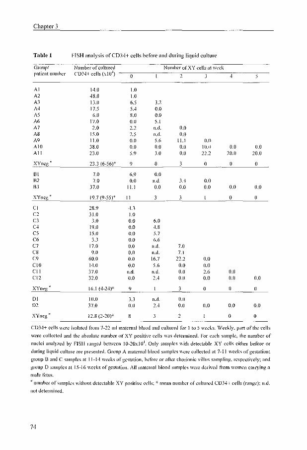
Chapter 3
Tuble 1 FISH analysis ofCD34+ cells before find during liquid culture
Group.! Number of cultured Number of Xl' cells at week patient number (:03<1+ cells (xlO~)
0 2 3 4 5
Al 14.0 1.0 A2 4S.0 1.0 Al 13.0 6.5 3.2 A4 17.5 5,4 0.0 AS 6.0 8.0 0.0 A6 17.0 0.0 5.1 A7 2.0 2.2 n.u. 0.0 AS 15.0 2.5 n,d. 0.0 A9 11.0 0.0 5.6 11.1 0.0 A10 38.0 0.0 0.0 0.0 IO.t) 0.0 0.0 All 23.0 5.9 1.0 0.0 22.2 20.0 20.0
XYneg 23.3 (6-56)* 9 0 1 0 0 0
III 7.0 6.9 0.0 H2 7.0 0.0 n.d. 3,4 0.0 Bl 37.0 11.1 0.0 0.0 0.0 0.0 0.0
XYncg · 19.7 (9-55)* 11 3 3 0 0
e1 28.9 4.3 C2 31.0 1.0 Cl 3.0 0.0 6.0 C4 19.0 0.0 4.8 C5 15.0 0.0 5.7 C6 3.3 0.0 6.6 C7 17.0 0.0 n,d, 7.0 CS 9.0 0.0 n.tI. 7.1 C9 60.0 0.0 \6.7 22.2 0.0 C10 14.0 0.0 5.6 0.0 0.0 ell 37.0 n.d, n.u. 0.0 2.6 0.0 el2 32.0 0.0 2.4 0.11 0.0 0.11 0.0
XYneg · 16.1 (4-24)' 9 3 0 0 0
D1 lU,O 1.3 n.d. 0.0 D2 37.0 0.0 2.4 0.0 0.0 0.0 0.0
XYneg · 12.8 (2-20)* 8 1 2 0 0
C034+ cells wcre isolated from 7-2'2 Illl rnatemal blood and cultured for I to 5 wccJ\s, Weekly, part urlhe cells
were collected and tlw absolute number of XY positive cells was determined. For each sample, the number of
nuclei analyzed by FISH ranged between IO-20xlO~. Only samples with detectable Xl' cells either before or
during liquid culture are presented. Group A maternal blood samples were collected <It 7-11 \\'1.'1.'1\" of gest<ltion;
group B amI C s<lmples <It 11-14 weeks of gestation, betore or aner chorionic villus sampling, respectively; and
group 0 samples at 15-16 weeks of gestation. All mntcmal blood sampll's were deri\'ed from wOlllen cllITying a
male fetlls . . lHulIber ofsnmpics without dctcctnble XY positi\'e c",'II~; * mean number of cultured CD34+ cells (range); n.d.
not determined.
74

Usefulness of ill vitm expansion of fetal CD34+ progenitor cells
XY positive cells, respectively, whercas no male cells were found in group Band D. After
4 and 5 weeks of liquid culture, only in group A, in one out of two patients 20.0 XY positive
cells were found. These results illllicate that fetal hemopoietic progenitor cells, if present at
all, may not preferentially expand over the maternal CD34+ component, which is in contrast
with the observations in the spike experiments.
As negative controls to determine the background level for FISH analysis, a total of
39 peripheral blood samples from 35 control patients eanying a female fetus and 4 female
non-pregnant volunteers (nulligravidae) were analyzed for the presence of male cells. From
all 39 blood samples CD34+ cells were isolated. CD34+ cell fractions from 20 pregnant and
4 non-pregnant women were cultured during 0 to 3 weeks, whereas from the other 15 samples
only FISH analysis was performed after MACS isolation. In none of the non-pregnant
samples male cells could be detected, whereas in 2 out of 35 pregnant wOlllen (5.7 %)
XV positive cells were found; 3.3 XY cells were detected all week 0 in one patient and
5.2 XY cells werc detected after 2 weeks of liquid culture in an other patient. In both cases a
prior male pregnancy was ascel1ained. These data suggest that the presence of male cells in
female pregnancies may be caused by aspecitic binding of the Y chromosomal probe or
might be the result of persisting fetal cells from earlier pregnancies.
DISCUSSION
The development of a non-invasive prenatal diagnostic test lIsing fetal cells present in the
maternal circulation is hampered by the low ti'cquency of these cells in maternal blood. The
idea to increase the number of fetal cells by amplification of progenitor cells has been
discllssed for a long time. In most recent studies, the expansion of fetal el,)1hroid cells is
described (Lo c/ (//., 1994; Valerio e/ (//., 1996, 1997; Chell e/ (//.,1998; Hall c/ (//.,1999),
whereas less is known about the amplification of fetal CD34+ progenitors (Little el al.,
1997). In the present study, we evaluated the lIsefulness of ill vitto expansion of CD34+ fetal
cells isolated IhHll maternal bloou samples for diagnostic purposes. Two major methods of
cell separation enable fetal cell isolation lI'om maternal blood: tluorescence activated cell
sorting (F ACS) (Bianchi el al., 1990) and magnetic activated cell sorting (ivIACS) (Ganshirt
Ahlert ct 01.,1992), Both isolation techniques have the disadvantage that the purified lI'action
still contains many maternal cells. In order to evaluate whether limited numbers of fetal
CD34+ cells present in an excess of maternal cells are able to overgrow the maternal
component, we lIscd a Illodel system in which CD34+ cells derived frolllillale umbilical cord
blood (UeB) were diluted up to 3 in 400,000 with CD34+ cells derived t1'om non-pregnant
female volullteers (nulligravidae) and in vitro expanded in liquid culture. We showed that
even vel)' low numbers of CD34+ cells derived from UCB (3 XY positive cells) diluted into
75
Chapter 3
an excess of PB cells were able to expand to high cellilumbers, corresponding to the il1l'il}'o
expansion ofCD34+ nucleated cells derived from undiluted UCB samples. As the magnitude
of in \'itro expansion of CD34+ cells derived from 20-week FB samples corrcspondcu to the
growth of hemopoietic progenitor cells derived from UCB, it is suggested that the grO\\1h
capacities of fetal hemopoietic progenitor cells of between 7 and 16 weeks of gestationnl age
may show similar expansion patterns, Consequently, the limHcd numbers of fetal
hemopoietic progenitor cells present in maternal blood samples may show the same growth
profiles. In cuntrast to the tlndings in the model system, we were not able to show abundant
growth of fetal male CD34+ cells isolated from maternal blood samples. In only a few
samples in group A and C, cell grO\\1h of male fetal cells could be detected. These results are
in concordance with the observations described by Little et al. (1997). Using an other
e)10kine combination, they also showed a limited expansiun of C034+ fetal cells derived
from 10-l3 week maternal blood samples after 5 days of cuhure, but in most cases (10 out
of IS (55 %)) no XY cells could be detected.
In many studies it has been investigated at what time in pregnancy the number of fetal cells
has reached its maximum. Relevant information regarding the frequency of fetal nucleated
red blood cells (NRBCs) in maternal blood is contradictOlY, and the frequency of fetal
NRBCs Was rep0l1ed to villy significantly among individuals and throughout the three
trimcstcrs of pregnancy (Hamada et (fl., 1993; Siunga-Tallberg eI al., 1995; Smid et al., 1997;
Kuo, 1998). Fetal cell frequency in maternal blood is influenced hy a number of biological
parameters that are mainly unknown. Gestational age seems to be one of the factors involved.
The optimal period during pregnancy for detecting circulating fetal cells remains unclear.
Therefore, we have analyzed maternal blood samplcs of different gestational ages, val)'ing
between 7 and 16 weeks of gestation (group A, B and D). In most cases no male fetal cells
could be detected, whereas in 28 out of65 maternal blood samplcs of women can)'ing a male
fetus XY positive cells could be observed. However, there was no statistically significant
increase in the number of patients with one or more XY positive cells between 7 and 11
weeks of gestation (group A) compared to patients of later gestational ages (group B and D).
This suggests that the number of fetal hcmopoietic progenitor cells present in blood 11'0111 tirst
trimester pregnant women might 110t be high enough or that these cells are even absent in
1110st pregnant women.
Another question concerns the stability of fetal cell properties. Are fetal progenitor cells
present in the maternal circulation cionnl and possess unchangeable characteristics or are they
transient and respond to a changing environment? Until now, it is nol known whether fetal
cells continue to express the same marker antigens in the new maternal environment after
crossing the placenta, and respond to similar c)1okines. The cytokine combination used in
this study has previously been shown to be capable of extensive amplification and
self-renewal of human primitive hemopoietic progenitor cells derived ti'om umbilical cord
76
Usefulness of in vitro expnnsion of fetnl CD34+ progenitor cells
blood (Piacibello e/ al., 1997; Rappold e/ al., 1999). However, other, perhaps yet unidentified
(combinations or concentrations ot) grO\\1h fnctors Illay induce n more vigorous proliferative
response, promoting the in \'itI'O expnnsioll of fetal cells isolated from maternal blood
samples.
Another 1~lctor that intluences the incidence of fetal cells in maternal blood is chorionic villus
sampling (CVS), which has previously becn rcported to cause a fcto-maternal transfusion of
fetal NRBCs (Jansen el al., 1997). The qucstion arises as to whether it may also havc an
etfect on the number of fetal hcmopoietic progcnitor cells in the maternal circulation.
Although there was a significant incrcnse of male cells aftcr one wcek of liquid culturc in
samples obtained after chorionic villus sampling (CVS)(group C) compared to before CVS
(group B), in most of the post-CVS samples the Ilumber of male fetal hemopoietic
progcnitors docs not markedly exceed the number of male cells in blood samples obtaincd
before any invasive procedure (group A, B and D).
Male cells were found in 2 out of 35 pregnant women eallying a female fetlls. In both cases a
prior male pregnancy was confirmed. Therefore, these cells may either represent
H1lsc-positivcs dlle to non-specitic binding of the Y chromosomal probe to non-target
sequcnces, or rcpresent residual fetal cells persisting from prior male pregnancies, since fctal
progcnitor cells have been rep0l1ed to circulate in nwternal blood as long as 27 years after
birth (Bianchi el al., 1996b). FlII1hermore, a fcto-maternal transfusion may OCClll' at the time
of bh1h. This may establish fetal microchimerism in the mother, which has been reported to
be implicated in the subsequent development of diseases, such as scleroderma that are
common in womcn (Art lett et (fl., 1998; Nelson, 1998; Evans el al., 1999). Therefore, it is
important to know whether fetal cells might persist from prior pregnancies, not only for the
development of n non-invasive prenatal diagnostic test, but also to evaluate the
immunological consequences of feto~maternal transfusion.
Until now, most attention has been focllsed all fetal NRBCs which have been isolated lIsing
antibodies against membrane-bound markers (CD7l, glycophorin A) or intracellular antigens
(hemoglobin F) (l.oken e/ al., 1987; Bianchi e/ al., 1990; Zheng e/ al., 1995). The number of
fetal NRBCs in maternal blood is vel)' low, and expansion of these cells might enhance the
detectability of this cell type. A previously reportcd cell culture protocol for erythroid cells
(Janscn el al., 1999; von Lindern el al., 1999) was used for CD71+ cells in a spike
cxpcriment similar to that used for hemopoietic progenitor cells described in this paper (data
not shown). CD71+ cells dcrived from male UCB wcre dilutcd lip to 1 in 400,000 in
CD71+ cells derived from female PB samplcs. This ccll mixture was culturcd according to
the previously described el)1hroid cell culture protocol (Jansen el al., 1999; von Lindern ef
01., 1999) and the number of XY positive cells was determined weekly. However, preferential
expansion of CD71 + UCB cells as shown for CD34+ UCB cells could not be observed,
suggesting that the expnllsion potential of fetal NRBCs in maternal blood samples is not
77

Chapter 3
comparable with the expansion potential of hemopoietic progenitor cells, and therctbrc~ fetal
NRBCs may not be able to overgrow the excess of maternal cells. These results were similar
to those reported by others (Chell ef a/., 1998; Hall ef a/., 1999), who rccently showed that
culturing of fetal cl)1hroblasts derived from contaminating maternal blood mainly produced
erythroid colonies derived from maternal erythroid progenitors,
In summary, expansion of retal cells isolated n'om maternal blood samples for analytical
purposes is highly desirable given the rarity of these cells in the maternal circulation.
Amplii1cation of the number or fetal cells llsing cell culture protocols might facilitate
non~in\'asivc prenatal detection of genetic anomalies. In the current study, the described
protocol for the expansion of hemopoietic progenitor cells resulted ill a dramatic expansion of
even vel)' few CD34+ UCB cells spiked into PB samples, which also corresponded to a
similar marked increase in the numbcr of CD34+ cells from 20-wcck fetal blood. However,
the expansion of fetal CD34+ cells dcriveu from 7-16 week matcrnal blood samples did not
result in signifil:ant growth of these rare cells and therdorc is not yet suitable for uiagnoslic
purposes.
ACKNOWLEDGEMENTS
\Ve would like to thank Professor H. Galjaard for his supp0l1 and Dr. P.A. in '( Veld for
critical reading of the manllscript.
REFERENCES
Artlett CM, Smith JB, Jimenez SA. (1998). Identification of fetal DNA and cells in skin lesions from women with systemic sclerosis. N Eng!.! J\led 338: 1186-1191.
Bianchi DW, Flint AF, Pizzimenti MF, Knoll JH, LaU SA. (1990). Isolation of fetal DNA from nucleated eJ)1hroc)1es in maternal blood. Fmc Nal! ;lead Sci USA 87: 3279-3283.
Bianchi DW, Klinger KW, Vadnnis TJ, Dcmaria ~vlA, Shuber AP. Skoletsky .I, Midura P, Diriso ivl, Pelletier C, Genova ivl, Erikson MS, Williams JM. (1996a). Development of a model system to compare cell separation methods for the isolation of fetal cells from maternal blood. Prenal Diagn 16: 289-298.
Bianchi DW, Zickwolf GK, Weil GJ, Sylvestcr S, Demaria MA. (1996b). Male tetal progenitor cells persist in maternal blood for as loqg as 27 years postpartum. Proc Not! Aead Sci USA 93: 705-708.
78
Usefulness of inl'it}'() expansion or fetal CD34+ progenitor cells
Chell H, Griffin OK, Jestice K, Hackett G, Cooper l, Ferguson-Smith MA. (1998). Evaluating the culture of fetal el)1.hroblasts from maternal blood for non-invasive prenatal diagnosis. Prenal Diagn 18: 883-892.
Elias S, Price J, Dockter M, Wachtel S, Tharapel A, Simpson JL, Klinger KW. (1992). First trimester prenatal diagnosis of trisomy 21 in fetal cells fr(Hll maternal blood. Lancel 340: 1033.
Evans PC, Lambert N, Maloney S, Furst DE, Moore .1M, Nelson .lL. (1999). Long-term fetal mierochimerism in peripheral blood mononuclcar cell subsets in healthy women and women with scJerodenna. Blood 93: 2033-2037.
Ganshil1-Ahlert 0, BOljesson-Stoll R, Burschyk M, Dahl' A, Garritsen HS, Helmer E, Miny P, Velasco M, \\'alde C, Palterson D. (1993). Detection of fctal trisomies 21 and 18 from maternal blood using triple gradient and magnetic cell sorting. Am .! Reprod Immullo/ 30: 194-201.
Ganshil1-Ahlert D, Bursehyk M, Garritsen I-IS, Helmer L, MillY P, Horst .I, Schneider HP, Holzgreve \V. (1992). Magnetic cell sorting and the transfcrrin receptor as potential means of prenatal diagnosis from maternal blood. Am.! Ohstet Gyneco/ 166: 1350-1355.
Goodfellow CF, Taylor PV. (1982). Extraction and identification of trophoblast cells circulating ill peripheral blood during pregnancy. Br.J Ohstcl GYl1acco/89: 65-68.
Hamada Hj Arinami T, Kuho T, Hamaguchi H, Iwasaki H. (1993). Fetal nucleated cells in maternal peripheral blood: frequcncy and relationship to gestational age. Hum Genel 91: 427-432.
Han lY, Je GH, Kim IH, Rodgers GP. (1999). Culture of fetal cl)rthroid cells from maternal blood llsing a two-phase liquid system. Am J Afed Gcnel 87: 84-85.
Herzcnbcrg LA, Bianchi DW, Schroder J, Cann HM, Iverson GM. (1979). Fetal cells in the blood of pregnant women: detection and enrichment by fluorescence-activated cell sorting. Proc Natl AmdSci USA 76: 1453-1455.
Holzgrcvc \V, Ghezzi F, Di Naro E, Ganshirt D, ["Iaymon E, Hahn S. (1998). Disturbed fetomaternal cell traffic in preeclampsia. Ohstel Gyneco/9 I: 669-672.
Jansen i"I\VlC, Brandenburg H, Wildschut HI.!, rvlartens ACM, Hagcnaars AM, \Vladimiroff J\V, in 't Veld, PA. (1997). The effect of chorionic villus sampling 011 the number of fetal cells isolated from maternal blood and on maternal serum alpha-fetoprotein levels. PreJ1at Dia);11 17: 953-959.
Jansen i'vI\VJC, VOIl Linuern rvl, BClIg H, Brandenburg 1-1, \Vildsehut 1-111, \VladimiroffJ\V, in 't Veld PA. (1999). The lise of in \'itro expanded elythroid cells in a mouel system for the isolation of fetal cells from maternal blood, Prellat Diagn 19: 323-329.
Kuo P. (1998). Frequencies of fetal nucleated red bloou cells in maternal blood during different stages of gestation. Felal Diagll Ther 13: 375-379.
79

Chapler 3
Lillie MT, Langlois S, Wilson RD, Lansdorp PM. (1997). Frequency of fetal cells in sorted subpopulations of nucleated erythroid and CD34(+) hematopoietic progcnitm cells fi'ol11 maternal peripheral blood. Illood 89: 2347-2358.
Lo YM, Leung TN, Teiu MS, SargenllL, Zhang J, Lau TK, Haines CJ, Rcdman CW. (1999). Quantitative abnormalities of fctal DNA in maternal serum in preeclampsia. Clin Chem 45: 184-188.
Lo YM, Morey AL, Wainscoat JS, Fleming KA. (1994). Culture of Iclal el}1hroid cells li'om malel11al peripheral blood. Laacet 344: 264-265.
Loken MR, Shah VO, Dattilio KL, Civin CI. (1987). Flow cytomctric analysis of human bone marrow: I. Normal CI)1.hroid development. Blood 69: 255~263.
Metcalf D, jVloorc MAS. (1971), Hacmopoictic Cells. In: Frontiers in Biology, volume 24, Amsterdam: Norlh~Holland Publishing Company,
Nelson JL. (l998). Pregnancy immunology and autoimmune disease. J Repl'od J\Ied 43: 335-340.
Piacibcllo \V, Sanavio F, Garetto L, Severino A, Bergandi D, Ferrario J, Fagioli F, Berger M, Aglietta M. (1997). Extcnsive amplification and self-renewal of human primitive hematopoietic slem cells from cord blood. Blood 89: 2644-2653.
Rappold I, Watt SM, Kusadasi N, Rose-John S, Hatzfeld J, Plocmacher RE. (1999). Gp130-signaling synergizes with FL and TPO for the long-term expansion of cord blood progenitors. Leukemia 13: 2036-2048.
Siunga-Tallberg AJ EI-Rifai \V, Kcinanen MJ Ylinen K, Kurki T, Klinger K, Ylikorkala 0, Knuutila S. (1995). Maternal origin of Ilucleated cl)'throcytes in peripheral venous blood of pregnant women. HUIll Genet 96: 53-57.
Smid M, Lagana F, Papasergio N, Ferrari A, Ferrari M, Cremoncsi L. (1997). Influence of gestational age 011 fetal deoxyribonllcleic acid retrieval in maternal peripheral blood. Am J Dbstet Gyaecol!77: 1517-1522.
Valerio D, Aiello R, Altieri V, Ivlalato AP, Fortunato A, Canazio A. (1996). Culture of fetal erythroid progenitor cells from maternal blood for non-invasive prenatal genetic diagnosis. Pre/tat Diogll!6: 1073-1082.
Valerio D, Allieri V, Antonncei FR, Aiello R. (1997). Characlerizalion of tctal haematopoietic progenitors circulating in maternal blood of seven ancuploid pregnancies. PreJ1(/I Dioga!7: ll59-1169.
von Lindern ivI, Zauner \V, Mellitzer G, Steinlein P, Fritsch G, Huber K, Lowenberg B, Beug H. (1999). The glucocorticoid receptor coopcrates with the cl)1hropoietin receptor and c-Kit to enhm,ce and sustain proliferation of CI)lthroid progenitors in "Uta. Blood 94: 550-559.
80

Usefulness of in vitro expansion of fctal CD34+ progenitor cells
\Vessman M, Ylinen K, Knuutila S. (1992). Fetal granulocytes in matcrnal vcnous blood detected by ill silU hybridization. Pre/wI Diagll12: 993-1000.
Zheng YL, Demaria M, Zhcn DK, Vadnais TJ, Bianchi D\V. (1995). Flow sorting of fetal cry throb lasts lIsing intracytoplasmic anti-fetal haemoglobin: prcliminat)' observations on maternal samples. Prenal Diagn15: 897-905.
81

I
I
I
I I
I
I
I
I
I
I
I
I
I
J -t
Th<e <eiBf<ecll; ojf chorioJl1J.iic viiJlll1l.ll.s 51 aJl'lr.!J. P 11:ii.Jl1J.g 0 Jl1J. 1!;lbJ. <e Jl1J.1UlJl1rll. b <eJr 0 jf jf <e1ta11 c <e 11 11 51 iiso11mll;<ed ftOll:lIll ll:lIllmll;<eJrJl1J.m11 b1100d aJ.Jl1J.d OJl1J. ll:lIllmll;<eJrJl1J.mll s<eJr1l.ll.JJ:JIll mll phm-jf<ell;opJroll;<eiiJl1J. ll<ev<e 1151
Published in Prenatal Diagnosis (1997), vol. 17, p953-959
The presence of fetal cells in maternal blood is supposed to be the result of a feta-maternal
transfusion at the placental interface. It has previollsly been rcpol1cd that the introduction of a
biopsy needle into placental tissue to aspirate chorionic villi via the transabdominal route
might induce a feta-maternal transfusion. Such a transfusion has been demonstrated by
elevated levels of maternal serum alpha-fetoprotein after chorionic villus sampling. In this
chapter, the effect of an invasive procedure like chorionic villus sampling was investigated by
analyzing the number of fetal nucleated red blood cells in the maternal circulation before and
aftcr transabdominal chorionic villus sampling.
f) John Wiley & Sons Limited: reproduced with pemlission.

The effect of chorionic vilius sampling
The effect of chorionic villus sampling on the number of fetal cells
isolated from maternal blood and on maternal serum alpha
fetoprotein levels
Mieke W.J.C. Jansen'·2, Helen Brandenburg', Hajo I. J. Wildschut', Anton C. M. Martens3
, Adriana M. Hagenaars', Juriy W. Wladimiroff', Peter A. in 't Veld2
! Department (~f Obstetrics (fuel Gynecology, 2 DepartmeHf (d' Clinical Genetics amI 31nstitllte (!f Hematology, E'/'(/,mllls Unil'el'sity and Unil'ersity Hospital, Rotterdam; .fDepa}'tment (?f Teratology, Endocrinology alld Perinatal .')'creening, National InstNllte of Public Health (lnd Em'il'omnental Protection (RlVAl), Bitthoven, nrc Netlrerland~'
ABSTRACT
Fetal cells are present in the circulation of pregnant women and can be isolated lIsing density
gradient centrifugation and magnetic cell sorting, In the present study~ maternal cell
preparations were depleted for CD4S- and CD14-positivc -cells and enriched for
CD71-positive celis, The number of fetal nucleated cells was determined using fluorescence
in situ hybridization for X and Y chromosomes, Analysis of maternal blood samples taken
before and atter transabdominal chorionic villus sampling (TA-CVS) showed an increase in
the number of fetal cells in 10 out of 19 male pregnancies after the invasive procedure. This
cellular transfllsioll was found to correlate with elevated maternal serum alpha-fetoprotcin
levels, TA-CVS-induced cellular transfusion Illay form a good in vivo system to optimize
fetHI cell isolation procedures and to study fetal cell dynamics and characteristics.
INTRODUCTION
Cells of fetal origin have been isolated from blood of pregnant womcn and can be used tor
non-invasive prenatal detection of genetic abnormalities. Cell types isolatcd from maternal
blood include nuclcated rcd blood cells (NRBCs) (Bianchi e/ al., 1990), lymphoc)1es
(I-Ierzcnbcrg et aI" 1979)~ granuloc)1es (Wessman et (fl.~ 1992)~ and trophoblast cells
(Goodfellow anti Taylor, 1982), Attention has been focused primarily on NRBCs as they
express both mel1lbrane~bollnd markers (CD71, glycophorin A) and intracellular antigens
(hemoglobin f) which aliow their isolation and identification (Loken et ai" 1987; Bianchi et
al., 1990; Zheng e/ al., 1995).
The occurrence of fetal NRBCs in maternal blood is a rare event, and extensive enrichment
and purification procedures are necessary to detect these cells. Isolation tcchniqucs currcntly
85

Chapter 4
available have succeeded in the isolation of only a vel)' small number or fetal cells in SOI11;': of
the pregnancies investigated (Reading et (1/., 1995; Lewis et aI" 1996), Improvements in
isolation technology that will result in a larger number of fetal cells me necessaI)' to allow the
lise of these cells for diagnostic purposes.
The presence of fetal cells in the maternal circulation is supposed to be the resull of a
feto-maternnltransfusion (FMT) at the placental interface (Price el a/., 1991; Bianchi ef a/.,
1992), This FMT has also been postulated to occur as a consequence of transabdominal
chorionic villus sampling (TA-CVS), which was shown to induce an increase in maternal
serum alpha-fetoprotein (MSAFP) levels (Los el al., 1989, 1993; Smidt-Jensen el al., 1993;
Brezinka el al., 1995), In the present study, we investigated whether the transfer of fctal
plasma components after TA-CVS is accompanied by a concurrent tllcrcase in the number of
fctal nucleated cells in the matclllal circulation.
MA TERIALS AND METHODS
Patient samples
Peripheral venous blood samples (13-18 1ll1) were obtained 1'1'0111 32 pregnant women
(11-14 weeks of gestation) ref cITed for prenatal diagnosis because of advanced maternal age
(11=29), familial Down syndrome (n=2), or a previous child with congenital abnormalities
(11=1). Mean maternal age was 37 years. Two blood samples were taken fi'om each patient,
the first one immediately before TA-CVS, the second 5-20 minutes after the invasive
procedure. Blooti samples were collected into vacutaincrs containing cthylenediaminetclra
acetic acid (EDTA). TA-CYS was performed according to Jahoda el al. (1990). In all patients
a single needle puncture was needed to obtain 5-30 mg of chorionic villi. Fetal kaJ)'ot)'ping
was performed on semi-direct villus preparations, All samples were obtained with the
patient's intollned consent.
Cell prepm'afiOll
Venous blood samples were diluted to 30 ml with phosphate-buffered saline (PBS) and
l11onol1uelcated cells were isolated by Ficoll-Paque-Plus (1.077 glllll; Phannaeia Biotech,
Uppsala, Sweden) density gradient centrifugation at 750 G for 15 minutes. Cells were washed
twice with PBS and centrifuged at 250 G for 10 minutes. For each sample, human serum was
collected and stored at - 80 {IC until further analysis.
A /plra-feloproteil1 meas IItemen/
["Iaternal serulll AFP UvISAFP) was measured with Amerlex ~\'I second-trimester radio
illll11ullo-assay (RIA) kits for AFP (Kodak) and was expressed in kJU/1.
86
The effect of chorionic villus sampling
l\Iaglletic (fctiwlfed cell sorting (J\IACSj
Fetal cells were isolated according to a procedure described by BUsch et al. (l994) with
modifications. Briefly, cells were labeled for 15 minutes on ice with CD45-PE
(50 rdl20x106eells; KC 56, T-200 (lgG1), Coulter, Kretcld, Germany) in 200 rd PBNHS
(PBS containing I % bovine serum albumin (BSA), 0.01 % sodium azide (NaN3), and 10 %
autologous serum), washed once in PBNHS, and labeled with 30 ~d of CDl4-eonjugated
mierobcads and 30 ~tI of rat anti-Illollse IgGI-conjugated microbeads (Miltenyi Biotec,
Bergisch Gladbach, Germany) in 200 pi of PBNHS at 4 "c for 15 minutes. Labeled cells
were washed once ~n PBNHS, resuspended in 500 ~tI wash butTer (PBS containing I % BSA,
0.01 % NaN,. aud 5 mM EDTA), and applied to a pre-separation filter (30 fun; CLB,
Amsterdam, The Netherlands) in order to obtain a single cell sLlspension. The labeled cell
~uspension was applied to an AS-depletion column (Miltenyi) using a 26G needle as flow
resistor. The negative fraction was applied again to the column in order to achieve an optimal
depiction, followed by elution with 3 1111 wash buffer. After an additional wash using a 24G
needle, the column was removed from the magnetic device and CD45- and CDl4-positive
cells were collected,
The negative fraction was labeled tor 15 minutes on ice with CD71-FITC (10 ~d/l06 cells;
100 pg/ml; LOl.l, IgG2a, Becton Dickinson, San Jose, CA, USA) in 200 ftl of PBNHS.
Cells were washed once in PBNHS and labeled tor 15 minutes at 4 °c with 30 pi of rat
anti-mouse IgG2a+b-conjugated microbeads (Miltenyi) in 200 ~tI of PBNHS, and washed
again in PBNHS. Labeled cells wcre rcsllspended in 500 ~ll wash butTer and applied to a
pre-separation tilter (30 pm; CLB). The cell suspension was added to a mini MACS column
(type MS; Miltcnyi) and the non-magnetic cell fraction was collected, After removing the
column 11'0111 the magnet, the CD71+ cell fraction was eluted.
The number of viable cells in each fraction was calculated before and after loading the
MACS column using a BUrkeI' counting chamber.
Fluorescence in sUu hybridization (FISH)
Cell fractions were treated with 75 mM KCI, incubated tor 18 minutes at 37°C, fixed in
methanol: acetic acid (3:1), and stored at _20°C until further analysis. Cells Were dropped
onto 3-aminopropyltriethoxysilane (2 % in acetone) -coated slides and air-dried, Slides were
treatcd with 70 % tonnamide (pH 7.0) in 2X SSC, washed in PBS, and dehydrated in 70 %,
90 % and 100 % ethanol tor 5 minutes each. Atter heating the slides tbr 10 minutes at 80°C,
cells were pretreated with pepsin (lOO ~lg/ml) in 0.01 N HCI at 37 llC tor 15 minutes,
followed by post-fixation in 3.7 % formaldehyde in PBS for l5 minutes. Subsequently, slioes
were denatured for 5 minutes in 70 % f0I111amide (pH 7.5) in 2X SSC at 75°C, followed by
87
Chapter 4
dehydration in 70 %, 90 % and 100 % ethanol for 1 minute each. Slides were prewanned at
45 t1C until probe was applied.
Two~color FISH was performed using a SpcctrumOrange labeled alpha-satellite probe
(DXZI) for centromere region Xpll.l-qll.l and a SpeetrumGreen labeled satellite III probe
for the Ygl2 region (Vysis, Downers Grove, fL, USA). The probes were denatured for
5 minutes at 75°C and hybridization was allowed to continue overnight at 42 (lC in a
humidified chamber.
Slides were posHvashcd three times at 46 nC in 50 % formam ide in 2X sse (pH 7.5) for
10 minutes each, followed by one wash in 2X sse (pH 7.0) for 10 minutes and one wash in
2X SSC/O.I % NP-40 (pH 7.0) for 5 minnles, both al 46 "C. Slides were mounled in
Vcctashield mounting medium (Veclor Laboratories, Inc" Burlingame. CA, USA) containing
DAPI (4', 6-diamidino-2-phenylindolc) and analyzed under a Leica Aristoplan tluorescence
microscope using a triple band-pass filtcr block. Images were captured with a Genetisean
ProbeMaster system (PSI, Chester, UK) using a Xybion CCD 24-bit color camera.
RESULTS
Blood samples were obtained from 32 pregnant women at 11-14 weeks of gestation (table 1).
Cytogenetic analysis of the villus sample showed a female karyotype in 13 cases and a male
km)'otypc in 19 cases. Two chromosomal abnormalities (47,XXY; 47,XY, +13) were
encollntered in two pregnancies refen'ed for advanced maternal age.
C07l-positive cells were isolaled ti'Om maternal blood laken before and after TA -CVS using
MACS separation. First, mOlloeytes (CD14) and lymphoc)1es (CD45) were depleted,
followed by enrichment of CD7l-positive cells. The number of XY cells in the
C07I-positive fractions was dctennined by FISH using X and Y chromosome-specific
probes. The number of cells analyzed in the CD7I-positive fracttons ranged from 0.3 to
80 x 10' (table I).
In 3 out of the 13 female cases (23 %), olle XY positive cell pel' sample could be detected,
either before or after TA-CVS. in two alit of these three cases, a prior male pregnancy was
either ascertained or could not be excluded due to a previolls spontaneous abortion. In the
third case, only prior female prC!~,'llancies were reported. The number of XY -positive cells
before TA-CVS in the male cases ranged from 0 to 4 XY cells, with the highest amount of
X- and Y-positive cells in the 47,XXY case (table 1 and figure lA). The number of
XV-positive cells after TA-CVS in the male cases ranged fi'om 0 to 187, with the highest
alllount of cells in the ease with trisomy 13 (table I and figure 18). A total of ten out of
19 male eases (53 %) displayed an increase in the number of XY positive cells after
TA-CVS compared with the number ofXY cells before TA-CVS.
88

The effect of chorionic villus sampling
Table 1 Number of XY cells and a[pha~fetoprotein values before and after TA~CVS
Case Fetal Ge~latiollal Number of cells Number of XY cells AFP villucs (kIUlI) no. kal)'otype age analyzed* (x 1 O~)
(weeks) Befor~ cvs Allcr CVS Before CVS After CVS Before CVS After CVS I ;6. xx 12 17 22 0 0 7.9 14.6 2 ;6. xx 12 15 15 0 I 3.0 8.8 3 ;6. XX 11 13 15 II 0 1.0 1.0 ; 46. XX 12 13 I; 0 0 1.6 3.3 5 46. XX 12 11 12 0 0 1.0 32.2 6 ;6. XX 12 10 17 0 0 14.3 1J.8 7 46. XX 12 21 24 0 II 2.5 3.4 8 46. XX 12 13 12 I 0 8.0 10.3 9 46. XX 12 28 41 0 II 8.7 8.4 10 46. XX 12 29 25 0 0 7.4 12.0 11 46. XX 12 7 II 0 0 2.0 9.9 12 46. XX 12 8 20 0 1 1.4 3.3 13 46. XX 12 28 17 0 0 14.7 19.3
14 46. XY 12 II 12 II 0 3.0 2.3 15 46. XY 12 9 II 0 0 4.4 7.8 16 46. XY 12 27 8 0 0 1.0 9.0 17 46. XY 12 18 18 I I 7.2 17.9 18 46. XY 12 S 14 I 175 1.4 332.6 19 46. XY 12 29 32 0 185 3.8 720.0 20 46. XY 12 25 24 2 15 29.9 63.9 21 46. XY 12 11 9 I 0 2.9 16.8 22 46. XY 13 80 40 0 I 5.8 11.5 23 46. XY 12 6 0.8 0 0 10.8 11.1 24 46. XY 12 1.5 0.3 0 7 10.8 18.1 25 46. XY 12 14 25 0 0 4.6 6.3 26 46.XY 13 8 8 0 2 6.5 164.3 27 46.XY 12 21 11 0 I 1.4 2.5 28 46.XY 13 20 10 0 4 1J.8 3;.0 29 46. XY 12 14 10 0 II 4.5 28.8 )0 46. XY 12 17 22 0 0 6.4 10.9
3 I 47. XXY 12 19 25 4' 23' 4.4 26.4 32 47.XY.+13 IJ 28 21 2 187 4.2 72.2
* All cells obtained aner C07l enrichment were analyzed by FISH lIsing X and Y chromosome specific probes; t number of XXY cells
MSAFP levels before and after TA~CVS were determined and compared with the number of
XY cells before and after TA-CVS (lab Ie I). The mean level ofMSAFP before TA-CVS was
6.3 klU/l (combination of all male and female pregnancies) and 53.0 klU/1 after TA-CVS.
indicating a significant 8.4~fold increase in i\'ISAFP Icvels due to the TA~CVS procedure
(two~sal11ple Wilcoxon test: p < 0,01). For the male cases, ~vISAFP levels were found to be
positively correlated with the number of XY cells after TA~CVS (Speannan rank correlation
coetlicient r = 0.78; n = 19; P < 0.001). No significant difference could be observed in
MSAFP Icvels after TA-CVS between male and female eascs (Mann-Whitney U-tcst,
p=OA7). There was no correlation between the amollnt of villi taken (range 5-30 mg) and
increases in fetal cell counts or MSAf-P levels.
89
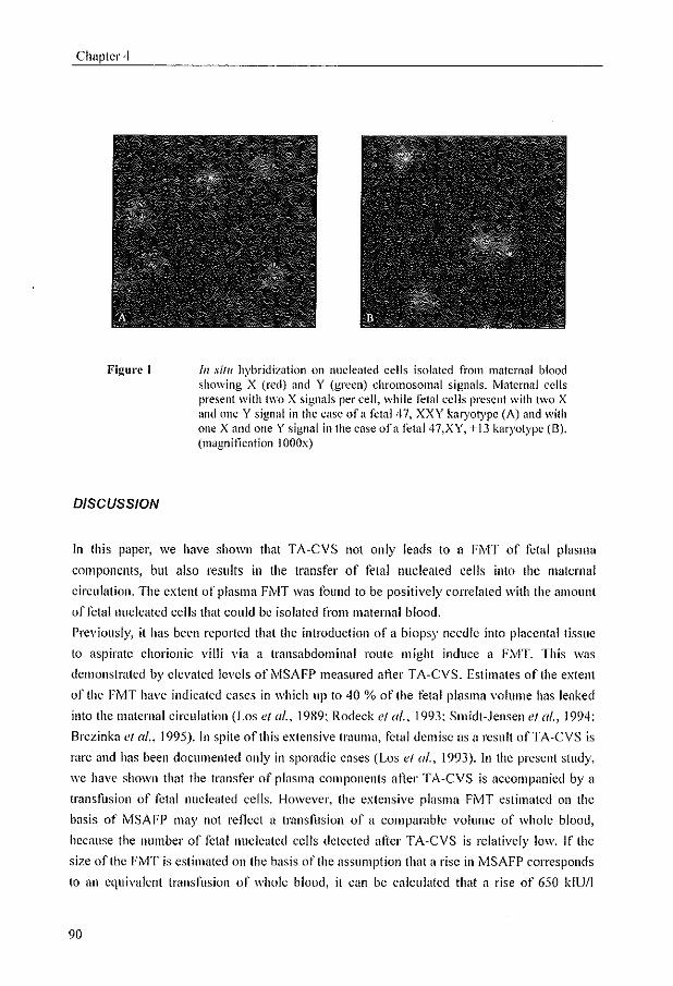
Chapter 4
Figun;'1
DISCUSSION
[11 silll hybridization on nucleated cells isolated from maternal blood showing X (red) <lnd Y (green) chromosomal signals. ivlatcrnal cells present with Iwo X signals per cell, while fetal cells present with two X and nne Y signal in the case ofa fetal 47, XXY karyotype (A) and with one X and one Y signal in the case ofa fetal 47,XY, +13 karyotype (8). (magnification IOOOx)
In this paper, we have shown that TA-CVS not only leads to a FMT of fetal plasma
components, but also results in the transfer of fetal nucleated cells into the maternal
circulation. The extent of plasma FiVlT was fOllnd to be positively correlated with the amount
offetal nucleated cells that could be isolated from maternal blood.
Previously, it has been reported that the introduction of a biopsy needle into placental tissue
to aspirate chorionic villi via a transabdominal route might induce a FrviT. This was
demonstrated by elevated levels of i\'ISAFP measured after TA-CVS. Estimates of the extent
of the FMT have indicated cases in which lip to 40 % of the fetal plasma volume has leaked
into the maternal circulation (Los ef a/., 1989; Rodeck et a/., 1993; Smidt-Jensen ef a/., 1994;
Brezinka et al., 1995). In spite of this extensive trauma, fetal demise as a result ofTA-CVS is
rare and has been documented only in sporadic cases (Los et a/., 1993). In the present study,
we have shown that the transfer of plasma components after TA-CVS is accompanied by a
transfllsion of fetal nucleated cells. However, the extensive plasma FMT estimated on the
basis of MSAFP may nut rellect a transfusion of a comparable volume of whole blood,
because the number of fetal nucleated cells detected after TA-CVS is relatively low. If the
size of the FMT is estimated on the basis of the assumption that a rise in i\'lSAFP corresponds
to an equivalent transfusion of whole blood, it can be calculated that a rise of 650 kIU/I
90

The effect of chorionic villus sampling
corrcsponds to a FMT of I 1111 (Los et al., 1989). A transfusion of this size will lead to a
leakage of at least 4 x 10⁶ fetal nucleated cells into the maternal circulation (Millar et al.,
1985). This estimated level does not correspond to the observed number of fetal nucleated
cells in the present study, which is at least an order of magnitude lower. This lower level of
fetal nucleated cells might be explained by removal of these fetal cells by the maternal
immune system, by retention of fetal cells in maternal tissucs, or by an inefficient isolation
procedure. Alternatively, it is possible that an extensive plasma FMT as indicated by a large
rise in MSAf-P is not accompanied by a prop0l1ionai cellular transfusion. In the prcscnt
study, the extent of plasma FMT after TA-CVS significantly correlates with the extent of
cellular FMT, although in individual cases an increase in plasma FMT was found that was not
accompanied by a demonstrable cellular FMT. Retention of the fetal blood cellular
component compared with the plasma FMT after TA-CVS might explain why relatively few
cases with extreme plasma Fi''vIT are accompanied by fetal death.
The number of fetal cells that could be isolated fi'om maternal blood before TA-CVS was
found to be very low in the male cases (0-4 cells per sample). This is in agreement with other
recent repolis in which similar levels of fetal cells were found (Reading el al., 1995; Lewis ct
al., 1996). In the two cases with a chromosome abnormality (47,XXY; 47,XY, +l3), a
relatively high number of fetal nucleated cells were isolated. It has previously been suggested
that a higher level of fetal cells can be found in maternal blood in cases with chromosome
abnormalities, possibly as a result of an altered placental structure (Simpson and Elias, 1994).
More studies are needed to investigate whether higher levels of cellular FMT occur in
abnormal pregnancies, as this will facilitate prenatal diagnostic procedures.
Some XY -positive cells were found in female pregnancies; these cells Illay either represent
false positives due to non-specific background staining of the V-chromosome probe, or
represent cells persisting from prior male pregnancies, since t(~tal lymphoc)1es were fOllnd to
circulate ill maternal blood as long as 27 years after birth (Bianchi el al., 1996).
Elevated ivlSAFP levels induced by TA-CVS were found in both male and female
pregnancies; although the three cases with the largest FMT were all male pregnancies, no
signiticant difference between the two groups could be observed.
The increase in fetal cell Humber after TA-CVS, resulting in the detection of XY cells in ten
out of 19 male pregnancies, represents an interesting experimental in vh'o system. 111 vilro
mooel systems bascd on artificial mixtures of neonatal conJ blood NRBCs and adult female
peripheral blood cells have been described (Andrews et al., 1995), but they have the
disadvantage that cell characteristics of el;1hroblasts at the 20 to 40-week stage may differ
from those at 12 weeks of gestation, The present in vivo modcl of TA-CVS-inouced cellular
Fi'vIT has the imp0l1ant advantage that fetal cells are derivcd from the correct gestational
stage and may be used to improve isolation procedures and to study fetal ccll dynamics and
characteristics.
91
Chapter 4
ACKNOWLEDGMENTS
We thank Professor H. Galjaard and the Foundation for Clinical Genetics in Rotterdam for
their financial support, and Armando Braat for technical assistance in generating figure 1.
REFERENCES
Andrews, K., Wienberg, J., f-ergllsonsmith, M.A., and Rubinsztein, D.C. (1995). Enrichment of fetal nucleated cells from maternal blood: mouel lest system lIsing cord blood, Prenat Diagl/, 15,913-919.
Bianchi, D.W., Plint, A.F., Pizzimenti, ~vI,F., Knoll, lH., and LaU, S.A. (1990). Isolation of fetal DNA from nucleated cl;1hroc)1CS in maternal blood, Proc NaIl Acad Sci U,~'A, 87, 3279-3283.
Bianchi, D.\V., l'vlahr, A" ZickwoU: G.K., Houseal, T.\V., Flint, A.F., and Klinger, K.\V. (1992), Detection of fetal cells with 47,XY,+21 kmyotype in maternal peripheral blood, Hum Gel/ef, 90,368-370.
Bianchi, D.W., Zickwolt; G.K., Weil, GJ., Sylvester, S., and DeMaria, M.A. (I 996). Male fetal progenitor cells persist in maternal blood for as long as 27 years postpmtum, Proc NaIl Aead Sci USA, 93, 705-708.
Brczinkn. c., Hagenaars, A.rvl., \Vladil11iroft~ l\V., and Los, FJ. (1995). Fetal ductus venosus flow velocity waveforms and maternal serum AFP before and aner Iirst-trinlCslcr transabdominal chorionic villus sampling, Prenal Diagll, 15,699-703.
BUsch, l, Huber, P., Pfluger, E., Miltenyi, S., Hoilz, .I., and Radbruch, A. (1994), Enrichment of fetal cells from maternal blood by high gradient magnetic cell sorting (double ivIACS) for PeR-based genetic analysis, Prellllf Diagll, 14, 1129-1140,
Goodfcllow, C.F., and Taylor, P.Y. (1982). Extraction and identitication of trophoblast cells circulating in peripheral blood during pregnancy, I1r.J Ohslet GYl1aeco/, 89,65-68.
Herzenberg, L.A., Bianchi, D.W., Schroder, J., Cnnll, H.M., and Iverson, (J,M. (1979). Fctal cells in the blood of pregnant women: detection and enrichment by tluorescence-activated cell sorling, Proc Naf! Aead Sci USA, 76, 1453-1455.
J[lhOU11, M.G., PUpers, L., Reuss, A., Brandenburg, 1-1., Cohen-Overbeek, T.E., Los, F.J., Sachs, E,S., and Wladimiroft~ J.\V. (1990). Transabdominal villus sampling in early secont! trimester: a safe sampling method for womcn of advanced age, Prenal Diagn, 10,307-311.
Lewis, D.L, Schobel', \V., Murrell, S" Nguycn, D., Scott, l, Boinoft~ J., Simpson, J.t., Bischoft~ F.Z., and Elias, S. (1996), Rarc cvent selection of fetal nucleated el')1hrocytes in matcrnal blood by now cytometl)', (}'lomen:)', 23, 218-227.
92

The effect of chorionic villus sampling
Loken, M.R., Civin, C.I., Bigbee, W.L., Langlois, R.G., and Jensen, R.H. (1987). Coordinate glycosylation and cell surface expression of glycophorin A during normal human el}'thropoiesis, Blood, 70, 1959-1961.
Los, F.J., .Iahoda, M.G., \vladillliroft~ J.\V., Brezinka, C, and Hagenaars, A.M. (1993). Fetal cxsanguination by chorionic villus sampling, Lallcel, 342, 1559.
Los, FJ., Pijpers, L., .lahoda, j'vI.G., Sachs, E.S., Reuss, A .• Hagenaars, A.M., and Wladimiroft~ .I.\V. (1989). Transabdominal chorionic villus sampling in the second trimester of pregnancy: feto-maternal transfusions in relation to pregnancy outcome, PrCHal DiagJl, 9, 521-526.
Millar, D.S., Davis, L.R., Rodeck, C.H., Nicolaides, K.H., and Mibashan, R.S. (1985). Normal blood cell valucs in the early mid-trimcstcr fetus, Prcnal Diagl1, 5,367-373.
Price, J.O., Elias, S., \Vachtcl, S.S., Klingcr, K., Dockter, M., Thampel, A., Shulman, L.P., Phillips, O.P., Meyers, C.M., Shook, D., and Simpson, J.L. (1991). Prcnatal diagnosis with fctal cells isolated from maternal blood by Illuitiparameter flow cytometry, Am J Obslel Gyl/eco/. 165, 1731-1737.
Reading, J.P., Huffman, .l.L., \VtI, .l.C., Palmer, F.T., Harton, G.L., Sisson, M.E., Keyvanfar, K., Gresinger, T.H., Cochranc, W,J., Fallon, L.A., Mcnnpncedrcw, G.F., Cummings, E.A., Jones, S.L., Black, S.H., Schulman, J.D., and Levinson, G. (1995). Nuclcated cl)'throcytes in maternal blood: quantity and quality of fetal cells in enriched populations, Hum Reprod, 10, 2510-2515.
Rodeek, CH., Sheldrake, A., Beattie, B., and Whittle, M..I. (1993). Maternal serlllll alphafetoprotein after placental damage in chorionic villus sampling, Lancel, 341, 500.
Simpson, .I.L., and Elias, S. (1994). Isolating fetal cells in I11nternai circulation tor prenatal diagnosis, Prenal Diagll, 14, 1229-1242.
Smidt-.Iellsen, S., Lllndsteen, C., Lind, A.M., Dinesen, K., and Philip, .I. (1993). Transabdominal chorionic vilius sampling in the second and third trimesters of pregnancy: chromosome quality, reporting timc, and fcta-maternal bleeding, Prenal Diagl1, 13,957-969.
Smidt-Jensen, S., Philip, .I., ZachalY, J.M., Fowler, S.E., and Norgaard-Pedersen, B. (1994). Implications of maternal serum alpha-fetoprotein elevation caused by transabdominal and transcervical CVS, PrCHal Diagu, 14,35-45.
"'essman, i'vl., Ylinen, K., and Knuutila, S. (1992). Fetal granulocytes in maternal venous blood dctccted by ill situ hybridization, Prenal Diagl1, 12,993-1000.
Zheng, Y.L., Demaria, M., Zhen, O.K., Vadnais, TJ., and Bianchi, D.W. (1995). Flow sorting of fetal cl)1hroblasts using intraC)1opiasmic anti-fetal haemoglobin: preliminary observations on matemal samples, Prcnal Diagn, 15,897-905.
93

J §iigltlllifica1ll1ltfiy lhliiglhlelr ltllumlbelr of feml ltll1Uldea1l:ed lred lblood ceH§ ill 1l:lhle ma1l:ernal dJrC1Ulhl11l:iioltll of womeltll wiilli plreedamp§iia
Submitted
Preeclampsia is a common, pregnancy-specific disease defined by clinical findings of
elevated blood pressure combined with proteinuria and edema. Although the etiology of
preeclampsia is not knowll, there are indications that abnormal placentation and endothelial
dysfunction are involved in the pathogenesis of preeclampsia, In this chapter, we investigated
whether this nbnormai placentation results in a transfusion of increased numbers of fetal
nucleated red blood cells in the maternal circulation.

feto-Illuternal cell trafficking in preeclampsia
Significantly higher number of fetal nucleated red blood cells in the
maternal circulation in women with preeclampsia
Mieke W.J.C. Jansen'·2, Karin Korver-Hakkennes', Dik van Leenen', Willy Visser', Peter A. in 't Veld2, Christianne J.M. de Groot', Juriy W. Wladimiroff' I Depal'tmeHl of Obstetrics ami Gynecolo,r;y, EmslIlus Unil'ersity (lnd lJnil'ersity Ho~pit(ll DUkzigt, Rotterdam, The Netherlmub": 2 Department of Clinical Genetics. Erasmlls University, Rotterdam, The Nether/wltls
ABSTRACT
Although the pathophysiology of preeclampsia is unknowll) several studies have described
that abnormal placentation early in pregnancy might play a key role, Therefore~ we
investigated whether this abnormal placentation results in a feto-maternal transfusion of fetal
nucleated red blood cells in wOlllen with preeclampsia. Male fetal nucleated red blood cells
were isolated using magnetic activated cell sorting from 20 women with preeclampsia and
20 controls, matched for gest. tl iOllal age and parity. The number of XY cells was determined
lIsing two-color fluorescence in situ hybridization for X and Y chromosomes. Significantly
more XY cells could be detected in women with preeclampsia (0.61 ± 1.2 XY cells/ml blood)
compared to women with ullcomplicated pregnancies (0.02 ± 0.04 XY eclls/ml blood)
(Manll-\Vhitney U test, p<O,OOI), These results suggest that fetal cell trafficking is enhanced
in women with preeclampsia and may contribute to the understanding of the disease,
INTRODUCTION
Preeclampsia is a pregnancy~specific syndrome particularly manifested in late pregnancy.
Preeclampsia is clinically characterized by hypertension and proteinuria, Although the
etiology of preeclampsia is unknown, in the last decade it has been proven that preeclampsia
is related to abnormal placentation early in pregnancy. Abnormal placentation might be due
to incomplete trophoblast invasion, The cause of failure of trophoblastic invasion is
unknown, but therc appears to be a combination of different tactors, including genetic and
environmental h1etors (Roberts and Rcdman, 1993).
Abnormal placentation has been suggested to play an important role in feto-maternal cell
traffic) which was first recognized in 1893, when Schmor! identified trophoblast cells in the
lung capillaries of women dying of eclampsia. For the last 20 years, investigators have lIsed
the presence of fetal cells in maternal blood in all attempt to develop a non-invasive prcnatal
diagnostic test (Simpson and Elias, 1994; Bianchi, 1998), Trafticking of fetal cells into the
97
Chaptcr 5
maternal circulation have raised several questions, pal1icularly regarding the types of cells
that arc able to pass the placental barrier, the number of fetal cells, and the consequences of
semi-allogeneic cells in the new hosl.
In several reccnt studies, the exchange of fetal and maternal blood cells in women with
preeclampsia has been investigated (Chua et al., 1991; Holzgrcvc et al., 1998; Knight et (fl.,
1998: La ef al., 1999), Recently, Holzgrcve et at, (1998) showed that <1 substantial propOI"tion
of erythroblast cells present in the blood of women with preeclampsia (n=8) were of fetal
origin, and that a significant increase in cross-placental tnllTic of fetal cells occurred in
wOlllen with preeclampsia compared to controls. In addition, Lo et al. (1999) recently
demonstrated a similar feto-maternal transfusion on the basis of abundance of fetal DNA in
maternal serum ofpaticnts suffering from preeclampsia (11=20).
To investigate whether abnormal placentation associated with preeclampsia results in a fcto
maternal transfusion, we isolated fetal nucleated red blood cells (NRBCs) from women with
preeclampsia compared to controls, both carrying a malc fetus, using a very efficient
magnetic activated cell sorting (MACS) protocol and two-color fluorescence in silu
hybridization (FISH) for X rmd Y chromosomes.
MATERIALS AND METHODS
Patfu/Us
The study protocol was approved by the local Ethics Review Board. After patients had given
infonned consent, matcl'Ilal blood samples (8-24 ml) were collected in vacutainers containing
ethylenedinlllinetetra-acetic acid (EDTA). Blood samples were obtained from 20 singleton
pregnancies associated with preeclampsia and 20 women with uncomplicated pregnancies,
both carrying [\ male fetus. Samples were matched for gestational age (± I week) and parity.
The gender of the Ictus was confirmed after delivel),. Preeclnmpsia was defined as an
absolute diastolic bloodpressllI'e 290 mmHg, combined with proteinuria, which was defined
as ;0:0.3 gil in 24 hours urine (Davey and MacGillivray, 1988). HELLP (hemolysis, elevated
liver enzymes, and low platelets) was defined as thromboc)1es <lOOx109/1, ASAT and ALAT
both >30 U/I. and haptoglobin < 0.28 gil.
Eighteen healthy pregnant controls carrying a female fetus werc recruited as negative
controls. These controls were included in order to determine the background level for fiSH
analysis.
,\laglletic activated cell sorting (AfAeS)
Maternal blood samples were diluted 1:1 with phosphate-buffered saline (PBS) and
Illononllcleated cells were isolated by Ficoll-Paque-Plus density gradient centrifugation at
98

Feto-maternal cell trafficking in preeclampsia
750 G for 10 minutes. Cells were washed twice in MACS buffer (PBS) containing 1 %
bovine serum albumin (BSA), 0.01 % sotliurn azide (NaN3). and 5 mM EDTA) and
centrifuged at 250 G for 10 minutes. The numbcr of viable cells was calculated using a
BUrker counting chamber.
Monollllclear cells derived fi'om maternal blood were labeled on ice with C071
(10 ftillO' cells) in 200 ftl MACS buffer for 15 minutes. Cells were washed in MACS burrel'
and labeled with IgG2a+b-conjugated microbeads (20 ~t1/107 cells: iVliltcnyi Biotec, Bergisch
Gladbach, Germany) for 15 minutes at 4 Pc. Cells were washed in MACS butfer,
resuspended in 1 III I ivlACS buffer and applied via a pre-separation filter (30 pill; CLB;
Amsterdam, The Netherlands) onto a miniMACS column (type ~vIS; Miltenyi). Atter
removing the colullln from the magnet, CD71 + cells were eluted with 15 III I MACS buffer.
The number of viable cells in each fraction was calculated using a Blirker counting cha;nber.
Fluorescence in situ hybridization (FISH)
Cell fractions were treated with 75 111M KCI, incubated for 18 minutes at 37°C, fixed in
methanol: acetic acid (3:1) and stored at _20°C until further analysis. Cells were dropped
onto Veetabond™ (Vector Laboratories, Inc, Burlingame, CA, USA) coated slides and
air-dried. Slides were pre-treated with pepsin (100 fIg/ill!) in 0.01 N HCI at 37 "c for
IS minutes, followcd by postfixation in 3.7 % formaldehydc in PBS for 15 minutes.
Subsequently, slides were denatured for 5 minutes in 70 % formam ide (pH 7,5) in 2X SSC at
75°C, followed by dehydration in 70 %, 90 % and 100 % ethanol for 1 minute each.
Two-coloI' FISH was performed llsing a SpcctrumOrange labekd alpha-satellite probe
(OXZ 1) for centromere region Xp 11, l-q 11.1 and a SpeetrumGreen labcled satell ite III probe
for the Yql2 region (Vysis, Downers Grovc, IL, USA). Samples were hybridized overnight at
37°C in a humidified chamber.
Slides were post-washed at 70 "c in O.4X SSe/0.3 % NP-40 (pH 7.2), followed by
5-60 seconds in 2X sse/o.1 % NP-40 (pH 7.2) at room temperature. Slides were mountcd in
Vectashicld mounting medium (Vector Laboratories, Inc.) containing OAPI (4', 6-Jiamidino-
2-phcnylindolc) and analyzed under a Leica Aristoplan tluoreseence microscope using a
triple band-pass filter block. Images were captured using: a Xybion CCO 24-bit color camera
with a Genetiscan ProbeMaster system and MacProbc 2.5 image analysis sot1ware (PSI,
Chester, UK).
All slidcs were nnalyzed without prior knowledge of the gender orthe teills .
.)'tatistic ({na(l'sis
Clinical data ofwol11cl1 with preeclampsia and their controls are presented in table 1 as mean
vailles ± SD. The unpaired Student t-test was used for a normal distribution, tested by
99

Chapter 5
Kolmogorov-Smirnov and Shapiro- Wilk tests. For nOll-normal distributions the
I'Vlann-\Vhitncy U lest was applied. Intra-uterine growth restriction is defined as a bit1h
weight below the fifth-percentile according to Kloosterman (l970). The exact Pearson chi
square test was lIsed to lest the fifth-percentile, Two-tailed tests with p<O.05 were accepted as
significant differences.
RESULTS
Patients characteristics
As expected, women with preeclampsia displayed a significantly higher mean systolic and
diastolic blood pressure and a lower gestational age at delivCI)l and fetal birth weight thun
women with ullcomplicated pregnancies (table 1). Preeclampsia was also associated with a
higher incidence of intra-uterine growth restriction. Unexpectedly, women with preeclampsia
had lower levels of hematocrit.
Table I Clinical characteristics
l'vtaternal age (years)
Nullipumc (N) Systolic blood pressure (mmHg)
Diastolic blood pressure (mmllg)
Proteinuria (gil)
Gestational age at blood sampling (days)
Gestational age at delivery (days)
Bil'thwcight (gram)
Intra-uterine gro\\1h restriction (N)
Hematocrit at blood sampling (vo1/vol %)
n.d. not determined
Pregnancies witll a male/etus
Women with preeclampsia
N=20
29.6 ± 5,4
14(70%)
169.2 ± 28.8
107.0± 13.3
5.07 ± 4.5
207.9± 21.3
213.3±21.8
1244 ± 530.2
6
32.7 ± 1.3
Control !,'TOUp
N=20
29.7±4.7
15 (75 %)
117± 8.0
75 ± 9.0
n.d.
207.7±20.5
276.5 ± 14.6
3206 ± 670.9
0
J5.I±J.6
p- vall!<:
0.95
n.d.
<0.001
<0.001
n.d.
0.88
<0.001
<0.001
0.02
0.014
XV positive cells were found in 70 % of women with preeclampsia (11=20, 0.61±1.2 XY
cells/ml blood), whereas this was in 10% of controls (IF20, 0.02 ± 0.04 XY eells/ml blood)
(figure 1). This difference in the number of XY cells is statistically significant (i'vIallll
Whitney lJ test, p<O.OOl). The total number of cells analyzed ill the CD71+ fraclion ranged
100
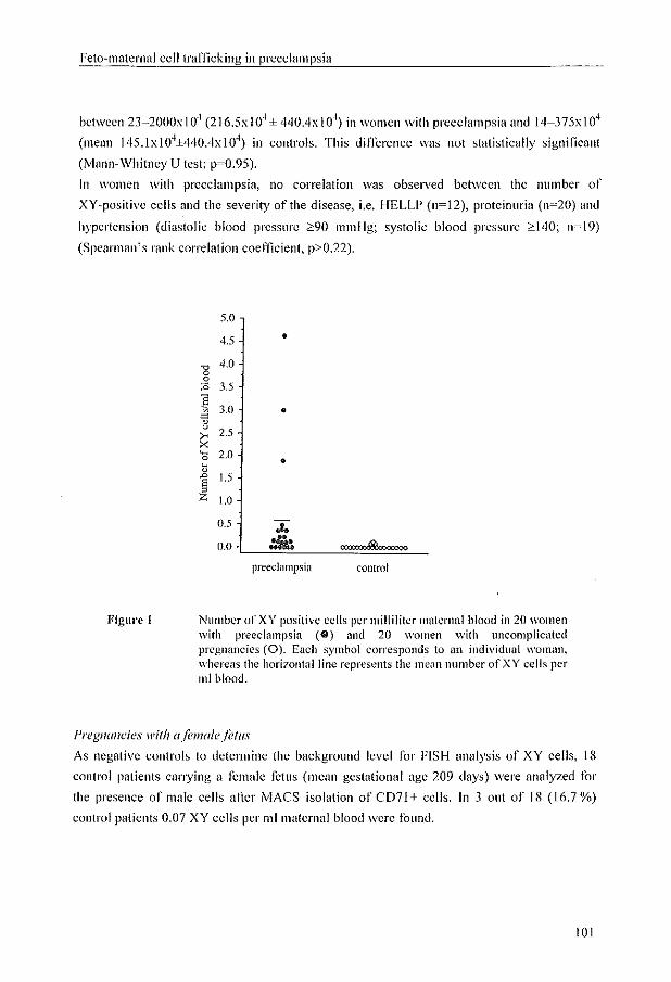
Feto-maternal cell trafficking in preeclampsia
bctween 23-2000x I 0' (216.5x 10' ± 440.4x 10') in wOlllen wilh preeclampsia and 14-375x10'
(mean 145.1x10⁴±440Ax104) in controls. This difference was not statistically significant
(Mann-Whitney U tesl; p~0.95).
In women with preeclampsia, no correlation was observed between the number of
XY~positivc cells and the severity of the disease, i.e. HELLP (n=12), proteinuria (n=20) and
hypertension (diastolic blood pressure :2:90 Il1mHg; systolic blood pressure :2:.140; n=19)
(Spearman's rank correlation coefficient, p>0.22).
Figul'e 1
5.0
4.5 •
'" 4.0
0 0 :0 3.5
~ "
3.0 • " u
2.5 ~ X ~ 2.0 0 • ~ L> 1.5 8 " Z i.O
U.5 ... 0.0 '* l'i> :::::
preeclampsia control
Number of XY positive cells per milliliter maternal blood in 20 women with preeclampsia Ce) and 20 women ""ith uncomplicated pregnancies (0). Each symbol corresponds to an individual woman, whereas the horizontal line represents the mean number of XY cells per 1111 blood.
Pregnancies with a/ema/e/ctus
As negative controls to determine the background level for FISH analysis of XY cells, 18
control patients can),illg a female fetus (mean gestational agc 209 days) were analyzed tor
the presence of male cells alier MACS isolation of CD7l + cells. In 3 out of 18 (16.7 %)
control patients 0.07 XY cells pel' ml maternal blood wcrc found.
101

Chapter 5
COMMENT
\Ve demonstrated an increased transfusion of fetal IlUclC<'llcd red blood cells in women with
preeclampsia compared with women with ullcomplicated pregnancies. The number of male
fetal cells found in the maternal circulation of women with preeclampsia was lip to 30-fold
higher than in controls, indicating a six times higher level of fetal cells compared to
previously repOited results (Holzgreve ef al., 1998; Lo ef al., 1999). Holzgrevc ef al. (1998)
isolated fetal nucleated red blood cells using the same MACS protocol as described in this
study, although they used directly labeled CD?I beads. This might explain our promising
higher rccovcl), of male cells since we have previollsly demonstrated in a model system lIsing
in vitro expanded erythroid cells (Jansen et al., 1999) that the isolation ofuucloated red blood
cells was more efi1cient using indirect beads labeling (unpublished results). In the study of Lo
el al. (1999), fetal DNA concentrations appear to be fivefold higher in maternal serum
samples derived from women with preeclampsia compared to controls, indicating that the
aillount of DNA in matcrnal blood samples Illay not correspond with the number of cells that
enter the maternal circulation in women with preeclampsia.
Some XY~I)()sitive cells were found in pregnancies can)'ing a female fetus. In 2 out of 3 of
these pregnancies a previolls pregnancy carrying a male fetus could not be excluded due to a
previolls spontaneous abol1ion, whereas in one case a previous male pregnancy was
ascel1ained. No XV-positive cells were found in primigravidae efln:Ang a female fetus. These
male cells in female pregnancies may either represent false positives due to nOll-specific
background staining of the Y chromosome probe, or represent fetal cells persisting from
previous pregnancies call)ling a male fetus, since fetal cells have been found in the maternal
circulation as long as 27 years allel' birth (Bianchi el al., 1996).
The increased feto-matcrnal transfusion observed in women with preeclampsia might be
explained by a decreased intravascular volume due to vasoconstriction and leading to an
increased absolute cell concentration and consequently. a higher number of fctal cells.
However, unexpectedly hematocrit valucs were significantly lower in women with
preeclampsia compared to conlrols. These lower le"els can be explained by Ireatmenl or women with preeclampsia with plasma volume expansion medication before the timc of
blood sampling.
Thc mechanism of fetal cell escape into the maternal circulation is unknown. Preeclampsia is
related to poor placental transfusion probably due to abnormal placentation early in
pregnancy. This might result in the release of unknown factors, which in turn will lead to
destruction of the maternal vascular endothelium. Thc abnormal placentation may explain the
increased transfusion of fetal products in preeclampsia. Furthermore, feto-maternal
transfusion has been observed in patients with chromosomal anomalies and aller chorionic
102
Feto~l11aternal cell traFficking in preeclampsia
villus sampling, possibly as a result of disrupted or altered placental morphology (Simpson
and Elias, 1994; Jansen e/ (/1., 1997).
The clinical findings of preeclampsia become manifested late in pregnancy, lIslially aner the
20th week of gestation and can develop in a life~thrcatcning disease. Precclampsia is a major
cause of fetal and maternal morbidity and 1ll0l1ality. Therefore, it is important to develop a
predictive screening test early in pregnancy. Relevant information regarding the frequency of
fetal nucleated red blood cells in maternal blood is contradictOl)I, and the frequency of fetal
nucleated red blood cells varies signitlcantly among individuals and throughout the three
trimesters of pregnancy (Hamada e/ at"~ 1993; Shmga~ Tall berg e/ a/" 1995; Smid et at"~ 1997;
Kuo, 1998), The question arises as to whether fetal cells are present in maternal blood of
women with preeclampsia early in pregnancy and whether they can be used for the
development of a prognostic test. Longitudinal studies are needed to invcstigate the
frequency of fetal cells in maternal blood throughout gestation and in particular in those
patients at risk for preeclampsia,
The presence of fetal cells in blood of women with preeclampsia may contribute to the
disease process. Although we did not demonstrate a significant correlation between the
number of male cclls in the circulation of women with prceclampsia and the severity of their
clinical symptoms, fetal cells are foreign to the maternal host and may have immunological
consequcnccs for mother and fetus. Furthermore, a feto~matcrnal transfusion may occur at the
time of labor and delivel)', This may establish fetal cell microchimerism in the mother, which
may be implicated in the subsequent developmcnt of diseases sllch as scleroderma that are
more common in fcmales (Artlett et a/., 1998; Nelson, 1998; Evans et aI" 1999).
In summa/Y, we dcmonstrated an increased number of male fetal nucleated red blood cells in
pregnancies complicated with preeclampsia, which could not be observcd in their matched
controls, This suggests that fetal cell trafficking is enhanced in women with preeclampsia and
may contribute to our unuerstanding orthe disease,
REFERENCES
Artlett C.M., Smith J.B., Jimenez SA (199S). Identification of fetal DNA and cells in skin lesions ii'om women with systcmic sclerosis, tV F1lg1 J J\Icd 338: 1186~ 1191,
Bianchi D.W., Ziekwolf G.K., Weil G.J., Sylvester S., Demaria M.A. (1996). Male fetal progenitor cells persist in maternal blood for as long as 27 years postpartulll. Proc Natl !lead Sci USA 93: 70S-70S.
Bianchi D,\V. (1998). Currcnt knowledge about fetal blood cclls in the maternal circulation. J Perillat Med 26: 175-1S5.
103

Chapter 5
Chua S.,. Wilkins T.~ Sargent I., Redman C. (1991). Trophoblast deportation in prc~eclalllptic pregnancy. 81' J Obstet Gynaecol98: 973-979.
Davey D.A., MacGillivray I. (1988), The classification and definition of the hypertensive disorders ofpregnuncy. Am J Obstet GYl1CCV{ 158: 892-898.
Evans P.c., Lambert N., Maloney S., Furst D.E., Moore J.M., Nelson J.L. (1999), Long-term fetal microchimerislll in peripheral blood mononuclear cell subsets in healthy women and women with scleroderma. Blood 93: 2033-2037.
Hamada H., Arinami T., Kubo T" Hamaguchi H., Iwasaki H. (1993), Fetal nucleated cells in maternal peripheral blood: frequency and relationship to gestational age. Hum Genet 91: 427-432.
Holzgreve \V., Ghezzi P., Oi Naro E., Ganshirt D., fvlaymon E" Hahn S. (1998), Disturbed feto-maternal cell trame in preeclampsia. Obstet Gyneco/91: 669-672.
Jansen M.\V,J.C., Brandenburg H" Wildschut H.I.J" Martens A,C.M" Hagenaars A.M" Wladimiroff J.W., In 't Veld P.A. (1997). The effect of chorionic villus sampling on the number of fetal cells isolated from maternal blood and on maternal serum alpha-fetoprotein levels. Prellat Diagll 17: 953-959.
Jansen M,\V,J,C., VOIl Lindern 1\'1., Beug H" Brandenburg H" Wildsehut H.I.J" Wladimiroff J.\V" In 't Veld P.A, (1999), The use of in vitro expanded el)1hroid cells in a model system for the isolation of fetal cells from maternal blood, frenal Diagn 19: 323-329.
Kloosterman GJ. (1970), On intrauterine growth, The significance of prenatal care. fnt J GYlleaco/ Dbstet 8: 895-912.
Knight M., Redman C.W., Linton E.A., Sargcntl.L. (1998). Shedding ofs),nc)1iotrophoblast microvilli into the maternal circulation in pre-eclamptic pregnancies, SI' J Obs'lel Gynaeco/ 105: 632-640.
Kuo P. (1998). Frequencies of fetal nucleated red blood cells in matemal blood during different stages ofgestatiun. Feta/ Diagll Ther13: 375-379.
Lo Y.M" Leung T,N" Tein M.S., Sargent'I.L., Zhang J., Lau T,K., Haines CJ" Redman C.\V, (1999). Quantitative abnormalities of fetal DNA in maternal serlllll in preeclampsia. Ciill Che1ll45: 184-188.
Nelson lL. (1998). Pregnancy immunology and autoimmune disease. J Reprod ,tJed 43: 335-340.
Roberts J .i\t, Redman C. \V. (1993). Pre-eclampsia: more than pregnancy-induced hypertension. Lallcet 341: 1447-1451.
Schmorl G. (1893). Pathologisch-anatomische untersllchungen ueber puerperal-ekl<lmpsie. Leipzig: Vogel.
104

Feto-maternal cell trafficking in preeclampsia
Simpson J.L., Elias S. (l994). Isolating fetal cells in maternal circulation for prenatal diagnosis. Prenlll Dillgn 14: 1229-1242.
Siunga-Tallbcrg A., el-Rifai \V., Keinanen M., Ylinen K., Kurki T., Klinger K., Ylikorkala 0., Knuutila S. (1995). Maternal origin of nucleated CI)1hrocytcs in peripheral venous blood of pregnant women. Hum Genet 96: 53-57.
Smid M., Lagona F., Papasergio N., Fermri A., Ferrari M., Cremoncsi L. (1997). Innuence of gestational age 011 fetal deoxyribonucleic acid retrieval in maternal peripheral blood. Am .! Dbslel Gyneco/ 177: 1517-1522.
105


(I II n···o 1 P T fj/ 'J ~~ 41 I :I 'J -
-


Gcncral discllssion and conclusions
The isolation of fetal cells from the matclllal circulation may have implications in prenatal
dingnosis since it would eliminate the sillall but significant risk to the fetus associated with
morc traditional procedures like chorionic villus sampling and amniocentesis. If true fetal
cells are conclusively isolated, clinical applications could includc screening for fetal
chromosome abnormalities by FISH and gene abnormalities by PCR. Most attention has been
focllssed on the isolation of fetal NRBCs. A small number ofNR13Cs occur in the peripheral
venolls blood of women during normal pregnancy, but they are less common in the blood of
healthy non-pregnant W0111el1 (Bianchi e/ al., 1991; Ganshil1-Ahlert el al., 1992; Siunga
Tallberg el al., 1994). It was, therefore, believed that these cells were of fetal origin and
could be a promising source of fetal material tor non-invasive prenatal diagnosis, especially
since the short lifespan of erythroid cells eliminatcs thc possibility of such cells to persist
fi'om previolls pregnallcies. However, most of these N RBCs in pregnant women appearcd to
be of maternal origin (Bianchi el al., 1994a; Busch el al., 1994; Siunga-Tallberg el al., 1995,
1996), leaving a vel)' small number of fetal NRBCs which may not be sufficient for a reliable
prenatal diagnostic test on a large scale. To cstnblish high purity and yield of these rare fetal
cells, morc specific and/or more efficient enrichment techniques arc needed, which have been
developed using different model systems (Andrews et al., 1995; Bianchi et al., 1996a;
Troeger el a/., 1999).
6.1. Model systems used for the isolation offetal cells from maternal blood
Attempts to isolate fetal cells from malel'llol blood has been frustrated due to very low yields
ofNRBCs or even no detectable fetal cells in many matcrnal blood samples. Absence of fetal
cells has been rcpolicd in numerous pttpers and might be caused by inefficicnt isolation
stmtegies or low sensitivity of the uscd identification methods. In ordcr to improve the
efficiencies of isolation techniques, dinerent model systcms have been described. In most of
these studies, m1iticialmixtures of maJe umbilical cord blood cells, fetal blood cells or male
fetal liver cells and adult female peripheral blood mononuclear cells were lIsed (Andrews e/
al., 1995; Bianchi el al., 1996a: Troeger el al., \999). In these model systems, different
isolation protocols were evaluated using density gradient centrifugation, i\llACS, F ACS or
illllllullomagnetic beads for the isolation of CD71 + NRBCs, after partial depletion of
contaminating maternal cells. In addition, Troeger e/ al. (1999) also compared the retrieval of
NRBCs lIsing antibodies against GPA, CD36 and the fetal Jivcr sLlrHlce antigen HAE9.
In the study described in Chapter 2, we have llsed in vitro expanded CI)rthroid cells derived
from male umbilical cord blood mixed with female peripheral blood mononuclear cells as a
model system Jor the isolation of fetal cells from maternal blood. In contrast to the previously
reported model systems (Andrews el al., 1995; Bianchi el 01., 1996a; Troeger t!I al., 1999),
109

Chapter 6
we were able to obtain a homogenolls erythroid cell population, that could be expanded in
vitro to high cell numbers, i.e. 107_108 el)1hroid cellsll111 cord blood after 10-15 days of
culture, and these cells could be maintained in an el)1hroblastic cell stage. Morc importantly,
the expanded CI)1hroiJ cells were imllllll1ophcnotypically identical to fetal NRBCs derived
Ii-om 10-20 week tetal liver (Bianchi, 1994b), expressing high levels of CD71. This might
indicate that the in vitro expanded CI)lthroid cells resemble fetal NRBCs circulating in
maternal blood. Nevertheless, it should be mentioned that the most ideal source of fetal
material lIsed in any model system tor the isolation of fetal cells n'ol11 matcl1lul blood would
be fetal blood of 8-15 weeks of gestation. However, it is technically difficult to obtain fetal
blood via cordocentesis before the gestational age of 20 weeks. To overcome this problem, a
1110re physiological model system, which is the isolation of fetal cells after chorionic vilius
sampling, can be used to optimize fetal cell isolation procedures. \Ve demonstrated, as
described in Chapter 4, a significant increase in the 11l1l11ber of detectable fetal NRBCs in the
maternal circulation directly after chorionic villus sampling. This indicates that maternal
blood samples obtained shortly after chorionic villus sampling arc 1110re likely to reveal fetal
NRBCs, and that these fctal cells are derived from the correct gestational age, i.e. at 12 weeks
of gestation, which may be an optimal time in pregnancy for the development of a
non-invasive prenatal diagnostic test. This ill vivo 1110del system has been used by several
investigators (Oosterwijk el al., 1996, 1998a, b; de Graaf el al., 1999).
For the isolation of fetal NRBCs, many investigators have lIsed antibodies against C071, the
transferrin receptor (Bianchi et al., 1990; Ganshirt-Ahlel1 el al., 1992; Lewis el al., 1996;
Sohda et al., 1997). C07l is IlOt only expressed on the entire el)1hroid lineage (Loken et al.,
1987), but also on activated lymphoc)1es, monoeytes, trophoblast cells and any cells
incorporating iron (Krantz, 1991). To improve overall efficiency for fetal cell isolation using
the CD7l antigcn, a strategy based on prior depletion of total white blood cells has recently
been devised using monoclonal antibodies against CD45 and CD14 for depletion of
lymphoc)1es and monocytcs, respectively (Bianchi el al., 1991; Busch el al., 1994; Reading
el ai" 1995; Lewis et al., 1996). In this thesis (Chaptcr 2), we show a signilicant two-fold
highcr yield of male NRBCs derived from il1 vitro expanded erythroid cells using a direct
enrichmcnt protocol for CD71 + cells compared to the combined depletion/cnrichment
protocol. This suggests that dcplction of lymphoc)1es and/or l11onoc)1es may also lead to loss
of fetal cells, which may comprise cells of the el)1hroid lincage that express CD45 at a low
level. Consequently, this will result in a lowcr yield of fetal NRBCs. A disadvantage of the
direct enrichment ofC071+ fetal cells is that the isolated fraction has a low purity due to the
prcscnce ofa high number of maternal cells. This might hamper the subsequent identification
of fetal cells by FISH or PCR analysis_
110

General discllssion and conclusions
6.2. In vitro expansion of fetal cells
For selective amplification of the target cell population for prenatal genctic tliagnosis, a
simple conceptual approach may be the in vitro cultivation of fetal cells. So far, the number
of fetal cells that have been isolated hom maternal blood samples is velY low, and theretore,
it is generally desirable that fetal cells can be selectively expanded in vitro. In order to
develop a non-invasive prenatal diagnostic test, it is impol1ant to obtain maternal blood as
early as possible. Fetal hemopoiesis stm1s in the yolk sac between days 16 and 19 tollowed
by hepatic hemopoiesis at approximately 6 weeks, indicating that fetal hemopoietic
progenitor cells may be present in the maternal circulation from week 3 of gestation (Metcalf
"nd Moore, 1971).
Several studies have suggested that fetal progenitor cells differ from those of adults (Forestier
et al., 1991; Weinberg ('I a/., 1992). This urges tor nn approach to exploit these differences
tbr the selective expansion of fetal progenitor cells. It may be possible to lise cultures of
mononuclear cells obtained from matel1lal blood to improve the proportion of fetal to adult
progeny by manipulation of the culture conditions, and thereby exploiting the grO\\1h
advantages of fetal cells over maternal ones. This may result in an enhancemcnt of both the
relative and absolute number of fetal cells.
The most suitable candidates among the variolls types of fetal cells that cross the placenta are
the hemopoietic progenitor cells. These cells are characterized by the expression of the
CD34 cell surface antigcn. Previolls studies lIsing in vitl'o colony-forming nssays as a
mcasure of felal hemopoietic progenitors have demonstrated that both early (l2-24 weeks of
gestation) and pre~term (25-32 weeks of gcstation) fetal blood samples have a highcr
frequency of hemopoietic progenitors compared with term umbilical cord blood, adult
peripheral blood, and bone marrow, and this frequency declines with advancing gestation
(Linch et al., 1982; Clapp et al., 1989; Andreu, et al., 1991; De Bruyn et al., 1995).
Moreover, Shields and Andrews (l998) recently demonstrated that early second trimester
tctal blood produced a significantly greater number of el;,1hroid burst-tbrming units (BFU-E)
compared with umbilical cord blood from term gestations. This higher number of BFU-E
may be related to a large Illimber of hemopoietic progenitors committed to red blood cell
differentiation bccause the fetal red blood cell mass is expanding rapidly enrly in gestation.
These early gcstation el)lthroid progenitors also appear to have a greater proliterative
response to a given growth factor stimulus relative to those in term umbilical cord blood.
Recently, Campagnoli et a/. (2000) investigated the number and cell characteristics of
circulating progenitor cells in first trimester fetal blood and demonstrated a higher number of
CD34+ cells compared to term cord blood. This suggests that circulating CD34+ cells are
likely to contribute significantly to hematopoiesis in early fetal life. Moreoyer, shurt-lenn
III
Chapter 6
liquid culture of first trimester fetal blood cells rcvealed extcnsive ill vitro proliferation in
response to various c)10kines.
In our study, described in Chapter 3, we evaluated whether fetal hemopoietic progenitor cells
derived t1'om maternal blood samples call be selectively expanded ill vitro in order to develop
a non-invasive prenatal diagnostic tcst. Although to date some success has been reported
(Valcrio el al., 1996), most groups havc found the degree of expansion insufficient against
the higher numbcr of background maternal cells (Little et al., 1997; Chen et al., 1998; Han el
al., 1999). To mimic this situation, i.e. low number of fetal cells in all excess of matemal
cells, we first lIscd a model system in which limiting numbers of CD34+ umbilical cord
blood (UCS) cells were diluted in up to I in 400,000 with CD34+ adult peripheral blood cells
to detennine whether these limited numbers can be selcctively expanded. \Ve demonstrated
that even very low numbers of CD34+ UCB cells could be expanded 1500-fold, which also
corresponded to the expansion of CD34+ cells derived from 20-week tetal blood.
Unfortunately, application of the same culturc protocol to maternal blood samplcs obtained at
7-16 weeks of gcstation did not show preferential growth of fetal cclls. This suggest that
primitive fetal cells might either not circulate in maternal blood or may require ditTerent
combinations and/or concentrations of c)10kines for their in \'iII'O expansion.
Another question concerns the stability of tetal cell propel1ies. If fetal hemopoietic progenitor
cells arc present in the maternal circulation, the question arises whether they are clonal and
possess unchangeablc characteristics or whether thcse cells arc transient and respond to a
changing cnvironment. \Vhen fetal cell propertics arc determined, for example surface
antigens, this is performed 011 cells recruited directly from fetal blood or umbilical cord blood
samples. However, it is not known whether these cells keep the same marker antigens
expressed in the new environmcnt, i.e. the IlHlternal circulation, after crossing the placenta.
Therefore, the question arises, especially for cclls ill culture, as to whether fetal cells after a
few rounds of proliferation and differentiation maintain or acquire properties thnt make them
distinguishnble from the abundant maternal counterparts.
FUl1her development of in vitro expansion approaches will involve detection of differcnccs in
optimal growth factor rcquirements bctween matcrnal and fetal hemopoietic progenitor cells
in order to exploit them for selective Ictal ccll expansion. F1II1hennorc, properties of fetal
cells circulating in maternal blood and after ex vil'O cultivation should be investigated.
6,3, Clinical implications offetal cells in the maternal circulation
A major point of interest about fetal cells in the maternal circulation is whcther their
existence may have an effect on the maternal immune system, especially in cascs with high
numbers of tetal cells. The incidence of fetal cells in mnternal blood has been reported to
112

General discussion and conclusions
increase in pregnancies in which the fetal and placental karyotype was abnormal (Elias et at..
1992: Ganshirt-Ahlcl1 et al., 1993: Simpson and Elias, 1993: Bianchi el al., 1997), aner
chorionic villus sampling (Chapter 4), and in women with preeclampsia (Chapter 5: Chua et
01., 1991: Gallshil1 el ill., 1994: Holzgreve el 01., 1998: Lo e/ al., 1999).
6.3.1. Aneuploidy
Almost all numerical chromosomal disorders have been detected in fetal cells isolated from
maternal blood. including trisomy 13, trisomy 18, trisomy 21, some of the sex chromosome
abnormalities and triploidy. In most aneuploid pregnancies, it has been shown that the
number of fetal cells in the maternal circulation is increased compared to normal pregnancies
(Elias e( al., 1992: Ganshit1-Ahlert et a/., 1993: Simpson and Elias, 1993: Bianchi e{ al.,
1997). This increased feto-maternal cell trafficking could be the result of altered placental
structures, i.e. destruction of the placental feto-maternal barrier (placenta membrane: see
Chapter 1. paragraph 1.1.1.), reduced vnscularization or disturbed ucvelopment or fUllction of
plncental villi (Kuhlmnnl1 el a/., 1990: Simpson nnd Elins, 1994; Genest el al., 1995;
Jmmiaux nnd Hustin, 1998). This elevated number of fetal cells in chromosomally abnormal
pregnnncies cnn also be explnined by nn incrensed isolntioll efficiency of fetal NRBCs, since
it hns previollsly been rep0l1ed that both CD71 expression as well ns the number ofNRBCs is
increased in fetal bloou snillpies of nnellploid fetllses (Thiiaganathull e( al .. 1995: Zheng el
al., 1999). Therefore, it might be expected that the isolation ofenoLlgh fetal cells is fncilitnted
in most nneuploid pregnancies, nlld hence, may be npplicnble tor prenatal dingnosis in the
near future.
6.3.2. The effect of chorionic villus sampling
It has previollsly been reported thnt the introduction ora biopsy needle into placentnl tissue to
aspirate chorionic villi via the trnnsabdominnl route might induce a feto-maternal transfusion.
This wns demonstrnted by elevated levels of mnlernal serum alpha-fetoprotein levels
measured before and nner trnnsabdominal chorionic villus sampling (Los ('( al., 1989:
Rodeck el (// .. 1993: Smidt-Jenscn e/ al .. 1994: Brezinka ('( a/., 1995). Although estimates of
the extent of the feto-maternal transttlsion indicnted thnt lip to 40 % of the fetal plasmn
volume may have leaked into the mnternal circulation, Ictal demise is a rare occurrence nnd
Iws been doellmcnteu only in sporadic cnses (Los e( aI., 1993). \Ve were interested in the
question whether this tmnsfer of fetal pin sma was accompanied by n concurrent incrense in
the number of fetnlnuclented cells in the maternnl circulntion (Chapter 4). We isolnted fetal
113

Chapter 6
NRBCs from maternal blood before and after chorionic villus sampling and were able to
demonstrate an increase in the number of fetal cells in 10 out of 19 male pregnancies
following the invasive procedure. This fcto·maternal transfusion not only leads to the transfer
of fetal nucleated cells but also results in the leakage of alpha·fetoprotein. However,
feto-maternal plasma trafJsfusion estimated on the basis of alpha-fetoprotcin concentrations
docs not reflect a transfusion of a comparable volume of whole blood! because the number of
fetal NRBCs detected after chorionic villus sampling is relatively low. This lower level of
fctal NRBCs might be explained by removal of these fetal cells by the maternal immune
system, by retention of fetal cells in maternal tisslles, or by an inefficient isolation procedure.
The question remains what the consequence is of these semi-allogeneic cells in the new host.
6.3.3. Feto-maternal cell trafficking in preeclampsia
In Chapter 5, we demonstrated a 30-fold higher number of fetal NRBCs in patients with
preeclampsia compared to women with uncomplicated pregnancies. The mechanism of this
fetal cell escnpe is not known. Preeclampsia is related to a poor placental transfusion
probably due to an abnormal placentation early in pregnancy. This might result in the release
of unknown factors, which in turn will lead to destruction of the maternal vascular
endothelium. The abnormal placentation may explain the increased transfusion of fetal
products in preeclampsia.
Although we did not demonstrate a significant correlation between the number of fetal cells
in the circulation of patients with preeclampsia nnd the severity of their clinical symptoms,
fetnl cells are foreign to their maternal host and may have immunological consequences for
mother and fetlls. Together with a feto-l11nternal transfusion occurring at the time of labor and
delivclY, these retal cells may establish microchimerism in the mother, which may be
implicated in the subscquent development of diseases sllch as scleroderma thnt nre more
C0l111110n in women (Artlett et al.! 1998; Nelson, 1998a; Evnns et a/., 1999). It would,
therefore! be of interest to investigate whether nutoilllll1UI1C diseases will occur more often in
patients with a previolls histOl)1 of preeclampsia.
6.4. Fetal-maternal immunology: tolerance versus autoimmune disease
The application of molecular biological tcchniques to the study of human pregnancy has
resulted in the recognition of two-directional cell trafficking of nucleated cells between the
fetus nnd the mother (Lo ct al., 1996). It has, therefore, been suggested that pregnancy may
estnblish as a long-term, low-grade chimeric state in the human Icmnle. Chimera is
114

General discllssion and conclusions
mythologically described as having the head of a lion, the body of a goat, and the tail of a
serpent. In medicine, the term "chimera" is lIsed to indicate a body that contains cell
populations derived from (a) different individual(s).
Fetal cells have been detected in the maternal circulation as early as 4 weeks and 5 days
postconccption (Thomas (II al., 1994). The origins of these cells are presently unknown. It has
been suggested that active cellular traffic across the placcnta early in gestation is important
and perhaps necessary for inducing tolerancc to the human fetus. Recently, Bianchi el al.
(J996b) demonstrated fetal progenitor cells in matcrnal blood as long as 27 years after bh1h
of a male infant. The establishment of fctal progcnitor cells in maternal lymphoid organs or
bone marrow may help to maintain tolerance of the fetal gran in a manner analogous to
allogeneic organ transplantation. Starzl el al. (I 993a, b) have demonstrated chimcrism
resulting t1'om widespread secding of donor dendritic and hemopoietic cells that were derived
from whole organs being transplanted, such as kidney, liver, or intestine. They have
postulated that bi-directional ceJlmigration and repopulation is the first step in the acquisition
of dOllor-specific tolerance, and, ultimately, successful graft acceptance. The human
pregnancy may thereforc benefit from similar one-way or even two-way traffic of fetal andlor
matcrnal cells.
On the other hand, it has previously been hypothesized that microchimerislll may contribute
to the pathogenesis of some autoimmune diseases, such as scleroderma, systemic IUPlls
el)1hematoslls (SLE), Sjogren's syndrome, Hashimoto's thyrioditis, and primm)' bilim)'
cirrhosis (rursz)1er ef al., 1970; Hochberg, 1985; Danielsson ef al., 1990; Kelly ef al., 1991;
Silman, 1991). Autoimmune diseases are thought of as disorders in whieh a body's cells
inexplicably attack its own tisslles. The exposure to fetal cells during pregnancy represents an
imlllullological event because these cells express gene products that are inherited from the
father, and are thus foreign to the pregnant woman.
An example of an autoimmunc diseasc in which microchimerislll Illay be involved in the
pathogenesis is scleroderma. Scleroderma has a strong predisposition to women with a
female-to-male ratio ranging from 3:1 to 8:1, and the highest incidence of scleroderma in
women occurs between the age of 35 and 54, generally alter the childbearing ycars (Silman el
al., 1988). Scleroderma also has clinical similarities to a known condition of human
chimerism: chronic gran-versus-host disease that occurs after allogeneic transplantation of
hematopoietic tissue (Furst el 01.,1979).
Recently, microchimerislll has been studied in women with scleroderma who had previously
given birth to at least one son prior to the diseasc onset. This patient group was compared
with healthy controls with at least one son (Nelson el al., 1998a). Male DNA was found more
frequently and in greater amounts in women with scleroderma than in healthy controls. This
observation was extended in a study by Artlett el al. (1998) addressing the important issue as
to whether microchimerism could be detected in the primal)' target organ of scleroderma,
115
Chapter 6
which is the skin, In their study, a Y-chromosome-specitic scqucnce was found more ottcn in
DNA extracted from skin biopsy specimens and pcripheral blood samples in women with
scleroderma than in controls. The majority of women with positive results had previously
given birth to a male child, although for some women with positive results the pregnancy
histOl)' Was unknown or included miscarriage but 110t the birth of a SOIL
Pregnancy presents an immullological challenge to a woman since half of the genes of the
fetus arc derivet! from the father, HLA genes are of particular intercst since they encode for
molecules that arc known to tlmction as classical transplantation lIntigens and also govern
immunc responses. Il has been suggested that HLA compatibility of a previously born child
might constitute a risk factor for thc subsequent development of scleroderma in the mother
(Nelson, 1996), Accordingly, a strong association between HLA-DRB 1 compatibility and
scleroderma has recently been demonstrated (Art lett et aI., 1997; Nelson et al., 1998b). No
association was observed for HLA class I antigens.
The finding of persistent microchimerism of fetal cells, however, docs not explain the
occlirrence of scJerodenna in men or in women who have never been pregnant. Alternative
sources of microchimerism in those patients include the engraftment of donor cells atter a
blood transfusion, or from a twin (Nelson, 1996). Another possibility is that microehimerism
is deri\'ed from maternal cells, since these cells have been detected in cord blood samples,
indicating that the traffic of cells during pregnancy is bi-direetional (l-Iall et al., 1995; La et
a/., 1996).
A mechanism by which microchimerislll might contribute to the pathogenesis of scleroderma
is unknown, but insight can be gained through knowledge acquired from studies of
microchimerislll in transplantation biology. It is also not known how a small degree of
microchimerism might result in either tolerance, breaking of tolerance or gratt-verslls-host
disease. Although fetal cells could be primarily sequestered in the affected tissues, the low
concentration of fetal cells argue against a role for these cells as direct effectors of damage to
host tissues. Another possibility is that a small popUlation of non-host cells (or peptides)
could start a process in which subsequent damage is caused by these host cells. Alternatively,
a small popUlation of nOll-host cells could down regulate host immunoregulatory cells, which
would allow damage by flutoreactive host cells (Fink et at"~ 1988).
In conclusioll, it seems that microchimerisl1l may account for a balance between a
host-verslls-graft reaction and a gran-versus-host reaction, leading to acceptance of the
allogeneic fetlls. Under some circumstances this halance has becn disturbed and an
autoimmulle disease might develop, The rcason for disturbance of this balance has not yet
been discovered,
116
General discussion and conclusions
6.5. Conclusions and future research
The isolation of fetal cells from the maternal circulation has the potential to allow early nOIl
invasive genetic analysis without endangering the fetus, Targeting these cells has proved to
be technically challenging because of their low frequency and the absence of suitable unique
fetal cell markers and corresponding monoclonal antibodies. To be useful in prenatal
diagnosis, fetal cells must be distinguished from the vast majority oflllatcrnal cells, they must
be enriched to an acceptable level of purity, and mllst then be identified as cells of fetal
origin. A major cOllcern is the identification of female fetal cells necessitating unique fetal
markers in combination with FISH or PCR.
Fetal cell detection and genetic analysis by PCR or FISH is hampered by the presence of
matemal sequences excluding the detection of maternally inherited and X-linked disorders.
Thereby, detection of aneuploidy and paternally inherited disorders is most accessible for
prenatal diagnosis lIsing tetal cells isolated t)'01ll maternal blood. In order to diflerentiate
between maternal and tetal sequences single fetal cells should be retrieved by
111 icrod issection,
Despite some promising results from investigations in this field, many of the basic questions
regarding the circulation of fetal cells in the maternal blood remain unanswered. Specifically,
numerous questions have to be addressed concerning the frequency and the type of fetal cells
that circulate in the maternal blood during pregnancy. Espccially in nornlal pregnancies, the
possibility of clonal expansion of fetal cells needs to be further explored, Furthermore,
techniques that facilitate the identification and isolation of fetal cells needs to be optimized
before this technique can be lIsed for prenatal diagnosis,
It has prcviollsly been reported by variolls groups that ancuploid pregnancies arc associated
with an increased feto-matcrnal transfusion as a result of altered placental structures,
Therefore, it may be expected that a non-invasive prenatal dingnostic test using N~lal cells in
maternal blood may only be applicable in cases of tetal chromosomal abnormalities, In our
opinion, it will not fully replace currently used invasive procedures like chorionic villus
sampling and mnniocentesis. Instead, the isolation of fctal cells may provide an additional
screening test adjunct to the current nOIl-invnsive tests, like ultrasound screening and
Illatelllul serqm screening.
Another question concerns the imlllunological consequences of the presence of fetal cells in
the circulatiOil of pregnant women and after pregnancy. Fetal cells may establish
microchimerism in the mother after the occurrence of a teto-maternal transfusion, e.g. after
chorionic villus sampling, in patients with preeclampsia, or in pregnancies with a
chromosomally abnonnni fetus, and even in normal pregnancies, These fetal cells could be
engraftcd in maternal lymphoid organs or bone marrow. Alternatively, fetal progenitor cells
117

Chapter 6
could remain in the circulation and continue to divide for years. It is also conceivable that in
subsequent pregnancies, in the presence of appropriate stimuli, these cells might begin active
cell cycling and feed cells of other hemopoietic lineages, sLlch as gnllluloc)1es, cl")1hrocytes,
macrophagcs and I11cgakaryocytes. The next question is whether these (persistent) fetal cells
might be implicated in the pathogenesis of preeclampsia andlor in the development of
autoimmune disorders.
In conclusion, the lise of fetal cells isolated from maternal blood remains a promising
approach for the development of a non-invasive test. Nevcl1heless, after more than two
decades of research numerous questions remain unsolved. i\'lost of these questions can be
answered once technical problems have been overcome, and sensitivity of detection methods
has increased, Only then, tetal cell detection in maternal blood may be applied as a screening
lest for prenatal diagnosis.
6.6. References
Andreux J.P., Renard M., Daftos F., Forestier r. (1991). EI)1hropoietic progenitor cells in human fetal blood. NOllV Rev Fr Hemala/33: 223-226.
Andrews K., \Vienberg l, Fergllsollsmith M.A., Rubinsztcin D.C. (1995). Enrichment of fetal nucleated cells tl'om maternal blood: model test system using cord blood. Pl'el1l1f Dhlgll 15: 913-919.
AI11ett C.M., Wclsh K.J., Black C.M., Jimenez S.A. (1997). Fetal-maternal liLA compatibility confers susceptibility to systemic sclerosis. ImmuNogel1etics 47: 17-22.
Artlett C.M., Smith lB., Jimenez S.A. (1998). Identitication of fctal DNA and cells in skin lesions from women with systemic sclerosis. N ElI?;1 J AIel! 338: 1186-1191.
Bianchi D.W., Flint A.F., Pizzimenti M.F., Knoll HI., Latt SA (1990). Isolation of fetal DNA tl'OI11 nucleated erythrocytes in maternal blood. Froc Natl Acad ,Sci USA 87: 3279-3283.
Bianchi D.W., StcWal1 .I.E., Garber M.F., Lueattc G., Flint A.F. (1991). Possiblc effect of gestational age on the detection of fetal nucleated el)1hroc)1CS in maternal blood. PreHat Dillgl/ 11: 523-528.
Bianchi D.W., Shuber A.P., DeMaria M.A., Fougner A.C., Klinger K.W. (1994a). Fetal cells in maternal blood: determination of purity and yield by quantitative polymerase chain reaction. Am J Obsfel G)llecol 171: 922-926.
Bianchi D.W. (1994b). Clinical trials and experience: Boston. AIIII N Y A"ad Sci 731: 92-102.
118

Gcncral disclIssion and conclusions
Bianchi D.\V.~ Klinger K.\V., Vadnais T.J., Demaria M.A., Shubcr A.P., Skoletsky.l., Midura P.~ Diriso M., Pelletier C., Genova M., Erikson M.S., \Villiams J.M. (J996a). Development of a model system to compare cell separation methods for the isolation of fctal cells from maternal blood. PreHal Diagl1 16: 289-298.
Bianchi D.W., Zickwolf G.K., Weil GJ., Sylvester S., Demaria M.A. (1996b). Male fetal progenitor cells persist in maternal blood for as long as 27 years postpm1Ul11. P/'o(' NaIl ;lead Sci USA 93: 705-70S.
Bianchi D.\V., \Villiams J.M., Sullivan L.M., Hanson F.\V., Klingel' K.\V., Shuber A.P. (1997). PCR quantitation of fetal cells in maternal blood in normal and aneuploid pregnancies. Am J HlIm Genel 61: 822-829,
Brczinka c., Hagenaars A.~"', \Vladimiroff J.\V" Los F.J. (1995). Fetal ductus venosus tlow velocity waveforms and maternal serum AFP before and aftcr lirst-trimesler transabdominal chorionic villus smnpling. Prenal Diagn 15: 699-703,
Busch l., Huber P., Pfluger E., Miltenyi S., Holtz 1., Radbruch A. (1994). Enrichment of fetal cells from maternal blood hy high gradient magnetic cell sorting (double MACS) for PCRbased genetic analysis, Prenal Diag1114: 1129-1140,
Campagnoli C., Fisk N., Ovel1on T., Bennett P., \Vatts T., Roberts I. (2000). Circulating hematopoietic progenitor cells in first trimester fetal blood. Blood 95: 1967-1972,
Chen H., Gritiln D.K., lestice K., Hackett G., Cooper 1., Ferguson-Smith M.A. (1998). Evaluating the culture of fetal el)1hroblasts from maternal blood tor non-invasive prenatal diagnosis. Pre1Uli Diagl1 18: 883-892.
Chua S., Wilkins T., Sargent I., Redman C. (1991). Trophoblast dep0l1ation in pre-eclamptic pregnancy. HI' J Ohstet GYllaccol98: 973-979,
Clapp D.\V., Baley J,E., Gerson S.L. (1989). Gestutional age-depcndent changes in circulating hematopoietic stem cells in newborn intants . .! Lab C/inlled 113: 422-427.
Daniclsson A., Boqvist L., LJddentcldt p, (1990), Epidemiology ofprilllat)' bilimy cirrhosis in a defined rmal population in the 1l0l1hern part of Sweden, Hepalology 11: 458-464.
de Bruyn C" Deltbrgc A., Bron D" Bernier 1'\'1., Massy M" Ley p" de Hemptinne D" StI),ckmans P. (1995). Comparison of the eoexpression of CD38, CD33 and HLA-DR antigens on C034+ purified cells from human cord blood and bone marrow, Stem Cells 13: 281-288.
de Graal' I.M., Jakobs M.E., Lesehot N.J., Ravkin I., Goldbard S., Hoovers 1.M. (1999). Enrichment, identification and analysis of fetal cells from maternal blood: cvaluation of a prenatal diagnosis system, Prenal Diagl1 19: 648-652,
Elias S., Price J., Dockter M., Wachtel S., Thampel A., Simpson 1.L., Klinger K.W. (1992). First trimester prenatal diagnosis of trisomy 21 in fetal cells from maternal blood, Lancet 340: 1033.
119
Chapter 6
Evans P.c., Lambert N., Maloney S., Furst D.E., Moore J.M., Nelson J.L. (1999). Long-term fetal microchimerism in peripheral blood mononuclear cell subsets in healthy women and women with scleroderma. Blood 93: 2033-2037.
Fink P.J., Shimonkevitz R.P., Bevan M.J. (1988). Veto cells. All/III Rev 11111111111016: 115-137.
FOl'eslier F., Danos F., Catherine N., Renard M., Andrcux J.P. (l991). Dcvelopmental hematopoiesis in normal human fetal blood. Blood 77: 2360-2363.
Furst D. E., Clcmcnts P..I., Graze P., Gale R., Robel1s N. (l979). A syndromc resembling progressive systemic sclerosis after bone marrow transplantation. A model for scleroderma? Artflritis RfleulII 22: 904-910.
FlII'szyfel' .I., Kurland L.T., \Voolnel' L.B., Elveback L.R., McConahey \V.M. (1970). Hashimoto's thyroiditis in Olmsted County, Minnesota, 1935 through 1967. Xfayo Clin Proc 45: 586-596.
Ganshil1-Ahlel1 D., Burschyk 1'vl., Garritsen H.S., Helmer L., Miny P., Horst .I., Schneider H.P., Holzgrevc \V. (1992). Magnetic cell s0l1ing and the transferrin receptor as potential means of prenatal diagnosis from maternal blood. Am J Obstel Gyl/eco/166: 1350-1355.
Ganshil1-Ahlert D., BOIjesson-Stoll R., Burschyk M., DohI' A., Garrilsen H.S., Helmer E., Miny P., Velasco M., Walde c., Patterson D. (1993). Detection of fetal trisomies 21 and 18 from maternal blood llsing triple gradient and magnetic cell sorting. Am J Repl'od I111111WIOI 30: 194-201.
Ganshil1 D., Garritsen H., Miny P., Holzgreve \V. (1994). Fetal cells in maternal circulation throughout gestation. Lalleet 343: 1038-1039.
Gencst D.R., Roberts D., Boyd T., Bieber F.R. (1995). Fetoplacentai histology as a predictor of karyotype: a controlled study of spontaneous first trimester ab0l1iolls. Hum PatllOl 26: 201-209.
Hall J.M., Lingenfelter P., Adams S.L., Lasser D., Hansen lA., Bean ~vI.A. (1995). Detection of maternal cells in human umbilical cord blood using fluorescence in sitll hybridization. Blood 86: 2829-2832.
Han J.Y., .Ie G.H., Kim I.H., Rodgers G.P. (1999). Culture of fetal erythroid cells from maternal blood lIsing a two-phase liquid system. Am.! j\Ied Genet 87: 84-85.
Hochherg iv1.C. (1985). The incidence of systemic lupus cl),thcmatosus in Baltimore, Mmyland, 1970-1977. Arthritis RheulI1 28: 80-86.
Holzgreve \V., Ghczzi r., Di Nar'o E., Ganshirt D., Mayman E., Hahn S. (1998). Disturbed feto-maternal cell traffic in preeclampsia. Obstet Gyn<!coI91: 669-672.
Jauniaux E., Hustin 1. (1998). Chromosomally abnormal early ongoing pregnancics: correlation of ultrasound and placental histological findings. Hum Pathol29: 1195-1199.
120
General discussion and conclusions
Kelly C.A., Fosler H., Pal B., Gardiner P., Malcolm A.J., Charles P., Blair G.S., Howe J., Dick \V.c., Griftlths 1.0. (1991). Primm), Sjogren's syndrome in north east England--a longitudinal study. BrJ Rhel1lllotol30: 437-442.
Krantz S.B. (1991). EI}1hropoietin. Blood 77: 419-434.
Kuhlmann R.S., \Verner A.L., Abramowicz .I., \Vnrsof S.L., Arrington J., Levy D.L. (1990). Placental histology in fetuses between 18 and 23 weeks' gestation with abnormal karyotype. Alii J Dbs!e! GYllccol163: 1264-1270.
Lewis D.E., Schober \V., iVlurrell S., Nguyen D., Scott J., Boinoff J., Simpson .I.L., Bischoff f.z., Elias S. (1996). Rare event selection of fetal nuclcated elythrocytes in maternal blood by now c)1ometJy. C,l'!ollle!l), 23: 218-227.
Linch D.C., Knoll L.J., Rodeck c.1-1., Hnchns E.R. (1982). Studies of circulating hemopoietic progenitor cells in human fetal blood. Blood 59: 976-979.
Littlc M.T., Langlois S., Wilson R.D., Lansdorp P.M. (1997). Frequency of fetal cells in sorted sUbpopulations of nucleated erythroid and CD34(+) hcmatopoietie progcnitor cells from maternal peripheral blood. Blood 89: 2347-2358.
La Y., La E., Watson N., Noakes L., Sargent I.L., Thilaganathan 8., \Vainscoat J.S. (1996). Two-way cell traffic between mother and fetlls: biologk and clinical implications. Blood 88: 4390-4395.
Lo Y.M., Leung T.N., Tein M.S., Sargcnt I.L., Zhang .I.) Lau T.K., Haines C..!., Rcdman C.W. (1999). Quantitative abnormalities of fetal DNA in maternal serum in preeclampsia. Cli" Chelll ~5: 184-188.
Loken M.R., Civin C.l., Bigbee \V.L., Langlois R.G., Jensen R.H. (1987). Coordinate glycosylation and cell surface expression of glycophorin A during normal human el}1hropoiesis. Blood 70: 1959-1961.
Los FJ., Pijpers L., .lahoda M.G., Sachs E.S., Reuss A., Hagenaars A.M., \Vladimiroff .I.\V. (1989). Transabdominal chorionic villus sampling in the seconu trimester of pregnancy: fetomaternal transfusions in relation to pregnancy outcome. Prena/ DhlfIlI 9: 521-526.
Los FJ., .Iahoda rv1.G., \Vladimirotf .1.\\'., Brezinka c., Hagenan('s A.M. (1993). Fetal exsnllguination by chorionic villus sampling. Lancet 342: 1559.
Metcalf 0, l'vloore MAS. (1971). Hacmopoietic Cells. In: Frontiers in Biology, volume 24, Amsterdam: North-Holland Publishing Company.
Nelson .I.L. (1996). Matcrnal-fctal immullology and autoimmunc disease: is some nutoillllllllne disease auto-alloilllillune or allo-autoimmune? Arthritis Rheum 39: 191-194.
Nelson .I.L. (1998a). Prcgnaney immunology and autoimmune disease. J Reprod Aled 43: 335-340.
121
Chapter 6
'Nelson J.L., Purst D.E., rVfaloncy S., Gooley T., Evans P.c., Smith A., Bean M.A., Obcr c., Bianchi D.\V. (l998b). Microchimcrism and HLA-coillpatible relationships of pregnancy in scleroderma. Lancet 351: 559-562.
Oostcrwijk J.e .• Mesker \V.E., Ouwerkerk M., Knepflc c., van del' Burg rv1., Wiesmcijer ec., Bevcrstock G,c., Losekoot M., Bernini L.F" van Ommen GJ., van de Kamp 1., Kanhai H" Tanke H,J. (1996). Detection of fetal cl)1hroblasts in maternal blood by onc- step gradient enrichment and immulloc)1ochemical recognition. Early Hlim De\' 47: S95-S97.
Oosterwijk J.C., Mesker \V.E., Ouwerkerk-van Vclzcn M,e., Knepfle C,F., WicsIllcijcr K.C., Beverstock G,e., van Ommen G.1., Kanhai H,H., Tanke 1-1.1. (1998a), fetal cell detection in maternal blood: a study in 236 samples lIsing erythroblast morphology, DAB and HbF staining, and FISH analysis. CylO/nellT 32: 178-185.
Oosterwijk J.C., Mesker W.E., Ouwerkerk-van Velzen M.e., Knepfle CF., \Vicsmeijer K.C, van den Burg M.1., Beverstock G.e., Bernini L.F., van Ommen GJ., Kanhai H.H., Tanke H.J. (l998b). Development of a preparation nnd staining method for fetal el)1hroblasts in maternal blood: simultaneolls immunocytochemical staining and FISH analysis. (l'tomctl), 32: 170-177.
Reading J.P., Huffman J.L., Wu J.C, Palmer F.T., Balian G.L., Sisson M.E., Kcyvanfar K., Gresinger T.H., Cochrane \V.1., Fallon L.A., Menapacedrcw G.F., Cummings E,A .• Jones S.L., Black S.H., Schulman J.D" Levinson G. (1995). Nucleated el)'tlll'oc)1es in maternnl blood: quantity and quality of fetal cells in enriched populations. Hum Reprot/ 10: 2510-2515.
Rodeck C.H., Sheldrake A., Beattie B., Whittle M,J, (1993). Maternal serum alphafetoprotein after placental damage in chorionic villus sampling. Lancet 341: 500.
Shields L.E., Andrews R.G. (1998). Gestational age changes in circulating CD34+ hematopoietic stem/progenitor cells in fetal cord blood, Am J Obstel Gynecol 178: 931-937.
Silman A., Jmmini S" SYl11mons D., Bacon P. (1988). An epidemiological study of scleroderma in the West Midlands, IJr J lI"elllll%/27: 286-290.
Silman AJ. (1991). Epidemiology of scleroderma. AWl Rheum Dis 50: 846-853.
Simpson J,L., Elias S. (1993). Isolating fetal cells from maternal blood. Advances in prcnatal diagnosis through molccular technology, .lama 270: 2357-2361.
Simpson J.L., Elias S. (1994), Isolating fetal cells in maternal circulation for prenatal diagnosis. Prcllal Diagll 14: 1229-1242.
Slunga-Tallherg A., "'essman M., Ylinen K., von Koskull H., Knuutila S. (1994). Nucleated CI)'throc)1es ill enriched and lIllcnriched peripheral venous blood samples from pregnant and nonpregnant women. Fetal Diagn Ther 9: 291-295.
122

General disclission and conclusions
Siunga-Tallbcrg A., el-Rifai \V., Keinanen M., Ylinen K., Kurki T., Klinger K., Ylikorkala 0., Knuutila S. (1995). Matem<li origin of nucleated el)1hroc)1es in peripheral vcnous blood ofprcgn<lnt women. Hlim Gellet96: 53-57.
Siunga-Tallberg A., Elrifai \V., Keinancn M., Ylinen K., Kurki T., Klinger K., Ylikorkala 0., Larramcndy M.L., Knllutila S. (1996). Maternal origin of transferrin receptor positive cells in venous blood of pregnant women. Clill Genel4lJ: 196-199.
Smidt-Jensen S., Philip J., ZaehUl)' J.M., Fowler S.E., Norgaard-Pedersen B. (1994). Implications of maternal serum alpha-fetoprotcin elevation caused by transabdominal and transcervical CVS. PreHal Diagn14: 35-45.
Sohda S., Arinami T., Hamada H., Nakauehi H., Hamaguchi H., Kubo T. (1997). The proportion of fetal nucleated red blood cells in maternal blood: estimation by FACS analysis. Prellal Diagll 17: 743-752.
StaiLI T.E., Demetris AJ., Trucco l\t, Zeevi A., Ramos H., Terasaki P., Rudert \V.A., Kocova M., Ricordi c., Jldstad S. (1993a). Chimcrism and donor-specific nonreactivity 27 to 29 years after kidney allotransplantation, 'lhimplalllation 55: 1272-1277.
St3l~LI T.E., Fung 1., Tzakis A" Todo S" Demetris AJ., Marino I.R" Doyle H., Zeevi A., \Varty V" Michaels M. (1993b), Baboon-to-human liver transplantation. Lancet 341: 65-71.
Thilaganathan B., Meher-Homji N.J,. Nicolaides K,H. (1995). Blood transfcrrin receptor expression in chromosomally abnormal fetuses. Prenal Diagn 15: 282-284,
Thomas M,R., \Villiamson R., Craft I., Yazdani N., Rodeck C.H, (1994), Y chromosome sequence DNA amplified from peripheral blood of women in early pregnancy, Lallcel 343: 413-414.
Trocgcr C., Holzgreve \V" Hahn S, (1999), A comparison of different density gradients and antibodies for enrichment of fetal ct,)1hroblasts by MACS. Prenal Diagl1 19: 521-526.
Valerio D., Aiello R., Altieri V., Malato A.P., Fortunato A., Canazio A. (1996). Culture of fetal el)1hroid progenitor cells from maternal blood for non-invasive prenatal gcnetic diagnosis. PreHat Diagl116: 1073-lO82,
\Veinberg R.S., He L.Y" Alter B.P. (1992). Erythropoicsis is distinct at each stage of ontogeny, Pet/ialr Res 31: 170-175.
Zheng Y.L., Zhcn D.K" Farina A" Beny S,M., Wapner RJ., \Villiams .I.M., Bianchi D.\V. (1999). Fetal cell identifiers: Results of microscope slide-based immulloc)1ochemical studies as a function of gestational age and abnormality, Am J Obslet GYHecol 180: 1234-1239,
123


Summary
Summary
Current methods for prenatal diagnosis of genetic abnormalities include chorionic villus
sampling and amniocentesis. Although these invasive methods arc accurate, there is a small
procedure-related risk for the fetus. The development of an equally reliable non-invasive
prenatal diagnostic test using fetal blood cells that leak through the placenta into the maternal
circulation would eliminate this small but significant risk.
Chapter 1 comprises a literature appraisal about the current knowledge of fetal cells in
maternal blood. The fetal cell types present in maternal blood that have been studicd by
numerous investigators include fetal lymphoc)1es, granuloC)1es, trophoblast cells and
nucleated red blood cells (NRBCs). Most altention has been focussed on the isolation of fetal
NRBCs, since they arc abundantly present in the fetlls during the first trimester of pregnancy,
they have a limited life span and Illay not persist from previolls pregnancies. Fetal NRBCs
have been isolated using antibodies against both membrane-bound markers, including the
transterrin receptor (CD?1) and glycophorin A (GPA) and intracellular antigens, like
embl)'onic (HbE) and fetal (HbF) hemoglobin. Despite all effOlis to develop enrichment and
purification strategies that would increase the detectability of tetal cells in a maternal blood
sample, the number of fetal cells recovered still remains vel)' low. Two l1l'lior methods of cell
separation enable fetal cell isolation from maternal blood: fluorescence-activated cell sorting
(FACS) and magnetic-activated cell sorting (MACS). If tetal cells are conclusively isolated,
genetic analysis of these cells include fluorescence il1 situ hybridization (FISH) using
cIlrol1losome~specific probes for the detection of chromosomal abnormalities, and polymerase
chain reaction (peR) to ampm).' unique fetal gene sequences for the detection of gene
abnormalities.
QUI' research has primarily been focussed on the technical and biological aspects of fetal cell
isolation. Since the number of fetal cells in maternal blood is very low, their isolation is
technically challenging and requires extensive cnrichment procedures before any analytical
procedure can bc performed. In Chapter 2, we describe the use of in vitro cxpanded erythroid
cells ill a model system for the isolation of fetal cells from maternal blood. ElY throb last cells
derived from male umbilical cord blood cells were in vitro expanded to high cell numbers.
These cells could be maintained in an erythroblastic stage and expressed high levels ofCD?l,
a marker frcquently lIsed for the isolation of fctal cells from maternal blood. Two approaches
of MACS isolation were evaluated using a mixture of in vitro expanded male NRBCs diluted
in up to 1 in 400,000 female peripheral blood monollucleated cells. The first strategy was
based Oil the direct enrichment of CD?1 + cells using a one-step MACS isolation protocol.
This isolation procedure was compared with a previously published and commonly used
two-step technique based on depletion of monoc)1es (CD14) and lymphocytes (CD45)
127

Summary
tollowed by the enrichment of CD71+ cells. The number of recovered male cells was
determined using two-color FISH with X and Y chromosomal probcs. We demonstrated a
significant two-fold higher yield of male NRBCs using the one-step enrichment protocol.
Application of these isolation stl'ategies to matcrnal blood samples resulted in a similarly
improved enrichment of male fetal cells after the direct enrichment of CD71+ cells. An
interesting question remains as to whether the described expansion protocol for erythroid
cells can also be used for expansion of fetal NR Bes derived from maternal blood samples.
The idea to increase the number of fetnl cells from maternal blood by amplificntion or fetal
hemopoietic progenitor cells has been discussed for a long time. If fetal cells could be
stimulatcd to proliferate in culture, thc technical limitations of working with vcry small
number of cells could be overcome. In Chapter 3, we evaluated the usefulness of the in vUro
expansion of fetal hemopoietic progenitor cells (CD34+) from maternal blood samples tor
diagnostic purposes. After ivlACS isolation, fetal cells are often contaminated with an exccss
of maternal cells and the question was whether fetal cells are able to ovcrgrow the maternal
component. Therefore, a model system was used in which limiting numbers of male
CD34+ umbilical cord blood (UCB) cells werc diluted in lip to 400,000 fen,"lc
CD34+ peripheral blood cells. \Ve demonstrated a 1500~fold increase of male UCB cells over
the female peripheral blood component after 3 weeks of liquid culture, which also
corresponded to the extent of expansion of CD34+ cells derivcd from 20-week fetal blood.
Unf0l1unately, we were not able to confirm these results lIsing fetal CD34+ cells isolated
from maternal blood samples obtained at 7-16 weeks of gcstation, indicating that hemopoietic
progenitor cells do either not circulate in maternal blood before 16 weeks of gestation) or
require different combinations and/or concentrations Ofc)10kines tor their in vitro expansion.
The presence of fetal cells in maternal blood is supposed to be the result of a feto-maternal
transfusion at the placental interface. The incidence of fetal cells in matcrnal blood has been
reported to increase in chromosomally abnormal pregnancies, after chorionic villus sampling
(Chapter 4), and in women with preeclampsia (Chapter 5). It has previously been rep0l1ed
that the introduction of a biopsy ncedle into placental tissue to aspirate chorionic villi via the
transabdominal route might induce a feto-maternnl transtlJsion. Such a transfusion has been
demonstrated by elevated levels of maternal serum alpha-fetoprotein Icvels nfter chorionic
villus sampling (CVS). In Chapter 4, we investigated whether this transfusion of alpha
fetoprotein was accompanied by a concurrent transfusion of fetal NRBCs in the maternal
circulntion. We isolated fetal CD71+ cells from maternal blood samples obtained betore and
after chorionic villus sampling, aner depletion of maternal lymphoc)1es (CD45+ cells) and
monoc)1cs (CD14+ cells). Analysis of these CD71+ t}actions revealcd an increase in the
number of tetal cells in 10 out of 19 male pregnancies after the invasive procedure, which
also correlated with elcvated maternal serum alpha~fetoprotein levels. Moreover, this
128

Summary
CVS-induced cellular transfusion may form an interesting in vivo model system, which call
be used to improve isolation procedures and to study fetal cell dynamics and characteristics.
Several recent reports suggest that fetal cell trafficking into the maternal circulation is
disturbed in preeclampsia. Preeclampsia is a common, pregnancy specific disease defined by
clinical findings of elevated blood pressure combined with proteinuria and edema. Although
the etiology of preeclampsia is not known, there are indications that abnormal placentation
and endothelial dysfunction are involved ill the pathogenesis of preeclampsia. In Chapter 5,
we investigated whether this abnormal placentation results in a transfusion of fetal NRBCs in
the maternal circulation of women with preeclampsia. We demonstrated a 30-fold higher
number of fetal NRBCs ill women with preeclampsia compared to WOlllen with
uncomplicated pregnancies, suggesting that fetal cell trafficking is enhanced in women with
preeclampsia. Furthermore, this feto-maternal transfusion may provide insight into the
pathophysiology of the disease.
In conclusion, the isolation of fetal cells from maternal blood has proved to be technically
challenging because of the low frequency of these cells in the maternal circulation requiring
extensive enrichment procedures for their subsequent isolation. To be useful in prenatal
diagnosis, fetal cells must be distinguished from the vast majority of maternal cells, they must
be enriched to an acceptable level of purity~ and must then be identified as cells of fetal
origin. One promising technique may be the il1 vitro expansion of fetal cells in order to
overcome the technical limitations of working with vel)' small numbers of celis. Erythroid as
well as other hemopoietic progenitor cells have been sllceessfully expanded, but the
technique has to be fllliher improved.
A major point of concern remains the possibility that fetal cells from previous pregnancies
persist in the maternal circulation, not only in view of the development of a non-invasive
prenatal diagnostic test, but also because of their immunological consequences. Especially in
cases with an increased teto-matel11al transfllsion, fetal cell microchimerism may have
immunological consequences and may subsequently result in the development of an
(auto)iml11l1lle disease in these women.
In conclusion, the use of tetal cells isolated from maternal blood remains a promising
approach for the development of a non-invasive prenatal diagnostic test. Nevertheless, after
more than two decades of research numerous questions remain unsolved. Most of these
questions can be answered once technical problems have been overcol11c~ and the sensitivity
of the detection methods has been improved. Only then, fetal cell detection in maternal hlood
may be applied as a screening test for prenatal diagnosis.
129


Samenvatting
Samenvatting
De huidige methoden voor prenataie diagnostiek voor de detectie van genetische afwijkingen
omvatten met name de vlokkentest en vruchtwaterpunctie. Hocwel deze invasieve melhoden
zeer nauwkeurig zijn, is er loch een klein risico voor de foetus gerclatcerd aan de procedure.
De ontwikkeling van een geIijkwaardig betrouwbare niet-invasieve prenatale diagnostische
test zal dit kleine, maar significante risico elimineren. Bij deze test wordt gcbruik gemaakt
van foetale cellen die via de placenta het perifere bloed van de moeder bereiken.
Hoofdstuk 1 omvat een literatuurstudie over de huidige kennis van de isolatie van foetale
cellen uit mocderlijk bloed. VerschiHende foetale celtypen circuleren in het moederlijke
bloed, zoals foetale lymfocyten, granulocyten, trofoblastcellen en kernhouclende rode
bloedcellen (KRBCs). De mceste aandacht wordt besteed aan de [oetale KRBCs, omdat zij in
grote aantallen voorkomen in het llloederlijke bloed, met name in het eerste trimester.
Bovendien hebben zij een korte levensduUf waardoor de aanwezigheid van KRBCs van een
cerdere zwangerschap vrijwel uitgesloten is. Foetale KRBCs worden ge'isoleerd door gebruik
te maken van antilichamen gerichl tegen lOwel membraan-gebonden merkers, zoals de
transferrine receptor (CD71) en glycoforine A (GPA), als tegen intracellulaire antigenen,
zoals embryonaal (HbE) en foetaal (HbF) hemoglobine. Ondanks aile inspanningen om
verrijkings- en zuiveringsstrategieen te ontwikkelcn die het aanlal [oetale cellen in moederlijk
bloed zouden vergroten, blijft het <Hmlal ge'isoleerJe 1'oelale cellen erg klein. 1\vce technieken
die veel gebruikl worden voor de isolatie van foetale cellen uit moederlijk hi oed zijn:
Fluorescent-Activated Cell Sorting (FACS) en Magnetic-Activated Cell Sorting (MACS).
Indien foelale cellen daadwerkeJijk gdsoleerd zijn, worden deze cellen op gene tisch niveau
gCi'dentificeerd. Chromosomale abnol'Inaliteiten worden gedetecteel'd middels fluorescentie ill
shu hybridisatie (FISH) door gebruik Ie maken van chromosoom-spedfieke probes. De
detectie vnn afwijkingen op genniveau geschiedt door middcl van amplitkatie van unieke
1'oetale gensequenties met bchulp van de polymerase ketting reactie (PCR).
Het onderzock beschreven in dit proefschrift richt zich met name op de technische en
biologische aspecten van de isolatie vall foetale cellen uit mocderlijk bloed. Aangezicn het
aanlal foetale cellen in l110edcrlijk bloed bijzonder klein is, wordt de isolatie van deze cellen
bel11oeilijkt, en zijn geavanceerde verrijkingsmethoden noodzakelijk voordat foe tale cellen
genetisch ge'identificcerd kunnen worden. In Hoofdstuk 2 wordt het gebruik van ill vitro
geexpandeerde erythroide cellen in een 1110delsysteem voor de isolatie van foetale cellen uit
moedcrlijk bloed beschreven, Erythroblast cellen ge'isoleerd uit mannelijk navelstrengbloed
\verden in \'itro geexpandeerd lot grote aantallen, Deze cellen konden behouden worden in
een erythroblast stadium en ze expresseren een hoog niveau van CD7l, een merkel' die vaak
gcbruikt wordt voor de isolatie van fnetale cellen nit l110ederlijk bloed. Twee MACS
133
Samcnvatting
isolatiestrategiecn werden gecvalueerd door gebruik to maken vall een mCllgsei van ill vitro
geexpandeerdc mallllelijkc KRBCs die 400,000 keel' verdund werden in kerndragcnde cellen
atkomstig van perifeer biDed van vrouwelijkc, niet-zwangcre vrijwilligers, De eerste
isolatiestmtegie was gebascerd 01' dc directe verrijking vall C071 + cellen door gebruik te
maken van een eell-staps MACS isolatieprotoeol. Oeze isolatieprocedure werd vergeleken
met een cerder besehrcven en een vank gebruikte methode, namelijk een twee-staps tcchniek
waarbij cerst de monoeyten (COI4+) ell Iymfocyten (C045+) verwijderd worden, gevolgd
door de verrijking Vall C071+ cellcn, Het aantal verkrcgen mannclijke CD71+ eellen lla
MACS isolatie werd bepaald mel behllip van een lwee-klcurcn f-ISH met X- en
Y-chromosomale probes, Het een-staps MACS pl'oloeol blcck een twce keel' zo grote
opbrengsl vall mannelUke KRBCs 01' te levcrell dan de twee-staps strategic, Toepassing van
beide isoiaticprotocollen 01' Illoederlijke bloedmonsters rcsulteerde in een vcrgelijkbaar
verbeterde vcrrijking van Illannelijkc foetalc cellen na de direete verrijking van
C071+ foctale cellen (een-staps protocol). Een interessante vmag is of het beschrevcn
expansieprolocol voor el)1hroide cellen ook geblllikt kan woruen voor de expansie van
foctale KRBCs gci'soleerd uit moederlijkc bloedmonsters,
Het idee om hct beperkte aantal foetale ccllen uit moederlijk bloed te verlllenigvuldigen door
toetale hemopoietische stamcellen (CD34+ cellen) te kwcken, wordt al een lange tijd
besproken, Als toetale cellen gekwcckt kunnen worden, dan zouden de lechnische
beperkillgcn door tc werken met hele kleillc aantallcn cellen omzeild kUllnen worden. In
Hoofdstuk 3 wordt gcevalueerd of de in \'itro expansie van foetalc hemopoietische stamcellen
uit Illoederlijkc bloedmonstcrs toegepast kan worden voor de ontwikkeling vall een
niet~invasieve prenatole diagnostische tcst. Aangezien na i'vlACS isolatie vall foctale cellen
uit 1110edcrlijk biDed vrijwel altijd cen ovennaat aan l110ederlijke ecllen aanwczig is, was de
eerste vmag of de gC'isoleerde foetalc cellen in staat zijn om boven de Illoedcrlijke componcnt
lIit te groeien. Om dit aall te {on en hebben we gcbruik gemaakt van een modelsystcem
waarbU hele kleine aantallcn CD34+ stamecllen uit mallnelijk navelstrcng bloed
400.000 maal verdulld zijn in CD34+ stmncellen afkomslig uit peri leer bloed vall een
vrouwelijkc, !liet zwangere, volwassen vrijwilliger. Na drie wcken kwcken bleken de
mannelijkc CD34+ stamcellen lIit navelstrcngbloed 1500 maal vcrmenigvuldigd tc Zijll in
vergelijking tot de vrouwelijkc volwassen C034+ stamcellen. Deze expallsic van mannelijke
C034+ nuvelstrcngbloed ccllen correspondeerde met de mate van expansie van
C034+ cellcn afkomstig uit toetaal bloed van 20 weken, Expansie van toetale
hell1opoietisehc stamcellcn (C034+) vcrkregen uit moederlijke bloedmonsters afgenomen
tussen 7 en 16 we ken van de zwangerschap was echter niet sllccesvol. Dit betckent enerzijds
uat toetalc hemopoietische stameellen niet aanwezig zijn ill het bloed van zwangcrcn VOOI'
16 weken van de zWllngcrschap, of anderzijds dat andcl'c combinaties of concentralies van
134
Samenvatting
groeifactoren noodzakclijk zijn voor de ill vitro expansie van foetalc stamcellen aanwezig in
moederlijk bloed.
De aanwezigheid van foctalc cellen in moedcrlijk bloed is Illogelijk het gcvolg van een
ccllulairc tmnsfusic tussell moeder Cil kind (feto-matcrnale tmnsfusie) op het niveau van de
placenta. Het is eerder beschrevcn dnt de frequcl1tic vall foetnle cellen in de mocdcrlijke
circulatie vcrhoogd is wnnneel' het cen zwangerschap bctrcft met een chrolllosomaal
afwijkcnd kind. Ook nn vlokkentcst (Hoofdstuk 4) en bij vrouwen mel pre-eclmnpsie
(Hoofdstllk 5) wordt cen verhoogd aanlal foetale cellen gcvonden in het moederlijk bloed.
Reccntelijk is gcblcken dnt de introductie van een biopticnaald in placenlaweefsel, 0111
choriollvlokken 01' te zuigell vin een transabdomillale route, mogclijk cell feto-maternale
transfusie veroOl'zaakt. Dit is aangetoond door de annwezigheid van vcrhoogde concentralies
van het alfa-foetoprote'ine in moederlijk serum na het uitvoeren van een vlokkcntest. In
Hoofdstuk 4 onderzoeken we of deze transfusie van foctale eiwitten samen gnat mct een
tmnsfusie van foetalc KRBCs in de 1110ederlijke perifel'e circulatie. Foetale CD7I + cellen zijn
gei'soleerd lIit moedcrJijke bloedmonstcrs die afgenolllen zijn zowcl VOOl' als na de
vlokkentest. Aliereersl zijn de moederlijke lymfoc)1en (CD45+ cellen) en monocyten
(CDI4+ cell en) gedepleteerd, gevolgd door de verrijking van CD71+ cellen middels
MACS-isolntie. Analyse vtln de CD7I+ cclfracties toonde aan dat na de invasicvc ingrccp
een verhoogd aantal foetale cellen aanwezig was in 10 vall de 19 zwangerschaPP'!Il. Deze
vcrhoogde tnillsfusic van foetale cellen correleerde met l'en verhoogde concentratie van
alfa-foetoproteInc in het moederlijke serum. Bovendien vormdc dcze door de vlokkentest
ge't'nduceerde cellulaire transfusic een goed in vivo modelsysteem, dat gebruikt knn worden
voor verbetering en optimalisatie van isolatieprocedurcs, cn VOOI' he! bestudercn vall de
dynamiek cn karaktcristiekcn van toctale cellen in helmoederlijk bloed.
In vCl'schiliende rcccnte studies is onderzocht of de uilwisseiing van foetale en Illoederlijke
cellen verstoord is bij patienten met pre-eclampsie (zwangerschapsvcrgiftiging).
Pre-eclampsie is een ernsligc, veelvoorkomendc aandoening in de zwangerschap, die
gekcnmerkt wordl door vcrhoogde bloeddruk en eiwitverlies in de urine. Dc ontstaanswijzc
van pre-eclampsic is onbekend, maar er zijn aanwijzingen dat abnormalc olltwikkeling van de
placenta en vaatwl.1ndbeschadiging (cndotheel) bij de mocder cen belangrijke rol spelen. In
Hoofdstuk 5 wordl ollderzocht of deze abnol'malc ontwikkeling van dc placenta rcsultecl1 in
een transfusie van foetalc cellen ill de pcrifcre circulntie. In hel bloed van vrOllwell met
prc-cclmnpsie blekcn 30 maal zovccl toetale KRBCs aanwezig te zijn in vergelijking met
bloed van vrouwen met een ongecompliceerde zwallgcrschap. Oil sllggerecrl dat cr bij
vrouwen met pre-eclampsie een verhoogde transfusie plaatsvindt van toetalc cellen. Mogelijk
zou deze verhoogde feto-matelllale transfusie meer illzicht kunnen geven in de
ontstaanswijze vall prc-eclmnpsie.
135

Samenvatting
Concluderend kan gesteld worden dat de isolatie van foctale cellen uit Illoederlijk bloed
crnstig bcmoeilijkt wordt door de zcer lage freqllcntie van deze cellen, am deze zecr
zeldzame foctale cellen te isolcren zijn effeclieve verrijkingsmethodcn Iloodzakclijk, Voor de
ontwikkclillg van eell nict~invasievc prenatale diagnostischc test Illoetcn foelalc cellen
onderschcidcn worden van moederlijke cell en, vcrvolgens nlClcten ze ge'isoicerd wordell lot
een accept abel zuivcrhcidniveau voor analyse, en tens lotte moclen ze gc'idcntiticeenJ worden
als zijnde toetaal. Een veelbelovende technick is de ill vitro expansic van foetalc eellen
waarmee de teehnische beperkingcn van het wcrkcn met zcer klcine aantllllen toetale cellen
omzcild kUBnen wordell. Zowel foetale el)1hro'idc cellen als hel11opoietischc slmllcellen zijn
rccentelijk sliecesvol geexpandccrd, Het blijft cehler noodzakelijk am dcze klonale expansie
verdeI' te evalueren en te verbctcrcn,
Een groat problcem bij dc isolatie vall foetale cellell uit mocdcrlijk bloed blijfl eehler de
mogelijkhcid dat toetale hemopoietische eellell van eell cerdere zwangcrsehap blijven
circuleren in de mocderlijke circulatie, Dit is niet aileen belangrijk met het oog op de
ontwikkeling vall cen betrouwbare niet~invasieve prenntale diagnostische test, maar zekcr
001\ met bctrekking tot de immul1ologische reaetie die de aanwczigheid van I'oetale cellen
lllogeJijk vcroOl~Laakt. Met name in gevallen van een verhoogde fcto-maternalc transfusie kan
microehimcrisllle van foetalc cellen il11l1lullologischc eonsequenties hebbcll, en l110gelijk
resultercn in de ontwikkeling van een (auto)immuunziekte bij deze vrouwen,
Dc ontwikkeling van een niet~invasieve prenatale test door gebruik te maken van toetalc
cellen uit Illoederlijk bloed blijt1 cen veelbclovende techniek, Desalnieltemin zijn na Illcer
dan 20 jaar onderzoek nag steeds een aantal belangrijke vragen onbeantwoord, De meeste
vragen zullcn bcantwoord worden wallneer de technische problem en van foctale cclisolatie
opgclost zijn, en de sensitiviteit van de detectiemethoden vcrbeterd is, HopelUk zal dam"door
in de toekomst toctnlc celdetectic gebruikt kUl1nen gaan worden als screeningstest voor
prenatale diagnostiek,
136


I
I
I
I
I
I
I
I
I
I
I
I
I
I
I
I
I
I

List of abbreviations
AGM BFU-E
CD71
CFS
CFU-E
CVS
FACS
FB
FISH FMT OPA HbA HbE
HbF
HELLP HLA KRBCs
MACS
MSAFP NRBCs
PB PCR
RhD
SRY
TA-CVS liCB
Aorta-gonad-mcsoncphros
Erythroid burst-forming unit
Transferrin receptor
Charge flow separation
Erythroid colony-forming unit
Chorionic villus sampling
Fluorescence activated cell sorting
Fetal blood
Fluorescence ;,/ shu hybridization
Felo-maternal transfusion
Glycophorin A
Adult hemoglobin
Embryonic hemoglobin
Fetal hemoglobin
Hemolysis, elevated liver enzymes, and low platelets
Human leukocyte antigen
Kernhoudcnde rode bloedcellen
Magnetic activated cell sorting
Maternal serum alpha-fetoprotein
Nucleated red blood cells
Peripheral blood
Polymerase chain reaction
Rhesus D
Sex-determining region Y
Transabdominal chorionic villus sampling
Umbilical cord blood
139

Curriculum Vitae
1968
1981-1988
1988-1993
1994-1995
1995-2000
2000-hcdcn
Gcborcn tc Spijkcnissc
VWO-B, OSG "De Ring van Putten" te Spijkenissc
Studic Biolllcdische Welenschappen, Rijksuiliversiteit Leiden
Keuzevakken: Kinucrgcnecskunde, 1I11111111loiogische Laboratoriulll, Academisch Ziekenhuis Leiden (Dr. M.J.O. van 1'01) Klinischc Oncologic, Academisch ZickcnlHlis Leiden (Dr. P.1. Schrier) Anthropogenetica, Sylvius Laboratoriul11, Lcidell (Dr. E. Bakker; IVI\\,. Dr. M. Skraastad) Inleiding beleid en organisa!ie (RUlJ) Inleiding management en organisatic (RUU)
Hoofdvak: Moleculaire en cellula ire immunologic, hnmullohaemaloiogie en Bloedbank, Academisch Ziekenhuis Leiden (Dr. P,E. van den Elsen; Mw. Dr. L. S!ruijk)
Wetenschappelijk Illedewerker bij de afdeling Immullohacmatologie en Blocdbank, Acadcmisch Zickcnhuis Lcidell
Assis!en! in Opleiding (AIO) bij de afdoling VCl'loskunde en Vrouwcnzicktcn aan de Erasmus Universiteil, o.l.v. Prof. Dr . .I.W. Wladimiroff.
Cursussen: Strategies for purification, sequencing, and structural analysis of proteins, gene expression and intracellular localization of gene prooucts: Oncogenesis and tumor biology; The Oxford examination in English as a foreign language, higher Icvcl
Post-doc bij de afdeling Verloskunde en Vrouwenziekten aan dc Erasllllls Universiteit, o.l.v. Prot~ Dr. J.\V. \Vladimiroffen Mw. Dr. C.J.M. de GroO!
141

Dankwoord
Zo, dit klusjc is geklaard, eindelijk komt de promotie nnbij. Vel's van de univcrsitcit, had ik
geen idee of wctcnschappclijk ondel~Loek iets yoor mij zall zijn. Mijn kells am uiteindelijk te
promovcrcn is zckcr de juistc gcwecst en dat komt met name omdat ik de afgelopen vijfjaal'
door zoveel mensen gesteund en gestimulccrd ben. En die wil ik in dit laatste hoofdstuk
hartelijk danken (plus iedereen die ik hier vergect te Hacmen!),
Allereerst wil ik Prof. Dr. J.\V. Wladimiroffhmiclijk bcdanken. Beste professor, u was voor
mij een hele bijzondere en unieke promotor. Mccstal spcclt de promotor pas III de laatste tbse
van het onderzoek cell belangrijke 1'01, met name bij de totstandkoming van het procfschrift.
Uw rol in mijn onderzoek was daarom uniek, omdat u degene was die mij wekclijks
begelciddc, en mct sucees!!! Ik hcb enorme bewondering VOOI' uw betrokkenheid bij het
onderzoek, ik wil u bed an ken voor het vel1rouwcn dat 1I in mij heeft gesteJd, en ik hoop nog
een lange tijd met u te Illogen samenwerken. Mijn promotor Prof. Dr. H. Galjaard wil ik
bijzonder bed an ken voor de mogelijkheid die hij mij heeft geboden om mijn ondcrzock bij de
afdclillg Klinisehe Gcnctica uit te voeren. Tevens wil ik 1I bedallken voor de constructieve
adviezell om1rcnl de inhoud van mijll proefschrift. Prot: Dr. R. Benner, Prof. Dr. H.A. van
Geijn en Dr. R.E. Ploemacher ben ik zcel' erkcntelijk voor het beool'delen van het
proefschrifl. Tevens wil ik Rob Plocmacher bedallkcn VOOl' Zijll cnthousiaste begeleiding op
het lab. Beste Rob, bedankt vaal' de gastvrijheid en de leerzame werkbcsprckingcn. Je wist
ahijd weer een gaatje in je agenda te vinden am met mij over de resullaten te praten.
Peter in 't Veld wil ik bedanken vaal' zijn bcgclciding in de eerste fase van het onderzoek.
Beste Peter, ik heb vee I van je geleerd. lk vond het zeer spijtig dat je besloot naal' Brllssel te
vCl1rekken, maar gelukkig was je na je vertrek ahijd bereid mijn manuscripten te beoordelen
en van hitiek te voorzicn.
Ik ben 01' vee I labs "Ie gasl" gewccst, was overal een vrccmdc cend in de bijt, Illaar ik heb dat
zeker nooit zo ervaren!!! De meesle lijd heb ik op het ciwit-lab van de Klinische Genetica
doorgebracht en dat was een gewcldigc lijd, danKzij mijn fijne eollega's. Ueve Manou, jij
was mijn eerste kamergenootje. Van jou heb ik aile fijne kneepjes van het "compulervak"
gclcerd (en Ill! nog steeds!!!), en ik ben blij dat ik die weer op anderen kan overdragen. Oak
naas! het werk dcden we vcle dingcn samen: we squash(d)en en zongen erop los. Ik ben zcer
blij dat jc dit bockje doorgeworslcld hebt op zock naal' "folltjes" alvorells het naar de drukkcr
ging en dat jij mijll paranimfwilt zijn!!! Lieve Leontine, alias CorIcone (hoc ik er aan kOI11
weet ik niel meer, volgens mij heeft het iets met een soap Ie makcn?), bedankt VOOI'
al die ontspanncn "ten-thirty cappuccino times", de gezelligheid op het lab, tijdens
balletvoorstellingen, BBQs, etc, Ik ben blij dat oak jij mijn paranimf wilt zijn. Ciao Filippo
(papa pippa, tlipjc), jammer dat je weg bent, het is wei erg stil in OilS hokje. Ik beloofje dat
143
ik nu weer meer tijd zal hebben om gezellig te gaan lunchen, Karin, hartelijk dank voor je
hulp tijdens mijn onderzoek, Toen je weg was kwam ik er achter hoevccl werk je verzet hebt,
oak al was het maar voor 2 dagen in de week!! Dik, bedankt dat jij daarna samen met mij de
eindsprint hebt willen inzetten, Oak aile overige (ex-)collega':-i van het eiwitlab wil ik
bedanken: Marian en Elly (bedankl dat jullie de receptic willen coordineren), Cecile ('t is zo
stil zonder jOll), Frans, Gracia, Andre, Arnold, Jocp (bedankt voor je nuchtere kijk op liel
leven en de vele pep-talks (het lijkt weI of iedereen van "ons kantoor" wegvlllcht)),
MarK (heb ik het goed geschreven?), Miriam, Paul, Joris, Fcnna, Debby, Agnes (de kleine
zal er IlU wei zijn!l), Coleta (zuurkoo! met vette jus,.,), Adrie, Daniela (ciao, comme staja,
bienne, spannend he).
De collegu's van het prenatale lab wil ik harlelijk danken voor het transporteren van de vele
huisjes bloed en de behulpzaamhcid aIs ik weer eens wilde weten of het een jongetje of een
meisje was, Beste Armando, jij was onmisbaar bij de lotstandkoming van dit proefschrift,
Mede dankzij jou ziet elit boekje er zo mooi uit, waren de dia's, piaatjcs en figuren (zelfs met
een log-schaal!!) altijd perfect en ga zo maar door, Ik sta nog heel wat kilo's drop bij jon in
het krijt!!! Cardi en Diane, bedankt voor het bijbrengen van de grondbeginselen van de FISH.
Cardi, bedankl voor je helpende hand ais ik weer eens gegevens nodig had, en vee I succes
met schrijven (misschien moet je toch cens overstappen op 'Word?), Beste Diane, bedankt
voor de gezellige, maar vooral leerzame uurtjes, Ik hoop dat we in de toekomst nog veel van
elkaar kunnen leren. Frans, bedankt voor de leerzame discussies en de gastvrijheid op het
prenatale lab, Annet en Bert (postnallllll), bedankt voor jullie nultige "FISH" en "mie"
adviezen,
Ecn aanlal medewerkers bij de afdeling Hcmalologie wil ik bedankcn voor de gezelligc lijd
die ik daar heb gehad, Marieke (von Lindem), bedankt voor je hnlp bij de erythroblastkweken
en je aanstekelijke enthousiasme voor het onderzoek. Paula en Nuray, bcdankt voor jullie
"stamcelkweektrainingen", en veel Sllcces bij de afronding vaIl jullie onderzoek. Irene,
\Vendy, Robin, Alexandra, Angeliyue en Elwin: bedankt voor de gezeIligheid op het lab,
Het onderzoek zoaIs beschreven in dit proefschrift had niet uitgevoerd kunnen worden zonder
de bereidwilligheid van de vele patiCnlen die bloed hebben afgestaan, Ik wil hen daarvoor
hartelijk danken, De medewerkers van de afdeling Verloskunde en Vrouwenziekten (sector
Prcnatale Diagnosliek) wiI ik bedanken voor hun bereidheid am patienlcn Ie motiveren bloed
at' te staan voor mijn onderzoek. Helen Brandenburg, Hajo \Vildschut, Annemarie
Westenreld, Roger Heydanus, Titia Cohen-Overbeck, Nicolette den Hollander, \VilIy Visser
en Christi<II1I1e de Groot: bedankt voor jullie bijdnlge nan het onderzoek.
Tijdens een proTl1otie-onderzoek zijn er vele mensen die een belangrijke rol or de
achtergrond speIen, Sylvia (bedankt dat je de afspraken mel de professor zo perfect regelde),
Jacqueline, Jeanette, Cilesta, Joyce en Hanneke: bcdankt voor aHe hulp bij secretarieel werk
en voor het beantwoorden van de vele vragen die ik op jullie afvuurde (moet de uitgaande
144

post IlU ill ofuit de zak?). Zonder schoon glaswerk en andere steriele benodigdheden bereikje
niets in het onderzoek. \Vilma, Jopie, Joke, Prema en Rob, hm1cIijk bcdankt voor aI het
"keukeuwerk". Rein, l'vlelle en Mieke, bedankt voor dc vclc bcstcliingen. Tom cn Ruuu,
bedankt voor dc fotografische ondcrstcuning.
Het laatste woord richt ik tot de bclangrijkste mensen in mijll leven. Licve mciden (de
Saskiaatjes Henzel, Knoop en Passchier, Debby, Chava, Stephanie en Helen). Dc vrielldschap
die wij hebbcl1 is uniek!!! \Ve kennen elkaar al bijna 20 jaar, hebben lief en Iced met elkaar
gcdecld CII ik hoop dOlt we dit nog lang zullen doell. 13edankt voor de broodnodige
ontspanning tus~cn de versehilicnde drukke beurijvcn door!!!
Lievc Jolandt, ondanks dat wc elkaar niet meer zo vaak zicn (Brazilie is weI heel vel' weg), is
onzc vriendschap altijd geblevell. Ik ben blij dat ik je hcb leren kennen en ik hoop dat jc cr dc
13c bij kan zijn.
Lieve mam en pap, bcdankt dat juliic mij de 1110gelijkheid hebben gegcven 0111 te studercn.
Ell pap, jc mag het nog een keel' zeggen, maar dam"na weet ik wei dat je trots op mij bent. Ik
bell oak trots op jullie en zekcr als ik naar de ol1lslag van dit proefschrift kijk. De combinatic
van mam's miistieke kUllsten en papa's computerkul1sten hebben de kroan op mijn
levenswerk gezet.
Lieve Ingmar, ik weet niet wat ik zonder jotl ZOU moeten. Dit boekje draag ik op aan jOll,
omdat zonder jou onvoorwaardelijke stelln dit boekje er nooi! gekomen zou zijn . .Ie hebt het
zwaar te verduren gehad met mij, als ik me opsloot in het studeerhok, de computer d,lg en
naeht bezetle en me onuitstaanbaar gedroeg. De laatste tijd Was het de promotie waal' alles
011 draaide, maar het klusje is IlU geklaard, we kunnen nu van andere dingen gaan gcnieten.
Ik hoop dat we samen hcel aud 1110gCI1 woruen.
145