temporalprocessingcapacityinhigh-levelvisualcortexis...
TRANSCRIPT
Behavioral/Cognitive
Temporal Processing Capacity in High-Level Visual Cortex IsDomain Specific
X Anthony Stigliani,1 X Kevin S. Weiner,1 and X Kalanit Grill-Spector1,2
1Department of Psychology and 2Stanford Neurosciences Institute, Stanford University, Stanford, California 94305
Prevailing hierarchical models propose that temporal processing capacity—the amount of information that a brain region processes ina unit time— decreases at higher stages in the ventral stream regardless of domain. However, it is unknown if temporal processingcapacities are domain general or domain specific in human high-level visual cortex. Using a novel fMRI paradigm, we measured tempo-ral capacities of functional regions in high-level visual cortex. Contrary to hierarchical models, our data reveal domain-specific process-ing capacities as follows: (1) regions processing information from different domains have differential temporal capacities within eachstage of the visual hierarchy and (2) domain-specific regions display the same temporal capacity regardless of their position in theprocessing hierarchy. In general, character-selective regions have the lowest capacity, face- and place-selective regions have an interme-diate capacity, and body-selective regions have the highest capacity. Notably, domain-specific temporal processing capacities are notapparent in V1 and have perceptual implications. Behavioral testing revealed that the encoding capacity of body images is higher thanthat of characters, faces, and places, and there is a correspondence between peak encoding rates and cortical capacities for characters andbodies. The present evidence supports a model in which the natural statistics of temporal information in the visual world may affectdomain-specific temporal processing and encoding capacities. These findings suggest that the functional organization of high-levelvisual cortex may be constrained by temporal characteristics of stimuli in the natural world, and this temporal capacity is a characteristicof domain-specific networks in high-level visual cortex.
Key words: domain specificity; extrastriate body area; fMRI; fusiform face area; parahippocampal place area; visual word form area
IntroductionVisual stimuli bombard us at different rates in our daily life. Forexample, words and scenes are typically stationary and vary at slowrates. In contrast, bodies are dynamic and typically change at fasterrates. While these effects may seem intuitive, little is known about
how temporal processing capacity is implemented in high-level vi-sual cortex and how it contributes to perception.
The prevailing hypothesis suggests that temporal processingcapacity, or the amount of information that a particular brainregion can process in a unit of time, is domain general and de-creases at progressively higher stages of the ventral stream hier-archy (Singh et al., 2000; Mukamel et al., 2004; McKeeff et al.,2007; Hasson et al., 2008; Gauthier et al., 2012). This hypothesis isderived from investigations of the temporal characteristics ofneurons in nonhuman primates providing evidence for lowercapacity in higher stages of the ventral stream processing hierar-chy (i.e., in inferotemporal cortex, IT) than primary visual cortex,namely V1. Three types of empirical evidence support this hier-archy hypothesis. First, the firing rate of V1 neurons decreases for
Received Nov. 21, 2014; revised July 1, 2015; accepted July 26, 2015.Author contributions: A.S. and K.G.-S. designed research; A.S., K.S.W., and K.G.-S. performed research; K.S.W. contrib-
uted unpublished reagents/analytic tools; A.S. and K.G.-S. analyzed data; A.S., K.S.W., and K.G.-S. wrote the paper.This research was funded by National Institutes of Health Grant 1R01EY02391501A1. We thank Samantha Guiry
for help generating stimuli.The authors declare no competing financial interests.Correspondence should be addressed to Anthony Stigliani, Department of Psychology, Jordan Hall, Stanford
University, Stanford, CA 94305. E-mail: [email protected]:10.1523/JNEUROSCI.4822-14.2015
Copyright © 2015 the authors 0270-6474/15/3512412-13$15.00/0
Significance Statement
Visual stimuli bombard us at different rates every day. For example, words and scenes are typically stationary and vary at slowrates. In contrast, bodies are dynamic and typically change at faster rates. Using a novel fMRI paradigm, we measured temporalprocessing capacities of functional regions in human high-level visual cortex. Contrary to prevailing theories, we find that differ-ent regions have different processing capacities, which have behavioral implications. In general, character-selective regions havethe lowest capacity, face- and place-selective regions have an intermediate capacity, and body-selective regions have the highestcapacity. These results suggest that temporal processing capacity is a characteristic of domain-specific networks in high-levelvisual cortex and contributes to the segregation of cortical regions.
12412 • The Journal of Neuroscience, September 9, 2015 • 35(36):12412–12424

presentation rates �10 Hz (Foster et al., 1985; Chance et al.,1998), while neuronal firing rates in IT decrease for slower rates�4 Hz (Keysers et al., 2001). Second, information accumulationin V1 is at least twice as fast as in IT (Optican and Richmond,1987; Richmond and Optican, 1990; Heller et al., 1995). Third,response latencies increase across the visual hierarchy from V1 toIT (Schmolesky et al., 1998), as does the dispersion of latenciesacross anatomically adjacent neurons (Vogels and Orban, 1994).fMRI studies in humans also support this hypothesis. Severalstudies reported that early visual areas (V1– hV4) process 10 –30items per second, whereas high-level regions in ventral temporalcortex (VTC) process only 4 – 6 items per second (Mukamel et al.,2004; Liu and Wandell, 2005; McKeeff et al., 2007; Gauthier et al.,2012).
Contrary to the prevailing hierarchy hypothesis, recent stud-ies found similar temporal processing in lateral occipital tempo-ral cortex (LOTC) and VTC (Mukamel et al., 2004; Gentile andRossion, 2014), even though LOTC is considered to precede VTCalong this hierarchy (Haxby et al., 2000; Peelen and Downing,2007; Schwarzlose et al., 2008; Dilks et al., 2013). These conflict-ing findings suggest a new unconsidered hypothesis that the tem-poral processing capacity of high-level visual regions may bedomain specific and related to the natural temporal regularities ofthe stimuli that they process rather than determined by their stagein the visual hierarchy. This temporal hypothesis predicts thatregions processing stimuli with fast dynamics in the naturalworld (such as bodies) will have a higher temporal processingcapacity than regions processing stimuli with slower dynamics(such as words).
To test these hypotheses, we scanned subjects with fMRI whilethey viewed stimuli from multiple domains (Fig. 1A, characters,bodies, faces, and places) at various rates (Fig. 1B, 1– 8 Hz).Crucially, the amount of information in a trial was maintainedacross rates by keeping the number of stimuli in a trial constant.We reasoned that selectivity in regions processing stimuli of aparticular domain would be highest when the presentation rate isaligned with their temporal processing capacity and would sub-stantially decrease when that capacity is surpassed. We consid-ered two hypotheses: (1) a domain-general hierarchy hypothesisthat predicts decreasing capacities from LOTC to VTC indepen-dent of category preference and (2) a domain-specific temporalhypothesis that predicts domain-varying temporal capacities in-dependent of processing stage in LOTC or VTC.
Materials and MethodsParticipantsTwelve right-handed subjects (five female, ages 19 – 44) with normalor corrected-to-normal vision were recruited from Stanford Univer-sity for the main experiment (Experiment 1). Eight of these subjects(four female) returned for both a control study (Experiment 2) and aretinotopic mapping experiment to functionally define primary vi-sual cortex (V1) using a population receptive field model (Dumoulinand Wandell, 2008). Participants gave their written informed con-sent. The Stanford Internal Review Board on Human Subjects Re-search approved all procedures.
MRIExperiment 1: fMRI temporal processing capacity experiment. Subjectsviewed gray-level stimuli, which consisted of two different types of fivedifferent domains: characters, bodies, faces, places, and objects (Fig. 1A,example stimuli in first row). Characters consisted of pseudowords(Glezer et al., 2009) and numbers. Bodies consisted of either whole bod-ies with obscured heads or limbs. Faces were either those of adults orchildren. Places were either indoor corridors or houses. Finally, objects
were either cars or guitars. The view, size, and retinal position of theimages varied. Each item was overlaid on a 10.5° phase-scrambled back-ground generated from a randomly selected image from the entire set tominimize low-level differences across categories.
We introduce a novel paradigm in which the number of stimuli (in-formation) in a trial is constant (eight stimuli per trial; Fig. 1B) and thepresentation rate, and consequently trial duration, varies across condi-tions. Prior studies (Mukamel et al., 2004; McKeeff et al., 2007; Gentileand Rossion, 2014) used constant block duration, confounding the num-ber of stimuli with rate: blocks with faster rates had more stimuli com-pared with blocks with slower rates. Consequently, with this design, it isimpossible to disentangle two competing effects on fMRI responses:stimulus rate versus the number of stimuli. In contrast, our new designmeasures the effect of rate alone on cortical responses. In each trial,stimuli of one category were shown in rapid serial visual presentation(RSVP) at a single rate of 1, 2, 4, or 8 items per second (Hertz). Stimulustrials were counterbalanced with blank baseline trials. In each run, stim-uli were shown at one rate. The order of runs at different rates wascounterbalanced across subjects. The total duration of visual presenta-tion at a particular rate was 11 min, which equated the statistical poweracross rates. The same set of 1440 images was shown across all rates, butimages did not repeat within a rate.
Subjects fixated on a central dot and pressed a button when a phase-scrambled oddball image appeared randomly in a trial. Trials contained0, 1, or 2 phase-scrambled images with probability of 0.25, 0.5, or 0.25,respectively.
Experiment 2: control study. The design of the second experiment wasidentical to the main experiment, except that we modified the stimulusset (Fig. 1A, example stimuli in second row) to simultaneously controlfor multiple low-level differences between stimuli of different categoriesincluding their (1) contrast, (2) luminance, (3) similarity to other stimuliof the same category, (4) visual field coverage, and (5) spatial frequencypower distributions. In doing so, we also considered the impact of imagemanipulations on high-level factors and made efforts to equate stimuli ofdifferent categories in terms of (6) familiarity and (7) memorability.Below, we detail the results from the quantification of these seven effectsfrom Experiment 2.
(1) Michelson Contrast: Calculated as the ratio between the differenceand sum of the maximum and minimum pixel intensities, was matchedacross all images by scaling the grayscale values of each image such that1% of pixels is saturated at the lowest and highest intensities, respectively.Further image processing by the SHINE toolbox (Willenbockel et al.,2010) and spatial filtering introduced a small amount of variability incontrast across images, but there is no significant difference in contrastlevel among images of different categories (no main effect of category,F(4,1435) � 0.73, p � 0.05; Fig. 1C, Contrast).
(2) Luminance: We used the SHINE toolbox (Willenbockel et al.,2010) to match histograms of grayscale values across all stimuli such thatthe mean luminance of images from different categories does not differ(no main effect of category, F(4,1435) � 0.97, p � 0.05; Fig. 1C,Luminance).
(3) Similarity: To estimate visual similarity among images of a cate-gory, we measured the mean Euclidean distance between normalizedgrayscale values (scaled to be between 0 and 1) of each stimulus and everyother stimulus of the same category (Grill-Spector et al., 1999). Thedistance between a pair of images was calculated using the followingformula in which p represents the pth pixel in an image, n represents thetotal number of pixels in an image, and Ip and Ip
' represent the pth pixel intwo different images:
D ���p�1
n�Ip � Ip
' �2
n
Similarity ( S) was defined as follows: S � 1 � D. Thus, pairwise similarityvalues range from 0 (images with inverted intensity difference at eachpixel) to 1 (identical images). Although there are numerically significantdifferences in mean similarity across categories (main effect of category,F(4,101529) � 788.52, p � 0.001), the range of similarity values is similaracross categories (Fig. 1C, Similarity), and differences in the mean values
Stigliani et al. • Domain-Specific Temporal Capacity in Visual Cortex J. Neurosci., September 9, 2015 • 35(36):12412–12424 • 12413

are not perceptually relevant because the size of the effect (0.0029, stan-dard deviation (SD) of category means in normalized grayscale values) isbelow the resolution of the display (0.0039, minimum pixelwise distanceresolved by the display in normalized values, corresponding to thechange in one gray-level value of a possible 255 increments). Boxplots inFigure 1C, Similarity, show distributions of similarities for all possiblepairwise comparisons in each category.
(4) Visual field coverage: While we varied the view, size, and position ofstimuli used in Experiment 1 to maximize variation within a category andreduce variation across categories, we further refined the cropping and place-
ment of stimuli in the visual field in Experiment 2. This refinement furtherreduced biases related to intrinsic regularities in the shape and aspect ratio ofdifferent categories. Figure 1C, Pixel Intensity, shows the mean luminance ofeach pixel location, averaged across all images in a category.
(5) Spatial frequency: Was controlled by filtering the images with alow-pass Gaussian filter with a visual angle cutoff frequency of 8 cycles/degree. After filtering, the mean spatial frequency distribution acrossimages of each category was matched. Figure 1C, Spatial Frequency,shows mean proportion of total power in each spatial frequency, aver-aged across all images in a category.
A
B
C
[
[
]
]
Figure 1. Experimental design. A, Example images used in experiments color coded by category for illustration. B, Partial stimulus sequences from fMRI studies. Images in a given run weredisplayed in trials of eight stimuli each, all from the same category. In different runs, images in a trial were shown at a single rate of 1 Hz, 2 Hz, 4 Hz, or 8 Hz. Stimuli presented at 8 Hz were shownfor 1 s followed by a 1 s blank screen to complete the 2 s TR. C, Image analysis of stimulus set used in Experiment 2. Contrast: Ratio between the difference and sum of the maximum and minimumpixel intensities of an image. Luminance: Average luminance across an image. Error bars indicate standard deviation (SD) across images. Similarity: 1 � (mean distance between all pairs of imagesin a category). Boxes depict the upper and lower interquartile range of values for a given category with the medians represented by black dashes; whiskers extend beyond this range �2 times thesize of the upper and lower interquartile ranges, respectively; black dots represent outlier data points falling outside of the range of the whiskers. Pixel Intensity: Mean luminance at each pixellocation averaged across all images in a category. Spatial Frequency: Spectrograms of the mean proportion of total power in each spatial frequency for images in each category.
12414 • J. Neurosci., September 9, 2015 • 35(36):12412–12424 Stigliani et al. • Domain-Specific Temporal Capacity in Visual Cortex

(6) Familiarity: To diminish familiarity effects across categories, allstimuli were selected to be equally unfamiliar to participants. None of theface or place stimuli were personally familiar to our participants, andpronounceable pseudowords and uncommon numbers were used ascharacter stimuli instead of more familiar English words and frequentlyencountered numbers.
(7) Memorability: We tested whether the memorability of stimuli be-longing to different categories was matched by comparing recognitionmemory for stimuli of various categories presented at 1 Hz (see below,Behavioral testing). Performance did not significantly differ across cate-gories at 1 Hz (no main effect of category, F(3,31) � 0.79, p � 0.05).
Functional localizer. All subjects participated in four runs of an inde-pendent functional localizer experiment (6 min per run) collected on adifferent day using 16 s blocks, 1 Hz presentation, and an oddball task(Weiner and Grill-Spector, 2010, 2011).
Data acquisition. Subjects were scanned on a GE 3 tesla Signa scannerat the Center for Cognitive and Neurobiological Imaging at StanfordUniversity using a custom-built, phase-array 32-channel head coil. Weacquired 34 slices covering occipitotemporal cortex (resolution: 2.4 2.4 2.4 mm; one-shot T2*-sensitive gradient echo acquisition se-quence: FOV � 192 mm, TE � 30 ms, TR � 2000 ms, and flip angle �77°). A whole-brain, anatomical volume was acquired (T1-weighted
BRAVO pulse sequence; resolution: 1 1 1 mm, TI � 450 ms, flipangle � 12°, 1 NEX, FOV � 240 mm).
Data analysis. Data were analyzed with MATLAB using the mrVistatoolbox (http://github.com/vistalab) as in our prior publications(Weiner and Grill-Spector, 2010, 2011).
Functional ROIs were defined in individual subjects from the func-tional localizer using anatomical and functional criteria (Weiner andGrill-Spector, 2010, 2012; Grill-Spector and Weiner, 2014). A commonthreshold (t � 4, voxel level) was used to define all regions across allsubjects, and subsequent analyses were conducted in ROIs identifiable inat least 9 of the 12 subjects (ROIs from a representative subject are shownin Fig. 3A).
Character-selective ROIs (characters � others) included a bilateralregion in the inferior occipital sulcus (IOS, N � 12), a bilateral region inthe posterior occipital temporal sulcus (pOTS; N � 12), correspondingto the visual word form area (Cohen et al., 2000; Ben-Shachar et al.,2011), and a more anterior patch often lateralized to the left hemispherelocalized to mid OTS (mOTS; N � 9). In our prior studies, we useanatomical nomenclature with the corresponding selectivity to refer toface-, place-, and body-selective ROIs (e.g., mFus-faces). Here, we extendthis nomenclature to character-selective regions. Thus, we use the label
A
B
Figure 2. Consistent GLM model fits between fast (8 Hz) and slow (1 Hz) presentation rates. The mean proportion of variance explained by the GLM fits in ROIs selective to characters, bodies, faces,and places in Experiment 1 (A) and Experiment 2 (B). Error bars indicate SEM across subjects. Regions in which there was a significant main effect of rate ( p�0.05) are plotted with thick lines. Resultsof paired t tests comparing the variance explained by 1 and 8 Hz GLMs in individual ROIs are summarized for sets of regions above each plot. These analyses rule out the possibility that any differencesthat we find across regions reflect differences in model fits between fast (8 Hz) and slow (1 Hz) presentation rates or between short (2 s) and long (8 s) trial durations.
Stigliani et al. • Domain-Specific Temporal Capacity in Visual Cortex J. Neurosci., September 9, 2015 • 35(36):12412–12424 • 12415
pOTS-characters for visual word form area 1 (VWFA-1) and mOTS-characters for VWFA-2.
Body-selective ROIs (bodies � others) were found bilaterally in thelateral occipital sulcus (LOS; N � 12), inferior temporal gyrus (ITG; N �12), and middle temporal gyrus (MTG; N � 12) as in Weiner and Grill-Spector (2011), collectively corresponding to the extrastriate body area(Downing et al., 2001). An additional body-selective region was definedin the OTS (N � 9), referred to elsewhere as the fusiform body area(Peelen and Downing, 2005; Schwarzlose et al., 2005).
Face-selective ROIs (faces � others) were observed on the inferioroccipital gyrus (IOG; N � 11), corresponding to the occipital face area(Gauthier et al., 2000), and on the posterior (pFus; N � 12) and mid(mFus; N � 12) lateral fusiform gyrus, collectively corresponding to thefusiform face area (Kanwisher et al., 1997).
Place-selective ROIs (places � others) were observed near the trans-verse occipital sulcus (TOS; N � 12) as described in previous studies(Hasson et al., 2003), collateral sulcus (CoS; N � 12), corresponding tothe parahippocampal place area (Epstein and Kanwisher, 1998), andmedial parietal/retrosplenial cortex (RSC; N � 10), corresponding to theretrosplenial complex (O’Craven and Kanwisher, 2000).
ROIs were considered in three anatomical sections arranged from pos-terior to anterior corresponding to putative stages of the visual hierarchy:LOTC (IOS-characters, LOS-bodies, IOG-faces, and TOS-places), pos-terior VTC (pOTS-characters, ITG-bodies, and pFus-faces), and midVTC (mOTS-characters, OTS-bodies, mFus-faces, and CoS-places).Two additional regions were located outside these anatomical expanses(MTG-bodies and RSC-places).
Estimating responses to experimental conditions. We ran a GLM usingthe experimental conditions to generate a design matrix that was thenconvolved with the HRF implemented in SPM (http://www.fil.ion.ucl.ac.uk/spm). This procedure was done on the time course of each voxel(units of percentage signal change) and separately for each rate. Responseamplitudes (betas) and residual variance of each voxel were estimatedfrom the GLM. The contrast effect size (CES) was measured as the dif-ference in response amplitudes to a preferred category versus all othercategories (in units of percentage signal change). Selectivity (t-value) wasmeasured as the ratio of the CES versus its standard error. The standarderror was estimated from the residual variance of the GLM without whit-ening (Worsley, 2001).
To examine the goodness of our model fits across rates, we comparedthe variance explained by the GLM fits in each voxel for each rate. Acrossboth experiments, this analysis revealed no significant differences in theproportion of variance explained by models fit separately for 1 and 8 Hzdata in any ROI (no significant differences, ts � 2.01, ps � 0.05, paired ttest for each ROI comparing proportion of variance explained at 1 and 8Hz; Fig. 2). Goodness of fit of the GLM tended to be highest at 4 Hz inmost category-selective ROIs even though these regions were definedusing a localizer with stimuli presented at 1 Hz. This fit at 4 Hz wassignificantly better than in other rates in some ROIs in Experiment 1(main effect of rate, Fs � 2.99, ps � 0.05, one-way ANOVA on propor-tion of variance explained by GLM for each ROI; Fig. 2A, thick lines) butnot in any ROI in Experiment 2 (no main effect of rate, Fs � 2.49, ps �0.05; Fig. 2 A, B, thin lines). These findings rule out the possibility thatour results reflect differences in model fits between fast (8 Hz) and slow(1 Hz) presentation rates or between short (2 s) and long (8 s) trialdurations.
Evaluating the effect of rate on category selectivity. We measured theeffect of rate on category selectivity using two different repeated-measures ANOVAs. Category selectivity was defined as the mean voxelt-value of preference to one category versus others, averaged across vox-els in a given ROI. The first ANOVA was conducted to determinewhether selectivity varies differentially as a function of rate across regionslocated in the same putative hierarchical tier. This ANOVA includedfactors of rate and preferred domain using data from subjects with allregions defined in a particular anatomical section for repeated-measuresanalyses (anatomical sections—LOTC: IOS-characters, LOS-bodies,IOG-faces, and TOS-places; posterior VTC: pOTS-characters, ITG-bodies, and pFus-faces; mid VTC: mOTS-characters, OTS-bodies,mFus-faces, and CoS-places; Fig. 3B). To determine the reproducibility
of domain-specific effects of rate on selectivity after controlling low-levelstimulus properties, we directly compared selectivity data from Experi-ments 1 and 2 for sets of regions in the same anatomical section using athree-way ANOVA with factors of rate, preferred domain, and experi-ment. The second ANOVA was conducted to determine whether selec-tivity varies differentially as a function of rate across regions with thesame category preference, but in different hierarchical tiers (preferredcategories— characters: IOS, pOTS, and mOTS; bodies: LOS, ITG, MTG,and OTS; faces: IOG, pFus, and mFus; places: TOS, CoS, and RSC; Fig. 4).
To gain a finer-grained estimate of the presentation rate producingmaximal selectivity in a given region within the range of rates measuredin the present study, we next fitted third-order polynomial functions tothe temporal frequency tuning curve of each ROI in individual subjects asin previous studies (McKeeff et al., 2007; Gauthier et al., 2012). We thenidentified the rate corresponding to the local peak of the function withinthe 1– 8 Hz range in which we presented our stimuli (Table 1, Selectivity).We compared the distributions of these local maxima across regions inthe same anatomical section and across regions with the same categorypreference using a series of one-way ANOVAs and data from all subjects.
Evaluating the effect of rate on response amplitude. Selectivity is affectedby (1) differences in response amplitudes across conditions and (2) thenoise level. Consequently, we measured the effect of rate on responseamplitudes to preferred and non-preferred categories in each ROI andthe effect of rate on the CES or the difference in amplitude betweenpreferred and non-preferred stimuli. We first determined whether theeffect of presentation rate on response amplitude differs for preferredand non-preferred stimuli in sets of regions preferring the same categoryusing a series of three-way repeated-measures ANOVAs with factors ofrate, preference, and ROI separately for Experiment 1 (Fig. 5) and Exper-iment 2 (Fig. 7). We then tested whether this interaction significantlydiffered between experiments using a series of four-way ANOVAs withfactors of rate, preference, ROI, and experiment. Likewise, we deter-mined the consistency of rate modulations for preferred stimuli acrossexperiments using a three-way ANOVA with factors of rate, ROI, andexperiment. For Experiment 2, we also examined if the effect of rate onresponse amplitudes in a given ROI varied across each individual stimu-lus domain using a two-way repeated-measures ANOVA with factors ofrate and domain for each ROI (Fig. 6).
The same curve-fitting procedures and statistical analyses used forselectivity data were also applied to amplitude data to estimate the rateeliciting peak responses to the preferred category (Table 1, Preferred) aswell as peak CES (Table 1, CES) for each ROI. To further quantify theseeffects, we compared these additional metrics of temporal capacity acrossregions processing different domains in the same hierarchical tier andacross regions processing the same domain in different hierarchical tiersusing a series of one-way ANOVAs. Results using amplitudes to thepreferred category or CES (Table 1, CES) were similar (no significantdifferences, t(12) � 0.50, p � 0.05, paired t test comparing estimated peakrates derived from amplitude to the preferred category and CES com-puted at the subject level and then averaged across subjects for each ROIin Experiment 1). Therefore, we focus our analysis on response ampli-tudes to the preferred category in the main text, but present all data inTable 1.
Finally, to assess the level of consistency between different neural mea-sures of temporal capacity within and across experiments, we comparedthe capacity estimates derived from various metrics (peak amplitude tothe preferred category, peak CES, and peak selectivity) in Experiments 1and 2 among sets of regions located in the same anatomical section usinga series of three-way ANOVAs with factors of preferred domain, experi-ment, and metric. Complementary analyses were then performed for setsof regions with the same category preference using three-way ANOVAswith factors of ROI, experiment, and metric.
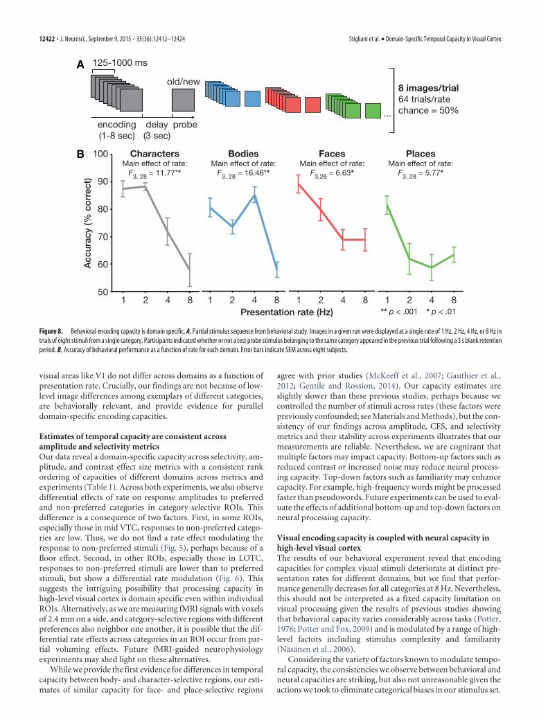
Behavioral testingEight of the 12 subjects participated in a behavioral experiment con-ducted on a different day outside the scanner to estimate subjects’ behav-ioral encoding capacities (Fig. 8). Encoding capacities were measured asa function of rate for a subset of the categories used in the fMRI experi-ments. These categories were as follows: characters, bodies, faces, and
12416 • J. Neurosci., September 9, 2015 • 35(36):12412–12424 Stigliani et al. • Domain-Specific Temporal Capacity in Visual Cortex

places. Subjects viewed trials comprised of three stages: (1) an encodingphase consisting of an RSVP sequence of eight stimuli from a singlecategory presented at a single rate of 1 Hz, 2 Hz, 4 Hz, or 8 Hz; (2) a 3 sdelay containing a blank screen; and (3) a probe image (Fig. 8A). Subjectswere instructed to indicate if the probe image occurred in the precedingsequence or not (0.5 probability). Subjects participated in four runs, eachconsisting of 64 trials at a single rate with 16 trials per category. The orderof runs at different rates was counterbalanced across subjects. In Figure8B, we plot accuracy for each domain as a function of the presentationrate during encoding.
We chose this type of behavioral test because it requires the subject toprocess all eight stimuli during the encoding phase. In contrast, havinga probe at the beginning of the trial would not require the subjects toprocess all images during encoding and pilot behavioral testing indicatedthat subjects’ performance was at ceiling for all rates, consistent withprior findings (Potter and Levy, 1969). Ideally, we would have had sub-jects respond and report their percept for each and every stimulus. How-ever, this is impractical for rates of 4 and 8 Hz, which are faster than theresponse times of typical subjects.
ResultsAre temporal processing capacities in high-level visual cortexdomain general or domain specific?We examined the effect of presentation rate on category selectiv-ity and tested if (1) presentation rate affects the degree of categoryselectivity, (2) regions in the same anatomical location and pro-cessing stage of the ventral stream have the same processing ca-pacity (domain-general hierarchy hypothesis), or (3) regionsprocessing the same category in different anatomical locationshave the same processing capacity (domain-specific temporal hy-pothesis). To address these questions, we independently definedfunctional ROIs selective to characters, bodies, faces, and placesin each subject and measured the degree of selectivity in each ROIas a function of presentation rate. We divided the ventral streaminto three anatomical sections: LOTC, posterior VTC, and midVTC (Fig. 3A). Each section corresponds to a putative tier of thevisual hierarchy and contains a cluster of character, body, face,and place ROIs (with the exception of posterior VTC that did notcontain a place-selective ROI).
We find that presentation rate significantly modulates the de-gree of category selectivity in all ventral stream ROIs regardless oftheir location in the hierarchy or category preference (main effectof rate, Fs � 25.28, ps � 0.001, repeated-measures ANOVA forregions in each anatomical section with factors of rate and pre-ferred domain). Thus, a property of category-selective regions isthat they are more selective to their preferred category when pre-sented at a certain rate.
Contrary to predictions of the hierarchy hypothesis, data fromExperiment 1 also reveal that each anatomical section containsregions with different temporal capacities (rate by domain inter-action, Fs � 6.16, ps � 0.001, repeated-measures ANOVA forregions in each anatomical section with factors of rate and pre-ferred domain; Fig. 3B). In LOTC, for example, regions selectiveto bodies, faces, and places show highest selectivity at 4 Hz, butthe character-selective IOS exhibits maximal selectivity at 2 Hz(Fig. 3B; LOTC). Likewise in mid VTC, regions selective to char-acters, bodies, faces, and places are anatomically adjacent (Fig.3A, mid VTC) yet show different peaks and troughs in their tem-poral processing characteristics (Fig. 3B, mid VTC). Movingfrom medial to lateral: CoS-places and mFus-faces have compa-rable selectivity at 2 and 4 Hz with a decline at 8 Hz, OTS-bodiesreveals higher selectivity at 4 and 8 Hz than 1 and 2 Hz, andmOTS-characters shows peak selectivity at 2 Hz with a decline at4 Hz.
Is temporal processing capacity organized hierarchicallyamong sets of regions processing the same domain?To further discount the hierarchy hypothesis, we must rule outthe possibility that domain-specific temporal processing capacitysystematically declines across the ventral stream hierarchy whenconsidering sets of regions with the same category preference.Therefore, for each ROI and subject we estimated the presenta-tion rate that produced maximal selectivity as a neural marker oftemporal capacity. We then examined if this peak varied acrossregions in the same anatomical section selective to different cat-egories and across regions at different stages of the hierarchyprocessing the same domain.
Our results indicate that the rates estimated to elicit maximalselectivity are similar across regions processing the same domainacross anatomical locations (no main effect of ROI, Fs � 0.89,ps � 0.05, ANOVA on peak rates estimated from selectivity forsets of regions with the same category preference; Fig. 4, dashedhorizontal bars), but vary considerably across regions processingdifferent domains at each stage of the hierarchy (main effect ofROI, Fs � 15.03, ps � 0.001, ANOVA on peak rates estimatedfrom selectivity for sets of regions in each anatomical section). Asshown in Table 1, regions processing characters show maximalselectivity at a similar rate in Experiment 1 (IOS: 1.94 � 0.15 Hz,average � SEM; pOTS: 1.97 � 0.14 Hz; mOTS: 2.07 � 0.30 Hz).The same is true for regions processing bodies (LOS: 5.12 � 0.14Hz; ITG: 4.84 � 0.41 Hz; MTG: 4.79 � 0.37 Hz; OTS: 5.10 � 0.55Hz), faces (IOG: 2.98 � 0.39 Hz; pFus: 3.20 � 0.36 Hz; mFus:3.09 � 0.30 Hz), or places (TOS: 2.28 � 0.35 Hz; CoS: 3.07 � 0.28Hz; RSC: 2.65 � 0.35 Hz). Consistent with these findings, selec-tivity does not differ as a function of rate across regions process-ing the same domain (no rate by ROI interaction, Fs � 2.22, ps �0.05, repeated-measures ANOVA for sets of regions with thesame category preference with factors of rate and ROI; Fig. 4),providing further evidence against the hierarchy hypothesis andin support of domain-specific temporal processing capacities inhigh-level visual cortex.
How does presentation rate affect response amplitude?In principle, selectivity for a preferred category (e.g., a t-valueof face selectivity in face-selective regions) and response am-plitudes to a preferred category (e.g., responses to faces inface-selective regions) are closely related. In practice, the pre-sentation rate eliciting peak selectivity may differ from thepresentation rate eliciting maximal amplitude if the effect ofrate differs across responses to preferred and non-preferredstimuli in that region.
In fact, we find differential effects of rate on responses topreferred and non-preferred stimuli in all category-selectiveregions in Experiment 1 (Fig. 5). Comparing the effect of rateon response amplitudes to preferred and non-preferred stim-uli among sets of regions selective to the same category, wefind that rate-dependent modulation of amplitude differs forthe preferred category and non-preferred categories [rate bypreference interaction, Fs � 4.55, ps � 0.05, repeated-measures ANOVA for sets of regions with the same categorypreference with factors of rate, preference (preferred/non-preferred stimuli), and ROI, but no significant three-way in-teraction, Fs � 0.93, ps � 0.05]. That is, presentation rategenerally has a more prominent effect on response amplitudesto the preferred than non-preferred stimuli.
Closely replicating the selectivity data, the rate producing thepeak response amplitude for the preferred category does not dif-fer across regions processing the same domain (no main effect of
Stigliani et al. • Domain-Specific Temporal Capacity in Visual Cortex J. Neurosci., September 9, 2015 • 35(36):12412–12424 • 12417

ROI, Fs � 0.72, ps � 0.05, ANOVA on peak rates estimated fromamplitude to the preferred category for sets of regions with thesame category preference; Fig. 5, dashed horizontal bars). In con-trast, the rate producing maximal response to the region’s pre-ferred category varies significantly across regions selective todifferent domains at each stage of the hierarchy (main effect of
ROI, Fs � 6.03, ps � 0.01, ANOVA on peak rates estimated fromamplitude for regions in each anatomical section). Likewise, therank ordering of processing capacities associated with differentdomains observed in selectivity data (Fig. 4) is replicated in theamplitude of response for the preferred category and for the CES(difference in response amplitudes between preferred and non-
A LOTC posterior VTC mid VTC
Preferred domain:Places Faces Bodies Characters
TOS
LOS
IOS
IOG
pFus
pOTS
ITG
CoS mFusOTS
mOTS
B
1 2 4 8 1 2 4 8Presentation rate (Hz)
Cat
ego
ry s
elec
tivity
(mea
n t-
valu
e)
1
0
2
3
4
5
6
1 2 4 8
Rate x Domain (N = 11):F9, 90 = 13.41*
Rate x Domain (N = 12):F6, 66 = 18.25*
Rate x Domain (N = 8):F9, 63 = 6.15*
* p < .001
LOTC posterior VTC mid VTC
BodiesFacesPlacesCharacters
Figure 3. Temporal processing capacity is domain specific. A, Functional regions of interest in LOTC, posterior VTC, and mid VTC selective to characters, bodies, faces, and places defined inindividual subjects with regions in each anatomical section shown in an example subject. B, Mean t-value of voxels in each region for the contrast of its preferred category � others as a function ofpresentation rate in Experiment 1. Error bars indicate SEM across subjects. See data analysis section in the Methods for definition of acronyms.
Figure 4. Temporal processing capacity is consistent across regions processing the same domain. Mean selectivity (t-value) of voxels in each ROI (preferred category � others). Error bars indicateSEM across subjects. Horizontal bars below curves represent estimates of the average rate producing peak selectivity for each ROI (white dashes) � 1 SEM across subjects (colored bars).
12418 • J. Neurosci., September 9, 2015 • 35(36):12412–12424 Stigliani et al. • Domain-Specific Temporal Capacity in Visual Cortex

preferred stimuli) as summarized in Table 1. That is, regardless ofthe metric used to estimate temporal capacity, we find that thepresentation rates estimated to elicit peak responses are slowestfor regions processing characters, intermediate among regionspreferring places and faces, and fastest for regions processingbodies.
Are domain-specific temporal frequencies in high-level visualcortex driven by low-level differences among stimuluscategories?Low-level differences among images of different categories maycontribute to the domain-specific temporal processing capacitiesthat we observe in high-level visual cortex. To address this con-cern, we conducted a second control experiment in eight of thesubjects who participated in Experiment 1 using a modified stim-ulus set designed to minimize categorical differences acrossmultiple low-level properties including contrast, luminance,within-category image similarity, visual field coverage, and spa-tial frequency (see Materials and Methods; Fig. 1C). Analyses ofExperiment 2 data indicate that it is unlikely that low-level imageproperties drive capacity differences across domains.
First, to validate the low-level controls implemented in Exper-iment 2, we examined V1 responses. There are no significantdifferences in the response amplitudes of V1 to different stimuluscategories across rates (no main effect of domain, F(3,21) � 1.46,p � 0.05, repeated-measures ANOVA with factors of rate anddomain; Fig. 6, V1), and we do not find variable effects of rate onV1 responses for different categories (no rate by domain interac-tion, F(9,63) � 1.79, p � 0.05). In contrast to V1, category-selective regions illustrate higher response amplitudes toparticular domains across rates (main effect of domain, Fs �16.26, ps � 0.001, repeated-measures ANOVA with factors ofrate and domain for each ROI; Fig. 6). Further, category-selectiveregions, even those in LOTC that are considered lower in thehierarchy compared with VTC, show a significant rate by domaininteraction (Fs � 2.05, ps � 0.05; Fig. 6).
Second, despite changing our stimuli in Experiment 2, we findthat presentation rate significantly modulates category selectivityin a domain-specific manner. Each hierarchical tier contains re-gions with different temporal capacities (rate by domain interac-tion, Fs � 2.10, ps � 0.03, ANOVA for regions in each anatomicalsection with factors of rate, preferred domain, and experiment),and these effects are consistent across experiments (no three-wayinteraction between rate, domain, and experiment, Fs � 0.92,ps � 0.05).
Third, examination of response amplitudes among regionsselective to the same category shows that presentation ratedifferentially modulates responses to preferred and non-preferred stimuli (rate by preference interaction, Fs � 2.89,ps � 0.04, ANOVA for sets of regions with the same categorypreference with factors of rate, preference, ROI, and experi-ment). These effects are similar across experiments (no three-way interaction between rate, preference, and experiment,Fs � 1.44, ps � 0.05; compare Figs. 5 and 7). Similar to Ex-periment 1, we also find that responses to the preferred cate-gory show the same rate modulation among regions preferringthe same category (main effect of rate, Fs � 9.32, ps � 0.001,ANOVA for sets of regions with the same category preferencewith factors of rate, ROI, and experiment, but there are notwo-way or three-way interactions, Fs � 2.73, ps � 0.05; Fig. 7,Preferred).
These analyses validate that the domain-specific temporalprocessing capacities observed in Experiment 1 are reproduc-
A
B
C
D
Figure 5. Differential effects of rate on response amplitudes to preferred and non-preferred cat-egories. Mean response amplitudes (percentage signal change) to the preferred category (left) andnon-preferred category (right) as a function of presentation rate in Experiment 1. A, Character-selectiveROIs. B,Body-selectiveROIs. C,Face-selectiveROIs. D,Place-selectiveROIs.Errorbars indicateSEM across subjects. Horizontal bars below responses to preferred categories represent estimates of theaveragerateproducingpeakamplitudeforeachROI(whitedashes)�1SEMacrosssubjects(coloredbars).
Stigliani et al. • Domain-Specific Temporal Capacity in Visual Cortex J. Neurosci., September 9, 2015 • 35(36):12412–12424 • 12419

ible and are not driven by low-level differences among stimulior inherited from V1.
Are domain-specific temporal processing capacity estimatesconsistent across experiments and metrics?To establish the overall impact of our stimulus manipulations,we compared capacity estimates across metrics and experi-ments (Table 1). Consistent with prior analyses, the largesteffect is that temporal capacity estimates differ between re-gions selective to different domains at each stage of the pro-cessing hierarchy. This differentiation is consistent acrossexperiments and metrics (main effect of domain, Fs � 22.60,ps � 0.001, ANOVA on all peak rate estimates from Experi-ments 1 and 2 for regions in the same anatomical section withfactors of preferred domain, experiment, and metric, butno main effect of experiment or metric, Fs � 3.80, ps � 0.05,and no two-way or three-way interactions, Fs � 0.61, ps �0.05).
Nevertheless, controlling for low-level differences amongstimuli does produce some numerical variations of peak ca-pacity estimates within a domain. For example, face-selective
ROIs show slightly higher capacities in Experiment 2 com-pared with Experiment 1, while body-selective ROIs show anopposite trend (main effect of experiment, Fs � 5.87, ps �0.05, ANOVA on all peak rate estimates from Experiments 1and 2 for body- and face-selective regions separately with fac-tors of ROI, experiment, and metric; Table 1). Also, somecharacter- and place-selective regions exhibit shifts in capacityestimates across experiments (ROI by experiment interaction,Fs � 5.23, p � 0.01, ANOVA on all peak rate estimates fromExperiments 1 and 2 for character- and place-selective regionsseparately with factors of ROI, experiment, and metric, but nomain effect of experiment, Fs � 1.63, ps � 0.05).
Critically, despite numerical shifts within domains, theranking of processing capacities for regions processing differ-ent domains across experiments is consistent: on average andacross all metrics, character-selective IOS, pOTS, and mOTSare the slowest three regions; body-selective LOS, ITG, MTG,and OTS are the fastest four regions; and all face-selectiveregions are intermediate, but faster than place-selective re-gions (Table 1).
Figure 6. Domain specificity is not driven by low-level image properties or inherited from V1. Mean response amplitude for each domain as a function of presentation rate in V1 andcategory-selective regions in LOTC. Error bars indicate SEM across eight subjects.
Table 1. Peak estimates derived from different capacity metrics retain the temporal rank ordering across domains
Experiment 1 Experiment 2
AverageaPreferred CES Selectivity Preferred CES Selectivity
Characters 1.99b 2.08b 1.99b 1.59b 1.93b 2.14b 1.95b
IOS 2.20 (0.26) 1.93 (0.16) 1.94 (0.15) 1.50 (0.15) 1.54 (0.06) 1.50 (0.07) 1.77pOTS 2.05 (0.28) 2.04 (0.16) 1.97 (0.14) 1.57 (0.15) 1.55 (0.12) 1.55 (0.13) 1.79mOTS 1.72 (0.30) 2.26 (0.33) 2.07 (0.30) 1.70 (0.23) 2.69 (0.90) 3.38 (0.90) 2.30
Places 2.83b 2.42b 2.67b 2.76b 2.31b 2.44b 2.57b
TOS 2.93 (0.38) 2.54 (0.37) 2.28 (0.35) 2.80 (0.45) 2.57 (0.54) 2.50 (0.59) 2.60CoS 2.91 (0.37) 2.62 (0.33) 3.07 (0.28) 2.72 (0.54) 2.04 (0.43) 2.37 (0.50) 2.62RSC 2.66 (0.66) 2.10 (0.29) 2.65 (0.35) 2.47
Faces 2.96b 3.08b 3.09b 3.12b 3.57b 3.99b 3.30b
IOG 2.79 (0.45) 2.99 (0.38) 2.98 (0.39) 3.26 (0.51) 3.33 (0.50) 4.51 (0.76) 3.31pFus 3.20 (0.37) 3.26 (0.31) 3.20 (0.36) 3.37 (0.47) 3.42 (0.48) 3.50 (0.50) 3.33mFus 2.90 (0.35) 3.00 (0.32) 3.09 (0.30) 2.74 (0.62) 3.95 (0.44) 3.95 (0.72) 3.27
Bodies 4.90b 4.92b 4.96b 4.15b 4.21b 4.04b 4.53b
LOS 4.91 (0.09) 5.06 (0.12) 5.12 (0.14) 4.10 (0.49) 4.09 (0.81) 4.42 (0.76) 4.62ITG 4.88 (0.10) 4.84 (0.41) 4.84 (0.41) 3.66 (0.66) 4.06 (0.94) 4.13 (0.95) 4.40MTG 5.05 (0.12) 4.76 (0.36) 4.79 (0.37) 4.96 (0.55) 4.14 (0.97) 4.28 (0.96) 4.66OTS 4.74 (0.51) 5.00 (0.50) 5.10 (0.55) 3.88 (0.94) 4.56 (0.83) 3.33 (0.68) 4.44
The mean presentation rate estimated to elicit peak amplitude to the preferred category, contrast effect size (CES), and selectivity in individual subjects from Experiments 1 and 2, with values in parentheses denoting the SEMof this peak estimate across subjects. aAverage peak estimates across metrics and experiments for each region (see rightmost column). bAverage peak estimates across regions selective to the same domain for each metric. Peaksare not estimated for RSC-places in Experiment 2 because of low signals.
12420 • J. Neurosci., September 9, 2015 • 35(36):12412–12424 Stigliani et al. • Domain-Specific Temporal Capacity in Visual Cortex

Does domain-specific temporal processing capacity have abehavioral impact?We hypothesized that if domain-specific temporal processing ca-pacities serve as bottlenecks for perception, there should also becategory-specific differences in temporal encoding capacity (i.e.,behavioral measures of temporal capacity). To test this hypothe-sis, we conducted a behavioral experiment during which our sub-jects viewed encoding stimuli varying in both presentation rateand domain. As in the fMRI experiments, all encoding trials con-tained eight stimuli, but trials varied in their presentation rate(Fig. 8A; see Materials and Methods). To evaluate temporal en-coding capacity, subjects were probed with an image 3 s after theend of the encoding interval and reported if the probe had ap-peared (or not) in the preceding encoding interval. We reasonedthat accuracy should be high for stimuli presented at rates at orbelow encoding capacity, but would decline at rates exceeding theencoding capacity.
Notably, rate affects encoding performance both when con-sidering accuracy for each category individually (main effect ofrate, Fs � 5.77, ps � 0.05, ANOVA on accuracy for each domainseparately; Fig. 8B) and when analyzing performance across alldomains (main effect of rate, F(3,21) � 57.18, p � 0.001, repeated-measures ANOVA with factors of rate and domain). Interest-ingly, the effect of rate on encoding capacity differs acrossdomains (rate by domain interaction, F(9,63) � 4.82, p � 0.001),and several aspects of behavioral encoding capacity mirror thetemporal processing capacity of high-level visual cortex. Theseeffects are apparent in three ways. First, behavioral and neuralcapacity measures for characters peak at presentation rates at 2Hz and drop sharply at faster rates. Second, both accuracy andcortical capacity for bodies are highest at 4 Hz. Third, encoding offaces at 8 Hz declines less than encoding of places at 8 Hz, con-sistent with the observation of higher selectivity to faces thanplaces at this rate in LOTC (Fig. 3).
Despite these striking consistencies, we also observe some de-viations between behavioral encoding and neural processing ca-pacities. For example, behavioral encoding capacity for faces andplaces is highest at 1 Hz, which is lower than the processingcapacity of these stimuli in high-level visual cortex both in selec-tivity and amplitude measures. Overall, these behavioral dataprovide striking evidence for domain-specific encoding capaci-ties that are largely consistent with our fMRI findings.
DiscussionAccording to present hierarchical models, the entirety of high-level visual cortex has a single, domain-general temporal process-ing capacity that is slower than earlier visual regions. Contrary tothis model, our results indicate that domain specificity is a betterpredictor of temporal processing capacity in high-level visualcortex than anatomical location or processing stage in the visualhierarchy. Specifically, body-selective regions illustrate the fast-est capacity, face- and place-selective regions exhibit an interme-diate capacity, and regions preferring characters manifest theslowest capacity. In comparison, neural responses within early
A
B
C
D
Figure 7. Response amplitudes to preferred and non-preferred categories in Experiment 2.Mean response amplitudes (percentage signal change) to the preferred category (left) andnon-preferred category (right) as a function of presentation rate in Experiment 2. A, Character-selective ROIs. B, Body-selective ROIs. C, Face-selective ROIs. D, Place-selective ROIs. Consider-ing data from Experiment 2 separately, rate differentially modulates response amplitudes forpreferred and non-preferred stimuli in character- and face-selective regions, with a similar
4
trend in place-selective regions but not in body-selective regions. However, the rate by prefer-ence interaction is not significantly different between experiments in any of these four domains(no three-way interaction between rate, preference, and experiment, Fs � 1.44, ps � 0.05,ANOVA on regions with the same domain preference, with factors of rate, preference, ROI, andexperiment). Error bars indicate SEM across subjects. Horizontal bars below responses to pre-ferred categories represent estimates of the average rate producing peak amplitude for eachROI (white dashes) � 1 SEM across subjects (colored bars).
Stigliani et al. • Domain-Specific Temporal Capacity in Visual Cortex J. Neurosci., September 9, 2015 • 35(36):12412–12424 • 12421
visual areas like V1 do not differ across domains as a function ofpresentation rate. Crucially, our findings are not because of low-level image differences among exemplars of different categories,are behaviorally relevant, and provide evidence for paralleldomain-specific encoding capacities.
Estimates of temporal capacity are consistent acrossamplitude and selectivity metricsOur data reveal a domain-specific capacity across selectivity, am-plitude, and contrast effect size metrics with a consistent rankordering of capacities of different domains across metrics andexperiments (Table 1). Across both experiments, we also observedifferential effects of rate on response amplitudes to preferredand non-preferred categories in category-selective ROIs. Thisdifference is a consequence of two factors. First, in some ROIs,especially those in mid VTC, responses to non-preferred catego-ries are low. Thus, we do not find a rate effect modulating theresponse to non-preferred stimuli (Fig. 5), perhaps because of afloor effect. Second, in other ROIs, especially those in LOTC,responses to non-preferred stimuli are lower than to preferredstimuli, but show a differential rate modulation (Fig. 6). Thissuggests the intriguing possibility that processing capacity inhigh-level visual cortex is domain specific even within individualROIs. Alternatively, as we are measuring fMRI signals with voxelsof 2.4 mm on a side, and category-selective regions with differentpreferences also neighbor one another, it is possible that the dif-ferential rate effects across categories in an ROI occur from par-tial voluming effects. Future fMRI-guided neurophysiologyexperiments may shed light on these alternatives.
While we provide the first evidence for differences in temporalcapacity between body- and character-selective regions, our esti-mates of similar capacity for face- and place-selective regions
agree with prior studies (McKeeff et al., 2007; Gauthier et al.,2012; Gentile and Rossion, 2014). Our capacity estimates areslightly slower than these previous studies, perhaps because wecontrolled the number of stimuli across rates (these factors werepreviously confounded; see Materials and Methods), but the con-sistency of our findings across amplitude, CES, and selectivitymetrics and their stability across experiments illustrates that ourmeasurements are reliable. Nevertheless, we are cognizant thatmultiple factors may impact capacity. Bottom-up factors such asreduced contrast or increased noise may reduce neural process-ing capacity. Top-down factors such as familiarity may enhancecapacity. For example, high-frequency words might be processedfaster than pseudowords. Future experiments can be used to eval-uate the effects of additional bottom-up and top-down factors onneural processing capacity.
Visual encoding capacity is coupled with neural capacity inhigh-level visual cortexThe results of our behavioral experiment reveal that encodingcapacities for complex visual stimuli deteriorate at distinct pre-sentation rates for different domains, but we find that perfor-mance generally decreases for all categories at 8 Hz. Nevertheless,this should not be interpreted as a fixed capacity limitation onvisual processing given the results of previous studies showingthat behavioral capacity varies considerably across tasks (Potter,1976; Potter and Fox, 2009) and is modulated by a range of high-level factors including stimulus complexity and familiarity(Nasanen et al., 2006).
Considering the variety of factors known to modulate tempo-ral capacity, the consistencies we observe between behavioral andneural capacities are striking, but also not unreasonable given theactions we took to eliminate categorical biases in our stimulus set.
A
B
Figure 8. Behavioral encoding capacity is domain specific. A, Partial stimulus sequence from behavioral study. Images in a given run were displayed at a single rate of 1 Hz, 2 Hz, 4 Hz, or 8 Hz intrials of eight stimuli from a single category. Participants indicated whether or not a test probe stimulus belonging to the same category appeared in the previous trial following a 3 s blank retentionperiod. B, Accuracy of behavioral performance as a function of rate for each domain. Error bars indicate SEM across eight subjects.
12422 • J. Neurosci., September 9, 2015 • 35(36):12412–12424 Stigliani et al. • Domain-Specific Temporal Capacity in Visual Cortex

Across categories, images were matched on low-level propertiessuch as contrast, luminance, and spatial frequency, and on high-level parameters such as familiarity and memorability. Whencontrolling for these factors, our results suggest that temporalcapacities for encoding stimuli with enough detail to allow accu-rate within-category recognition are closely coupled with neuralcapacity estimates in regions preferring characters and bodies.Nevertheless, it is possible that neural capacity estimates reflect-ing the rate at which selectivity or response amplitudes fall tofloor (instead of the rate at which they peak) are coupled withcapacities for more simple perceptual operations. This hypothe-sis can be examined in future research comparing behavioralcapacities for encoding compared with other tasks such as detec-tion and categorization (Grill-Spector and Kanwisher, 2005; Fei-Fei et al., 2007).
Temporal encoding capacity in high-level visual cortex isdomain specificAt the theoretical level, our results suggest a new view of high-level visual cortex in which regions processing related contenthave common temporal processing capacities that are not hier-archically organized from LOTC to VTC. In general, this findingis consistent with the notion that regions selective to particularcategories operate as networks that are synchronized to avoid aprocessing bottleneck. While the underlying source of capacitydifferences across high-level visual cortex is uncertain, we hy-pothesize that two factors may contribute: (1) regularities in thetemporal characteristics of exemplars of various domains in thereal world and (2) the amount of time required to processdomain-specific information.
For instance, lower temporal processing capacities of character-selective regions may be related to the stationary nature of charac-ters. These lower capacities may also be due to the recruitment ofadditional cortical areas processing linguistic or semantic informa-tion when viewing written characters. On the other hand, the highcapacity of body-selective regions may result from the necessity toperform fast computations of rapidly evolving, nonrigid bodymovements. Consistent with this speculation, computational mod-els of biological motion perception posit the existence of “snapshot”neurons selective to brief frames of action sequences (Giese and Pog-gio, 2003). Interestingly, behavioral encoding performance for pseu-dowords and bodies peaked at the same rates that produced thehighest selectivity in ROIs preferring these domains, respectively.This suggests that the temporal processing capacity of these regionsmay influence the rate of behavioral encoding specifically for itemsfrom these categories. Considering other examined domains, bothfaces and places contain more malleable features than characters, buttheir large-scale configurations are more rigid than those of bodies(e.g., eyes and clouds remain above most other features of faces andplaces, respectively). Thus, the present evidence supports the theorythat the natural statistics of temporal information within the visualworld may generate domain-specific temporal encoding capacities.
Temporal processing capacity contributes to the organizationof high-level visual cortexWe propose that temporal processing capacity contributes to thesegregation of category-selective regions in high-level visual cor-tex. It is interesting to speculate how this segregation may developacross the ventral stream hierarchy. From its origin in the retinaand lateral geniculate nucleus, the visual system contains segre-gated temporal channels for faster processing in the magnocellu-lar (M) pathway and slower processing in the parvocellular (P)pathway (Derrington and Lennie, 1984; Schneider et al., 2004).
This segregation is further propagated into cortex in V1 (Sun etal., 2007) and continues downstream to V2 (Munk et al., 1995)and the middle temporal visual area (MT; Maunsell et al., 1990).Thus, it is possible that the propagation of channels with separa-ble temporal processing capacity continues even further and in-fluences the organization of high-level visual cortex.
Other fundamental properties that prevail across the visualsystem such as segregation of neurons processing foveal and pe-ripheral stimuli have also been proposed to constrain the topol-ogy of high-level visual cortex. For example, viewing biases thatare associated with particular domains (e.g., people tend to fove-ate on faces and characters) may generate the particular eccen-tricity biases associated with category-selective regions (Levy etal., 2001; Hasson et al., 2002, 2003; Kay et al., 2015). Similar to theeccentricity bias hypothesis (Malach et al., 2002), the topology ofcategory-selective regions may also be determined by “temporalbiases” of stimuli in the natural world that are mapped ontoregions with differential contribution of M and P inputs. Theseare not mutually exclusive organizational principles. On the con-trary, we propose that the functional organization of high-levelvisual cortex may be constrained both by spatial and temporalcapacity limitations, the combination of which is domain spe-cific. Thus, we suggest that both eccentricity and temporal biasesmay contribute to the functional organization of high-level visualcortex. For example, regions selective to faces and places havesimilar temporal processing capacity, but remain segregated as aresult of their association with different eccentricity biases andviewing patterns. On the other hand, regions processing faces andcharacters have similar foveal biases but distinct temporal dy-namics, which may guide segregation of associated neural pro-cessing in lateral VTC. Our results suggest that temporalprocessing capacity is one of multiple dimensions (Huth et al.,2012) contributing to segregation of functional regions, and thattemporal capacity is a characteristic of domain-specific networksin high-level visual cortex.
NotesSupplemental material for this article is available at vpnl.stanford.edu/fLoc/.This material contains stimuli and code for a functional localizer exper-iment to define category-selective visual regions. This material has notbeen peer reviewed.
ReferencesBen-Shachar M, Dougherty RF, Deutsch GK, Wandell BA (2011) The de-
velopment of cortical sensitivity to visual word forms. J Cogn Neurosci23:2387–2399. CrossRef Medline
Chance FS, Nelson SB, Abbott LF (1998) Synaptic depression and the tem-poral response characteristics of V1 cells. J Neurosci 18:4785– 4799.Medline
Cohen L, Dehaene S, Naccache L, Lehericy S, Dehaene-Lambertz G, HenaffMA, Michel F (2000) The visual word form area: spatial and temporalcharacterization of an initial stage of reading in normal subjects and pos-terior split-brain patients. Brain 123:291–307. CrossRef Medline
Derrington AM, Lennie P (1984) Spatial and temporal contrast sensitivitiesof neurones in lateral geniculate nucleus of macaque. J Physiol 357:219 –240. CrossRef Medline
Dilks DD, Julian JB, Paunov AM, Kanwisher N (2013) The occipital placearea is causally and selectively involved in scene perception. J Neurosci33:1331–1336a. CrossRef Medline
Downing PE, Jiang Y, Shuman M, Kanwisher N (2001) A cortical area selec-tive for visual processing of the human body. Science 293:2470 –2473.CrossRef Medline
Dumoulin SO, Wandell BA (2008) Population receptive field estimates inhuman visual cortex. Neuroimage 39:647– 660. CrossRef Medline
Epstein R, Kanwisher N (1998) A cortical representation of the local visualenvironment. Nature 392:598 – 601. CrossRef Medline
Stigliani et al. • Domain-Specific Temporal Capacity in Visual Cortex J. Neurosci., September 9, 2015 • 35(36):12412–12424 • 12423

Fei-Fei L, Iyer A, Koch C, Perona P (2007) What do we perceive in a glanceof a real-world scene? J Vis 7(1):10. CrossRef Medline
Foster KH, Gaska JP, Nagler M, Pollen DA (1985) Spatial and temporalfrequency selectivity of neurones in visual cortical areas V1 and V2 of themacaque monkey. J Physiol 365:331–363. CrossRef Medline
Gauthier B, Eger E, Hesselmann G, Giraud AL, Kleinschmidt A (2012) Tem-poral tuning properties along the human ventral visual stream. J Neurosci32:14433–14441. CrossRef Medline
Gauthier I, Tarr MJ, Moylan J, Skudlarski P, Gore JC, Anderson AW (2000)The fusiform “face area” is part of a network that processes faces at theindividual level. J Cogn Neurosci 12:495–504. CrossRef Medline
Gentile F, Rossion B (2014) Temporal frequency tuning of cortical face-sensitive areas for individual face perception. Neuroimage 90:256 –265.CrossRef Medline
Giese MA, Poggio T (2003) Neural mechanisms for the recognition of bio-logical movements. Nat Rev Neurosci 4:179 –192. CrossRef Medline
Glezer LS, Jiang X, Riesenhuber M (2009) Evidence for highly selective neu-ronal tuning to whole words in the “visual word form area.” Neuron62:199 –204. CrossRef Medline
Grill-Spector K, Kanwisher N (2005) Visual recognition: as soon as youknow it is there, you know what it is. Psychol Sci 16:152–160. CrossRefMedline
Grill-Spector K, Weiner KS (2014) The functional architecture of the ven-tral temporal cortex and its role in categorization. Nat Rev Neurosci 15:536 –548. CrossRef Medline
Grill-Spector K, Kushnir T, Edelman S, Avidan G, Itzchak Y, Malach R(1999) Differential processing of objects under various viewing condi-tions in the human lateral occipital complex. Neuron 24:187–203.CrossRef Medline
Hasson U, Levy I, Behrmann M, Hendler T, Malach R (2002) Eccentricitybias as an organizing principle for human high-order object areas. Neu-ron 34:479 – 490. CrossRef Medline
Hasson U, Harel M, Levy I, Malach R (2003) Large-scale mirror-symmetryorganization of human occipito-temporal object areas. Neuron 37:1027–1041. CrossRef Medline
Hasson U, Yang E, Vallines I, Heeger DJ, Rubin N (2008) A hierarchy oftemporal receptive windows in human cortex. J Neurosci 28:2539 –2550.CrossRef Medline
Haxby JV, Hoffman EA, Gobbini MI (2000) The distributed human neuralsystem for face perception. Trends Cogn Sci 4:223–233. CrossRef Medline
Heller J, Hertz JA, Kjaer TW, Richmond BJ (1995) Information flow andtemporal coding in primate pattern vision. J Comput Neurosci 2:175–193. CrossRef Medline
Huth AG, Nishimoto S, Vu AT, Gallant JL (2012) A continuous semantic spacedescribes the representation of thousands of object and action categories acrossthe human brain. Neuron 76:1210–1224. CrossRef Medline
Kanwisher N, McDermott J, Chun MM (1997) The fusiform face area: amodule in human extrastriate cortex specialized for face perception.J Neurosci 17:4302– 4311. Medline
Kay KN, Weiner KS, Grill-Spector K (2015) Attention reduces spatial un-certainty in human ventral temporal cortex. Curr Biol 25:595– 600.CrossRef Medline
Keysers C, Xiao DK, Foldiak P, Perrett DI (2001) The speed of sight. J CognNeurosci 13:90 –101. CrossRef Medline
Levy I, Hasson U, Avidan G, Hendler T, Malach R (2001) Center-peripheryorganization of human object areas. Nat Neurosci 4:533–539. Medline
Liu J, Wandell BA (2005) Specializations for chromatic and temporal signalsin human visual cortex. J Neurosci 25:3459 –3468. CrossRef Medline
Malach R, Levy I, Hasson U (2002) The topography of high-order humanobject areas. Trends Cogn Sci 6:176 –184. CrossRef Medline
Maunsell JH, Nealey TA, DePriest DD (1990) Magnocellular and parvocel-lular contributions to responses in the middle temporal visual area (MT)of the macaque monkey. J Neurosci 10:3323–3334. Medline
McKeeff TJ, Remus DA, Tong F (2007) Temporal limitations in object pro-cessing across the human ventral visual pathway. J Neurophysiol 98:382–393. CrossRef Medline
Mukamel R, Harel M, Hendler T, Malach R (2004) Enhanced temporal
non-linearities in human object-related occipito-temporal cortex. CerebCortex 14:575–585. CrossRef Medline
Munk MH, Nowak LG, Girard P, Chounlamountri N, Bullier J (1995) Vi-sual latencies in cytochrome oxidase bands of macaque area V2. Proc NatlAcad Sci U S A 92:988 –992. CrossRef Medline
Nasanen R, Ojanpaa H, Tanskanen T, Paallysaho J (2006) Estimation oftemporal resolution of object identification in human vision. Exp BrainRes 172:464 – 471. CrossRef Medline
O’Craven KM, Kanwisher N (2000) Mental imagery of faces and places ac-tivates corresponding stimulus-specific brain regions. J Cogn Neurosci12:1013–1023. CrossRef Medline
Optican LM, Richmond BJ (1987) Temporal encoding of two-dimensionalpatterns by single units in primate inferior temporal cortex. III. Informa-tion theoretic analysis. J Neurophysiol 57:162–178. Medline
Peelen MV, Downing PE (2005) Selectivity for the human body in the fusi-form gyrus. J Neurophysiol 93:603– 608. Medline
Peelen MV, Downing PE (2007) The neural basis of visual body perception.Nat Rev Neurosci 8:636 – 648. CrossRef Medline
Potter MC (1976) Short-term conceptual memory for pictures. J Exp Psy-chol Hum Learn 2:509 –522. CrossRef Medline
Potter MC, Fox LF (2009) Detecting and remembering simultaneous pic-tures in a rapid serial visual presentation. J Exp Psychol Hum PerceptPerform 35:28 –38. CrossRef Medline
Potter MC, Levy EI (1969) Recognition memory for a rapid sequence ofpictures. J Exp Psychol 81:10 –15. CrossRef Medline
Richmond BJ, Optican LM (1990) Temporal encoding of two-dimensionalpatterns by single units in primate primary visual cortex. II. Informationtransmission. J Neurophysiol 64:370 –380. Medline
Schmolesky MT, Wang Y, Hanes DP, Thompson KG, Leutgeb S, Schall JD,Leventhal AG (1998) Signal timing across the macaque visual system.J Neurophysiol 79:3272–3278. Medline
Schneider KA, Richter MC, Kastner S (2004) Retinotopic organization andfunctional subdivisions of the human lateral geniculate nucleus: a high-resolution functional magnetic resonance imaging study. J Neurosci 24:8975– 8985. CrossRef Medline
Schwarzlose RF, Baker CI, Kanwisher N (2005) Separate face and body se-lectivity on the fusiform gyrus. J Neurosci 25:11055–11059. CrossRefMedline
Schwarzlose RF, Swisher JD, Dang S, Kanwisher N (2008) The distributionof category and location information across object-selective regions inhuman visual cortex. Proc Natl Acad Sci U S A 105:4447– 4452. CrossRefMedline
Singh KD, Smith AT, Greenlee MW (2000) Spatiotemporal frequency anddirection sensitivities of human visual areas measured using fMRI. Neu-roimage 12:550 –564. CrossRef Medline
Sun P, Ueno K, Waggoner RA, Gardner JL, Tanaka K, Cheng K (2007) Atemporal frequency-dependent functional architecture in human V1 re-vealed by high-resolution fMRI. Nat Neurosci 10:1404 –1406. CrossRefMedline
Vogels R, Orban GA (1994) Activity of inferior temporal neurons duringorientation discrimination with successively presented gratings. J Neuro-physiol 71:1428 –1451. Medline
Weiner KS, Grill-Spector K (2010) Sparsely-distributed organization of faceand limb activations in human ventral temporal cortex. Neuroimage 52:1559 –1573. CrossRef Medline
Weiner KS, Grill-Spector K (2011) Not one extrastriate body area: usinganatomical landmarks, hMT�, and visual field maps to parcellate limb-selective activations in human lateral occipitotemporal cortex. Neuroim-age 56:2183–2199. CrossRef Medline
Weiner KS, Grill-Spector K (2012) The improbable simplicity of the fusi-form face area. Trends Cogn Sci 16:251–254. CrossRef Medline
Willenbockel V, Sadr J, Fiset D, Horne GO, Gosselin F, Tanaka JW (2010)Controlling low-level image properties: the SHINE toolbox. Behav ResMethods 42:671– 684. CrossRef Medline
Worsley KJ (2001) Statistical analysis of activation images. In: FunctionalMRI: an introduction to methods (Jezzard P, Matthews PM, Smith SM,eds.), pp 251–270. New York: Oxford UP.
12424 • J. Neurosci., September 9, 2015 • 35(36):12412–12424 Stigliani et al. • Domain-Specific Temporal Capacity in Visual Cortex