tfcc tears and repair - coa.org · •ecu sheath –arises from dorsal fovea –distal radioulnar...
TRANSCRIPT

TFCC Tears and Repair
Jeffrey Yao, M.D.
Associate Professor
Department of Orthopaedic Surgery
Stanford University Medical Center
Disclosures
• The following relationships exist:
1. Grants
American Foundation for Surgery of the Hand
2. Royalties and stock optionsArthrex
3. Consulting incomeSmith and Nephew Endoscopy, Arthrex, Axogen
4. Research and educational support
Arthrex
5. Editorial Honoraria
Elsevier, Lippincott
6. Speakers Bureaus
Arthrex, Trimed
Introduction
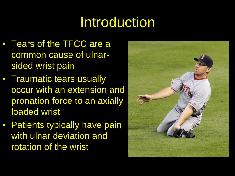
• Tears of the TFCC are a
common cause of ulnar-
sided wrist pain
• Traumatic tears usually
occur with an extension and
pronation force to an axially
loaded wrist
• Patients typically have pain
with ulnar deviation and
rotation of the wrist
Functions of DRUJ
• Distal link between radius and ulna
• Allows radius and attached carpus to pivot smoothly around ulna
• TFCC
– major stabilizer of the DRUJ
– provides suspensory mechanism for ulnar carpus
– central articular disk is the load-bearing component of TFCC
– allows transmission of axial load from carpus to forearm

Anatomy - The TFCC
Thanks to Rebecca Yu, MD
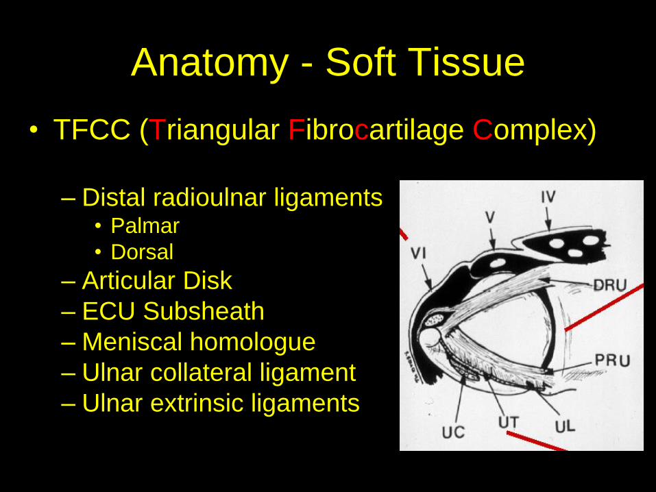
Anatomy - Soft Tissue
• TFCC (Triangular Fibrocartilage Complex)
– Distal radioulnar ligaments• Palmar
• Dorsal
– Articular Disk
– ECU Subsheath
– Meniscal homologue
– Ulnar collateral ligament
– Ulnar extrinsic ligaments

Anatomy of the TFCC

Anatomy of the TFCC
• Dorsal and palmar
radioulnar ligaments
– Ulna fovea to palmar and
dorsal margins of sigmoid
notch
– Ligamentum subcruentum:
deep and strong vertical
foveal insertion

Anatomy of the TFCC
• Fibrocartilaginous articular disk
– Load transmission
– Transitions to hyaline cartilage radially, does
not insert into distal sigmoid notch

Anatomy of the TFCC
• ECU sheath
– Arises from dorsal
fovea
– Distal radioulnar
ligament splits to
form the ECU
tendon sheath

Anatomy of the TFCC
• Meniscal homologue
– Ulnar leash of tissue sweeps distally from
surface of articular disk to the triquetrum
(90%) or triquetrum + lunate (10%)

Anatomy of the TFCC
• Ulnar Collateral Ligament
– loose fibers passing from
tip of ulnar styloid to
triquetrum, pisiform, and
articular disk
– Resists radial deviation
Anatomy of the TFCC
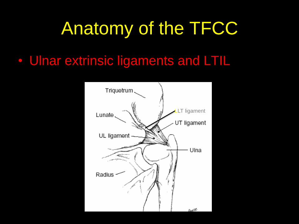
• Ulnar extrinsic ligaments and LTIL
LLT ligament
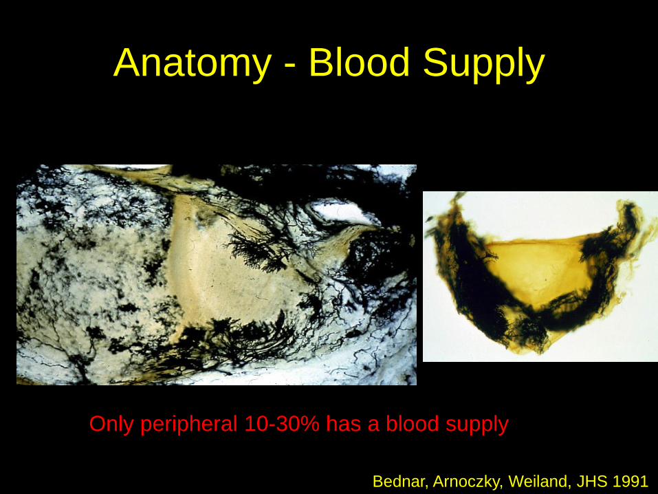
Anatomy - Blood Supply
Only peripheral 10-30% has a blood supply
Bednar, Arnoczky, Weiland, JHS 1991

Biomechanics
• Force Transmission
– Typically 80% of compressive force from the
wrist is borne through distal radius
• 20% through ulna

Biomechanics
Force transmission changes with ulnar variance
+2 mm ulnar variance results in increase to
40% through ulna

Clinical Evaluation

Clinical Evaluation - History
•Fall on an axially loaded pronated wrist
•Pain with forced pronation or supination
•Pain with gripping in ulnar deviation

Clinical Evaluation- Physical Exam
• Fovea sign– Focal tenderness to palpation at
ulnar styloid base
• TFCC stress test– Axial load, ulnar deviation,
rotation
• Test for DRUJ stability (piano key & shuck test) in all positions - neutral, pronation & supination

Examination

Imaging
• Standard Radiographs
• “Zero Degree - PA”
– Elbow flexed to 90
– Shoulder abducted to 90
– Hand flat on X-ray cassette
– Standard for measuring
ulnar variance

Imaging
• Ulnar Variance PA

Ulnar Positive Variance Ulnar Negative Variance
Ulnar Variance

Imaging - CT
• Visualizes:
– Sigmoid notch depth
– Congruency of ulnarhead
– Arthritic changes
• Both wrists should be scanned in
– Pronation
– Neutral
– Supination
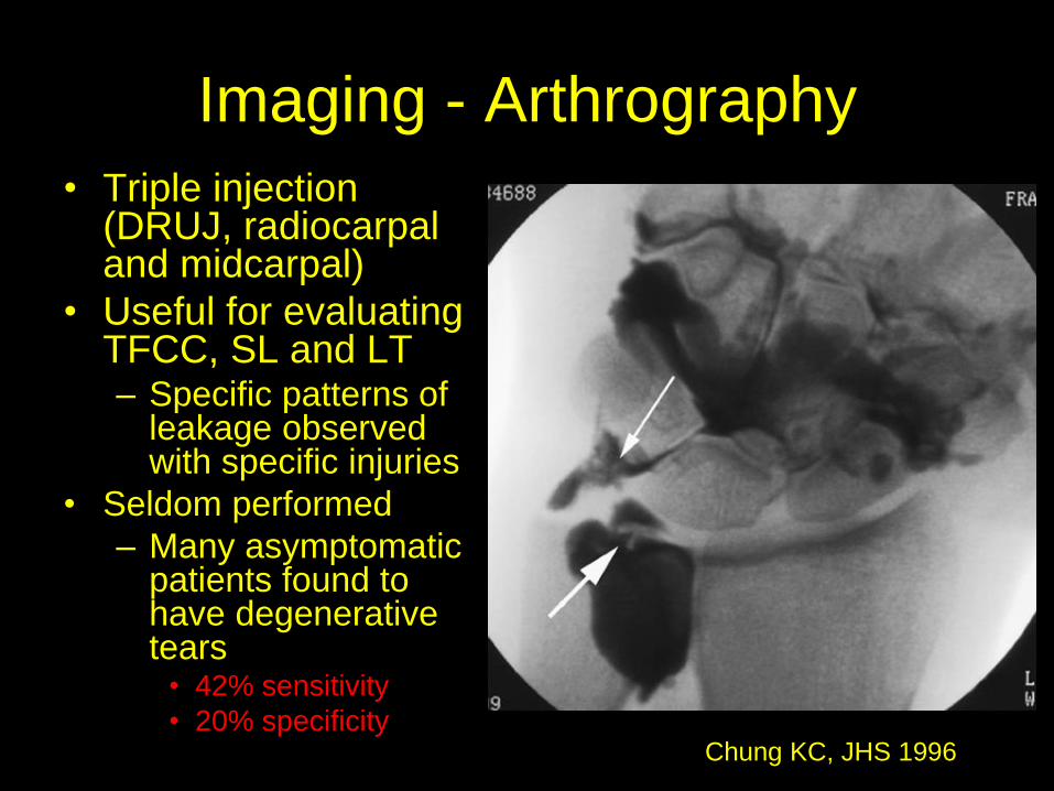
Imaging - Arthrography
• Triple injection (DRUJ, radiocarpal and midcarpal)
• Useful for evaluating TFCC, SL and LT– Specific patterns of
leakage observed with specific injuries
• Seldom performed
– Many asymptomatic patients found to have degenerative tears
• 42% sensitivity
• 20% specificityChung KC, JHS 1996

Imaging - MRI
Articular diskRadial attachment of
articular disk at
sigmoid notch
Ligamentum
subcruentum
Foveal
insertion
Styloid insertion

Imaging - MRI
• ± MR Arthrography
(Intra-articular
injection)
• ± Indirect MR
Arthrography (IV
contrast)
• 1.5T: 85% sensitive
• 3.0T: 94% sensitive
Traumatic
Ulnar
Avulsion
Anderson et al JHS 2008, Faber et al JHS 2010

Imaging - Arthroscopy
• Gold Standard
• Diagnostic as well as therapeutic
• Can detect TFCC tears as well as other pathology– chondral lesions
– other ligamentous injuries
• TFCC Tears: • Loss of resiliency to probing (Trampoline test)
– Indirect visualization of a peripheral tear• Hyperemia along periphery
• Tears of LT ligament
• ECU sheath injury

Imaging - Arthroscopy

Imaging - Arthroscopy
• Trampoline Test
– Ballottment of articular
disk
TFCC Tears
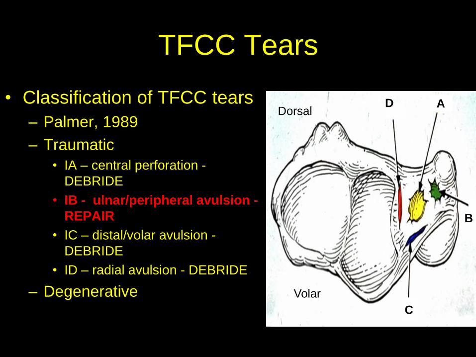
• Classification of TFCC tears
– Palmer, 1989
– Traumatic
• IA – central perforation -
DEBRIDE
• IB - ulnar/peripheral avulsion -
REPAIR
• IC – distal/volar avulsion -
DEBRIDE
• ID – radial avulsion - DEBRIDE
– Degenerative
A
C
B
D
Volar
Dorsal

TFCC Tears
• Can result in isolated ulnar sided wrist pain as
well as DRUJ instability
• Mechanism of injury:
– Extension with pronation to axially loaded wrist
– Can also occur with hypersupination
• More common in patients who are ulnar positive
or neutral
– Ulnar negative patients have thicker articular disks

TFCC Classification

Palmer Traumatic Tears - Type 1
A
B
C
D

TFCC Treatment
• History, clinical findings, and studies are all used to formulate a plan
• Non-operative management is the initial treatment– Unless there is gross instability
– Immobilization in for 4-6 weeks may allow healing of a TFCC tear
• 57% versus 43%
• Acute peripheral tears would be expected to heal given their vascularity
• Otherwise, surgical intervention– Debridement vs repair
– Based on location of tear

Palmer 1A Tear
• Central tear
• Unlikely to heal (avascular)
• May be debrided– up to 2/3 of disk
without affecting load transfer
• Typically ulnar positive variance:– Consider ulnar
recession (wafer) or shortening osteotomy

Palmer 1C Tear
• Usually treated non-operatively or with
debridement
• If repair is necessary be mindful of
ulnar artery and nerve in region whether
repairing through scope or open

Palmer 1D Tear
• Controversial
• Little if any vascularity to area
• Open and arthroscopic(difficult) treatments report good outcomes– Scope:
• Meniscal repair sutures used
• Exit between 1st and 2nd wrist extensor compartments (radial sensory nerve)
– Open :• Dorsal approach between 5th and 6th
extensor compartments
• Simple debridement has satisfactory results

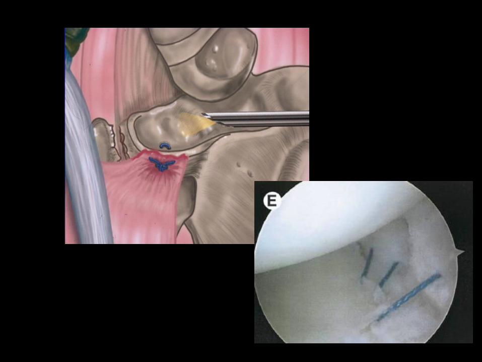
Palmer 1D Repair
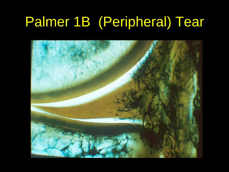
Palmer 1B (Peripheral) Tear
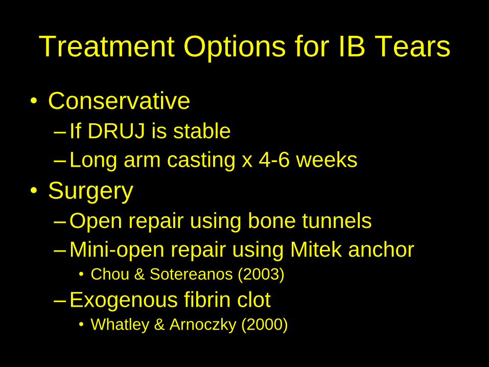
Treatment Options for IB Tears
• Conservative
– If DRUJ is stable
– Long arm casting x 4-6 weeks
• Surgery
– Open repair using bone tunnels
– Mini-open repair using Mitek anchor• Chou & Sotereanos (2003)
– Exogenous fibrin clot• Whatley & Arnoczky (2000)

Open vs Arthroscopic TFCC
Repair: What’s the Evidence?
• Anderson and Berger, et al. (JHS 2008)
– 75 patients over 10 years
– 36 arthroscopic, 39 open
– Mean f/u: 43 months
– NO significant differences in objective and subjective outcomes
– Non-significant trend toward increased ulnarnerve irritation with open repair
– 17% total reoperation rate for DRUJ instability• 8 open, 5 arthroscopic

Atzei and Luchetti, Hand Clinics 2011

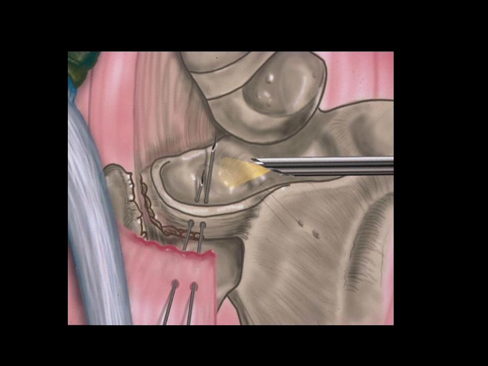
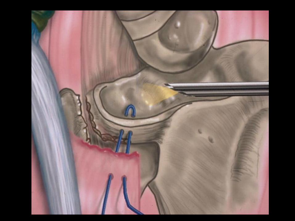
Open Repair

Treatment Options for IB Tears
• Arthroscopic repair
– Outside-inside using meniscal repair needles
• Whipple & Geissler (1993)
• Knot tied over a button
• Knot tied under the dorsal/ulnar skin

Treatment Options for IB Tears
• Arthroscopic Repair, cont
– Inside-outside using meniscal repair needles• Trumble (1996)
– Inside-outside using a Tuohy needle• Araujo & Poehling (1996)
– All-arthroscopic• Bohringer et al, Arthroscopy (2002)
• Conca & Dalla Pria (2004)

Arthroscopic -Assisted Repair


Disadvantages of Current
Techniques
• Extra/larger incision
• Prominent subcutaneous
suture knots
• Patient intolerance of
buttons
– Painful, unsightly,
malodorous, skin changes
– Septic arthritis
• Possible nerve injury

Introduction
• All-Arthroscopic Method of Repair• Yao et al, Arthroscopy, 2007
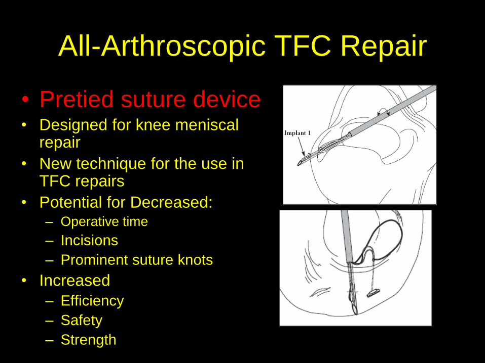
All-Arthroscopic TFC Repair
• Pretied suture device• Designed for knee meniscal
repair
• New technique for the use in TFC repairs
• Potential for Decreased:– Operative time
– Incisions
– Prominent suture knots
• Increased
– Efficiency
– Safety
– Strength
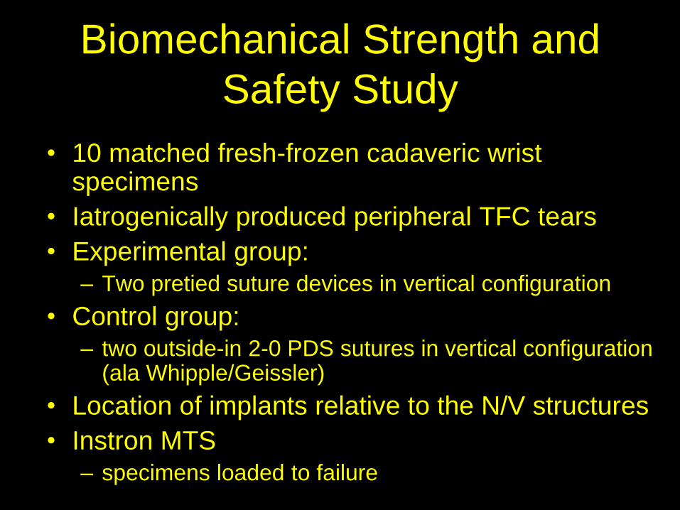
Biomechanical Strength and
Safety Study
• 10 matched fresh-frozen cadaveric wrist specimens
• Iatrogenically produced peripheral TFC tears
• Experimental group:– Two pretied suture devices in vertical configuration
• Control group:– two outside-in 2-0 PDS sutures in vertical configuration
(ala Whipple/Geissler)
• Location of implants relative to the N/V structures
• Instron MTS– specimens loaded to failure

Ulnar Dissection
Whipple/Geissler (PDS)
Distance from UNB: 1.9 cm
Distance from DBUN: 4.6 mm
Suture Device
Distance from UNB: 1.8 cm
Distance from DBUN: 17.1 mm


Biomechanical Study
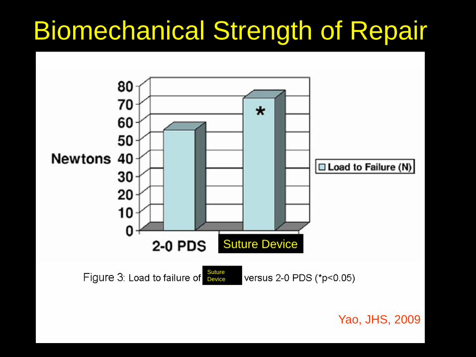
Biomechanical Strength of Repair
Yao, JHS, 2009
Suture Device
Suture
Device

Arthroscopy Set-Up

Portals
Lister’s tubercleECU over
ulnar head
EPL
3-46-R

Arthroscope in 3-4
Probe in 6R
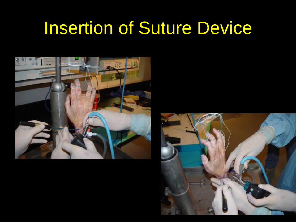
Insertion of Suture Device

Arthroscope in 6R
Suture device in 3-4

Pre-RepairPost-Repair
Arthroscope in 3-4
Probe in 6R
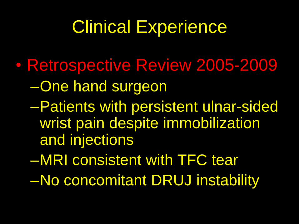
Clinical Experience
• Retrospective Review 2005-2009
–One hand surgeon
–Patients with persistent ulnar-sided wrist pain despite immobilization and injections
–MRI consistent with TFC tear
–No concomitant DRUJ instability

Methods
• Objective data: – range of motion
–grip strength
– return to activity
–post operative complications
• Subjective data: –quickDASH
–PRWE questionnaires

Results
• 14 patients
• Mean f/u: 16.1 months
• Supination: 81 (+/- 13.1)
• Grip strength: 66% (+/- 13.8)
• quickDASH: 10.2 (+/- 11.4)
• PRWE: 18.8 (+/- 13.5)
• Mean time to full activity: 5.2 months
• 0 surgical complications

Conclusion
• All-arthroscopic repair of peripheral TFC tears
show excellent short term results
– 1 year followup, 93% achieved excellent subjective
outcomes based on quickDASH and PRWE
• Benefits of this technique are– ease of use
– lack of prominent suture knots or button
– no extra incisions
– safety
– strength of repair
• reduced immobilization from long arm Munster cast (6 wks)
to short arm cast (4 wks)

Thank You!