the anatomy of tubercles: a corrosion study in a fresh ... · the anatomy of tubercles: a corrosion...
TRANSCRIPT

Materials and Corrosion 2010, 61, No. 12 DOI: 10.1002/maco.201005739 993
The anatomy of tubercles: A corrosion study in a fresh waterestuary
Dedicated to Professor Dr. Wolfgang Sand on the occasion of his 60th birthday
R. I. Ray, J. S. Lee, B. J. Little* and T. L. Gerke
The structure and mineralogy of corrosion products formed on carbon steel
coupons exposed in Duluth Superior Harbor (DSH, USA), were investigated and
compared with corrosion products on similar substrata from other locations.
Corrosion products in DSH formwithin a fewmonths each year and are removed
by ice scour and reform. The corrosion products formed in DSH are tubercles
with an outer surface, an inner shell of magnetite, and a core of iron(III)
oxyhydroxides, goethite, and lepidocrocite, in association with stalks produced
by bacteria. In general, the tubercles formed in DSH are similar in morphology
and mineralogy to corrosion products described for carbon steel and cast iron
exposed to treated waters in decades-old drinking water and cooling water
systems. DSH tubercles are unique in several structural details. DSH tubercles
increase areal coverage of the substratum by consolidation of tubercles.
Furthermore, the core material extends into the pit and is an exact replica of the
pit profile.
1 Introduction
The term tubercle, meaning a small rounded prominence, has
been used to refer to iron corrosion products on steel surfaces
exposed in treated (i.e., chlorinated and heated) fresh waters.
Herro [1] working with iron corrosion products formed in cooling
water systems, defined tubercles as structurally complex
corrosion cells in which accumulations of metal oxides, deposits,
and corrosion products cap localized regions of metal loss. He
concluded that differential aeration cells caused tuberculation,
suggesting that oxygen deficient regions below the accumulated
corrosion products were anodic sites, while surrounding areas
were cathodic. He indicated that tubercles grew as a result of both
internal (anodic) and external (cathodic) reactions, i.e., anodic
dissolution of metal resulted in the accumulation of iron(II)
(ferrous) and iron(III) (ferric) ions and cathodic reactions outside
the tubercle reduced the pH and caused the precipitation of
carbonate and other species whose solubility decreases with
R. I. Ray, J. S. Lee, B. J. Little
Naval Research Laboratory, Code 7303, Bldg. 1009, Stennis Space
Center, MS 39529 (USA)
E-mail: [email protected]
T. L. Gerke
Department of Geology, University of Cincinnati, Cincinnati, OH 45221
(USA)
www.matcorr.com
increasing pH. Herro [1] suggested that tubercle morphology
depended on water chemistry, dissolved oxygen concentration,
temperature, flow, and corrosion rates.
In contrast, Gerke et al. [2] described tubercles formed in a
90-year old chlorinated drinking water distribution system
(DWDS) that were not associated with significant localized
corrosion. Furthermore, the tubercles within a single DWDS,
exposed to the same water had different structures and differing
amounts of associated heavy metals. They proposed that
parameters other than water quality could control tubercle
formation and growth.
The role of microorganisms in tubercle formation is also
controversial. Several investigators have demonstrated bacteria
and bacterial stalks within tubercles [3–5]. Tuovinen and Hsu [6]
suggested that there were zones within some tubercles that
contained enough organic carbon and other nutrients to support
the growth of microorganisms and microcosms with symbiotic
relationships and nutrient cycling. Miller and Tiller [7] indicated,‘‘iron bacteria, which, together with the ferric hydroxide they
produce can form extensive deposits called tubercles on the inside
of water pipes.’’ Tiller [4] suggested that iron-oxidizing bacteria
‘‘encouraged’’ the formation of tubercles. However, bacteria are
not the only cause of tubercle development and the presence of
tubercles cannot be used to conclude the involvement of bacteria
or microbiologically influenced corrosion. Menzies [8] describedabiotic tubercle formation at breaks or discontinuities in an oxide
scale exposed in an oxygenated environment. ‘‘Anodic dissolution
� 2010 WILEY-VCH Verlag GmbH & Co. KGaA, Weinheim

994 Ray, Lee, Little and Gerke Materials and Corrosion 2010, 61, No. 12
takes place and as metal ions concentrate in the solution the
solubility product of the solid hydroxide is exceeded locally and
hydroxide precipitates out as a hemispherical membrane which
surrounds and covers the original discontinuity. This results in
effective screening of the anodic area from available oxygen and
the metal at the discontinuity remains anodic.’’ Tubercles have
been reported in boilers where oxygen is dissolved in water at
high pressures and temperatures exceeding 100 8C and in
sulfuric acid baths.
Tubercles were recently identified in association with pitting
on carbon steel pilings in Duluth Superior Harbor (DSH), MN
and WI, a freshwater estuary [5]. Ray et al. [5] identified bacteria,
bacterial stalks and iron, in addition to other heavy metals, within
the DSH tubercles. Copper, isolated in a distinct stratum at the
base of the tubercles, was the most abundant heavy metal (other
than iron). They concluded that the aggressive corrosion in DSH
was due to the galvanic couple between the deposited copper and
the carbon steel pilings. DSH is icebound frommid-December to
mid-April and during that time has a durable, well-defined ice
cover. Freeze ice thicknesses in DSH range from 0.5 to 1.4m in
addition to snow ice, stack ice, and ice from wave and splash
action along harbor walls. Ice scour breaks and removes tubercles
each year and tubercles reformwithin a fewmonths. Experiments
described in this paper were designed to examine the structure,
microbiology, and mineralogy of tubercles on carbon steel pilings
in DSH. In addition the characteristics of the DSH tubercles were
compared to tubercles of varying ages isolated from other
freshwater environments.
2 Materials and Methods
Coupons of 0.9525 cm thick A328 (0.035% max P, 0.04% max S,
and 0.20% min Cu) cold rolled sheet pile were cut to an average
size of 19.3 cm� 11.6 cm and placed in holders at locations
throughout DSH. Divers collected coupons annually (prior to ice
formation) for three consecutive years. Details related to coupon
Figure 1. Map of the DSH with exposure locations
� 2010 WILEY-VCH Verlag GmbH & Co. KGaA, Weinheim
placement, retrieval, and shipping have been reported elsewhere
[5]. Exposure locations are shown in Fig. 1. Coupons were imaged
using a Nikon S-700 digital camera prior to and after cleaning
with a solution of hydrochloric acid and distilled water (1:1) with
3.5 g/L of hexamethylene–tetramine [9]. Pit profiles (depth and
area) were collected from five (25mm� 25mm) locations on
each of three coupons every year using aMicrophotonics Nanovea
PS50 non-contact optical profiler with a 3.5mm optical pen. Pit
depth and area were used to calculate mass loss. Tubercles were
removed from DSH coupons and embedded in epoxy resin and
sectioned using a diamond blade, slow speed saw [5]. The cross-
sections were imaged with a digital camera and the dimensions of
the tubercles were recorded. Tubercles (not embedded) were
examined using environmental scanning electron microscopy
(ESEM) coupled with energy dispersive X-ray spectrometry (EDS)
as previously described [5]. The mineralogy of the tubercles was
analyzed using a Siemens D-500 automated diffractometer
system using a Cu Ka radiation at 30mA and 40 kV. The 2u
ranged from 5 to 608, with a 0.028 step, and a 2 s count time at
each step. Crystalline phase identifications were made on the
basis of peak position and peak intensities using the American
Mineralogist Crystal Structure Database, the Mineral Database,
and the International Center for Diffraction Data 2002 PDF-2 (see
the following link: http://rruff.geo.arizona.edu/AMS/amcsd.php,
http://webmineral.com/).
3 Results
Well-formed tubercles were observed at all DSH exposure
locations over a 3-year period. The surface area covered by
individual tubercles increased each year [2 cm� 3 cm – year-1
(Fig. 2), 3 cm� 5 cm – year-2, and 6 cm� 10 cm – year-3].
Tubercle height, 2–5mm (above the surface of themetal) however
remained constant over the 3-year exposure period. The general
internal morphology of these tubercles, regardless of age,
consisted of a surface layer, overlying a hard shell layer that
typically enclosed a core region.
www.matcorr.com

Materials and Corrosion 2010, 61, No. 12 The anatomy of tubercles 995
Figure 2. Tubercle on carbon steel after 1-year exposure in DSH
Figure 4. (a) Underside of tubercle core, showing black magnetite shell.
(b) Twisted bacterial sheaths with deposited iron within tubercle core
The surface layer of the DSH tubercles was made up of a
reddish brown material composed of iron(III) oxyhydroxides,
primarily goethite with trace amounts of lepidocrocite. A black
hard shell that had both metallic and non-metallic luster under
the surface layer was composed predominantly of magnetite with
trace amounts of goethite and lepidocrocite. The core region,
yellowish–brown in color and composed of goethite and
lepidocrocite, extended into the area of localized corrosion
(Fig. 3). The internal morphologies of the year-3 tubercles were a
bit more complex in that they tended to contain multiple cores. In
addition the cores had a fibrous appearance. ESEM imaging of the
core regions demonstrated that the iron(III) oxyhydroxides were
associated with twisted bacterial stalks (Fig. 4a and b). All DSH
tubercles were associated with localized corrosion, i.e., pitting.
Pit volume, i.e., mass loss (depth� area) increased with time
over the 3-year exposure in all locations. Averaged pit volume data
are shown for the Midwest Energy location (Fig. 5a–c). Acid
cleaning of pre-exposed carbon steel coupons resulted in shallow
pitting (metal loss) – 42.56mm3 over 25mm� 25mm (625mm2)
area (Fig. 5a). Pit volume for the year-1 coupon was typically
86.12mm3 over 625mm2 with a range of pit depths (70–350mm)
Figure 3. Year-3 tubercle with pit profile
www.matcorr.com
(Fig. 5b). By year-3, the distribution of pit depths had increased
(70–630mm) with a metal loss of 215.25mm3 over 625mm2
(Fig. 5c). Pit depth varied among the locations (Table 1) and
increase in pit depth with time was not linear. The deepest pits at
� 2010 WILEY-VCH Verlag GmbH & Co. KGaA, Weinheim

996 Ray, Lee, Little and Gerke Materials and Corrosion 2010, 61, No. 12
Figure 5. Three-dimensional representations, with accompanying pit density histograms, of carbon steel surfaces (625mm2) exposed at
Midwest Energy Dock illustrating increased pit depth and metal loss after acid cleaning; (a) prior to exposure, after (b) 1 year and (c) 3 years
exposure
year-3 were measured in coupons collected from the Cutler–
Magner location. Weight loss increased with time with an
approximate 3–4% weight loss at all exposure sites after 3 years
(Table 1).
4 Discussion
Carbon steel pilings in DSH that are over 30 years old are either
completely or partially perforated by localized corrosion. Mass
loss, a measure of general corrosion, for coupons exposed in DSH
� 2010 WILEY-VCH Verlag GmbH & Co. KGaA, Weinheim
was approximately 3–4% after 3 years. Average pit depth, a
measure of localized corrosion in the year-3 coupons examined in
this study, ranged between 670 and 788mm, 7–8% of the total
thickness of the coupons. Pit depth varied with location and
increase in pit depth was not linear over the 3-year exposure. The
rate of localized corrosion during the first year was faster than
that measured in either of the next 2 years. Sontheimer et al. [10]found that corrosion of iron pipes occurred quickly during the
first few years after placement into a DWDS and then slowed.
However, the decrease in corrosion rate over time in a DWDSwas
influenced by the formation of an intact scale layer [11], a
www.matcorr.com

Materials and Corrosion 2010, 61, No. 12 The anatomy of tubercles 997
Table 1. Compilation of corrosion data by location
Location Average pit depth (mm) Weight loss (%)
10 months Year-2 Year-3 Year-1 Year-2 Year-3
Oliver Bridge 445.8 469.5 2.4
Hallett 7 Dock 0.9 2.1
Hallett 5 Dock 1.1 3.8
Midwest Energy Dock 393.6 610.6 668.4 1.1 2.5 3.3
DSPA Berth 4 738.2 1.2 3.0 3.8
Cutler Magner 387.1 787.8 2.0 3.4 4.4
situation that does not apply to the ice-scoured pilings in DSH. In
the absence of a better understanding of the relationship between
water chemistry, tubercle formation, and corrosion, there are no
scientific reasons to expect that the penetration rate will remain
linear over decades.
Relative rates of tubercle growth could be evaluated by
comparing the size of the DSH tubercles with those formed in
other environments. The maximum DSH tubercle size after 3
years was 6 cm� 10 cm. DSH tubercle height was remarkably
similar for all three 3 years, approximately 2–5mm. By
comparison, tubercles from a 90-year old DWDS were approxi-
mately 4–6 cm in diameter and 2–3 cm high [2]. Tubercles formed
in a few months each year in DSH were comparable in surface
area to the undisturbed, older tubercles. Pit depth and height of
the DSH tubercle above the surface of the metal were not related.
DSH tubercle growth appears to include the possibility of
merging tubercles as indicated by the multiple core regions after
3 years.
Several authors have described the internal morphologies of
tubercles [1, 2, 12] and provided schematics of tubercles.Herro [1]indicated that tubercles should contain the following structural
features: outer crust, inner shell, core material, fluid cavity, and
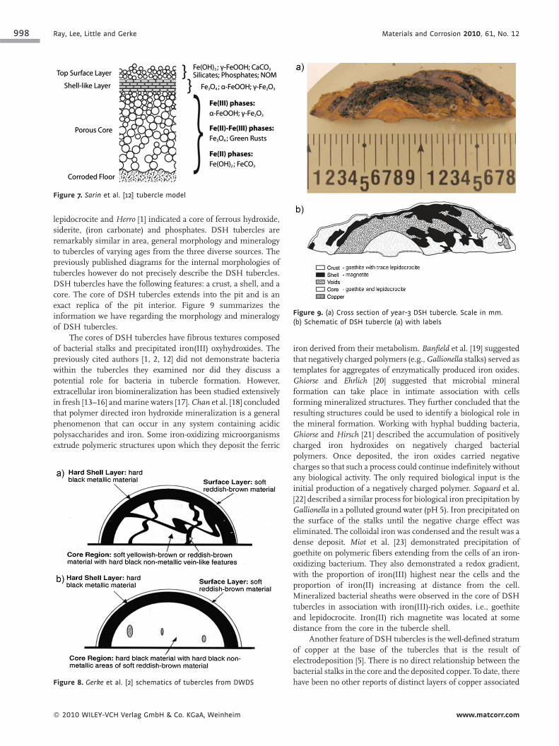
corroding floor (Fig. 6). Sarin et al. [11, 12] indicated a surface
Figure 6. Herro [1] schematic of tubercle
www.matcorr.com
layer, a shell-like layer and a porous core over a corroding floor
(Fig. 7). The significant difference between theHerro [1] and Sarinet al. [12] model is the absence of a fluid filled cavity in the Sarinet al. [12] model. Instead Sarin et al. [12] suggested that porosity
within the tubercle determined the ease with which ions migrate
within the core. Their diagram indicated increased porosity at the
base of the core. Gerke et al. [2] examined the morphology,
mineralogy, and chemistry of five tubercles from 90-year-old cast
iron piping in a single DWDS. The overall morphology of all five
DWDS samples was similar – a core with a hard shell layer,
covered with surface material (Fig. 8). Magnetite, lepidocrocite,
and goethite were the three predominant iron minerals in all five
tubercles, but in different proportions. Gerke et al. [2] reported
magnetite veins within core regions of some tubercles that
produced a marbled appearance. They demonstrated that heavy
metals were concentrated in regions of the tubercles. Tubercles
exposed to the same water supply contained different metals or
the same metals at different concentrations. The Herro [1] and
Gerke et al. [2] schematics are similar in the following details: a
magnetite shell overlain with a reddish brown crust, and a core.
The fluid filled cavity between the core and the corroding floor,
described by Herro [1], were not observed in the Gerke et al. [2]tubercles. Gerke et al. [2] indicated a core of either goethite or
� 2010 WILEY-VCH Verlag GmbH & Co. KGaA, Weinheim
998 Ray, Lee, Little and Gerke Materials and Corrosion 2010, 61, No. 12
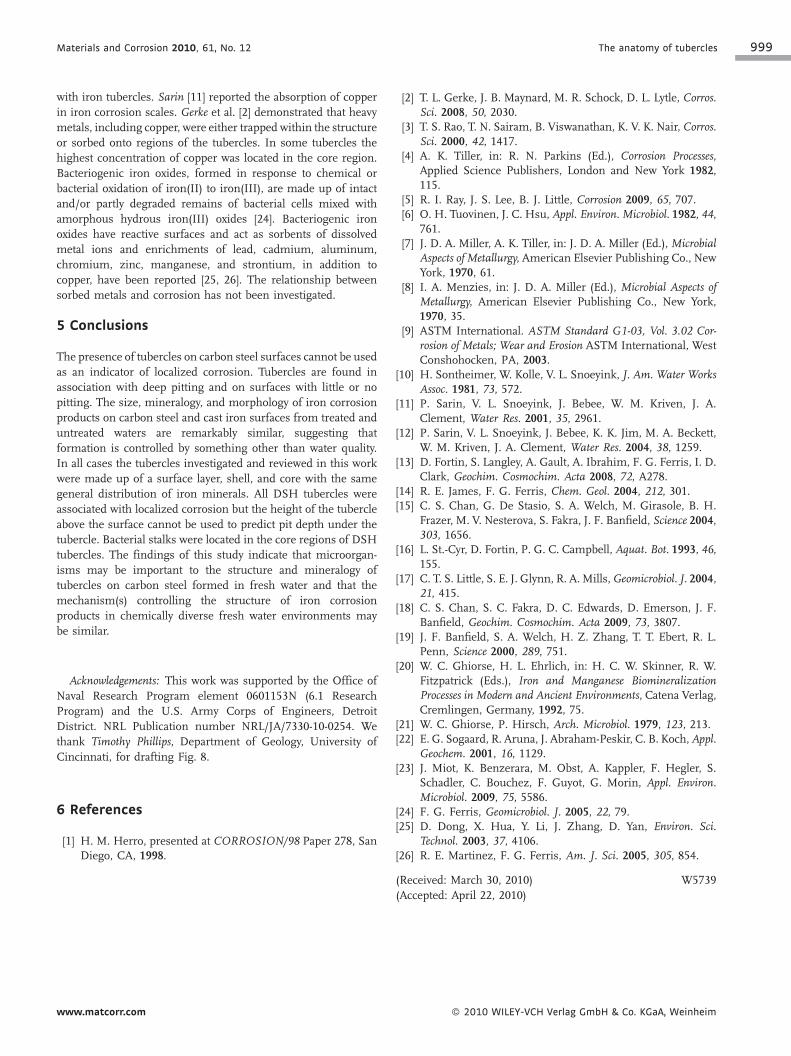
Figure 9. (a) Cross section of year-3 DSH tubercle. Scale in mm.
(b) Schematic of DSH tubercle (a) with labels
Figure 7. Sarin et al. [12] tubercle model
lepidocrocite and Herro [1] indicated a core of ferrous hydroxide,
siderite, (iron carbonate) and phosphates. DSH tubercles are
remarkably similar in area, general morphology and mineralogy
to tubercles of varying ages from the three diverse sources. The
previously published diagrams for the internal morphologies of
tubercles however do not precisely describe the DSH tubercles.
DSH tubercles have the following features: a crust, a shell, and a
core. The core of DSH tubercles extends into the pit and is an
exact replica of the pit interior. Figure 9 summarizes the
information we have regarding the morphology and mineralogy
of DSH tubercles.
The cores of DSH tubercles have fibrous textures composed
of bacterial stalks and precipitated iron(III) oxyhydroxides. The
previously cited authors [1, 2, 12] did not demonstrate bacteria
within the tubercles they examined nor did they discuss a
potential role for bacteria in tubercle formation. However,
extracellular iron biomineralization has been studied extensively
in fresh [13–16] andmarine waters [17]. Chan et al. [18] concludedthat polymer directed iron hydroxide mineralization is a general
phenomenon that can occur in any system containing acidic
polysaccharides and iron. Some iron-oxidizing microorganisms
extrude polymeric structures upon which they deposit the ferric
Figure 8. Gerke et al. [2] schematics of tubercles from DWDS
� 2010 WILEY-VCH Verlag GmbH & Co. KGaA, Weinheim
iron derived from their metabolism. Banfield et al. [19] suggestedthat negatively charged polymers (e.g.,Gallionella stalks) served astemplates for aggregates of enzymatically produced iron oxides.
Ghiorse and Ehrlich [20] suggested that microbial mineral
formation can take place in intimate association with cells
forming mineralized structures. They further concluded that the
resulting structures could be used to identify a biological role in
the mineral formation. Working with hyphal budding bacteria,
Ghiorse and Hirsch [21] described the accumulation of positively
charged iron hydroxides on negatively charged bacterial
polymers. Once deposited, the iron oxides carried negative
charges so that such a process could continue indefinitely without
any biological activity. The only required biological input is the
initial production of a negatively charged polymer. Sogaard et al.
[22] described a similar process for biological iron precipitation by
Gallionella in a polluted ground water (pH 5). Iron precipitated on
the surface of the stalks until the negative charge effect was
eliminated. The colloidal iron was condensed and the result was a
dense deposit. Miot et al. [23] demonstrated precipitation of
goethite on polymeric fibers extending from the cells of an iron-
oxidizing bacterium. They also demonstrated a redox gradient,
with the proportion of iron(III) highest near the cells and the
proportion of iron(II) increasing at distance from the cell.
Mineralized bacterial sheaths were observed in the core of DSH
tubercles in association with iron(III)-rich oxides, i.e., goethite
and lepidocrocite. Iron(II) rich magnetite was located at some
distance from the core in the tubercle shell.
Another feature of DSH tubercles is the well-defined stratum
of copper at the base of the tubercles that is the result of
electrodeposition [5]. There is no direct relationship between the
bacterial stalks in the core and the deposited copper. To date, there
have been no other reports of distinct layers of copper associated
www.matcorr.com
Materials and Corrosion 2010, 61, No. 12 The anatomy of tubercles 999
with iron tubercles. Sarin [11] reported the absorption of copper
in iron corrosion scales. Gerke et al. [2] demonstrated that heavy
metals, including copper, were either trapped within the structure
or sorbed onto regions of the tubercles. In some tubercles the
highest concentration of copper was located in the core region.
Bacteriogenic iron oxides, formed in response to chemical or
bacterial oxidation of iron(II) to iron(III), are made up of intact
and/or partly degraded remains of bacterial cells mixed with
amorphous hydrous iron(III) oxides [24]. Bacteriogenic iron
oxides have reactive surfaces and act as sorbents of dissolved
metal ions and enrichments of lead, cadmium, aluminum,
chromium, zinc, manganese, and strontium, in addition to
copper, have been reported [25, 26]. The relationship between
sorbed metals and corrosion has not been investigated.
5 Conclusions
The presence of tubercles on carbon steel surfaces cannot be used
as an indicator of localized corrosion. Tubercles are found in
association with deep pitting and on surfaces with little or no
pitting. The size, mineralogy, and morphology of iron corrosion
products on carbon steel and cast iron surfaces from treated and
untreated waters are remarkably similar, suggesting that
formation is controlled by something other than water quality.
In all cases the tubercles investigated and reviewed in this work
were made up of a surface layer, shell, and core with the same
general distribution of iron minerals. All DSH tubercles were
associated with localized corrosion but the height of the tubercle
above the surface cannot be used to predict pit depth under the
tubercle. Bacterial stalks were located in the core regions of DSH
tubercles. The findings of this study indicate that microorgan-
isms may be important to the structure and mineralogy of
tubercles on carbon steel formed in fresh water and that the
mechanism(s) controlling the structure of iron corrosion
products in chemically diverse fresh water environments may
be similar.
Acknowledgements: This work was supported by the Office of
Naval Research Program element 0601153N (6.1 Research
Program) and the U.S. Army Corps of Engineers, Detroit
District. NRL Publication number NRL/JA/7330-10-0254. We
thank Timothy Phillips, Department of Geology, University of
Cincinnati, for drafting Fig. 8.
6 References
[1] H. M. Herro, presented at CORROSION/98 Paper 278, SanDiego, CA, 1998.
www.matcorr.com
[2] T. L. Gerke, J. B. Maynard, M. R. Schock, D. L. Lytle, Corros.Sci. 2008, 50, 2030.
[3] T. S. Rao, T. N. Sairam, B. Viswanathan, K. V. K. Nair, Corros.Sci. 2000, 42, 1417.
[4] A. K. Tiller, in: R. N. Parkins (Ed.), Corrosion Processes,Applied Science Publishers, London and New York 1982,115.
[5] R. I. Ray, J. S. Lee, B. J. Little, Corrosion 2009, 65, 707.[6] O. H. Tuovinen, J. C. Hsu, Appl. Environ. Microbiol. 1982, 44,
761.[7] J. D. A. Miller, A. K. Tiller, in: J. D. A. Miller (Ed.), Microbial
Aspects of Metallurgy, American Elsevier Publishing Co., NewYork, 1970, 61.
[8] I. A. Menzies, in: J. D. A. Miller (Ed.), Microbial Aspects ofMetallurgy, American Elsevier Publishing Co., New York,1970, 35.
[9] ASTM International. ASTM Standard G1-03, Vol. 3.02 Cor-rosion of Metals; Wear and Erosion ASTM International, WestConshohocken, PA, 2003.
[10] H. Sontheimer, W. Kolle, V. L. Snoeyink, J. Am. Water WorksAssoc. 1981, 73, 572.
[11] P. Sarin, V. L. Snoeyink, J. Bebee, W. M. Kriven, J. A.Clement, Water Res. 2001, 35, 2961.
[12] P. Sarin, V. L. Snoeyink, J. Bebee, K. K. Jim, M. A. Beckett,W. M. Kriven, J. A. Clement, Water Res. 2004, 38, 1259.
[13] D. Fortin, S. Langley, A. Gault, A. Ibrahim, F. G. Ferris, I. D.Clark, Geochim. Cosmochim. Acta 2008, 72, A278.
[14] R. E. James, F. G. Ferris, Chem. Geol. 2004, 212, 301.[15] C. S. Chan, G. De Stasio, S. A. Welch, M. Girasole, B. H.
Frazer, M. V. Nesterova, S. Fakra, J. F. Banfield, Science 2004,303, 1656.
[16] L. St.-Cyr, D. Fortin, P. G. C. Campbell, Aquat. Bot. 1993, 46,155.
[17] C. T. S. Little, S. E. J. Glynn, R. A. Mills,Geomicrobiol. J. 2004,21, 415.
[18] C. S. Chan, S. C. Fakra, D. C. Edwards, D. Emerson, J. F.Banfield, Geochim. Cosmochim. Acta 2009, 73, 3807.
[19] J. F. Banfield, S. A. Welch, H. Z. Zhang, T. T. Ebert, R. L.Penn, Science 2000, 289, 751.
[20] W. C. Ghiorse, H. L. Ehrlich, in: H. C. W. Skinner, R. W.Fitzpatrick (Eds.), Iron and Manganese BiomineralizationProcesses in Modern and Ancient Environments, Catena Verlag,Cremlingen, Germany, 1992, 75.
[21] W. C. Ghiorse, P. Hirsch, Arch. Microbiol. 1979, 123, 213.[22] E. G. Sogaard, R. Aruna, J. Abraham-Peskir, C. B. Koch,Appl.
Geochem. 2001, 16, 1129.[23] J. Miot, K. Benzerara, M. Obst, A. Kappler, F. Hegler, S.
Schadler, C. Bouchez, F. Guyot, G. Morin, Appl. Environ.Microbiol. 2009, 75, 5586.
[24] F. G. Ferris, Geomicrobiol. J. 2005, 22, 79.[25] D. Dong, X. Hua, Y. Li, J. Zhang, D. Yan, Environ. Sci.
Technol. 2003, 37, 4106.[26] R. E. Martinez, F. G. Ferris, Am. J. Sci. 2005, 305, 854.
(Received: March 30, 2010)
(Accepted: April 22, 2010)
W5739
� 2010 WILEY-VCH Verlag GmbH & Co. KGaA, Weinheim