the cardiovascular system -...
TRANSCRIPT

PowerPoint® Lecture Slides
prepared by
Karen Dunbar Kareiva
Ivy Tech Community College© Annie Leibovitz/Contact Press Images
Chapter 17 Part A
The
Cardiovascular
System
© 2017 Pearson Education, Inc.
Why This Matters
• How the anatomy and physiology of the heart
affect the sounds heard through a stethoscope
© 2017 Pearson Education, Inc.

17.1 Heart Anatomy
The Pulmonary and Systemic Circuits
• Heart is a transport system consisting of two
side-by-side pumps
– Right side receives oxygen-poor blood from
tissues
• Pumps blood to lungs to get rid of CO2, pick up O2, via
pulmonary circuit
– Left side receives oxygenated blood from lungs
• Pumps blood to body tissues via systemic circuit
© 2017 Pearson Education, Inc.

The Pulmonary and Systemic Circuits (cont.)
• Receiving chambers of heart
– Right atrium
• Receives blood returning from systemic circuit
– Left atrium
• Receives blood returning from pulmonary circuit
• Pumping chambers of heart
– Right ventricle
• Pumps blood through pulmonary circuit
– Left ventricle
• Pumps blood through systemic circuit
© 2017 Pearson Education, Inc.

Figure 17.1 The systemic and pulmonary circuits.
© 2017 Pearson Education, Inc.
Capillary bedsof lungs wheregas exchangeoccurs
Capillary bedsof all bodytissues where gasexchange occurs
Pulmonaryveins
Pulmonaryarteries
Systemic Circuit
Aorta and branches
Heart
Rightatrium Right
ventricle
Venaecavae
Leftatrium
Leftventricle
Pulmonary Circuit
Oxygen-rich,CO2-poor blood
Oxygen-poor,CO2-rich blood

Size, Location, and Orientation of Heart
• Approximately the size of a fist
– Weighs less than 1 pound
• Location
– In mediastinum between second rib and fifth
intercostal space
– On superior surface of diaphragm
– Two-thirds of heart to left of midsternal line
– Anterior to vertebral column, posterior to sternum
© 2017 Pearson Education, Inc.

Figure 17.2a Location of the heart in the mediastinum.
© 2017 Pearson Education, Inc.
Location ofapicalimpulse
Diaphragm
Sternum
2nd rib
Midsternal line

Figure 17.2b Location of the heart in the mediastinum.
© 2017 Pearson Education, Inc.
Heart
Posterior
Left lung
Body of T8
vertebra
Mediastinum
Size, Location, and Orientation of Heart
(cont.)
• Base (posterior surface) leans toward right
shoulder
• Apex points toward left hip
• Apical impulse palpated between fifth and sixth
ribs, just below left nipple
© 2017 Pearson Education, Inc.

Figure 17.2a Location of the heart in the mediastinum.
© 2017 Pearson Education, Inc.
Location ofapicalimpulse
Diaphragm
Sternum
2nd rib
Midsternal line

Figure 17.2b Location of the heart in the mediastinum.
© 2017 Pearson Education, Inc.
Heart
Posterior
Left lung
Body of T8
vertebra
Mediastinum

Coverings of the Heart
• Pericardium: double-walled sac that surrounds
heart; made up of two layers
1. Superficial fibrous pericardium: functions to
protect, anchor heart to surrounding structures,
and prevent overfilling
2. Deep two-layered serous pericardium
• Parietal layer lines internal surface of fibrous
pericardium
• Visceral layer (epicardium) on external surface of
heart
• Two layers separated by fluid-filled pericardial cavity
(decreases friction)
© 2017 Pearson Education, Inc.

Figure 17.3 The layers of the pericardium and of the heart wall.
© 2017 Pearson Education, Inc.
Fibrous pericardium
Parietal layer of serous
pericardium
Pericardial cavity
Epicardium (visceral
layer of serous
pericardium)
Myocardium
Endocardium
Pulmonarytrunk
Heart chamber
Pericardium
Myocardium
Heart
wall

Clinical – Homeostatic Imbalance 17.1
• Pericarditis
– Inflammation of pericardium
– Roughens membrane surfaces, causing
pericardial friction rub (creaking sound) heard
with stethoscope
– Cardiac tamponade
• Excess fluid that leaks into pericardial space
• Can compress heart’s pumping ability
• Treatment: fluid is drawn out of cavity (usually with
syringe)
© 2017 Pearson Education, Inc.

Layers of the Heart Wall
• Three layers of heart wall
1. Epicardium: visceral layer of serous
pericardium
2. Myocardium: circular or spiral bundles of
contractile cardiac muscle cells
• Cardiac skeleton: crisscrossing, interlacing layer of
connective tissue
– Anchors cardiac muscle fibers
– Supports great vessels and valves
– Limits spread of action potentials to specific paths
© 2017 Pearson Education, Inc.
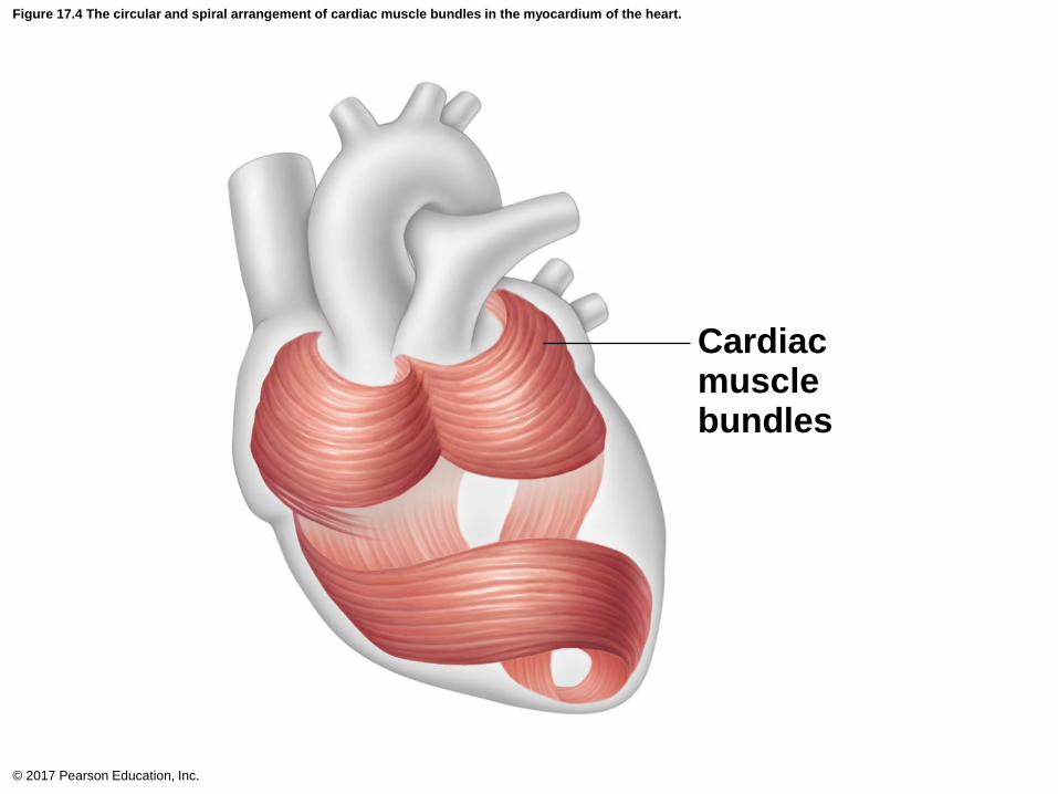
Figure 17.4 The circular and spiral arrangement of cardiac muscle bundles in the myocardium of the heart.
© 2017 Pearson Education, Inc.
Cardiacmusclebundles
Layers of the Heart Wall (cont.)
3. Endocardium: innermost layer; is continuous
with endothelial lining of blood vessels
• Lines heart chambers and covers cardiac skeleton of
valves
© 2017 Pearson Education, Inc.
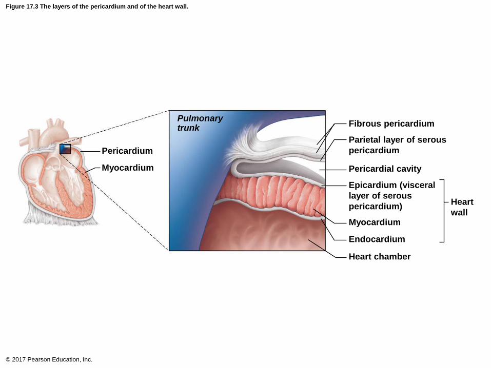
Figure 17.3 The layers of the pericardium and of the heart wall.
© 2017 Pearson Education, Inc.
Fibrous pericardium
Parietal layer of serous
pericardium
Pericardial cavity
Epicardium (visceral
layer of serous
pericardium)
Myocardium
Endocardium
Pulmonarytrunk
Heart chamber
Pericardium
Myocardium
Heart
wall

Chambers and Associated Great Vessels
• Internal features
– Four chambers
• Two superior atria
• Two inferior ventricles
– Interatrial septum: separates atria
• Fossa ovalis: remnant of foramen ovale of fetal heart
– Interventricular septum: separates ventricles
© 2017 Pearson Education, Inc.

Figure 17.5e Gross anatomy of the heart.
© 2017 Pearson Education, Inc.
Superior vena cava
Right atrium
Right pulmonary artery
Pulmonary trunk
Right pulmonary veins
Fossa ovalis
Pectinate muscles
Tricuspid valve
Right ventricle
Chordae tendineae
Trabeculae carneae
Inferior vena cava
Frontal section
Aorta
Left pulmonary artery
Left atrium
Left pulmonary veins
Mitral (bicuspid) valve
Aortic valve
Pulmonary valve
Left ventricle
Papillary muscle
Interventricular septum
Epicardium
Myocardium
Endocardium
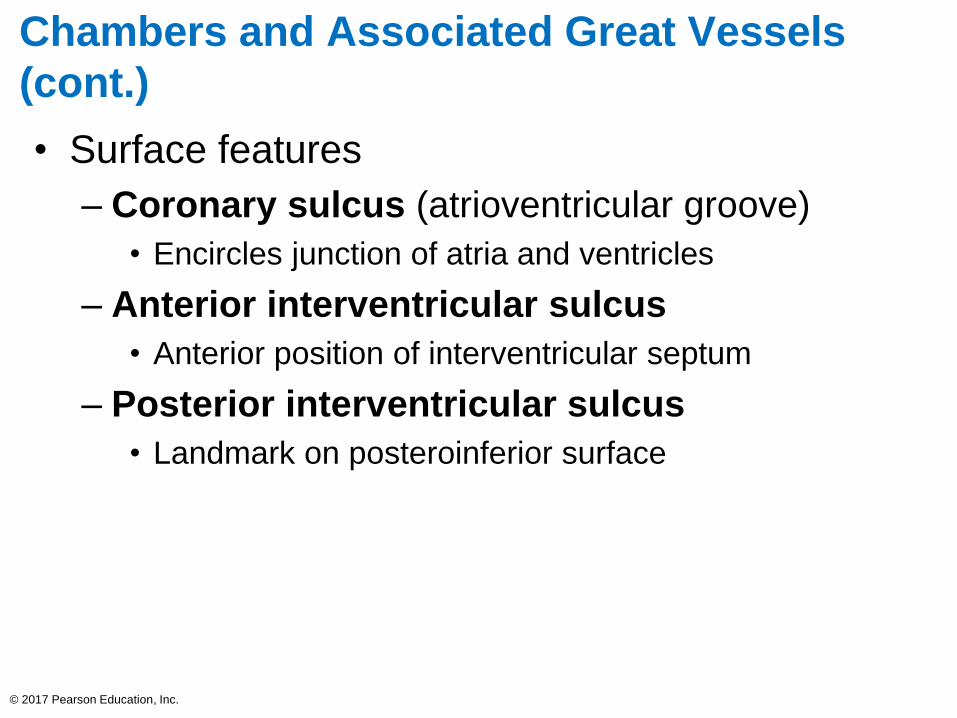
Chambers and Associated Great Vessels
(cont.)
• Surface features
– Coronary sulcus (atrioventricular groove)
• Encircles junction of atria and ventricles
– Anterior interventricular sulcus
• Anterior position of interventricular septum
– Posterior interventricular sulcus
• Landmark on posteroinferior surface
© 2017 Pearson Education, Inc.

Figure 17.5b Gross anatomy of the heart.
© 2017 Pearson Education, Inc.
Brachiocephalic trunk
Superior vena cava
Right pulmonary artery
Ascending aorta
Pulmonary trunk
Right pulmonary veins
Right atrium
Right coronary artery(in coronary sulcus)
Anterior cardiac vein
Right ventricle
Right marginal artery
Small cardiac vein
Inferior vena cava
Anterior view
Left common carotidartery
Left subclavian artery
Aortic arch
Ligamentum arteriosum
Left pulmonary artery
Left pulmonary veins
Auricle ofleft atrium
Circumflex artery
Left coronary artery(in coronary sulcus)
Left ventricle
Great cardiac vein
Anterior interventricularartery (in anteriorinterventricular sulcus)
Apex
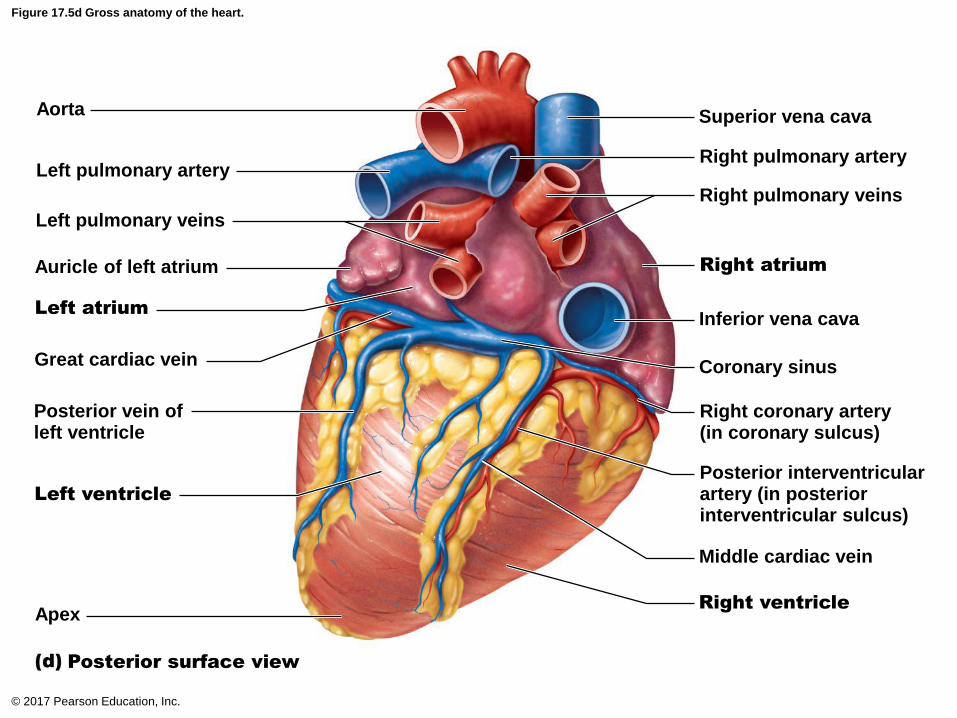
Figure 17.5d Gross anatomy of the heart.
© 2017 Pearson Education, Inc.
Aorta
Left pulmonary artery
Left pulmonary veins
Auricle of left atrium
Great cardiac vein
Posterior vein ofleft ventricle
Apex
Left atrium
Left ventricle
Posterior surface view
Superior vena cava
Right atrium
Right pulmonary artery
Right pulmonary veins
Inferior vena cava
Coronary sinus
Right coronary artery(in coronary sulcus)
Posterior interventricularartery (in posteriorinterventricular sulcus)
Middle cardiac vein
Right ventricle
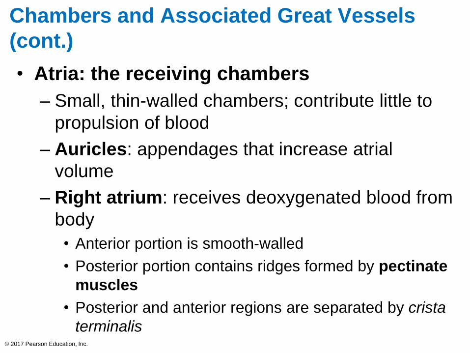
Chambers and Associated Great Vessels
(cont.)
• Atria: the receiving chambers
– Small, thin-walled chambers; contribute little to
propulsion of blood
– Auricles: appendages that increase atrial
volume
– Right atrium: receives deoxygenated blood from
body
• Anterior portion is smooth-walled
• Posterior portion contains ridges formed by pectinate
muscles
• Posterior and anterior regions are separated by crista
terminalis© 2017 Pearson Education, Inc.
Chambers and Associated Great Vessels
(cont.)
• Atria: the receiving chambers (cont.)
• Three veins empty into right atrium:
– Superior vena cava: returns blood from body regions
above the diaphragm
– Inferior vena cava: returns blood from body regions
below the diaphragm
– Coronary sinus: returns blood from coronary veins
– Left atrium: receives oxygenated blood from
lungs
• Pectinate muscles found only in auricles
• Four pulmonary veins return blood from lungs
© 2017 Pearson Education, Inc.

Figure 17.5e Gross anatomy of the heart.
© 2017 Pearson Education, Inc.
Superior vena cava
Right atrium
Right pulmonary artery
Pulmonary trunk
Right pulmonary veins
Fossa ovalis
Pectinate muscles
Tricuspid valve
Right ventricle
Chordae tendineae
Trabeculae carneae
Inferior vena cava
Frontal section
Aorta
Left pulmonary artery
Left atrium
Left pulmonary veins
Mitral (bicuspid) valve
Aortic valve
Pulmonary valve
Left ventricle
Papillary muscle
Interventricular septum
Epicardium
Myocardium
Endocardium

Chambers and Associated Great Vessels
(cont.)
• Ventricles: the discharging chambers
– Make up most of the volume of heart
– Right ventricle: most of anterior surface
– Left ventricle: posteroinferior surface
– Trabeculae carneae: irregular ridges of muscle
on ventricular walls
– Papillary muscles: project into ventricular cavity
• Anchor chordae tendineae that are attached to heart
valves
© 2017 Pearson Education, Inc.
Chambers and Associated Great Vessels
(cont.)
• Ventricles: the discharging chambers (cont.)
– Thicker walls than atria
– Actual pumps of heart
– Right ventricle
• Pumps blood into pulmonary trunk
– Left ventricle
• Pumps blood into aorta (largest artery in body)
© 2017 Pearson Education, Inc.
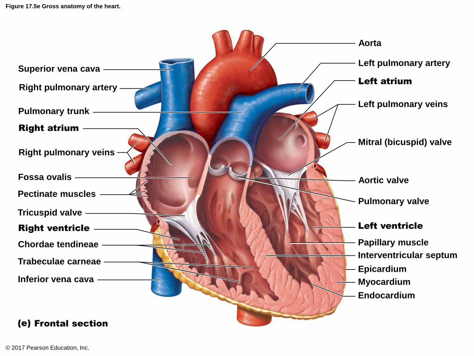
Figure 17.5e Gross anatomy of the heart.
© 2017 Pearson Education, Inc.
Superior vena cava
Right atrium
Right pulmonary artery
Pulmonary trunk
Right pulmonary veins
Fossa ovalis
Pectinate muscles
Tricuspid valve
Right ventricle
Chordae tendineae
Trabeculae carneae
Inferior vena cava
Frontal section
Aorta
Left pulmonary artery
Left atrium
Left pulmonary veins
Mitral (bicuspid) valve
Aortic valve
Pulmonary valve
Left ventricle
Papillary muscle
Interventricular septum
Epicardium
Myocardium
Endocardium

Figure 17.5c Gross anatomy of the heart.
© 2017 Pearson Education, Inc.
Superior vena cava
Pectinate muscles
Crista terminalis
Interatrial septum
Opening of inferior vena
cava (cut)
Fossa ovalis
Opening of coronary sinus
Interventricular sulcus
Ascendingaorta
Myocardium ofleft ventricle
Internal aspect of the right atrium, anterior view
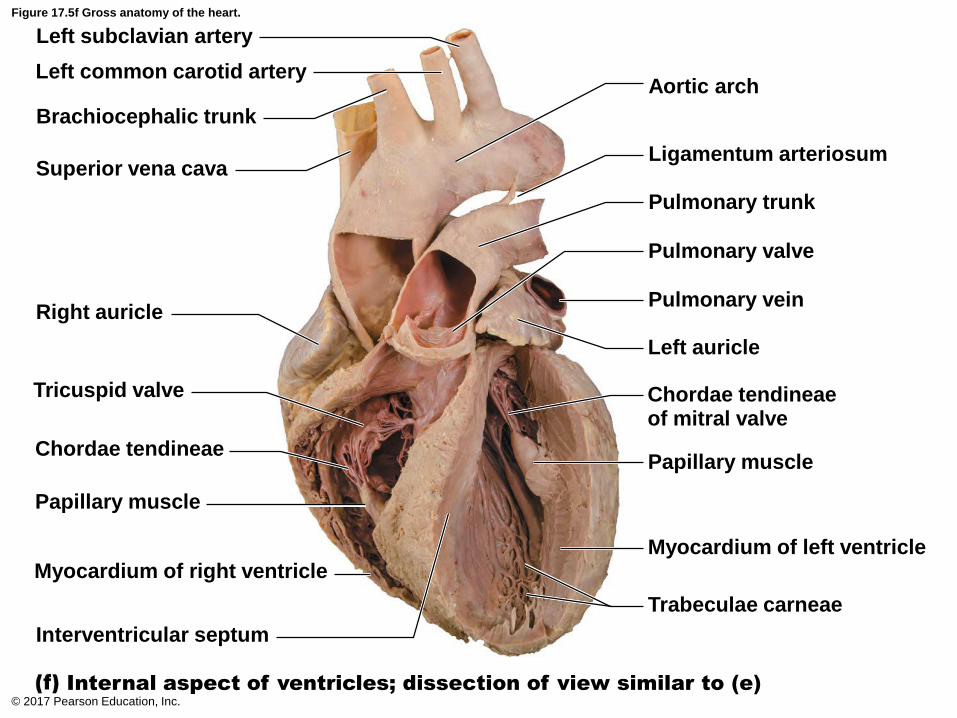
Figure 17.5f Gross anatomy of the heart.
© 2017 Pearson Education, Inc.
Left subclavian artery
Left common carotid artery
Brachiocephalic trunk
Superior vena cava
Right auricle
Tricuspid valve
Chordae tendineae
Papillary muscle
Myocardium of right ventricle
Interventricular septum
Internal aspect of ventricles; dissection of view similar to (e)
Aortic arch
Ligamentum arteriosum
Pulmonary trunk
Pulmonary valve
Pulmonary vein
Left auricle
Chordae tendineaeof mitral valve
Papillary muscle
Myocardium of left ventricle
Trabeculae carneae

Animation – Rotating Heart Sectioned
© 2017 Pearson Education, Inc.

Figure 17.5a Gross anatomy of the heart.
© 2017 Pearson Education, Inc.
Left subclavian artery
Left common carotid artery
Brachiocephalic trunk
Ascending aorta
Right atrium
Right coronary artery(in coronary sulcus)
Right ventricle
Anterior aspect (pericardium removed)
Aortic arch
Ligamentum arteriosum
Pulmonary trunk
Auricle of left atrium
Anterior interventricular artery(in anterior interventricularsulcus)
Additional branch off leftcoronary artery, normalvariation
Left ventricle
Apex of heart(left ventricle)

Animation – Rotating Heart
© 2017 Pearson Education, Inc.

17.2 Heart Valves
• Ensure unidirectional blood flow through heart
• Open and close in response to pressure
changes
• Two major types of valves
– Atrioventricular valves located between atria
and ventricles
– Semilunar valves located between ventricles
and major arteries
© 2017 Pearson Education, Inc.
17.2 Heart Valves
• No valves are found between major veins and
atria; not a problem because:
– Inertia of incoming blood prevents backflow
– Heart contractions compress venous openings
© 2017 Pearson Education, Inc.

Figure 17.6-1 Heart valves.
© 2017 Pearson Education, Inc.
Pulmonary valve
Aortic valve
Area of cutaway
Mitral valve
Tricuspid valve
Myocardium
Tricuspid(right atrioventricular) valve
Mitral(left atrioventricular)valve
Aorticvalve
Pulmonaryvalve
Cardiacskeleton Anterior
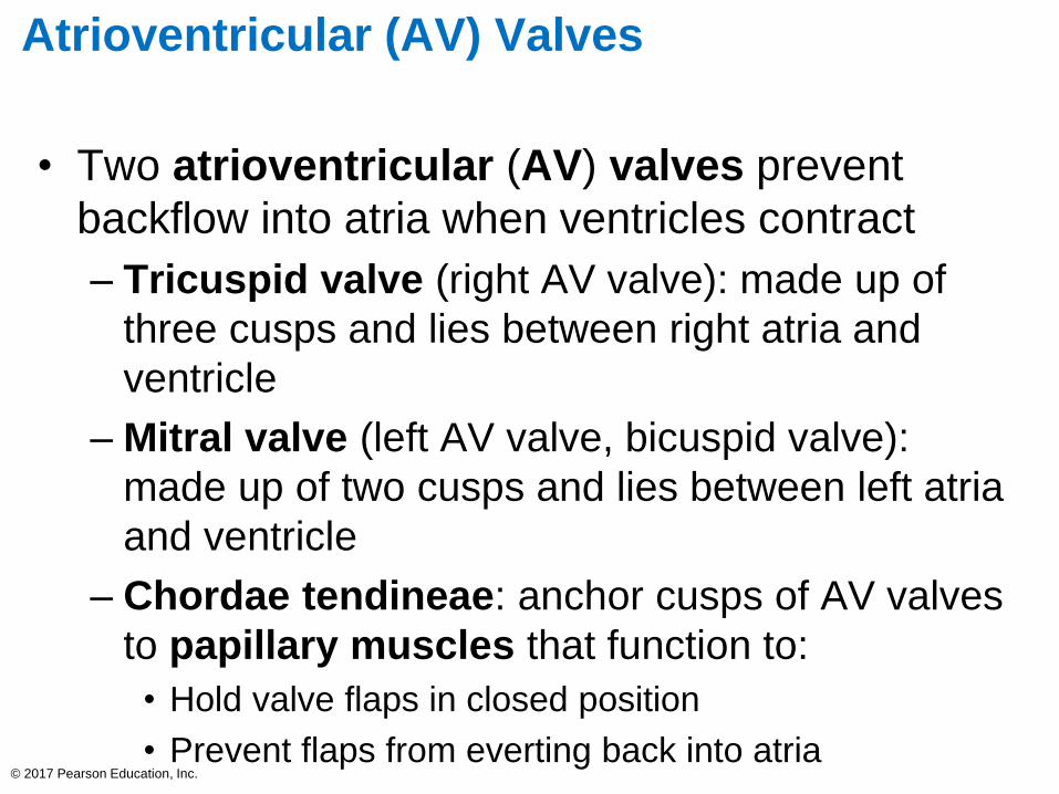
Atrioventricular (AV) Valves
• Two atrioventricular (AV) valves prevent
backflow into atria when ventricles contract
– Tricuspid valve (right AV valve): made up of
three cusps and lies between right atria and
ventricle
– Mitral valve (left AV valve, bicuspid valve):
made up of two cusps and lies between left atria
and ventricle
– Chordae tendineae: anchor cusps of AV valves
to papillary muscles that function to:
• Hold valve flaps in closed position
• Prevent flaps from everting back into atria© 2017 Pearson Education, Inc.

Figure 17.6-2 Heart valves (continued).
© 2017 Pearson Education, Inc.
Chordae tendineae
attached to tricuspidvalve flap
Papillary muscle
Mitral valveChordae tendineae
Tricuspidvalve
Interventricularseptum
Myocardium of leftventricle
Rightatrium
Left atrium
Papillary muscles
Myocardium of right ventricle

Figure 17.7a The function of the atrioventricular (AV) valves.
© 2017 Pearson Education, Inc.
Direction of
blood flow
Atrium
Ventricle
Cusp of
atrioventricular
valve (open)
Chordae
tendineae
Papillary
muscle
Atria contract, forcingadditional blood into ventricles.
As ventricles fill, AV valveflaps hang limply into ventricles.
Blood returning to theheart fills atria, pressingagainst the AV valves. Theincreased pressure forcesAV valves open.
3
2
1
AV valves open; atrial pressure greater than ventricular pressure

Figure 17.6a Heart valves.
© 2017 Pearson Education, Inc.
Pulmonary valve
Aortic valve
Area of cutaway
Mitral valve
Tricuspid valve
Myocardium
Tricuspid(right atrioventricular) valve
Mitral(left atrioventricular)valve
Aorticvalve
Pulmonaryvalve
Cardiacskeleton Anterior

Semilunar (SL) valves
• Two semilunar (SL) valves prevent backflow
from major arteries back into ventricles
– Open and close in response to pressure
changes
– Each valve consists of three cusps that roughly
resemble a half moon
– Pulmonary semilunar valve: located between
right ventricle and pulmonary trunk
– Aortic semilunar valve: located between left
ventricle and aorta
© 2017 Pearson Education, Inc.

Figure 17.8a The function of the semilunar (SL) valves.
© 2017 Pearson Education, Inc.
Semilunar valves open
As ventricles contractand intraventricularpressure rises, bloodis pushed up againstsemilunar valves,forcing them open.
Pulmonarytrunk
Aorta
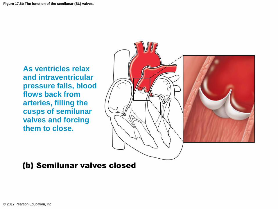
Figure 17.8b The function of the semilunar (SL) valves.
© 2017 Pearson Education, Inc.
Semilunar valves closed
As ventricles relaxand intraventricularpressure falls, bloodflows back fromarteries, filling thecusps of semilunarvalves and forcingthem to close.

Clinical – Homeostatic Imbalance 17.2
• Two conditions severely weaken heart:
– Incompetent valve
• Blood backflows so heart repumps same blood over
and over
– Valvular stenosis
• Stiff flaps that constrict opening
• Heart needs to exert more force to pump blood
• Defective valve can be replaced with
mechanical, animal, or cadaver valve
© 2017 Pearson Education, Inc.

17.3 Pathway of Blood Through Heart
• Right side of the heart
– Superior vena cava (SVC), inferior vena cava
(IVC), and coronary sinus →
– Right atrium →
– Tricuspid valve →
– Right ventricle →
– Pulmonary semilunar valve →
– Pulmonary trunk →
– Pulmonary arteries →
– Lungs
© 2017 Pearson Education, Inc.

17.3 Pathway of Blood Through Heart
• Left side of the heart
– Four pulmonary veins →
– Left atrium →
– Mitral valve →
– Left ventricle →
– Aortic semilunar valve →
– Aorta →
– Systemic circulation
© 2017 Pearson Education, Inc.

Focus Figure 17.1-1 The heart is a double pump, each side supplying its own circuit.
© 2017 Pearson Education, Inc.
Rightatrium
To heart
To lungs
Pulmonaryarteries
Tricuspidvalve
Tricuspidvalve
Pulmonarytrunk
Rightventricle
Pulmonarysemilunarvalve
Pulmonarysemilunar valve
IVC
SVCCoronarysinus
Oxygen-poor blood is carriedin two pulmonary arteries tothe lungs (pulmonary circuit)to be oxygenated.
Oxygen-rich bloodreturns to the heart viathe four pulmonary veins.
Pulmonarycapillaries
Superior vena cava (SVC)Inferior vena cava (IVC)
Coronary sinus
Rightatrium
Rightventricle
Pulmonarytrunk
Oxygen-poor blood
Oxygen-rich blood

Focus Figure 17.1-2 The heart is a double pump, each side supplying its own circuit.
© 2017 Pearson Education, Inc.
Aorta
To body
To heart
Mitralvalve
Leftventricle
Leftatrium
Pulmonaryveins
Aorticsemilunarvalve
Mitralvalve
Aorticsemilunar valve
Oxygen-poor bloodreturns from the bodytissues back to the heart.
Oxygen-rich blood isdelivered to the bodytissues (systemic circuit).
Systemiccapillaries
LeftventricleAorta
Leftatrium
Four pulmonary
veins
Oxygen-poor blood
Oxygen-rich blood

Focus Figure 17.1 The heart is a double pump, each side supplying its own circuit.
© 2017 Pearson Education, Inc.
Right
atrium
Aorta
To body
To heart
To heart
To lungs
Mitralvalve
Leftventricle
Left
atrium
Pulmonary
veins
Pulmonary
arteries
Aorticsemilunarvalve
Tricuspid
valve
Tricuspid
valve
Pulmonary
trunk
Right
ventricle
Pulmonary
semilunar
valve
Pulmonary
semilunar valve
IVC
SVC
Mitralvalve
Aorticsemilunar valve
Coronary
sinus
Oxygen-poor blood is carriedin two pulmonary arteries tothe lungs (pulmonary circuit)to be oxygenated.
Oxygen-poor bloodreturns from the bodytissues back to the heart.
Oxygen-rich blood isdelivered to the bodytissues (systemic circuit).
Oxygen-rich bloodreturns to the heart viathe four pulmonary veins.
Pulmonarycapillaries
Systemic
capillaries
Superior vena cava (SVC)Inferior vena cava (IVC)
Coronary sinus
Rightatrium
Rightventricle
Pulmonarytrunk
LeftventricleAorta
Leftatrium
Four pulmonary
veins
Oxygen-poor blood
Oxygen-rich blood

17.3 Pathway of Blood Through Heart
• Equal volumes of blood are pumped to
pulmonary and systemic circuits
• Pulmonary circuit is short, low-pressure
circulation
• Systemic circuit is long, high-friction circulation
• Anatomy of ventricles reflects differences
– Left ventricle walls are 3× thicker than right
• Pumps with greater pressure
© 2017 Pearson Education, Inc.

Figure 17.9 Anatomical differences between the right and left ventricles.
© 2017 Pearson Education, Inc.
Rightventricle
Leftventricle
Interventricularseptum

Coronary Circulation
• Coronary circulation
– Functional blood supply to heart muscle itself
– Shortest circulation in body
– Delivered when heart is relaxed
– Left ventricle receives most of coronary blood
supply
© 2017 Pearson Education, Inc.

Coronary Circulation (cont.)
• Coronary arteries
– Both left and right coronary arteries arise from
base of aorta and supply arterial blood to heart
– Both encircle heart in coronary sulcus
– Branching of coronary arteries varies among
individuals
– Arteries contain many anastomoses (junctions)
• Provide additional routes for blood delivery
• Cannot compensate for coronary artery occlusion
– Heart receives 1/20th of body’s blood supply
© 2017 Pearson Education, Inc.

Coronary Circulation (cont.)
• Coronary arteries (cont.)
– Left coronary artery supplies interventricular
septum, anterior ventricular walls, left atrium,
and posterior wall of left ventricle; has two
branches:
• Anterior interventricular artery
• Circumflex artery
– Right coronary artery supplies right atrium and
most of right ventricle; has two branches:
• Right marginal artery
• Posterior interventricular artery
© 2017 Pearson Education, Inc.

Figure 17.10a Coronary circulation.
© 2017 Pearson Education, Inc.
Rightventricle
Aorta
Anastomosis(junction ofvessels)
Superiorvena cava
Left atrium
Pulmonarytrunk
Rightatrium
Rightcoronaryartery
Rightmarginalartery
Posteriorinterventricularartery
Anteriorinterventricularartery
Circumflexartery
Leftventricle
Leftcoronaryartery

Coronary Circulation (cont.)
• Coronary veins
– Cardiac veins collect blood from capillary beds
– Coronary sinus empties into right atrium;
formed by merging cardiac veins
• Great cardiac vein of anterior interventricular sulcus
• Middle cardiac vein in posterior interventricular
sulcus
• Small cardiac vein from inferior margin
– Several anterior cardiac veins empty directly
into right atrium anteriorly
© 2017 Pearson Education, Inc.

Figure 17.10b Coronary circulation.
© 2017 Pearson Education, Inc.
Superiorvena cava
Anteriorcardiacveins
Smallcardiac
vein
Middlecardiacvein
Coronarysinus
Greatcardiacvein

Figure 17.5d Gross anatomy of the heart.
© 2017 Pearson Education, Inc.
Aorta
Left pulmonary artery
Left pulmonary veins
Auricle of left atrium
Great cardiac vein
Posterior vein ofleft ventricle
Apex
Left atrium
Left ventricle
Posterior surface view
Superior vena cava
Right atrium
Right pulmonary artery
Right pulmonary veins
Inferior vena cava
Coronary sinus
Right coronary artery(in coronary sulcus)
Posterior interventricularartery (in posteriorinterventricular sulcus)
Middle cardiac vein
Right ventricle
Clinical – Homeostatic Imbalance 17.3
• Angina pectoris
– Thoracic pain caused by fleeting deficiency in
blood delivery to myocardium
– Cells are weakened
• Myocardial infarction (heart attack)
– Prolonged coronary blockage
– Areas of cell death are repaired with
noncontractile scar tissue
© 2017 Pearson Education, Inc.

17.4 Cardiac Muscle Fibers
Microscopic Anatomy
• Cardiac muscle cells: striated, short, branched,
fat, interconnected
– One central nucleus (at most, 2 nuclei)
– Contain numerous large mitochondria (25–35%
of cell volume) that afford resistance to fatigue
– Rest of volume composed of sarcomeres
• Z discs, A bands, and I bands all present
– T tubules are wider, but less numerous
• Enter cell only once at Z disc
– SR simpler than in skeletal muscle; no triads© 2017 Pearson Education, Inc.
Microscopic Anatomy (cont.)
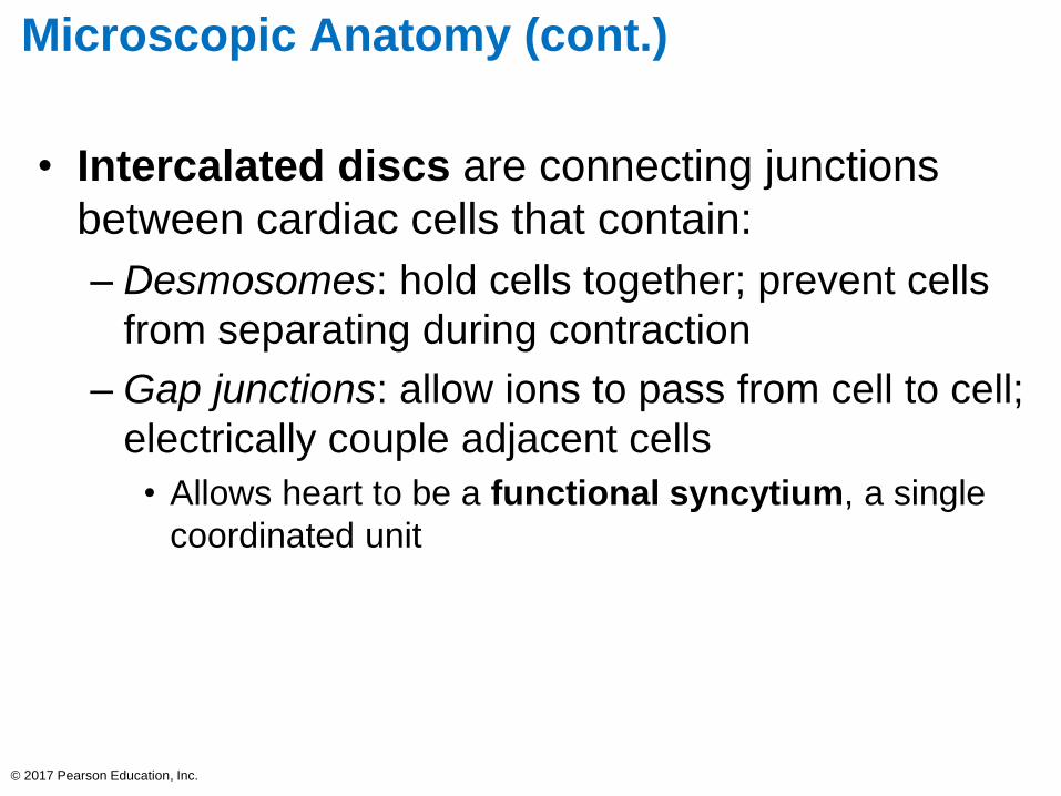
• Intercalated discs are connecting junctions
between cardiac cells that contain:
– Desmosomes: hold cells together; prevent cells
from separating during contraction
– Gap junctions: allow ions to pass from cell to cell;
electrically couple adjacent cells
• Allows heart to be a functional syncytium, a single
coordinated unit
© 2017 Pearson Education, Inc.

Microscopic Anatomy (cont.)
• Intercellular space between cells has connective
tissue matrix (endomysium)
– Contains numerous capillaries
– Connects cardiac muscle to cardiac skeleton,
giving cells something to pull against
© 2017 Pearson Education, Inc.

© 2017 Pearson Education, Inc.
Desmosomes (keep
myocytes from pulling apart)
Gap junctions (electrically
connect myocytes)Nucleus
Intercalated
discs
Cardiac
muscle cell
Figure 17.11a Microscopic anatomy of cardiac muscle.

Figure 17.11b Microscopic anatomy of cardiac muscle.
© 2017 Pearson Education, Inc.
Nucleus
Nucleus
I band
Cardiac muscle cell
A bandSarcolemma
Z disc
Mitochondrion
Mitochondrion
T tubule
Sarcoplasmic
reticulum
I band
Intercalated disc

How Does the Physiology of Skeletal and
Cardiac Muscle Differ?
• Similarities with skeletal muscle
– Muscle contraction is preceded by depolarizing
action potential
– Depolarization wave travels down T tubules;
causes sarcoplasmic reticulum (SR) to release
Ca2+
– Excitation-contraction coupling occurs
• Ca2+ binds troponin causing filaments to slide
© 2017 Pearson Education, Inc.

How Does the Physiology of Skeletal and
Cardiac Muscle Differ? (cont.)
• Differences between cardiac and skeletal
muscle
– Some cardiac muscle cells are self-excitable
• Two kinds of myocytes
– Contractile cells: responsible for contraction
– Pacemaker cells: noncontractile cells that
spontaneously depolarize
» Initiate depolarization of entire heart
» Do not need nervous system stimulation, in contrast
to skeletal muscle fibers
© 2017 Pearson Education, Inc.

How Does the Physiology of Skeletal and
Cardiac Muscle Differ? (cont.)
– Heart contracts as a unit
• All cardiomyocytes contract as unit (functional
syncytium), or none contract
• Contraction of all cardiac myocytes ensures effective
pumping action
• Skeletal muscles contract independently
– Influx of Ca2+ from extracellular fluid triggers
Ca2+ release from SR
• Depolarization opens slow Ca2+ channels in
sarcolemma, allowing Ca2+ to enter cell
• Extracellular Ca2+ then causes SR to release its
intracellular Ca2+
• Skeletal muscles do not use extracellular Ca2+
© 2017 Pearson Education, Inc.
How Does the Physiology of Skeletal and
Cardiac Muscle Differ? (cont.)
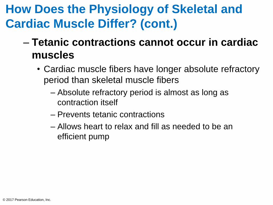
– Tetanic contractions cannot occur in cardiac
muscles
• Cardiac muscle fibers have longer absolute refractory
period than skeletal muscle fibers
– Absolute refractory period is almost as long as
contraction itself
– Prevents tetanic contractions
– Allows heart to relax and fill as needed to be an
efficient pump
© 2017 Pearson Education, Inc.

How Does the Physiology of Skeletal and
Cardiac Muscle Differ? (cont.)
– The heart relies almost exclusively on
aerobic respiration
• Cardiac muscle has more mitochondria than skeletal
muscle so has greater dependence on oxygen
– Cannot function without oxygen
• Skeletal muscle can go through fermentation when
oxygen not present
• Both types of tissues can use other fuel sources
– Cardiac is more adaptable to other fuels, including
lactic acid, but must have oxygen
© 2017 Pearson Education, Inc.

Table 17.1 Key Differences between Skeletal and Cardiac Muscle
© 2017 Pearson Education, Inc.