the cell cycle, dna replication, and mitosisclasspages.warnerpacific.edu/bdupriest/bio 330/ch 19...
TRANSCRIPT

© 2012 Pearson Education, Inc.
Lectures by
Kathleen Fitzpatrick Simon Fraser University
The Cell Cycle,
DNA Replication,
and Mitosis
Chapter 19

© 2012 Pearson Education, Inc.
Figure 19-1B

© 2012 Pearson Education, Inc.
Figure 19-1A

© 2012 Pearson Education, Inc.
Figure 19-20A

© 2012 Pearson Education, Inc.
Figure 19-21A

© 2012 Pearson Education, Inc.
Figure 19-22A

© 2012 Pearson Education, Inc.
Figure 19-20B
© 2012 Pearson Education, Inc.
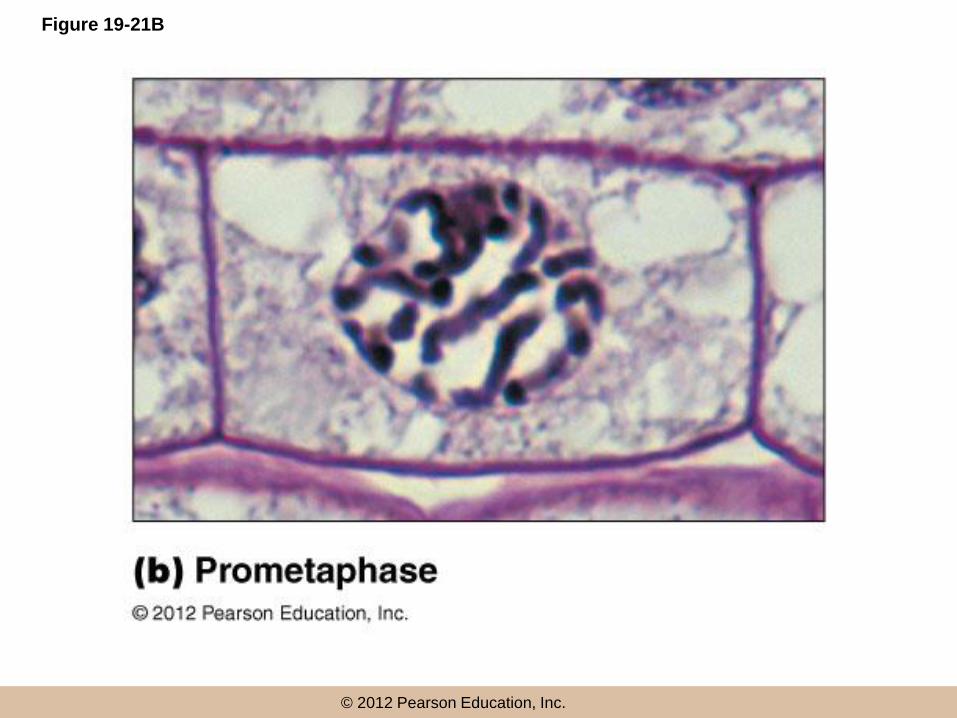
Figure 19-21B

© 2012 Pearson Education, Inc.
Figure 19-20C

© 2012 Pearson Education, Inc.
Figure 19-21C

© 2012 Pearson Education, Inc.
Figure 19-22B

© 2012 Pearson Education, Inc.
Figure 19-23

© 2012 Pearson Education, Inc.
Figure 19-20D

© 2012 Pearson Education, Inc.
Figure 19-21D

© 2012 Pearson Education, Inc.
Figure 19-24


© 2012 Pearson Education, Inc.
Figure 19-20E

© 2012 Pearson Education, Inc.
Figure 19-21E

© 2012 Pearson Education, Inc.
Figure 19-25

© 2012 Pearson Education, Inc.
Figure 19-26
© 2012 Pearson Education, Inc.
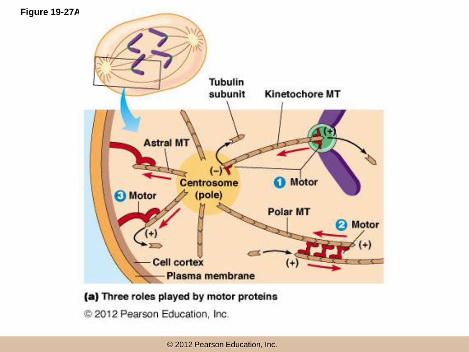
Figure 19-27A

© 2012 Pearson Education, Inc.
Figure 19-27B

© 2012 Pearson Education, Inc.
Figure 19-27C

© 2012 Pearson Education, Inc.
Figure 19-28

© 2012 Pearson Education, Inc.
Figure 19-29A

© 2012 Pearson Education, Inc.
Figure 19-29B

© 2012 Pearson Education, Inc.
Figure 19-30

© 2012 Pearson Education, Inc.
Figure 19-31

© 2012 Pearson Education, Inc.
Progression Through the Cell Cycle Is
Controlled at Several Key Transition
Points
• Control of the cell cycle must
– 1. Ensure that events of each phase are carried out in the correct order and at the appropriate time
– 2. Ensure that each phase is completed before the next one begins
– 3. Respond to external conditions

© 2012 Pearson Education, Inc.
Figure 19-32

© 2012 Pearson Education, Inc.
Mitotic Cdk-Cyclin Drives Progression
Through the G2-M Transition by
Phosphorylation Key Proteins Involved
in the Early Stages of Mitosis
• Evidence of a control molecule triggering mitosis came from experiments involving frog eggs (Masui)
• Mature eggs develop from oocytes through meiosis; the oocyte arrests shortly after meiosis begins until a hormone signal is received
• Injecting the cytoplasm of a mature egg into an immature oocyte causes it to immediately proceed through meiosis

© 2012 Pearson Education, Inc.
Figure 19-34

© 2012 Pearson Education, Inc.
Figure 19-35
© 2012 Pearson Education, Inc.
Activation of mitotic Cdk
• Activation of mitotic Cdk involves
phosphorylation and dephosphorylation
• The binding of mitotic cyclin to mitotic Cdk forms
a cyclin-Cdk complex that is initially inactive (1)
• Inhibiting kinases phosphorylate two sites on the
Cdk, blocking the active site (2)

© 2012 Pearson Education, Inc.
Figure 19-36

© 2012 Pearson Education, Inc.
Figure 19-37
© 2012 Pearson Education, Inc.
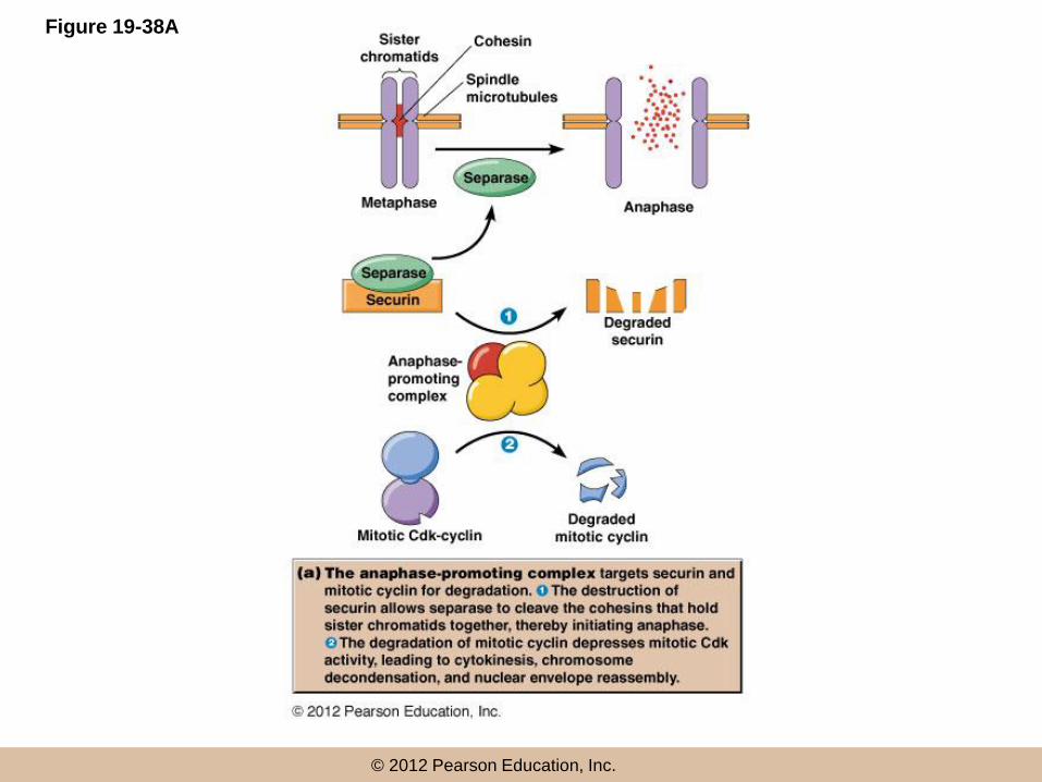
Figure 19-38A

© 2012 Pearson Education, Inc.
Figure 19-39

© 2012 Pearson Education, Inc.
Figure 19-38B

© 2012 Pearson Education, Inc.
Figure 19-40

© 2012 Pearson Education, Inc.
Figure 19-42

© 2012 Pearson Education, Inc.
Figure 19-43
© 2012 Pearson Education, Inc.
Inhibitory Growth Factors Act Through
Cdk Inhibitors
• Some growth factors inhibit cell proliferation, e.g.,
transforming growth factor (TGF)
• TGFbinding to its receptor phosphorylates Smad
proteins that move into the nucleus and activate
expression of genes coding for proteins that inhibit
proliferation
• Two Cdk inibitors that block cell cycle progression
are p15 and p21

© 2012 Pearson Education, Inc.
Apoptosis
• Damaged or diseased cells need to be eliminated
• In such cases, the process must not damage
surrounding cells
• Multicellular organisms accomplish this through a
programmed cell death—apoptosis

© 2012 Pearson Education, Inc.
Apoptosis and necrosis
• Cell death called necrosis sometimes follows
tissue injury
• Necrosis involves swelling and rupture of
injured cells, whereas apoptosis involves a
specific series of events that lead to
dismantling of the cell contents

© 2012 Pearson Education, Inc.
Steps of apoptosis
• The cell’s DNA segregates near the periphery of
the nucleus and the cytoplasm decreases (1)
• The cell produces small cytoplasmic extensions
and the nucleus begins to fragment (2)
• DNA is cleaved by an apoptosis-specific
endonuclease and the cell is dismantled into
small pieces called apoptotic bodies

© 2012 Pearson Education, Inc.
Steps of apoptosis (continued)
• Inactivation of a phospholipid translocator
(flippase) causes accumulation of
phosphatidylserine in the outer leaflet of the
plasma membrane
• This serves as a signal for the remnants of
the affected cell to be engulfed by nearby
cells via phagocytosis (3)

© 2012 Pearson Education, Inc.
Figure 19-44A

© 2012 Pearson Education, Inc.
Figure 19-44B,C,D
© 2012 Pearson Education, Inc.
Caspases
• Apoptosis proceeds through the activation of
a series of enzymes called caspases
• They are produced as inactive precursors
called procaspases and are cleaved to
create active enzymes
© 2012 Pearson Education, Inc.
Apoptosis Is Triggered by Death
Signals or Withdrawal of Survival
Factors
• There are two main routes by which cells can
activate caspases and enter apoptosis
• Activation can occur directly, e.g., when human
cells are infected by viruses, cytotoxic T
lymphocytes are activated and induce apoptosis
• This is triggered when cells receive cell death
signals

© 2012 Pearson Education, Inc.
Apoptosis in cell infected by viruses
• Two death signals are tumor necrosis factor and
CD95/Fas
• CD95 is a protein on the surface of infected cells;
lymphocytes have a protein on their surfaces that
binds CD95, causing it to aggregate
• Adaptor proteins attach to the CD95, which
recruits procaspase-8 to the sites of clustering

© 2012 Pearson Education, Inc.
Initiator and executioner caspases
• When the procaspase is activated it acts as an
initiator caspase, initiating the cascade
• Initiator caspases also activate an executioner
caspase, caspase-3, which is important for
activating many steps in apoptosis

© 2012 Pearson Education, Inc.
The second type of apoptosis
• One of the best-studied cases of the second type of apoptosis involves survival factors
• When survival factors are withdrawn, a cell may enter apoptosis
• The site of action is the mitochondrion
• Healthy cells have several anti-apoptotic proteins in the outer mitochondrial membrane

© 2012 Pearson Education, Inc.
The second type of apoptosis (continued)
• The proteins are structurally related to a protein called Bcl-2 which, together with other proteins, counteracts proteins that promote apoptosis (pro-apoptotic proteins)
• When cellular signals shift in balance toward pro-apoptotic proteins, the cell is more likely to undergo apoptosis
• One pro-apoptotic protein is called Bad (Bcl-2-associated death promoter)

© 2012 Pearson Education, Inc.
Mitochondria trigger apoptosis
• Mitochondria trigger apoptosis by releasing cytochrome c into the cytosol after accumulation of pro-apoptotic proteins lead to formation of channels in the outer mitochondrial membrane
• Cytochrome c stimulates calcium release from mitochondria and ER, where it binds IP3 receptors
• It also activates an initiator procaspase, procaspase-9, which then activates caspase-3

© 2012 Pearson Education, Inc.
Damaged cells can trigger their own
apoptosis
• If a cell suffers such damage that it can’t repair
itself, it may trigger its own demise
• It can enter apoptosis through the activity of
p53, which acts through the protein Puma,
which binds and inhibits Bcl-2

© 2012 Pearson Education, Inc.
Figure 19-45