the cleavage of phospholipides by brain … · the cleavage of phospholipides by brain tissue* by...
TRANSCRIPT

THE CLEAVAGE OF PHOSPHOLIPIDES BY BRAIN TISSUE*
BY WARREN M. SPERRY
(From the Departments of Biochemistry, New York State Psychiatric Institute and Hospital, and the College of Physicians and Surgeons, Columbia University,
New York)
(Received for publication, July 16, 1947)
Studies of brain metabolism with isotopic indicators are unanimous in showing that the rate of turnover of lipides in the adult rat brain is exceed- ingly slow in vivo (l-6). The results obtained by several investigators with P32 are particularly convincing in demonstrating a low rate of phospholipide metabolism. It might be expected from these findings that no change in lipides, detectable with conventional methods, would occur during short periods of incubation of brain preparations in vitro. However, the chemical behavior of isolated tissue is not necessarily the same as it is in its natural environment in the living animal, and it seemed worth while to include some measurements of lipides in a study of brain respiration under way in our laboratory.
These preliminary experiments showed an apparent decrease in the phos- phorus of the fraction of brain homogenates, soluble in organic solvents, during a 4 hour period of incubation. This finding was confirmed and stud- ied in considerable detail.
EXPERIMENTAL
Brain tissue from rats was homogenized with the all-glass apparatus of Potter and Elvehjem (7) in saline-carbonate buffer, saturated with 5 per cent CO*-95 per cent 02. In the experiments through Experiment 45 (Table I) the buffer was at room temperature and the homogenizer was run from 40 to 50 seconds. The tube was moved up and down continuously during the homogenization. The temperature did not increase perceptibly to the touch. However, to minimize possible effects of frictional heating, from Experiment 45 through the rest of the investigation cold buffers were used and the time of homogenization was reduced to 25 to 30 seconds. For control determinations portions of the homogenates, as shown in Table I, were pipetted into 25 cc. volumetric flasks and extracted at once with alcohol-ether (3: 1). In Experiments 10 to 14, inclusive, other portions were employed for respiratory measurements in the Warburg apparatus,
* Preliminary reports of this investigation were presented to the Division of Biological Chemistry of the American Chemical Society at Buffalo, 1942, and to the American Society of Biological Chemists (Federation Proc., 2, 70 (1943)).
675
by guest on September 12, 2018
http://ww
w.jbc.org/
Dow
nloaded from

676 PHOSPHOLIPIDE CLEAVAGE BY BRAIN
TABLE I
Decrease in Lipide Phosphorus during Incubation of Brain I’issue Homogenates in Carbonate Buffer for .J Hours with Shaking at 87.6”
Cerebral hemispheres “ ‘I
10 Whole brain 11 “ ‘I
12 ‘I I‘
13 “ “
14 ‘I ‘I
15 16 17
‘I “
27 I‘ ‘I
29 ‘I “
30 “ “
31 “ “
32 ‘I ‘I
33§ I‘ ‘I
348 “ ‘I
35§ “ “
36 37 38 39 40 41 41
428 43 45 46 47 53 55 55 56
Cerebellum + brain stem* Cerebral hemispheres
‘I “ “ “ “ “ “ “
Cerebellum + brain stem* Whole brain
‘I “ “ “ “ “ ‘I “ “ “
Cerebral hemispheres Cerebellum + brain stem* Whole brain
* Including medulla and pons.
fw. 131 130 131 131 48
133 131 131 83 91 90
158 173 168 189 199 153 148 168 153 167 167 166 167 141 177 164 147 165 162 137 172
cc.
1.0 1.0 1.0 1.0 2.0 1.0 1.0 1.0 2.0 2.0 2.0 0.5 0.5 0.5 0.5 0.5 0.5 0.5 0.5 0.5 0.5 0.5 0.5 2.0 0.5 0.5 0.5 0.5 0.5 0.5 0.5 0.5
Phosphorus per mg. brain
Control ncubatec
Y Y per cent
2.64 2.40 9.1 2.57 2.33 9.3 2.61 2.52 3.4 2.64 2.24 15.2 2.53 2.29 9.5 2.53 2.15 15.0 2.48 2.31 6.9 2.48 2.27 8.5 2.61 2.41 7.7 2.57 2.49 3.1 2.25 2.15 4.4 2.37 1.96 17.3 2.33 2.16 7.3 1.61 1.35 16.1 1.76 1.56 11.4 1.76 1.67 5.1 2.51 2.28 9.2 2.27 2.18 4.0 2.30 1.99 13.5 2.60 2.25 13.5 2.37 2.29 3.4 2.27 2.18 4.0 2.55 2.45 3.9 1.62 1.53 5.6 2.50 2.39 4.4 2.52 2.30 8.7 2.35 2.24 4.7 2.49 2.29 8.0 2.44 2.16 11.5 2.30 2.13 7.4 2.69 2.55 5.2 2.39 2.17 9.2
DeCIeaSe
t The data in this column are only approximate, but errors in this measurement have no effect on the percentage decrease, since they apply equally to bothof the phosphorus values.
$ The volumes shown were pipetted into 25 cc. volumetric flasks for control determinations and for incubation, except in Experiments 10 to 14, in which the incubated samples were pipetted at the end of the incubation period.
$ Age of rats: Experiment 33,18 days; Experiments 34 and 35,20 days; Experiment 42, 16 days; other rats adults.
by guest on September 12, 2018
http://ww
w.jbc.org/
Dow
nloaded from

W. M. SPERRY 677
and at the end of a 4 hour period of shaking in the bath at 37.5” samples were pipetted for extraction exactly as in the control analyses. In the re- maining experiments “control” and “incubated” portions were pipetted into 25 cc. volumetric flasks at the start of the experiment. The “control” samples were extracted at once; the experimental samples were shaken for 4 hours in the Warburg apparatus and then extracted. The stoppers were removed and the flasks were flushed out with CO*-O2 at intervals of about 3 hour during the period of incubation.
In making the extractions about 15 cc. of solvent were added and brought to a boil and the flasks were allowed to stand, usually overnight. Solvent was added to the mark and the content,s were thoroughly mixed and filtered. Phosphorus was determined in 1 cc. portions of the filtrate by Sperry’s micro modification (8) of the Fiske and Subbarow method (9).
In most of the experiments two control and two incubated samples were extracted and each sample was analyzed in duplicate; i.e., each phosphorus value, with a few exceptions, is the average of four determinations. An est,imate of the error of the procedure for determining phosphorus in the extracts was obtained by calculating the st.andard deviation of the per- centage deviations of the duplicate estimations from their respective means. The standard deviation of 344 such values was f 1.1 per cent. This es- timate does not include the errors of preparing the extract’s, particularly of pipett.ing samples of the homogenates which usually contained air bubbles and were not entirely homogeneous.’ An est,imate of the over-all error was obtained by calculating the standard deviation of the four analyses (two on each of two samples) from their respective means. The value was f1.9 per cent.
Although in some of the experiments the decreases in lipide phosphorus content (Table I) were not significant, the majority were too large to be fortuitous and the fact that a change in the same direction was observed in every experiment leaves no doubt that the over-all result is significant. The wide variation in the magnitude of the change is probably related to variations in the preparation of the homogenates.
The loss of lipide phosphorus during incubation of brain t.issue appears to demonstrate the presence in brain of a fairly active mechanism capable of cleaving phospholipides, but other explanations were considered. That non-lipide phosphorus was extracted in the control but not in the incubated samples appeared unlikely from some experiments in which the filtered alcohol-ether extracts from replicate control and incubated samples were taken to dryness and reextracted with ethylene chloride. The solutions were made up to the original volume and phosphorus was determined in
1 In the latter part of the work, starting with Experiment 71, the samples of the emulsions were weighed to minimize this error.
by guest on September 12, 2018
http://ww
w.jbc.org/
Dow
nloaded from

678 PHOSPHOLIPIDE CLEAVAGE BY BRAIN
1 cc. portions. A comparison of the results (Table II) with the correspond- ing values found in alcohol-ether extracts of the same homogenates (Table I) shows that although only about three-fourths of t,he alcohol-ether-soluble phosphorus was extracted by ethylene chloride the percent,age differences between control and incubated homogenates were as large as, or in some experiments considerably larger than, those found in the alcohol-ether extracts.
More serious is the possibility that a portion of the phospholipides be- came insoluble during incubation through the formation of stable eombina- tions with protein, or in some other way. Three types of evidence, bearing on this point, were obtained. In one series of experiments a different and somewhat more efficient solvent mixture, methyl alcohol-chloroform (1: l),
TABLE II
Reextraction with Ethylene Chloride
I 7 I 7 / per cent / I y I y I per cent
* In this experiment rat blood serum was added to the homogenate. In the cor- responding experiment, in which alcohol-ether was used as the only solvent, the decrease was 10.0 per cent.
was compared with alcohol-ether. The technique was t.he same as that described except that incubation was continued for 21 to 24 hours,.with or without shaking during the first 6 or 7 hours, and the more complex carbonat,e buffer of Krebs and Henseleit (lo), with phosphate omit,t.ed, was used in Experiments 79 to 88. Merthiolate was added in the proportion of 10 mg. to 100 cc. of buffer. Each phosphorus value (Table III) is the average of at least four determinations, two on each of two samples of the brain homo- genate. Although methyl alcohol-chloroform extracted a little more phos- phorus than alcohol-ether, the decreases were about the same with the two solvent mixtures, except in Experiment 87 in which a very high apparent decrease was found in the samples extracted with alcohol-ether. This value is so far out of line with other results obtained in t,his investigation that it is open to serious question, but the replicate determinations agreed closely and there is no evident reason for excluding it
In a further effort to rule out a change in solubility as an explanation of the results an entirely different method of extraction was employed, viz., the
by guest on September 12, 2018
http://ww
w.jbc.org/
Dow
nloaded from

W. M. SPERRY 679
colloidal iron procedure of Folch and Van Slyke (11). In the first attempts to apply this t~echnique to brain homogenates the agreement among repli- cates was poor and the amounts of lipide phosphorus found were only about half of those obtained with direct alcohol-ether extraction. The method was studied in considerable detail, with particular attention to the procedure for extraction from the colloidal iron precipitate. Various amounts of water were added to the precipitate and various combinations of solvents
TABLE III Comparison of Solvents
51 51B* 52 52B* 7% 81 82 87 88
Alcohol-ether (3: 1)
.-
-
Phosphorus per mg. brain Phosphorus per mg. brain Decrease Decrease
Control Incubated Control Incubated
Y Y per cent Y Y per cent
2.32 1.97 15.1 2.44 2.12 13.1 2.46 1.88 23.6 2.65 2.19 17.4 2.35 2.03t 13.6 2.55 2.20 13.7 2.31 1.74 24.7 2.66 2.05 22.9 0.98 0.82 16.3 1.06 0.88 17.0 2.42 2.08 14.0 2.53 2.18 13.8 2.09 1.97 5.7 2.38 2.18 8.4 2.33 0.90 61.45 2.78 2.49 10.4 2.16 1.87 13.4 2.28 1.97 13.6
Methyl alcohol-chloroform (1: 1)
* Rat blood was added to the brain homogenates in these experiments. t The phosphorus value found in one of the samples was so low (1.56 y) thatan
error seemed likely and the value was omitted from the average. $ The brains of 5 day-old rats were used. $ The individual phosphorus values were as follows: control Flask 1,2.32 and 2.33;
control Flask 2, 2.34, and 2.34; incubated Flask 1, 0.91 and 0.89; incubated Flask2, 0.89 and 0.90.
were tried. Fairly uniform results were obtained with the following pro- cedure: Approximately 0.2 cc. portions of the homogenate were placed in tared 15 cc. centrifuge tubes, weighed, and treated as described by Folch and Van Slyke. The precipitate was washed twice with magnesium sulfate solution and once with water. Either 0.8 or 1.1 cc. of water and 2 cc. of alcohol were added and the precipitate was stirred into suspension and transferred to a 10 cc. volumetric flask. Two 1 cc. portions of alcohol followed by 2 and 1 cc. portions of ether were used in washing. The flask was filled to the mark with ether and 1.0 cc. portions of the filtrate were taken for phosphorus determination. In the preliminary experiments the quantity of water added was varied from none to 1.6 cc. Maximal yields of lipide phosphorus were obtained with 0.8 and 1.1 cc. and both of these
by guest on September 12, 2018
http://ww
w.jbc.org/
Dow
nloaded from
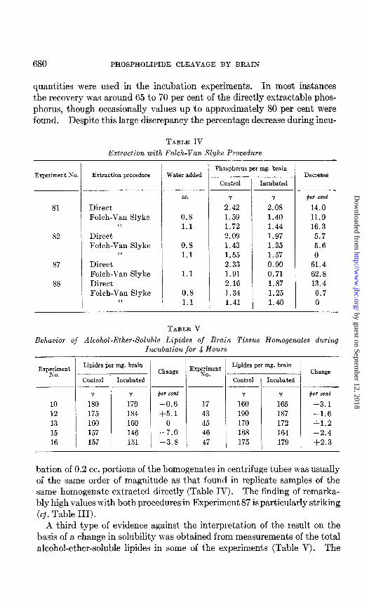
680 PHOSPHOLIPIDE CLEAVAGE BY BRAIN
quantities were used in the incubation experiments. In most instances the recovery was around 65 to 70 per cent of the directly extractable phos- phorus, though occasionally values up to approximately 80 per cent were found. Despite this large discrepancy the percentage decrease during incu-
TABLE IV
Extraction with Folch-Van Slyke Procedure
Experiment No. Extraction procedure Water added
cc.
81 Direct Folch-Van Slyke
“ 0.8 1.1
82 Direct Folch-Van Slyke
“
87
88
Direct Folch-Van Slyke Direct Folch-Van Slyke
I‘
0.8 1.1
1.1
0.8 1.1
Phosphorus per mg. brain
Control Incubated
Y 7
2.42 2.08 1.59 1.40 1.72 1.44 2.09 1.97 1.43 1.35 1.55 1.57 2.33 0.90 1.91 0.71 2.16 1.87 1.34 1.25 1.41 1.40
-
--
Decrease
per cent
14.0 11.9 16.3
5.7 5.6 0
61.4 62.8 13.4
6.7 0
TABLE V
Behavior of Alcohol-Ether-Soluble Lipides of Brain Tissue Homogenates during Incubation for Q Hours
Expekwt
10 12 13 15 16
Lipides per mg. brain Change
Control Incubated
Y Y
180 179 175 184 160 160 157 146 157 151
per cent Y Y per cent
-0.6 17 160 165 t-3.1 +5.1 43 190 187 -1.6
0 45 170 172 f1.2 -7.0 46 168 164 -2.4 -3.8 47 175 179 +2.3
Explement Lipides per mg. brain
. I Control 1 Incubated Change
bation of 0.2 cc. portions of the homogenates in centrifuge tubes was usually of the same order of magnitude as that found in replicate samples of the same homogenate extracted directly (Table IV). The finding of remarka- bly high values with both procedures in Experiment 87 is particularly striking (cl. Table III).
A third type of evidence against the interpretation of the result on the basis of a change in solubility was obt,ained from measurements of the total alcohol-ether-soluble lipides in some of the experiments (Table V). The
by guest on September 12, 2018
http://ww
w.jbc.org/
Dow
nloaded from

W. M. SPERRY 681
total extract remaining after the two 1 cc. samples for phosphorus deter- mination were removed was transferred quantitatively to a weighed flask and taken to dryness in a stream of carbon dioxide. Although the quan- tities were small, close agreement between duplicate determinations was obtained with the use of a uniform technique for drying and weighing the flasks. The apparent changes were about equal in both directions and were within the error of the procedure; the largest apparent difference (7 per cent) represented a weight of only 1.4 mg.
TABLE VI
Effect of Blood Added to Homogenates of Brain Tissue on Cleavageof Phospholipides
37
38
39
48
49
51
52
-
i .-
Time of ncubation
-
- hrs. CC.
4 0 4 0.85 4 0 4 0.25 4 0 4 0.41*
24 0 24 0.24 24 0 24 0.25 24 0 24 0.24 24 0 24 0.29
- I F
-
hxreasein ,hosphorus
per cent 4.0 6.5
13.5 7.6
13.5 10.0 25.1 24.1
7.5 17.4 15.1 23.6 13.6 24.7
- 1 Expe&ment
53
54
56
Time of ncubation
Blood per =m. brain
hrs.
4 4
24 24
7 7
24 24
4 4
21 21
CC.
0 0.22 0 0.22 0 0.23 0 0.23 0 0.24 0 0.24
-
! -
‘: - per cent
11.5 4.9
16.0 20.9
9.3 7.0
14.8 24.2
9.2 6.3 9.4
13.6
* Blood serum from another rat added in this experiment
Effect of Added Blood-The possibility that phospholipide-splitting en- zymes of the blood were responsible for the decrease in lipide phosphorus was investigated through a study of the effect of a large excess of blood. The brain was divided longitudinally into approximately equal parts, of which one was homogenized in carbonate buffer, and the other in buffer to which had been added blood from the same rat,, or in one case serum from another rat. Samples of the t,\vo homogenates were carried through the procedure side by side. The flasks were shaken in the Warburg bath for the times indicated in Table VI, except that in the 21 or 24 hour experiments shaking was continued for 4 to 7 hours, after which the flasks were placed in an incubator at 37.5” for the remainder of the period. The data show no effect of blood or serum in increasing the cleavage of phospholipides during a 4 or 7 hour period of incubation; indeed in five of the six esperi- ments the decrease in phosphorus was less in t.he presence of an excess of
by guest on September 12, 2018
http://ww
w.jbc.org/
Dow
nloaded from

682 PHOSPHOLIPIDE CLEAVAGE BY BRAIN
blood than in the emulsions to which no blood had been added. This result indicates that enzymes of blood were not responsible for the cleavage of phospholipides found in short time experiments. On the other hand in six of the seven 21 or 24 hour experiments the decrease was greater in the presence than in the absence of added blood, a finding which suggests that enzymes of the blood may be responsible, at least in part, for the increased splitting of phospholipides usually observed during autolysis for a long period.
E$ect of Time of Incubation and of Shaking-In several experiments the effect of time of incubation (1 to 24 hours), of shaking, or of both was studied on replicate samples of the same homogenate. The data are too variable to justify detailed presentation, but they show a definite tendency for the degree of cleavage to increase with increasing time and little, if any, effect of shaking.
E$ect of Heating-Replicate samples of the homogenates were heated at 100” for 2 to 10 minutes before incubation in several of the experiments. In all cases the splitting of phospholipides was reduced, usually to less than half that of the unheated samples, but in only one instance was there com- plete inactivat.ion. Some of this cleavage probably occurred during the heating; in four experiments in which samples were heated to 100” for 10 minutes and then analyzed without incubation, there was a decrease in phosphorus averaging 3.2 per cent. However, not all of the splitting in the heated and incubated samples can be accounted for in this way, and it must be concluded that if the reaction is catalyzed by enzymes they must be unusually resistant to heat. There is some support for this possibility: the lecithinase A of snake venom and of pancreas is remarkably stable to heat (12, 13).
Effect of Various Solutions Used in Preparing Brain Homogenates-h the experiments described above either a simple saline-carbonate buffer or the more complex carbonate buffer described by Krebs and Henseleit (lo), with phosphate omitted, was used. These buffers were saturated with 95 per cent 02-5 per cent COZ. They were compared in eight experiments in which half the brain was homogenized in one and the other half in the other buffer. The two homogenates were incubated side by side for different periods of time, with or without shaking. The average percentage decreases in phosphorus were 12.1 and 13.8, respectively. In several of these experiments the pH was determined with the glass electrode in replicate samples which had been carried through the incubation procedure. The values ranged from 7.0 to 7.9 and agreed closely for the two homogenates in each experiment.
In three experiments homogenates made from halves of the same brain in simple carbonate buffer and in water tt-ere compared. The average de-
by guest on September 12, 2018
http://ww
w.jbc.org/
Dow
nloaded from
W. M. SPERRY 683
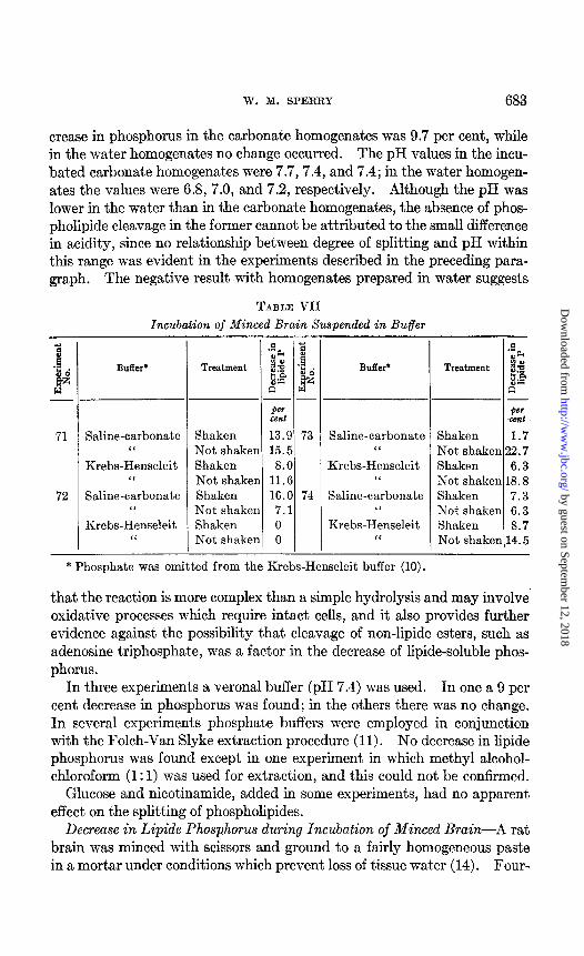
crease in phosphorus in the carbonate homogenates was 9.7 per cent, while in the water homogenates no change occurred. The pH values in the incu- bated carbonate homogenates were 7.7,7.4, and 7.4; in the water homogen- ates the values were 6.8, 7.0, and 7.2, respectively. Although the pH was lower in the water than in the carbonate homogenates, the absence of phos- pholipide cleavage in the former cannot be attributed to the small difference in acidity, since no relationship between degree of splitting and pH within this range was evident in the experiments described in the preceding para- graph. The negative result with homogenates prepared in water suggests
TABLE VII
Incubation of Minced Brain Suspended in Buffer
1 g
71
72
-
BUff&
Saline-carbonate “
Krebs-Henseleit ‘I
Saline-carbonate ‘I
Krebs-Henseleit “
-
_-
-
Treatment
Per cent
Shaken 13.9 73 Not shaken 15.5 Shaken 8.0 Not shaken 11.6 Shaken 16.0 74 Not shaken 7.1 Shaken 0 Not shaken 0
Saline-carbonate “
Krebs-Henseleit “
Saline-carbonate “
Krebs-Henseleit ‘I
Shaken 1.7 Not shaken 22.7 Shaken 6.3 Not shaken 18.8 Shaken 7.3 Not shaken 6.3 Shaken 8.7 Not shaken 14.5
* Phosphate was omitted from the Krebs-Henseleit buffer (10).
that the reaction is more complex than a simple hydrolysis and may involve. oxidative processes which require intact cells, and it also provides further evidence against the possibility that cleavage of non-lipide esters, such as adenosine triphosphate, was a factor in the decrease of lipide-soluble phos- phorus.
In three experiments a Verona1 buffer (pH 7.4) was used. In one a 9 per cent decrease in phosphorus was found; in the others there was no change. In several experiments phosphate buffers were employed in conjunction with the Folch-Van Slyke extraction procedure (11). No decrease in lipide phosphorus was found except in one experiment in which methyl alcohol- chloroform (1: 1) was used for extraction, and this could not be confirmed.
Glucose and nicotinamide, added in some experiments, had no apparent effect on the splitting of phospholipides.
Decrease in Lipide Phosphorus during Incubation of Minced Brain-A rat brain was minced with scissors and ground to a fairly homogeneous paste in a mortar under conditions which prevent loss of tissue water (14). Four-
by guest on September 12, 2018
http://ww
w.jbc.org/
Dow
nloaded from

684 PHOSPHOLIPIDE CLEAVAGE BY BRAIN
teen portions were drawn up in a glass tube fitted with a glass plunger, transferred to tared 25 cc. volumetric flasks, and weighed. Simple saline- carbonate buffer was added to half the flasks and the Krebs-Henseleit car- bonate buffer to the other half. Of each set of seven, three were extracted at once with alcohol-ether as controls and the other four were incubated for about 21 hours, with or without shaking for the first 5 to 6 hours. Each sample was analyzed for phosphorus in duplicate.
The results (Table VII) show that wit.h three exceptions the lipide phos- phorus decreased as it did in similar experiments with brain homogenates. There was no consistent difference in results with the two buffers. Shaking usually, though not always, appeared to have an adverse effect on the reaction.
DISCUSSION
The results of this investigation demonstrate that a significant decrease, which may be of considerable magnitude, in the quantity of phosphorus extractable with lipide solvents occurs during incubation of homogenized or minced brain tissue in carbonate buffer, and the findings show further that the decrease is in all probability the result of an active mechanism present in brain tissue with the ability to split phospholipides at a relatively rapid rate.
Other investigators have presented indirect evidence for the presence of “lecithinase” in the brain. Coriat (15) found that choline was liberated during autolysis of a sample of minced brain for 72 hours in the presence of chloroform, and Simon (16) in similar experiments observed an increase in inorganic phosphorus. King (17) incubated aqueous extracts of rabbit and chicken brains with a lecithin emulsion for 48 hours and observed a small and variable increase in the t,otal free and acid-soluble phosphorus. The only indirect study in which short periods of incubation were employed was reported by Stamm (18), who incubated suspensions of minced calf brain in Locke’s solution for periods of 3 to 14 hours and measured the increase in inorganic phosphorus. In nine to fifteen such experiments he found an average increase of 0.13 y per mg. of brain at 3 hours, 0.20 y at 43 hours, and 0.24 y at 6 hours. These values may be compared with the average decrease in lipide phosphorus of 0.19 y per mg. of brain found in the 4 hour experiments (Table I) of the present investigation.
In a publication which appeared after most of the experiments listed in Table I had been completed, Fries, Schachner, and Chaikoff (19) reported direct studies, similar to those reported here, on one young (15 gm.) and one adult (200 gm.) rat. Homogenat’es of brain tissue in carbonate buffer were incubated for 1, 2, and 4 hours. The quantity of brain per unit vol- ume of solution (about 60 mg. per cc.) was considerably less than that
by guest on September 12, 2018
http://ww
w.jbc.org/
Dow
nloaded from

TV. M. SPERRY 685
employed in most of my experiments and the methods of measurement were also different: changes in the quantity of phospholipide were determined by oxidative procedures and by t.he isotope labeling technique. Both methods showed a decrease of 10 to 15 per cent in the phospholipide present.
The phospholipide-splitting mechanism of brain appears to be relatively very slow or inoperative in the living animal. All available evidence indi- cates that the rate of turnover of phospholipides in the adult brain is far slower than would be expected if this mechanism were active during life. Furthermore, the findings on the brain tissue of young rats cannot be cor- related with studies of living animals. Fries, Changus, and Chaikoff (4) and Waelsch, Sperry, and Stoyanoff (20) showed that the rate of lipide metabolism in the brain is very rapid during the first few days of life. In contrast, Fries, Schachner, and Chaikoff (19) found no difference in phbs- pholipide-splitting ability of brain of the young rat and the adult rat which they studied, and the same result was obtained in the present investigation (cf. Experiments 33, 34, 35, and 42, Table I, and Experiment 79, Table III).
The author is indebted to Katharina Newerly, David Aaron, and V. A. Stoyanoff for technical assistance.
SUMMARY
Homogenates of rat brain tissue in saline-carbonate buffer were incubated at 37.5” with shaking for 4 hours. A consistent, though variable decrease, averaging about 8 per cent, in the amount of phosphorus extractable with alcohol-ether took place. Increasing the time of incubation up to 24 hours usually increased the magnitude of the change.
Extraction with different solvents and with an entirely different tech- nique (11) gave similar results, and rat blood, added in excess, had no con- sistent effect. Total lipides were not changed significantly. These findings indicate that the decrease in extractable phosphorus was the result of a cleavage of phospholipides by a mechanism present in brain tissue.
Homogenates of brain tissue in the more complex carbonate buffer of Krebs and Henseleit (lo), with the phosphate omitted, responded in the same way. Results obtained with other buffers were inconclusive. No change in extractable phosphorus occurred during incubation of homogen- ates made in water.
Brains of young rats, in which myelination with an active lipide metab- olism was going on in viuo, showed no more phospholipide cleavage in vitro than brains from adult rats.
A considerable decrease in alcohol-ether-soluble phosphorus, comparable with that found in homogenates, usually occurred during incubation of minced brain suspended in carbonate buffer.
by guest on September 12, 2018
http://ww
w.jbc.org/
Dow
nloaded from
686 PHOSPHOLIPIDE CLEAVAGE BY BRAIN
BIBLIOGRAPHY
1. Hahn, L., and Hevesy, G., Skand. Arch. Physiol., 77, 148 (1937). 2. Artom, G., Sarzana, G., and Segri, E., Arch. internat. physiol., 47,245 (1938). 3. Changus, G. W., Chaikoff, I. L., and Ruben, S., J. Biol. Chem., 126, 493 (1938). 4. Fries, B. A., Changus, G. W., and Chaikoff, I. L., J. Biol. Chem., 132,23 (1940). 5. Sperry, W. M., Waelsch, H., and Stoyanoff, V. A., J. Biol. Chem., 136,281 (1940). 6. Waelsch, H., Sperry, W. M., and Stoyanoff, V. A., J. Biol. Chem., 136,291 (1940). 7. Potter, V. R., and Elvehjem, C. A., J. Biol. Chem., 114,495 (1936). 8. Sperry, W. M., Ind. and Eng. Chem., Anal. Ed., 14,88 (1942). 9. Fiske, C. H., and Subbarow, Y., J. BioZ. Chem., 66, 375 (1925).
10. Krebs, H. A., and Henseleit, K., 2. physiol. Chem., 210, 33 (1932). 11. Folch, J., and Van Slyke, D. D., Proc. Sot. Exp. BioZ. and Med., 41, 514 (1939). 12. Hughes, A., Biochem, J., 29, 437 (1935). 13. Gronchi, V., Sperimentale, 90, 223 (1936). 14. Sperry, W. M., and Brand, F. C., Proc. Sot. Exp. BioZ. and Med., 42, 147 (1939). 15. Coriat, I. H., Am. J. Physiol., 12,353 (1905). 16. Simon, F., 2. physiol. Chem., 72, 463 (1911). 17. King, E. J., Biochem. J., 26, 799 (1931). 18. Stamm, W., Arch. exp. Path. u. Pharmakol., 111, 133 (1926). 19. Fries, B. A., Schachner, H., and Chaikoff, I. L., J. BioZ. Chem., 144, 59 (1942). 26. Waelsch, H., Sperry, W. M., and Stoyanoff, V. A., J. Biol. Chem., 136,297 (1940) ;
140, 835 (1941).
by guest on September 12, 2018
http://ww
w.jbc.org/
Dow
nloaded from

Warren M. SperryBY BRAIN TISSUE
THE CLEAVAGE OF PHOSPHOLIPIDES
1947, 170:675-686.J. Biol. Chem.
http://www.jbc.org/content/170/2/675.citation
Access the most updated version of this article at
Alerts:
When a correction for this article is posted•
When this article is cited•
alerts to choose from all of JBC's e-mailClick here
tml#ref-list-1
http://www.jbc.org/content/170/2/675.citation.full.haccessed free atThis article cites 0 references, 0 of which can be
by guest on September 12, 2018
http://ww
w.jbc.org/
Dow
nloaded from