the clinical benefits of calcium-releasing dental products
TRANSCRIPT

Calcium-releasing materials have taken the dental world by storm, but the concept is nothing new. The exploration of relationships between pulp structure
and calcium hydroxide (CaOH) was undertaken as far back as the 1930s. Because calcium hydroxide has the ability to increase pH, resulting in the release of growth factors that can help repair dentin, calcium hydroxide liners have long been regarded as a go-to solution for pulp capping.1
However, early liners had their limitations. Poorly sealed liners were vulnerable to dissolution and could not bond to tooth structure. To combat this, mineral trioxide aggregate (MTA) was developed in the 1990s. When mixed with water, the compound (comprised of tricalcium silicate, tricalcium aluminate, dicalcium silicate and bismuth oxide) forms calcium hydroxide—with the added bonus of being able to form a seal with tooth structure. While this development brought advantages, it also had a laboriously long setting time that made the process cumbersome.2
“Materials for pulpotomy procedures have improved greatly in recent years,” says Dr. Carla Cohn, a pediatric dentist in Winnipeg, Manitoba. “Now, focus has turned to the option of calcium silicates.”
Enter this new generation of materials. While similar to previous solutions, the new calcium silicates are much easier to use, and not as water soluble as calcium hydroxide, which is susceptible to washing out over time. In fact, studies have found calcium silicate liners, which have higher degrees of moisture tolerance (are more hydrophilic) and are therefore less likely to wash out, have higher success rates for direct pulp capping than calcium hydroxide liners.1
These resin-modified calcium silicates are designed to aid the regenerative process by stimulating apatite and secondary dentin formation. The calcium silicates’ high alkaline pH enables the extended calcium ion release necessary for pulpal healing.3 Additionally, fast light-curing capabilities eliminate the lengthy setting time of MTAs. And there’s still room for more evolution in calcium-releasing products as well.
“Every aspect of restorative dentistry can benefit from this technology,” Dr. Rolando Nuñez says. “Restorative composites, dental adhesives, pit and fissure sealants and varnishes are materials that are being researched as we speak.”
Exploring how the THERA line of products from BISCO can improve restorations.
SPONSORED BY:
The Clinical Benefits of Calcium-releasing Dental Products

SPONSORED BY:2 || THE CLINICAL BENEFITS OF CALCIUM-RELEASING DENTAL PRODUCTS
Solving clinical problems therapeutically The leader in the resin-modified calcium silicate market is the THERA line of materials from BISCO, which have numerous benefits for multiple clinical indications. They contain a unique hydrophilic matrix that facilitates the release and exchange of calcium ions between the material and the dentin.*
“THERA materials work because the resin matrix in these products allows for calcium ion exchange and delivery with low solubility in the oral environment,” Dr. Nuñez says. “This makes them unique.”
The THERA family includes three primary products: TheraCal LC (a pulp cap and liner), TheraCal PT (a pulpotomy treatment) and TheraCem (a self-adhesive resin cement).
Designed for use as a liner and for pulp capping, TheraCal LC is a light-cured, resin-modified calcium silicate filler, designed as a highly effective alternative to calcium hydroxide, resin-modified glass ionomers, CaOH glass ionomers, and IRM/ZOE materials. Moisture tolerant and radiopaque, TheraCal LC’s thixotropic properties make it easy to place. This ease of placement makes it ideal for use in deep cavity preparations, and it acts as a protective seal over pulp and releases calcium ions to promote apatite formation.3,4
“TheraCal LC is an ideal liner for direct and indirect pulp capping,” says Dr. James Chae, a general and cosmetic dentist in private practice in Diamond Bar, California. “It can be used as a liner where a cavity may have been very deep (for indirect pulp capping), to pinpoint pulpal exposure (for direct pulp capping).”
“For both primary and permanent teeth, TheraCal LC can be used effectively as a liner for an indirect pulp cap in clinical situations of deep caries,” Dr. Cohn explains further. “It can also be used successfully as a direct pulp cap in clinical situations of traumatic, mechanical or carious pulpal exposure—or near exposure—to treat or protect the pulpal complex.
TheraCal PT, a biocompatible, dual-cured, resin-modified calcium silicate pulp filler, is indicated for pulpotomies
and deep cavity preparations. Its synthetic calcium
silicate particles in a hydrophilic matrix promote calcium
release, and its pH level reaches alkaline levels one week
after placement.*
“TheraCal PT is primarily used for pulpotomy treatment,
due to its dual-cure ability and thixotropic properties,
but it can be used as a liner in indirect and direct pulp
capping as well,” says Dr. Cohn, who finds the material
especially beneficial for use with her pediatric patients,
as TheraCal PT’s working time of 45 seconds and set time
of five minutes greatly speeds up procedures. This results
in greatly reduced chair time—a particular concern when
treating children.
Released in 2017, BISCO’s self-adhesive resin cement,
TheraCem, contains an MDP monomer that can bond to
most substrates including zirconia, metal and composite,
without the need for adhesives, etchant or a dedicated
primer. The cement’s pH becomes alkaline just minutes
after placement, and it continuously releases fluoride and
calcium into the surrounding tooth structure. 5,6
“TheraCem can be used without an adhesive due to its
self-adhesive ability,” Dr. Nuñez explains. “And since it
incorporates MDP into its formation, there’s no need to
use a separate primer when bonding to zirconia.”
*BISCO has data on file
“Both TheraCal LC and TheraCal PT are ideal to use in any pediatric patient, but especially useful in situations in which we must work quickly,” says Dr. Cohn. “The ability to command set with a light cure allows for the practitioner to continue with the restoration immediately after placement.”
SPONSORED BY:3 || THE CLINICAL BENEFITS OF CALCIUM-RELEASING DENTAL PRODUCTS
Clinical outcomes with THERA materialsThe clinical benefits of the calcium-releasing THERA materials are numerous, both in the short and long term.
“Short-term benefits of TheraCal LC include calcium release, hydroxyapatite formation and stimulation of dentinal bridging,” Dr. Cohn explains. “In a pulpotomy procedure, TheraCal PT helps maintain the vitality of the radicular pulp, as well as sealing the coronal pulp chamber. TheraCal LC and TheraCal PT also protect the pulpal complex in an indirect or direct pulp cap procedure.”
In addition to TheraCal LC and PT, TheraCem also releases calcium and fluoride, and changes from an acidic to an alkaline pH in minutes.5,6 The self-adhesive resin cement eliminates the need for many of the clinical steps (such as etching) that can lead to post-operative sensitivity.
“A lack of post-operative sensitivity is a big benefit,” Dr. Chae agrees. “Plus, once it is placed in a thin layer, it does not show through the translucency of the composite, which is beneficial esthetically.”
Simplifying pulpal proceduresBeyond the restorative outcomes of the calcium-releasing THERA line, there are benefits for practitioners. THERA materials are known for their ease of use, durability, versatility and consistency.
“TheraCal LC is simply easy to use,” Dr. Chae says. “It is packaged in syringes like a regular flowable composite, which is easy to handle. It’s also not too runny and has good consistency without forming voids or bubbles. After light curing, it adheres to the dentin very nicely.”
Dr. Chae particularly recommends TheraCal LC for cases where the cavity preparation is very deep, when pinpoint exposure exists or in situations where dark amalgam stains develop. If pinpoint exposure develops after a clean and neat cavity preparation, Dr. Chae controls the bleeding and then places the TheraCal LC directly to the exposure area, extending 1 mm beyond the exposure onto the dentin. Even when there is no pinpoint exposure, Dr. Chae is meticulous in his procedure.
“For indirect pulp capping, I place TheraCal LC very slowly and spread it evenly to the area, then light cure for 20 seconds,” Dr. Chae outlines.
Dr. Nuñez has another important tip that practitioners should keep in mind.
“TheraCal LC requires moist dentin and the application should be in layers less than 1 mm in thickness,” he says.
Dr. Cohn seconds the sentiment.
“It works best on a slightly moist tooth,” she says. “My number-one tip for placing TheraCal LC is to be sure not to desiccate the tooth prior to applying or the material will flake off.”
She also has suggestions when it comes to working with TheraCal PT.
“For TheraCal PT, I place the tip of the syringe at the very base of the pulp chamber and slowly back fill to the desired level, all the while leaving the tip of the syringe within the material,” Dr. Cohn says. “This will ensure a voidless pulp-chamber filling.”
In addition to the good workability and handling of the materials, the delivery systems themselves help simplify procedures. For example, TheraCal PT’s double-barreled delivery system has an injection mixing mechanism that eliminates the tedious and time-consuming extra mixing steps required by other materials. Additionally, the light-cured set enables practitioners to perform prompt placement and condensation of restorative materials.
“They are, without question, the easiest calcium silicate materials to use,” Dr. Cohn says. “They are easy to dispense, quick to light cure and they are effective. Once placed, the tooth can be immediately restored. And, TheraCal LC and PT are compatible with all restorative materials. In addition, both TheraCal LC and TheraCal PT are radiopaque, and easily identifiable on radiographs.”
In short, the THERA line of materials are at the forefront of the calcium-releasing product field.
“Both TheraCal LC and TheraCal PT have superior handling, ability for precise placement and improved mechanical properties,” Dr. Cohn concludes. “They come out on top when compared to other materials that are meant to be used in the same clinical indications, such as calcium hydroxide or MTA materials.”
SPONSORED BY:4 || THE CLINICAL BENEFITS OF CALCIUM-RELEASING DENTAL PRODUCTS
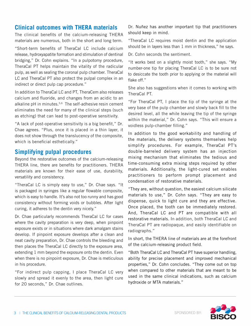
CASE STUDIESCASE STUDY #1 – Carla Cohn, DMDA six-year-old patient presented with severe early childhood caries and required oral rehabilitation of crowns, pulpotomies, and extractions.
Quadrant 3 is shown pre-operatively in Figure 1. Teeth K and L present with gross caries extending to the coronal pulp. Signs and symptoms include only transitory discomfort upon chewing, no radiographic radiolucency or soft tissue indication of infection.
1. Diagnosis was made as a reversible pulpitis treatable with a pulpotomy and ultimately full coverage with stainless-steel crowns. The quadrant is isolated with a rubber dam clamp (Ivory 2A) and a slot style rubber dam (Hedy).
2. In Figure 2, gross decay has been excavated and the roof of the pulp chamber is removed with straight cross-hatched carbide #556 friction grip bur (NSK handpiece) and copious water. Care is taken to make a wide enough access to visualize and remove all coronal pulpal tissue with #8 slow speed round bur (NSK handpiece) until the radicular pulp can be visualized (Fig. 3).
3. The chamber is irrigated with sterile saline (Fig. 4) and a saline moistened pellet is placed with pressure in order to achieve hemostasis (Fig. 5). Radicular pulp stumps with hemostasis achieved is shown in Figure 6.
4. Once hemostasis is achieved, TheraCal PT (BISCO) is placed. The syringe is bled, and then dispensed directly into the empty coronal pulp chamber. The tip of the syringe is placed at the very base of the chamber and the TheraCal PT is slowly extruded, while keeping the tip of the syringe submerged in order to avoid creating any bubbles. The material is of an ideal viscosity to easily fill the coronal chamber. Working time is 45 seconds, which is more than enough time to extrude the material.
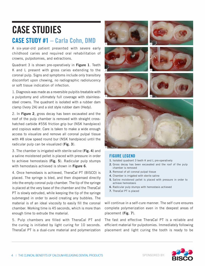
5. Pulp chambers are filled with TheraCal PT and the curing is initiated by light curing for 10 seconds. TheraCal PT is a dual-cure material and polymerization
will continue in a self-cure manner. The self cure ensures complete polymerization even in the deepest areas of placement (Fig. 7).
The fast and effective TheraCal PT is a reliable and efficient material for pulpotomies. Immediately following placement and light curing the tooth is ready to be
7
5 6
3 4
1 2
FIGURE LEGEND1. Isolated quadrant 3 teeth K and L pre-operatively2. Gross decay has been excavated and the roof of the pulp
chamber is removed3. Removal of all coronal pulpal tissue4. Chamber is irrigated with sterile saline5. Saline moistened pellet is placed with pressure in order to
achieve hemostasis6. Radicular pulp stumps with hemostasis achieved7. TheraCal PT is placed
SPONSORED BY:5 || THE CLINICAL BENEFITS OF CALCIUM-RELEASING DENTAL PRODUCTS
restored. TheraCal PT is compatible with a variety of common dental restorative materials including composite, glass ionomers, full coverage options. Radiographically TheraCal PT is easily visible and identifiable. TheraCal PT has changed the way we deliver pulpotomies. It is the first simple-to-place pulpotomy treatment. The efficiency and efficacy make this a valuable material for any dentist performing pulpotomies.
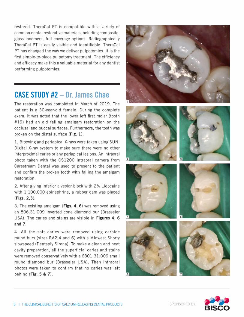
CASE STUDY #2 – Dr. James ChaeThe restoration was completed in March of 2019. The patient is a 30-year-old female. During the complete exam, it was noted that the lower left first molar (tooth #19) had an old failing amalgam restoration on the occlusal and buccal surfaces. Furthermore, the tooth was broken on the distal surface (Fig. 1).
1. Bitewing and periapical X-rays were taken using SUNI Digital X-ray system to make sure there were no other interproximal caries or any periapical lesions. An intraoral photo taken with the CS1200 intraoral camera from Carestream Dental was used to present to the patient and confirm the broken tooth with failing the amalgam restoration.
2. After giving inferior alveolar block with 2% Lidocaine with 1:100,000 epinephrine, a rubber dam was placed (Figs. 2,3).
3. The existing amalgam (Figs. 4, 6) was removed using an 806.31.009 inverted cone diamond bur (Brasseler USA). The caries and stains are visible in Figures 4, 6 and 7.
4. All the soft caries were removed using carbide round burs (sizes RA2,4 and 6) with a Midwest Shorty slowspeed (Dentsply Sirona). To make a clean and neat cavity preparation, all the superficial caries and stains were removed conservatively with a 6801.31.009 small round diamond bur (Brasseler USA). Then intraoral photos were taken to confirm that no caries was left behind (Fig. 5 & 7). 7
5
6
3
4
1
2
SPONSORED BY:6 || THE CLINICAL BENEFITS OF CALCIUM-RELEASING DENTAL PRODUCTS
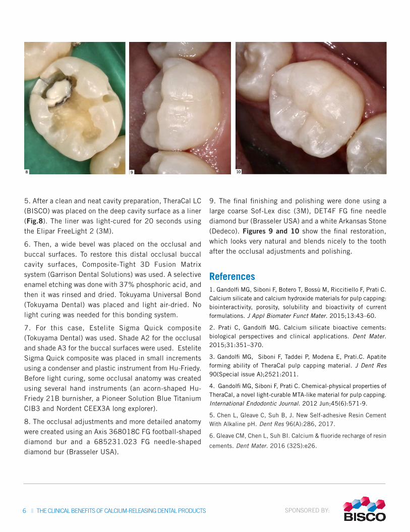
5. After a clean and neat cavity preparation, TheraCal LC (BISCO) was placed on the deep cavity surface as a liner (Fig.8). The liner was light-cured for 20 seconds using the Elipar FreeLight 2 (3M).
6. Then, a wide bevel was placed on the occlusal and buccal surfaces. To restore this distal occlusal buccal cavity surfaces, Composite-Tight 3D Fusion Matrix system (Garrison Dental Solutions) was used. A selective enamel etching was done with 37% phosphoric acid, and then it was rinsed and dried. Tokuyama Universal Bond (Tokuyama Dental) was placed and light air-dried. No light curing was needed for this bonding system.
7. For this case, Estelite Sigma Quick composite (Tokuyama Dental) was used. Shade A2 for the occlusal and shade A3 for the buccal surfaces were used. Estelite Sigma Quick composite was placed in small increments using a condenser and plastic instrument from Hu-Friedy. Before light curing, some occlusal anatomy was created using several hand instruments (an acorn-shaped Hu-Friedy 21B burnisher, a Pioneer Solution Blue Titanium CIB3 and Nordent CEEX3A long explorer).
8. The occlusal adjustments and more detailed anatomy were created using an Axis 368018C FG football-shaped diamond bur and a 685231.023 FG needle-shaped diamond bur (Brasseler USA).
9. The final finishing and polishing were done using a large coarse Sof-Lex disc (3M), DET4F FG fine needle diamond bur (Brasseler USA) and a white Arkansas Stone (Dedeco). Figures 9 and 10 show the final restoration, which looks very natural and blends nicely to the tooth after the occlusal adjustments and polishing.
References1. Gandolfi MG, Siboni F, Botero T, Bossù M, Riccitiello F, Prati C. Calcium silicate and calcium hydroxide materials for pulp capping: biointeractivity, porosity, solubility and bioactivity of current formulations. J Appl Biomater Funct Mater. 2015;13:43–60.
2. Prati C, Gandolfi MG. Calcium silicate bioactive cements: biological perspectives and clinical applications. Dent Mater. 2015;31:351–370.
3. Gandolfi MG, Siboni F, Taddei P, Modena E, Prati.C. Apatite forming ability of TheraCal pulp capping material. J Dent Res 90(Special issue A);2521:2011.
4. Gandolfi MG, Siboni F, Prati C. Chemical-physical properties of TheraCal, a novel light-curable MTA-like material for pulp capping. International Endodontic Journal. 2012 Jun;45(6):571-9.
5. Chen L, Gleave C, Suh B, J. New Self-adhesive Resin Cement With Alkaline pH. Dent Res 96(A):286, 2017.
6. Gleave CM, Chen L, Suh BI. Calcium & fluoride recharge of resin
cements. Dent Mater. 2016 (32S):e26.
98 10