the clinical utility of tomosynthesis in lung cancer diagnosisinterpretation tests was 1.2 mgy, but...
TRANSCRIPT
R/F
The Clinical Utility of Tomosynthesis in Lung Cancer Diagnosis
Division of Thoracic Oncology, National Cancer Center Hospital East1
National Cancer Center Hospital
Masami Ito, MD
Research Center for Cancer Prevention and Screening, National Cancer Center2
Masami Ito1, Hironobu Ohmatsu
1, Yoichi Naito
1, Hirotsugu Kenmotsu
1, Yuki Yamane
1,
Kiyotaka Yo1, Seiji Niho
1, Koichi Goto
1, Yuichiro Ohe
1, Yutaka Nishiwaki
1, Noriyuki Moriyama
2
1. Background
The number of fatalities due to lung cancer is
increasing. Therefore, the rapid detection of lung
cancer through screening is extremely important.
The chest radiography conventionally used for
screening has the benefit of convenience and low
exposure dose, but detection of cancer is difficult
from the small, low-density shadow images.
The introduction of helical CT for screening
enhanced the sensitivity of shadow detection and
increased the proportion of lung cancers detected
early. However, CT with high shadow detection
sensitivity can only be introduced into limited
facilities and it suffers from problems with high
exposure dose.
We focused on a new imaging technology,
tomosynthesis. This is a simple technique that
offers high shadow detection sensitivity at a low
exposure dose.
This paper reports on our investigations into the
clinical utility of tomosynthesis in the diagnosis of
lung cancer.
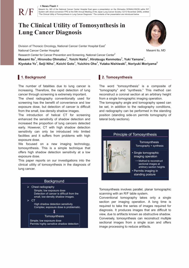
2. Tomosynthesis
The word “tomosynthesis” is a composite of
“tomography” and “synthesis.” This method can
reconstruct a coronal section at an arbitrary height
from a single tomographic imaging operation.
The tomography angle and tomography speed can
be set, in addition to the radiography conditions,
and radiography can be performed in the standing
position (standing side-on permits tomography of
lateral body sections).
Tomosynthesis involves parallel, planar tomographic
scanning with an R/F table system.
Conventional tomography takes only a single
section per imaging operation. A long time is
required to take the series of images required for
diagnosis. It produces images that are difficult to
view, due to artifacts known as obstructive shadow.
Conversely, tomosynthesis can reconstruct multiple
sectional images from a single scan and offers
image processing to reduce artifacts.
Masami Ito, MD of the National Cancer Center Hospital East gave a presentation on the Shimadzu SONIALVISION safire R/F
System with direct-conversion FPD at the 50th Annual Meeting of the Japan Lung Cancer Society (12/13 November 2009), entitled
“The Clinical Utility of Tomosynthesis in Lung Cancer Diagnosis.” The contents of her presentation are introduced below.
< News Flash >
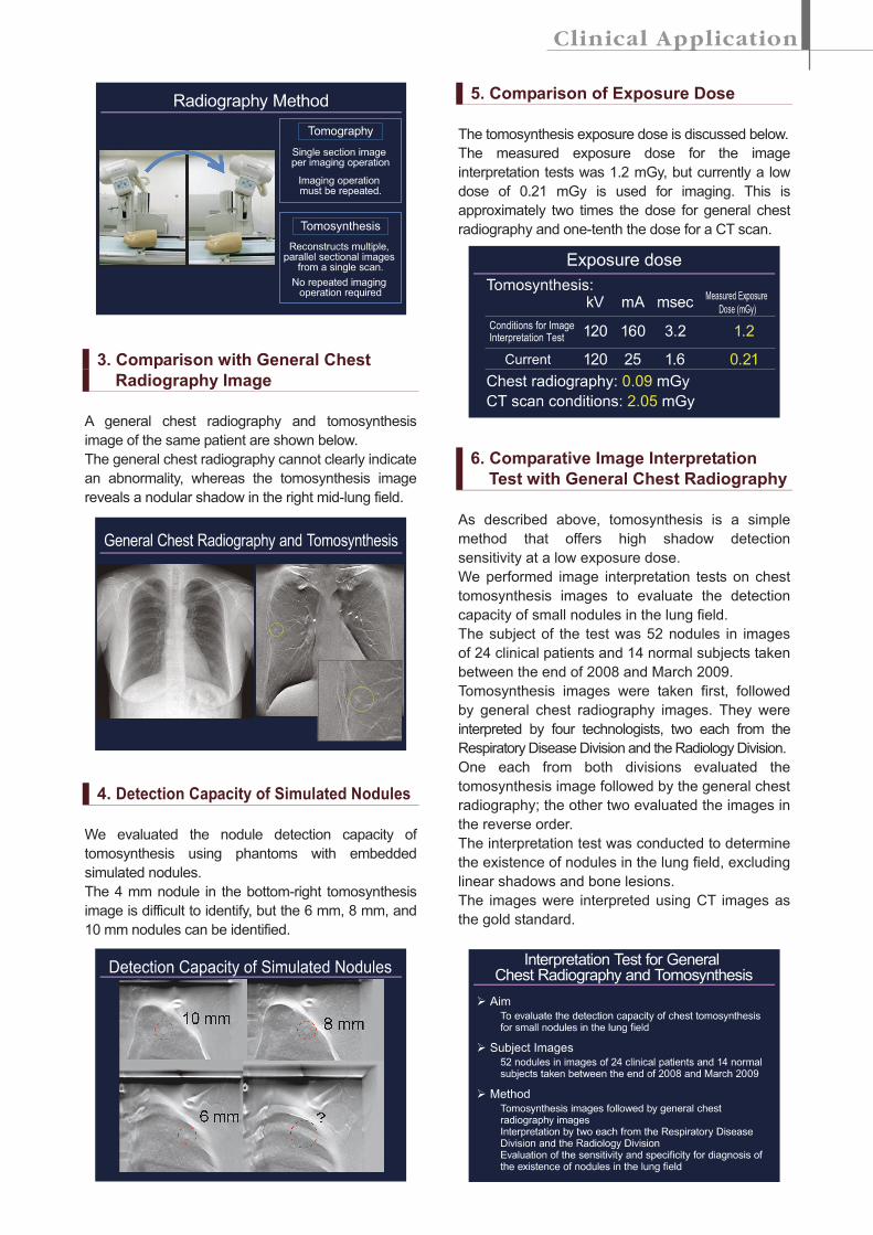
3. Comparison with General Chest
Radiography Image
A general chest radiography and tomosynthesis
image of the same patient are shown below.
The general chest radiography cannot clearly indicate
an abnormality, whereas the tomosynthesis image
reveals a nodular shadow in the right mid-lung field.
4. Detection Capacity of Simulated Nodules
We evaluated the nodule detection capacity of
tomosynthesis using phantoms with embedded
simulated nodules.
The 4 mm nodule in the bottom-right tomosynthesis
image is difficult to identify, but the 6 mm, 8 mm, and
10 mm nodules can be identified.
5. Comparison of Exposure Dose
The tomosynthesis exposure dose is discussed below.
The measured exposure dose for the image
interpretation tests was 1.2 mGy, but currently a low
dose of 0.21 mGy is used for imaging. This is
approximately two times the dose for general chest
radiography and one-tenth the dose for a CT scan.
6. Comparative Image Interpretation
Test with General Chest Radiography
As described above, tomosynthesis is a simple
method that offers high shadow detection
sensitivity at a low exposure dose.
We performed image interpretation tests on chest
tomosynthesis images to evaluate the detection
capacity of small nodules in the lung field.
The subject of the test was 52 nodules in images
of 24 clinical patients and 14 normal subjects taken
between the end of 2008 and March 2009.
Tomosynthesis images were taken first, followed
by general chest radiography images. They were
interpreted by four technologists, two each from the
Respiratory Disease Division and the Radiology Division.
One each from both divisions evaluated the
tomosynthesis image followed by the general chest
radiography; the other two evaluated the images in
the reverse order.
The interpretation test was conducted to determine
the existence of nodules in the lung field, excluding
linear shadows and bone lesions.
The images were interpreted using CT images as
the gold standard.
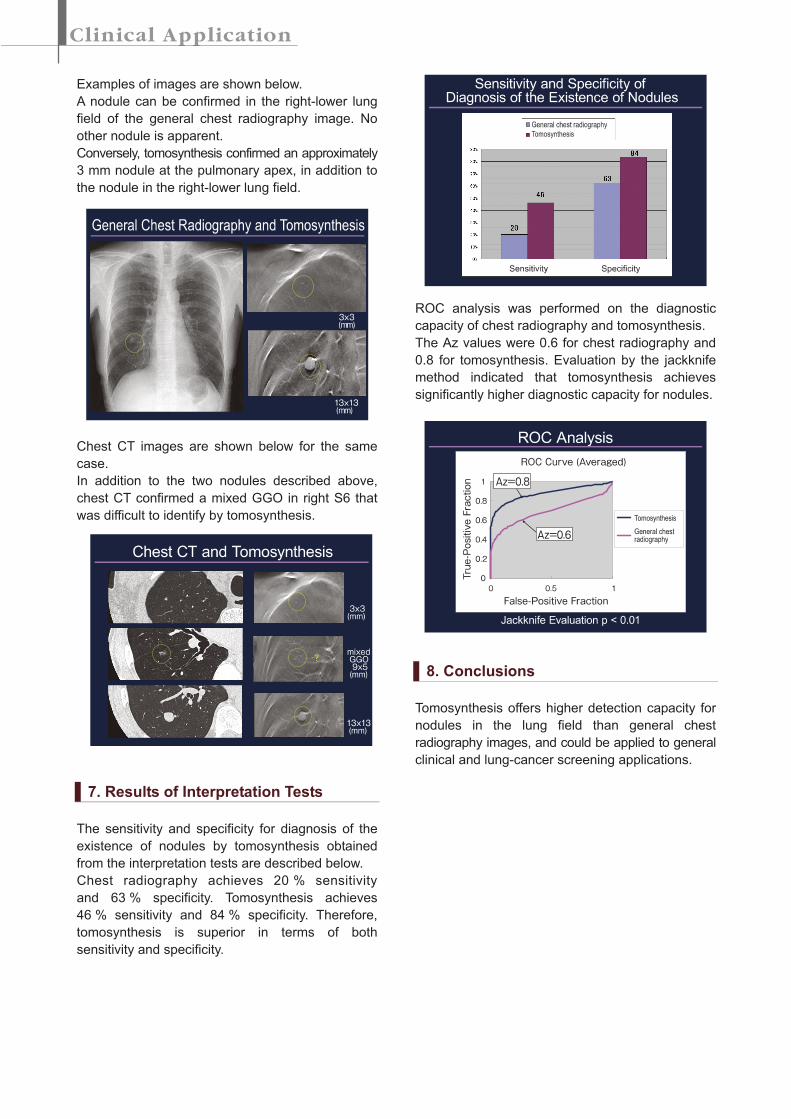
Examples of images are shown below.
A nodule can be confirmed in the right-lower lung
field of the general chest radiography image. No
other nodule is apparent.
Conversely, tomosynthesis confirmed an approximately
3 mm nodule at the pulmonary apex, in addition to
the nodule in the right-lower lung field.
Chest CT images are shown below for the same
case.
In addition to the two nodules described above,
chest CT confirmed a mixed GGO in right S6 that
was difficult to identify by tomosynthesis.
7. Results of Interpretation Tests
The sensitivity and specificity for diagnosis of the
existence of nodules by tomosynthesis obtained
from the interpretation tests are described below.
Chest radiography achieves 20 % sensitivity
and 63 % specificity. Tomosynthesis achieves
46 % sensitivity and 84 % specificity. Therefore,
tomosynthesis is superior in terms of both
sensitivity and specificity.
ROC analysis was performed on the diagnostic
capacity of chest radiography and tomosynthesis.
The Az values were 0.6 for chest radiography and
0.8 for tomosynthesis. Evaluation by the jackknife
method indicated that tomosynthesis achieves
significantly higher diagnostic capacity for nodules.
8. Conclusions
Tomosynthesis offers higher detection capacity for
nodules in the lung field than general chest
radiography images, and could be applied to general
clinical and lung-cancer screening applications.