the control of gaze - cocila.files.wordpress.com · 4/9/2017 · the control of gaze. which part of...
TRANSCRIPT

P R I N C I P L E S O F N E U R A L
S C I E N C E 5 T H E D I T I O N
THE CONTROL OF GAZE

WHICH PART OF BODY ALWAYS MOVES?

WHAT’S THE POINT?We want to understand the mechanisms of life.
How?
Using the simple system as much as possible, we can find THE PRINCIPLES of life.

WHAT’S THE POINT?We want to understand the mechanisms of life.
How?
Using the simple system as much as possible, we can find THE PRINCIPLES of life.
Then, what’s your question in this chapter?
1. How do the brain control its final effector, the muscle?
2. Plus, how do the brain differently guide its effector, the muscle according to different conditions?

WHAT’S THE POINT?
Then, what’s your question in this chapter?
1. How do the brain control its final effector, the muscle?
2. Plus, how do the brain differently guide its effector, the muscle according to different conditions?
Eye movement!

WHAT WILL WE LEARN TODAY.
1. Basic anatomy to understand the eye movement1. Eye muscles2. Cranial nerves for eye movement
2. Eye movement1. Types of eye movements2. Basic circuit3. Brain structure and function in eye movement

E Y E M U S C L E

11-5 AXIAL MUSCULATURE• Six Extrinsic Eye Muscles (Oculomotor Muscles)
1. Inferior rectus
2. Medial rectus
3. Superior rectus
4. Lateral rectus
5. Inferior oblique
6. Superior oblique
Total only 12 muscle!
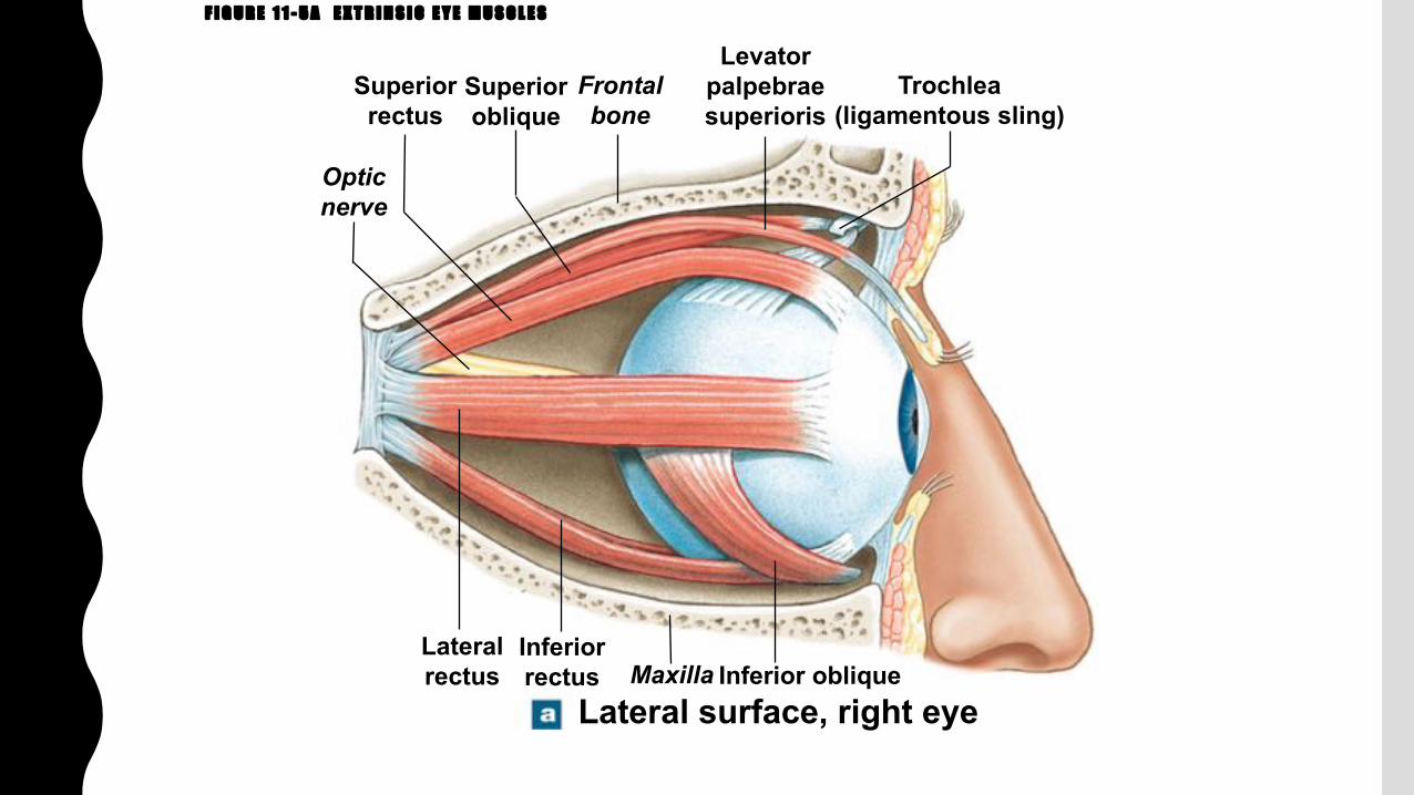
F I G U R E 1 1 - 5 A E X T R I N S I C E Y E M U S C L E S
Inferior oblique
Opticnerve
Superiorrectus
Superioroblique
Frontalbone
Levatorpalpebraesuperioris
Trochlea(ligamentous sling)
Lateralrectus
Inferiorrectus Maxilla
Lateral surface, right eye

F I G U R E 1 1 - 5 B E X T R I N S I C E Y E M U S C L E S
TrochleaSuperiorrectus
Levatorpalpebraesuperioris
Superioroblique
Medialrectus
Inferiorrectus
Opticnerve
Medial surface, right eye

F I G U R E 1 1 - 5 C E X T R I N S I C E Y E M U S C L E S
Superiorrectus
Lateralrectus
Inferioroblique
TrochleaSuperioroblique
Medialrectus
Inferiorrectus
Anterior view, right eye

F I G U R E 1 1 - 5 D E X T R I N S I C E Y E M U S C L E S
Levator palpebraesuperioris
Superior rectusOculomotornerve (N III)
Lateral rectusAbducens
nerve (N VI)
Inferior oblique
Trochlearnerve (N IV)Trochlea
SuperiorobliqueMedial rectus
Optic nerve (N II)
Inferior rectus
Anterior view, right orbit
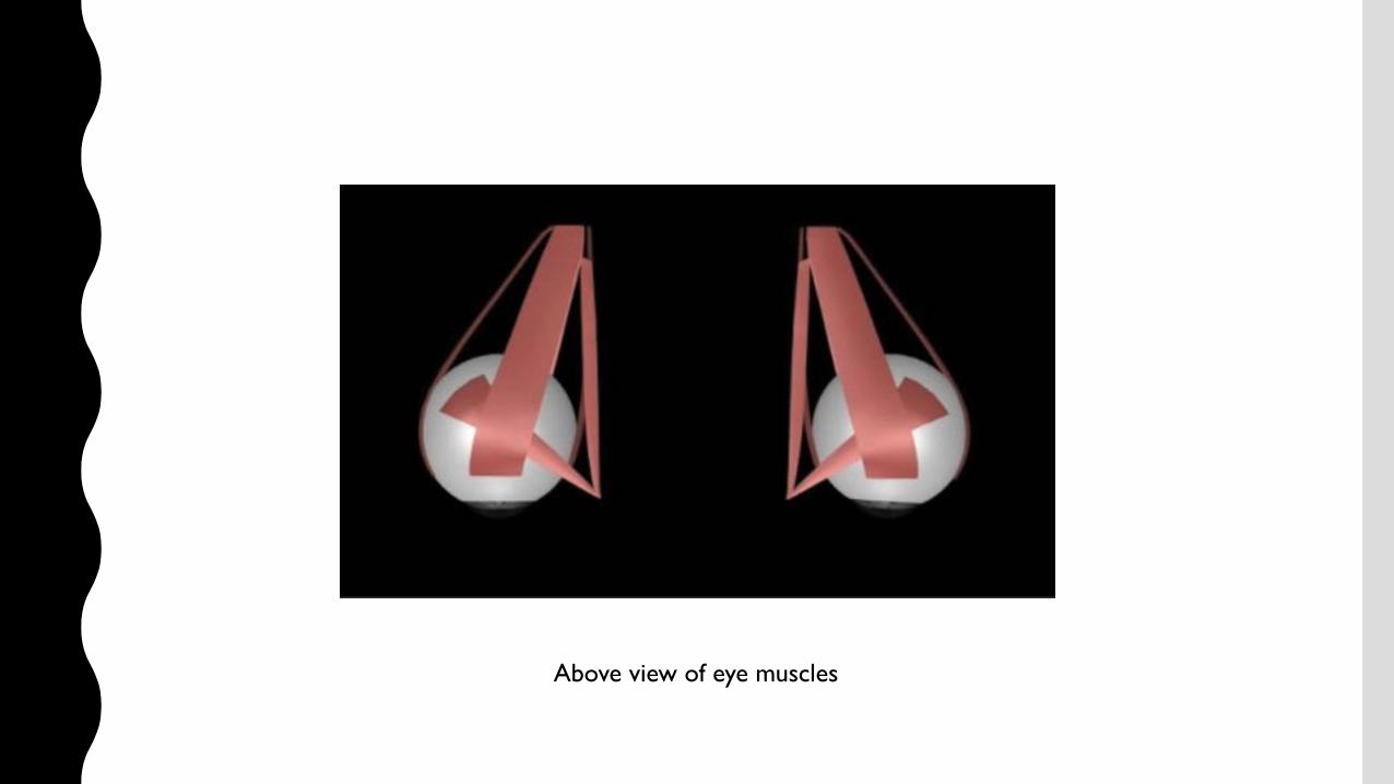
Above view of eye muscles

Eyes in actionhttps://www.youtube.com/watch?v=vd7OOJ7c1q4
Abduction Adduction
Elevation
Depression


T A B L E 1 1 - 3 E X T R I N S I C E Y E M U S C L E S ( F I G U R E 1 1 – 5 )

C R A N I A L N E R V E S

F I G U R E 1 4 - 1 8 O R I G I N S O F T H E C R A N I A L N E R V E S
Olfactory bulb: terminationof olfactory nerve (I)
Olfactory tract
Optic nerve (II)
Infundibulum
Oculomotor nerve (III)
Pons
Basilar artery
Vertebral artery
Cerebellum
Medulla oblongata
Spinal cord
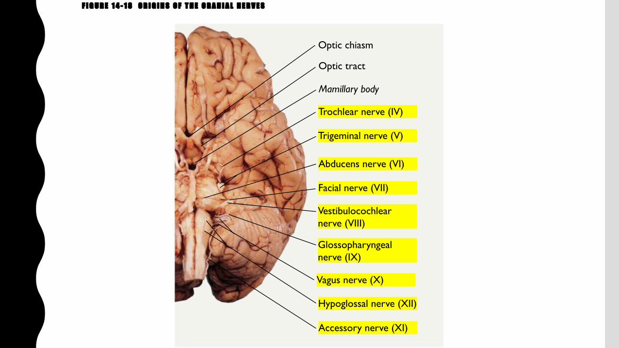
F I G U R E 1 4 - 1 8 O R I G I N S O F T H E C R A N I A L N E R V E S
Optic chiasm
Optic tract
Mamillary body
Trochlear nerve (IV)
Trigeminal nerve (V)
Abducens nerve (VI)
Vestibulocochlearnerve (VIII)
Facial nerve (VII)
Glossopharyngealnerve (IX)
Vagus nerve (X)
Hypoglossal nerve (XII)
Accessory nerve (XI)
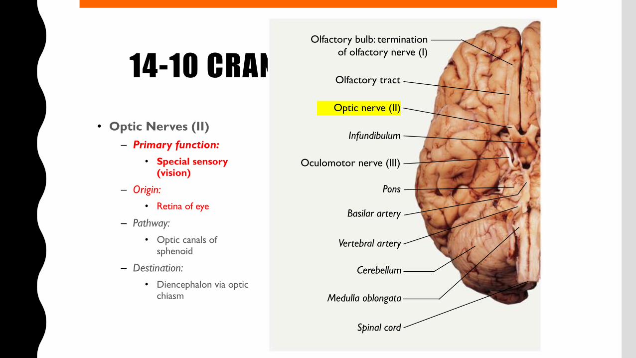
14-10 CRANIAL NERVES• Optic Nerves (II)
– Primary function:• Special sensory
(vision)
– Origin:• Retina of eye
– Pathway:• Optic canals of
sphenoid
– Destination:• Diencephalon via optic
chiasm
Olfactory bulb: terminationof olfactory nerve (I)
Olfactory tract
Optic nerve (II)
Infundibulum
Oculomotor nerve (III)
Pons
Basilar artery
Vertebral artery
Cerebellum
Medulla oblongata
Spinal cord

14-10 CRANIAL NERVES
• Optic Nerve Structures
– Optic chiasm
• Where sensory fibers converge
• And cross to opposite side of brain
– Optic tracts
• Reorganized axons
• Leading to lateral geniculate nuclei

F I G U R E 1 4 - 2 0 T H E O P T I C N E R V E
Pituitarygland
Midbrain(cut)
Visual cortex(in occipital lobes)
Optic projectionfibers
Optic tract
Optic chiasm
OPTIC NERVE(N II)
Olfactory bulb
Olfactory tract
Eye
Lateralgeniculatenucleus (inthalamus)
14-10 CRANIAL NERVES
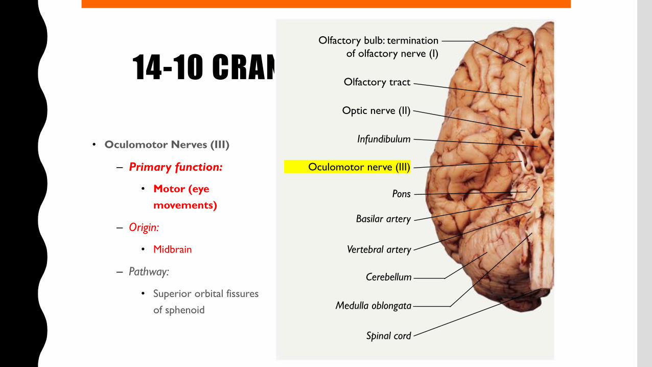
• Oculomotor Nerves (III)
– Primary function:
• Motor (eye movements)
– Origin:
• Midbrain
– Pathway:
• Superior orbital fissures of sphenoid
Olfactory bulb: terminationof olfactory nerve (I)
Olfactory tract
Optic nerve (II)
Infundibulum
Oculomotor nerve (III)
Pons
Basilar artery
Vertebral artery
Cerebellum
Medulla oblongata
Spinal cord

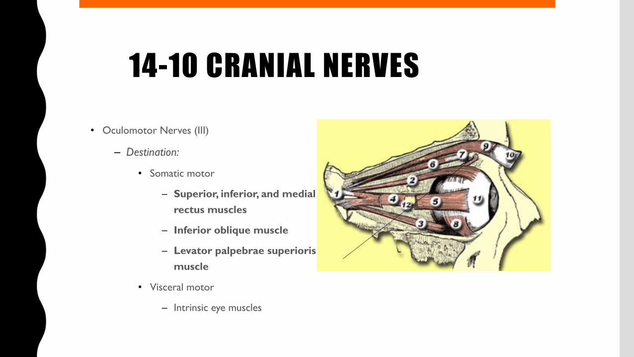
14-10 CRANIAL NERVES• Oculomotor Nerves (III)
– Destination:
• Somatic motor
– Superior, inferior, and medial rectus muscles
– Inferior oblique muscle
– Levator palpebrae superioris muscle
• Visceral motor
– Intrinsic eye muscles
14-10 CRANIAL NERVES• Oculomotor Nerves (III)
– Destination:
• Somatic motor
– Superior, inferior, and medial rectus muscles
– Inferior oblique muscle
– Levator palpebrae superioris muscle
• Visceral motor
– Intrinsic eye muscles
14-10 CRANIAL NERVES
• Oculomotor Nerve Structures
– Oculomotor nerve
• Controls four of six eye-movement muscles
• Delivers autonomic fibers to ciliary ganglion
– Ciliary ganglion controls intrinsic muscles of iris and lens

14-10 CRANIAL NERVES• TheTrochlear Nerves (IV)
– Primary function:
• Motor (eye movements)
– Origin:
• Midbrain
– Pathway:
• Superior orbital fissure of sphenoid
– Destination:
• Superior oblique muscle
Optic chiasm
Optic tract
Mamillary body
Trochlear nerve (IV)
Trigeminal nerve (V)
Abducens nerve (VI)
Vestibulocochlearnerve (VIII)
Facial nerve (VII)
Glossopharyngealnerve (IX)
Vagus nerve (X)
Hypoglossal nerve (XII)
Accessory nerve (XI)

14-10 CRANIAL NERVES• TheTrochlear Nerves (IV)
– Primary function:
• Motor (eye movements)
– Origin:
• Midbrain
– Pathway:
• Superior orbital fissure of sphenoid
– Destination:
• Superior oblique muscle

14-10 CRANIAL NERVES
• The Abducens Nerves (VI)– Primary function:
• Motor (eye movements)
– Origin:• Pons
– Pathway:• Superior orbital fissures of
sphenoid
– Destination:
• Lateral rectus muscle
Optic chiasm
Optic tract
Mamillary body
Trochlear nerve (IV)
Trigeminal nerve (V)
Abducens nerve (VI)
Vestibulocochlearnerve (VIII)
Facial nerve (VII)
Glossopharyngealnerve (IX)
Vagus nerve (X)
Hypoglossal nerve (XII)
Accessory nerve (XI)

F I G U R E 1 4 - 2 1 C R A N I A L N E R V E S C O N T R O L L I N G T H E E X T R A - O C U L A R M U S C L E S
Superiorobliquemuscle
Trochlea
Superiorrectusmuscle
Levatorpalpebraesuperioris
muscle
Inferiorrectusmuscle
Ciliaryganglion
Lateral rectusmuscle (cut)
ABDUCENSNERVE (N VI)
OPTICNERVE (N II)
Opticchiasm
OCULOMOTORNERVE (N III)
TROCHLEARNERVE (N IV)
Trigeminalnerve (N V), cut
Vestibulocochlearnerve (N VIII), cut
Facial nerve (N VII), cutMedial
rectusmuscle
Inferiorobliquemuscle

T A B L E 1 4 - 9 C R A N I A L N E R V E B R A N C H E S A N D F U N C T I O N S

T A B L E 1 4 - 9 C R A N I A L N E R V E B R A N C H E S A N D F U N C T I O N S

G A Z E C O N T R O L

What is this eye movement?
Saccadic eye movement

Yarbus, 1960

WHY IS IT NECESSARY?
http://anstislab.ucsd.edu/2012/11/20/peripheral-acuity/

http://anstislab.ucsd.edu/2012/11/20/peripheral-acuity/
WHY IS IT NECESSARY?

What is this eye movement?
What’s the purpose of eye movement?
Place an object at the center of fovea.

TYPES OF EYE MOVEMENTS
1.Saccade2.Smooth-pursuit3.Vergence4.Vestibulo-ocular reflex5.Optokinetic movement6.Fixation

SACCADE

SMOOTH PURSUIT

VERGENCE

VERGENECE

VESTIBULO-OCULAR REFLEX
https://www.youtube.com/watch?v=06TOLDbcdds


OPTOKINETIC MOVEMENTThe optokinetic reflex is a combination of a saccade and smooth pursuit eye movements

OPTOKINETIC MOVEMENT
https://www.youtube.com/watch?v=KSJksSA6Q-A

Activity of Vestibular Nuclei Neurons During Vestibular and Optokinetic Stimulation in the Alert MouseM. Beraneck, K. E. CullenJournal of Neurophysiology Published 1 September 2007Vol. 98 no. 3, 1549-1565 DOI: 10.1152/jn.00590.2007

IMPORTANCE OF EYE MOVEMENT?
https://www.youtube.com/watch?v=0SbGsg4twHs
e.g. Congenital nystagmus
Fixation impairment
Good vision but bad sight.

CONGENITAL NYSTAGMUS
Congenital nystagmus (also known as infantile nystagmus) is a clinical sign that may take many different forms. Involuntary, rhythmic eye movements are characteristic

SACCADE

SACCADE, PLOTTING IN 2D SPACE
Which one can you control and which one cannot?

SACCADE
Saccade direction and distance are controlled voluntarily.
BUT, the velocity is not.

SMOOTH PURSUIT
Slower than the saccade. (100 deg./s)
Interesting point?!?
Imagine the eye movement!

SMOOTH PURSUIT
Slower than the saccade. (100 deg./s)
Interesting point?!?
What’s the first?

EYE MUSCLES & CIRCUITS

REMINDER

REMINDER

EYE MUSCLES AND THE CIRCUIT

Abducens nerve => lateral rectus
Trochlear nerve => superior oblique
Oculomotor nerve => medial, superior, inferior rectus and inferior oblique& levator muscle

Abducens nerve => lateral rectus
Trochlear nerve => superior oblique
Oculomotor nerve => medial, superior, inferior rectus and inferior oblique& levator muscle

Which nerve could be impaired?

Which nerve could be impaired?
Abducense nerve palsy

Which nerve can be impaired?
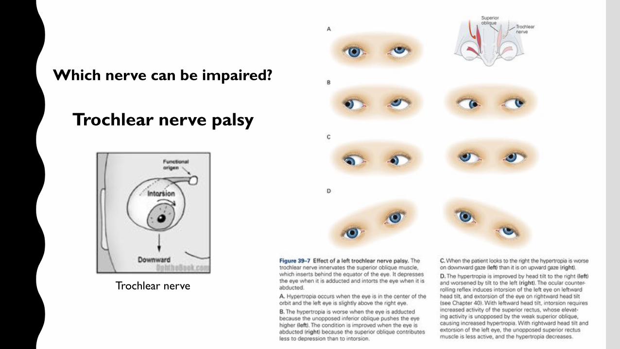
Which nerve can be impaired?
Trochlear nerve
Trochlear nerve palsy

MOVING THE EYES!

Basic neuronal activity for eye movement
Horizontal eye movement

Basic neuronal activity for eye movement
What’s encoded in the abducens motor neuron?

Basic neuronal activity for eye movement
What’s encoded in the abducens motor neuron?

Basic neuronal activity for eye movement
Burst firingSaccadic pulse
Saccadic step
What’s encoded in the abducens motor neuron?

Basic neuronal activity for eye movement
Burst firingSaccadic pulse
Saccadic step
What’s encoded in the abducens motor neuron?

Eye movement circuitBasic for horizontal eye movement

Eye movement circuitBasic for horizontal eye movement
Omnipause neuronIn dorsal raphe

Eye movement circuitBasic for horizontal eye movement
Omnipause neuronIn dorsal raphe
Excitatory burst neuronIn paramedian pontine reticular formation
Motor neuronIn abducense nucleus
The paramedian pontine reticular formation, also known as PPRF or paraabducens nucleus, is part of the pontine reticular formation, a brain region without clearly defined borders in the center of the pons.

Eye movement circuitBasic for horizontal eye movement
Omnipause neuronIn dorsal raphe
Excitatory burst neuronIn paramedian pontine reticular formation
Tonic neuronIn nucleus prepositus hypoglossi
Motor neuronIn abducense nucleus

Eye movement circuitBasic for horizontal eye movement
Omnipause neuronIn dorsal raphe
Excitatory burst neuronIn paramedian pontine reticular formation
Tonic neuronIn nucleus prepositus hypoglossi
Motor neuronIn abducense nucleus

Eye movement circuitBasic for horizontal eye movement
HOW TO CONTROL THE SACCADE BY CORTEX?


COLLICULUS

SUPERIOR & INFERIOR COLLICULUS

LAYERS OF SC
superficial layers (SZ, stratum zonale; SGS, stratum griseumsuperficiale; SO, stratum opticum); the intermediate layers (SGI, stratum griseum intermediale; SAI, stratum album intermediale); and the deep layers (SGP, stratum griseum profundum; and SAP, stratum album profundum)
Visual response
Motor response
Visuomotor response
Striate cortex (V1)
Prestriate cortex (V2)Middle temporal cortexParietal cortexFrontal eye field
Pulvinar

SUPERIOR COLLICULUS RETINOTOPIC MAP
Medial
Lateral

SUPERIOR COLLICULUSRetinotopic map
Electrical stimulation experiment

Activity of SC and SNduring saccade
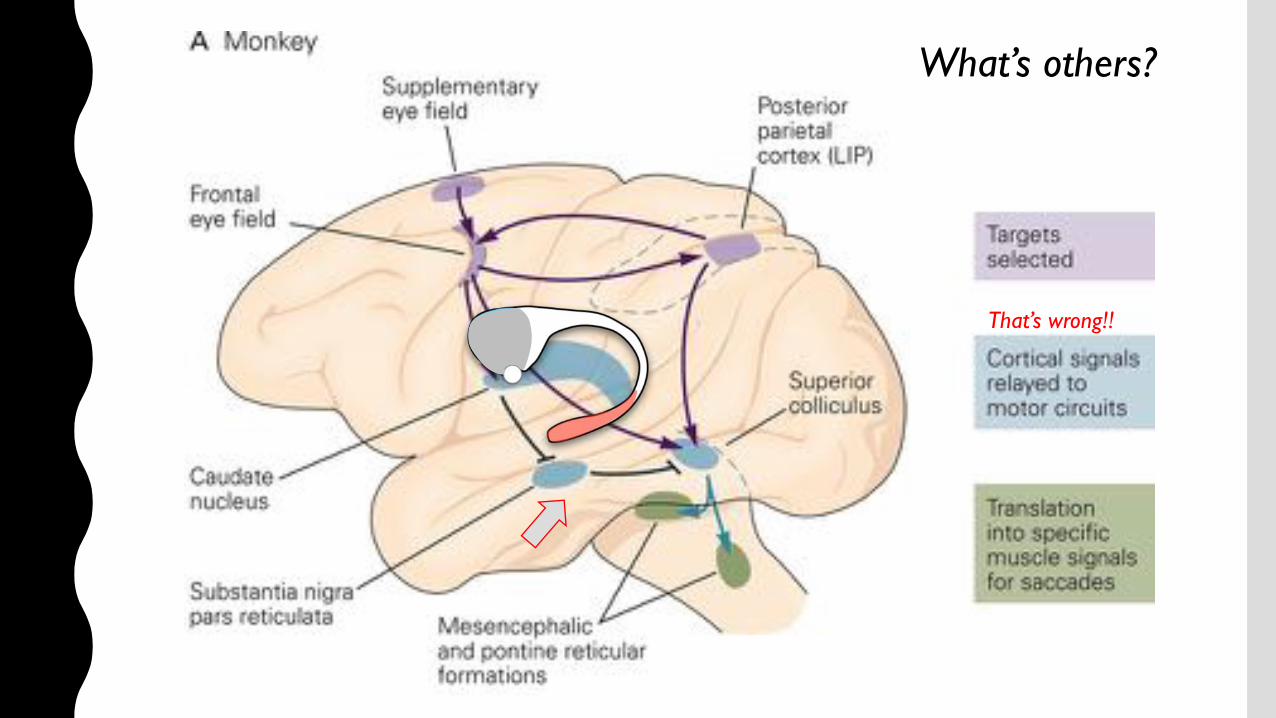
What’s others?
That’s wrong!!

fMRI during saccadeLet’s see the FEF, SEF and PS.

Target codingby the parietal cortex
That’s the target!

Frontal eye field activity during saccade
Two types of neurons in the FEF
Visual neuronMotor neuron

Supplementary eye field activity during saccade
Spatial location rather than the direction
More abstract informationMovement planning

nature neuroscience • volume 1 no 5 • september 1998 413
third type of response is illustrated by a neuron in the left cau-date nucleus (Fig. 4), which showed sustained activity in ADRafter the cue was presented in the right-up (RU) direction; theactivity reached its peak at the time of the saccade. In 1DR, theneuron’s activity for the RU direction was reduced considerablywhen this direction was not rewarded (columns 2–4), whereassome activity appeared in the right-down (RD) and left-up (LU)directions when they were rewarded.
The cells shown in Figs 2–4 were notexceptional. Most caudate neuronsshowed either a strong enhancement(data points close to the ordinate) or areduction (data points close to theabscissa) of response by expectation ofreward (Fig. 5a). A statistically signifi-cant modulation was found in 76 of 87neurons (87%) in visual or memory-related responses: visual response, 36/45(80%); memory response, 43/50 (86%;two-way ANOVA (reward condition ×cued direction), main effect of rewardcondition; p < 0.01). Among the 76modulated neurons, 64 neurons (visual,31; memory, 36) showed an enhance-ment (‘reward-facilitated neurons’),whereas 12 neurons (visual, 5; memo-ry, 7) showed a reduction of response(‘reward-suppressed neurons’). Theresults were the same for exclusive 1DRand relative 1DR.
The caudate contributes to the initi-ation of saccades with its connection tothe superior colliculus through the sub-stantia nigra11. The modulation of cau-date neuron activity by rewardexpectation might therefore producechanges in the characteristics of the sub-sequent saccade to the remembered cuelocation. Our data confirmed this pre-diction; the latencies were shorter
(Fig. 5b) and the peak velocities were higher (Fig. 5c) when thesaccades were followed by reward than when they were not (pairedt-test, p < 0.0001). The saccade latencies were significantly dif-ferent in the two monkeys, but the difference between the reward-ed and non-rewarded conditions was evident for each monkey.In addition, saccades to the rewarded direction were more accu-rate than those to the non-rewarded directions; the monkeys occa-sionally made incorrect saccades on non-rewarded trials.
articles
Fig. 4. Reward-dependent memory response (reward-facilitated type) of a neuron recorded inthe left caudate nucleus. See legend to Fig. 2 for format. Here the neuron discharge was alignedon saccade onset. Cued/rewarded direction: RU, right-up; LU, left-up; LD, left-down; RD, right-down. Target eccentricity was 20 degrees. The neuron showed sustained memory-relatedactivity and phasic saccadic activity for the right-up direction, both of which were strongerwhen this direction was rewarded.
Fig. 5. Effects of reward expectation on caudate neuron activity (a), saccade latency (b), and saccade velocity (c). Values in the rewarded(ordinate) and non-rewarded (abscissa) conditions are compared. After determining the preferred direction for each neuron, we calculatedthe mean magnitude (test-control, Hz) of the neuron’s response to its preferred cue in two conditions: when the preferred direction wasrewarded (one block) and when the preferred direction was not rewarded (three blocks). Data from two monkeys are shown with differentsymbols. Both visual and memory-related responses are included. Arrows 2–4 indicate the data for the neurons shown in Figs 2–4, respec-tively. The saccade parameters were obtained for each neuron by averaging across saccades to the neuron’s preferred direction separatelyfor the rewarded and non-rewarded conditions (b and c).
1DR ADRRewarded direction
Cued
dire
ctio
n
Cell activity, non-rewarded (Hz)
Cell
activ
ity, r
ewar
ded
(Hz)
a
Sacc
ade
late
ncy,
rew
arde
d (m
s)
b
Sacc
ade
velo
city
, rew
arde
d (d
eg/s
)
c
Saccade velocity, non-rewarded (deg/ s)Saccade latency, non-rewarded (ms)
© 1998 Nature America Inc. • http://neurosci.nature.com
© 1
998
Nat
ure
Am
eric
a In
c. •
http
://ne
uros
ci.n
atur
e.co
m
nature neuroscience • volume 1 no 5 • september 1998 411
Visual responses of neurons can be modulated by changes inbehavioral contexts. Many widely used behavioral tasks requirethe subject to respond by choosing one stimulus among manyor by choosing one feature (for example, color) of a stimulusamong many features1–3. In this type of procedure, the samereward is given for all correct trials, so that the motivational stateof the subject is assumed to be the same no matter which stimu-lus (or stimulus feature) represents the correct response. Thismodel is therefore ideal for investigating the cognitive aspect ofaction or attention, but not the motivational aspect.
Action is controlled by both cognition and motivation4,5, andmotivational states vary considerably. The same action can lead todifferent reward outcomes in different behavioral contexts. Bothneural and behavioral responses (for example, speed of action)may co-vary with such motivational changes, which may havedifferent consequences in the subsequent decision-makingprocesses. However, there have been few physiological studiesthat manipulated the outcome of an action in terms of reward(its amount or kind) while keeping the subject’s actions con-stant6. Consequently, little is known about neural mechanismsof the motivational aspect of attention or action selection.
To investigate how expectation of reward affects cognitiveinformation processing, we devised a memory-guided saccadetask in which the subject had to make a saccade to a rememberedcue location. However, correct performance was only rewardedwhen the cue had appeared at one of the four possible locations.
The cognitive requirement was always the same, in that the sub-ject had to attend to the cue stimulus, remember its location andmake a saccade to the location, but the motivational significancevaried. Using this model, we studied single-neuron activity inthe monkey caudate nucleus, a major input zone of the basal gan-glia, as the basal ganglia may be involved in control of actionbased on motivation5,7–9. We found that visual or memory-relat-ed responses of presumed projection neurons were frequentlymodulated by expectation of reward, either as an enhancement oras a reduction of the response.
Result sWe trained two monkeys on a memory-guided saccade task intwo reward conditions: ‘all directions rewarded’ (ADR) and ‘onedirection rewarded’ (1DR). In ADR, which is the conventionalreward schedule, the monkeys were rewarded each time they madea memory-guided saccade to the cued location for that trial. In1DR, which we devised specifically for this study, the monkeyswere rewarded for making correct memory-guided saccades onlyin one direction, termed the rewarded direction (Fig. 1). Mon-keys were not rewarded (exclusive 1DR) or were rewarded with asmaller amount (relative 1DR) when they made a correct responsein one of the other three directions, but they had to make a correctsaccade to proceed to the next trial. The rewarded direction wasfixed in a block of 60 successful trials, and a total of four blockswere done, with four different rewarded directions. Thus, the cue
articles
Expectat ion of reward modulatescognit ive signals in the basal gangliaReiko Kawagoe, Yoriko Takikawa and Okihide Hikosaka
Department of Physiology, School of Medicine, Juntendo University, 2-1-1 Hongo, Bunkyo-ku, Tokyo 113-8421, JapanCorrespondence should be addressed to O.H. ([email protected])
Action is controlled by both motivation and cognition. The basal ganglia may be the site wherethese kinds of information meet. Using a memory-guided saccade task with an asymmetric rewardschedule, we show that visual and memory responses of caudate neurons are modulated by expect-ation of reward so profoundly that a neuron’s preferred direction often changed with the changein the rewarded direction. The subsequent saccade to the target was earlier and faster for therewarded direction. Our results indicate that the caudate contributes to the determination ofoculomotor outputs by connecting motivational values (for example, expectation of reward) tovisual information.
Fig. 1. Memory-guidedsaccade task in the ‘onedirection rewarded’ condi-tion (1DR). Throughout ablock of experiment (60trials), only one directionwas rewarded. (Here theright direction wasrewarded.) Differentdirections were rewardedin different blocks.
Eye position
Fixationpoint
Target point
Tone
Reward
1 s
No reward
No reward
No reward
Reward
© 1998 Nature America Inc. • http://neurosci.nature.com
© 1
998
Natu
re A
mer
ica
Inc.
• ht
tp://
neur
osci
.nat
ure.
com
MSN also encoded the target & goal!

What’s others?
That’s wrong!!
Remember the motor circuit?

Oculomotor nucleus & Abducens nucleus
Speed signal in medial vestibular nucleus & prepositus hypoglossi
Flocculus in cerebellum
MT/MST
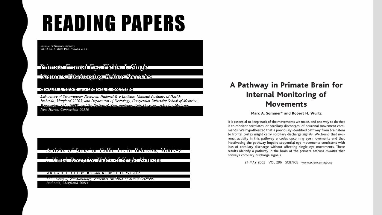
READING PAPERS
Activity of Superior Colliculus in Behaving Monkey.
I . Visual Receptive Fields of Single Neurons
MICHAEL E. GOLDBERG AND ROBERT H. WURTZ
Laboratory of Neurobiology, Nntional Institute of Mental Health, Bethesda, Maryland 20014
THE SIJPERIOR COI,LICIJT,I :S has long been
thought to participate in the neural events preceding visually guided eye movements, but the nature of this participation has never been understood. The monkey col- liculus receives visual input directly from the retina and indirectly from visual cortex (42) and cells in the superior colliculus have been shown to respond to visual stimulation in many species (see review by Sprague et al., ref 32) including monkeys (14, 16, 17, 28). While the colliculus ha i no known direct anatomical connections to the motor nuclei of the extraocular muscles in the monkey, it does send fibers to possible intermediate nuclei (20). Eye- movement elects in the monkey were demonstrated through stimulation studies (23) and through the discovery of cells that discharge before saccades (28, 47). How- ever, these cells are not necessary for the actual production of eye movements since monkeys with lesions of the superior col- liculus appear to have the full range of normal eye movements (1, 21).
In this series of experiments we used awake behaving monkeys to rci rives tiga te the superior colliculus by first examining the relationship of single neurons to visual stimuli ancl eye movements under varying behavioral conditions, by then using the properties of these cells to formulate a hypothesis of collicular function, and by finally testing the hypothesis. In this paper, the first of a series, we describe the visual receptive fields of the cells in the super- ficial <gray and optic layers of the monkey superior colliculus. In the next paper (7) we describe the enhanced effect that a visual stimulus can have on some of these same
Received for publication J~luary G, 1!372.
542
cells when the monkey is going to use the stimulus as a target for a saccade. In the third paper (48) we describe two classes of cells in the intermediate gray and white layers of the superior colliculus that dis- charge before eve movements of given distance and direction. In the final paper (49) we describe the effect of destruction of these cells in the superior colliculus on the monkey’s ability to make visually guided eye movements.
The visual receptive fields considered in this paper were studied in awake monkeys that had been trained to hold their eyes still by fixating a small spot of light (45). During this fixation period we were able to map the receptive field of the neuron under study using stationary and moving stimuli. In this manner we could study the neurons in the absence of voluntary eye movements but in the presence of the in- voluntary microsaccades and drifts with which the visual system must normally contend. All cells we studied in the super- ficial gray and optic layers of the superior colliculus responded to light and, unlike the majority of cells described in the col- liculus bf other animals, most of these cells responded well to stationary spots of light or to spots of light moving in any direction.
METHODS
Rhesus monkeys (Macrrca mulatta) were trained to fixate a spot of light on a tangent screen 57 cm in front of them. The training procedures used have been described previously (45). The result of this training was that each monkey learned to press a bar to turn on a fixation light which appeared 0.5 set after the bar was pressed. The point of light stayed on for a randomly selected variable time, between
A Pathway in Primate Brain forInternal Monitoring of
MovementsMarc A. Sommer* and Robert H. Wurtz
It is essential to keep track of the movements we make, and one way to do thatis to monitor correlates, or corollary discharges, of neuronal movement com-mands. We hypothesized that a previously identified pathway from brainstemto frontal cortex might carry corollary discharge signals. We found that neu-ronal activity in this pathway encodes upcoming eye movements and thatinactivating the pathway impairs sequential eye movements consistent withloss of corollary discharge without affecting single eye movements. Theseresults identify a pathway in the brain of the primate Macaca mulatta thatconveys corollary discharge signals.
When the brain initiates a movement, it alsogenerates internal information that is used bysensory systems to adjust for resultant chang-es to peripheral receptors and by motor plan-ning systems to prepare subsequent move-ments (1–7 ). Information about an impendingmovement arises as a correlate, or corollarydischarge, of the neuronal movement com-mand (8 ). The concept of corollary dischargehas been invaluable for understanding dispar-ate behaviors such as the circling of insects,fish, and amphibians after visual field inver-sion (9 , 10 ), electrolocation in fish (4 ), andsong learning in birds (11). In humans, psy-chophysical studies have demonstrated thatcorollary discharge signals exist (3, 5, 6 ) andlesion studies have emphasized that the thal-amus and cerebral cortex are crucial for usingcorollary discharge information (2, 12–14 ).Some neurons in the cerebral cortex of non-human primates receive corollary dischargesignals (15–17 ), but where these signalscome from has remained unknown.
To identify neurons as conveying corol-lary discharge signals, one must show thatthey have movement-related activity andproject upstream, away from motor neurons,instead of downstream, toward motor neu-rons. That is, their activity must transmitinformation about movement without causingmovement. A promising system in which tolook for such neurons is that for producingsaccadic eye movements. An important nodein this system is the superior colliculus—specifically its intermediate layer, which con-tains neurons that fire just before saccadegeneration (18 ). Some projections of the in-termediate layer go downstream to saccade-generating circuits in the midbrain and pons
(19 ), and some go upstream to mediodorsalthalamus (MD) relay neurons that project to afrontal lobe region known as the frontal eyefield (20 ). Our hypothesis is that the ascend-ing pathway carries corollary discharges ofsaccadic commands.
To test this hypothesis, first we recordedfrom 46 MD relay neurons in Macaca mu-latta (21), all physiologically verified asreceiving input from the superior colliculusand projecting to the frontal eye field (Fig.1A). We studied their activity while mon-keys made delayed saccades to visual tar-gets (Fig. 1B), a procedure that facilitatesdetermining whether the activity is relatedto vision or to movement (21). Most neu-rons (74%; 34/46) had increased activityjust before saccades (Fig. 1B) that began onaverage 144 ms before saccade generation(SD, 106 ms; median, 101 ms; range, 23 to392 ms). Because this activity began beforemovement, it could not have representedproprioception. Most of the saccade-relatedneurons (82%; 28/34) were spatially tuned,firing most strongly for saccades madewithin a restricted range of amplitudes anddirections. For all tuned neurons, the bestdirection was contraversive.
Second, we reversibly inactivated MD byinjecting muscimol unilaterally at the sites ofpreviously recorded MD relay neurons (Fig.2A) (21). Muscimol is a !-aminobutyric acidtype A (GABAA) agonist and inhibits neuroncell bodies, not axons (22), so it should sup-press MD relay neurons without affectingtransthalamic fibers passing nearby. We test-ed monkeys on a double-step task, in whichthey had to make successive saccades to twoflashed targets (Fig. 2B, top) (21, 23, 24 ).Correct execution of the second saccade re-quires knowledge about the first saccade’smetrics. Visual feedback regarding perfor-mance is not available, because the saccadesbegin after the targets disappear, and propri-oception is unlikely to contribute because it
plays little if any role in the online control ofsaccades (25–28 ). The ability to make a cor-rect second saccade, therefore, is thought torely critically on corollary discharge informa-tion about the first saccade (12–14 ). If inac-tivation totally disrupts corollary discharge(Fig. 2B, bottom), a monkey will be able tomake a first saccade correctly but will haveno internal information that the saccade wasmade. If the monkey then tries to completethe trial by making a saccade to the secondtarget location, the second saccade will travelas if starting from the fixation point. Theobserved effect will be a contraversive shiftof the second saccade end points.
Results from an example injection areshown in Fig. 2C. Before inactivation, themonkey made saccadic sequences correct-ly. Because the saccades were made in totaldarkness, first saccades were shifted slight-ly upward (29 ). Second saccades went near-ly straight up, which indicates that corol-lary discharge was intact. During inactiva-tion, the second saccade end points shiftedcontraversively, which indicates that corol-lary discharge was impaired. Quantitatively(Fig. 2D) (21), the second saccade endpoints were indeed shifted horizontally[contraversive 2.5° shift; P " 0.001 (21)]but not vertically during the injection. Nei-ther the initial fixation locations nor thefirst saccade end points were shifted signif-icantly in either direction.
We performed a total of seven muscimolexperiments in which there were a total of 22cases of before versus during saccadic se-quence pairs to analyze (21). In every case,the principle for identifying a corollary dis-charge deficit was the same as in Fig. 2. In82% of the cases (18/22), there was a contra-versive shift in second saccade end points(Fig. 3A), and the overall mean shift (1.12°)was significantly greater than zero. The con-traversive shift in 11 of these cases was in-dividually significant [and always reversible(30 )]. First saccade end points did not exhibita significant mean horizontal shift (Fig. 3B);neither did initial fixation locations (# 0.09°shift; P $ 0.025). In the vertical direction,there were no mean shifts in any of the data(31). For controls, we randomly interleavedtrials in which targets appeared ipsilaterally.Identical target configurations were used butwere flipped across the vertical meridian. Inthese trials, the first saccades were ipsiver-sive, a direction poorly represented by MDrelay neurons. Accordingly, we found no cor-ollary discharge deficits: the mean horizontalshift for second saccade end points was notsignificantly different from zero (# 0.41°;P $ 0.025).
We also considered whether inactivationmight have degraded a monkey’s ability tosee the second target or to remember itslocation. If visual or memory deficits oc-
Laboratory of Sensorimotor Research, National EyeInstitute, National Institutes of Health, Bethesda, MD20892, USA.
*To whom correspondence should be addressed. E-mail: [email protected]
R E P O R T S
24 MAY 2002 VOL 296 SCIENCE www.sciencemag.org1480
on
June
2, 2
016
http
://sc
ienc
e.sc
ienc
emag
.org
/D
ownl
oade
d fr
om
JOURNALOF NEUROPHYSIOLOGY Vol. 53. No. 3, March 1985. Printed in U.S.A.
Primate Frontal Eye Fields. I. Single Neurons Discharging Before Saccades CHARLES J. BRUCE AND MICHAEL E. GOLDBERG
Laboratory of Sensorimotor Research, National Eye Institute, National Institutes of Health, Bethesda, Maryland 20205; and Department of Neurology, Georgetown University School of Medicine, Washington, D.C. 20007; and the Section of Neuroanatomy, Yale University School of Medicine, New Haven, Connecticut 06510
SUMMARY AND CONCLUSIONS
1. We studied the activity of single neurons in the frontal eye fields of awake macaque monkeys trained to perform several oculo- motor tasks. Fifty-four percent of neurons discharged before visually guided saccades.
2. Three different types of presaccadic ac- tivity were observed: visual, movement, and anticipatory. Visual activity occurred in re- sponse to visual stimuli whether or not the monkey made saccades. Movement activity preceded purposive saccades, even those made without visual targets. Anticipatory activity preceded even the cue to make a saccade if the monkey could reliably predict what sac- cade he had to make.
3. These three different activities were found in different presaccadic cells in different proportions. Forty percent of presaccadic cells had visual activity (visual cells) but no move- ment activity. For about half of the visual cells the response was enhanced if the monkey made saccades to the receptive-field stimulus, but there was no discharge before similar saccades made without visual targets.
4. Twenty percent of presaccadic neurons discharged as briskly before purposive sac- cades made without a visual target as they did before visually guided saccades, and had weak or absent visual responses. These cells were defined as movement cells. Movement cells discharged much less or not at all before saccades made spontaneously without a task requirement or an overt visual target.
5. The remaining presaccadic neurons (40%) had both visual and movement activity (visuomovement cells). They discharged most
briskly before visually guided eye movements, but also discharged before purposive eye movements made in darkness and responded to visual stimuli in the absence of saccades. There was a continuum of visuomovement cells, from cells in which visual activity pre- dominated to cells in which movement activ- ity predominated. This continuum suggests that although visual cells are quite distinct from movement cells, the division of cell types into three classes may be only a heuristic means of describing the processing flow from visual input to eye-movement output.
6. Twenty percent of visuomovement and movement cells, but fewer than 2% of visual cells, had anticipatory activity. Only one cell had anticipatory activity as its sole response.
7. When the saccade was delayed relative to the target onset, visual cells responded to the target appearance, movement cells dis- charged before the saccade, and visuomove- ment cells discharged in different ways during the delay, usually with some discharge fol- lowing the target and an increase in rate immediately before the saccade. Presaccadic neurons of all types were actively suppressed following a saccade into their response fields.
8. All presaccadic activity was stimulus dimensions . We plotted
selective for movement-
and visuomovement-cell discharge as a func- tion of saccade direction and amplitude. Di- rection plots were fit to a Gaussian function and amplitude plots fit to a log-Gaussian function. Movement cells were less sharply tuned to direction and amplitude than were visual cells. Visuomovement cells had inter- mediate tuning.
0022-3077/85 $1 SO Copyright 0 1985 The American Physiological Society 603
A Pathway in Primate Brain forInternal Monitoring of
MovementsMarc A. Sommer* and Robert H. Wurtz
It is essential to keep track of the movements we make, and one way to do thatis to monitor correlates, or corollary discharges, of neuronal movement com-mands. We hypothesized that a previously identified pathway from brainstemto frontal cortex might carry corollary discharge signals. We found that neu-ronal activity in this pathway encodes upcoming eye movements and thatinactivating the pathway impairs sequential eye movements consistent withloss of corollary discharge without affecting single eye movements. Theseresults identify a pathway in the brain of the primate Macaca mulatta thatconveys corollary discharge signals.
When the brain initiates a movement, it alsogenerates internal information that is used bysensory systems to adjust for resultant chang-es to peripheral receptors and by motor plan-ning systems to prepare subsequent move-ments (1–7 ). Information about an impendingmovement arises as a correlate, or corollarydischarge, of the neuronal movement com-mand (8 ). The concept of corollary dischargehas been invaluable for understanding dispar-ate behaviors such as the circling of insects,fish, and amphibians after visual field inver-sion (9 , 10 ), electrolocation in fish (4 ), andsong learning in birds (11). In humans, psy-chophysical studies have demonstrated thatcorollary discharge signals exist (3, 5, 6 ) andlesion studies have emphasized that the thal-amus and cerebral cortex are crucial for usingcorollary discharge information (2, 12–14 ).Some neurons in the cerebral cortex of non-human primates receive corollary dischargesignals (15–17 ), but where these signalscome from has remained unknown.
To identify neurons as conveying corol-lary discharge signals, one must show thatthey have movement-related activity andproject upstream, away from motor neurons,instead of downstream, toward motor neu-rons. That is, their activity must transmitinformation about movement without causingmovement. A promising system in which tolook for such neurons is that for producingsaccadic eye movements. An important nodein this system is the superior colliculus—specifically its intermediate layer, which con-tains neurons that fire just before saccadegeneration (18 ). Some projections of the in-termediate layer go downstream to saccade-generating circuits in the midbrain and pons
(19 ), and some go upstream to mediodorsalthalamus (MD) relay neurons that project to afrontal lobe region known as the frontal eyefield (20 ). Our hypothesis is that the ascend-ing pathway carries corollary discharges ofsaccadic commands.
To test this hypothesis, first we recordedfrom 46 MD relay neurons in Macaca mu-latta (21), all physiologically verified asreceiving input from the superior colliculusand projecting to the frontal eye field (Fig.1A). We studied their activity while mon-keys made delayed saccades to visual tar-gets (Fig. 1B), a procedure that facilitatesdetermining whether the activity is relatedto vision or to movement (21). Most neu-rons (74%; 34/46) had increased activityjust before saccades (Fig. 1B) that began onaverage 144 ms before saccade generation(SD, 106 ms; median, 101 ms; range, 23 to392 ms). Because this activity began beforemovement, it could not have representedproprioception. Most of the saccade-relatedneurons (82%; 28/34) were spatially tuned,firing most strongly for saccades madewithin a restricted range of amplitudes anddirections. For all tuned neurons, the bestdirection was contraversive.
Second, we reversibly inactivated MD byinjecting muscimol unilaterally at the sites ofpreviously recorded MD relay neurons (Fig.2A) (21). Muscimol is a !-aminobutyric acidtype A (GABAA) agonist and inhibits neuroncell bodies, not axons (22), so it should sup-press MD relay neurons without affectingtransthalamic fibers passing nearby. We test-ed monkeys on a double-step task, in whichthey had to make successive saccades to twoflashed targets (Fig. 2B, top) (21, 23, 24 ).Correct execution of the second saccade re-quires knowledge about the first saccade’smetrics. Visual feedback regarding perfor-mance is not available, because the saccadesbegin after the targets disappear, and propri-oception is unlikely to contribute because it
plays little if any role in the online control ofsaccades (25–28 ). The ability to make a cor-rect second saccade, therefore, is thought torely critically on corollary discharge informa-tion about the first saccade (12–14 ). If inac-tivation totally disrupts corollary discharge(Fig. 2B, bottom), a monkey will be able tomake a first saccade correctly but will haveno internal information that the saccade wasmade. If the monkey then tries to completethe trial by making a saccade to the secondtarget location, the second saccade will travelas if starting from the fixation point. Theobserved effect will be a contraversive shiftof the second saccade end points.
Results from an example injection areshown in Fig. 2C. Before inactivation, themonkey made saccadic sequences correct-ly. Because the saccades were made in totaldarkness, first saccades were shifted slight-ly upward (29 ). Second saccades went near-ly straight up, which indicates that corol-lary discharge was intact. During inactiva-tion, the second saccade end points shiftedcontraversively, which indicates that corol-lary discharge was impaired. Quantitatively(Fig. 2D) (21), the second saccade endpoints were indeed shifted horizontally[contraversive 2.5° shift; P " 0.001 (21)]but not vertically during the injection. Nei-ther the initial fixation locations nor thefirst saccade end points were shifted signif-icantly in either direction.
We performed a total of seven muscimolexperiments in which there were a total of 22cases of before versus during saccadic se-quence pairs to analyze (21). In every case,the principle for identifying a corollary dis-charge deficit was the same as in Fig. 2. In82% of the cases (18/22), there was a contra-versive shift in second saccade end points(Fig. 3A), and the overall mean shift (1.12°)was significantly greater than zero. The con-traversive shift in 11 of these cases was in-dividually significant [and always reversible(30 )]. First saccade end points did not exhibita significant mean horizontal shift (Fig. 3B);neither did initial fixation locations (# 0.09°shift; P $ 0.025). In the vertical direction,there were no mean shifts in any of the data(31). For controls, we randomly interleavedtrials in which targets appeared ipsilaterally.Identical target configurations were used butwere flipped across the vertical meridian. Inthese trials, the first saccades were ipsiver-sive, a direction poorly represented by MDrelay neurons. Accordingly, we found no cor-ollary discharge deficits: the mean horizontalshift for second saccade end points was notsignificantly different from zero (# 0.41°;P $ 0.025).
We also considered whether inactivationmight have degraded a monkey’s ability tosee the second target or to remember itslocation. If visual or memory deficits oc-
Laboratory of Sensorimotor Research, National EyeInstitute, National Institutes of Health, Bethesda, MD20892, USA.
*To whom correspondence should be addressed. E-mail: [email protected]
R E P O R T S
24 MAY 2002 VOL 296 SCIENCE www.sciencemag.org1480
on
June
2, 2
016
http
://sc
ienc
e.sc
ienc
emag
.org
/D
ownl
oade
d fr
om

Vestibulo-ocular reflex