the different connections and motor …jeb.biologists.org/content/jexbio/54/2/391.full.pdfthe...
TRANSCRIPT
J. Exp. Biol. (1971), 54, 391-402 291With 2 plates and 5 text-figures
Printed in Great Britain
THE DIFFERENTCONNECTIONS AND MOTOR OUTPUTS OF LATERAL
AND MEDIAL GIANT FIBRES IN THE CRAYFISH
BY JAMES L. LARIMER, ALAN C. EGGLESTON,LEONA M. MASUKAWA AND DONALD KENNEDY
Department of Zoology, The University of Texas, Austinand the Department of Biological Sciences,Stanford University, Stanford, California
(Received 8 September 1970)
INTRODUCTION
The giant fibres of crayfish comprise a pre-motor system that secures rapid con-traction of phasic flexor muscles in the abdomen. The result of their activation is asudden flexion of all abdominal segments that propels the animal backward in an'escape' response; repeated sequences of similar tail-flips, with intervening fastextensions, result in sustained swimming.
The adequacy of single medial or lateral giant fibres for producing such tail-flipswas demonstrated by Wiersma (1938) who isolated axons in the circumoesophagealconnectives and stimulated them. He also (1947) showed that either kind of centralgiant produced short-latency discharge of flexor motor neurones in the abdominalthird roots. It was not clear from Wiersma's results, however, whether the actions ofthe two types of giant fibres were really equivalent. It is known that medial and lateralgiants differ both in their morphology and in the location of presynaptic input. Medialgiant fibres (MG) are single cells whose somata are located in the brain; branches anda cross-connexion also occur there (Horiuchi, Hayashi & Takahashi, 1966), but theaxons run caudally without branches or septa to the last abdominal ganglion. Lateralgiants (LG), on the other hand, consist of segmental units linked end-to-end in eachganglion by septa across which electrical propagation occurs (Kao & Grundfest, 1956;Watanabe & Grundfest, 1961). A contralateral soma and dendritic branches wereshown to occur in the third abdominal ganglion (Remler, Selverston & Kennedy,1968). MG are said to be excited by stimuli applied to the head region (Wiersma,1961), whereas the LG are discharged by afferents entering the abdominal ganglia viathe second roots (Krasne, 1969).
The experiments we shall describe show that the two types of giant fibres differdistinctively in their motor output, as well as in their input. MG produce a full,symmetrical flexion of the abdomen without flaring of the uropods, whereas LG evokea flexion in which intermediate abdominal segments are relatively uninvolved and theuropods are strongly promoted. These differences, which are readily seen in thebehavioural response to giant-fibre stimulation in the intact animal, are accounted forby specific connexions made by the giant fibres with motor neurones innervating thedifferent muscles involved.

392 J. L. LARIMER AND OTHERS
METHODS
Crayfish (Procambarus clarkii) were prepared in a variety of ways to gain access tothe giant fibres for different purposes. In behavioural experiments the objective wasto stimulate medial and lateral giants selectively with minimal interference to theabdomen. They were therefore isolated in the circumoesophageal connectives, whichwere exposed by removing the rostral-dorsal portion of the carapace and the under-lying hepatopancreas and stomach. During this dissection and subsequent recordingsessions the animal was immersed in cold van Harreveld's (1936) solution which wasfrequently replaced in order to avoid exposure to enzymes escaping from the digestiveorgans. A Lucite rod was then fixed to the mid-dorsal carapace so that the animalcould be held suspended in Ringer in its normal primary orientation. Alternatively,the thorax was pinned in a wax dish which was shaped to allow the abdomen to movefreely. Micromanipulated platinum wire hooks were used to stimulate the MG andLG axons after they had been dissected out of one of the connectives. The movementsof the abdomen were recorded by photographing them at 1000 frames/s with a high-speed Hycam rotating-prism camera (Red Lake Labs., Sunnyvale, Ca.). The positionsof the abdomen at chosen times during a tail-flip could be traced from single framesusing a stop-frame projector equipped with a frame counter.
For studying the influence of giant-fibre impulses on muscles in the abdomen andin the appendages of the last segment, we pinned the animal ventral side up in aLucite dissecting dish partially filled with paraffin, and exposed the abdominal nervecord so as to leave the roots intact in each segment. This preparation is essentiallyidentical to that described by Evoy & Kennedy (1967), except that to record fast flexormotor outflow we recorded from the transverse, anterior oblique, and posterior obliquemuscles in the main abdominal segments with suction electrodes (for a description ofthese muscles, see Rayner & Wiersma, 1967). Various muscles of the uropods andtelson (Larimer & Kennedy, 1969 a) were exposed and recorded from in the same way,or dissected peripheral branches of the roots innervating them were attached to suctionelectrodes for recording impulses in specific motor axons. In all experiments of thissort, the giant fibres were isolated for stimulation through suction electrodes in arostral abdominal connective.
Specific motor neurones leaving the sixth ganglion via its sixth root and innervatingthe telson flexor and anal compressor muscles were also studied anatomically in orderto verify the physiological observation that they made differential synaptic contactswith LG and MG axons. The motor axons in this root were impaled with micro-pipettes filled with a saturated (c. 4 %) aqueous solution of Procion Yellow M-4RAN(Imperial Chemical Industries America, Inc.). This fluorescent dye remains withincells and diffuses into even relatively fine processes, and is thus suitable for directvisualization of the ganglionic architecture of such neurones (Stretton & Kravitz,1968; Remler et al. 1968). In our experiments it was injected by pressure; the prepara-tion was then kept in the cold for about 12 h to insure sufficient time for diffusion intothe ganglion. The sixth ganglion was then fixed in Lavodowsky's solution, dehydrated,and cleared in methyl salicylate. Whole mounts were made in Fluoromount andphotographed in a Zeiss fluorescence microscope using excitation filter I and barrierfilter 50. Such a preparation could later be resuspended in xylol to free it from the
Output connexions of giant fibres 393
mounting medium, embedded in paraffin, and sectioned at 10 ji. Complete recon-structions of the branching system were not made, but each cell was analysed to seewhich of the giant fibres its processes contacted. Similar methods were used to injectthe giant fibres in the 5-6 connective, so as to visualize their terminations in the sixthganglion.
RESULTS
1. Cinematographic analysis of giant-fibre responses
The movements of the abdomen in response to stimulation of LG and MG axonin the circumoesophageal connectives were photographed at 1000 frames/s; tracingsof frames from such sequences are shown in Fig. 1. The differences between the twokinds of tail flexion are obvious and consistent. LG responses involve promotion ofthe exopodites of both uropods and a relatively incomplete flexion of the abdomenexcept at the junction of the thorax and in the last segment. Specifically, there is strongflexion of the anterior abdominal segments, but very little flexion of segment 4 inrelation to 3 or 5 upon 4. The result is a ' sculling' stroke in which much of the thrustis vertical rather than horizontal. MG impulses, on the other hand, generate verycomplete flexion of all abdominal segments and the telson, but this movement isunaccompanied by flaring of the uropods. The stroke ends with the abdomen in atightly curled position, and so produces a primarily horizontal thrust. Similar resultshave been obtained by Wine (personal communication), who also showed that LGflips in untethered animals actually produced the expected upward movement.
Flicks produced spontaneously or by natural stimulation were also photographedin the hope that the central pathway involved could be deduced by comparing themwith those evoked by giant-fibre stimulation. Some movements, especially thosestimulated by tapping in the abdominal region, resembled LG responses in that theflexion of intermediate segments was poorly developed and the exopodites of theuropods were promoted. Others were more complete, like MG flips, but differed fromMG responses in the involvement of uropod appendages. We were left with theimpression that pure responses of either kind rarely occurred spontaneously, and thatmost such movements probably involved pathways other than the giant fibres. At thetime, we demonstrated that single fast flexions could be evoked by natural stimuliwithout giant-fibre involvement; and Schrameck (1970) has since shown that giant-fibre impulses are not usually involved in swimming.
2. Electrical responses of abdominal muscles to giant-fibre activity
The behavioural data suggested that the LG and MG axons made selective centralconnections with motor neurones innervating the muscles of the abdomen and of thetail appendages. We therefore recorded from individual muscles or the peripheralmotor nerve branches innervating them while stimulating giant axons isolated fromone of the central connectives.
Before presenting these results, we find it necessary to deal in a more general waywith the junctions that connect the central giants and fast flexor motor neurones.Since it was first discovered that such a pathway exists (Wiersma, 1938, 1947), it hasbeen shown to be a duplex one. A large cell, the motor giant, makes connections withboth lateral and medial giant fibres in each abdominal segment. The connexions are

394 J- L. LARIMER AND OTHERS
rectifying electrotonic junctions (Furshpan & Potter, 1959), and the motor giant cellinnervates each fibre of the main flexor muscles of one side. The peripheral neuro-muscular junctions, however, show rapid antifacilitation at low frequencies of stimu-lation, so that only the impulses in a well-rested preparation are capable of recruiting
M L
Fig. 1. Tracing from high-speed 16 mm cin£ film of the abdominal movements of crayfishresulting from stimulating the isolated medial giant (M) and lateral giant (L) in the circumoeso-phageal connectives. The films were taken at 1000 frames/s; animals were held firmly by a rodattached to the thorax, but the abdomen was free to move. The tracings were made at 15 msintervals.
substantial tension in the muscles (Kennedy & Takeda, 1965; Bruner & Kennedy,1970). The eight other motor axons in each main abdominal flexor root innervatesingle muscle groups or parts of them, and maintain their efficacy even at fairly high

Output connexions of giant fibres 395
frequencies. The central junctions between the giant fibres and these motor neuronesare located in the ganglia rather than at the root exit, where the motor giant contactsthe central giants; they are formed by branches from the motor neurones that contactthe axis cylinders of the central giants (Remler, Selverston & Kennedy, 1968). It isnot yet known whether these are electrotonic or chemical synapses, but they exhibita delay that is longer than that for the motor giant synapse.
One expects, then, that records from a third root in response to stimulation of acentral giant axon will show two components: a short-latency discharge consisting ofthe single motor giant impulse, and a slightly delayed compound action potential
Fig. 2. Responses of the flexor muscles in the third abdominal segment following separatestimulation of MG and LG axons in the first abdominal connective. Upper trace, giant axonspike; lower trace, muscle responses; both were recorded extracellularly with suction electrodes.A, Anterior oblique muscle, following MG and LG stimulation: B, Posterior oblique; C,Transverse muscles. Only the MG axons were effective in driving all of the main flexor musclesof the abdomen. Time calibration, 10 ms.
composed of impulses in several of the 'non-giant' motor axons. Such records have,in fact, been published (Kennedy & Takeda, 1965), but the predicted output is oftennot obtained. The early records obtained from third roots by Wiersma (1947), forexample, fail to exhibit two components, and most of them appear to consist of spikesfrom one to several 'non-giant' neurones. We have frequently failed to observe themotor giant response ourselves, and in fact Furshpan & Potter (1959) report thatin the majority of their experiments on the LG-motor giant junction, it was nottransmitting.
This perplexing unreliability also applies to the junctions between central giantsand the non-giant motor neurones. The number of units responding in the main rootsto activity in a given giant fibre is somewhat variable, as is the response of identified

396 J. L. LARIMER AND OTHERS
motor neurones recorded in the periphery. For this reason a large number of experi-ments on each muscle or motor neurone was necessary to determine the presence orabsence of a central connexion with the giant fibres. In none of the cases in which wereport the absence of a connexion was one ever found; but some of the demonstratedconnexions were not functional in some preparations. We do not know whether theseinconsistencies demonstrate a real variation in the efficacy of junctions, or whetherthey are due to some experimental variable that we have been unable to control.
Figure 2 shows the responses of the major groups of phasic flexor muscles in thethird abdominal segment to MG (left) and LG (right) impulses, following repetitive
MG
Fig. 3. Activity of muscles in the telson-uropod segment in response to electrical stimulationof MG and LG axons. Upper trace, giant axon spike; the lower trace, muscle potential; bothwere recorded externally with suction electrodes. A, Productor exopodite muscle; B, telson-uropodalis posterior; C, Posterior telson flexor; D, anterior telson flexor; E, anal compressor(ventral telson flexor); F, lateral remoter; and G, Ventral rotator muscle. Muscles involved inrecords A—F are innervated from the sixth ganglion; the ventral rotator is innervated from thefifth ganglion. Time calibration, 10 ms.
stimulation at low frequency. The MG strongly drive both the anterior and posterioroblique muscles as well as the transverse muscle, whereas the LG excite only thetransverse muscle.
An even clearer differentiation of giant-mediated output occurs in the fifth and sixthganglia (Fig. 3). Here, the MG axons drive the major flexors of the telson-uropodsegment; i.e. the anal compressor (Fig. 3E; the suggested name given by Larimer &Kennedy, 1969 a, for this muscle is the ventral telson flexor), the posterior telson flexor(Fig. 3 C) and the ventral rotator muscle (Fig. 3 G). The first two of these muscles areinnervated from the sixth root of the sixth ganglion, while the latter is innervated viathe third root of the fifth ganglion. All three of these muscles are probably homologousto the oblique series. In addition, however, the LG mediate contraction of threesmaller phasic muscles in this region, viz. the productor exopodite of the uropods
Output connexions of giant fibres 397
(Fig. 3 A), the telson uropodalis posterior muscle (Fig. 3B) and the lateral remotormuscle (Fig. 3 F). None of these muscles receive motor neurones that are driven byboth LG and MG axons.
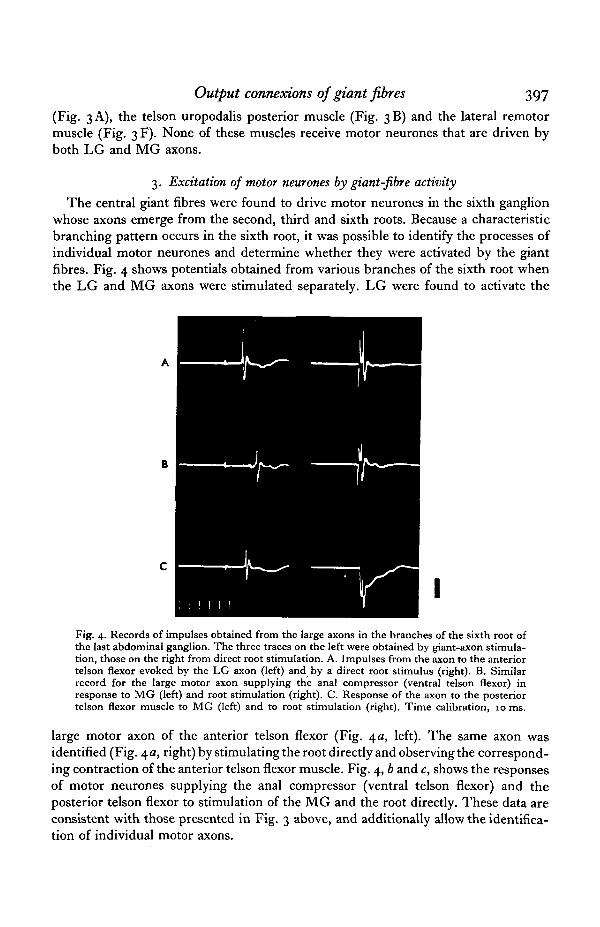
3. Excitation of motor neurones by giant-fibre activity
The central giant fibres were found to drive motor neurones in the sixth ganglionwhose axons emerge from the second, third and sixth roots. Because a characteristicbranching pattern occurs in the sixth root, it was possible to identify the processes ofindividual motor neurones and determine whether they were activated by the giantfibres. Fig. 4 shows potentials obtained from various branches of the sixth root whenthe LG and MG axons were stimulated separately. LG were found to activate the
Fig. 4. Records of impulses obtained from the large axons in the branches of the sixth root ofthe last abdominal ganglion. The three traces on the left were obtained by giant-axon stimula-tion, those on the right from direct root stimulation. A. Impulses from the axon to the anteriortelson flexor evoked by the LG axon (left) and by a direct root stimulus (right). B. Similarrecord for the large motor axon supplying the anal compressor (ventral telson flexor) inresponse to MG (left) and root stimulation (right). C. Response of the axon to the posteriortelson flexor muscle to MG (left) and to root stimulation (right). Time calibration, 10 ms.
large motor axon of the anterior telson flexor (Fig. \a, left). The same axon wasidentified (Fig. 4a, right) by stimulating the root directly and observing the correspond-ing contraction of the anterior telson flexor muscle. Fig. 4, b and c, shows the responsesof motor neurones supplying the anal compressor (ventral telson flexor) and theposterior telson flexor to stimulation of the MG and the root directly. These data areconsistent with those presented in Fig. 3 above, and additionally allow the identifica-tion of individual motor axons.
398 J. L. LARIMER AND OTHERS
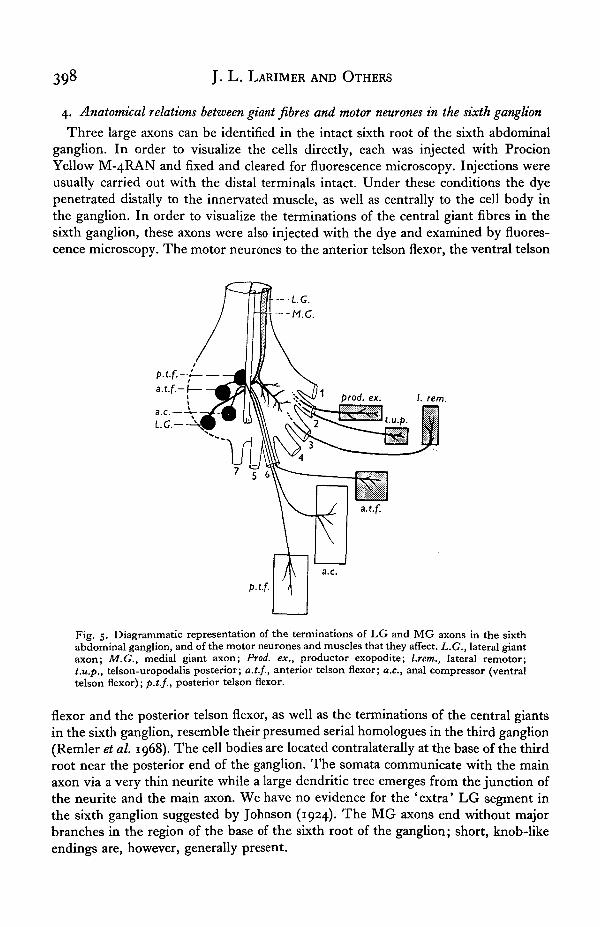
4. Anatomical relations between giant fibres and motor neurones in the sixth ganglion
Three large axons can be identified in the intact sixth root of the sixth abdominalganglion. In order to visualize the cells directly, each was injected with ProcionYellow M-4RAN and fixed and cleared for fluorescence microscopy. Injections wereusually carried out with the distal terminals intact. Under these conditions the dyepenetrated distally to the innervated muscle, as well as centrally to the cell body inthe ganglion. In order to visualize the terminations of the central giant fibres in thesixth ganglion, these axons were also injected with the dye and examined by fluores-cence microscopy. The motor neurones to the anterior telson flexor, the ventral telson
/. rem.
Fig. 5. Diagrammatic representation of the terminations of LG and MG axons in the sixthabdominal ganglion, and of the motor neurones and muscles that they affect. L.G., lateral giantaxon; M.G., medial giant axon; Prod, ex., productor exopodite; l.rem., lateral remotor;t.u.p., telson-uropodalis posterior; a.t.f., anterior telson flexor; a.c, anal compressor (ventraltelson flexor); p.t.f., posterior telson flexor.
flexor and the posterior telson flexor, as well as the terminations of the central giantsin the sixth ganglion, resemble their presumed serial homologues in the third ganglion(Remler et al. 1968). The cell bodies are located contralaterally at the base of the thirdroot near the posterior end of the ganglion. The somata communicate with the mainaxon via a very thin neurite while a large dendritic tree emerges from the junction ofthe neurite and the main axon. We have no evidence for the 'extra' LG segment inthe sixth ganglion suggested by Johnson (1924). The MG axons end without majorbranches in the region of the base of the sixth root of the ganglion; short, knob-likeendings are, however, generally present.

Output connexions of giant fibres 399
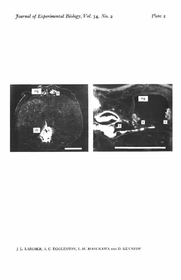
Some of the injected motor axons (PI. 1) were sectioned at 10 /i and examined toconfirm the morphology of the synaptic connections indicated in the electrophysio-logical experiments (Figs. 3, 4). As an example, contacts between the motor axon tothe posterior telson flexor and MG is shown in PL 2. This axon enters the dorsal sideof the ganglion through root 6. After coursing forward a short distance it becomesnarrow, turns ventrally, passes near and sends out numerous small contacts that endon the MG. These contacts are assumed to be the functional synapses between thetwo neurones, since similar contacts are not made upon LG processes that lie nearby(cf. Kennedy, Selverston & Remler, 1969; Davis, 1970). Similar observations weremade upon other identified motor neurones to the major telson muscles.
A summary diagram of the motor neurones, the giant fibres, and the musclesactivated by the latter is given in Fig. 5. The LG axons supply excitation to fourmuscles in the terminal segments and appendages of the animal: the anterior telsonflexor, the lateral remotor, the productor exopodite and the telson-uropodalis pos-terior. The MG axons activate the ventral telson flexor (anal compressor) and posteriortelson flexor. LG axons of the two sides have a functional commissural synapse in thesixth as they do in the ganglia of more anterior segments, while the MG are notcross-connected.
DISCUSSION
The different central connections made by LG and MG neurones adequatelyexplain the differences in motor behaviour observed when the central giant fibres arestimulated separately. A comparison of the movements commanded by MG activity(Fig. 1) with data on the muscles that are selectively activated by these interneurones(Figs. 2, 3) shows that the complete flexion of the abdomen is the result of a uniformactivation of the oblique muscles of each anterior abdominal segment, with simul-taneous excitation of the homologous flexor muscles of the telson-uropod segment(ventral rotator, and ventral and posterior telson flexors). The movements resultingfrom LG activity are somewhat more complex than those evoked by MG. They too,however, can be accounted for in terms of central connexions between the giant fibresand identified motor neurones. The incomplete abdominal flexion (Fig. 1) is probablythe result of weak activation by the LG of motor neurones supplying oblique musclesin the central abdominal segments (Fig. 2); uropod promotion comes about throughactivity in motor neurones innervating the productor exopodite muscles.
The neural network involving motor neurones and central giant fibres in theterminal ganglion is more complete than that in more rostral segments of the abdomen.In the third ganglion, for example, all flexor motor axons are collected into the thirdroot and all extensors into the second; whereas in the sixth ganglion phasic motorneurones of the several sorts emerge from roots 1, 2, 3 and 6. The MG axons synapsewith the sixth root axons, while LG connect with motor neurones leaving roots 2 and3 as well as 6. Since root 6 leaves the dorsal surface of the ganglion and contains mostof the large phasic flexor motor axons, it is probably homologous with the third rootof the anterior abdominal ganglia. The homologue of the motor giant neurone in thesixth ganglion, if present, would therefore be expected to send its axon out via thesixth root. We have, however, been unable to identify a neurone in the sixth ganglionthat possesses the unique characteristics of the motor giants of anterior segments. For
400 J. L. LARIMER AND OTHERS
example, none of the 6th root axons is driven by both MG and LG axons; further-more, the 6th root contains three or four especially large axons instead of one, and allmake en passant synapses with the giant fibres.
Several ' escape' behaviours that differ from one another in detail may be presentin crayfish. High-speed photography of animals producing tail flips with theircircumoesophageal connectives cut reveals that these movements differ from thoseobtained from intact animals. Even in the same intact animal spontaneous flips are notconsistent in form from trial to trial. Since giant axons are not required for at leastsome of these responses (Schrameck, 1970), it is likely that several other interneuronesor combinations of them, in addition to the central giants, can synchronously activatethe fast flexor muscles. For the phasic extensors of the abdomen and several othermuscles—including the rotators, remotors and adductors of the telson-uropod region(Larimer & Kennedy, 1969 a)—the normal pre-motor elements are unknown. Certainspecific responses, such as swimming or steering, may incorporate these additionalmotor elements only under very occasional and specific commands.
The demonstration that specific interneurones (the giant fibres and perhaps others)can activate ensembles of phasic motor neurones in different combinations is ofinterest because it is now clear (Larimer & Kennedy, 19696) that a comparablesystem of interneurones exists for the tonic system of uropod muscles. Such a groupof interneurones acting upon the phasic elements could provide for a wide range ofrapid, stereotyped movements comparable to those known to be available in thepostural system.
SUMMARY
1. High-speed cinematography was used to analyse the abdominal movements ofcrayfish in response to separate stimulation of medial and lateral giant axons. Thesefilms showed that the medial giant fibres command complete abdominal flexions withlittle flaring of the tail appendages. The lateral giants, in contrast, evoked a relativelyweak flexion of the middle abdominal segments, accompanied by promotion of theexopodites of the uropods.
2. An examination of the muscles activated by the two types of giant fibres showsthat differences in the connexions between the giant fibres and specific motor neuronescan account for the behavioural differences observed.
3. The output of the giant fibres was determined in the sixth abdominal ganglion,where their differential effects are most pronounced. The medial giants activate motorneurones whose axons emerge from root 6 of the sixth ganglion. The lateral giantsactivate motor neurones whose axons emerge via roots 2 and 3, as well as thoseemerging via root 6.
4. The larger motor neurones associated with the giant axons in the sixth root ofthe sixth ganglion have been mapped by Procion Yellow injection, and the terminationsof the central giant axons in the sixth ganglion have also been determined. Theconnexions revealed by this technique are consistent with the physiological findings.
5. The evidence suggests that root 6 of the sixth ganglion is homologous with root3 of the more anterior ganglia. However, the giant motor neurone of the sixth ganglionhas not been identified.
6. The medial and lateral giant fibres, and perhaps other specific 'command'
Output connexions of giant fibres 401
interneurones, can thus drive specific ensembles of phasic motor neurones to providea range of stereotyped quick movements. In this respect the organization of the phasicsystem of interneurones and motor neurones resembles that in the tonic system.
The experiments on cinematography and on the influence of LG and MG stimula-tion on abdominal and uropod muscles were carried out at Stanford during 1967-68.This work was supported by grants from the U.S. Public Health Service (NB-02944)and the Air Force Office of Scientific Research (AFOSR-68-1373) to D. Kennedy andby a Guggenheim Fellowship award to J. Larimer. The experiments on identifiedsixth-ganglion motor neurones and the dye injection studies were carried out at Texasduring 1969 and 1970, and were supported by a grant to J. Larimer from the U.S.Public Health Service (NS-05423) and by The NIH Biomedical Sciences SupportGrant 5-S05 FR-07091-04. L. M. Masukawa and A. C. Eggleston were supportedby NIH Training grant 2 Toi GM 00836-07. We acknowledge the unfailing technicalassistance of Joanna Hanawalt and Gary Shelton, and thank J. Schrameck and thelate Dr D. M. Wilson for helpful discussions and a critical reading of the manuscript.
REFERENCES
BRUNER, J. & KENNEDY, D. (1970). Habituation: Occurrence at a neuromuscular junction. Science 169,92-4.
DAVIS, W. J. (1970). Motoneuron morphology and synaptic contacts: Determination by intracellulardye injection. Science 168, 1358—60.
EVOY, W. H. & KENNEDY, D. (1967). The central nervous organization underlying control of antagon-istic muscles in the crayfish. I. Types of command fibers. J. exp. Zool. 165, 223—8.
FURSHPAN, E. J. & POTTER, D. D. (1959). Transmission at the giant motor synapses of the crayfish.J. Physiol. 145, 289-325.
VAN HARREVELD, A. (1936). A physiological solution for freshwater crustaceans. Proc. Soc. exp. Biol.Med., N. Y. 34, 428-32.
HORIUCH, E., HAYASHI, H. & TAKAHASHI, I. (1966). Median giant fibre system in the crayfish cephalicganglion. Nature, Lond. 212, 831-2.
JOHNSON, G. E. (1924). Giant nerve fibers in crustaceans with special reference to Cambarus andPaleomonetes. J. comp. Nenrol. 36, 323-73.
JOHNSON, G. E. (1926). Studies on the function of the giant nerve fibers of crustaceans with specialreference to Cambarus and Paleomonetes. J. comp. Neurol. 43, 19-33.
KAO, C. Y. & GRUNDFEST, H. (1956). Conductile and integrative functions of crayfish giant axons.Fed. Proc. 15, 104.
KENNEDY, D. & TAKEDA, K. (1965). Reflex control of abdominal flexor muscles in the crayfish. I. Thetwitch system. J. exp. Biol. 43, 211-27.
KENNEDY, D., SELVERSTON, A. I. & REMLER, M. P. (1969). Analysis of restricted neural networks.Science 164, 1488-96.
KRASNE, F. B. (1969). Excitation and habituation of the crayfish escape reflex: The depolarizingresponse in lateral giant fibres of the isolated abdomen. J. exp. Biol. 50, 29-46.
LARIMER, J. L. & KENNEDY, D. (1969a). Innervation patterns of fast and slow muscle in the uropodsof crayfish. .7. exp. Biol. 51, 119-33.
LARIMER, J. L. & KENNEDY, D. (19696). The central nervous control of complex movements in theuropods of crayfish. J. exp. Biol. 51, 135-50.
RAYNER, M. D. & WIERSMA, C. A. G. (1967). Mechanisms of the crayfish tail flick. Nature, Lond. 213,1231-3.
REMLER, M., SELVERSTON, A. & KENNEDY, D. (1968). Lateral giant fibers of crayfish: location of somataby dye injection. Science 162, 281-3.
SCHRAMECK, J. E. (1970). Crayfish swimming: alternating motor output and giant fiber activity. Science169, 698-700.
STRETTON, A. O. W. & KRAVITZ, E. A. (1968). Neuronal geometry: determination with a technique ofintracellular dye injection. Science 162, 132—4.
WATANABE, A. & GRUNDFEST, H. (1961). Impulse propagation at the septal and commissural junctionsof crayfish lateral giant axons. J. gen. Physiol. 45, 267-308.

Journal of Experimental Biology, Vol. 54, No. 2 Plate 1
J. L. LARIMER, A. C. EGGLESTON, L. M. MASUKAWA AND D. KENNEDY {Facing p. 402)
Journal of Experimental Biology, Vol. 54, No. 2 Plate 2
J. L. LARIMER, A. C. EGGLESTON, L. M. MASUKAWA AND D. KENNEDY
402 J. L. LARIMER AND OTHERS
WIERSMA, C. A. G. (1938). Function of giant fiber of the central nervous system of the crayfish. Proc.Soc. exp. Biol. Med. 38, 661-2.
WIERSMA, C. A. G. (1947). Giant nerve fiber system of the crayfish. A contribution to comparativephysiology of synapse. J. Neurophysiol. 10, 23—38.
WIERSMA, C. A. G. (1961). Reflexes and the central nervous system. In Physiology of Crustacea, ed.T. H. Waterman. Volume n, pp. 241^79. New York: Academic Press.
EXPLANATION OF PLATES
PLATE I
Fluorescence photomicrographs of Procion Yellow-injected giant fibres and motor neurones in wholemounts of the sixth ganglion, (a) Upper left: MG axon. There is some leakage of dye into one of thelarge motor neurones of the sixth root, probably at the junctions. (6) LG. (c) The large motor neuronesupplying the anterior telson flexor (retouched), (d) The motor neurone supplying the anal compressormuscle (ventral telson flexor muscle), (e) Side view of the large motor neurone to the posterior telsonflexor. ( / ) The same p.t.f. neurone as seen from the ventral side of the ganglion. Injections of this typeallowed the placement of the neurones in the map of Fig. 5. Scale: 100 /i.
PLATE 2
Photomicrographs of cross-sections of the sixth abdominal ganglion, showing a Procion Yellow-injected posterior telson flexor motor neurone cell body (cb) and neurite («) with synapses (s) on MG.Scale left and right: 200 fi, 50 fi.