the fat compartments of the face: anatomy and clinical...
TRANSCRIPT

Seediscussions,stats,andauthorprofilesforthispublicationat:http://www.researchgate.net/publication/6312494
TheFatCompartmentsoftheFace:AnatomyandClinicalImplicationsforCosmeticSurgery
ARTICLEinPLASTICANDRECONSTRUCTIVESURGERY·JULY2007
ImpactFactor:2.99·DOI:10.1097/01.prs.0000265403.66886.54·Source:PubMed
CITATIONS
156
READS
1,494
2AUTHORS,INCLUDING:
RodJRohrich
UniversityofTexasSouthwesternMedical…
769PUBLICATIONS11,016CITATIONS
SEEPROFILE
Availablefrom:RodJRohrich
Retrievedon:17November2015

COSMETIC
The Fat Compartments of the Face: Anatomyand Clinical Implications for Cosmetic Surgery
Rod J. Rohrich, M.D.Joel E. Pessa, M.D.
Dallas, Texas
Background: Observation suggests that the subcutaneous fat of the face ispartitioned as distinct anatomical compartments.Methods: Thirty hemifacial cadaver dissections were performed after methyl-ene blue had been injected into specified regions. Initial work focused on thenasolabial fat. Dye was allowed to set for a minimum of 24 hours to achieveconsistent diffusion. Dissection was performed in the cadaver laboratory usingmicroscopic and loupe magnification.Results: The subcutaneous fat of the face is partitioned into multiple, inde-pendent anatomical compartments. The nasolabial fold is a discrete unit withdistinct anatomical boundaries. What has been referred to as malar fat is com-posed of three separate compartments: medial, middle, and lateral temporal-cheek fat. The forehead is similarly composed of three anatomical units in-cluding central, middle, and lateral temporal-cheek fat. Orbital fat is noted inthree compartments determined by septal borders. Jowl fat is the most inferiorof the subcutaneous fat compartments. Some of the structures referred to as“retaining ligaments” are formed simply by fusion points of abutting septalbarriers of these compartments.Conclusions: The subcutaneous fat of the face is partitioned into discrete an-atomic compartments. Facial aging is, in part, characterized by how these com-partments change with age. The concept of separate compartments of fatsuggests that the face does not age as a confluent or composite mass. Shearingbetween adjacent compartments may be an additional factor in the etiology ofsoft-tissue malposition. Knowledge of this anatomy will lead to better under-standing and greater precision in the preoperative analysis and surgical treat-ment of the aging face. (Plast. Reconstr. Surg. 119: 2219, 2007.)
Clinical observation and laboratory investi-gation suggest that the subcutaneous fat ofthe face exists in distinct anatomical com-
partments (Fig. 1). When the operating surgeonperforms a face lift, zones of adherence areencountered that alternate with zones wheredissection proceeds with relative ease. This sug-gests that barriers exist between different zonesof facial fat.
Patients with facial atrophy and midface hol-lowing consistently show preservation of the na-solabial fold and jowl fat (Fig. 2). This commonclinical observation suggests that regions of fatbehave differently during the aging process.
In the cadaver laboratory, dye injected intothe upper forehead flows down the cheek andinto the neck in a distinct and reproducible man-ner. This test has been repeated at least a dozentimes. Moreover, dye injected into the nasolabialfold partitions in a discrete fashion (Figs. 3).
Taken as a whole, these clinical and laboratoryobservations suggest that the subcutaneous fat ofthe face is highly partitioned, that it is not aconfluent mass, and that further study is war-ranted to investigate this concept as it pertains tofacial aging and cosmetic surgical techniques.
MATERIALS AND METHODSThirty hemifacial fresh cadaver dissections
were performed on 18 male and 12 female spec-imens ranging in age from 47 to 92 years. Pre-liminary work was performed on multiple spec-imens to determine the best dye stainingtechnique. Letraset, Bombay India Ink, indocya-nine green, and methylene blue were all evalu-
From the Department of Plastic Surgery, University of TexasSouthwestern Medical Center.Received for publication July 27, 2006; accepted October 13,2006.Copyright ©2007 by the American Society of Plastic Surgeons
DOI: 10.1097/01.prs.0000265403.66886.54
www.PRSJournal.com 2219

ated. Methylene blue consistently displayed thebest tissue diffusion.1 In other studies, rehydratingsome of the specimens was found to improve dyediffusion.
Dye was allowed to set for a minimum of 24hours to allow for adequate tissue diffusion. Al-lowing the dye to set for 48 to 72 hours actuallyimproved distribution and facilitated dissection.Each compartment was verified by injecting a min-imum of three and a maximum of 10 cadaverhemifaces. All work was performed in the cadaverlaboratory.
Microscopic and 4.5- and 6.0-power loupemagnification facilitated dissection. Photographicdocumentation was obtained with a Canon 20Dsystem and F2.8 macro lens. Images were scannedinto Adobe Photoshop (CS2; Adobe Systems, Inc.,San Jose, Calif.). All results are shown from thecadaver’s left side for the sake of consistency.
RESULTS
Nasolabial Fat CompartmentThe nasolabial fat was injected in 10 hemifaces
from three male and two female cadavers. Thecadaver face was allowed to set at least 24 hours,although immediate staining of a distinct areacould be seen through the skin. A distinct com-partment was noted in all specimens (Fig. 4). Thenasolabial fat lies anterior to medial cheek fat, andoverlaps jowl fat. The orbicularis retaining liga-ment represents the superior border of this com-
partment. Nasolabial fat can be noted medial tothe deeper fat of the suborbicularis fat compart-ment. The lower border of the zygomaticus majormuscle is adherent to this compartment.
As an incidental observation, the volume ofthis compartment did not vary much between ca-davers, regardless of age or sex. The only variablenoted was that medial cheek fat overlapped naso-labial fat to a greater degree in certain cadavers.
Cheek Fat CompartmentsThere are three distinct cheek fat compart-
ments: the medial, middle, and lateral temporal-cheek fat.
Medial cheek fat is lateral to the nasolabial fold(Fig. 5). This compartment is bordered superiorlyby the orbicularis retaining ligament and the lat-eral orbital compartment. Jowl fat lies inferior tothis fat compartment.
Middle cheek fat lies superficial in its midpor-tion (Fig. 6). This fat compartment is found an-terior and superficial to the parotid gland. At itssuperior portion, the zygomaticus major muscle isadherent. A confluence of septa occurs at thislocation where three compartments meet, andforms a dense adherent zone where the zygomaticligament has been described.2
The fusion of septal boundaries is an anatom-ical principle and can be simply illustrated bycross-sectional anatomy (Fig. 6, right). Middlecheek fat abuts medial cheek fat, and their septal
Fig. 1. An artist’s rendition of the subcutaneous compartmentsof the face.
Fig. 2. Lipoatrophy and midface hollowing (red arrow) are notedwith preservation of the nasolabial and jowl fat (black arrows).
Plastic and Reconstructive Surgery • June 2007
2220

Fig. 3. (Left) Methylene blue dye injected into the forehead flows down the cheek in a spe-cific and reproducible manner. The nasolabial fat also stains as a specific region. (Right) Anartist’s rendition of how dye flows from the forehead to the neck with a distinct medial bound-ary (arrow). Dye partitioning would not occur if the face were a confluent mass.
Fig. 4. The nasolabial fat compartment is the most medial of themajor cheek compartments. Blue dye has stained this region. Theorbicularis retainingligament is thesuperiorboundary (ORL), andthe suborbicularis fat is a lateral and deep boundary (SOOF). Me-dial cheek fat has been reflected off the nasolabial compartment.The zygomaticus major is tethered at its inferior border (ZM).
Fig. 5. Malar fat iscomposedofthreecompartments: themedial,middle, and lateral temporal-cheek. The medial fat, shown here,lies adjacent to the nasolabial fat. The superior boundary is againthe orbicularis retaining ligament (ORL). The lateral boundaryis the middle cheek septum. The red arrow represents a pointof fixation.
Volume 119, Number 7 • The Fat Compartments of the Face
2221

boundaries fuse into a dense fascial network (Fig.6, right, arrow). Again, this corresponds to what hasbeen described as the zygomatic ligament. Thezone where the medial fat abuts the middle cheekfat corresponds to the location of the parotido-masseteric ligaments.3
The lateral temporal-cheek compartment isthe most lateral compartment of cheek fat (Fig. 7).This fat lies immediately superficial to the parotidgland and connects the temporal fat to the cervicalsubcutaneous fat.
A true septum can be located anterior to thiscompartment. This septum, the lateral cheek sep-tum, can be dissected and clearly identified as avertical septal barrier with loupe magnification.This is the first transition zone encountered dur-ing a face lift when proceeding medially from thepreauricular incision.
Forehead and Temporal Fat CompartmentsThe subcutaneous fat of the forehead is com-
posed of three compartments. The central com-partment is located in the midline region of theforehead (Fig. 8). It has a consistent location thatabuts the middle temporal compartments on ei-ther side and has an inferior border at the nasaldorsum. The lateral boundary probably is a septal
barrier and could be referred to as the centraltemporal septum.
The middle temporal fat compartments lie oneither side of the central forehead fat (Fig. 9). Theinferior border is the orbicularis retaining liga-ment, and the lateral border corresponds to thesuperior temporal septum.4
The lateral temporal-cheek compartment haspreviously been described. It connects the lateralforehead fat to the lateral cheek and cervical fat(Fig. 7).
Orbital Fat CompartmentThree subcutaneous fat compartments exist
around the eye. The most superior compartmentis bounded by the orbicularis retaining ligamentas it courses around the superior orbit (Fig. 10).The orbicularis retaining ligament is a truly cir-cumferential structure that spans the superiorand inferior orbits and blends into the medial andlateral canthi. Dye injected into the superior com-partment does not stain the inferior orbital com-partment.
The inferior orbital fat is a thin, subcutaneouslayer that lies immediately below the inferior lidtarsus (Fig. 11). Its inferior boundary is the orbic-ularis retaining ligament or malar septum. Themedial and lateral extents are, again, the canthi.
Fig. 6. (Left) The middle cheek fat compartment lies between medial and lateral temporal-cheek fat. Thesuperior border is defined by the superior cheek septum (SCS). A zone of fixation (red arrow) is noted where thiscompartment adjoins the middle compartment and inferior orbital compartment. (Right) The cross-sectionalanatomy illustrates the anatomic principle that fusion planes exist between adjacent fat compartments. Adense fascial system (red arrow) exists where the medial and middle fat compartments meet. The zygomaticusmajor muscle is noted deep to this fusion plane.
Plastic and Reconstructive Surgery • June 2007
2222

Fig. 7. The lateral temporal-cheek fat spans the forehead to thecervical region. It is the most lateral of the cheek fat compart-ments and has an identifiable septal barrier medially called thelateral cheek septum (LCS). The superior and inferior temporalsepta (STS and ITS, respectively) represent the superior bound-aries. This cadaver dissection is noteworthy because several fatcompartments are seen without dye staining, including the in-ferior orbital fat (IOF) and medial cheek fat (M). Nasolabial fat hasbeen stained with methylene blue dye.
Fig. 8. Three forehead fat compartments have been identified todate. The central fat is a midline region. It has an inferior boundary atthe nasal dorsum. The lateral border is a dense fascial plane thatappears to be a septum, termed the central temporal septum.
Fig. 10. Therearethreeperiorbital fatcompartments.Thesuperiororbital fat is shown here. The boundary is the orbicularis retainingligament (ORL), a truly circumferential membrane that inserts at themedial and lateral canthi. The superior and inferior orbital compart-ments are, however, distinct from one another.
Fig. 9. The middle forehead fat compartments are situated oneither side of the central fat and are located medially to the su-perior temporal septum (STS). The inferior border is the orbicu-laris retaining ligament of the superior orbit. The lateral tempo-ral-cheek fat has already been described and is the third of theforehead fat compartments.
Volume 119, Number 7 • The Fat Compartments of the Face
2223

The lateral orbital fat compartment is thethird of the subcutaneous orbital fat compart-ments (Fig. 12). Its superior border is the inferiortemporal septum4; the inferior border is desig-nated the superior cheek septum. The zygomati-cus major muscle is again noted to be adherent tothis compartment. Transitioning the zygomaticusmajor muscle plays a major role in adequatelyreleasing soft tissues if one attempts to elevatemedial fat or jowl fat.
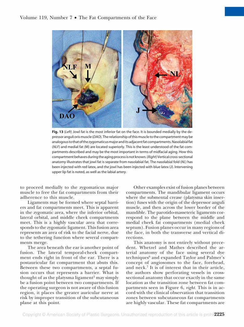
Jowl Fat CompartmentJowl fat is separate from nasolabial fat (Fig. 13,
left). Jowl fat adheres to the depressor anguli orismuscle. The medial boundary of this compart-ment is the lip depressor muscle, and the inferiorboundary is determined by a membranous fusionof the platysma muscle. The fusion point betweenthese two muscles occurs at the region of the man-dibular retaining ligament.2 The difference be-tween nasolabial fat and jowl fat can be shown bycross-sectional anatomy (Fig. 13, right).
DISCUSSIONThis study suggests that facial subcutaneous
fat is highly compartmentalized. Because theface is composed of multiple discrete anatomi-cal regions, it is unlikely that it ages as a con-fluent mass.
A youthful face is characterized by a smoothtransition between subcutaneous compartments:aging leads to abrupt contour changes betweenthese regions. This may occur due to volume lossas described by Lambros5 or to malposition ofspecific compartments from a number of causes.Attenuation of ligaments alone would be insuffi-cient to explain compartment changes, especiallyin light of the septated architecture of the fatcompartments noted herein.
This anatomical arrangement is in agree-ment with that noted clinically. The operatingsurgeon encounters areas of fixation or adher-ence in dissecting from lateral to medial. Theseareas are vascular and occur in the transitionregion between compartments. The zygomaticmajor muscle provides an important area of fix-ation to three compartments, and dissection be-comes much easier once the zygomaticus muscleis transitioned. The plane between the lateraland middle cheek compartments can easily leadinto the buccal fat; again, the zygomaticus majormuscle is a major landmark in avoiding thispitfall. As a clinical point, if one chooses todissect medially to elevate jowl fat, dissection has
Fig. 12. The lateral orbital fat lies below the inferior temporalseptum (ITS) and above the superior cheek septum (SCS). Thezygomaticus major muscle is adherent superiorly (ZM). Inter-estingly, the zygomaticus muscle is adherent to several fatcompartments. This is clinically important, because dissectionpast the zygomaticus major is necessary to move or alter me-dial cheek fat.
Fig. 11. The inferior orbital fat compartment is analogous to thesuperior orbital fat and, again, is bordered by the orbicularis re-taining ligament (ORL). This compartment is noted clinically withperiorbital ecchymosis.
Plastic and Reconstructive Surgery • June 2007
2224
to proceed medially to the zygomaticus majormuscle to free the fat compartments from theiradherence to this muscle.
Ligaments may be formed where septal barri-ers and fat compartments meet. This is apparentin the zygomatic area, where the inferior orbital,lateral orbital, and middle cheek compartmentsmeet. This is a highly vascular area that corre-sponds to the zygomatic ligament. This fusion arearepresents an area of risk to the facial nerve, dueto the tethering function where several compart-ments merge.
The area beneath the ear is another point offusion. The lateral temporal-cheek compart-ment ends right in front of the ear. There is apostauricular fat compartment that abuts this.Between these two compartments, a septal fu-sion occurs that represents a barrier. What isthought of as the platysma ligament2 may simplybe a fusion point between two compartments. Ifthe operating surgeon is not aware of this fusionregion, it places the greater auricular nerve atrisk by improper transition of the subcutaneousplane at this point.
Other examples exist of fusion planes betweencompartments. The mandibular ligament occurswhere the submental crease (platysma skin inser-tion) fuses with the origin of the depressor angulimuscle, and then across the lower border of themandible. The parotido-masseteric ligaments cor-respond to the plane between the middle andmedial cheek fat compartments (medial cheekseptum). Fusion planes occur in many regions ofthe face, in both the transverse and vertical di-rections.
This anatomy is not entirely without prece-dent. Whetzel and Mathes described the ar-terial anatomy of the face using several dyetechniques6 and expanded Taylor and Palmer’sconcept of angiosomes to the face, forehead,and neck.7 It is of interest that in their article,the authors show perforating vessels in cross-sectional anatomy that occur exactly in the samelocation as the transition zone between fat com-partments seen in Figure 6, right. This is in ac-cord with the clinical observation that transitionzones between subcutaneous fat compartmentsare highly vascular. These fat compartments are
Fig. 13 (Left) Jowl fat is the most inferior fat on the face. It is bounded medially by the de-pressor anguli oris muscle (DAO). The relationship of this muscle to the compartment may beanalogous to that of the zygomaticus major and its adjacent fat compartments. Nasolabial fat(NLF) and medial fat (M) are located superiorly. This is the least understood of the fat com-partments described and may be the most important in terms of midfacial aging. How thiscompartment behaves during the aging process is not known. (Right) Vertical cross-sectionalanatomy illustrates that jowl fat is separate from nasolabial fat. The nasolabial fold (NL) hasbeen injected with red latex, and the jowl has been injected with blue latex (J). Interveningupper lip fat is noted, as well as the labial artery.
Volume 119, Number 7 • The Fat Compartments of the Face
2225

not angiosomes; rather, they occur between vas-cular perforating vessels that supply the skin.More evidence is the important role played bythe zygomaticus major and depressor anguli orismuscles, which may have myocutaneous perfo-rators traveling in septa.
Deep layers of facial fat, including the sub-orbicularis, retro-orbicularis, and buccal fat,have been studied in detail.8 –11 This study sug-gests that additional deep fat compartments ex-ist (Fig. 6, right). For example, a fat compart-ment surrounds the levator anguli muscle, andthere exists a fat compartment beneath the lipelevator muscles. A tenet of facial anatomy isthat fat is noted both above and below mostfacial muscles, probably to facilitate the neces-sary gliding mechanism.
With this knowledge in mind, the aging facecan be analyzed as a change in volume and posi-tion of these separate compartments, both super-ficial and deep (Fig. 2). The cadaver shown inFigure 2 has a loss of midfacial projection, prom-inence of the nasojugal crease, malar moundshow, jowl prominence, and a deep nasolabialfold. These findings are not unrelated.
Jowl prominence may occur from malposi-tion of the labiomandibular compartment. Inaddition, loss of volume of the deep midfacial fat(Fig. 14) may be a primary determinant of mid-facial aging. This decreases support for the me-dial cheek compartment and results in dimin-ished midface projection. The nasolabial fold isunmasked, just as is the malar mound. A cascadeoccurs from malposition of this compartment.The negative vector, caused by loss of supportfor the medial cheek compartment and volumeloss of the deep compartment fat, allows excesstraction to be placed on the lower eyelid.12 Thisleads to scleral show. Confirmatory evidence forthis last statement is noted clinically by the snaptest13: if a prolonged amount of time is noted foran individual’s lower lid to return to the normalposition, this can be improved by simple medialcheek elevation. Lid laxity, orbicularis laxity,and loss or attenuation of the canthi may play asmall or no role in what is actually encounteredclinically. Rather, subcutaneous fat malpositionand atrophy simply place downward traction onthe lower lid and distorts its position.
We suggest that jowl fat may be an important keyto rejuvenating the midface. In the cadaver labora-tory, dissection of the nasolabial fat and reposition-ing of this compartment beneath the medial cheekfat can efface the nasolabial fold (Fig. 14). Perhaps
this technique will find application in repositioningattempts of the jowl fat.
The eye sees what the mind knows. The con-cept of subcutaneous fat compartments shiftsone’s perspective from visualizing the face as a
Fig. 14. The nasolabial fat compartment can be repositioned be-neath the medial cheek fat compartment experimentally to di-minish the contour deformity. Understanding the anatomiccompartments explains why simple lateral traction of skin or skinand fat has little overall effect on the nasolabial fold prominence.This technique may be applicable to jowl fat and midface pro-jection. (Above) The nasomaxillary fat is seen medially (black ar-row). The deep compartment fat—the deep midfacial fat—isnoted by the red arrow. This has not been previously described.There is likewise a lateral deep compartment fat (red arrow later-ally) beneath the lateral temporal-cheek compartment (ZM, zy-gomaticus major muscle). (Below) The deep midfacial fat hasbeen injected with methylene blue dye (red arrow), which stainsaround the levator anguli oris muscle. This deep compartmentmay be a primary determinant of a youthful, anteriorly projectingmidface. Loss of volume in this deep compartment, similar towhat may occur in the temporal fat, may lead to the cascade ef-fect described.
Plastic and Reconstructive Surgery • June 2007
2226
discrete mass to viewing the face as a set of archi-tectural compartments. These compartments mayage independently, and mass shifts of facial softtissue may not sufficiently address the complexityof the aging process. The answers to this and manyother questions suggested by this study await fur-ther research.
Joel E. Pessa, M.D.Department of Plastic Surgery
5323 Harry Hines BoulevardDallas, Texas 75390-9132
ACKNOWLEDGMENTSThe authors thank Melinda Mora of the University
of Texas Southwestern Willed Body Program. This studywould not have been possible without her help. Kindthanks are also extended to Holly Smith, from the medicalillustration department, for her help in preparing thearticle.
DISCLOSUREThe authors have no financial interests in this re-
search project or in any of the techniques or equipmentused in this study.
REFERENCES1. Ahlberg, K. M., Assavanop, P., and Tay, W. M. A comparison
of the apical dye penetration patterns shown by methylene
blue and India ink in root-filled teeth. Int. Endod. J. 28: 30,1995.
2. Furnas, D. W. The retaining ligaments of the cheek. Plast.Reconstr. Surg. 83: 11, 1989.
3. Stuzin, J. M., Baker, T. J., and Gordon, H. L. The relationshipof the superficial and deep facial fascias: Relevance to rhyt-idectomy and aging. Plast. Reconstr. Surg. 89: 441, 1992.
4. Moss, C. H., Mendelson, B. C., and Taylor, G. I. Surgicalanatomy of the ligamentous attachments in the temple andperiorbital regions. Plast. Reconstr. Surg. 105: 1475, 2000.
5. Lambros, V. Personal communication. July 2006.6. Whetzel, T. P., and Mathes, S. J. Arterial anatomy of the face:
An analysis of vascular territories and perforating cutaneousvessels. Plast. Reconstr. Surg. 89: 591, 1992.
7. Taylor, G. I., and Palmer, J. H. The vascular territories (an-giosomes) of the body: Experimental study and clinical ap-plications. Br. J. Plast. Surg. 40: 113, 1987.
8. Aiache, A. E., and Ramirez, O. H. The suborbicularis oculifat pads: An anatomic and clinical study. Plast. Reconstr. Surg.95: 37, 1995.
9. May, J. W., Jr., Fearon, J., and Zingarelli, P. Retro-orbicularisoculi fat (ROOF) resection in aesthetic blepharoplasty: A6-year study in 63 patients. Plast. Reconstr. Surg. 86: 682, 1990.
10. Stuzin, J. M., Wagstrom, L., Kawamoto, H. K., Baker, T. J., andWolfe, S. A. The anatomy and clinical applications of thebuccal fat pad. Plast. Reconstr. Surg. 85: 29, 1990.
11. Jackson, I. T. Anatomy of the buccal fat pad and its clinicalsignificance. Plast. Reconstr. Surg. 103: 2059, 1999.
12. Jelks, G. W., and Jelks, E. B. Preoperative evaluation of theblepharoplasty patient: Bypassing the pitfalls. Clin. Plast.Surg. 20: 213, 1993.
13. Furnas, D. W. Festoons, mounds, and bags of the eyelids andcheek. Clin. Plast. Surg. 20: 367, 1993.
Volume 119, Number 7 • The Fat Compartments of the Face
2227