the genetic science behind hereditary colorectal cancer · the genetic science behind hereditary...
TRANSCRIPT

The Genetic Science Behind Hereditary Colorectal Cancer
Larry Geier, MD Medical Oncologist and Director,
Genetic Risk Evaluation and Testing Program Kansas City Cancer Center
University of Kansas Cancer Center

Disclosure
Speaker Bureau – Myriad Genetics

Relatively Common Hereditary Cancer Syndromes
• Hereditary Breast/Ovary Syndrome (BRCA genes)
• Breast, ovary, others
• Lynch Syndrome (Mismatch Repair genes)
• Colon, uterus, ovary, stomach, others
• Colon polyposis syndromes (APC, MUTYH genes) • Colon, upper GI, others

Hallmarks of Hereditary Cancer
• Family clustering of specific types of cancer among siblings or across multiple generations
• Younger age at diagnosis compared to non-hereditary cases of the same cancer • Multiple cancers in the same person
• Typical phenotypes in some cancers
4

“FAMP” and Hereditary Cancer
Family Age
Multiplicity Phenotype

Take-Home Messages For Today
• Hereditary CRC is much more common than previously realized
• Historically, doctors do a rather poor job of recognizing these patients/families before it is too late
• Many of the resulting cancers could have been prevented, or at least found in an earlier, more curable stage
• The syndromes involve high risk for cancers other than CRC, and providers must be familiar with this spectrum of cancers

Why should we care?
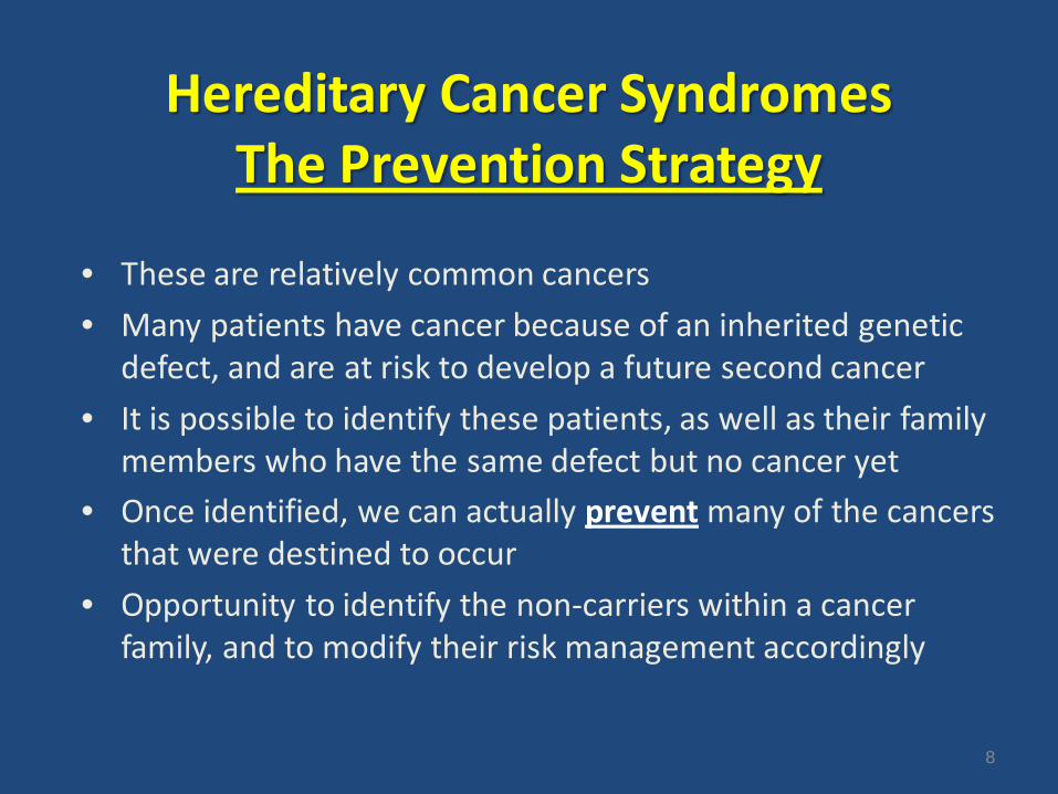
Hereditary Cancer Syndromes The Prevention Strategy
• These are relatively common cancers • Many patients have cancer because of an inherited genetic
defect, and are at risk to develop a future second cancer • It is possible to identify these patients, as well as their family
members who have the same defect but no cancer yet • Once identified, we can actually prevent many of the cancers
that were destined to occur • Opportunity to identify the non-carriers within a cancer
family, and to modify their risk management accordingly
8
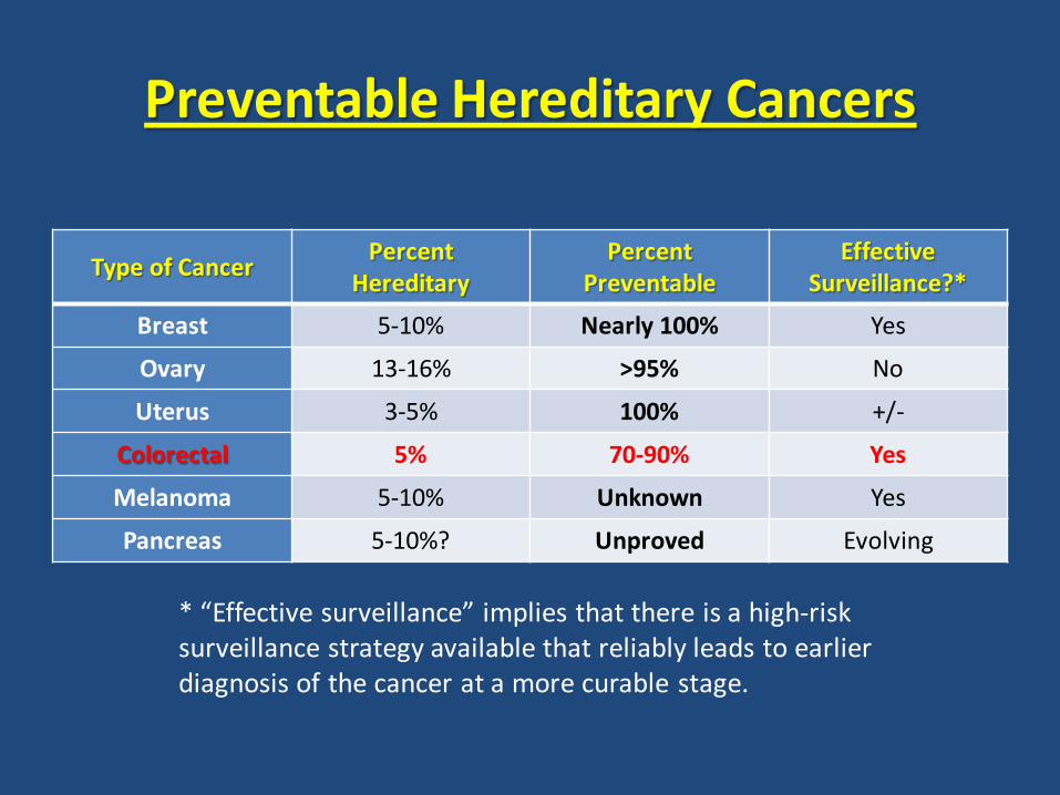
Preventable Hereditary Cancers
Type of Cancer Percent Hereditary
Percent Preventable
Effective Surveillance?*
Breast 5-10% Nearly 100% Yes
Ovary 13-16% >95% No
Uterus 3-5% 100% +/-
Colorectal 5% 70-90% Yes
Melanoma 5-10% Unknown Yes
Pancreas 5-10%? Unproved Evolving
* “Effective surveillance” implies that there is a high-risk surveillance strategy available that reliably leads to earlier diagnosis of the cancer at a more curable stage.

Hereditary Cancer Syndromes Management Implications
• Some hereditary cancers may be biologically distinct from their sporadic counterparts, and this may have a significant effect on:
• Overall prognosis • Surgical decision-making • Chemotherapy alternatives
10

Hereditary Colorectal Cancer Management Implications
• Lynch CRC has a better prognosis than sporadic
• Consideration of total colectomy in Lynch or FAP pts
• Consideration of prophylactic hysterectomy in Lynch
patients undergoing colon resection for CRC
• 5FU not effective as the dominant drug in Lynch CRC
11

Epidemiology of Colorectal Cancer
145,000 new U.S. cases/year

Epidemiology of Colorectal Cancer
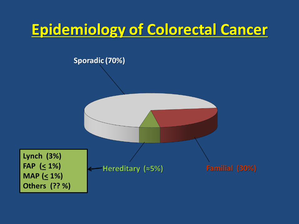
Epidemiology of Colorectal Cancer
Lynch (3%) FAP (< 1%) MAP (< 1%) Others (?? %)
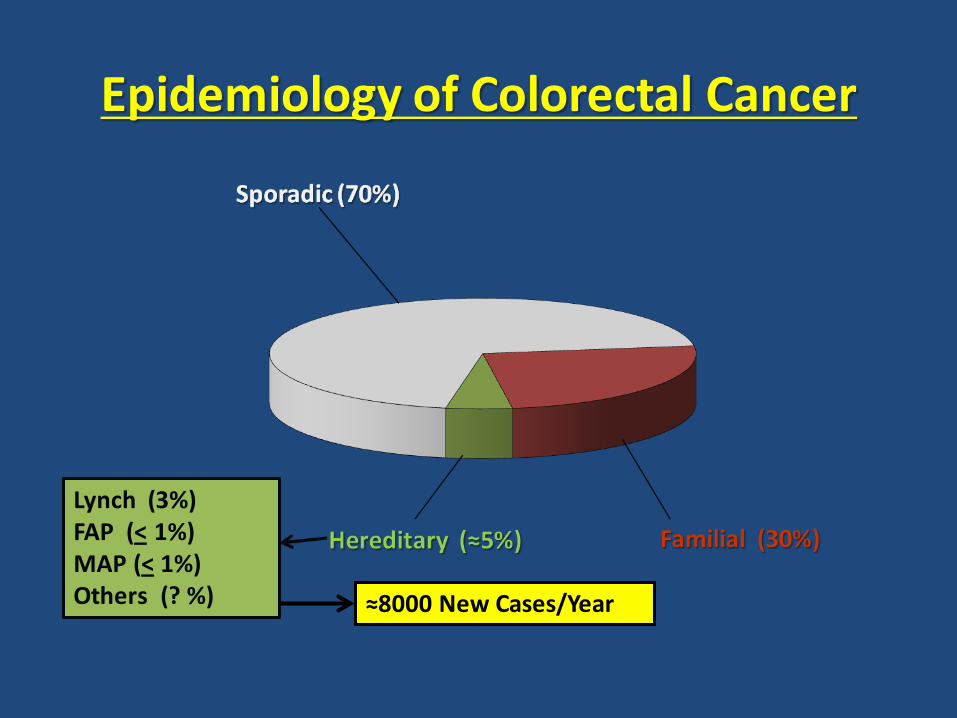
Epidemiology of Colorectal Cancer
Lynch (3%) FAP (< 1%) MAP (< 1%) Others (? %) ≈8000 New Cases/Year

Hereditary Colorectal Cancer: Common Syndromes
POLYPOSIS (many colon polyps): Familial Adenomatous Polyposis (Classic FAP)
Attenuated FAP (AFAP) MUTYH-Associated Polyposis (MAP) Serrated, juvenile, Peutz-Jeghers, Cowden, other rare non-adenomatous syndromes
NON-POLYPOSIS (relatively few polyps): Lynch Syndrome (formerly “HNPCC”)
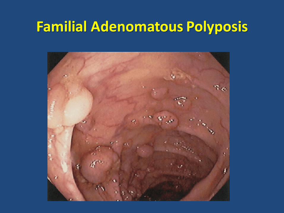
Familial Adenomatous Polyposis

Familial Adenomatous Polyposis (Classic FAP)
• Hundreds to thousands of polyps • Early age of onset, frequently in teen years • Mutation in the APC gene • Autosomal dominant
• 30% of affected individuals represent de novo rather
than inherited mutations, and therefore may have no family history of polyps or cancer

Familial Adenomatous Polyposis (Classic FAP)
• Virtually 100% chance of developing colorectal cancer unless preventive total colectomy is performed
• Average age for colon cancer approximately 35-40 yrs • 7% occur by age 21, 90% by age 50
• Gastric cancer in 2-5% (may be higher in Asians) • Duodenal, periampullary cancer 4-12% • Thyroid cancer 4-6% • Variety of other extraintestinal manifestations:
• Osteomas, desmoid tumors (Gardner syndrome) • CNS medulloblastoma (Turcot syndrome) • CHRPE

Congenital Hypertrophy of the Retinal Pigment Epithelium

Attenuated FAP (AFAP)
• “FAP Lite” • Typically less severe but highly variable degree of polyposis • Often involves the right side of the colon more than the left • Later age of onset, often 30’s or older
• Lower penetrance for colorectal cancer, estimated 80% • Patients with relatively low polyp density can be managed
with annual surveillance colonoscopy • Some will still eventually require preventive surgery
Attenuated FAP (AFAP)
• Same gene as classic FAP, with same rate of de novo mutations • The upper GI cancer risks are not attenuated • Extracolonic manifestations similar to classic FAP, although
CHRPE and desmoid tumors are not seen as commonly
• These patients are much harder to diagnose than classic FAP • Critically important to track the cumulative number of
adenomatous polyps removed over time • Ten is the consensus number to trigger genetic evaluation • Desmoid tumors should also lead to genetic testing

Case Study FM
• 52 y/o male who has been under high-risk surveillance for colon cancer since age 42
• At 42 he was found to have 2 sigmoid adenomas on his first screening colonoscopy
• Repeat scope q 2-3 yrs, with 1-3 polyps each time • Scope at 52 shows 8 adenomas, mostly ascending and
transverse. One is a villous adenoma with dysplasia.
• Mother died from colon cancer at 49 • No other history of CRC or polyps known in the family

Case Study FM
24
52 Polyps 42 43 50
49
70
Never scoped No Polyps
Colon 72 66

Case Study FM
• What is the key question to be asked for this patient?

Case Study FM
• What is the key question to be asked for this patient?
• Cumulative number of adenomas is now 23 (two years earlier the number had been 15)

Case Study FM
• What is the key question to be asked for this patient?
• Cumulative number of adenomas is now 23 (two years earlier the number had been 15)
• Genetic consultation leads to germline testing, and he is found to carry a mutation in the APC gene

Case Study FM
28
52 Polyps 42 43 50
49
70
Never scoped No Polyps
Colon 72 66

Case Study FM
29
52 Polyps 42 43 50
49
70
Never scoped No Polyps
Colon 72 66
30 27
NEG
NEG

Case Study FM
30
52 Polyps 42 43 50
49
70
First scope reveals 7 adenomas, one
with dysplasia No Polyps
Colon 72 66
30 27
NEG
NEG 21
NEG ?

MUTYH-Associated Polyposis (MAP)
• MUTYH gene aka MYH • Recessive trait rather than dominant • 1-2% of Americans carry an MYH mutation, esp Europeans
• Patients who are doubly heterozygous typically have a
phenotype similar to attenuated FAP, with highly variable degree of polyposis and age of onset, and increased incidence of duodenal polyps
• Other potential cancers include thyroid, ovary, breast, uterus, bladder, and skin – risks are not yet well characterized

MUTYH-Associated Polyposis (MAP)
• “Average” patient has onset of polyps in 40’s, and cumulative number of ten by age 50
• Some patients never reach ten polyps, but start earlier in life
• Some have developed CRC, including at young age, without ever demonstrating “polyposis” per se
• The spectrum of MAP remains poorly defined, and the syndrome is likely to be highly underdiagnosed


s.
LYNCH
FAP
AVERAGE

Lynch Syndrome Lifetime Risk of Cancers (vs Normal)
• Colorectal 80% (5-6) • Endometrial 40-60% (2.5) • Ovary 8-12% (1.5) • Stomach 8-10% (<1) • Urothelial 4-5% (<1) • Biliary/Pancreas 2-4% (<1) • Small intestine 1-2% (<1) • CNS (GBM) 1-3% (<1) • Breast? Prostate?
Prevalence of Hereditary Cancer: BRCA vs Lynch Syndrome
Which ratio most closely approximates the number of U.S. patients affected with BRCA mutations compared
to the number affected by Lynch syndrome? A) 20 to 1 B) 10 to 1 C) 5 to 1 D) 1 to 1

Prevalence of Hereditary Cancer: BRCA vs Lynch Syndrome
Which ratio most closely approximates the number of U.S. patients affected with BRCA mutations compared
to the number affected by Lynch syndrome? A) 20 to 1 B) 10 to 1 C) 5 to 1 D) 1 to 1

Prevalence of Hereditary Cancer: BRCA vs Lynch Syndrome
• Prevalence of BRCA mutations in U.S. ≈ 1 in 400
• Prevalence of BRCA in American Jews ≈ 1 in 40
• Prevalence of Lynch syndrome in U.S. ≈ 1 in 450
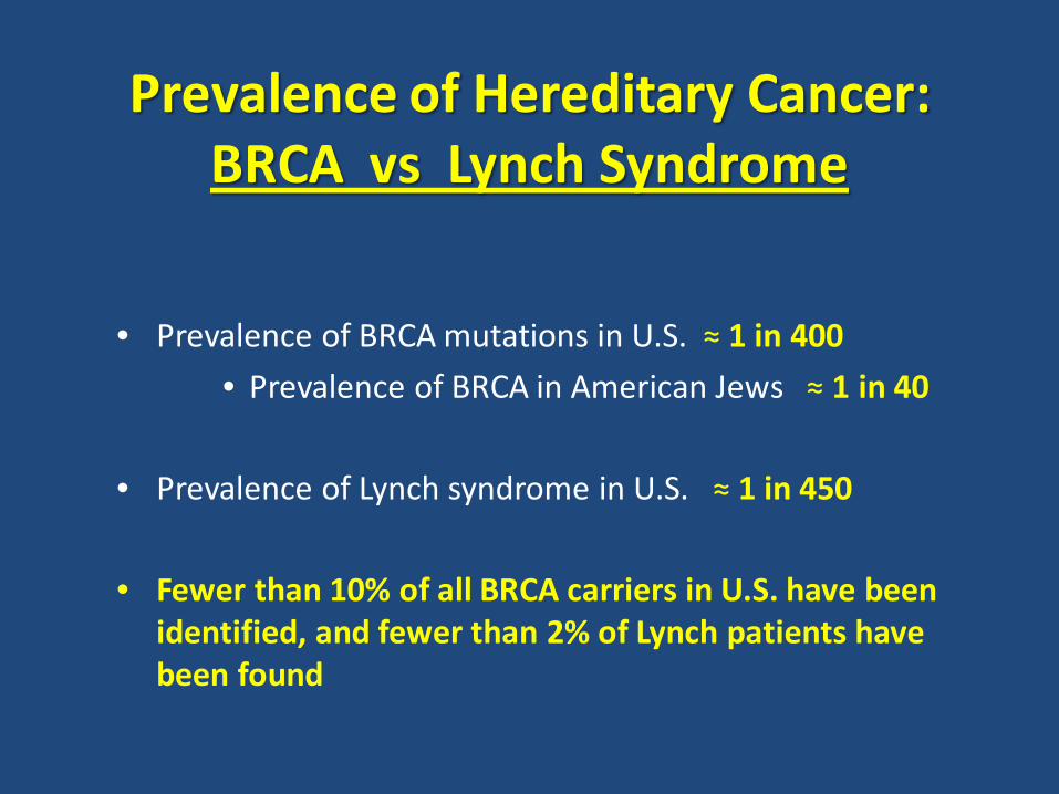
Prevalence of Hereditary Cancer: BRCA vs Lynch Syndrome
• Prevalence of BRCA mutations in U.S. ≈ 1 in 400
• Prevalence of BRCA in American Jews ≈ 1 in 40
• Prevalence of Lynch syndrome in U.S. ≈ 1 in 450
• Fewer than 10% of all BRCA carriers in U.S. have been identified, and fewer than 2% of Lynch patients have been found

Colorectal Cancer Phenotype Sporadic Lynch
• Avg age 60-65 • 2/3 left-sided • Variable histology • Slow evolution from
polyp to cancer • MSI 10-15%
• Avg age 45-55 • 2/3 right-sided • Mucinous, signet ring • Rapid evolution from
polyp to cancer • MSI 90%

Genetics of Lynch Syndrome
• Caused by an inherited defect in any one of several “mismatch repair” genes (MMR). Five genes are currently available for clinical testing:
• MLH1 (most common gene involved) • MSH2 (second most common, freq assoc with MTS) • MSH6 (excess of uterine cancers) • PMS2 (new, prevalence and features are uncertain) • EPCAM (not MMR, but adjacent to MSH2)
• Prior to 2011, clinical testing of the last two genes was not available, ie, suspicious patients who were previously “negative” may need to be retested

Genetics of Lynch Syndrome
• These mismatch repair genes normally function as part of the “spell-check” system to correct DNA mismatch mutations that occur naturally during the DNA replication phase of cell division
• Failure of the system leads to more rapid accumulation of these mutations – “Genomic Instability”
• Cancer occurs when a sufficient number of these mutations occur in critical genes – it’s just a matter of time

Lynch Syndrome Accelerated Timeline For CRC
• Genomic instability in Lynch syndrome greatly accelerates the timeline from colon polyp to CRC
• Instead of the usual 7-10 years, it may be only 1-3 years
• Beware of the colon cancer that seemed to come out of nowhere, within 2-3 years of a normal colonoscopy
• This is not only the basis for the annual colonoscopy recommendation, but also an important clue to underlying Lynch syndrome

How do we find the people with Lynch syndrome?
• Traditional clinical criteria: • Amsterdam criteria • Bethesda criteria
• Rigorous application of “red flags” to newly diagnosed patients with colon or endometrial cancer
• Systematic review of cancer survivors • Universal pathology screening of CRC and endometrial ca • Computer prediction models (PREMM, MMPro, etc)
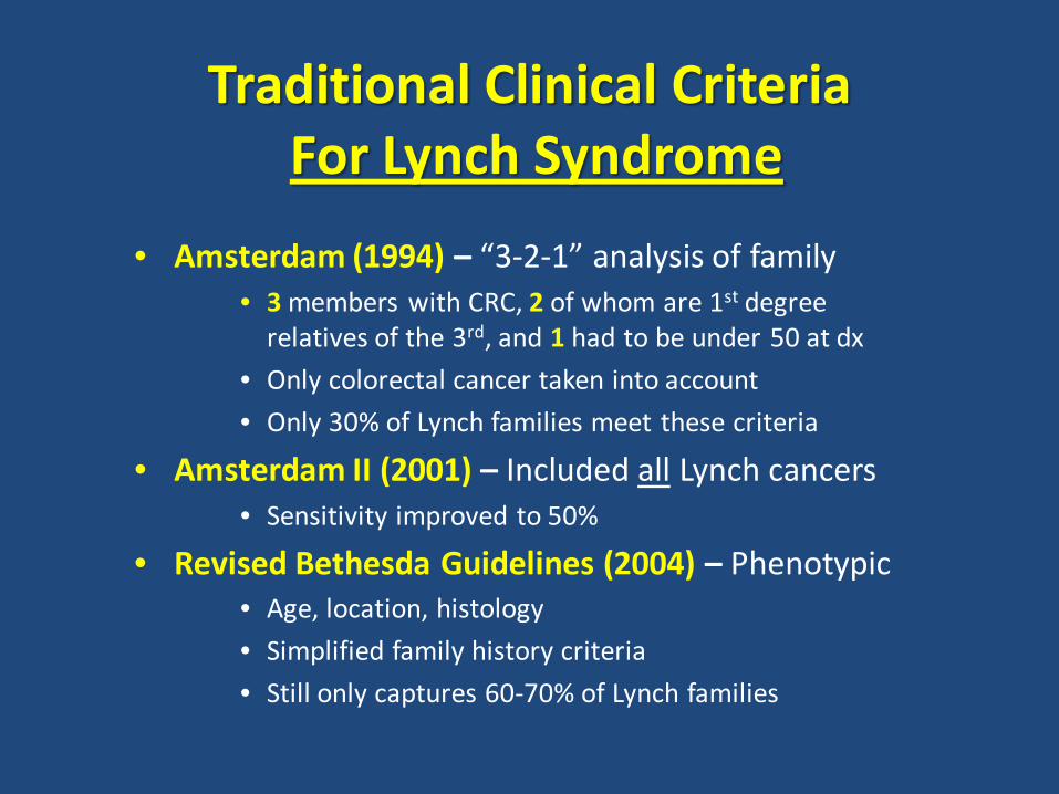
Traditional Clinical Criteria For Lynch Syndrome
• Amsterdam (1994) – “3-2-1” analysis of family • 3 members with CRC, 2 of whom are 1st degree
relatives of the 3rd, and 1 had to be under 50 at dx • Only colorectal cancer taken into account • Only 30% of Lynch families meet these criteria
• Amsterdam II (2001) – Included all Lynch cancers • Sensitivity improved to 50%
• Revised Bethesda Guidelines (2004) – Phenotypic • Age, location, histology • Simplified family history criteria • Still only captures 60-70% of Lynch families
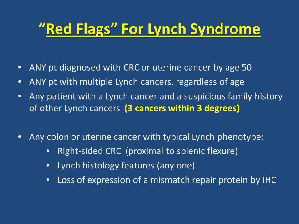
“Red Flags” For Lynch Syndrome
• ANY pt diagnosed with CRC or uterine cancer by age 50 • ANY pt with multiple Lynch cancers, regardless of age • Any patient with a Lynch cancer and a suspicious family history
of other Lynch cancers (3 cancers within 3 degrees)
• Any colon or uterine cancer with typical Lynch phenotype: • Right-sided CRC (proximal to splenic flexure) • Lynch histology features (any one) • Loss of expression of a mismatch repair protein by IHC

“Pink Flags” For Lynch Syndrome
• Cancer of the ureter or renal pelvis
• Cancer of the small intestine
• Development of colorectal cancer less than three years out from a clean colonoscopy
• Sebaceous tumors (adenomas, carcinomas) • Muir-Torre syndrome (MTS)

Computer Models For Assessing Likelihood of Lynch Syndrome
• Several different models to determine the statistical likelihood that a given patient has Lynch syndrome
• Variables taken into account include: • Type and age of cancer in the patient • Multiplicity of cancers • Weighted family pattern for cancers in first and
second degree relatives • PREMM, MMRPro, others • NCCN 2014: Patients with > 5% likelihood are appropriate
for DNA testing

Diagnostic Tools For Lynch Syndrome
Tumor Testing: • Microsatellite Instability (MSI) • Immunohistochemistry (IHC) for MMR proteins
• Useful for automatic screening of all CRC patients at the pathology level
Germline DNA Testing: • Direct DNA analysis of one or more of the five genes • This is the only way to diagnose LS, and the only way
to track the mutation through the family
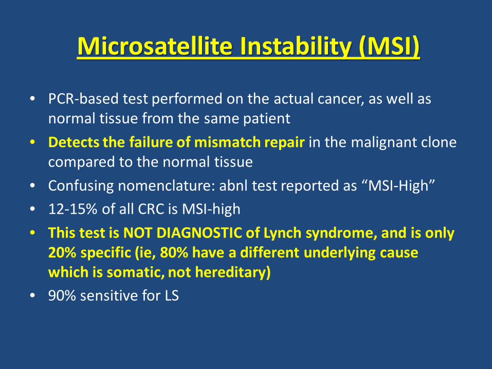
Microsatellite Instability (MSI)
• PCR-based test performed on the actual cancer, as well as normal tissue from the same patient
• Detects the failure of mismatch repair in the malignant clone compared to the normal tissue
• Confusing nomenclature: abnl test reported as “MSI-High” • 12-15% of all CRC is MSI-high • This test is NOT DIAGNOSTIC of Lynch syndrome, and is only
20% specific (ie, 80% have a different underlying cause which is somatic, not hereditary)
• 90% sensitive for LS

Immunohistochemistry (IHC)
• Performed on the cancer tissue, looking for the presence or absence of the four mismatch repair proteins in the tumor
• Theoretically, the defective gene will not produce the corresponding MMR protein
• An abnormal test is NOT DIAGNOSTIC of Lynch syndrome, particularly if the missing protein is MLH1
• Similar to MSI, IHC is 20% specific and 90% sensitive for LS, but the 10% it misses is not the same 10% that MSI misses – together the tests are about 98% sensitive
• Useful for screening population groups with colon and endometrial cancer

Germline DNA Testing
• Performed on blood or saliva
• Testing for mutations in any of the five Lynch genes that would render that gene defective, and therefore unable to produce the corresponding MMR protein
• This is the only way to confirm the diagnosis of LS, and the only way to track a mutation through the family

How Many CRC Patients Should Be Tested For Lynch Syndrome?
• NCCN guidelines and other consensus recommendations are set to trigger testing when the likelihood of being positive is approximately 10% or higher
• When guidelines are applied to large groups of patients, at least 20-25% of patients with breast cancer or colorectal cancer appear to be appropriate for testing
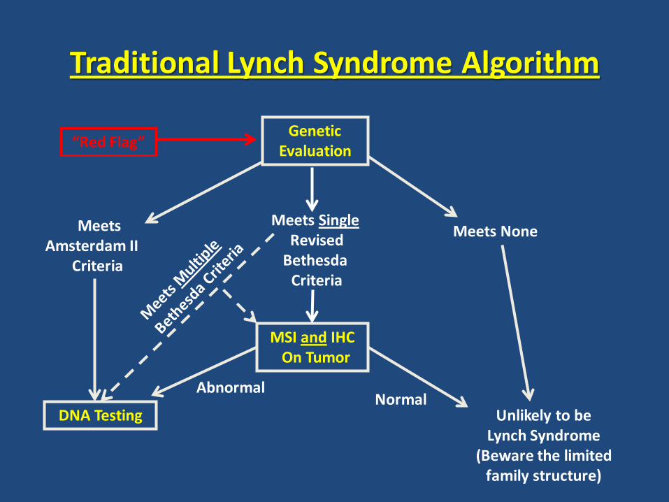
Traditional Lynch Syndrome Algorithm
“Red Flag” Genetic
Evaluation
Meets Amsterdam II
Criteria
Meets Single Revised Bethesda Criteria
Meets None
MSI and IHC On Tumor
Normal Abnormal
DNA Testing Unlikely to be Lynch Syndrome
(Beware the limited family structure)
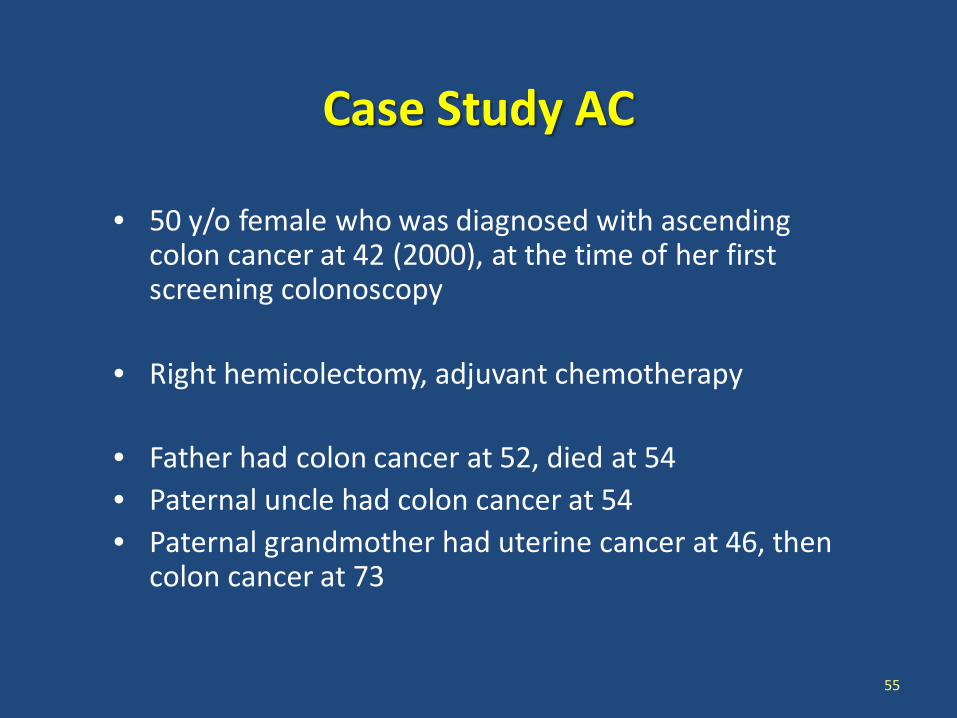
Case Study AC
• 50 y/o female who was diagnosed with ascending colon cancer at 42 (2000), at the time of her first screening colonoscopy
• Right hemicolectomy, adjuvant chemotherapy
• Father had colon cancer at 52, died at 54 • Paternal uncle had colon cancer at 54 • Paternal grandmother had uterine cancer at 46, then
colon cancer at 73
55

Case Study AC – “Red Flags”
• 50 y/o female who was diagnosed with ascending colon cancer at 42, at the time of her first screening colonoscopy
• Right hemicolectomy, adjuvant chemotherapy
• Father had colon cancer at 52, died at 54 • Paternal uncle had colon cancer at 54 • Paternal grandmother had uterine cancer at 46, then
colon cancer at 73
56

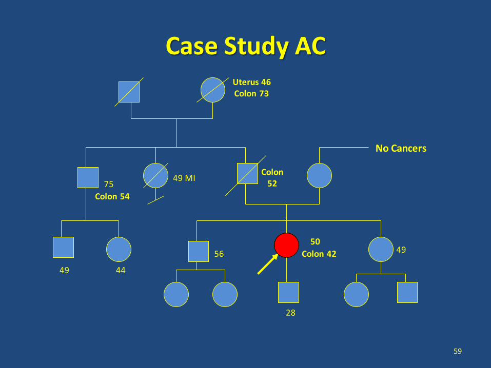
Case Study AC - FAMP
57
No Cancers
50 Colon 42 49 56
Colon 54
Uterus 46 Colon 73
Colon52
44
49 MI
49
75
28

Case Study AC
• This patient and her family were missed at the time of her diagnosis at 42, and at every follow-up visit with multiple physicians over the next 7 years
• She was identified by systematically applying the “red flags” to our CRC survivor population
• She was found to carry a mutation in the MSH2 gene
58
Case Study AC
59
No Cancers
50 Colon 42 49 56
Colon 54
Uterus 46 Colon 73
Colon52
44
49 MI
49
75
28

Case Study AC
60
No Cancers
50 Colon 42 49 56
Colon 54
Uterus 46 Colon 73
Colon52
44
49 MI
49
75
28
NEG NEG
? ?

Lifetime Risk of Lynch-Related Cancers (vs Normal)
• Colorectal 80% (5-6) • Endometrial 40-60% (2.5) • Ovary 8-12% (1.5) • Stomach 8-10% (<1) • Urothelial 4-5% (<1) • Biliary/Pancreas 2-4% (<1) • Small intestine 1-2% (<1) • CNS (GBM) 1-3% (<1) • Breast?, Bladder?

Case Study AC
• She was found to carry a mutation in the MSH2 gene
• She went on to have a prophylactic complete hysterectomy/oophorectomy
• This procedure could have been done as part of her hemicolectomy if her Lynch diagnosis had been timely
62

How do we actually manage cancer risk in
patients with Lynch syndrome?

Lynch-Related Cancers With Effective Risk-Reducing Strategies
• Colorectal 80% (5-6) • Endometrial 40-60% (2.5) • Ovary 8-12% (1.5) • Stomach 8-10% (<1) • Urothelial 4-5% (<1) • Biliary/Pancreas 2-4% (<1) • Small intestine 1-2% (<1) • CNS (GBM) 1-3% (<1) • Breast?, Bladder?

Cancer Risk Management In Lynch Syndrome
• Screening colonoscopy starting at 25*, annual after age 30 • Transvaginal sono and CA-125 starting at 30*, then annually • Complete hysterectomy after child-bearing is complete (35-40) • EGD starting at 30, then q 3 yrs unless gastric cancer in the
family, or gastric polyps identified • Annual U/A +/- urine cytology starting at 30 • ? Evolving role for EUS in screening for pancreas
• *Age to start screening may need to be modified in families
with cancers occuring at very young age

Case Study ME
• 39-year-old moderately obese female with menorrhagia • Endometrial bx shows carcinoma • Hysterectomy reveals Stage IA endometrioid carcinoma
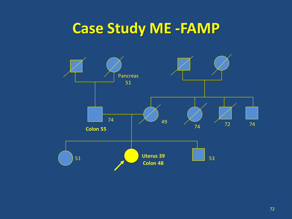
• Family history reveals no other GYN malignancies • Father had colon cancer at 55 • Paternal grandmother had pancreas cancer at 51

Case Study ME - FAMP
67
39 Uterus 45 43
49 70
Colon 55 72 66
66
Pancreas 51

Case Study ME
• 39-year-old moderately obese female with Stage IA endometrial carcinoma
• At 44 she underwent her first screening colonoscopy (10-year rule), and was found to have a tubulovillous adenoma in the ascending colon

Case Study ME
• 39-year-old moderately obese female with Stage IA endometrial carcinoma
• At 44 she underwent her first screening colonoscopy (10-year rule), and was found to have a tubulovillous adenoma in the ascending colon
• Repeat colonoscopy one year later was normal
• When would you scope her again?
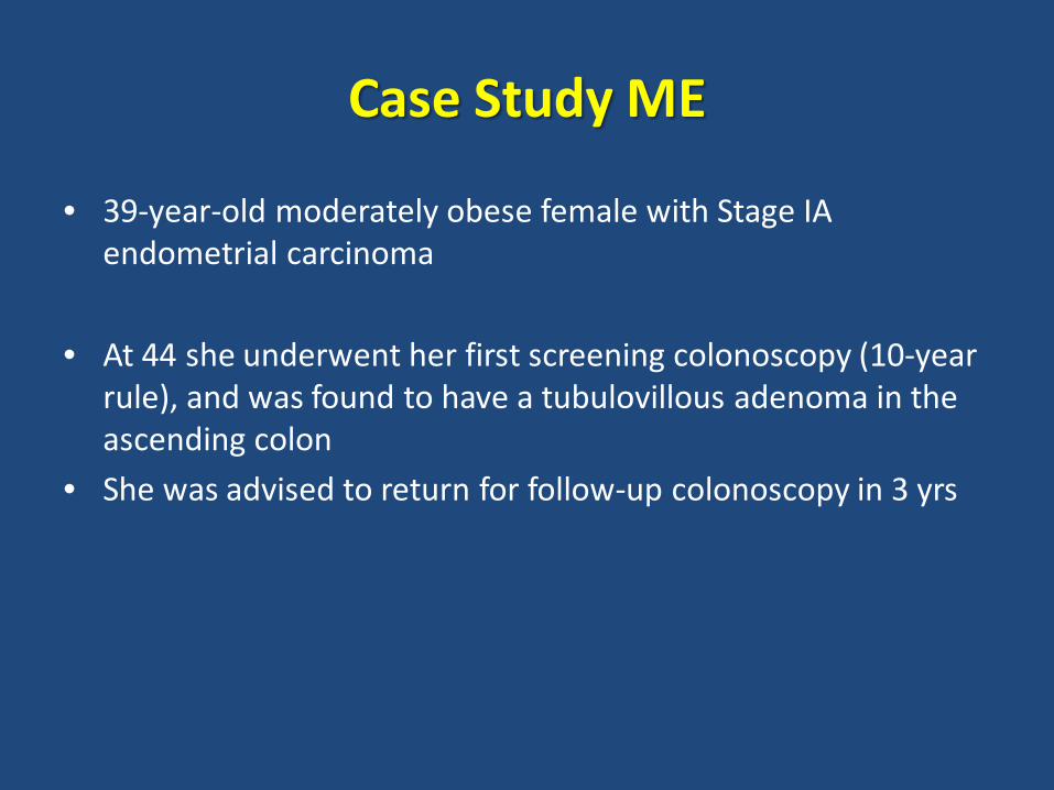
Case Study ME
• 39-year-old moderately obese female with Stage IA endometrial carcinoma
• At 44 she underwent her first screening colonoscopy (10-year rule), and was found to have a tubulovillous adenoma in the ascending colon
• She was advised to return for follow-up colonoscopy in 3 yrs

Case Study ME
• 39-year-old moderately obese female with Stage IA endometrial carcinoma
• At 44 she underwent her first screening colonoscopy (10-year rule), and was found to have a tubulovillous adenoma in the ascending colon
• She was advised to return for follow-up colonoscopy in 3 yrs
• At 48, she was found to have adenocarcinoma in the cecum • Right hemicolectomy for Stage III-A cancer
Case Study ME -FAMP
72
Uterus 39 53 51
49 74
Colon 55 72 74
74
Pancreas 51
Colon 48
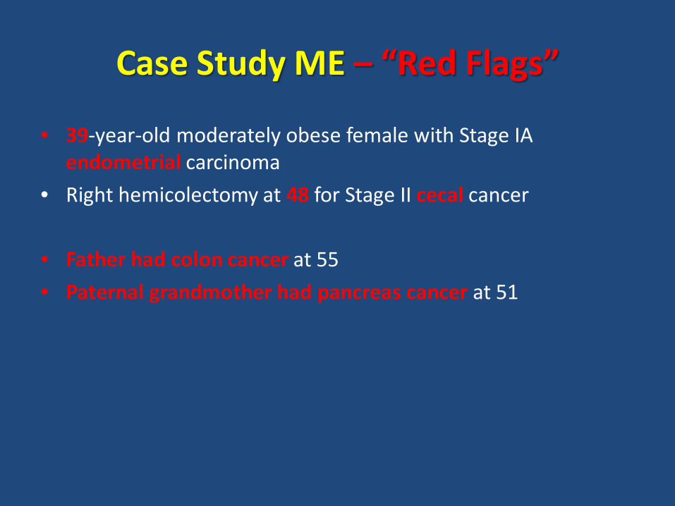
Case Study ME – “Red Flags”
• 39-year-old moderately obese female with Stage IA endometrial carcinoma
• Right hemicolectomy at 48 for Stage II cecal cancer • Father had colon cancer at 55 • Paternal grandmother had pancreas cancer at 51

Case Study ME – “Red Flags”
• 39-year-old moderately obese female with Stage IA endometrial carcinoma
• Right hemicolectomy at 48 for Stage II cecal cancer • Father had colon cancer at 55 • Paternal grandmother had pancreas cancer at 51
• DNA testing revealed a mutation in MSH6

Case Study ME
75
Uterus 39 53 51
49 74
Colon 55 72 74
74
Pancreas 51
Colon 48
48

Case Study ME
76
Uterus 39 53 51
49 74
Colon 55 72 74
74
Pancreas 51
Colon 48
48
30 27 28 26 34
NEG
NEG NEG NEG

Case Study ME – The Sister
77
Uterus 39 53 51
49 74
Colon 55 72 74
74
Pancreas 51
Colon 48
48
30 27 28 26 34
NEG
NEG NEG NEG
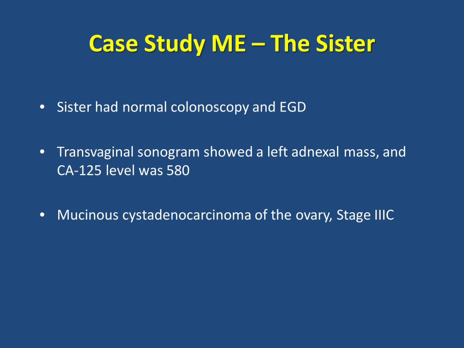
Case Study ME – The Sister
• Sister had normal colonoscopy and EGD
• Transvaginal sonogram showed a left adnexal mass, and CA-125 level was 580
• Mucinous cystadenocarcinoma of the ovary, Stage IIIC

Case Study ME Time-Line
Patient’s Uterine Ca
Age 39
Patient’s Cecal Ca Age 48
Sister’s Ovarian Ca
Age 51
Father’s Colon Ca Age 55
2000
1995 2009
2011 2005
Patient’s Ascending TVA
Age 44
Case Study ME Time-Line
Patient’s Uterine Ca
Age 39
Patient’s Cecal Ca Age 48
Sister’s Ovarian Ca
Age 51
Father’s Colon Ca Age 55
2000
1995 2009
2011
When should this family have been diagnosed?
2005
Patient’s Ascending TVA
Age 44
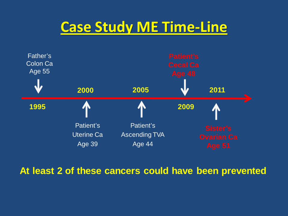
Case Study ME Time-Line
Patient’s Uterine Ca
Age 39
Patient’s Cecal Ca Age 48
Sister’s Ovarian Ca
Age 51
Father’s Colon Ca Age 55
2000
1995 2009
2011
At least 2 of these cancers could have been prevented
2005
Patient’s Ascending TVA
Age 44

Case Study ME Time-Line
Patient’s Uterine Ca
Age 39
Patient’s Cecal Ca Age 48
Sister’s Ovarian Ca Died at 54
Father’s Colon Ca Age 55
2000
1995 2009
2011
At least 2 of these cancers could have been prevented ……..and 1 death
2005
Patient’s Ascending TVA
Age 44

Case Study RT
• 51 y/o woman with Stage II-B cancer of the cecum • Presented with abdominal cramping only 18 months out
from a normal screening colonoscopy
• First colonoscopy at age 25, and q 3 yrs since then • Brother diagnosed with Stage IV colon ca at age 23
• Right hemicolectomy performed
83

Case Study RT
• Brother died of colon cancer at 23 • No colon polyps in either sister with q3yr surveillance
• Father had ureteral ca at 45, then prostate ca at 70 • Paternal aunt died of pancreas ca at 46 • Paternal GM had uterine ca at 44
84

Case Study RT – “Red Flags”
• 51 y/o woman with Stage II-B cancer of the cecum, diagnosed only 18 months after a normal colonoscopy
• Brother diagnosed with Stage IV colon ca at age 23 • Father had ureteral ca at 45, then prostate ca at 70 • Paternal aunt died of pancreas ca at 46 • Paternal GM had uterine ca at 44
85

Case Study RT - FAMP
86
MI 55 Pancreas 46
Cecal Ca 51 46 Colon Ca 23
Uterine 44 Stroke 51
26 28 18 19 14 18
76 Ureter 45 Prostate 70
54
79 74
Unknown
22

Case Study RT
• Genetic evaluation revealed a mutation in MLH1
87

Case Study RT
88
MI 55 Pancreas 46
Cecal Ca 51 46 Colon Ca 23
Uterine 44 Stroke 51
26 28 18 19 14 18
76 Ureter 45 Prostate 70
54
79 74
Unknown
22

Case Study RT
• Genetic evaluation revealed mutation in MLH1
• Patient needed a second operation to remove her uterus
and ovaries 89
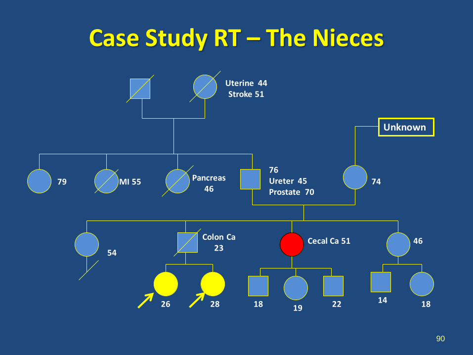
Case Study RT – The Nieces
90
MI 55 Pancreas 46
Cecal Ca 51 46 Colon Ca 23
Uterine 44 Stroke 51
26 28 18 19 14 18
76 Ureter 45 Prostate 70
54
79 74
Unknown
22

Case Study RT – The Nieces
91
MI 55 Pancreas 46
Cecal Ca 51 46 Colon Ca 23
Uterine 44 Stroke 51
26 28 18 19 14 18
76 Ureter 45 Prostate 70
54
79 74
Unknown
22
NEG
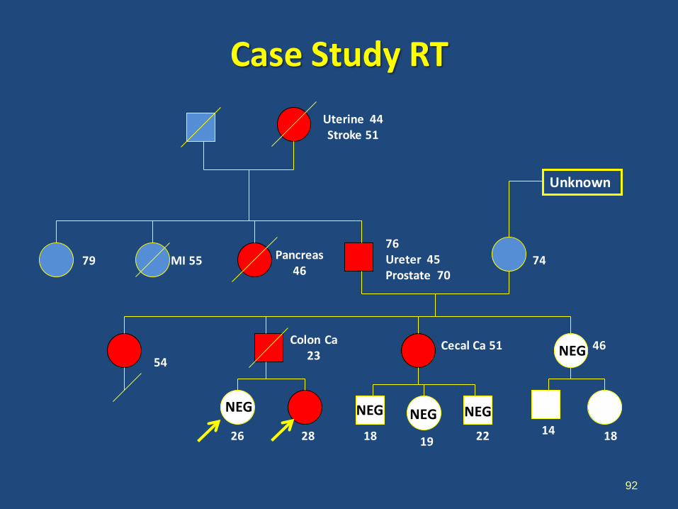
Case Study RT
92
MI 55 Pancreas 46
Cecal Ca 51 46 Colon Ca 23
Uterine 44 Stroke 51
26 28 18 19 14 18
76 Ureter 45 Prostate 70
54
79 74
Unknown
22
NEG NEG NEG
NEG
NEG

Lynch Syndrome: How Are We Doing?
• Fewer than 2% of all patients affected with Lynch syndrome have yet been identified
• Approximately 20-25% of all colorectal and endometrial cancer patients are suitable for focused genetic evaluation

Screening For Lynch Syndrome: The Traditional Approach
• Traditional dependence on providers to identify these patients has been largely ineffective: • Wide spectrum of cancers and physicians • Providers underestimate the prevalence of these
syndromes, and the associated cancer risks • Too much reliance on the “slam dunk” family history • Too little attention to the phenotype that is typical for
Lynch colon cancer
• Families get missed, cancers continue to occur

Screening For Lynch Syndrome: The Pathology Approach
• Automatic screening of colon or endometrial cancers at the pathology level using MSI or IHC testing
• 15% of all colon cancers will exhibit MSI or abnormal IHC , and 90% of the Lynch colon cancers will be within this group
• Finding the 3% that are Lynch syndrome within this 15% is much easier than finding the 3% within the 100%
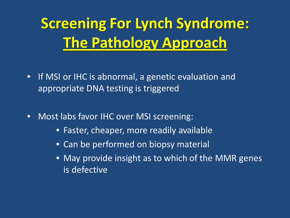
Screening For Lynch Syndrome: The Pathology Approach
• If MSI or IHC is abnormal, a genetic evaluation and appropriate DNA testing is triggered
• Most labs favor IHC over MSI screening: • Faster, cheaper, more readily available • Can be performed on biopsy material • May provide insight as to which of the MMR genes
is defective

Lynch Syndrome Algorithm: The CRC Pathology Approach
Suspicious Phenotype Automatic
Pathology Screening:
IHC
Family Cancer History Analysis Suspicious
DNA Testing Unlikely to be
Lynch Syndrome
Genetic Evaluation
Mucinous, Right-sided
OR < 70
OK
Positive
BRAF Testing
Absent MSH2, MSH6, or PMS2
Negative

Lynch Syndrome Algorithm: The Universal Pathology Approach
Automatic Pathology Screening:
IHC
Family Cancer History Analysis Suspicious
DNA Testing Unlikely to be
Lynch Syndrome
Genetic Evaluation
OK
ALL CRC And Endometrial
Cancer Patients
Positive
BRAF Testing
Absent MSH2, MSH6, or PMS2
Negative
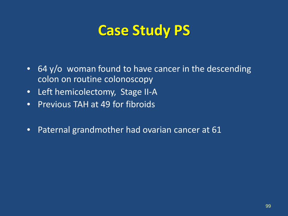
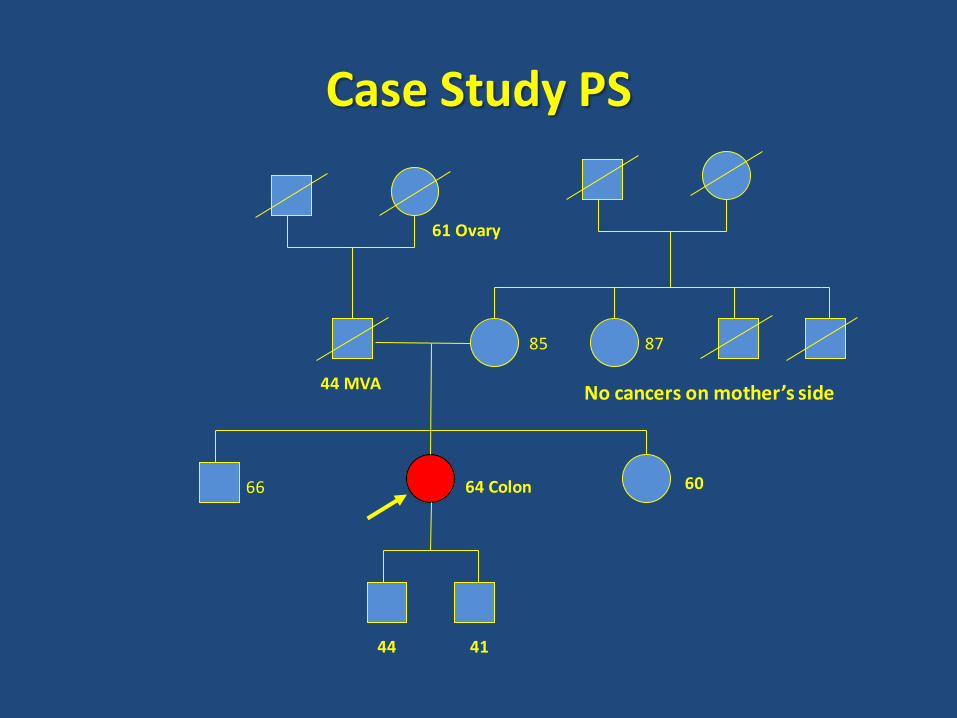
Case Study PS
• 64 y/o woman found to have cancer in the descending colon on routine colonoscopy
• Left hemicolectomy, Stage II-A • Previous TAH at 49 for fibroids
• Paternal grandmother had ovarian cancer at 61
99

Case Study PS
64 Colon 60 66
85 87
No cancers on mother’s side
61 Ovary
44 MVA
41 44
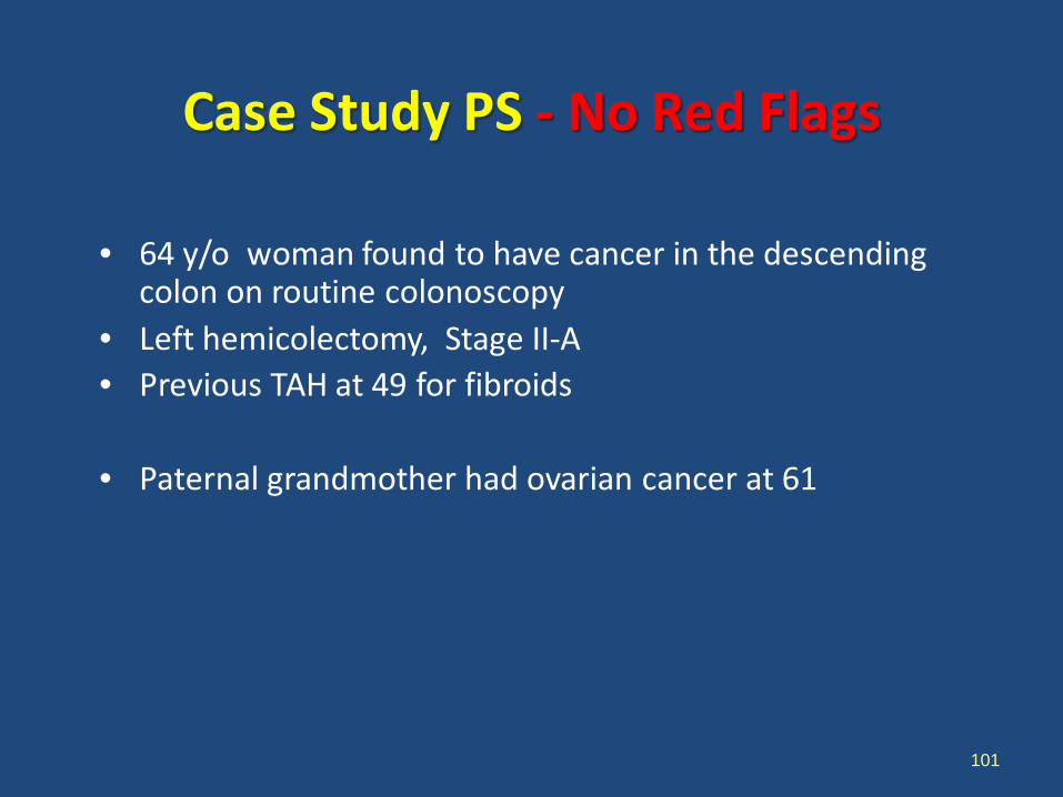
Case Study PS - No Red Flags
• 64 y/o woman found to have cancer in the descending colon on routine colonoscopy
• Left hemicolectomy, Stage II-A • Previous TAH at 49 for fibroids
• Paternal grandmother had ovarian cancer at 61
101

Case Study PS
• 64 y/o woman found to have cancer in the descending colon on routine colonoscopy
• Left hemicolectomy, Stage II-A • Previous TAH at 49 for fibroids • Automatic pathology screening with IHC shows that the
MSH6 protein is not expressed in the cancer
• DNA testing confirms a mutation in the MSH6 gene
102
Case Study PS
64 Colon 60 66
85 87
No cancers on mother’s side
61 Ovary
44 MVA
41 44

Case Study PS
64 Colon 60 66
85 87
No cancers on mother’s side
61 Ovary
44 MVA
41 44
NEG
NEG
NEG

Screening For Lynch Syndrome: Limitations of The Pathology Approach
• It will still miss the 10% of Lynch CRC and endometrial cancer who have normal tumor testing with IHC
• Still need proper attention to the family history, requiring providers to know the spectrum of Lynch cancers
• This strategy will not help to identify the many Lynch patients who are among the cancer survivors, or the carriers who have not yet had cancer

Summary and Call To Action
• These syndromes are more common than you may realize, and they are easily missed
• The potential impact on both cancer prevention and cancer management can be huge
• The cumulative number of colon adenomas needs to be systematically tracked in all patients with polyps
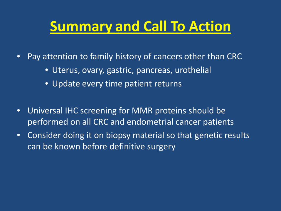
Summary and Call To Action
• Pay attention to family history of cancers other than CRC • Uterus, ovary, gastric, pancreas, urothelial • Update every time patient returns
• Universal IHC screening for MMR proteins should be
performed on all CRC and endometrial cancer patients • Consider doing it on biopsy material so that genetic results
can be known before definitive surgery

Summary and Call To Action
• Rigorous application of the “red flags” will capture the majority of families in newly diagnosed patients
• Testing should be considered soon after diagnosis in appropriate patients
• A systematic process is required for providers to find the affected patients among their survivor populations

Summary and Call To Action
• Cancer genetics is now a critical element in providing high
quality comprehensive cancer care
• Every physician and nurse has a role to play in identifying these patients and their families
• We can prevent many cancers that were destined to occur