the immune system chapter 39.1 & 39.2 (pgs. 1022-1041)
TRANSCRIPT
The Immune System
Chapter 39.1 & 39.2
(Pgs. 1022-1041)
Introductory Questions #11. Your Immune system protects your body from
many kinds of pathogens. Give three examples of a pathogen. Where do these pathogens come from?
2. Are all micoorganisms harmful to your body? Briefly explain your answer.
3. Looking at Table 39.1, determine if the following illnesses are caused by a virus or a bacteria.
-Tetanus -Influenza
-Chicken pox -Tuberculosis
Pathogens
• Meaning “pathos”- to suffer & “geneia” refers to producing
• Cause disease and illness• Disrupts homeostasis in the body• Includes agents such as:
– Bacteria– Protozoans– Fungi– Viruses– Other parasites
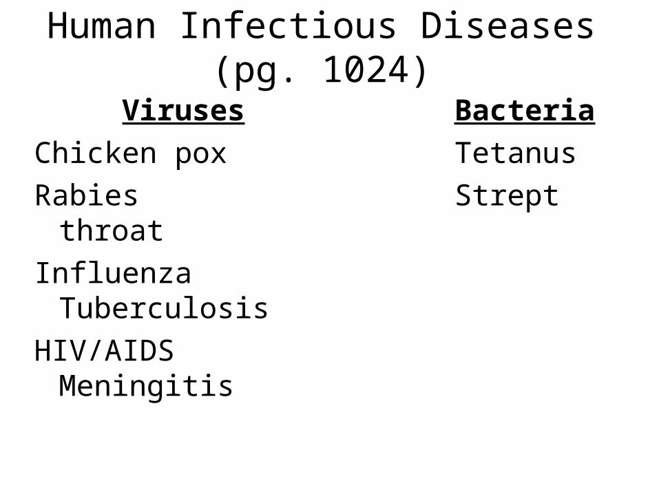
Human Infectious Diseases (pg. 1024)
Viruses Bacteria
Chicken pox Tetanus
Rabies Strept throat
InfluenzaTuberculosis
HIV/AIDS Meningitis
Video #1: Killer Viruses• While watching the video, write 15 key statements
on a separate sheet of paper.• Be sure to number each statement.
Introductory Questions #2
1. Some disorders are not caused by pathogens. Give three examples. (see pg. 1024) Explain how these are acquired by a person.
2. Name the rod-shaped pathogen Robert Koch discovered in the blood of sheep and cattle in 1876. What 4 steps are taken to identify a pathogen with a disease? Write these on the back of your cover sheet.
3. Do these steps only work with bacteria? Explain your answer.
Non-Pathogenic Disorders
• Hemphilia: (sex-linked trait) called bleeder’s disease.
• Osteoarthritis: degeneration from wear and tear in joint spaces. (aging process)
• Cirrhoisis: exposure to a chemical/toxin such as alcohol.
• Scurvy: Malnutrition (deficiency in Vitamin C) causing gums to bleed, teeth to losen, and wounds to heal poorly.
Pathogens• 1st proof was a pathogen was identified by
Robert Koch in 1876. • Developed postulates (method) of relating
a pathogen to a specific disease.– Pathogen must be found in host in every case– Pathogen must be isolated & grown in culture– Pathogen must cause disease if take from this
pure culture– Must be able to isolate the pathogen from new
host and shown to be the original pahtogen.
Exceptions of Koch’s Process
• STD’s: sexually transmitted diseases
• Viruses
Homework (Mon.)
• Reading guide:– Research a disease on the internet– Answer the questions posed on the guide for
the section “Before You Read”
Do Question #1 & #3
Homework Due (Tues.)
• Study Guide/Reinforcement Quest. #1-10
• Have research ready to share about a particular disease. (printout of website)
Introductory Questions #3
1. People who harbor pathogens without exhibiting any signs of the illness and unknowingly transmit it to others are called _________. (see pg. 1026)
2. The symptom-free period of time when a pathogen is transmitted and multiplying rapidly within the body is called the ___________ period.
3. Name three diseases that can be transferred from animals to humans. What are some non-living reservoirs of infectious disease?
Spreading of Infectious Diseases
• Pathogens begin from a reservoir which can include: Human body, Animals, and non-living sources such as water & soil.
• Achieved by a living organism (human body itself) or an inanimate object.
• People can “carry” the disease and not know they have it without showing any signs of illness.
• The first stage for the spread of a disease is called the incubation period.– Humans unknowingly pass it on to others– Influenza, Strept throat, STD’s like gonorrhea, and AIDS
are all examples
Transmission of Disease (pg. 1027)
• Pathogens are transmitted from a reservoir to a host in four main ways:– Direct contact (flu, cold, & STD’s)– An object (can house bacteria & other microorganisms)– The air (coughing or sneezing)– Intermediate organisms called “vectors” which include:
• Insects• Arthropods• Ticks, fleas, mosquitos, and flies
Homework (Wed.)
• Study Guide/Reinforcement Quest. #1-10
• Have research ready to share about a particular disease. (printout of website)
Homework Due: (Thurs.)
• Reading Guide 39.1 – Quest #4-10– Final Page “After You Read” #1 & 2 (Table)
• Disease research from those that didn’t share yesterday. (2pts)
(Note: must have a printout of website)
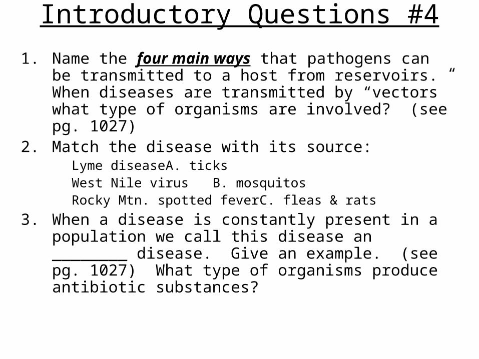
Introductory Questions #4
1. Name the four main ways that pathogens can be transmitted to a host from reservoirs. When diseases are transmitted by “vectors” what type of organisms are involved? (see pg. 1027)
2. Match the disease with its source:Lyme disease A. ticksWest Nile virus B. mosquitosRocky Mtn. spotted fever C. fleas & rats
3. When a disease is constantly present in a population we call this disease an ________ disease. Give an example. (see pg. 1027) What type of organisms produce antibiotic substances?
Video #3: Parasites
• Write 10 key Statements from the video.
**Be sure to number each statement.
Tonight’s Homework
• Textbook pg. 1030 Quest. #1-4
• Review for tomorrow’s quiz
• Bring in SSR reading for 2 extra credit pts.
• Review by taking a practice quiz from textbook website:
www.ca.bdol.glencoe.com
Assignment Packet Due Fri. 4/3
• Cover sheet (w/all four IQ’s answered)
• Reading guide 39.1
• Study Guide/Reinforcement (one page-front)
• Videos x3– Killer Viruses (15 statements)– Bacteria (fill in answers)– Parasites (10 statements)
• SSR- Your five key points & Presenters five points

SSR Guide for Presenters
• Give your name
• Hold up the your source
• FIVE key points with a brief summary of what you read.
• The rest of us need to write points discussed in the SSR boxes on your cover sheet.
The Immune System (Part II)
Chapter 39.2
(pgs. 1031-1047)
Schedule
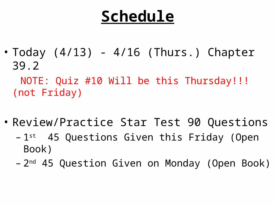
• Today (4/13) - 4/16 (Thurs.) Chapter 39.2 NOTE: Quiz #10 Will be this Thursday!!! (not Friday)
• Review/Practice Star Test 90 Questions – 1st 45 Questions Given this Friday (Open Book)– 2nd 45 Question Given on Monday (Open Book)

Introductory Questions #1
1. Name three non-specific defenses to a foreign invader that enters the body.
2. Give two purposes that mucus serves in the body. Name two areas where mucus is secreted.
3. Name two white blood cells (WBC’s) that engulfs (phagocytize) foreing invaders. Which WBC releases histamine. What does histamine do?
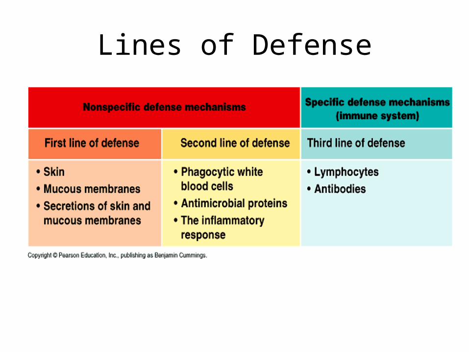
Lines of Defense
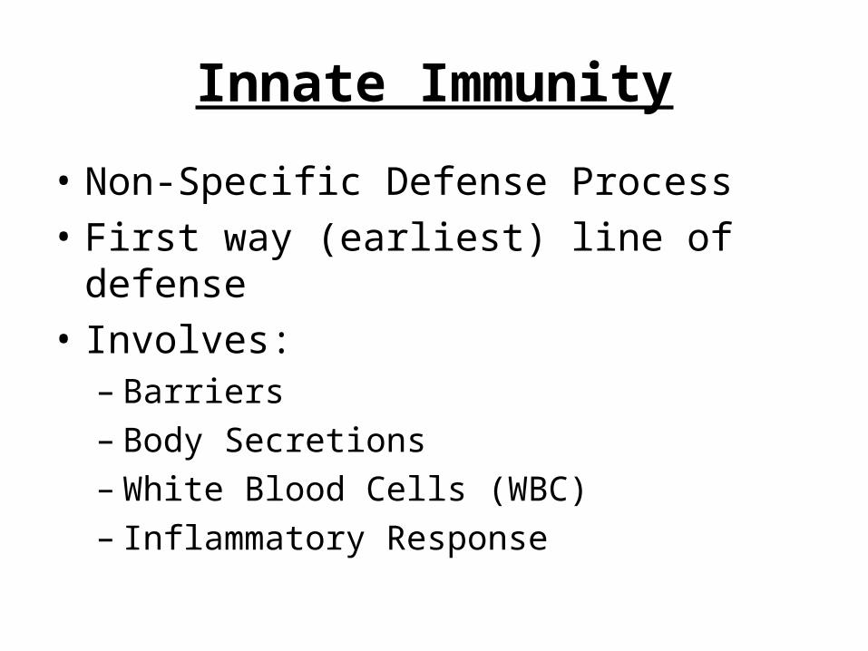
Innate Immunity
• Non-Specific Defense Process
• First way (earliest) line of defense
• Involves:– Barriers– Body Secretions– White Blood Cells (WBC)– Inflammatory Response
Skin & Body Secretions
• Skin is a dry desolate place that is always replacing itself.*It’s been estimated that we slough off and replace
about 90 lbs. of skin in our lifetime!!!
• Secretions:– Mucus– Oil– Sweat– Tears– Saliva
Mucus
• Thick (viscous) fluid that is slightly sticky
• Traps microorganisms & foreign particles
• Prevents membranes from drying out
• Secreted by cells that line the – Digestive tract– Respiratory tract– Sinus cavities
Sweat, Tears, and Saliva
• All contain Lysozymes– Breaks down the cell wall of bacteria cells
• Tears & Saliva - have a small amount of antibiotic
• Sweat - has a high amount of salt
Inflammatory Response
• Characterized by:– Redness – Swelling– Pain– Heat
• Cells Involved:– Mast cells – Basophil: releases Histamine– Monocytes (Macrophages): WBC– Neutrophil: (WBC
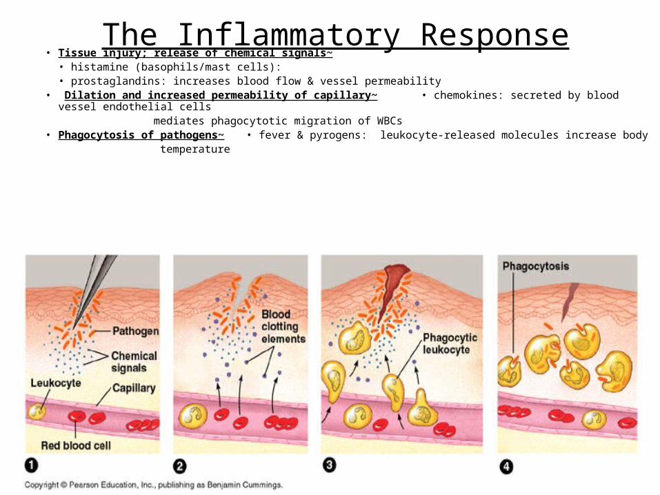
The Inflammatory Response• Tissue injury; release of chemical signals~
• histamine (basophils/mast cells): • prostaglandins: increases blood flow & vessel permeability
• Dilation and increased permeability of capillary~ • chemokines: secreted by blood vessel endothelial cells
mediates phagocytotic migration of WBCs• Phagocytosis of pathogens~ • fever & pyrogens: leukocyte-released molecules increase body temperature
Histamine
• Substance that cause blood vessels to dilate
• Released by Mast cells & Basophil cells
• Cause the vessels to be more permeable and allow cells & substances to leak out.
• Causes redness & swelling
Phagocytic cells
• Surround & Engulf foreign invaders
• Present in body tissues & blood
• Include the following White blood Cells– Monocytes ----- become macrophages– Neutrophils– Eosinophils
Video #1: “The Immune System”(10 Statements)
Introductory Questions #1
1. Name three non-specific defenses to a foreign invader that enters the body.
2. Give two purposes that mucus serves in the body. Name two areas where mucus is secreted.
3. Name two white blood cells (WBC’s) that engulfs (phagocytize) foreign invaders. Which WBC releases histamine. What does histamine do?
Introductory Questions #2
1. Looking at the mini-lab 39.2 (pg. 1035), list the five white blood cells that we could look for in a prepared slide. Name the type of enzyme that a macrophage uses to digest particles it engulfs. (see pg. 1033)
2. When a wound is healing pus can form. What is pus composed of? Name the protein that protects cells from viruses. Briefly explain what an antigen is. (pg. 1035)
3. Name three organs that make up the lymphatic system. Which white blood cells make up a lymph node? Where can you find a high number of lymph nodes in the body?

Lines of Defense
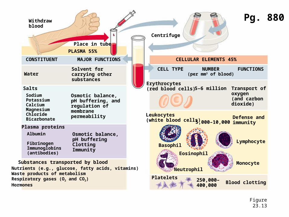
Figure 23.13
Withdrawblood
Place in tube
PLASMA 55%
CONSTITUENT MAJOR FUNCTIONS
WaterSolvent forcarrying othersubstances
Salts
Osmotic balance,pH buffering, andregulation ofmembranepermeability
SodiumPotassiumCalciumMagnesiumChlorideBicarbonate
Plasma proteins
Osmotic balance,pH bufferingClottingImmunity
Albumin
FibrinogenImmunoglobins(antibodies)
Substances transported by bloodNutrients (e.g., glucose, fatty acids, vitamins)Waste products of metabolismRespiratory gases (O2 and CO2)Hormones
Centrifuge
CELLULAR ELEMENTS 45%
CELL TYPE NUMBER(per mm3 of blood)
FUNCTIONS
Erythrocytes(red blood cells) 5–6 million Transport of
oxygen (and carbon dioxide)
Leukocytes(white blood cells) 5,000–10,000
Defense andimmunity
Basophil
Eosinophil
Neutrophil
Lymphocyte
Monocyte
Platelets 250,000–400,000
Blood clotting
Pg. 880
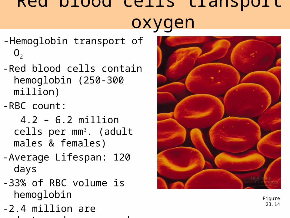
Red blood cells transport oxygen
Figure 23.14
-Hemoglobin transport of O2
-Red blood cells contain hemoglobin (250-300 million)
-RBC count:
4.2 – 6.2 million cells per mm3. (adult males & females)
-Average Lifespan: 120 days
-33% of RBC volume is hemoglobin
-2.4 million are destroyed per second and are replaced in the bone marrow
-No nucleus or mitochondria
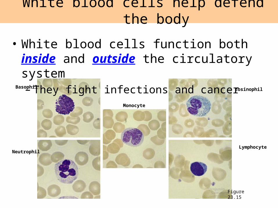
White blood cells help defend the body
Figure 23.15
• White blood cells function both inside and outside the circulatory system – They fight infections and cancer
Basophil
Neutrophil
Monocyte
Eosinophil
Lymphocyte
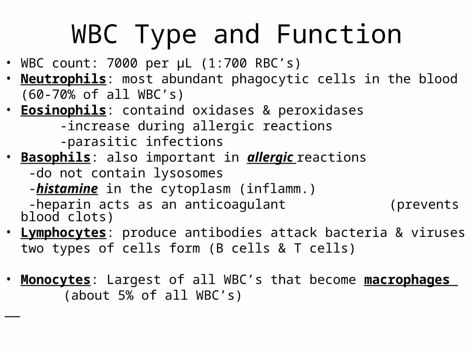
WBC Type and Function• WBC count: 7000 per µL (1:700 RBC’s)• Neutrophils: most abundant phagocytic cells in the blood
(60-70% of all WBC’s)• Eosinophils: containd oxidases & peroxidases
-increase during allergic reactions -parasitic infections
• Basophils: also important in allergic reactions -do not contain lysosomes -histamine in the cytoplasm (inflamm.) -heparin acts as an anticoagulant (prevents blood clots)
• Lymphocytes: produce antibodies attack bacteria & virusestwo types of cells form (B cells & T cells)
• Monocytes: Largest of all WBC’s that become macrophages (about 5% of all WBC’s)
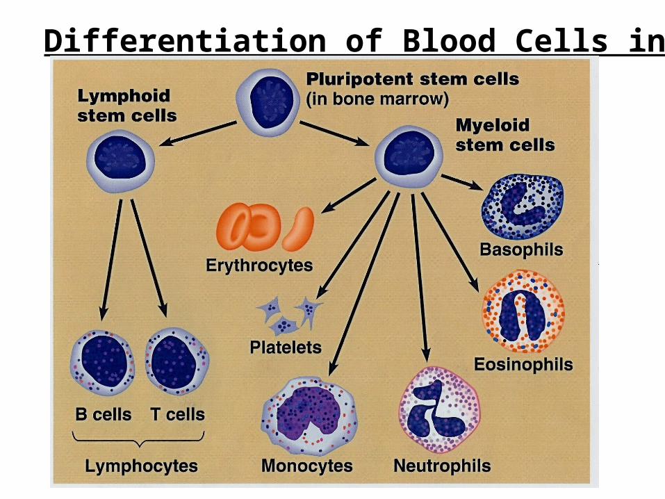
Differentiation of Blood Cells in the Bone Marrow
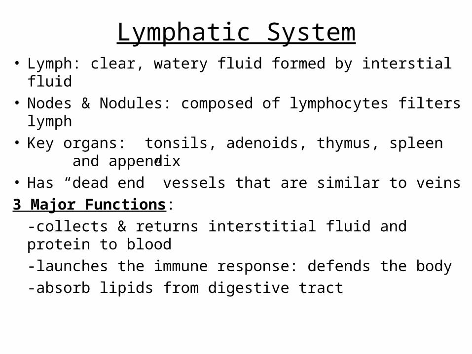
Lymphatic System• Lymph: clear, watery fluid formed by interstial fluid• Nodes & Nodules: composed of lymphocytes filters
lymph• Key organs: tonsils, adenoids, thymus, spleen
and appendix• Has “dead end” vessels that are similar to veins
3 Major Functions:
-collects & returns interstitial fluid and protein to blood
-launches the immune response: defends the body
-absorb lipids from digestive tract
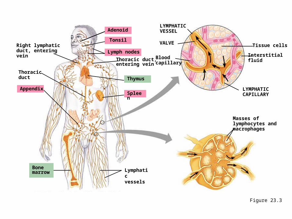
Figure 23.3
Right lymphaticduct, enteringvein
Thoracicduct
Appendix
Adenoid
Tonsil
Lymph nodes
Thoracic duct,entering vein
Thymus
Spleen
Bonemarrow Lymphatic
vessels
LYMPHATICVESSEL
VALVE
Bloodcapillary
Tissue cells
Interstitialfluid
LYMPHATICCAPILLARY
Masses oflymphocytes andmacrophages
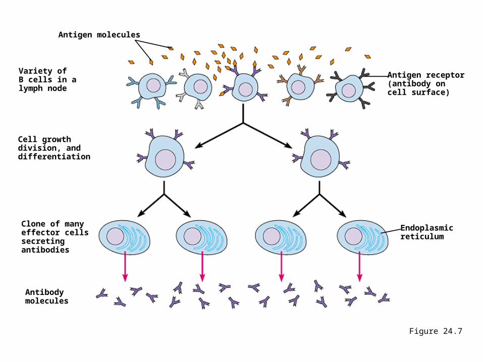
Figure 24.7
Antigen molecules
Variety ofB cells in a lymph node
Cell growthdivision, anddifferentiation
Clone of manyeffector cellssecretingantibodies
Antibodymolecules
Antigen receptor(antibody oncell surface)
Endoplasmicreticulum
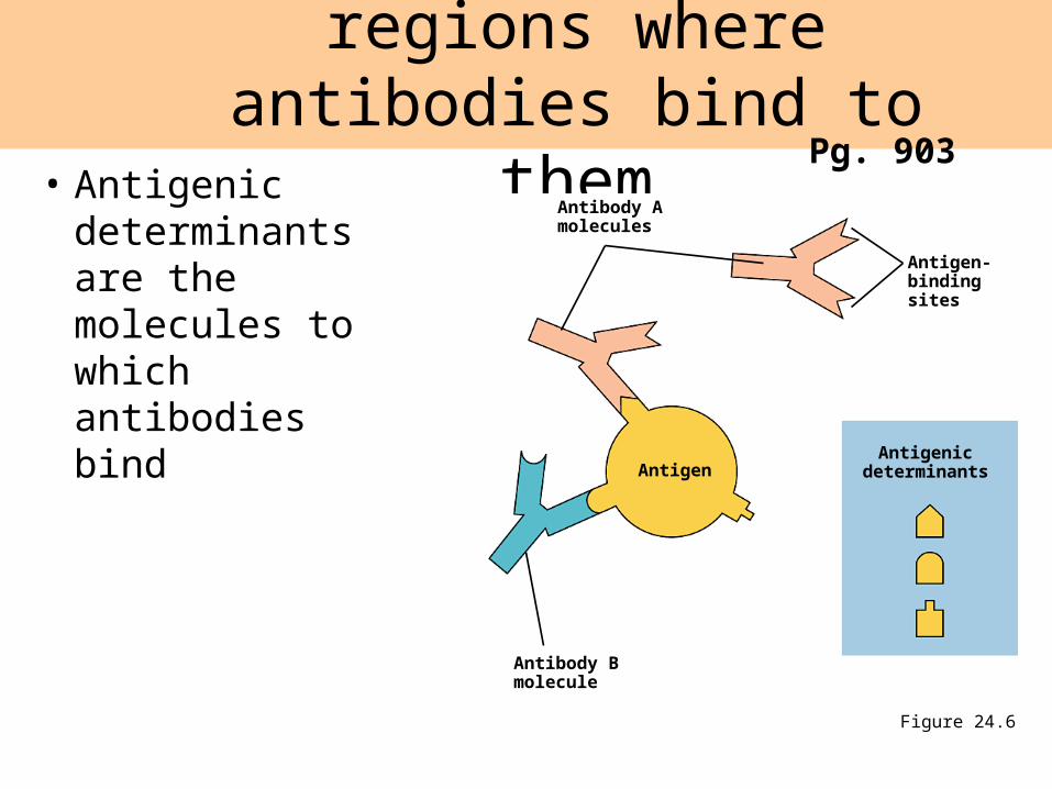
• Antigenic determinants are the molecules to which antibodies bind
Antigens have specific regions where antibodies bind to them
Figure 24.6
Antibody Amolecules
Antigen
Antibody Bmolecule
Antigenicdeterminants
Antigen-bindingsites
Pg. 903
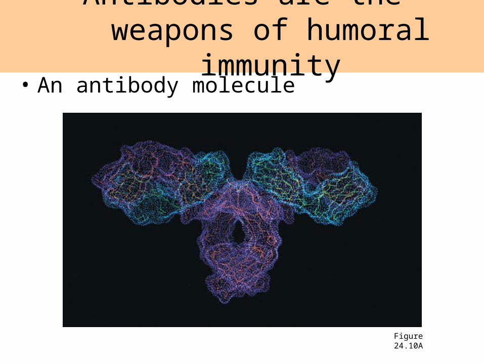
• An antibody molecule
Antibodies are the weapons of humoral immunity
Figure 24.10A
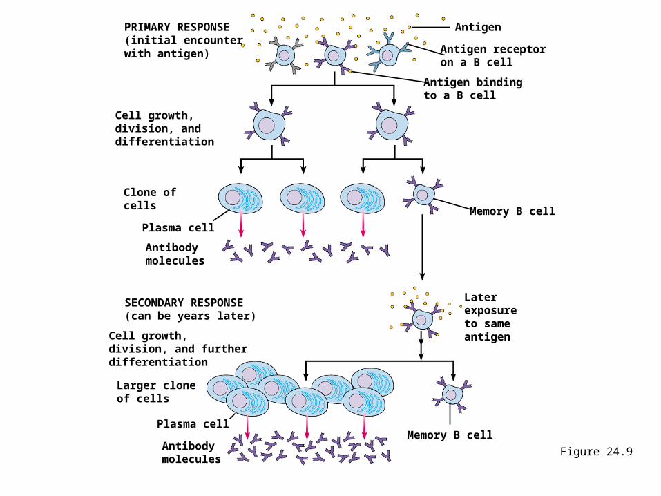
Figure 24.9
PRIMARY RESPONSE(initial encounterwith antigen)
Antigen
Antigen receptoron a B cell
Antigen bindingto a B cell
Memory B cell
Antibodymolecules
Plasma cell
Cell growth,division, anddifferentiation
SECONDARY RESPONSE(can be years later)
Cell growth,division, and furtherdifferentiation
Larger cloneof cells
Plasma cell
Antibodymolecules
Later exposure to same antigen
Memory B cell
Clone ofcells
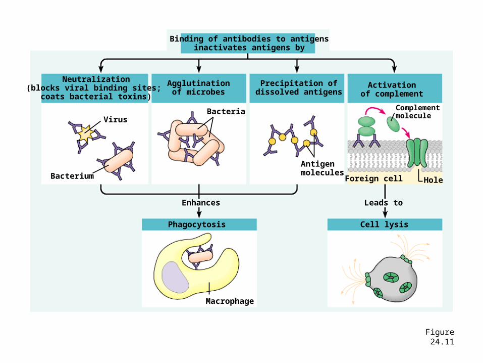
Figure 24.11
Binding of antibodies to antigensinactivates antigens by
Neutralization(blocks viral binding sites;
coats bacterial toxins)
Agglutinationof microbes
Precipitation ofdissolved antigens
Activationof complement
Virus
Bacterium
Bacteria
Antigenmolecules
Complementmolecule
Foreign cell Hole
Enhances
Phagocytosis
Macrophage
Cell lysis
Leads to

Video Segment #2: Minding Your B’s & T’s
• Write 10 Key statements
Introductory Questions #3
SSR Guide for Presenters
• Give your name
• Hold up the your source
• FIVE key points with a brief summary of what you read.
• The rest of us need to write points discussed in the SSR boxes on your cover sheet.
Assignment Packet Due Fri. 3/27• Cover sheet (last week)• Reading guide 36.1• Reflex lab w/meter stick• Draw/Label of a Neuron• SSR#1-5 key points of your reading only
• Cover sheet #2 (this week)• Reading Guide 36.2• Drawings & labels of the brain (2 pgs)• Study guide/Reinforcement guide• Text Review Q’s Pgs 968-969 #1-11 & #19-24• Video guide sheets (x2)• Word List• SSR #2 (5 key points of yours & presenter)
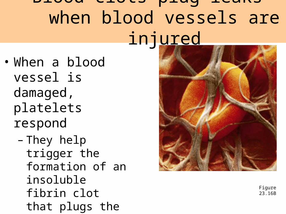
Blood clots plug leaks when blood vessels are injured
Figure 23.16B
• When a blood vessel is damaged, platelets respond– They help trigger the
formation of an insoluble fibrin clot that plugs the leak