the incidence of antibiotic resistant bacteria in …sangita jivan, johan habig and jaco...
TRANSCRIPT

THE INCIDENCE OF ANTIBIOTIC RESISTANT
BACTERIA IN CHICKEN AND PORK
Eugenie van Wijk
B.Sc.; B.Sc. (Honours)
Dissertation submitted in the School for Environmental
Sciences and Development, Potchefstroom University for
Christian Higher Education, in partial fulfillment for the degree
of Magister Scientiae
( Microbiology )
Supervisor : Dr. HA Esterhuysen
Co-supervisor Ms. AM van der Walt
November 2003

TABLE OF CONTENTS
Acknowledgements
Abstract
Opsomming
Chapter 1 Introduction
Chapter 2 Literature review
Introduction
Medical, veterinary and agricultural uses of antibiotics
Classes of antibiotics based on the mechanism of their antimicrobial
action
Agents that inhibit the synthesis of the bacterial cell wall
Agents that act directly on the cell membrane of microorganisms
Agents that bind to ribosome subunits causing inhibition of protein
synthesis
Agents that bind to the ribosomal subunits and alter protein synthesis
Agents that affect nucleic acid metabolism
Agents that act as nucleic acid analogues
Agents that interfere with important metabolic pathways
Non-medical uses of antibiotics
The use of antibiotics in animal feed
The use of antibiotics in food preservation
Bacterial resistance to antibiotic action
Biochemical basis for resistance to antibiotics
Alteration of the target to which antimicrobial agents bind
Role of cell wall and membrane permeability in antibiotic resistance
Production of enzymes that destroy the inhibitory capacity of
antibiotics
Alterations in the enzymes targeted by antibiotic action
Alteration in metabolic pathways affected by antibiotic action
Genetic aspects of antibiotic resistance in bacteria

Antibiotic resistance associated with the bacterial genome
Antibiotic resistance associated with extrachromosomal genetic
elements
The transmission of genetic information
Conjugation
Transduction
Transformation
Prevalence of antibiotic resistance among pathogenic bacteria
The misuse and abuse of antibiotics
Addressing the problem of antibiotic resistance
The aim of the present study
Chapter 3 Materials and Methods
Sampling and processing of meat
Enrichment and isolation of the different groups of bacteria on
selective media
Isolation of Pseudomonas
Isolation of Staphylococcus aureus
Isolation of Enterobacteriaceae
Isolation of Escherichia coli
Isolation of Salmonella
Antibiotic susceptibility testing with the Calibrated Dichotomous
Sensitivity (CDS) method
Preparation of isolates for use in the Calibrated Dichotomous
Sensitivity (CDS) method
Inoculation of plates
Measurement and interpretation of growth inhibition zones
Processing of data
Chapter 4 Results
Isolation of bacteria from samples collected from retail outlets and farms
Antibiotic susceptibility testing using the Calibrated Dichotomous
Sensitivity (CDS) method
Reading the inhibition zones
Level of antibiotic resistance in Pseudomonas isolates
Level of antibiotic resistance in Staphylococcus isolates
Level of antibiotic resistance in Enterobacteriaceae isolates
Level of antibiotic resistance in Escherichia coli isolates
Level of antibiotic resistance in Salmonella isolates
Cluster analysis
Antibiotic resistance in Pseudomonas isolates
Antibiotic resistance in Staphylococcus isolates
Antibiotic resistance in Enterobacteriaceae isolates
Antibiotic resistance in Escherichia coli isolates
Antibiotic resistance in Salmonella isolates
Major antibiotic resistant clusters
Ranking of the antibiotics
Chapter 5 Discussion and Conclusion
5.1 Isolation
5.2 Antibiotic susceptibility testing using the CDS method
5.2.1 Pseudomonas
5.2.2 Staphylococcus
5.2.3 Entero bacteriaceae
5.2.4 Escherichia coli
5.2.5 Salmonella
5.3 Cluster analysis
5.4 Major clusters
5.5 Ranking of antibiotics
5.6 Conclusion

REFERENCES
Appendix A
Appendix B
Appendix C
Appendix D
Appendix E
78
I
I1
IV
VI
VII

ACKNOWLEDGEMENTS
I am greatly indebted to the following persons:
God my Creator. He who will never fail me.
My supervisor, Dr HA Esterhuysen, for his encouragement and guidance during my research,
and assistance in the preparation of this dissertation.
Ms AM van der Walt, for her support and assistance as co-supervisor.
Prof Karl-Heinz Riedel, chair of Microbiology at the Potchefstroom University for financial
support of the study.
Prof HS Steyn and Statistical Services of the Potchefstroom University for statistical
processing of data.
Sangita Jivan, Johan Habig and Jaco Bezuidenhout for assistance during the execution of
experiments and the electronic preparation of this dissertation.
Haagner & Serfontein Chicken Farms for kindly providing samples of chicken, chicken feed,
chicken manure and eggs for this investigation.
My parents for their ongoing moral support and financial assistance during the years of study.
Most of all, Gary Outram for love, encouragement and understanding during a stressful
period.

ABSTRACT
The emergence of antibiotic resistance in important human pathogens has globally become a
public health concern. Consumption of contaminated meat and meat products constitute a
major route for the transmission of antibiotic resistant organisms and the dissemination of
resistance genes in the human environment. The aim of this study was to determine the level
of antibiotic resistance in potentially pathogenic bacteria associated with pork, chicken meat,
chicken manure, chicken feed and eggs. Standard procedures were employed for the selective
enrichment and isolation of Escherichiu coli, Staphylococcus, Enterobacteriaceae,
Pseudomonas and Salmonella, and to determine the level of their susceptibility for penicillin,
oxytetracycline, tetracycline, streptomycin, as well as aminoglycoside antibiotics. It was
found that 10,1% of the total number of isolates were Pseudomonas, 25,3% Staphylococcus,
2 1,2% Enterohacteriuceae, 7,0% E. coli and 36,4% Salmonella. Statistical analysis of results
showed clusters of isolates exhibiting similar patterns of antibiotic resistance. Except for
resistance to penicillin, Pseudomonas isolates were largely susceptible to the antibiotics
tested. Staphylococcus isolates were relatively susceptible, with the highest levels of
resistance, in this case to oxytetracycline and tetracycline, observed in those from pork and
chicken manure. High levels of resistance to oxytetracycline (71%), tetracycline (79%),
streptomycin (52%), and penicillin (1 00%) were detected in Enlerohucteriuceue isolates from
chicken meat samples. It was found in addition that E. coli from chicken meat samples 100%
resistant to oxytetracycline, tetracycline, and penicillin, while Salmonella showed resistance
to gentamycin (63%), tetracycline (46%), oxytetracycline and penicillin (99%). Indexing of
multiple antibiotic resistance (MAR) confirmed the relatively high levels of resistance in E.
coli and Salmonella from the chicken meat samples. Overall, results from the present study
indicated that relatively high levels of resistance towards tetracycline, oxytetracycline and
penicillin was observed in potentially pathogenic bacteria associated with pork, chicken meat,
and the environment of the chicken industry. It was, however found that isolates from the
respective bacterial groups were largely susceptible to the aminoglycoside antibiotics, as well
as streptomycin and erythromycin.

OPSOMMING
Daar bestaan wereldwyd bekommernis oor die uitwerking wat die verskyning van
antibiotikumweerstandbiedendheid in belangrike menslike patogene op openbare gesondheid
kan he. Die inname van gekontamineerde vleis en vleisprodukte verteenwoordig 'n hoofroete
vir die oordrag van antibiotikumweerstandbiedende organisms en die verspreiding van
weerstandigheidsgene in die mens se omgewing. Die doe1 van hierdie studie was om die
vlakke van antibiotikumweerstandbiedendheid in potensieel-patogene bakteriee wat met
varkvleis, hoendervleis, hoendermis, hoendervoer en eiers geassosieer word, te bepaal.
Standaard prosedures is gebruik vir die selektiewe verryking en isolering van Escherichia
coli, S~aphylococcus, Enterobactericrceae, Pseudomonus en Salmonella, en om die vlakke
van hul vatbaarheid vir penisillien, oksitetrasiklien, tetrasiklien, streptomisien, sowel as die
aminoglikosied antibiotikums, te bepaal. Daar is in hierdie studie gevind dat 10,1% van die
totale getal isolate Pseudomonas was, 25,3% Staphylococcus, 21,2% Enterobucteriaceae,
7,0% E. coli en 36,4% Salmonella. Statistiese ontleding van die resultate het die isolate in
trosse met soortgelyke patrone van antibiotikumweerstandbiedendheid geplaas. Behalwe vir
die weerstandbiedendheid teen penisillien, was Pseudomonus vatbaar vir die ander
antibiotikums wat getoets is. Staphylococcus was ook vatbaar vir hierdie antibiotikums, met
die hoogste vlakke van weerstandbiedendheid, in hierdie geval teen oksitetrasiklien en
tetrasiklien, in isolate uit varkvleis en hoendermis. Hoe vlakke van weerstandbiedendheid
teen oksitetrasklien (71%), tetrasiklien (79%), streptomisien (52%), and penisillien (1 00%) is
egter waargeneem in Enterohacteriuceae isolate uit hoendervleis. Daar is verder gevind dat
100% van die E. coli isolate uit hoendervleis weerstandbiedendheid is teen oksitetrasiklien,
tetrasiklien, and penisillien, terwyl Salmonellu weerstandbiedendheid teen gentamisien
(63%), tetrasiklien (46%), oksitetrasiklien en penisillien (99%) getoon het. Indeksering van
meervoudige antibiotikum-weerstandbiedendheid (MAW) het die relatiewe hoe vlakke van
weerstandbiedendheid in E. coli en Salmonella uit hoendervleismonsters bevestig. In die
geheel gesien, toon die resultate van hierdie studie dat relatiewe hoe vlakke van
weerstandbiedendheid teen tetrasiklien, oksitetrasiklien and penisillien in potensieel-patogene
bakteriee wat met varkvleis, hoendervleis en die omgewing van die hoendervleisnywerheid
geassosieer word, voorgekom het. Daar is egter gevind dat isolate verteenwoordigend van die
onderskeie bakteriese groepe we1 tot 'n groot mate vatbaar was vir
aminoglikosiedantibiotikums, sowel as vir streptomisien en eritromisien.
vii

CHAPTER 1
INTRODUCTION
The discovery of antibiotics with therapeutic applications in both human and veterinary
medicine has been among the great achievements of our time (Vasquez-Moreno et ul., 1990).
The modern era of antimicrobial chemotherapy began in 1929 with Fleming's discovery of
the powerful bactericidal substance penicillin, and Domagk's discovery in 1935 of synthetic
chemicals (sulfonamides) with broad antimicrobial activity (Todar, 1996a). In the early
1940's, spurred partially by the need for antibacterial agents in World War 11, penicillin was
isolated, purified and injected into experimental animals, where it was found not only to cure
infections, but also to possess low toxicity towards animals (Harrison and Svec, 1998). The
subsequent discovery, development, and clinical use of other antibiotics that followed this
major event, resulted in the effective treatment of infection caused by major bacterial
pathogens to the extent that many experts considered bacterial infectious diseases to be under
complete therapeutic control (Harrison and Svec, 1998).
The emergence of antibiotic resistance in important human pathogens has globally become a
continuing public health concern (Newsome et al., 1987). Acquired antibiotic resistance
enables pathogens to survive in the presence of antibiotics to which they were previously
sensitive (Newsome et al., 1987). The development of resistance in bacteria is generally
related to a combination of factors including the abuse or misuse of antibiotics in human and
veterinary medicine, the application of antibiotics in agriculture as growth promoters in
animal feed, as well as the remarkable genetic plasticity of bacteria (Vasquez-Moreno et al.,
1990). Furthermore, the application of antibiotics in the treatment of viral infections,
prescription by physicians of erroneous dosage, or failure of patients to complete the
prescribed course are factors that probably had a major role in rendering many important
antibiotics ineffective (Hardman and Limbird, 1996). Extensive use of antibiotics as the
"miracle drug" to cure infection of any kind could have created a selective pressure for
microorganisms to develop mechanisms to escape the inhibitory effect of the antimicrobial
agents. The injudicious use of antibiotic treatment may thus lead to the selection of resistance
in the target organism that causes the infection and against which antibiotic therapy was
directed (Lerner, 1998).

In livestock and poultry production sub-therapeutic amounts of various antibiotics are used
in animal feed mixtures for growth promotion, improved feed efficiency and for the control
and prevention of diseases (Vasquez-Moreno et al., 1990; Schwarz and Chaslus-Dancla,
2001). Various studies have reported that a reservoir of resistant microorganisms exists in
food animals that were fed antibiotic-containing feed while being raised under intensive
growth conditions (Newsome et al, 1987). In addition, the widespread use of antibiotics in
veterinary practice is believed to be a major factor contributing to the establishment of a
reservoir of antibiotic resistant enteric pathogenic and/or non-pathogenic bacteria in animals.
These bacteria may transfer their resistance genes to unrelated human and animal pathogens
(Newsome et al., 1987; Schwarz and Chaslus-Dancla, 2001).
Meat and meat products contaminated with antibiotic resistant organisms constitute the
primary means by which antibiotic resistant organisms are transmitted to humans (Mattila el
ul., 1988). Humans may also acquire antibiotic resistant bacterial strains from environments
where food animals were raised intensively on antibiotic-containing feed. Direct exposure to
the manure, or contact with pets, rodents, flies and cockroaches that were exposed to the
waste of these animals also constitute a possible route for the transmission of resistant
bacteria to humans (Feinman, 1999).
According to Schwarz and Chaslus-Dancla (2001), the emergence and spread of
antimicrobial resistance among human pathogens could be significantly reduced if published
guidelines aimed at promoting the prudent and judicious use of antibiotics were followed in
both medical and veterinary practice, as well as by agriculture and manufacturers of animal
feed.
Because commercial feed used in the large scale production of chickens and pigs intended for
human consumption probably contain various types of antibiotics in varying degrees, and fed
animals raised under these conditions could be a factor contributing to the widespread
problem of antibiotic resistance in modern society, the present study was designed to screen
for potential human pathogenic bacteria from different settings of the chicken and pork
industry. Standard procedures were employed for the isolation of Pseudon~onas,
Sfuphylococcus, Enferobucferiuceae, Escherichia coli and Salmonella from feed and manure
samples collected from different farm settings, as well as meat products and eggs obtained
from different retail outlets. Isolates were subsequently subjected to standard procedures to

determine the level of antibiotic resistance in the different bacterial groups and to compare
the various samples in this regard.
In the present study, the number of isolates of the respective bacterial groups obtained from
chicken meat was comparable to that from pork. The highest number of isolates picked of
from the selective mediums was Salmonella organisms, followed by Staphylococcus aureus,
En/erohacleriaceae, Pseuomonus and Escherichiu coli organisms. Salmonella in particular,
exhibited high resistance to three antibiotics with the highest MAR index followed by
Escherichia coli. Findings from the present study seem to be in consensus with literature
reports in suggesting that food animals may serve as reservoirs of resistant enteric bacteria
from which antibiotic resistance factors could be disseminated into the environment.

CHAPTER 2
LITERATURE REVIEW
2.1 Introduction
Antibiotics are chemical compounds produced as secondary metabolites of microbial
metabolism. Although many antibiotics used today are produced by microorganisms, some are
manufactured partly or entirely by chemical synthesis. The term antimicrobic is often used to
include agents produced entirely by microorganisms, as well as commercial antibiotics that have
been chemically altered to improve potency or to increase the range of species they affect (Atlas,
1997; Elliot et ul., 1997; Jacob, 1999).
Due to their antimicrobial activity, antibiotics are widely applied in medicine as
chemotherapeutic agents to treat bacterial infections. Antibiotics act on bacteria by interfering
with essential biological processes, such as cell wall synthesis, DNA replication and transcription
of protein (Atlas, 1997). Bacteria vary in their sensitivity to various antibiotics. Bacteria in which
the structure or function targeted by a specific antibiotic, will exhibit an intrinsic or natural
resistance to the action of the specific drug. Due to their genetic plasticity, however, bacteria
may also acquire antibiotic resistance after having been exposed to low levels of the drug over an
extended period of time. Some antibiotics are referred to as narrow-spectrum, because they are
very specific in their action and may target Gram-negative bacteria only, as opposed to wide-
spectrum antibiotics that are effective in the treatment of infections caused by a wide range of
bacteria, including both Gram-negative and Gram-positive bacteria (Brock et ul., 1994; Lancini
et ul., 1995).
2.2 Medical, veterinary and agricultural uses of antibiotics
Antibiotics are used to treat diseases of microbial origin in both humans and animals. Their
application in veterinary medicine, however, has been confined mainly to those diseases in
animals that cause major economic losses, such as bovine mastitis (Atlas, 1997). Antibiotics

probably find its most important application in the treatment of infectious human diseases and
these agents accounts for roughly half the antibiotics consumed every year (Jacob, 1999).
Antimicrobials used in both animal or human medicine include amikacin, ampicillin,
amoxicillin-clavulanic acid, apramycin, ceftiofur, cefriaxone, cephathin, chloramphenicol,
ciprofloxacin, gentamycin, kanamycin, nalidixic acid, streptomycin, sulfamethoxazole,
tetracycline, trimeyhoprim-sufamethoxazole and ticarcillin (Torrence, 200 1 ).
In addition to its medical applications, a variety of antibiotics are used prophylactically and as
growth promoters in commercial feed formulations used in food animal production. However,
concern has repeatedly been expressed by public health authorities, infectious disease specialists
and plasmid biologists that the practice of using antibiotics routinely at subtherapeutic levels in
animal feed has contributed to the establishment of a reservoir of resistant zoonotic bacteria,
including Salmonella and coliforms, to which humans may become exposed (DuPont and Steele,
1987; Teuber, 200 1 ; Torrence, 200 1).
2.3 Classes of antibiotics based on the mechanism of their antimicrobial action
Most of the therapeutically useful antimicrobial agents are effective against bacterial infections
because of significant differences between the prokaryotic cells of the infecting bacteria and the
eukaryotic cells of the infected human or animal. Various sites in a bacterial cell that are targeted
by antibiotic action are absent in eukaryotic cells so that selective toxicity for bacteria can
effectively be achieved. Antibiotics typically prevent bacterial reproduction by entering the
bacterial cells and interfering with the production of components needed to form new bacterial
cells (Jacob, 1999). Structures and macromolecules of prokaryotic cells that serve as potential
targets for antimicrobial action include cell walls, ribosomes for protein synthesis, membranes
and nucleic acids (Elliot et al., 1997). Based on their mode of action and the specific structures
they target in the bacterial cell, antibiotics can be arranged in five major groups (Atlas, 1997).
The different classes of antibiotics include agents that affect (i) cell wall synthesis, (ii)
membrane integrity, (iii) protein synthesis, (iv) DNA replication and (v) important methabolic
pathways (Katzung and Trevor,1998). The different antibiotic classes, and the mode of action of

different chemotherapeutic agents belonging to each class, are discussed in the subsequent
paragraphs.
2.3.1 Agents that inhibit the synthesis of the bacterial cell wall
Unlike mammalian cells, bacteria have cell walls. Some antimicrobial agents, such as the P-
lactam and glycopeptide antibiotics, act selectively on the bacterial cell wall (Elliot et al., 1997).
For normal growth and development, the formation of peptide cross-linkages within the
peptidoglycan backbone is essential to maintain the rigidity of the bacterial cell wall. This
transpeptidation reaction is catalysed by certain enzymes collectively known as penicillin-
binding proteins (PBPs). Beta-lactam antibiotics act by inhibiting the carboxypeptidase and
transpeptidase enzymes that are required for the formation of cross-linkages to form between the
peptidoglycan chains (Mandell and Petri, 1996; Atlas, 1997; Elliot et al., 1997; Bush and
Mobashery, 1998). Glycopeptides, such as the antibiotic vancomycin, appear to inhibit both
transglycosylation and transpeptidation reactions during peptidoglycan assembly (Mims et al.,
1993).
Penicillins have been shown to be most effective against Gram-positive cocci, Neisseria and
Treponema pallidum, but not against Gram-negative pathogens such as Salmonella, Shigella and
Pseudomonus and the Gram-negative organism, Neisseria gonorrhoeae (Todar, 1996a; Elliot et
ul., 1997). Chlamydia and Mycoplasma species are insensitive to the action of penicillin and
cephalsporins because they lack peptidoglycan-containing cell walls. Bacteria resistant to
penicillins most often produce enzymes that are collectively referred to as P-lactamases because
of their ability to open the P-lactam ring of penicillins (Neu, 1976; Atlas, 1997).
In contrast to penicillins, cephalosporins are broad spectrum antibiotics relatively resistant to
penicillinase, and therefore these drugs have become useful chemotherapeutic agents in the
treatment of infections caused by either Gram-positive or Gram-negative bacteria (Atlas, 1997).
The cephalosporin, cefamandole, is widely used in the treatment of infections caused by strains
of Klebsiella pneumoniae, causing pneumonia and urinary tract infections (Atlas, 1997). Most
Gram-positive cocci, with the exception of enterococci and methicillin-resistant Staphylococcus

aureus, are susceptible to cephalosporins (Mandell and Petri, 1996). Cephalosporins are used as
alternatives to penicillins for patients who are allergic to penicillin and for those pathogens that
are not penicillin sensitive. The cephalosporins may be used also in place of penicillins for the
prophylaxis of infection by Gram-positive cocci following surgical procedures (Atlas, 1997).
2.3.2 Agents that act directly on the cell membrane of microorganisms
Polipeptide antibiotics are chemotherapeutic agents that target the microbial cell membrane.
These include polymyxins, polyenes and imidazoles that damage or disorganise the structure or
inhibit the function of the bacterial membrane. The integrity of the cytoplasmic membrane is
vital to bacteria, and compounds that disorganise the membranes, resulting in loss of cytoplasmic
content, rapidly kill bacterial cells. Antibiotics of clinical importance that act by this mechanism
are the polymyxins produced by Bacillus polymyxa (Todar, 1996a). Polypeptide antibiotics
disrupt the phospholipids that make up the structure of the membrane whereas polyene acts on
the sterol components of the cell membrane. The antimicrobial activities of polymyxin b and
colistin are similar and are restricted to Gram-negative bacteria such as Enterobacter,
Escherichia coli, Klebsiella, Salmonella, Pasteurella, Bordetella, and Shigella (Mandell and
Petri, 1996).
2.3.3 Agents that bind to ribosomal subunits causing inhibition of protein synthesis
The selectivity of this class of antibiotics is based on their ability to target either the 30s and 50s
subunits of 70s bacterial ribosomes, leaving human ribosomes consisting of 80s subunits
unaffected (Elliot et al., 1997). Many therapeutically useful antibiotics owe their antibacterial
action to the inhibition of some step in the complex process of protein synthesis (Todar, 1996a).
The most important antibiotics with this mode of action are the tetracyclines, chloramphenicol,
and the macrolides (Elliot et al., 1997; Katzung and Trevor, 1998).
According to Elliot and coworkers (1997) chloramphenicol binds to the 50s subunit and
interferes with the linkage of amino acids in the peptide chain formation, or combines with the
bacterial ribosome to prevent the assembly of amino acids into a protein chain. Chloramphenicol

is active against many species of Gram-negative bacteria. Chloramphenicol is used for treating
typhoid fever and various infections caused by Salmonella. Chloramphenicol is also effective
against rickettsia and are used for the treatment of typhus fever caused by this agent, as well as
conditions caused by anaerobe pathogens, e.g Bacteriodes fragilis (Elliot et al., 1997).
Tetracyclines bind to the 30s subunit, preventing binding of aminoacyl transfer RNA to the
acceptor site in the ribosome, thereby inhibiting amino acid chain elongation (Elliot et al., 1997).
At least two processes appear to be required for these antibiotics to gain access to the ribosomes
of Gram-negative bacteria, namely (i) passive diffusion through the hydrophilic channels formed
by the porin proteins of the outer membrane and (ii) active transport by an energy-depending
system that pumps all tetracyclines through the inner cytoplasmic membrane (Mandell and Petri,
1996). Tetracyclines have a broad spectrum of activity against many Gram-positive and some
Gram-negative bacteria (Elliot et al., 1997). Tetracyclines are effective against various
pathogenic bacteria, including Rickettsia and Chlamydia species. Tetracyclines are useful also
against various other bacterial infections, including Mycoplasma pneumoniae and the causative
agents of brucellosis, tularemia and cholera.
The macrolide antibiotic, erythromycin, exert it's action on the bacterial cell by binding to the
50s subunit of ribosomal RNA and inhibiting the formation of the initiation complex (Elliot et
al., 1997). Although not active against most aerobic Gram-negative rods, erythromycin does
exhibit antibacterial activity against some Gram-negative organisms including Legionella
pneumophila, Mycoplasma pneumoniae, Corynebacterium and Chlamydia trachomatis (Atlas,
1997). Erythromycin may serve as an important alternative treatment for streptococcal infections
in patients who are allergic to penicillin, e.g. infection caused by Streptococcus pyogenes
(Mandell and Petri, 1996; Todar, 1996a).
2.3.4 Agents that bind to ribosomal subunits and alter protein synthesis
Aminoglycoside antimicrobial agents include chemotherapeutic agents such as streptomycin,
gentamicin, neomycin, kanamycin, tobramycin, and amikacin. After entering the bacterial cell,
binding of the aminoglycoside to the 30s ribosomal subunit of the 70s bacterial ribosomes

interferes with the formation of the initiation complexes, the first step in the translation genetic
code, thus affecting the fidelity of translation into a protein (Mims et al., 1993; Atlas, 1997).
These antimicrobics are used almost exclusively in the treatment of infections caused by Gram-
negative bacteria. Aminoglycosides are relatively ineffective against anaerobic bacteria,
facultative anaerobes and Gram-positive bacteria (Elliot et al., 1997).
2.3.5 Agents that affect nucleic acid metabolism
The nucleic acids, DNA and RNA, of the bacterial cell are informational macromolecules that
carry important biological information, and the means to process this information, in the highly
specific sequence of the amino acids they are composed of (Brock et al., 1994; Atlas, 1997).
Some antibiotics have the ability to interfere with some processes involved in nucleic acid
biosynthesis, whether at the stage of nucleotide biosynthesis, or nucleotide polimerization
(Betina, 1983). The quinolone antibiotic, nalidixic acid, interferes with the DNA gyrase,
preventing the formation of a replication fork, an essential step in the replication of DNA needed
for cell proliferation. Although DNA synthesis is blocked, transcription and translation can still
proceed (Betina, 1983; Mann and Grabbe, 1996; Atlas, 1997). Quinolones are effective against a
broad range of Gram-positive and Gram-negative bacteria including the mycobacteria (Mims et
al., 1993).
Rifampin, a semi-synthetic derivative of rifamycin B, inhibits DNA-dependent RNA polymerase
of mycobacteria and other microorganisms by forming a stable drug-enzyme complex, leading to
the suppression of initiation of chain formation in RNA synthesis (Mandell and Petri, 1996).
Rifampin is used in combination with other antibiotics in the treatment of Mycohacterium
tuberculosis (Elliot et al., 1997). Rifampin inhibits the growth of most Gram-positive bacteria as
well as many Gram-negative microorganisms such as Escherichia coli, Pseudomonas, indole-
positive and indole-negative Proteus, and Klebsiella. Rifampin is very active against
Staphylococcus aureus and coagulase-negative staphylococci (Mandell and Petri, 1996).

2.3.6 Agents that act as nucleic acids analogues
Nucleic acid analogues such as zidovudine, ganciclovir, vidarabine and acyclovir inhibit viral
replication by affecting enzymes that are essential for viral DNA synthesis (Mandell and Petri,
1996). Acyclovir is an acyclic guanine nucleoside analogue that lacks a 3'-hydroxyl on the side
chain. The clinically useful antiviral spectrum of acyclovir is limited to Herpes viruses. By a
mechanism termed suicide inactivation, the terminated DNA template containing acyclovir binds
to the enzyme and leads to irreversible inactivation of the DNA polymerase (Mandell and Petri,
1996). Ganciclovir is an acyclic guanine nucleoside analogue. This agent has inhibitory activity
against all herpes viruses but especially against cytomegalovirus (CMV) (Mandell and Petri,
1996). Vidarabine is an adenosine analogue with an altered sugar, and although this drug was
originally developed for the treatment of leukaemia, it has proven to be more effective in treating
herpes simplex virus (HSV), encephalitis and keratoconjunctivitis. Intravenous vidarabine is
approved for use in HSV encephalitis, neonatal herpes, and zoster or varicella in
immunocompromised patients, but can be replaced by acyclovir for these applications (Mandell
and Petri, 1996; Atlas, 1997). Zidovudine is a thymidine analogue with antiviral activity against
HIV-1, HIV-2, human T lymphotropic (or leukaemia) virus and other retroviruses. Following
diffusion into host cells, the drug is initially phosphorylated by cellular thymidine kinase
(Mandell and Petri, 1996). Being an analogue of the DNA base thymidine, azidothymidine
triphosphate (AZT) inhibits viral reverse transcriptase thus terminating DNA chain elongation
prematurely (Mims et al., 1993).
2.3.7 Agents that interfere with important metabolic pathways
Folic acid, consisting in part of para-aminobenzoic acid (PABA) has an important role as an
essential co-substrate in the biosynthesis of amino acids, purines and pirimidines. In contrast to
mammalian cells, bacteria are responsible for synthesising the required amount of folic acid for
themselves. The antimicrobial action of drugs such as sulphonamides and trimethoprim is due to
their ability to disrupt the folic acid metabolism in bacterial cells (Atlas, 1997). Because folic
acid cannot take up PABA in the presence of sulphonamides, dysfunctional molecules that are
unable to perform their essential metabolic functions are formed as a result. The one-carbon
transfer required for the synthesis of thymidine and purines does not occur in the presence of
thrimethoprim, an antimicrobial agent that prevents the transformation of dihydrofolic acid
(DHF) into tetrahydrofolic acid (THF) (mann and Grabbe, 1996).
Many Gram-positive cocci, including Staphylococcus aureus, streptococci and to a variable
extent the enterococci, are susceptible to trimethoprim. The enterobacteria, including E. coli and
Salmonella species, are also sensitive to the action of trimethoprim. The wide-spectrum activity
of trimethoprim favors the application of this drug in the treatment of gastroenteritis as well as
respiratory and urinary tract infections caused by susceptible organisms. Sulphonamides and
trimethoprim often are used in combination to combat bacterial infections (Mann and Grabbe,
1996; Atlas, 1997).
2.4 Non-medical uses of antibiotics
Statistics for the year 1997, released by the Federation of Animal Health (FEDESA), indicated
that over 11.5 x lo6 kg of antibiotics were used for animal growth promotion in the European
Union countries and Switzerland (Ungemach, 2000). Antibiotics are also employed in the
preservation of human food in order to reduce human risk of being exposed to pathogenic
microorganisms (Khachatourians, 1998). Food is an excellent substrate for the growth of
microorganisms, and consumption of food contaminated with pathogens could put humans at
risk of food poisoning, or disease outbreaks of epidemiological proportions (Frazier and
Westhoff, 1996). To ensure food safety, antimicrobials such as tetracycline and natamycin are
often used as food preservatives (Jay, 1992).
2.4.1 The use of antibiotics in animal feed
In the 1940s researchers at Lederle Laboratories (USA) seeking nutritional factors that would
improve the growth rate of animals raised for human consumption, discovered that small
amounts of the antibiotic chlorotetracycline significantly improved the growth of food animals.
An entirely new industry was generated which opened a large new market for pharmaceutical
companies manufacturing antibiotics (Harrison and Svec, 1998). In agriculture, low levels of

various antibiotics are presently added to animal feeds to promote growth and improve the
efficiency of feed conversion into meat (Moro et ul., 1998). According to Du Pont and Steele
(1987), the addition of subtherapeutical amounts of certain antimicrobial agents to animal feed,
not only prevents infectious diseases caused by bacteria or protozoa, but also decreases the
amount of feed needed while increasing the rate of weight gain. In 1948, it was noted that
chlorotetracycline containing vitamin B12 promoted the growth of chickens even when chicken
feed contained more than sufficient amounts of all known vitamins (Cooke, 1974). The addition
of tetracycline or penicillin to commercial swine and poultry feeds at the rate of 5 to 20 grams
per ton of feed, was found to have increased the growth rate of young animals by at least 10%
and sometimes more. This probably occurred because the added drugs destroyed pathogenic
bacteria and intestinal parasites that could cause mild forms of disease that affect the growth and
development of young animals (Pelczar et ul., 1993; Khachatourians, 1998). Antibiotics in
animal feed could also improve the performance of animals under conditions of stress such as
poor ventilation or overcrowding during transit. Chronic respiratory disease in poultry, scouring
in pigs, and chronic diarrhoea1 disease in pigs commonly occur under these conditions (Cooke,
1974). The use of subtherapeutic levels of antimicrobial agents is one of the tools that had
facilitated confinement housing, allowing larger numbers of animals to be maintained in a
production facility of a given size. The practice of adding subtherapeutical amounts of antibiotics
to the feed of food animals probably contributes to lower costs of animal care and ultimately
could lower costs to the consumers of meat, milk and eggs (Du Pont and Steele, 1987). However,
a major concern about the use of antimicrobial agents to raise food animals is the possibility that
illegal antibiotic residues of the agents may be found in meat, especially liver or kidney.
Whether or not the drug to which the animals were exposed will reach the consumer, depends
upon a number of factors including (i) the specific drug involved, (ii) its absorbability and
pharmacokinetics, (iii) the interval from administration of the last dose of the drug until
slaughtering, (iv) the tissue to be eaten and (v) the degree of cooking of the meat (Du Pont and
Steele, 1987; Harrison and Svec, 1998).

2.4.2 The use of antibiotics in food preservation
Upon prolonged storage, even at refrigerator temperatures, microbial spoilage of food occurs.
According to Jay (1992), internal temperatures of food are not reduced to within the refrigerator
range, and spoilage that is likely to occur is caused by bacteria such as from Clostridium
perpingens and genera of the Enterobacteriaceae family, originating from internal sources.
Bacterial spoilage of refrigerated-stored meats may also be reflective of external conditions and
sources of contamination. With respect to fungal spoilage of fresh meats, especially beef, a
diverse range of molds have been recovered from various spoilage conditions of whole beef
including Thamnidium, Mucor, and Rhizopus, molds that typically produce "whiskers" on beef.
Cladosporium produces black spots, Penicillium green patches, and Sporotrichum and
Chrysosporium white spots on the meat. Molds apparently do not grow on meat if the storage
temperature is below -5 "C. Among the yeast genera associated with spoiled beef, Candidu and
Rhodotorula are well known (Jay, 1992).
The skin of live birds, as well as the feet, feathers and faeces contain a variety of
microorganisms. Contamination of poultry usually occurs during washing, plucking and
evisceration (Frazier and Westhoff, 1996). Whole poultry tends to have a lower microbial count
than cut up poultry (Jay, 1992). Freshly laid eggs, although sterile inside, become contaminated
on the outside by faecal matter from the hen, cage, nest, wash-water if the eggs are washed and
by handling (Frazier and Westhoff, 1996). Fresh eggs may exhibit cracks and any breaks in the
shell or dirt on the egg will favour spoilage on storage or transmission of pathogenic bacteria to
the consumer (Frazier and Westhoff, 1996). Infected eggs can also contribute to the problem of
meat contamination with potential human pathogens, since infected chicks hatched from lightly
infected eggs may survive and grow into birds that continue to excrete salmonellae (Jay, 1992).
Preservation methods that have been developed to reduce the risk of food-borne outbreaks of
infectious diseases include physical procedures such as irradiation, freezing, vacuum packaging
or chilling (Frazier and Westhoff, 1996). Food can also be preserved using chemicals such as
benzoic acid, the parabens, sorbic acid, nitrites or nitrates, sulphites or sulphur dioxide, or by
increasing carbon dioxide concentrations. Nisin, a bacteriocin produced by some strains of

Lactococcus lactis, as well as antibiotics such as tetracycline, natamycin and subtilins, are often
applied to preserve food (Jay, 1992).
2.5 Bacterial resistance to antibiotic action
According to Chubb (2000) infectious diseases are the third leading cause of death in the United
States and the number one cause worldwide. For several decades following their discovery in the
1940's, antibiotics have been most reliable as drugs to control infectious diseases. The
development of antimicrobial agents for clinical use has brought unquestionable benefits for the
treatment of individual as well as community-acquired infections, because many infections that
formerly were frequently fatal, have since become routinely curable. However, it soon became
apparent that the frequent use of antibiotics have lead to the selection of resistance in organisms
causing infection and against which antibiotic therapy was directed (Lerner, 1998). Concerns
have been raised regarding the possibility of bacteria becoming increasingly antibiotic resistant
due to the abuse and misuse of this miracle drug. Due to the increased in the incidence of
infections caused by resistant bacteria, in particular nosocomial infections, and the importance of
R-factors in clinical medicine, many studies have been focused on the biochemical mechanisms
of drug resistance and the epidemiology of resistant bacteria (Mitsuhashi, 1975).
Antibiotic resistance refers to the phenomenon that bacteria causing infection are not killed or
inhibited by the antibiotic to which they were previously susceptible and, therefore, they do not
respond to treatment, but survive and continue to multiply and causing symptoms in the patient
(Mims et al., 1993; Mitcher et al., 1999). Some bacteria are intrinsically resistant to a certain
antibiotic. Other bacteria, although intrinsically susceptible, may develop resistance when they
tolerate concentrations of an antibiotic that is significantly higher than the concentration which
inhibits the growth of susceptible strains of the same species in vitro (WHO, 1978; Pelczar et al.,
1993).
Resistant strains of bacteria emerge through evolutionary selection. Whenever antibiotics are
used there is selective pressure for resistance to occur, since cells that can survive exposure to
the antibiotic gradually replace the more vulnerable strains. Antibiotics cause resistance, but also

create conditions for an existing strain of resistant bacteria to become dominant (Jacob, 1999;
Mitcher et al., 1999). Due to their ability to develop resistance, bacteria that once appeared to be
under control, or at least were believed to be potentially controllable, are now causing infections
that are increasingly difficult to treat. Relatively common strains of infectious agents such as
Staphylococcus, Enterococcus, Pseudomonas aeruginosa, Mycobacterium and several
pneumococci are becomming increasingly resistant to multiple antibiotics, while strains of
Enterobacter faecium and Pseudomonas cepacia exist that are insensitive to any presently
available antibiotics (Mitcher et al., 1999).
2.5.1 Biochemical basis for resistance to antibiotics
Antibiotic resistant bacteria owe their drug insensitivity to antibiotic resistance genes that encode
mechanisms that prevent the drug from reacting with its target in the bacterial cell (Jacob, 1999;
Mitcher et al., 1999). In some bacteria, the antibiotic is inactivated when resistant genes encode
enzymes that degrade or chemically modify the drug in such a way that it no longer react with its
target (Jacob, 1999). Aminoglycoside antibiotics are inactivated by bacterial enzymes that
facilitate the acetylation, phosphorylation or adenylation of the drug (Mitsuhashi, 1975; Schwarz
and Chaslus-Dancla, 2001). In the case of tetracyclines, enzymes encoded by resistant genes
prevent the drug from reaching the target of its action inside the bacterial cell. Drug inactivation
may also occur when its target inside the bacterial cell was modified (Jacob, 1999; Mitcher et al.,
1999; Schwarz and Chaslus-Dancla, 2001). Some bacteria develop resistance because of their
ability to eliminate entry ports for the antibiotic or, more effectively may manufacture pumps
that export antibiotics out of the cell before they could react with the target molecule (Jacob,
1999). Bacteria may have an alternative biochemical pathway that allows them to bypass the
particular reaction that is inhibited by the antibiotic (Mandell and Petri, 1996). It has also been
recognised that resistant microorganisms may carry genes encoding resistance by more than one
mechanistic class. Biochemical mechanisms responsible for antibiotic resistance will be
discussed in the subsequent paragraphs.

2.5.1.1 Alteration of the target to which antimicrobial agents bind
In some bacteria the structure targeted by the antibiotic is altered due to a mutation in either the
genomic or plasmid DNA. By modifying targets such as the cell wall peptidoglycan, genomic
DNA, ribosomal RNA or any of the functional or structural proteins, resistant bacteria prevent
binding of the antimicrobic to these targets and so avoid the disruption of cell function (Black,
1996; Atlas, 1997). Bacterial resistance to antibiotics such as erythromycin, rifamycin, and the
antimetabolites such as trimethoprim has developed by this mechanism (Black, 1996).
Resistance to rifampin, for instance, occurs in bacterial cells that have altered the target of this
drug, namely DNA-dependent RNA polymerase (Mandell and Petri, 1996). Although the degree
of modification enables the bacterial cell to avoid drug action, the functionality of these
structures usually remain unaffected by these changes (Black, 1996; Atlas, 1997). Plasmid-
encoded dihydrofolate reductases with altered affinity for trimethoprim, allows the synthesis of
tetrahydrofolic acid (THFA) to proceed unhindered in the presence of trimethoprim. The
modified enzymes, although less susceptible to trimethoprim, retain their affinity for the normal
substrate, namely dihydrofolate (Mims et al., 1993). Some bacteria develop resistance to
vancomycin by encoding an enzyme that removes the alanine residue from the peptide portion of
peptidoglycan. In this case, vancomycin cannot bind to the altered peptide, but the latter remains
functional in the formation of the cross-linkages during synthesis of peptidoglycan (Black, 1996;
Atlas, 1997).
2.5.1.2 Role of cell wall and membrane permeability in antibiotic resistance
Resistance to bacterial beta-lactam antibiotics, such as penicillin, is caused by the inability of the
antibacterial agent to penetrate to reach the site of its action. In Gram-positive bacteria the
peptidoglycan polymer is very near the cell surface and beta-lactam antibiotic molecules can
penetrate easily to reach the outer layer of cytoplasmic membrane and the penicillin-binding
proteins (PBPs) where the final stages of cell wall synthesis occur. Gram-negative bacteria, on
the other hand, are intrinsically resistant to beta-lactams, because their surface structure is more
complex. An outer membrane consisting of lipopolisaccharide occurs on the outside of the
peptidoglycan layer, which in some bacteria is surrounded by a capsule as well. This

composition renders Gram-negative cell walls impenetrable to the large molecules of the beta-
lactam and other antibiotics (Atlas, 1997).
Small, hydrophilic antibiotics such as the aminoglycosides, however, can diffuse through
aqueous channels that are formed by proteins, known as porins, in the outer membrane of the
Gram-negative cell wall (Mandell and Petri, 1996; Atlas, 1997). Alteration of membrane
permeability may occur when new genetic information changes the nature of proteins or pores of
the outer membrane, thus preventing the antimicrobial agent from entering the bacterial cell and
reach its target. Resistance to tetracyclines, quinolones, and some aminoglycosides occurs via
this mechanism (Black, 1996; Mitcher et al., 1999).
Bacterial resistance to tetracyclines is based on the transport of tetracyclines out of the cell fast
enough to prevent the accumulation of toxic levels of tetracycline so that bacterial protein
synthesis is not inhibited (Atlas, 1997). The efflux pump mechanism function in association with
the inner membrane and occurs in both Gram-positive and Gram-negative bacteria.
2.5.1.3 Production of enzymes that destroy the inhibitory capacity of antibiotics
The presence of enzymes that can either destroy or inactivate the antimicrobial agent is a
common cause of resistance in bacteria. Some bacteria produce enzymes that are called beta-
lactamases because of their capability to hydrolyse the p-lactam ring of antimicrobial agents
such as penicillins and some cephalosporins (Black, 1996; Mitcher et al., 1999). Many Gram-
positive microorganisms release relatively large amounts of p-lactamase into the surrounding
medium. There are three classes of p-lactamase: penicillinases, oxacillinases, and
carbenicillinases (Atlas, 1997). The location of the p-lactamase enzyme in the periplasmic space
probably makes it more effective in destroying cephalosporins than penicillins, because the
targets for cephalosporins also are located on the inner membrane of the bacterial cell (Mandell
and Petri, 1996).
Certain Gram-negative bacteria possess enzymes that can destroy aminoglycosides and
chloramphenicol (Black, 1996). These enzymes may act either by hydrolyzing the antimicrobial

agent or by adding a chemical group causing the enzyme to loose its inhibitory activity (Atlas,
1997). For instance, some bacterial cells produce enzymes that could either add a phosphate, an
acetate, or adenyl group to an aminoglycoside antibiotic. The modified aminoglycoside cannot
bind to 30s ribosomal subunits to block protein synthesis (Atlas, 1997; Schwarz and Chaslus-
Dancla, 2001). Chloramphenicol resistance most often is caused by enzymatic acetylation of the
antibiotic. Modified chloramphenicol is no longer effective in blocking protein synthesis since it
cannot bind to the 50s ribosomal subunit of the bacterial ribosome. Most clinical isolates that are
resistant to chloramphenicol possess a plasmid that carries a gene that encodes chloramphenicol
acetyltransferase, leading to the inactivation of the drug soon after it has crossed the cytoplasmic
membrane (Atlas, 1997; Schwarz and Chaslus-Dancla, 200 1).
2.5.1.4 Alterations of enzymes targeted by antibiotic action
Certain sulfonamide resistant bacteria are capable of modifying enzymes so that the reaction that
is supposed to be inhibited as a result of antibiotic action may proceed. In these organisms the
modified enzyme has a higher affinity for the substrate para-amino benzoic acid (PABA), a
precursor in folic acid metabolism, than for sulfonamide. Consequently, even in the presence of
sulfonamide, the enzyme works well enough to allow the bacterium to function (Black, 1996).
2.5.1.5 Alterations in metabolic pathways affected by antibiotic action
Some sulphonamide resistant bacteria may alter a metabolic pathway in order to bypass the
reaction inhibited by the antimicrobial agent. These organisms have acquired the ability to use
ready-made folic acid from their environment and no longer need to make it from PABA (Black,
1996; Schwarz and Chaslus-Dancla, 200 1).

2.5.2 Genetic aspects of antibiotic resistance in bacteria
Bacteria have evolved diverse mechanisms to transmit antibiotic resistance traits to members of
their own, or other species in their environment. Genetic traits for antibiotic resistance are
encoded by genes occurring either on the bacterial genome, or on extrachromosomal genetic
elements called plasmids (Khachatourians, 1998). Mobile genetic elements capable of
transferring resistant genes from the bacterial genome to plasmids, and from one plasmid to
another, contribute to the rapid rise of R-plasmids that contain multiple genes for antibiotic
resistance (Atlas, 1997). According to Harrison and Svec (1 998), the frequent exchange of these
R-plasmids is a major factor in the rapid distribution of resistance genes among bacteria in the
environment.
2.5.2.1 Antibiotic resistance associated with the bacterial genome
In bacteria genetic information is passed on to the progeny during the process of binary division
(Harrison and Svec, 1998). However, during replication of DNA, copying errors, called
mutations, may occur leading to changes in the sequence of nucleic acids. Mutations may either
arise spontaneously, or could be induced by external stress factors in the environment, including
chemical agents such as antibiotics, heat or irradiation (Todar, 1996b; Elliot et al., 1997;
Harrison and Svec, 1998). Mutations causing a change in only a single nucleotide with no
detectable alteration in the end product, namely the transcribed protein, are referred to as point
mutations. Point mutations probably are of less consequence to the problem of antibiotic
resistance compared to the major genetic changes that cause significant alterations in bacterial
cells. Such alterations often are detrimental and the mutant organism may not survive. However,
alterations in the bacterial genome may also result in mutant cells having new properties of
significant advantage under particular environmental conditions allowing them to outcompete
other daughter cells. For example, antibiotic resistance mutants may become dominant in an
environment where the presence of an antibiotic exerts the selective pressure (Todar, 1996b;
Elliot et al., 1997). Mutations that cause chromosomal genes to encode resistance in stead of
sensitivity occur at a rate of one per million to one per billion cells (Khachatourians, 1998).

2.5.2.2 Antibiotic resistance associated with extrachromosomal genetic elements
In bacteria extrachromosomal genetic material occur in plasmids and transposons. Plasmids carry
those genes that encode properties or functions that are not essential for growth and
multiplication, but rather give the organism an advantage for survival in environments where
they are exposed to a particular stress factor, such as antibiotics (Harrison and Svec, 1998).
Transposons, often referred to as jumping genes, are mobile genetic elements that move from
one site to another, inevitably causing the amino acid sequence in these sites to change.
(Harrison and Svec, 1998). At each end of the transposon is specific base sequences known as
insertion sequences which allow the transposon DNA to be inserted into existing DNA strands.
Transposons allow genetic information to be transferred rapidly between plasmids and
chromosomal DNA, and also facilitate the dissemination of genetic information among bacteria
in the environment (Elliot et al., 1997; Harrison and Svec, 1998).
Plasmids are relatively large, independent, self-replicating genetic units carrying several genes
that control the activities of the plasmid itself as well as those of the parent cell, such as plasmid
replication, production of sex pili, conjugation, DNA transfer, antibiotic resistance and toxin
production (Mims et al, 1993). R-factors are extrachromosomal genetic elements which are
conjugationally transmissible and capable of conferring resistance to various chemotherapeutic
agents and heavy metal ions on their host bacteria. All genetic characters of the R-factor are
conjugationally transmissible as a whole or are jointly transduced with bacteriophage when the
phage has the same (or larger) size of DNA as that of the R-factor (Mitsuhashi, 1976). R-factor
mediated multivalent resistance was noted in the 1960s, as was the emergence of resistance
during the course of chemotherapy (Mitcher et al., 1999).
2.6 The transmission of genetic information
Genetic information, including resistance genes, are exchanged at relatively high frequency
between different bacterial species sharing the same environment (Feinman, 1999). Genetic
transfer between bacteria is facilitated by three mechanisms, namely conjugation, transformation

and transduction (Ho et al., 1998). These mechanisms will be discussed in subsequent
paragraphs.
2.6.1 Conjugation
According to Sirotnek (1976), genetic material is transferred from one cell to another through a
process known to involve the transfer of a large portion, if not all, of the chromosome from the
donor (F') to a recipient cell. Close contact between donor and recipient is one of the major
requirements for efficient conjugation (Schwarz and Chaslus-Dancla, 2001). This process of
genetic transfer, termed conjugation, is recognised as an extremely important mechanism for the
spread of antibiotic resistance. Genes coding for resistance to multiple drugs may be transferred
in this manner (Mandell and Petri, 1996). This process also allows for genetic transfer between
different species of organisms and the transfer of resistance to several antibiotics at the same
time (Cooke, 1974; Schwarz and Chaslus-Dancla, 2001).
It is known that genes for antibiotic resistance, located on conjugative R-plasmids in bacteria
such as E. coli, can be readily transferred to various other bacterial cells (Pelczar et a1.,1993).
Conjugative plasmids contain genes which control the formation of pili, which allow the
bacterial cell to attach to a second cell via a cytoplasmic bridge. Following attachment, the
conjugative plasmid divides and a copy is transferred across the cytoplasmic bridge into the
recipient cell (Elliot et al., 1997). R-plasmids contain a resistance transfer factor (RTF) that
controls conjugative transfer of resistant genes (Atlas, 1997). Organisms that serve as recipients
of R-plasmids from E. coli include species of Enterobacter, Klebsiella, Salmonella, and Shigellu
(Pelczar et al, 1993). Genetic transfer by conjugation occurs predominantly among Gram-
negative bacilli, and resistance is conferred on a susceptible cell as a single event. Enterococci
also contain broad host range conjugative plasmids which are involved in the transfer and spread
of resistance genes among Gram-positive organisms. Conjugation can take place in the intestinal
tract between non-pathogenic and pathogenic microorganisms. While the efficiency of transfer is
low in vitro and still lower in vivo, antibiotics can exert a powerful selective pressure to allow
emergence of the resistant strain (Mandell and Petri, 1996).

R-plasmids have contributed largely to the problems of multiple antimicrobic resistance since
they can be transferred from one species to another, giving rise to antimicrobic resistant strains
during an infection which then may become difficult to treat.
2.6.2 Transduction
Transduction is the transfer of DNA from a donor cell to a recipient cell by bacteriophages. In
most cases only a small segment of the host (the donor) DNA is transferred. Two kinds of
transduction can be distinguished, namely (i) non-specific or generalised transduction during
which any part of the host DNA is transferred, and (ii) specialised transduction which is
restricted to the transfer of specific DNA segments. In non-specific transduction the host DNA
segment is integrated into the virus particle, either in addition to, or in place of the phage
genome. In specific transduction some of the phage genes are replaced by the host genes
(Schlegel, 1992; Mitcher et al., 1999). If these altered particles successfully infect another cell
then the donated DNA can be integrated into the chromosome of the recipient cell. Since
bacteriophages infect only a narrow range of bacteria, this form of DNA recombination can
occur only between closely related bacterial strains. A bacteriophage can also incorporate its own
viral DNA into the bacterial chromosome; occasionally, this can result in the bacteria
synthesizing new proteins (Mandell and Petri, 1996; Elliot et al., 1997).
2.6.3 Transformation
Gene transfer by soluble DNA, which has been extracted, or otherwise liberated from donor
bacterium, to a recipient bacterium is called transformation (Schlegel, 1992; Mitcher et ul.,
1999). In the case of chromosomal DNA transfer, the process is initiated by direct transfusion of
the protoplasts. This brings together genome sections from different parent cells, resulting in
mutant or recombinant cells under certain experimental conditions. The recombinant cells
obtained from such fusion exhibit properties of both parents, by virtue of homologous
recombination (Schlegel, 1992). Some bacteria are able to take up soluble DNA fragments
derived from other, normally closely related, species directly across their cell wall (Elliot et al.,
1997). According to Mims et al. (1993), lysing of bacteria could release DNA fragments of

competent cells into the environment. After the released DNA have been broken up in smaller
lengths, the double-stranded donor DNA is reduced to single strand, which can then be
incorporated into the recipient's chromosome (Mims et al. 1993; Schwarz and Chaslus-Dancla,
200 1).
2.7 Prevalence of antibiotic resistance among pathogenic bacteria
The following important organisms have been shown to be resistant to one or more of the
presently available antibiotics, to the extent that treatment of infections caused by them has
become extremely difficult: Staphylococcus aureus (toxic shock syndrome, postoperative
infections), Streptococcus pneumoniae (pneumonia), Streptococcus pyogenes (rheumatic fever),
Haemophilus injluenzae (meningitis), Mycobacterium leprae (leprosy), Neisseria gonorrhoeae
(gonorrhea), Shigella dysenteriae (dysentry) and several other species of bacteria that infect the
human gut including E. coli, Klebsiella, Proteus, Salmonella, Serrratia marcescens,
Pseudomonas, Enterococcus faecium, Enterobacteriaceae and Vibrio cholerae (Khan et al.,
2000).
Pseudomonas ueruginosa, a human opportunistic pathogen, is a common cause of nosocomial
infections and can be found growing in a large variety of environmental locations (Kaiser, 1999).
This organism characteristically causes infections in hospitalised patients, particularly those who
are immunocompromised. In addition to it being a normal commensal organism in the human
gastrointestinal tract, Pseudomonas aeruginosa may also colonise other sites when host defences
are compromised (Elliot et al., 1997). Opportunistic infections caused by P. aeruginosa skin
infections associated with burns and venous ulcers, urinary tract infections particularly with
associated long-term urethral catheterisation and destructive infections of the eye. Contact lens
wearers are one group at increased risk for such infections (Baron et al., 1994; Elliot et al.,
1997). Clinically important antibiotics used for control of Pseudomonas infection include
aminoglycosides such as gentamycin, cephalosporins such as ceftazidime and the quinolones
(Elliot et al., 1997). Pseudomanas species are genetically complex, often possessing one or more
plasmids in addition to their chromosomal genes. These plasmids often contain antibiotic
resistance genes that frequently are transferred to neighbouring bacteria sharing the same

environment transfer (Elliot et al., 1997). Resistance in Pseudomonas has been shown to involve
antibiotics such as gentamicin, kanamycin, streptomycin, tetracycline and sulfonamide. An
antibiotic that can be used with caution against Pseudomonas is polymyxin, which is not
ordinarily used in human therapy because of its toxicity. Although effective, polymyxin may
cause damage to the kidneys and other organs, and should therefore only be given under close
supervision in the hospital (Todar, 1996a). Some newer antibiotics have been designed
specifically to combat P. aeruginosa (Elliot et al., 1997).
Staphylococci cause diseases because of their ability to spread in tissues and form abscesses,
produce extracellular enzymes or exotoxins and combat host defences (Elliot et al., 1997).
Staphylococcus aureus produces the enzyme, hyaluronidase that may contribute to the spread of
infections through tissues (Baron et al., 1994). Infections caused by staphylococci include deep
and superficial abscesses, endocarditis, mastitis, pneumonia, meningitis, wound infections and
sepsis. Several toxins are produced by Staphylococcus and are the cause of diseases such as
staphylococcal food poisoning and toxic shock syndrome (Atlas, 1997). Among the toxins
produced by S. aureus are alpha, beta, gamma and delta toxins and leukocidan, which act on the
red and white blood cell membranes of some species. The frequencies of infection caused by
penicillin-resistant staphylococci is reported to increase (Atlas, 1997) and in most cases this
resistance is attributed to p-lactamase production due to genes located on extrachromosomal
plasmids (Baron et al., 1994). Strains of S. aureus were found to be resistant to macrolide
antibiotics, aminoglycosides and tetracyclines (Thomson and Holding, 1986; Schwarz et al.,
1989). Antimicrobial agents such as flucloxacillin or erythromycin with fusidic acid remain the
first line treatment for S. aureus infections (Elliot et al., 1997).
Many genera of the family Enterobacteriaceae are normal biota of the human intestinal tract and
are considered opportunistic pathogens when they are introduced into body locations where they
are not normally found, especially if the host is debilitated or immunocompromised. The most
common genera of Enterobacteriaceae causing opportunistic infections in humans are
Escherichia coli, Proteus, Enterobacter, Klebsiella, Citrobacter, and Serratia. Infections,
associated with Enterobacteriaceae include urinary tract infections, wound infections,
pneumonia, and septicemia (Kaiser, 1999). Multiple antibiotic resistance occur in hospital strains
of Enterobacteriaceae most commonly involved in serious nosocomial infections. Treatment of
these infections usually consists of a P-lactam or quinolone, either alone, or in combination with
another agent such as one of the aminoglycosides (Baron et al., 1994). Environmental strains of
Enterobacteriaceae usually are susceptible to aminoglycosides, imipenem, quinolones and third
generation cephalosporins (Baron et al., 1994).
Most strains of E. coli are harmless commensals that help protect us against infection by
competing with enteric pathogens such as Shigella and Salmonella (Ingraham and Ingraham,
2000). Although E. coli is a commensal of the human intestine, it can cause a variety of
important infections, including infections of the gastrointestinal tract, urinary tract, bilary tract,
lower respiratory tract and septicaemia (Elliot et al., 1997). E. coli strains are also involved in
foodborne illnesses. Enterohemorrhagic E. coli strains (EHEC) associated with consumption of
undercooked hamburgers can cause disease outbreaks, including infections that cause bloody
diarrhoea which may eventually may develop into hemolytic uremic syndrome leading to
anaemia and renal failure (Atlas, 1997). Infection by enteropathogenic E. coli (EPEC) is
typically associated with epidemic diarrhoea in areas of the developing world, particularly in
infants younger than 6 months (Kaiser, 1999). E. coli "0" serotypes (EPEC) cause direct damage
to the intestinal villi (Elliot et al., 1997). Enterotoxigenic E. coli (ETEC) produces enterotoxins
that cause the loss of sodium ions in water from the intestines resulting in watery diarrhoea
(Kaiser, 1999). ETEC is an important cause of travellers' diarrhoea and diarrhoea disease in
children in developing countries (Elliot et ul., 1997). Enteroinvasive E. coli (EIEC) produces a
shigella-like toxin, with invasion of the intestinal mucosa, resulting in diarrhoea containing
blood, pus and mucus (Elliot el al., 1997). The toxin inhibits protein synthesis and eventually
kills cells in the intestinal lining, which leads to dysentery and colitis (Ingraham and Ingraham,
2000). E. coli is commonly resistant to penicillin and ampicillin due to production of P- lactamases by those organisms. Interestingly, a higher degree of resistance to these antibiotics
was reported for strains isolated from hospital patients. Therefore, infections caused by E. coli
commonly is treated using antibiotics not affected by P-lactamases such as cephalosporins,
trimethoprin and aminoglycosides (Petrosino et al., 1998).

Several Salmonella species are pathogenic to humans. Typhoid fever, enterocolitis and various
other gastrointestinal diseases are caused by Salmonella species (Atlas, 1997). Other infections
associated with salmonellae are osteomylitis, septic arthritis or abscesses and enteric fever
caused by S. typhi or S. paratyphi. (Elliot et al., 1997).
2.8 The misuse and abuse of antibiotics
Antibiotics are vital to treat human infections, but their effectiveness is threatened by their
overuse and often misuse which have been shown to contribute to bacteria developing resistance
to the action of these type of drugs (Feinman, 1999). Antibiotics are often prescribed for
infections caused by agents, such as viruses, that have been shown to be insensitive for their
action. The majority of diseases caused by viruses are self-limited and generally do not respond
to most of the currently available anti-infective compounds (Mandell and Petri, 1996).
Erroneous dosage of antimicrobial agents by physicians prescribing them, whether in excessive
amounts or suboptimal quantities, is a problem in modem medicine, since overdosage may harm
the patient. Applying anti-infective agents in sub-optimal concentrations can also lead to
overdosage (Mandell and Petri, 1996). Another misuse is failure to finish an antibiotic
prescription, because if the infectious agent is not eliminated completely, exposure to the
antibiotic probably would induce resistance in surviving cells. All the afore-mentioned problems
are prevalent in countries where antibiotics are available without prescription by a doctor. Many
scientists believe that poorly regulated use of antibiotics in medicine as well as agriculture,
whether for growth promotion, prophylaxis or therapy, has contributed to a build-up of reservoirs
of antibiotic resistant bacteria of animal origin (Mandell and Petri, 1996; Harrison and Svec,
1998; Khachatourians, 1998; Jacob, 1999). The danger of this to humans is that (i) antibiotic
resistant pathogens common to both animals and humans may reach the latter by cross-infection,
and (ii) antibiotic resistant non-pathogenic zoonotic organisms occurring in the gastrointestinal
tract of both humans and animals, may carry R-plasmids into the human environment. These R-
plasmids may subsequently be transferred to human pathogens or to indigenous biota in the
human body (Moro et al., 1998). According to a World Health Organisation report in 1978
(WHO, 1978) the rapid emergence and spread of drug-resistant Salmonella resulted from the
antibiotic use in animals during the 1960's. Transmission of these resistant Salmonella strains to

man resulted in many human infections and the resistance was acquired in the animal host. In
the CDC Annual Salmonella Surveillance (1974), it was reported that 6 of the most common
human Salmonella serotypes were among the 10 most common animal serotypes. In fact,
according to Novick (1 978), it is probable that virtually all salmonellae of farm animal origin are
capable of infecting humans as well.
In 1998 the American National Research Council's report stated that the use of antibiotics to
control and treat diseases in animals improves the safety of our food supply by providing
healthier sources of meat, cheese, milk and eggs, but cases have also been reported of resistant
bacteria that were passed on to humans through the consumption of these foods (Jacob, 1999).
Thus, although the evidence is unclear, many scientists believe that the use of antibiotics in
animal feed should be discontinued (Jacob, 1999).
2.9 Addressing the problem of antibiotic resistance
The often unrestricted and indiscriminate use of antimicrobial agents in humans, animals and
agriculture in past decades probably has contributed largely to present conditions that apparently
promote antibiotic resistance in bacterial species that often are of clinical significance. The
widespread occurrence of multiple antibiotic resistance has rendered many useful antimicrobials
ineffective for the treatment of infections thought to have been under control. Since relatively
few new classes of antimicrobial agents are being developed, the need for prudent use of the
available agents is becoming increasingly apparent.
When novel antibacterial compounds are developed, early studies should be directed to the
investigation of mechanisms of resistance that are already present in microorganisms and of
resistance that can be selected in vitro, since such findings might detect novel resistance
mechanisms that might arise in targeted or other bacteria once the drug has come into clinical use
(Lerner, 1998).
Levy and co-workers have warned in 1975 that new antibiotic development lagged several years
behind the need, and that "no magic bullet around the corner" could be expected to solve the

problem. According to the authors, doctors, who often are pressurised by patients, should refrain
from prescribing antimicrobial drugs for minor infections. They also suggested that fortified
cleansers could contribute to the selection of resistance in bacteria and should, therfore, be
avoided.
Nobel laureate and the National Institute of General Medical Sciences (NIGMS) grantee Sidney
Altman and his colleagues at Yale University are taking an entirely different approach
(Machalek, 1997). Instead of developing new antibiotics, they are forcing bacteria to submit once
again to existing antibiotics. Using one of the same genetic methods that spread resistance, plus
some biochemical wizardry of their own, the scientists destroyed bacterial genes that confer
resistance, rendering previously resistant microbes susceptible to certain antibiotics.
There can be no argument that the ultimate responsibility for the excessive human therapeutic
use of antibiotics in developed countries lies with the world's health care providers, primarily
physicians and dentists. Clinicians must be educated to understand the resistance problem, accept
the importance of their role in both the problem and the solution and resolve to use these drugs in
a far more appropriate manner (Harrison and Svec, 1998).
The indiscriminate use of antibiotics in agriculture also have far-reaching medical consquences,
because the selective pressure induced by these practices promotes the establishment of
resistance reservoirs in food animals. In view of the risks for human health, the uncontrolled of
antibiotics as growth promoters in animal feed, particular those that are valuable in human
medicine, has become unacceptable and should be regulated (Teuber, 2001). Treatment of
bacterial infections in herds or flocks consisting of food animals should also be limited. This
practice is aimed at preventing the spread of infection among animals of the herd of flock, and
more often than not, all animals are dosed with antibiotcs regardless of whether or not they are
infected. To minimise the risk that resistant strains will be selected, this mode of treatment
should be carefully considered and performed in a controlled manner in order to prevent the
transfer of low levels of antibiotics to uninfected animals (Gustafson and Bowen, 1997;
Johnston, 2001). Care must also be taken to limit or avoid routes of transmission that could
facilitate the dissemination of resistance genes into environments shared by both food animals

and humans, such as human contact with animal feces, or contaminated water and utensils
(Teuber, 2001).
The American Veterinary and Medical Association has established a steering committee for
antimicrobial use. The purpose of this committee was the development of guidelines for
judicious therapeutic use of antimicrobials by veterinarians, and the development of educational
materials and programs to increase awareness the problem of antibiotic resistance among
veterinarians (Torrence, 2001). These Guidelines have been published to assist all those people
involved in the effort towards prudent and judicious use of antimicrobials. By complying to these
guidelines, medical and veterinary practitioners, manufacturers of animal food and meat
producers could help to contain the problem of antibiotic resistance. At this stage this seems to
be the only broad scale approach available to retain the efficiency of antimicrobial agents for
medical use (Schwarz and Chaslus-Danla, 2001).
2.10 The aim of the present study
There seems to be general consensus in scientific literature concerning the important role of food
animals in the development of antibiotic resistance in human pathogenic bacteria worldwide. In
view of the problem antibiotic resistance is causing in human medicine, the present study was
aimed at determining the level of antibiotic resistance in bacteria derived from chicken meat and
pork, as well as from the environment of the respective meat-producing industries.
The first objective was to collect representative samples of the the different products, namely
various parts of chicken, eggs and pork, as well as feed and manure samples.
The second objective was to isolate Pseudomonas, Staphylococcus, Enterobacteriuceue, E. coli
and Salmonella from the respective samples.
Following isolation, the third objective was to test isolates for potential resistance to one or more
of eight commonly used antibiotics, using the Calibrated Dichotomous Sensitivity Method.

Based on the outcome of the Calibrated Dichotomous Sensitivity Test, the fourth objective was
to subject potential antibiotic resistant strains to antibiotic susceptibility testing, employing the
standard Kirby Bauer Susceptibility Test.
The fifth objective was to statistically process data derived from the Kirby Bauer Susceptibility
Test and present the processed data in dendograms that can be used to infer trends in the
antibiotic resistance levels of isolates.

CHAPTER 3
MATERIALS AND METHODS
3.1 Sampling and processing of meat
Meat samples were collected and handled according to prescribed procedures (SABS
Method 758, 1975a). Chicken and pork samples were obtained from two different
chain stores, as well as a poultry and pig farm respectively. The chicken and pork
samples were not derived from the farms. Eggs were collected from a chain store as
well as two different farms from where the chicken feed and manure samples also
derived. All samples were transported and stored under refrigerated conditions (4 "C)
and processed within 24 hours to prevent changes in the composition of bacterial
populations.
Refrigerated samples were aseptically processed and prepared for use in subsequent
isolation procedures. chicken and pork were aseptically cut into smaller pieces before
a 10 g mass of each sample was grounded aseptically for 60 seconds using a sterile
Drehzahiregler CH 6000 Kriens-Lu homogeniser. The contents of one egg per sample
locality was homogenised for 60 seconds. A 10 g mass of chicken feed and chicken
manure sample respectively was aseptically crushed in a crucible until fine before it
was grounded aseptically for 60 seconds using a sterile homogeniser. Subsequently, a
one to ten dispersion of each sample was aseptically prepared for use in the respective
standard isolation procedures.
3.2 Enrichment and isolation of the different groups of bacteria on selective
media
Standard procedures of the British Pharmacopoeia (1 993) and South African Bureau
of Standards (SABS, 1975a, 1975b and 1975c) respectively were used for the
enrichment and subsequent isolation of the experimental organisms, namely
Pseudomonas, Staphylococcus, Enterobacteriaceae, Salmonella and Escherichia coli.

3.2.1 Isolation of Pseudomonas
Enrichment and isolation of Pseudomonas from respective samples were carried out
according to the procedure described in the British Pharmacopoeia (1993:A197). A
1:10 (m/v) dispersion was prepared for each sample by aseptically transferring a 5 g
mass of the prepared sample to a sterile flask and making that up to 50 ml with sterile
NaCl peptone. Subsequently a 5 ml volume of the prepared dispersion was transferred
aseptically to a sterile tube containing 50 ml of Triptone Soy Broth. After 24/48 hours
of incubation at 37 "C tubes showing growth were used for the isolation of
Pseudomonas.
To obtain single colonies, a serial dilution of to 1 0 ' ~ were prepared for each
enriched Triptone Soy Broth cultures that was used for the plating of the to 1 o-' Cetrimide Agar spread plates. After incubation at 37 "C for 24/48 hours,
Pseudomonas colonies on Cetrimide Agar were well develop and typically showed a
blue-green fluorescence colour. A statistically representative number of typical
colonies (i.e. all the colonies on plates from the highest dilution that showed growth
plus half of those on plates from the second highest dilution that showed
growth) were selected and subjected to Gram-staining to confirm pureness of colonies
and a Gram-negative reaction. Pure cultures of Gram-negative rods were transferred to
Nutrient Agar slants for subsequent use in antibiotic susceptibility testing.
3.2.2 Isolation of Staphylococcus aureus
Enrichment and isolation of Staphylococcus aureus from the respective samples were
carried out according to procedures described in the SABS method 760 (1975~). To
prepare the required 1: 10 (m/v) dispersion for each sample, a total mass of 5 g of the
prepared sample were aseptically transferred to a sterile flask, and made up to 50 ml
using Physiological Salt Solution. Subsequently a 5 ml volume of dispersion was
transferred aseptically to sterile tubes containing 10 ml volumes of Staphylococcus
broth. After 48 hours of incubation at 37 "C tubes were examined for the typical
colour change to yellow or orange.

To obtain single colonies a serial dilution of to was prepared from the
enriched Stuphylococcus broth cultures that were used for the plating of to lo-'
Mannitol Salt Agar spread plates. After incubation at 37 "C for 48 hours,
Staphylococcus aureus colonies were well developed and turned the Mannitol Salt
Agar yellow. To confirm pureness of colonies and a Gram-positive reaction, a
statistically representative number of typical colonies (i.e. all the colonies on plates
from the highest dilution that showed growth plus half of those on plates from
the second highest dilution that showed growth) were selected and subjected to Gram-
staining. Pure cultures of Gram-positive cocci were transferred to Nutrient Agar slants
for subsequent use in antibiotic susceptibility testing.
3.2.3 Isolation of Enterobacteriaceae
Enrichment and isolation of Enterobucteriaceae from respective samples were carried
out according to procedures described in the British Pharmacopoeia (1 993 :A 196). To
prepare the required 1: 10 (mlv) dispersion of each sample, a total mass of 5 g of the
prepared sample were aseptically transferred to a sterile flask, and made up to 50 ml
using Lactose Broth. The dispersion was mixed well using a vortex, and incubated at
37 "C for 5 hours that would have allowed sufficient time for reviving bacteria, but
was insufficient to allow multiplication. After incubation 5 ml of the homogenate
were transferred to sterile tubes containing 50 ml volumes of Enterobacteriuceue
Enrichment Broth-Mossel and incubated at 37 "C for 18/24 hours.
After incubation tubes containing 10 ml of Violet-Red Bile Broth as a selective
medium for Enterobacteriuceue were inoculated from the enriched cultures.
Following incubation at 37 "C for 18/48 hours, flasks showing growth were used to
prepare serial dilutions of lo-' to that were used for the plating of 1 ~ " l to
spread plates on Violet-Red Bile Agar containing glucose and lactose. After
incubation at 37 "C for 18/48 hours plates were examined for the presence of colonies
with red or reddish colour that were typical of potential Enterobacteriaceae. To
confirm pureness of colonies and a Gram-negative reaction, a statistically
representative number of typical colonies (i.e. all the colonies on plates from the

highest dilution that showed growth plus half of those on plates from the second
highest dilution that showed growth) were selected and subjected to Gram-staining.
Pure cultures of Gram-negative bacilli were transferred to Nutrient Agar slants for
subsequent use in antibiotic susceptibility testing.
3.2.4 Isolation of Escherichia coli
Enrichment and isolation of Escherichia coli from respective samples were carried out
according to the SABS method 758 (1975a). To prepare the required 1:10 (m/v)
dispersion of each sample, a total mass of 5 g of the prepared sample were aseptically
transferred to a sterile flask, and made up to 50 ml using Physiological Salt Solution.
Subsequently 5 ml volumes the dispersion was transferred to a sterile tube with a 50
ml volume of MacConkey Broth containing a Durham tube. Following 18/24 hours of
incubation at 37 "C, tubes were examined for production of acid and gas, typical of
Escherichia coli due to fermentation of lactose in MacConkey Broth.
To obtain single colonies from the MacConkey Broth cultures, a serial dilution of
to lo-' was prepared from the MacConkey Broth cultures and used for the plating of
the to lo-' MacConkey Agar spread plates. After incubation at 37 "C for 24/48
hours, Escherichia coli colonies on MacConkey Agar spread plates were well develop
and typically showed a red colour. A statistically representative number of typical
colonies (i.e. all the colonies on plates from the highest dilution that showed growth
plus half of those on plates from the second highest dilution that showed
growth) were selected and subjected to Gram-staining to confirm pureness of colonies
and a Gram-negative reaction. Pure cultures of Gram-negative rods were transferred to
Nutrient Agar slants for subsequent use in antibiotic susceptibility testing.
3.2.5 Isolation of Salmonella
To facilitate the isolation of Salmonella from the respective samples, enrichment prior
to isolation was carried out according to the procedure described by the British
Pharmacopoeia, (1 993:Al97). The required 1 : 10 (mlv) dispersion of each sample
was prepared by transferring aseptically 5 g of the prepared sample to a sterile flask,

which was then made up to 50ml using Buffered Peptone Water. Following
incubation at 37 "C for 16/20 hours, a lOml volume of the dispersion was aseptically
transferred to a tube containing a lOOml volume of Tetrathionate broth, and
subsequently incubated at 43 "C for 48 hours. A 10 ml volume of the dispersion was
also used to inoculate a tube containing 100 ml of Selinite broth that was subsequently
incubated at 37 "C for 48 hours. After 18/24 hours of incubation, Selinite broth
cultures had been shaken at 160 revolutions per minute for the rest of the incubation
period. To obtain single colonies, a serial dilution of lo-' to lom7 was prepared from
each of the Tetrathionate broth and Selinite broth cultures that were subsequently used
to plate Brilliant Green agar and Desoxycholate Citrate agar spread plates respectively
(SABS method 759, 1975b). Following incubation of the Brilliant Green agar and
Desoxycholate Citrate agar plates at 37 "C for 18/24 hours, Salmonella colonies were
well developed and typically small, transparent and colourless. A statistically
representative number of typical colonies (i.e. all the colonies on plates from the
highest dilution that showed growth (1 0") plus half of those on plates from the second
highest dilution that showed growth) were selected and subjected to Gram-staining to
confirm pureness of colonies and a Gram-negative reaction. Pure cultures of Gram-
negative rods were transferred to Nutrient agar slants for subsequent use in antibiotic
susceptibility testing.
3.3 Antibiotic susceptibility testing with the Calibrated Dichotomous
Sensitivity (CDS) method
Susceptibility of isolates for eight antibiotics of different classes was assayed using
the CDS method (Bell et al., 1999). All antibiotics were used in 30 pg.l-'
concentrations as recommended in the Standard Antimicrobial Disc Susceptibility
Tests (NCCLS, 1997; Bell et al., 1999). The antibiotics used in the present study, and
the classes they represent are listed in Table 3.1.

Table 3.1 - Antibiotics used in the present study
I Antibiotic I Antibiotic Class I I Erythromycin USP (Ery) I Macrolides /
I Kanamycin Acid Sulphate (Kan) I Aminoglycosides I
I
I Neomycin Sulphate (Neo) I Aminoglycosides 1
Gentamicin Sulphate (Gen)
I Oxyteracycline (Oxy) I Tetracyclines I
Aminoglycosides
I Streptomycin (Strep) 1 Aminoglycosides I I
I Tetracycline (Tet) / Tetracyclines I
Potassium Penicillin (Pen)
3.3.1 Preparation of isolates for use in the Calibrated Dichotomous Sensitivity
(CDS) method
Beta -1actam
To prepare isolates for susceptibility testing employing the Calibrated Dichotomous
Sensitivity (CDS) procedure, Nutrient agar slant cultures were inoculated into
volumes of 5 ml Triptone Soy broth and incubated at 37 "C until the absorbency
measured 0, I at 640 nm on the spectrophotometer (Bell et ul., 1999).
3.3.2 Inoculation of plates
A dried (half an hour in a laminar flow cabinet) Mueller-Hinton agar plate containing
20 ml agar in a 90 mm petridish was flooded with 1 ml of the inoculum described in
par. 3.3.1 (Bell et ul., 1999). The plate was swivelled carefully to distribute the
suspension evenly over the surface of the petridish. The excess bacterial suspension
was removed from the plate by pouring it off. The inoculated plate was dried in a
laminar flow cabinet for 10 minutes. With the aid of a sterile cork bore, four holes of
7 mm in diameter were made in the agar plate. Holes were made one and a half
centimetre from the edge of the plate. The bottom of each hole was sealed with 0,01

p1 molten agar. Wells were filled with 20 p1 of antibiotic. After antibiotics were
allowed to diffuse into the medium surrounding the wells, plates were incubated at 35
"C for 18 hours. Following incubation, plates were examined for clear zones in the
lawn of bacterial growth where the antibiotic diffused from the well into the
surrounding medium.
3.3.3 Measurement and interpretation of growth inhibition zones
Zones of growth inhibition were measured using a vernier to determine the annular
radius, i.e. the shortest distance from the edge of the well to the edge of the confluent
growth. The diameter of the growth inhibition zone around the well containing the
antibiotic correlates inversely with minimum inhibitory concentration of the antibiotic
indicated by the Standard Antimicrobial Disc Susceptibility Tests WCCLS, 1997).
According to Bell et al. (1 999), an annular radius equal or larger than 6 mm indicated
susceptibility for the antibiotic tested, and a radius smaller than 6 mm, resistance.
3.3.4 Processing of data
Results from the isolation of Pseudomonas, Staphylococcus, Enterobacleriaceae,
Sulmonellu and Escherichia coli from the respective meat, egg, feed and manure
samples, as well as the antibiotic susceptibility testing are presented in tables. To
determine the level of multiple antibiotic resistance in isolates, antibiotic
susceptibility data were statistically processed using Ward's method*. Results from
isolation, susceptibility testing and statistical processing are presented in Chapter 4.
An analysis of variance approach that attempts to minimise the Sum of Squares (SS) of any two (hypothetical) clusters that can be formed at each step. This method is regarded as very efficient, although it tends to create clusters of small size (Ward, 1993).

CHAPTER 4
RESULTS
4.1 lsolation of bacteria from samples collected from retail outlets and farms
A total of 198 organisms were isolated from chicken meat, pork, chicken manure, chicken
feed and eggs (par. 3.2). Of these organisms, 20 was Pseudomonas isolates, 50
Sluphylococcus isolates, 42 Enierobacteriaceae isolates , 14 Escherichia coli isolates and
72 Salmonella isolates ( see Table 4.1).
Table 4.1 - The total number of isolates from representative of the various bacterial groups
The relative numbers of isolates representative of each of the respective bacterial groups
Pseudomonas
obtained from each sample are illustrated in Table 4.2.
Table 4.2 - The relative numbers of isolates of the different bacterial groups obtained from the various samples
Staphylococcus Enterobacteriaceae
Sample
E. coli
Staphylococcus Pseudomonas
Salmonella
Enterobacteriaceae E. coli Salmonella

Table 4.2 (continued)
CH = chicken meat from Chain store C S 1 = eggs from Farm S
CV = pork meat from Chain store C S2 = chicken manure from Farm S
PH = chicken meat from Chain store P S3 = chicken feed from Farm S
PV = chicken meat from Chain store P DA = chicken liver from Farm D
P = eggs from Chain store P DB = chicken stomach from Farm D
H1 = eggs from Farm H WL = pork liver from Farm W
H2 = chicken manure from Farm H WV = pork meat from Farm W
H3 = chicken feed from Farm H
S2
S3
DA
DB
WL
wv Total
Table 4.3 depicts the relative numbers of isolates representative of the respective bacterial
groups obtained from chicken meat from different retail outlets, namely chain stores C and
P, as well as farm D respectively.
Table 4.3 - The relative numbers of the different bacterial groups isolated from chicken meat obtained from two chain stores and a farm respectively
2
1
2
3
3
0
2 0
sampling point I Pseudomonas I Staphylococcus l~nterobacteriaceael E. coli I Salmonella I
15
5
0
1
4
3
50
6
2
4
7
4
1
42
Chain store C I 0
Chain store P
Farm D
Total
5 2
4
3
2
1
0
1
14
1
5
6
17
3
8
6
5
9
72
1 5
7
1
13
5
11
18
1
3
5
2
14
2 1

According to results depicted in Table 4.3, a total of 63 isolates representative of all five
bacterial groups were obtained from chicken meat collected at the respective sampling
points. Percentages of different bacterial groups relative to total number of isolates were
as follows:
Pseudomonas: 9.5 %
Staphylococcus: 20.6 %
Enterohacteriaceae: 28.6 %
Escherichia coli: 7.9 %
Salmonella: 33.4 %.
Table 4.4 depicts the relative numbers of isolates representative of the respective bacterial
groups obtained from pork collected at the different retail outlets, namely chain stores C
and P, as well as farm W, respectively.
Table 4.4 - The relative numbers of the different bacterial groups isolated from pork obtained from chain stores and a farm respectively
Chain store C I 3 I 4
Sampling point
Chain store P 1 4 I 3
Farm W I 3 I 7
Pseudomonas Stap/ylococcus
I I
According to results depicted in Table 4.4, a total of 53 isolates representative of all five
bacterial groups were obtained from pork collected at the respective sampling points.
Percentages of different bacterial groups relative to total number of isolates were as
follows:
Pseudomonas: 18.9 %
Stuphylococcus: 26.4 %
Enterobacteriuceue: 1 5.1 %
Escherichiu coli: 3.8 %
Salmonella: 35.8 %.
Enterobacteriaceae
Total
E. coli
10
Salmonella
14

Table 4.5 shows the relative numbers of isolates representative of the respective bacterial
groups obtained from chicken feed and chicken manure collected at the different chicken
farm settings, farm H and farm S respectively.
Table 4.5 - The relative numbers of the different bacterial groups isolated from feed and manure obtained from the farms respectively
Farm S-manure 1 2 1 15 I 6 1 4 1 1 7 1
Sampling point
Farm H-feed
Farm S-feed
Farm H-manure
Total 1 4 1 2 3 I 13 1 7 1 3 2 1
E. coli
According to results depicted in Table 4.5, a total of 79 isolates representative of all five
bacterial groups were obtained from pork collected at the respective sampling points.
Percentages of different bacterial groups relative to total number of isolates were as
follows:
Pseudomonas: 5.1 %
Stapl~ylococcus: 29.1 %
Enterobacteriaceae: 16.5 %
Escherichia coli: 8.8 % and
Salmonella: 40.5 %.
Salmonella Pseudomonas
1
1
0
From eggs the only isolates obtained were 3 representative of the family
Entero bacteriaceae
Staphylococcus
3
5
0
Enterobacteriaceae
2
2
3
0
3
0
12
3
0

4.2 Antibiotic susceptibility testing using the Calibrated Dichotomous Sensitivity
(CDS ) method
Antibiotic susceptibility testing of isolates from various samples were performed
according to the Calibrated Dichotomous Sensitivity (CDS) method (Bell et al., 1999).
Results were interpreted based on the diameter of the zones of growth inhibition caused by
the presence of the specific antibiotic in the growth medium.
4.2.1 Reading the inhibition zones
The diameter of the growth inhibition zone around the well correlate inversely with
minimum inhibitory concentration from standard dilution tests (NCCLS, 1997). According
to the standard interpretation suggested by Bell et al. (1999) an annular radius equal or
larger than 6 mm, indicates that the isolate is susceptible to the action of the antibiotic and
smaller than 6 mm, that the isolate is resistant.
4.2.2 Level of antibiotic resistance in Pseudomonas isolates
According to the results depicted in Table 4.6, 10 % of the total of 20 Pseudomonas
isolated from the respective samples were found resistant to three antibiotics, namely
kanamycin (Kana), erythromycin (Ery) and neomycin (Neo), 5 % to both tetracycline
(Tetra) and streptomycin (Strep), 20 % to gentarnicin (Genta), 25 % to oxytetracycline
(Oxy) and 90 % to penicillin (Pen).
Table 4.6 - The relative numbers of Pseudomonas isolates resistant or susceptible to the different antibiotics
The number of the resistant Pseudomonas isolated from each of the respective samples is
Resistant Susceptible
shown in Table 4.7.
Kana 2 18
Ery 2 18
Neo 2 18
Genta 4 16
Oxy 5 15
Tetra 1
19
Strep 1
19
Pen 18 2

Table 4.7 - The relative numbers of Pseudomonas isolates from chicken meat and pork showing resistance to the different antibiotics
4.2.3 Level of antibiotic resistance in Staphylococcus isolates
Results depicted in Table 4.8 showed that 12 % of the total of 50 Staphylococcus isolated
from the respective samples were resistant to both kanamycin (Kana) and gentamicin
(Genta), 44 % to erythromycin (Ery), 20 % to both neomycin (Neo) and streptomycin
(Strep), 76 % to oxytetracycline (Oxy), 52 % to tetracycline (Tet) and 8 % to penicillin
(Pen).
Table 4.8 - The relative numbers of Staphylococcus isolates resistant or susceptible to the different antibiotics
The number of the resistant Staphylococus isolated from each of the respective samples is
shown in Table 4.9.
Resistant Susce~tible
Table 4.9 - The relative numbers of Staphylococcus isolated from chicken meat and pork showing resistance to the different antibiotics
Samples I Kana / Ery I Neo I Genta I Oxy I Tetra / Strep 1 Pen 1
Kana 6
44
I Pork 1 1 1 8 1 1 1 1 l 1 1 1 8 1 3 1 1 1
Ery 22 2 8
Chicken
Neo 10 40
1
Manure Feed
Genta 6
44
8
3 1
Oxy 3 8 12
1
3 3
Tetra 2 6 24
1
7 1
Strep 10 4 0
9
3 1
Pen 4
46
7
12 6
1
7 4
3
6 0
0 0

4.2.4 Level of antibiotic resistance in Enterobacteriaceae isolates
According to results depicted in Table 4.10, 14 % of the total of 42 Enterobucteriuceue
isolated from the respective samples were resistant to kanamycin (Kana), 39 % to
erythromycin (Ery), 21 % to neomycin (Neo), 7 % to gentamicin (Genta), 71 % to
oxytetracycline (Oxy), 79 % to tetracycline (Tetra), 52 % to streptomycin (Strep), while
100 % of the isolates were resistant to penicillin (Pen).
Table 4.10 - The relative numbers of Enterobacteriaceae isolates resistant o r susceptible to the different antibiotics
The number of the resistant Enterobucteriuceue isolated from each of the respective
samples is shown in Table 4.1 1.
l~es i s t an t
Table 4.11 - The relative numbers of Enterobacteriaceae isolates from chicken meat and pork showing resistance to the different antibiotics
I
L Samples I Kana I Ery I Neo I Genta I Oxy I Tetra / Strep 1 Pen /
Kana 6
4.2.5 Level of antibiotic resistance in Escherichia coli isolates
Results depicted in Table 4.12 showed that 43 % of the total of the 14 Escherichia coli
isolated from the respective samples were resistant to erythromycin (Ery), 21 % to
neomycin (Neo), 36 % to both gentamicin (Gen) and streptomycin (Strep) and 100 % of
isolates the to three antibiotics, namely oxytetracycline (Oxy), tetracycline (Tet) and
penicillin (Pen).
Ery 16
Neo 9
Genta 3
Oxy 30
Tetra 3 3
Strep 22
Pen 42

Table 4.12 - The relative numbers of Escherichia coli isolates resistant o r susceptible to the different antibiotics
The number of the resistant E. coli isolated from each of the respective samples are shown
in Table 4.13
Resistant Susceptible
Table 4.13 - The relative numbers of Escherichia coli isolates from chicken meat and pork meat showing resistance to the different antibiotics
Kana 0 14
4.2.6 Level of antibiotic resistance in Salmonella isolates
Ery 6 8
Samples Chicken
Pork Manure
Feed
According to results shown in Table 4.14, 33 % of the total of 72 Salmonella isolated from
the various samples were resistant to kanamycin (Kana), 38 % to erythromycin (Ery),
11 % to neomycin (Neo), 63 % to gentamicin (Gen), 99 % to oxytetracycline (Oxy), 88 %
to tetracycline (Tetra), 99 % to penicillin (Pen) and 46 % to streptomycin (Strep).
Kana 0 0 0
Table 4.14 - The relative numbers of Salmonella isolates resistant o r susceptible to the different antibiotics
Neo 3 11
Ery 2 1 1
0
Genta 1 0 1
Neo 2 1 0
The number of the resistant Salmonella isolated from each of the respective samples are
shown in Table 4.15.
Genta 5 9
2
Resistant Susceptible
Oxy 5 2 4
Oxy 14 0
0
Kana 24 4 8
Tetra 5 2 4
Tetra 14 0
3
Ery 2 7 45
Strep 1 0 2
Strep 5 9
Pen 12 2 0
3
Neo 8
64
Pen 14 0
3
Genta 45 2 7
2
Oxy 7 1 1
0
Tetra 6 3 9
Strep 3 3 39
Pen 7 1 1

Table 4.15 - The relative numbers of Salmonella isolates from chicken meat and pork showing resistance to the different antibiotics
4.3 Cluster analysis
Cluster analysis was done by the Statistical Consulting Services (PU for CHE). The Wards
Method was applied using STATISTICA for Windows Release 5.1 (1998) StaSofi, Inc,
Tulsa, USA. Isolates were grouped together in clusters on the basis of similar pattern of
antibiotic resistance.
4.3.1 Antibiotic resistance in Pseudomonas isolates
Strep 13 5 1 1 4
Tetra 2 3 12 15 13
Clusters of Pseudomonas isolates exhibiting similar patterns of antibiotic resistance are
Pen 2 4 16 16 15
Samples Chicken
Pork Manure
Peed
illustrated in Figure 4.1. A large cluster consisting of 10 isolates has been identified.
Neo 1 2 4 1
Tree Diagram for 20 Cases
Ward's method
Percent disagreement
Kana 5 5 8 6
Figure 4.1 - Dendogram illustrating clusters of antibiotic resistant Pseudomonas
isolates
Genta 15 11 1 1 8
Ery 8 4 5 10
Oxy 24 16 17 14

The susceptibility/resistance pattern to the various antibiotics together with the Multiple
Antibiotic Resistance ( MAR ) Index for each individual isolate is showed in Table 4.16.
Table 4.16 - Antibiotic susceptibility/resistance patterns of the individual Pseudomonas isolates
* The MAR Index for each individual isolate was calculated. For an individual isolate the MAR Index = the number of antibiotics
/ to which the isolate was resistant, divided by the total number o f antibiotics tested. I
According to Figure 4.1 and Table 4.16, two clusters of antibiotic resistant isolates were
identified for Pseudomonas: one large cluster (Pse 1) contained 10 isolates namely P2, P4,
P7, P 10, P 1 1, P 14, P 15, PI 6, P 19, and P20. The other cluster (Pse 2) contained 2 isolates,
namely P1 and P6. Eight single isolates exhibited each a different resistant pattern where
isolate PI8 (from chicken feed) 87.5 % resistance showed. Cluster Pse 1 contain 40%
isolates from pork meat, 10 % isolates from pork liver, 20 % chicken stomach and 10 %
from chicken liver and 20 % from chicken manure while cluster Pse 2 consisted of 100 %

pork liver isolates (See Appendix A). The individual isolates were obtained from chicken,
pork, chicken feed and manure (See Appendix A).
The antibiotic resistant patterns for the two major clusters for the Pseudomonas isolates
are showed in Table 4.17.
Table 4.17 - Antibiotic resistant patterns of the two clusters identified for Pseudomonas
4.3.2 Antibiotic resistance in Staphylococcus isolates
Clusters of Stuphylococcus isolates exhibiting similar patterns of antibiotic resistance are
illustrated in Figure 4.2. Two relatively large clusters consisting of 9 isolates each have
been identified, as well as one cluster consisting of three isolates and three clusters of two
Cluster
Pse 1 Pse 2
isolates each.
Tree Diagram for 50 Cases
Ward's method
Percent disagreement
Kana
s s
Neo
s s
Figure 4.2 - Dendogram illustrating clusters of antibiotic resistant Staphylococcus isolates
Ery
s s
MAR Index 0.125 0.25
Genta
s R
Strep
s s
Pen
R R
Oxy
s s
Tetra
s s

The susceptibility/resistance pattern to the various antibiotics together with the MAR
Index for each individual isolate is showed in Table 4.18.
Table 4.18 - Antibiotic susceptibility/resistance patterns of Staphylococcus isolates

Table 4.18 (continued)
According to Figure 4.2 and Table 4.18, seven clusters of antibiotic resistant isolates were
identified for Staphylococcus. One large cluster, (Sta 4) contained the following isolates:
S3, S12, S3 1, S32, S33, S34, S38, S39 and S4l. The other large cluster (Sta 5) contained 9
isolates namely S5, S6, S21, S28, S29, S35, S36, S42 and S46. Cluster Sta 7 contained the
following 3 isolates: S1, S27, S43 while clusters Sta 1 (S4, S26), Sta 2 (S7, S30), Sta 3
(S37, S45) and Sta 6 (S48, S49) each 2 isolates contained. Twenty-one single isolates
exhibited each a different resistant pattern. The single isolate S23 (isolated from chicken
manure) showed a very high resistance of 62.5 % to the antibiotics. Twenty-two percent of
cluster Sta 4 consisted of isolates from chicken meat and chicken feed each, 44.4 %
isolates from pork meat and 1 1.1 % isolates from chicken manure. Cluster Sta 5 contained
22.2 % isolates from pork liver, 44.4 % isolates from chicken meat and pork meat each.
Chicken feed and chicken manure contained 1 1.1 % isolates. The smaller cluster, Sta 7
contained 33.3 % isolates from pork meat, chicken meat and chicken manure respectively
(See Appendix B).

The antibiotic resistant patterns of the 7 clusters for the Staphylococcus isolates are
showed in Table 4.19.
Table 4.19 - Antibiotic resistant patterns of the seven clusters identified for Staphylococcus
4.3.3 Antibiotic resistance in Enterobacteriaceae isolates
Clusters of Enterobacteriaceae isolates exhibiting similar patterns of antibiotic resistance
are illustrated in Figure 4.3. Clusters consisting of 7 and 5 isolates respectively have been
identified, as well as two clusters of 4 isolates, one of three isolates and four small clusters
of two isolates each.
Strep
s s s s s R s
Cluster
Sta I Sta 2 Sta 3 Sta 4 Sta 5 Sta 6 Sta 7
Tree Diagram for 42 Cases
Ward's method
Percent disagreement
Genta
s s s s s s s
I
Figure 4.3 - Dendogram illustrating clusters of antibiotic resistant
Pen
s s R s s s s
Kana
s s s s s s s
Enterobacteriaceae isolates
Oxy
s s R R R R R
MAR Index 0.125 0.0
0.375 0.25 0.375 0.375 0.25
Tetra
s s R R R s s
Ery
R s s s R R R
Neo
s s s s s s s

The susceptibility/resistance pattern to the various antibiotics together with the MAR
Index for each individual isolate is showed in Table 4.20.
Table 4.20 - Susceptible/resistance pattern of the individual Enterobacteriaceae isolates and MAR Index values
Isolate
El
E5 E6
Kana
s
E7 E8 E9 El0 E l 1
s s
El2 E l 3
Ery
R
s s s R s
s s
s R
Neo
R
s s s R s
s s
s R
Genta
s
s s s s s
s s
R s
Oxy
s
s s s s R
R R
s s
Tetra
s
s R s R R
s R
R s
Strep
R
s R s R R
s R
R R
Pen
R
s s s R s
R R
s s
MAR Index 0.125
R R R R R
Cluster n r.
Ent 9
0.25 0.5
R R
Ent 3 0.125 0.375 0.125 0.75 0.5
Ent 1 Ent 5 Ent 1
Ent 4 0.5 0.5 Ent 7

E37 s s s s R R s R 1 0.375 1 Ent 5 ] Table 4.20 (continued)
According to Figure 4.3 and Table 4.20 nine clusters of antibiotic resistant isolates were
identified for Enterobacteriaceae. Cluster (Ent 3) contained 7 isolates namely E6, E23,
E24, E25, E30, E32 and E36. The other large cluster (Ent 5) contained five isolates
namely E8, E18, E33, E34 and E37. Clusters Ent 1 (E7, E9, E27 and E28) and Ent 2 (E14,
E 16, E 1 7 and E29) contained 4 isolates, clusters Ent 8 (E 1 8, E39 and E40) contained 3
isolates and clusters Ent 4 (El 1 and E20), Ent 6 (E22 and E38), Ent 7 (El 3 and E42) and
Ent 9 (El and E21) contained 2 isolates each. Eleven single isolates exhibited each a
different resistant pattern. Isolate El 0 (from chicken feed) and E41( from pork liver) each
showed an 87.5% resistance to the different antibiotics. Cluster Ent 3 consisted of 14.24 %
organisms isolated from chicken manure, chicken stomach, chicken meat, chicken liver
and eggsrepestively. Cluster Ent 5 contained 20 % isolates from chicken manure, 60 %
isolates from chicken stomach and 20 % isolates from chicken liver (See Appendix C).
The antibiotic resistant patterns of the 9 different clusters for the Enterobacteriuceue
isolates are showed in Table 4.2 1.
Table4.21- Antibiotic resistant patterns of the seven clusters identified for Enterobacteriaceae and MAR Index values
Cluster
Ent 1 Ent 2 Ent 3 Ent 4 Ent 5 Ent 6 Ent 7 Ent 8
Kana
s s s s s s R s
Ery
s R s s s R R R
Neo
s s s s s s s R
Genta
s s s R s s s s
Oxy
s R R R R R s R
Tetra
s R R R R R R R
Strep
s R R s s s s R
Pen
R R R R R R R R
MAR
0.125 0.625
0.5 0.5
0.375 0.5 0.5 0.75

4.3.4 Antibiotic resistance in Escherichia coli isolates
Clusters of Escherichia coli isolates exhibiting similar patterns of antibiotic resistance are
illustrated in Figure 4.3. One clusters consisting of 3 isolates, and two of two isolates each
have been identified for Escherichia coli isolates.
Tree Diagram far 14 Cases
Ward's method
Percent disagreement
Figure 4.4 - Dendogram illustrating clusters of antibiotic resistant Escherichia coli isolates
The pattern of susceptibility for, or resistance to the different antibiotics together with the
MAR Index for each individual isolate are shown in Table 4.22.

Table 4.22 - Antibiotic susceptibility/resistance patterns of the individual Escherichia coli isolates
According to Figure 4.4 and Table 4.22 three clusters of antibiotic resistant isolates were
identified for Escherichia coli. Cluster (Esc 3) contained the following isolates: C l , C3 and
C11. The other clusters (Esc 2) contained 2 isolates namely C4 and C8 and Esc 1 contained
C 13 and C 14. Seven single isolates exhibited each a different resistant pattern. Cluster Esc 1
contained 100 % isolates from chicken feed while Esc 2 100 % isolates from chicken
manure contained. Thirty-three percent of cluster Esc 3 was isolates from chicken manure,
pork meat and chicken liver each (See Appendix D).
The antibiotic resistant patterns of the 3 different clusters for the Escherichia coli isolates
are showed in Table 4.23.
Table 4.23 - Antibiotic resistant patterns of the seven clusters identified for Eschericliia coli
luster I Kana I Ery 1 Neo I Genta I Oxy I Tetra I Strep I Pen I MAK Tnrleu
Esc 1 Esc 2 Esc 3
s s s
R S S
s s s
R s s
R R R
R R R
R R s
R R R
m. .u - -
0.75 0.5
0.375

4.3.5 Antibiotic resistance in Salmonella isolates
Clusters of Salmonella isolates exhibiting similar patterns of antibiotic resistance are
illustrated in Figure 4.5. One cluster consisting of 10 isolates, two of seven isolates each,
two clusters of six isolates each, three of four isolates each and four clusters of two
isolates each have been identified for Salmonella isolates.
Tree Diagram for 72 Cases
Ward's method
Percent disagreement
Figure 4.5 - Dendogram illustrating clusters of antibiotic resistant Salmonella isolates
The susceptibility/resistance pattern to the various antibiotics together with the MAR
Index for each individual isolate is showed in Table 4.24.

Table 4.24 - Antibiotic susceptibility/resistance patterns of the individual Salmonella isolates

According to Figure 4.5 and Table 4.24 thirteen clusters of antibiotic resistant isolates
were identified for Sulmonellu. Cluster Sal 1 1 (isolates 6, 9, 10, 11, 14, 29, 47, 50, 52 and
58) contained 10 isolates while cluster Sal 7 (isolates 19, 28, 37, 38, 39, 40 and 42) and

Sal 12 (isolates 2, 3, 4, 8, 17 45 and 56) each 7 isolates and, Sal 9 (isolates 53, 55, 61, 69,
71 and 72) and Sal 13 (isolates 1, 7, 16, 23, 48 and 57) each 6 isolates contained. Three
clusters contained each four isolates, namely Sal 1 (isolates 36, 62, 66 and 67), Sal 6
(isolates 21, 22, 3 1, 63) and Sal 10 (isolate 12, 26, 44, 70). Cluster Sal 2 contained 3
isolates (isolates 34, 54, 60) and clusters Sal 3 (isolates 43, 64), Sal 4 (isolates 33,59), Sal
5 (isolates 25, 46) and Sal 8 (isolates 15, 24) contained 2 isolates each. Thirteen single
isolates exhibited each a different resistant pattern. Sal 11 contained 30 % chicken manure
isolates, 20 % chicken stomach isolates and 10 % isolates from chicken meat, pork meat,
chicken feed, pork liver and chicken liver each. Fifty-seven percent of cluster Sal 7 was
pork meat isolates, 28.57 % chicken feed isolates and 14.28 % isolates from chicken
stomach. Sal 12 contained 42.85 % chicken liver isolates, 28.57 % pork liver isolates and
14.28 % isolates from chicken feed and chicken meat each. Fifty percent of cluster Sal 9
contained chicken manure isolates, 33.3 % isolates was from chicken liver and 16.6 % was
from pork meat. Cluster Sal 13 contained 33.3 % chicken manure isolates and 16.6 %
isolates from chicken liver, chicken meat, pork meat and chicken stomach (See Appendix
El.
The antibiotic resistant patterns of the 13 different clusters for Salmonella isolates are
showed in Table 4.25.
Table 4.25 - Antibiotic resistant patterns of the thirteen clusters identified for Salmonella
Sal 12 Sal 13
s s
s s
s s
R R
R R
R R
s R
R R
0.5 0.625

4.4 Major antibiotic resistant clusters
Table 4.26 shows the different major clusters for each isolated bacterial group with the
represented number of isolates in each cluster.
Table 4.26 - Major clusters with the number of isolates in each cluster
Salmonella I Pseudomonas I Staphylococcus I Enterobacteriaceae / Escherichia coli I
Thirteen different clusters are present in the Salmonella group, with 9 clusters in the
Enterobacteriaceue, 7 in the Staphylococcus isolates, 3 in the Escherichia coli group and
2 in the Pseudomonas isolates. Cluster Pse 1 and Sal 11 exhibit each 10 isolates, while 2
clusters (Sta 5 and Sta 6) each 9 isolates separately contain. Three clusters (Ent 3, Sal 7
and Sal 12) each contain 7 isolates. Clusters Sal 9 and Sal 13 each contain 6 isolates. The
rest of the major clusters contain 5 or less isolates. The total number of isolates present in
the major clusters is 138 with 60 individual isolates. The antibiotic resistant patterns of the
Clusters
Pse 1 Pse 2
6 Single isolates (not contained in a cluster)
No. of isolates
10 2
Clusters
Sta 1 Sta 2 Sta 3 Sta 4 Sta 5
No. of isolates
2 2 2 9 9
Clusters
Ent 1 Ent 2 Ent 3 Ent 4 Ent 5
No. of isolates
4 4 7 2 5
Clusters
Esc 1 Esc 2 Esc 3
Clusters
Sal 1 Sal 2 Sal 3 Sa14 Sal 5
No. of isolates
2 2 3
No. of isolates
4 3 2 2 2

thirty-three clusters (see Table 4.26) are showed in Table 4.27. They were named, M1 to
M33.
Table 4.27 - Antibiotic resistant patterns for clusters M1 to M33
Representive clusters for each group
of organisms Sal 10
Esc 1, Sal 1 1 Ent 9
Tetra
R R s
Genta
R R s
Cluster
M1 M2 M3
Strep
R R R
Oxy
R R S
Kana
R s s
Ery
R R R
Pen
R R R
Neo
s s R

* Individual isolate Ent 4 (see Table 4.20) and Esc 6 (see Table 4.22).
* * Individual isolate Ent 19 (see Table 4.20) and Pse 8 (see Table 4.16).
*** Individual isolate Sal49 (see Table 4.24) and Sta 13 (see Table 4.18).
**** Individual isolate Pse 12 (see Table 4.16) and Sal 32 (see Table 4.24).
***** Individual isolate Pse 5 (see Table 4.16), Ent 5 (see Table 4.20), Sal 5 (see
Table 4.24) and Sta 10 (see Table 4.18).
**** ** Individual isolate Pse 9 (see Table 4.16) and Sta 2 (see Table 4.18).
According to the number of isolates, 11 large clusters with five or more organisms were
present (see Table 4.28). The rest of the 22 major clusters contain less than 5 isolates.
Table 4.28 - Number of isolates in each of the major clusters
Single isolates (not contained in a cluster) 7 7
4.5 Ranking of the antibiotics
Number of
Isolates 3 2 3
Using the data depicted in Tables 4.6 to 4.14, the respective antibiotics were ranked
according to the number of isolates resistant to each of them. Results shown in Table 4.29
give an indication of the overall level of resistance towards each of the respective
antibiotics that was observed amongst isolates from the various groups of bacteria.
Number of
Isolates 4 6 17 9 9 14 2
Clusters
M 1 M 2 M 3 M 4 M 5 M 6 M 7
Clusters
M 31 M 32 M 33
Number of
Isolates 2 3 2 6 10 2 2
Clusters
M 21 M 22 M 23 M 24 M 25 M 26 M 27
Number of
Isolates 4 12 2 3 6 3 2
Clusters
M 11 M 12 M 13 M 14 M 15 M 16 M 17
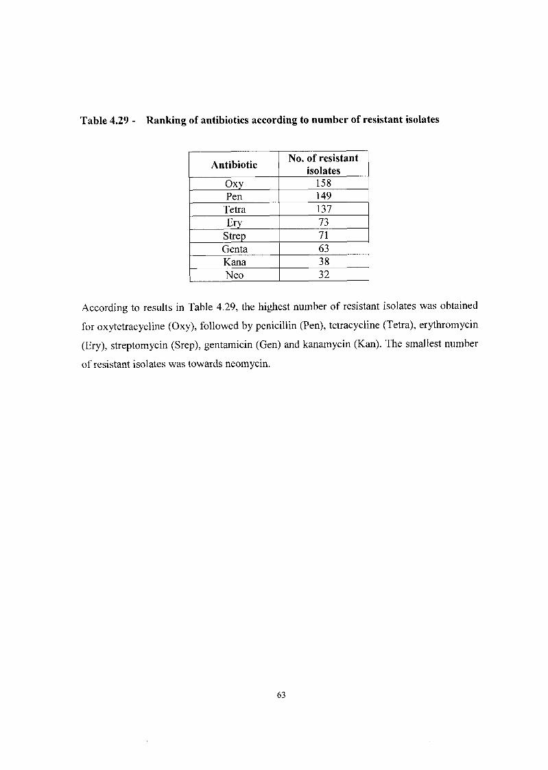
Table 4.29 - Ranking of antibiotics according to number of resistant isolates
ltes was obtai
Antibiotic
Oxv
No. of resistant isolates
158 Pen
Tetra 149 137
E ~ Y Strep
According to results in Table 4.29, the highest number of resistan
73 7 1
Genta Kana Neo
ned
63 3 8 32
for oxytetracycline (Oxy), followed by penicillin (Pen), tetracycline (Tetra), erythromycin
(Ery), streptomycin (Srep), gentamicin (Gen) and kanamycin (Kan). The smallest number
of resistant isolates was towards neomycin.

CHAPTER 5
DISCUSSION AND CONCLUSION
5.1 Isolation
Single and multiple antibiotic resistance (MAR) in microorganisms raise serious concerns
in researchers all over the world. In consequence of studies about MAR bacteria in other
countries (Newsome et al., 1987; Mattila et al., 1988; Epling and Carpenter, 1990;
Vazquez-Moreno et al., 1990; Krumperman, 1993; Salmon et al., 1995; Klein et al.,
1998; Moro et al., 1998) this investigation of MAR bacteria in pork and chicken meat as
well as chicken manure, feed and eggs was conducted.
A total of 198 organisms were isolated from various samples of chicken and pork meat,
chicken manure, chicken feed and eggs (see par. 3.2). Seventy-two of the total isolates
picked off were Salmonella species (see Table 4.1). Studies conducted by Khachatourians
(1998), showed that poultry that are given antibiotics often carry antibiotic-resistant
strains of Salmonellu or antibiotic-resistant transposons, which eventually reach humans
through the meat and eggs. It has been found repeatedly that chickens, pigs, and cattle are
frequent asymptomatic chronic Salmonella carriers and that these animals, especially
pigs, infect others en route to the abattoir as they travel in crowded and unsanitary
conditions and are stressed by such crowding and also by food and water deprivation. As
a consequence, they excrete large number of Salmonella organisms that contaminate the
carcasses and meat products: The Salmonella organisms isolated from animals and their
products show a high frequency of multiple resistance (Novick, 1978).
The rest of the total percentage of the isolates picked off during this study was
Stuphylococcus species (50), followed by the Enterobacteriaceae (42), Pseudomonas
species (20) and E. coli isolates (1 4) (see Table 4.1).
The frequency of microorganisms isolated from chicken tissues according to Vazquez-
Moreno et ul. (1990) were as followed: E. coli (80%), Staphylococcus epidermis (70%)
and Hafnia ulvei (.SO%), while Salmonella and Enterobacter agglomerans were present in

20-30% of the samples. Other microorganisms, for example Pseudomonas and
Staphylococcus aureus, were present in less than 10% of the samples. According to
Novick (1 978) during the slaughtering process, contamination of carcasses with intestinal
microorganisms cannot be prevented. Meat and meat products are often contaminated
with antibiotic-resistant E. coli and these often reach the human consumer. It was
demonstrated that 52 percent of the bovine (beef) and 83 percent of porcine (pork)
carcasses slaughtered at commercial abattoirs are contaminated with E. coli.
The highest number of isolates during this investigation was obtained from chicken feed
and manure (79 isolates, see Table 4.5). This was followed by isolates from chicken meat
(63 isolates, see Table 4.3), pork meat (53 isolates, see Table 4.4) and eggs (3 isolates).
In previous studies antibiotic resistance was investigated in 474 E. coli isolates recovered
from animal faeces (broilers, pigs, pets, bulls and horses), human faeces (patients and
healthy volunteers) and food products of animal origin. E. coli isolates (3260) recovered
from human significant infectious samples were also included. There was a high
frequency of nalidixic acid, ciprofloxacin and gentamicin resistance in E. coli isolates
from broilers (88%, 38% and 40%, respectively), and from foods (53, 13 and 17%). High
levels of resistance to trimethoprim-sulphamethoxazole and tetracycline have been found
in E. coli isolates from broilers, pigs and foods. These data raise important questions
about the potential impact of antibiotic use in animals and the possible entry of resistant
pathogens into the food chain (SaCnz et al., 2001).
5.2 Antibiotic susceptibility testing using the CDS method
5.2.1 Pseudomonas
Pseudomonas species occur as free-living bacteria or in association with plants and
animals (Atlas, 1997). Pseudomonas often possess one or more plasmids in addition to
their chromosomal genes (Atlas, 1997). In this study little resistance was observed to
kanamycin (lo%), erythromycin (lo%), neomycin (lo%), tetracycline (5%) and
streptomycin (5%) while moderate resistance was indicated to gentamicin (20%) and
oxytetracycline (25%) (see Table 4.6). From the 20 isolates of Pseudomonas, 90%
showed resistance to penicillin (see Table 4.6). Isolates from chicken and pork meat were

observed to be most resistant to penicillin while little resistance was observed to the other
antibiotics (see Table 4.7). It is clear from Table 4.7 that the highest number of resistant
Pseudomonas isolates was obtained from pork samples (meat and liver) (1 5 isolates),
followed by chicken samples (meat, stomach and liver) (9 isolates) and feed (9 isolates)
and 2 isolates from chicken manure. Research done by Vazquez-Moreno et al. (1990)
showed penicillin, streptomycin and tetracycline resistant Pseudomonas species were also
isolated from chicken tissue. They, however, found none gentamicin resistant
Pseudomonas species during their investigation.
5.2.2 Stapltylococcus
Staphylococcus aureus has been recognized to encode for a variety of antibiotic
resistance determinants (Thomson and Holding, 1986). In this study all the
Staphylococcus isolates show little resistance to kanamycin (1 2%), gentamicin (12%) and
penicillin (8%), whereas 76% showed resistance to oxytetracycline (see Table 4.8). The
chicken manure isolates (41 isolates) show multiple antibiotic resistance to all the
antibiotics except penicillin while the isolates from chicken samples (meat, stomach and
liver) (31 isolates), chicken feed (16 isolates) and pork samples (meat and liver) (34
isolates) contribute less to resistance (see Table 4.9).
Studies done by Newsome et al. (1987) showed that 67% of 160 staphylococci isolates
from beef steaks were sensitive to 13 antimicrobial agents tested. The study also revealed
that antibiotic resistance was greater for staphylococci from restructured steaks (6%) than
from conventional steaks (16%). Thirty eight percent of the restructured and 15% of the
conventional steak isolates were resistant to tetracycline.
5.2.3 Enterobacteriaceae
In the Netherlands, the emergence of resistance to the fluoroquinolone antibiotics
coincide with their increased use in human and veterinary medicine to treat infections
caused by Enterobacter and Cumpylobacter species (Khachatourians, 1998).
In this study little resistance was observed to kanamycin (14%), neomycin (21%) and
gentamicin (7%) while moderate resistance was indicated to erythromycin (39%) (see

Table 4.10). Isolates were observed to be most resistant to oxytetracycline (71%),
tetracycline (79%) and streptomycin (52%) and 100 % resistant to penicillin (see Table
4.10). Isolates of chicken samples (meat, stomach and liver) (68 isolates) show high
resistance to oxytetracycline, tetracycline and penicillin while the isolates from chicken
manure (36 isolates), chicken feed (16 isolates) pork samples (meat and liver) (34
isolates) and eggs (6 isolates) contribute less to resistance (see Table 4.11).
A study done by Newsome et al. (1987) showed that 63% of 97 Enterobacteriaceae steak
isolates were resistant to cephalothin. 10% was resistant to streptomycin and 11% was
resistant to tetracycline. Less than 10% had multiple resistance.
According to Bonfiglio and coworkers (2002), the prevalence of extended-spectrum 0- lactamase (ESBL) production by consecutive non-repeated isolates of
Enterobacteriaceae was determined over a 6-month period. A total of 801 5 strains were
isolated from ten Italian laboratories and 509 (6.3%) of these were designated ESBL
producers from the results of a double-disc synergy test. Escherichia coli was the most
isolated microorganism, followed by Klebsiella pneumoniue and Proteus mirabilis.
Providenciu stuartii (28.1%) was the most frequently isolated ESBL producer. K.
pneumoniue (38.2%) was the most represented followed by P. mirabilis (25.7%).
5.2.4 Escherichia coli
Escherichia coli is ubiquitous and is also found in the gastrointestinal tracts of humans
and warmblooded animals (Atlas, 1997). Antibiotic resistance is very common among
gut coliforms, and it is likely that everyone, well or ill, carries some resistant coliforms at
all times (Richmond, 1972). E. coli can harbour antibiotic resistant plasmids which can
spread naturally among species and can colonise the intestinal tracts of humans and other
species who live in the same environment (Feinman, 1999).
In this study the E. coli isolates shows 100% resistance to oxytetracycline, tetracycline
and penicillin and no resistance to kanamycin (see Table 4.12). Isolates of all the samples
also show resistance to erythromycin (43%) (see Table 4.13). It is clear from Table 4.13
that the highest number of resistant E. coli isolates was obtained from chicken samples

(meat, stomach and liver) (28 isolates) followed by chicken feed (13 isolates), chicken
manure (12 isolates) and pork samples (meat and liver) (8 isolates).
Moro et ul. (1 998) showed antimicrobial resistance patterns for E. coli isolated from pigs,
ranged from 5 to 10% for ampicillin and from 25 to 55% for tetracycline. According to
studies done by Vazquez-Moreno et ul. (1990) E. coli isolated from chicken carcasses
had similar multiple resistance except for lower frequency to ampicillin and gentamicin.
Levy et al. (1975) found that 78% of E. coli isolates from faecal matter of chickens was
resistant to chloramphenicol. The isolates also showed resistance to streptomycin and
sulphonamides, but not to tetracycline.
5.2.5 Salmonella
During the past decade, numerous investigations have been initiated to examine the
effects of sub-therapeutic doses of antibiotics in animal feeds on Sulmonella (Epling and
Carpenter, 1990). An earlier study (Novick, 1978) shows it is very probable that virtually
all Salmonella of farm origin are capable of infecting humans. A single specific serotype,
namely S. lyphimurium, is by far the most common cause of salmonellosis both in
animals and in man, moreover, certain other serotypes are very commonly isolated from
both man and animals. For example, in the CDC 1974 Annual Salmonella Surveillance
(CDC, 1974), it is reported that 6 of the most common human serotypes were among the
10 most common animal serotypes. Similar results have been reported for European
isolates. It has even been granted that there are some biotypes that are more commonly
isolated from chickens, others from pigs, etc and most or all strains and serotypes can
cause illness both in man and in farm animals (Novick, 1978).
In this study little resistance was observed to neomycin (1 l%), while moderate resistance
was indicated to kanamycin (33%), erythromycin (38%) and streptomycin (46%) (see
Table 4.14). Lack of resistance to neomycin, kanamycin, erythromycin and streptomycin
does not mean that the bacteria were not exposed to antibiotics at all, but may suggest
that bacterial strains have been exposed to these agents for a short while (Vazquez-
Moreno et ul., 1990). Isolates were observed to be most resistant to gentamicin (63%),
tetracycline (88%) and 99% resistance to oxytetracycline and penicillin (see Table 4.14).
Isolates of chicken samples (meat, stomach and liver) (1 13 isolates) and chicken manure

(87 isolates) show high resistance to all antibiotics except to kanamycin (33%),
erythromycin (38%) and neomycin (1 I%), while isolates from pork samples (meat and
liver) (71 isolates) and chicken feed (71 isolates) show less resistance (see Table 4.1 5).
Epling and Carpenter (1 990) found that Salmonella isolates from pork carcasses showed
resistance to penicillin (95%), trimethoprim (80%), ampicillin (80%) and tetracycline
(65%). No resistance to gentamicin and neomycin was detected. Chloramphenicol
showed a 12% resistance in all strains. Research done by Vazquez-Moreno et ul. (1 990)
showed that Salmonella species isolated from chicken tissues presented resistance mainly
to penicillin, tetracycline and streptomycin.
An epidemiological survey of antibiotic resistance in Salmonella recovered from market-
age swine at five different Texas farms showed during preliminary analysis of the first
183 samples out of approximately 400 Salmonella samples that 183 (100%) of the
Salmonellu samples were resistant to penicillin G, and 122 (66.7%) were resistant
chlortetracycline. Six (3.3%) were resistant to four antibiotics (chlortetracycline,
penicillin G , streptomycin, and sulfisoxazole), and 25 (13.7%) were resistant to three
antibiotics (chlortetracycline, penicillin G, and either streptomycin, sulfisoxazole, or
ampicillin) (Farrington et al., 1999).
5.3 Cluster analysis
MAR is the ability to resist the effects of two or more unrelated antibiotics by bacterial
strains generally containing R-plasmids. The higher the MAR index, the greater the
resistance and the lower the MAR index, the greater the susceptibility (Krumperman,
1983). This study showed that the average MAR for the different groups of organisms
isolated during the study are as follow: Pseudomonas (0.219), Staphylococcus (0.288),
Enterobacteriaceae (0.473), Escherichia coli (0.545) and Salmonella (0,582) (see Tables
4.16 - 4.24). Two isolates (P3 and P18) of Pseudomonas showed high resistance to the
antibiotics with a MAR of 0.5 and 0.875 respectively (see Table 4.16). Five isolates of
Staphylococcus aureus showed a MAR value equal or higher than 0.5: (S8 and 523 =
(0.625); S17, S19 and S47 = (0.5) (See Table 4.18). Twenty-eight isolates of
Enterohacteriaceae showed high resistance to the antibiotics with a MAR equal or higher
than 0.5. Sixteen isolates showed a value of 0.5. seven isolates showed a value of 0.624

and five isolates showed high resistance of 0.725 (see Table 4.20). Eleven isolates of
Escherichia coli showed high resistance to the antibiotics. Five isolates (C4, C5, C7, C8
and C 12) showed a MAR of 0.5. Four isolates (C2, C6, C9 and C 10) showed a MAR of
0.625, while two isolates (C13 and C 14) showed a MAR of 0.75 (see Table 4.22). Sixteen
isolates of Salmonella showed a MAR of 0.5 and 14 isolates showed a MAR of 0.625.
Nineteen isolates showed a MAR of 0.75 and 6 isolates showed a MAR of 0.875 (see
Table 4.24).
Krumperman (1983) demonstrated that the sub-therapeutic use of antibiotics in the mass
production of poultry, eggs and pork had promoted the emergence of resistance and has
maintained the prevalence of MAR E. coli in the faecal environment of these animals.
The wide use and abuse of antibiotics in human therapy has also produced MAR E. coli
in the faeces of humans as well. These practices have resulted in the coexistence of MAR
E. coli within these major reservoirs of enteric disease for humans. The consequence of
these practices may provide a fortuitous opportunity to identify E. coli contamination of
food originating from these high-risk environments by MAR indexing of E. coli isolates
obtained from food (Krumperman, 1983).
Research done by Vazquez-Moreno et al. (1990) showed that MAR was more common
than single resistance among bacteria isolated from chicken and beef tissues. Epling and
Carpenter (1990) found that multiple resistance was most often observed among
Salnzonella isolates from chickens, cattle and swine with 91% resistance to 2 or more
antibiotics. Krumperman (1 983) suggested that an unexpected increase in the MAR index
of isolates from any sample should prompt an immediate investigation even though the
number of isolates present was below the established guideline or standard. He also
suggested MAR indexing was likely to provide a useful tool for better risk assessment by
identifying contamination from high-risk environments.
Multiple drug resistance to antibacterial agents, antifungals, antivirals, antiprotozoals,
and antitumor agents has risen spectacularly in the last decade and presently threatens to
put an end to successful chemotherapy in all of the above fields. Multiple drug resistance
against chemotherapy of infections and other diseases characterized by rapid growth of
genetically versatile cells has developed in recent years. Development of carefully chosen

new drugs devising more intelligent ways of administering them, will reduce the negative
impact of multiple drug resistance, but will not abolish it (Mitcher et al., 1999).
5.4 Major clusters
From the 198 isolates, 33 clusters containing 165 isolates and 33 individuals were
obtained (see Table 4.26). Of the 165 isolates present in the major clusters, 67 of the
isolates were from the Salmonella species. Thirty-two isolates were from Staphylococcus
species, 36 isolates from the Enterobacteriuceae species, 18 from Pseudomonas species
and 12 from the Escherichia coli species (see Table 4.26).
According to Tables 4.27 and 4.28 the 33 major clusters can be divided as follows:
2 clusters (M1 and M6, represent 7 isolates) were resistant to 7 antibiotics (87.5%
resistance).
4 clusters (M2, M4, M7 and M18, represent 23 isolates) were resistant to 6 antibiotics
(75% resistance).
3 clusters (M5, M8 and M14, represent 15 isolates) were resistant to 5 antibiotics
(62.5% resistance).
8 clusters (M3, M9, M11, M15, M19, M20, M21 and M22, represent 42 isolates)
were resistant to 4 antibiotics (50% resistance).
6 clusters (M10, M12, M16, M23, M24 and M27, represent 35 isolates) were resistant
to 3 antibiotics (37.5% resistance).
6 clusters (M13, M17, M25, M28, M29 and M33, represent 22 isolates) were resistant
to 2 antibiotics (25% resistance).
3 clusters (M26, M30 and M32, represent 18 isolates) were resistant to 1 antibiotics
(12.5% resistance).
1 cluster (M3 1, represent 3 isolates) was resistant to 0 antibiotics (0% resistance).
Of the individual isolates that did not cluster (See Table 4.27), 2 was resistant to 5
antibiotics (62.5%), 2 resistant to 3 antibiotics (37.5%), 8 resistant to 2 antibiotics (25%)
and 2 was resistant to 1 antibiotic (1 2.5%).

5.5 Ranking of antibiotics
This study showed a high percentage of resistance of the isolates to oxytetracycline,
tetracycline and penicillin. Little resistance was shown to kanamycin and neomycin. Of
the 198 isolates, 158 (80%) of the isolates were resistant to oxytetracycline, 149 (75%) of
the isolates resistant to penicillin and 137 (70%) resistant to tetracycline (see Table 4.29).
Isolates showed resistance between 37% and 32% for erythromycin, streptomycin and
gentamicin (see Table 4.29). Of the 198 isolates, 38 (19%) showed resistance to
kanamycin and 32 (1 6%) showed resistance to neomycin (see Table 4.29).
Epling and Carpenter (1990) examined isolates of Salmonella from pork carcasses, for
resistance to antimicrobial agents and found that the most commonly resistance was to
penicillin (95%), trimethoprim (80.2%), ampicillin (80. I%), chloramphenicol (1 1.6%)
and tetracycline (64.5%). No resistance to gentamicin and neomycin was detected.
Khachatourians (1998) showed antibiotic resistant Salmonella isolates associated with
human infections had a 93% resistance to chlorarnphenicol, streptomycin, sulfonamides
and tetracyclines for each antibiotic respectively.
Newsome et al. (1987) determined antibiotic resistance of Staphylococcus and
Enterobacteriaceae isolated from restructured and conventional steaks. Thirty eight
percent of the restructured and 15% of the conventional steak isolates were resistant to
tetracycline. One percent of the restructured steak isolates was resistant to erythromycin,
kanamycin and penicillin. No resistance to chlorarnphenicol and neomycin was detected.
Ten percent of the Enterobacteriaceae isolates was resistant to streptomycin and 11%
was resistant to tetracycline.
Vazquez-Moreno et al. (1990) showed antibiotic resistance in various bacteria isolated
from chicken tissues. E. coli was resistant to penicillin (71%), tetracycline (71%),
streptomycin (78%) and gentamicin (2 1 %) and no resistance to chlorarnphenicol was
detected. Staphylococcus was resistant to penicillin (38%), tetracycline (38%) and
streptomycin (19%) and not resistant to chloramphenicol and gentamicin. Enterobacter
was resistant to penicillin (80%), tetracycline (70%) and streptomycin (60%) and no
resistance to chloramphenicol and gentamicin was detected. Salmonella was resistant to

penicillin (75%), tetracycline (SO%), streptomycin (38%) and chloramphenicol (50%)
and not resistant to gentamicin.
According to Geornares et al. (2001), E. coli strains from poultry carcasses were analysed
to determine their susceptibility to antimicrobial agents (chlortetracycline,
oxytetracycline and neomycin) used in the South African poultry industry. The majority
(76%) of the strains were resistant to chlortetracycline and oxytetracycline only, while
14% resistant to both tetracyclines as well as neomycin. Six percent of the isolates were
resistant to neomycin only, while the remaining 2% of the isolates were susceptible to
neomycin.
Animal-adapted bacterial strains harbouring antibiotic resistant plasmids may spread
naturally among animals and, at least transiently, can colonise the intestinal tracts of
humans and other species (Feinman, 1999). Humans may also acquire antibiotic resistant
strains from the environment through direct exposure to manure or contact with pets or
mice that are in contact with waste of farm animals. It can also spread to humans from
pets fed contaminated poultry products; by human contact with farm animals, freshwater
fish and wild animals or from animals to humans by vectors, such as flies, cockroaches
and rats (Feinrnan, 1999).
The genetic material coding for antibiotic resistance is termed the R-factor and is usually
carried on plasmids, which can be transferred between strains of the same species of
bacteria, and also between different species (Mattila et al., 1988). According to Mattila et
al. (1988) there are three mechanisms by which R-factors of animal origin could be
transferred to man. The first is the direct transmission of E. coli harbouring the R-factor
to the human gut, where it may colonise. The second is the transmission of the organism
to the human gut where it does not colonise but may transfer R-factors to resident strains
of E. coli in which they are maintained. This may also occur, especially if antibiotics are
present to provide selective pressure, a phenomenon in which the more susceptible
bacteria are destroyed and the more resistant bacteria survive (Harrison and Svec, 1998).
Epidemiological evidence indicates it must occur occasionally, and this may be all that is
required to allow the slow but steady spread of R-factors through a range of bacterial
species in the environment where the use of antibiotics is widely practised. The third
mechanism is indirect, where the organism, after leaving the host animal, may transfer R-

factors to other organisms, such as another strain of E. coli or Salmonella species which
may subsequently colonise the human gut (Mattila et al., 1988).
Epidemiological studies (Mitsuhashi, 1976) of resistant bacteria have produced the
following findings:
many resistant bacteria carry transmissible plasmids (R-factor) or nontransmissible
plasmids (r-plasmid).
there are two groups of drug resistant bacterial species, one group which easily
acquires multiple resistance and is isolated frequently from clinical specimens, and
another which is still sensitive to various antibiotics.
biochemical mechanisms of resistance in clinical isolates are mostly different from
those in resistant mutants developed in vitro and are common in many clinical
isolates which are isolated all over the world.
5.6 Conclusion
This study has revealed that bacteria are common in chicken and pork meat as well as
chicken feed and manure and the surrounding area. Isolates from these samples, tested
against 8 different antibiotics, exhibit resistance towards most of the antibiotics tested.
These antibiotics are mainly used in human therapy, but some are also used in
subtherapeutic levels in animal feed. From this survey it is evident that most of the
isolates from the samples exhibit multiple antibiotic resistance. A variety of resistance
patterns were observed when all the isolates were compared. When comparing the major
resistant patterns (33 patterns) only 1 pattern consist of 17 isolates. The rest of the
patterns contain either 14 isolates to 1 isolate. This study showed that antibiotic resistance
to the 8 antibiotics can vary from 87.5% to 0% resistance. From the total of 198 isolates,
seven isolates (3.5%) showed a MAR of 0.875. Twenty-six isolates (13.13%) with a
MAR of 0.75, twenty-seven isolates (13.6%) with a MAR of 0.625 and 40 isolates
(20.2%) have a MAR of 0.5. Thus 50.5% of the total isolates showed multiple antibiotic
resistance to 4 or more antibiotics.
The first antimicrobial agents were introduced in the 1940s and a large number of new
compounds were discovered in the following decades. Shortly after their introduction,
resistance began to emerge and over the years bacteria have acquired resistance to all

known antimicrobial agents (Aarestrup et al., 1998). Emergence of antimicrobial
resistance in bacteria from food animals may compromise the treatment of infections in
animals, but may also be of consequence for human health (Aarestrup et al., 1998). The
wide use and abuse of this wonderful drug in human therapy and subtherapeutic use of
antibiotics, has produced MAR E. coli in human faeces (Krumperman, 1983). The
bacteria became resistant and the antibiotic inadequate against the bacteria. Extensive and
sometimes careless usage of antibiotics in medicine, dentistry, and agriculture is
rendering these wonder drugs less reliable for treating important infections (Feinman,
1999). Clinicians must be educated to understand the resistance problem, accept the
importance of their role in both the problem and the solution, and resolve to use these
drugs in a far more appropriate manner. Humans have ignored clear warnings that
excessive or improper use of antibiotics would lead to a world filled with human carriers
of bacterial resistant to most known antibiotics (Harrison and Svec, 1998).
The consequence of the abuse and subtherapeutic use of antibiotics has resulted in the
coexistence of MAR E. coli within major reservoirs of enteric disease for humans. The
consequence of these practices may provide opportunity to identify E. coIi contamination
of food originating from these high-risk environments by MAR indexing of E. coli
isolates obtained from food (Krumperman, 1983).
The role of the veterinary profession in the control and prevention of disease in animals is
critical. Ideally all farms should have a veterinary health plan that is based on the results
from the good surveillance of the disease trends on the farm including antibiotic
resistance. This surveillance should also enable emerging antibiotic resistance to be
recognized. To reduce the need for antibiotics, the method of production may have to
change. Methods for rapid diagnosis must be developed to enable improved empirical
therapy of sick animals. The use of antibiotics as part of the farm veterinary health plan is
the way forward and must be an essential component of any Farm Quality Assurance
Scheme (Johnston, 200 1).
According to Novick (1978) the problem stem from the fact that the meat producers tend
to give antibiotics throughout the entire life of the animal with the consequence that the
normal (and abnormal) bacterial flora becomes highly resistant to many antibiotics
simultaneously, so that the benefits of the drugs at times of stress are drastically reduced.

The conclusion is that it is unquestionably true that many clinically effective antibiotics
have distinct growth promoting properties for a variety of animals, when fed at low levels
as part of their food, and undoubtedly the use of antibiotics for this purpose can make a
substantial difference, to the efficiency of conversion of food to edible protein. But at
whose expense? Where do we draw the line? So, in summary, all our efforts should be
directed towards reducing the selection pressure as much as possible. Only in this way
can we hope to reduce the reservoir of resistant bacteria (Richmond, 1972).

Bladsy 77

REFERENCES
AARESTRUP, F.M, BAGER, F., JENSEN, N.E., MADSEN, M., MEYLING, A. and
WEGENER, H.C. 1998. Resistance to antimicrobial agents used for animal
therapy in pathogens-, zoonotic- and indicator bacteria isolated from different food
animals in Denmark: A baseline study for the Danish integrated antimicrobial
resistance monitoring programme (DANMAP). APMIS, 106 : 745-770.
ATLAS, R.M. 1997. Principles of microbiology. 2nd ed. Boston : McGraw-Hill. 1 185 p.
BARON, E.J., PETERSON, L.R. & FINEGOLD, S.M. 1994. Bailey & Scott's
diagnostic microbiology. 9"'ed. St. Louis : Mosby. 407 p.
BELL, S.M., GATUS, B.J. & PHAM, J.N. 1999. Antibiotic susceptibility testing by
the CDS method. A concise laboratory manual. Sydney: Arthur Productions.
32 p.
BETINA, V. 1983. The chemistry and biology of antibiotics. Amsterdam : Elsevier
Scientific Publishing Company
BLACK, J.G. 1996. Microbiology principles and application. 3rd ed. London :
Prentice-Hall. 790 p.
BONFIGLIO, G., PERILLI, M., STEFANI, S., AMICOSANTE, G. & NICOLETTI,
G. 2002. Prevalence of extended spectrum p-lactamase among Enterobucteriuceue: an
Italian survey. International Journal of Antimicrobial Agents, 19 : 2 13-2 17.
BRITISH PHARMACOPOEIA see GREAT BRITIAN. GENERAL MEDICAL
COUNCIL
BROCK, T.D., MADIGAN, M.T., MARTINKO, J.M. & PARKER, J. 1994. Biology
of microrganisms. 7Ih ed. London : Prentice-Hall. 876p.
BUSH, K. & MOBASHERY, S. 1998. How p-lactamases have driven pharmaceutical
drug discovery. ( In Rosen, B. P.& Mobashery, S., eds. Resolving the antibiotic
paradox. New York : Kluwer Academic/Plenum. p.71-98. )
CDC (CENTER FOR DISEASE CONTROL). 1974. Annual Salmonella Surveillance,
CDC, Atlanta.

CHUBB, L. 2000. Research fuels anti-antibacteria debate.
[ Web: ] http://www.enn.com/extras/printer-friendly.asp?storyid= 15 1 16
[ Date of access: 18 Oct.2000 1. COOKE, E.M. 1974. Esherichiu coli and man. London : Churchill Livingstone. 82 p.
DUPONT, H.L. & STEELE, J.H. 1987. Use of antimicrobial agents in animal feeds:
Implications for human health. Reviews of Infectious Diseases, 9 : 447-449.
ELLIOT, T., HASTINGS, M. & DESSELBERGER, U. 1997. Lecture notes on
medical microbiology. 3rd ed. London : Blackwell Science. 332 p.
EPLING, L.K. & CARPENTER, A. 1990. Antibiotic resistance of Salmonella
isolated from pork carcasses in Northeast Georgia. Journal of Food Protection,
53 : 253-254.
FARRINGTON, L.A., HARVEY, R.B., BUCKLEY, S.A., STANKER, L.H & INSKIP,
P.D. 1999. A preliminary survey of antibiotic resistance of Salmonella in market-
age swine. Advances in Experimental Medicine and Biology, 473 : 29 1-297.
FEINMAN, S.E. 1999. Antibiotics in animal feed - Drug resistance revisited.
American Society Microbiolo~y News, 64 : 24-30.
FRAZIER, W.C. & WESTHOFF, D.C. 1996. Food microbiology. 4td ed. New York :
McGraw-Hill. 503 p.
GEONARAS, I., HASTINGS, J.W. & VON HOLY, A. 2001. Genotypic analysis of
Escherichia coli strains from poultry carcasses and their susceptibility to
antimicrobial agents. Applied and Environmental Microbiology, 67 : 1940- 1944.
GREAT BRITAIN. GENERAL MEDICAL COUNCIL. 1993. British Pharmacopoeia,
Vol. 1. London : Spottiswoorde. 1 128 p.
GUSTAFSON, R.H. & BOWEN, R.E. 1997. Antibiotic use in animal agriculture.
Journal of Applied Microbiolony, 83 : 53 1-541.
HARDMAN, J.G. & LIMBIRD, L.E. 1996. Goodman and Gilman's
pharmacological basis of therapeutics. 9"' ed. New York : McGraw-Hill. 11 20 p.
HARRISON, J.W. & SVEC, T. A. 1998. The beginning of the end of the antibiotic
era. Part 1. The problem : Abuse of the "miracle drugs" Quintessance International,
29 : 151-162.

HO, M-W., TRAAVIK, T., OLSVIK, O.,TAPPESER, B., HOWARD, C.V., VON
WEIZSACHAR, C. & MCGAVIN, G.C. 1998. Gene technology and gene
ecology of infectious diseases. Microbial Ecoloav in Health and Disease,
10 : 33-59.
INGRAHAM, J. & INGRAHAM, C.A. 2000. Introduction to microbiology. 2"d ed.
Belmont : Wadsworth. 570 p.
JACOB, J. 1999. What is the impact of antibiotic use in poultry. Dairy and poultry
sciences, University of Florida.
[ Web:] http://www.dps.ufl.edu/Pub/Proceeding p 98. htm# Toc4328442 1 9
[Date of access: 18 Oct 20001.
JAY, J.M. 1992. Modern food microbiology. 41h ed. New York : Chapman & Hall. 553 p.
JOHNSTON, A.M. 200 1. Animals and antibiotics. International Journal of Antimicrobial
Agents, 18 : 291-294.
KAISER, G. 1999. Isolation and identification of Enterobacteriaceae and
Pseudomonas, Part 1.
[ Web: ] http://www.cat.cc.md.us/-gkaiser/labmanua~labl2/lab 12.html
[ Date of access: 22 Aug. 2001 1. KATZUNG, B.G. & TREVOR, A.J. 1998. Examination and board review of
pharmacology. 51h ed. London : Prentice Hall. 334 p.
KHACHATOURIANS, G. 1998. Agricultural use of antibiotics and the evolution and
transfer of antibiotic-resistant bacteria. Department of Applied Microbiology and
Food Science, College of Agriculture. [ Web: ] khachatouria@,sask.usask.ca
[ Date of access: 10 Aug. 2000 1. KHAN, A.A., NAWAZ, M.S., KHAN, S.A. & CERNIGLIA, C.E. 2000. Transfer of
erythromycin resistance from poultry to human clinical strains of StaphyIococcus
uureus. Journal of Clinical Microbiology, 38 : 1832-1 838.
KLEIN, G., PACK, A. & REUTER, G. 1998. Antibiotic resistance patterns of
enterococci and occurrence of vancomycin-resistant enterococci in raw minced beef
and pork in Germany. Applied and Environmental Microbiology, 64 : 165 p.
KRUMPERMAN P. H. 1983. Multiple antibiotic resistance indexing Escherichiu coli
to identify high-risk sources of fecal contaminations of foods. Applied and

Environmental Microbioloav, 46 : 165- 170.
LANCINI, G., PARENTI, F. & GALLO, G.G. 1995. Antibiotics: A multidisciplinary
approach. New York : Plenum. 278p.
LERNER, S. A. 1998. Clinical impact of antibiotic resistance. (In Rosen, B. P. &
Mobashery, S., eds. Resolving the antibiotic paradox. New York : Kluwer
Academic/Plenum. p.7-16. )
LEVY, S.B., FITZGERALD, G.B. & MACONE, A.B. 1975. Spread of antibiotic-
resistant plasmids from chicken to chicken and from chicken to man. Nature, 260 :
40-42.
MACHALEK A.Z. 1997. Outwitting antibiotic resistant bacteria. National institute of
general medical sciences.
[Web:] http://www.nih.novlninms/news/release/altman.html [Date of access: 20
March. 19981.
MANDELL, G.L. & PETRI, W.A. 1996. Antimicrobial agents: sulfonamides,
trimethoprim-sulfamrthoxazole, quinolones, and agents for the urinary tract
infections. (In Hardman, J.G., Limbird, L.E., Molinoff, P.B., Ruddon, R.W. &
Gilman, A.G., eds. The pharmacological basis of therapeutics. New York :
McGraw-Hill. p. 1057-1 07 1 .)
MANN, J. & GRABBE, M.J.C. 1996. Bacteria and antimicrobial agents. United States :
Spektrum. 74 p.
MATTILA, T., O'BOYLE, D. & FROST, A. J. 1988. Inability to transfer antibiotic
resistance in Escherichia coli on meat surfaces. Journal of Food Science, 53 :
1309-1310.
MIMS, C.A, PLAYFAIR, J.H.L., ROITT, I.M., WAKELIN, D. & WILLIAMS, R.
1993. Medical microbiology. St. Louis : Mosby. 39.18 p.
MITCHER, L.A, SEGARAN, P.P., GENTRY, E.J. & SHANKEL, D.M. 1999.
Multiple drug resistance. Departments of Medicinal Chemistry and Molecular
Biosciences. Medical Research Reviews, 19 : 477-496.
MITSUHASHI, S. 1975. Epidemiology and genetics of aminoglycoside antibiotic
resistance in pathogenic bacteria. ( In Mitsuhashi, S., ed. Drug action and drug
resistance in bacteria. Japan : University of Tokyo Press. p. 180-204. )

MITSUHASHI, S. 1976. Genetic R Factors. ( In Hahn, F.E., ed. Antibiotics and
chemotherapy : Acquired resistance of microorganisms to chemotherapeutic
drugs. Vol. 20. Basil, Switzerland : S. Karger. p. 133-1 66. )
MORO, M.H., BERAN, G.W., HOFFMAN, L.J. & GRIFFITH, R.W. 1998. Effects of
cold stress on the antimicrobial drug resistance of Escherichiu coli of the intestinal
flora of swine. Letters of Applied Microbiology, 27 : 25 1-254.
NCCLS see NATIONAL COMMITTEE FOR CLINICAL LABORATORY
STANDARDS.
NATIONAL COMMITTEE FOR CLINICAL LABORATORY STANDARDS.
1997. Performance standards for antimicrobial disk susceptibility tests : Approved
standards. 6"' ed. Vol. 17. Pensylvania : NCCLS. 104 p.
NEU, H.C. 1976. Molecular modifications of antimicrobial agents to overcome drug
resistance. ( In Hahn, F.E., ed Antibiotics and chemotherapy : Acquired resistance
of microorganisms to chemotherapeutic drugs. Vol. 20. Basil, Switzerland : S.
Karger. p.87- 106. )
NEWSOME, R. L., LANGLOIS, B.E. & MOODY, W.G. 1987. Identification and
antibiotic resistance of Staphylococci and Enterobucteriaceae isolated from
restructured and conventional steaks. Journal of Food Science, 52 : 1 169-1 173.
NOVICK, R.P. 1978. The development and spread of antibiotic resistant bacteria as a
consequence of feeding antibiotics to livestock. Annals New York Academy of
Science, 368 : 23-55.
PELCZAR, M.J., CHAN, E.C.S. & KRIEG, N.R. 1993. Microbiology : concepts and
application. New York : McGraw-Hill. 896 p.
PETROSINO, J., CANTU, C., 111 & PALZKILL, T. 1998. P-Lactamase : protein
evolution in real time. Trends in Microbiology, 6 : 323-327.
RICHMOND, M.H. 1972. Some environmental consequences of the use of antibiotics: or
' what goes up must come down'. Journal of Applied Bacteriology, 35 : 155-1 76.
SAENZ, Y., ZARAZAGA, M., BRINAS, L., LANTERO, M., RUIZ-LARREA, F. &
TORRES, C. 2001. Antibiotic resistance in Escherichiu coli isolates obtained
from animals, food and humans in Spain. International Journal of Antimicrobial
Agents, 18 : 353-358.

SALMON, S.A, WATTS, J.L., CASE, C.A, HOFFMAN, L.J., WEGENER, H.C &
YANCEY, J.R. 1995. Comparison of MICs of ceftiofur and other antimicrobial agents
against bacterial pathogens of swine from the United States, Canada, and Denmark.
Journal of Clinical Microbiology, 33 : 24-35.
SCHLEGEL, H.G. 1992. General microbiology. 7'l' ed. Cambridge : University Press.
507-5 10.
SCHWARZ, S., CARDOSO, M. & BLOBEL, H. 1989. Plasmid-mediated
chloramphenicol resistance in Staphylococcus hycus. Journal of General
Microbiology, 135 : 3329-3336.
SCHWARZ, S. & CHASLUS-DANCLA, E. 2001. Use of antimicrobials in veterinary
medicine and mechanisms of resistance. Veterinary Research, 32 : 20 1-225.
SIROTNEK, F.M. 1976. Chromosomal mutations to drug resistance in bacteria.
( In Hahn, F.E., ed. Antibiotics and chemotherapy : Acquired resistance of
microorganisms to chemotherapeutic drugs. Vol. 20. Basil, Switzerland : S.
Karger. p.67-80.)
SABS see SUID-AFRIKAANSE BURO VIR STANDAARDE
SUID-AFRIKAANSE BURO VIR STANDAARDE. 1975a. Ondersoek vir die
aanwesigheid van lewensvatbare Esherichia coli organismes in voedselsoorte. Suid-
Afrikaanse Buro Vir Standaarde. Pretoria. (Standaardmetodes: SABS-metode 758.)
SUID-AFRIKAANSE BURO VIR STANDAARDE. 1975b. Ondersoek vir die
aanwesigheid van lewensvatbare Salmonella organismes in voedselsoorte. Suid-
Afrikaanse Buro Vir Standaarde. Pretoria. (Standaardmetodes: SABS-metode
759.)
SUID-AFRIKAANSE BURO VIR STANDAARDE. 1975c. Ondersoek vir die
aanwesigheid van lewensvatbare Staphylococcus aureus organismes in
voedselsoorte. Suid-Afrikaanse Buro Vir Standaarde. Pretoria.
( Standaardmetodes: SABS-metode 760.)
TEUBER, M. 2001. Veterinary use and antibiotic resistance. Current Opinion in
Microbiolo~y, 4 : 493-499.

THOMSON, J.K. & HOLDING, A.J. 1986. Plasmids of Staphylococcus aureus
associated with live and processed poultry. Journal of Applied Bacterioloay,
60 : 277-287.
TODAR, K. 1996a. Bacteriology 330 Lecture topics: Antimicrobial agents.
[ Web: ] http://www.bact.wisc.edu/Bact33O/lectureama [ Date of access: 20 Mrc.
1 9981.
TODAR, K. 1996b. Bacteriology 330 Lecture topics: Bacterial resistance to antibiotics
[ Web: ] http://www.bact.wisc.edu/Bact33O/lectureama [ Date of access: 20 Mrc.
19981.
TORRENCE, M.E. 2001. Activities to address antimicrobial resistance in the United
States. Preventive Veterinary Medicine, 5 1 : 37-49.
UNGEMACH, F.R. 2000. Figures on quantities of antibacterials used for different
purposes in the EU-countries and interpretation. Acto Vet Scand, 93 : 89-98.
VASQUEZ-MORENO, L., BERMDEZ, M.C., LANGURE, A.A., HIGUERA-
CIAPARA, I., DIAZ DE AGUAYO, M. & FLORES, E. 1990. Antibiotic residues
and drug resistant bacteria in beef and chicken tissues. Journal of Food
Science, 55 : 632-635.
WARD, JH. 1993. Hierarchical grouping to optimise an objective function. Journal of the
American Statistical Association, 58 : 239.
WHO see WORLD HEALTH ORGANISATION
WORLD HEALTH ORGANISATION. 1978. Surveillance for the prevention and
control of health hazards due to antibiotic-resistant enterobacteria. Technical
report series. Geneve. 54 p.

Appendix A
Table A: The source of the Pseudomonas isolates obtain from the different
samples
Isolate Source of isolation P 1 P2
Pork meat Pork meat
P3 Pork meat P4 P5
Pork meat Pork liver
P6 P7
Pork liver Pork liver
P8 Chicken meat P9 PI0
Chicken liver
Chicken stomach Chicken stomach
PI1 Chicken liver
P13 P14 PI5
1 PI8 Chicken feed
Chicken feed Chicken manure Chicken stomach
P16 P17
Pork meat -
Pork meat
P19 P20
Pork meat Chicken manure

Appendix B
Table B: The source of the Staphylococcus aureus isolates obtain from the
different samples
Isolate S1 S2
Source of isolation Chicken meat Chicken meat
S3 S4
Chicken meat Chicken meat
S5 S6
Pork liver Pork liver
S7 Pork liver S8 S9
Pork liver Chicken feed
S10 S11 S12
Chicken feed Chicken feed Chicken feed
S13 S14
Chicken manure Chicken manure
S15 S16
1 S20 Chicken manure
Chicken manure Chicken manure
S17 S18 S19
Chicken manure Chicken manure Chicken manure
S21 S22
Chicken manure Chicken manure
S23 S24
Chicken manure Chicken feed
S25 S26
Pork meat Chicken feed
S27 Pork meat S28 S29
Pork meat Chicken meat
S30 S3 1 S32
Chicken meat Pork meat Pork meat
S33 S34
Pork meat Pork meat
S35 Chicken meat S36 S37 S38
Chicken meat Chicken meat Chicken meat

Appendix B (continued) S39 S40 S41
Chicken manure Chicken stomach
Chicken feed S42 S43 S44 S45 S46 S47
Chicken feed Chicken manure Chicken manure Chicken manure
Chicken meat Chicken meat - - .
S48 S49
Pork meat Pork meat
S50 Pork meat

Appendix C
Table C: The source of the Enterobacteriaceae isolates obtain from the different
samples
Isolate E l E2
Source of isolation Pork meat
Chicken meat E3 E4
Chicken meat Chicken manure
E5 E6
Eggs Chicken manure
E7 Chicken manure E8 E9
Chicken manure Chicken feed
E l 0 E l l
Chicken feed Chicken manure
El2 El3
Chicken manure Chicken feed
E l 4 E l 5 El6
Chicken stomach Chicken stomach
Pork liver El7 Chicken feed E l 8 E l 9
Chicken stomach Pork liver
E20 E2 1
I E24 Chicken liver
Chicken stomach Pork meat
E22 E23
Pork meat Chicken stomach
E25 E26
Eggs Chicken manure
E27 Chicken liver E28 E29
Pork meat
Eggs E30 E3 1
Chicken meat Chicken meat
E32 E33 E34
Chicken meat Chicken stomach Chicken stomach
E35 E36 E37
Chicken meat Chicken meat Chicken liver
E38 Chicken liver
IV

Appendix C (continued)
I E42 Pork liver
E39 E40 E4 1
Chicken manure Chicken manure
Pork liver

Appendix D
Table D: The source of the Escherichia coli isolates obtain from the different
samples
I Isolate Source of isolation C1 C2
Chicken manure Chicken meat
C3 C4
Pork meat Chicken manure
C5 Chicken feed C6 Pork meat C7 Chicken liver
C9 C10
Chicken manure Chicken meat
C11 C12 C13
C8
Chicken liver Chicken stomach
Chicken feed - -
Chicken manure
C14 Chicken feed

Appendix E
Table E: The source of the Salmonella isolates obtain from the different samples
Isolate 1
4 Chicken liver 1
Source of isolation Chicken liver
2 3
Chicken liver Chicken liver
5 6
Pork liver Chicken meat
7 8 9
Chicken meat Pork liver
Chicken manure 10 11
15 Pork meat J
Chicken manure Chicken manure
12 13 14
Chicken manure Pork meat Pork meat
16 17
Pork meat Chicken feed
18 19
Chicken feed Chicken feed
20 Chicken feed 21 22
Chicken manure Chicken manure
23 Chicken manure 24 25
Chicken manure Chicken feed
26 2 7
Chicken feed Chicken feed
28 29 30
Chicken stomach Chicken stomach
Chicken meat 3 1 Pork meat 32 33
36 Chicken meat 1
Pork meat Chicken feed
34 35
Pork liver Chicken meat
VII
3 7 38 39
Pork meat Pork meat Pork meat

Appendix E (continued) 40 41
Pork meat Chicken feed
42 43
Chicken feed Chicken feed
44 45
Chicken meat Chicken meat
46 47
Chicken stomach Chicken stomach
48 Chicken stomach 49 50
Chicken stomach Chicken feed
51 52
Chicken feed Pork liver
53 54
1 57 I Chicken manure I
Chicken liver Chicken liver
55 56
Chicken liver Pork liver
58 59
Chicken liver Chicken feed
6 0 6 1
I 64 I Pork meat I
Chicken feed Pork meat
62 63
Chicken manure Chicken manure
65 66
Pork meat Pork meat
67 68 69
Chicken manure Chicken manure Chicken manure
70 7 1
VIII
Chicken manure Chicken manure
1 72 Chicken manure