the infrared trigemino-tectal pathway in the rattlesnake ... ean pubs/j comp neurol 191 465... ·...
TRANSCRIPT

THE JOURNAL OF COMPARATIVE NEUROLOGY 191:465-477 (1980)
The Infrared Trigemino-Tectal Pathway in the Rattlesnake and in the Python
ERIC A. NEWMAN, EDWARD R. GRUBERG AND PETER H. HARTLINE Eye Research Institute of Retinn Foudation, Boston, Massachusetts 02114 ( E A N . , P.H.H.), Research Labomtoly of Ehtronics, Massathusetts Institute of Technology, Cambridge, Massachusetts 02139 (E.R.G.)
ABSTRACT We have studied the infrared trigemino-tectal pathway of the rattlesnake (Crotalus viridis) and the python (P. reticulatus). In the rattlesnake, horseradish peroxidase (HRP) injections into the nucleus reticularis caloris (RC) result in retrograde filling of cells in the ipsilateral nucleus of the lateral descending trigeminal tract (LTTD) and in the anterograde labelling of terminal fields in the contralateral optic tectum, confirming our previous finding of an RC-tectal projection. The primary projection of the pit organ of the rattlesnake was traced by injecting cobalt chloride into the pit, demonstrating that the pit organ projects exclusively to the ipsilateral LTTD. Electrophysiological recording from single units in the RC shows that these cells respond to infrared stimulation. Taken together, these results demonstrate that the infrared pathway in the rattlesnake proceeds from the pit organ to the LTTD, to the RC, to the contralateral tectum. In contrast, HRP injection into the tectum of the python results in the retrograde filling of the large cells of the contralateral LTTD. Thus, a direct LTTD-tectal projection occurs in the python. The cells of the rattlesnake RC and the larger cells of the python LTTD stain heavily for acetylcholinesterase activity and have a similar multipolar appearance, sug- gesting that the tectal-projecting cells in the two species may have a common origin.
The infrared-sensitive pit organs of pit vi- pers (subfamily Crotalinae) and boids (family Boidae) are innervated by branches of the trigeminal nerve (Lynn, '31; Bullock and Diecke, '56; Bullock and Barrett, '68; Mole- naar, '78b). These branches project t o the ipsilateral medullary nucleus of the lateral descending trigeminal tract, (LTTD) (Mole- naar, '74, '78a; Schroeder and Loop, '76). The LTTD in turn is connected to the optic tectum; infrared-driven activity can be recorded in the frared-driven activity can be recorded in the tecta of both snake families (Hartline, '72, '74; Goris and Terashima, '73; Terashima and Gor- is, '75; Haseltine, '78).
In a previous horseradish peroxidase (HRP) study of the pit viper (Crotalus viridis) (Grub- erg et al., '79a), we found that only one cir- cumscribed nucleus in the medulla, the nucle- us reticularis caloris (RC), projects to the tectum. We proposed that the RC serves as an intermediate link between the LTTD and the tectum in this species. This hypothesis is sup- ported by the work of Stanford and Schroeder
('79), who used degeneration techniques to demonstrate that the LTTD projects to the RC but not to the tectum in the rattlesnake. This indirect projection differs from the scheme proposed by Molenaar for boids. His degener- ation study of the python (Molenaar and Fi- zaan-Oostveen, '78) indicated that the LTTD projects directly to the contralateral tectum in this species.
We now report the results of several addi- tional anatomical and histochemical experi- ments, which define the similarities and dif- ferences between the LTTD-tectal pathways in these two groups of snakes. The results support our earlier proposal that the RC func- tions as a link between the LTTD and the tectum in the rattlesnake. In addition, we have recorded the electrical activity of single RC units to determine their infrared sensitiv- ity. We have also injected HRP into the tectum of the python to study the infrared projection in this species. Preliminary results of this work have appeared elsewhere (Newman et al., '79; Gruberg et al., '79b).
0021-9967/80/1913-0465$02.60 0 1980 ALAN R. LISS, INC.

466 E.A. NEWMAN, E.R. GRUBERG, AND P.H. HARTLINE
METHODS
pacific Rattlesnakes (Crotalus viridis) 40-120 cm in length were obtained from Western Zoological Supply, Monrovia, Ca. Reticulated Pythons (P. reticulatus) 80-140 em in length were obtained from Stoneham Aquarium Sup- ply, Stoneham, Mass. Both species were main- tained at 27°C in a 12 hour-12 hour light-dark cycle.
Surgical procedures All surgical procedures were performed un-
der Metofane (Methoxyfluorane) anesthesia (Pitman-Moore, Inc.). The RC of the rattle- snake was approached by removing the inner ear structures and exposing the dorso-lateral medulla a t the level of the VIII nerve. Snakes were cooled by packing them in ice to mini- mize bleeding. Once the dorsolateral medulla was exposed and the meningial layers had been removed, the RC could be reliably located from surface landmarks. The nucleus lay ap- proximately 2 mm directly below the surface in an area just caudal to the entry of the VIII nerve root.
The tectum of one python was exposed by removing the overlying skull using a dental drill. l b o meningial membranes covering the tectum were removed.
Horseradish peroxidase injections HRP was injected into the RC of five rat-
tlesnakes and the tectum of a single python. Both iontophoresis and pressure injection of HRP was used for rattlesnake injections. The iontophoresis method is described elsewhere (Gruberg et al., '79a). HRP was pressure-in- jected from micropipettes (2-5 pm tip diame- ters), with pressure applied to a syringe con- nected to the back of the pipette. In both methods, a freshly prepared, 2Wo solution of HRP (sigma type VI) in 0.05M Tris buffer a t pH 8.6, was used. The two injection methods gave similar results but pressure injection proved more reliable. In the python, HRP was pressure injected at seven widely spaced lo- cations in one tectal lobe at depths ranging from 200 to 800 pm below the surface-the expected location of infrared afferent termi- nals (Haseltine, '78). In five rattlesnakes, HRP was injected into the RC in two closely spaced locations per animal. Electrophysiological re- cordings were first made to ascertain the lo- cation and depth of the RC in each animal. HRP filled pipettes were then lowered through the same penetration sites to the depths in- dicated by the unit recordings. Multiunit in- frared-evoked activity was recorded from
these pipettes when they were at the proper depth.
Snakes were maintained post-operatively at 27°C for times ranging from two to five days for the rattlesnakes and eight days for the python. The animals were then anesthetized, perfused first with saline and then with 2.5% glutaraldehyde and WO paraformaldehyde in pH 7.4 phosphate buffer. The brains were removed, fixed for an additional three hours, and placed overnight in phosphate buffer with 10% sucrose. On the following day, the brains were rinsed in water, quick frozen, and cut a t 40 pm in a cryostat. Sections were collected on subbed slides, thoroughly dried, and treat- ed by the blue benzidinelsodium nitroferri- cyanide method (Mesulam, '76).
Cobalt The primary afferent fibers innervating the
pit organ were labelled by direct application of CoC1, to the pit. The external cornified layer of the pit membrane was peeled away from the membrane surface. The membrane was then lacerated at several points and cov- ered with H,O. The H,O was soaked up and the external cavity of the pit filled with 300 mM CoC1,. The pit was then sealed with petroleum jelly and tape. Following survival times of six to eight days at 27"C, animals were perfused as described above. Brains were removed, treated with ammonium sulfide (2 dropdl0 ml) for 10 min, rinsed several times in H,O, frozen and cut at 40 pm in a cryostat. The sections were collected on subbed slides and processed using Timm's intensification technique (Tyrer and Bell, '74).
Acetylcholinesterase Animals were anesthetized and perfused as
above, using fixative containing 1% gluteral- dehyde and Wo paraformaldehyde buffered to pH 7.4. Brains were removed and placed over- night in phosphate buffer with lWo sucrose. Brains were then frozen and cut a t 40 pm in a cryostat. Sections were collected on subbed slides, throughly dried and stained using a procedure similar to that of Mesulam and Van Hoesen ('76). Sections were incubated in a solution containing 100 mg acetylthiocholine iodide (AcThChI), 75 mg glycine, 50 mg cupric sulfate, 410 mg sodium acetate (anhydrous), and 100 ml H,O titrated to pH 5.5 with acetic acid, until a visible white precipitate formed on the surface of the sections. Following in- cubation, the tissue was stained in 1Wo potas- sium ferricyanide for 1-5 min. Controls were

467 INFRARED PATHWAY IN R A ' M " A K E AND PYTHON
run by omitting the substrate (AcThChI) and by the addition of the specific acetylcholines- terase inhibitor BW 284Crjl. Sections were pre-incubated for 10 min in BW 284C51 and then incubated in a solution containing the substrates and the inhibitor.
The distribution of cells within the infrared nuclei in both species was studied from cresyl violet sections cut in a transverse plane at 15 CLm.
Electrophysiology and lesioning Single unit recordings were made from the
RC of the rattlesnake using metal-filled mi- cropipettes flashed with gold and plated with platinum black (Dowben and Rose, '53; Gest- land et al., '59) and with standard recording techniques (Newman and Lettvin, '78). A hot tungsten filament, gated by an electromag- netic shutter (Vincent Associates) and filtered to remove visible wavelengths (Wratten filter no. 87b) was used as a stimulus. Flux of the stimulus at the pit membrane was 3 mW/cm2. Electrolytic lesions were made to mark the location of single unit infrared activity by passing a 5 pA current through the electrode for 15-30 sec (electrode negative). Two to seven lesions were made in each of nine rat- tlesnakes. The brains were removed, fixed overnight in 1oo/o formalin in saline, dehy- drated, cleared in cedarwood oil, paraffin embedded, and cut at 15 pm. The lesions were apparent after cresyl violet staining of the sections.
RESULTS
HRP injection in rattlesnake RC We injected HRP into the nucleus RC of the
rattlesnake in order to identify its afferent input and its efferent projections. Figure 1 summarizes the projections resulting from one such HRP-RC injection. The cell groups and fiber tracts shown in the figure stained in all of our five HRP-RC injection experiments. Figure 2A shows the appearance of an HRP injection site after two days of survival. HRP injections extended over a diameter of approx- imately 250 pm. Because of the small size of the RC, the injections spread beyond the bor- der of the nucleus, generally in a dorsal direc- tion.
Cell staining HRP injection into the RC invariably led to
retrograde filling of cells in the ipsilateral LTTD (Figs. lF, 2B). Figure 2C shows the morphology of these labeled cells, which were distributed throughout the LTTD. They are
approximately 15-20 pm in diameter. In many cases, labelling was intense and spread into the dendrites. Under these conditions some labeled cells were multipolar and others bipolar. The fiber tract from these LTTD cells to the RC was clearly delineated (Figs. lE, F). It ran in a ventro-rostra1 direction from the LTTD, along the lateral surface of the medulla to the RC. The stained fibers were approxi- mately 1-2 Fm in diameter.
Additional cells were stained in the caudal end of the common descending trigeminal nu- cleus (NDs), in the region medial and posterior to the LTTD (Figs. lG, HI. A handful of cells were stained in the dorsal horn of the spinal cord (Fig. 1 I) and in a region corresponding to the motor nucleus of the trigeminal.
Fiber staining
Crossed RC-Tectal Tract (rct). Ankrograde HRP transport delineated an RC projection to the contralateral optic tectum. As illustrated in Figures 1B-E, fibers from the RC crossed the midline tranversely and collected in the ventral medulla, where they coursed forward into the tegmentum. They then coursed dorsad near the lateral surface of the tegmentum into layer 7a of Ram6n (18961, in the stratum griseum centrale, where they branched into smaller diameter segments (Fig. 2D). This pathway is identical to the RC-tectal tract we described previously (Gruberg et al., 79a) on the basis of retrograde transport of HRP fol- lowing tectal injections.
Descending Tracts. Large diameter fibers, approximately 3-7 p m in diameter, coursed dorsad and then caudad and mediad from the RC (Figs. lF, G). The fibers ended abruptly in a terminal field (CRF) immediately posterior to the L " D and ventral to the ipsilateral descending nucleus of the trigeminal (NDs) (Fig. 1G).
Cb CD CRF lttd LlTD NDs on RC rct RJ RM TeO ttd V VIII XMD
Abbreviations cerebellum dorsal horn central reticular field tractus descendens lateralis n. trigemini nucleus descendens lateralis n. trigemini nucleus descendem n. trigemini optic nerve nucleus reticularis caloris nucleus RC-tectal tract nucleus reticularis inferior lateralis nucleus reticularis medius lateralis optic tectum tractus descendens n. trigemini fiRh nerve root eighth nerve root nucleus motorius dorsalis n. vagi

468 E.A. NEWMAN, E.R. GRUBERG, AND P.H. HARTLINE
B
C
I t t d + t i d
G
H
Fig. 1. Camera lucida diagrams of the distribution of HRP-stained material aRer injection into the rattlesnake nucleus RC. A11 sections are from a two-day-survival animal. Filledcircles represent stained cells; dots and lines represent stained fibers. The injection site is shown in E. The level of each section within the brain is indicated in the inset.

INFRAFED PATHWAY IN RATTLESNAKE AND PYTHON 469
Fig. 2. Staining following HRP injection into the rattlesnake nucleus RC. A) HRP injection site in the Rc. Scale: 500 pm. B) HRP-stained cells in the LlTD (within boxed area) ipsilateral to the injection site. Section at the level of Figure 1F. Scale: 250 pm. C) Enlargement of boxed area in B, showing L " D cells. Scale: 50 pm. D) HRP-stained fibers and terminal aborizations in the intermediate layers of the tectum contralateral to injection site. Oval cells are stained erythrocytes. Stained glial cells are distributed throughout the section. Section at approximately the level of Fig. 1B. Scale: 200 pm.

470 E.A. NEWMAN, E.R. GRUBERG, AND P.H. HARTLINE
Other, thinner fibers, 1-2 pm in diameter, took a slightly more lateral route than the large diameter fibers. These fibers collected at the dorsal surface of the extreme caudal me- dulla and continued into the spinal cord (Figs.
Another discrete set of fibers coursed dorsad and slightly rostrad from the RC before cours- ing mediad through the ventral cerebellum, traversing to the opposite side of the medulla (Fig. 1C). We could not ascertain the termi- nation site of this tract from our material. They can not be true commissural fibers since no cells were stained in the contralateral RC.
No HRP-stained fibers or cell bodies were found in the diencephalon following RC injec- tions.
Cobalt Schroeder and Loop ('76) traced the projec-
tion of one of three nerve branches innervat- ing the pit organ in the rattlesnake, the su- perficial branch of the maxillary, using cobalt iontophoresis. In order to extend their results, we introduced cobalt into the terminal branch- es of the nerves within the pit membrane itself, thus simultaneously labelling all nerve branches innervating the pit. Following six to eight day survival times, the ipsilateral lat- eral trigeminal tract and nucleus showed co- balt staining. We found no evidence of a pro- jection from the pit organ to any area other than the L'ITD.
Single unit activity in the RC We recorded the electrical activity of ap-
proximately 40 single units from the nucleus RC. These units could only be driven by in- frared stimulation of the ipsilateral pit organ. They did not respond to visual, tactile, audi- tory, or vibrational stimulation. Figure 3A shows activity typical of the neurons sampled. Onset of a sustained infrared stimulus of mod- erate intensity led to a transient response, whose initial burst of spikes lasted for approx- imately 1.5 sec. Some units showed a low rate of spontaneous activity which was sensitive to the level of anesthesia.
Electrolytic lesions made following the re- cording of single unit activity allowed us to localize the recording site. These lesions (a total of 30 in nine animals) were within the RC. Figure 3B shows the lesion made follow- ing the recording illustrated in 3A.
HRP injection in the python tectum HRP injection into the tectum of the python
led to the retrograde filling of cells within the
1G-I).
A
1 I ON I
OFF
Fig. 3. Infrared response of the rattlesnake RC. A) Top trace: The electrical activity of a single RC neuron to infrared stimulation of the ipsilateral pit organ. Bottom trace: time cour~e of the infrared stimulus (2.4 sec). B) Lesion (arrow) in the nucleus RC made following the electrical recording shown in A. Approximate boundary of RC is indicated by dashed line. Nissl-stained section at a level between Figures 1E and 1F. Scale: 250 pm.
contralateral LTTD. Figure 4 shows these stained cells. They are multipolar, varying from 35 to 55 pm in diameter. They are uniformly distributed throughout the main body of the LTl'D, but are not found in the caudal portion of the nucleus, which extends into the spinal cord. The small cells within the LlTD were not stained with HRP. We found no stained cells along the ventro-lateral margin of the medulla, where the nucleus RC is located in the rattlesnake.
The tract from the L'ITD to the contralat- era1 tectum in the python coursed ventrad and

INFRARED PATHWAY IN RATIU3NAKE AND PYTHON 47 1
Fig. 4. A) Dark field photomicrograph of a transverse hemisection of the python medulla contralateral to the HRP- injected tectal lobe (dorsal upwards, midline to the left). Stained cells appear exclusively in the L"D (area outlined by broken line). Survival time: eight days. Scale: 500 pm. B> Enlargement of boxed area in A, showing LlTD cells. Small oval cells are erythrocytes. Scale: 50 pm.
mediad from the LTTD, crossing the midline in the ventral medulla. The fibers formed a discrete bundle near the lateral edge of the medulla, coursing forward into the tegmen- tum, then dorsad into the tedum. This path- way is similar to the one followed by the rattlesnake projection from LTTD to RC to contralateral tectum.
Cell morphology Figures 5A, B, and C show Nissl-stained
sections of the python LTTD, the rattlesnake RC, and the rattlesnake LTTD respectively. The python LTTD consists of small cells, ap- proximately 5-8 pm in diameter, and large, multipolar cells, approximately 20-30 pm in diameter. The LTTD cells stained following tectal-HRP injections are presumably from the large cell population. We attribute the difference in size between the HRP-stained cells and the Nissl-stained cells to differences in tissue treatment. The large cells do not occur in the caudal tail of the nucleus.
The rattlesnake RC consists of a population of multipolar cells approximately 25-45 pm in diameter. It is these cells that stain follow- ing HRP injections into the rattlesnake tec-
tum. The rattlesnake LTTD consists of a pop- ulation of small cells, approximately 5-8 pm in diameter, similar to the small cells of the python LTTD. In addition, there is a popula- tion of medium-sized cells, approximately 15-25 pm in diameter.
Acetylcholinesterase activity We investigated the distribution of acetyl-
cholinesterase activity in the infrared system of both the python and the rattlesnake. In the python, the large, multipolar cells of the LTTD stained heavily for acetylcholinesterase activ- ity (Fig. 6A). The noncellular background of the LTTD stained lightly as well. It is difficult to determine whether the small cells of the LTTD stain because of this background activ- ity.
The LTTD of the rattlesnake showed a sim- ilar light stain extending over its entire ex- tent. The medium-sized cells of the LTTD were lightly stained. The cells of the rattlesnake RC (Fig. 6B) stained heavily for cholinesterase activity; they stood out clearly against an unstained fiber tract which passes through the area.

472

INFRARED PATHWAY IN RA’ITLESNAKE AND PYTHON
Fig. 6. Distribution of acetylcholinesterase activity in the LlTD of the python (A) and the nucleus RC of the rattlesnake (B). Arrows indicate some of the stained cells. Both scales: 50 pm.
473
474 E.A. NEWMAN, E.R. GRUBERG, AND P.H. HARTLINE
Control experiments verified the specificity of the cholinesterase stain in both species. Removal of the substrate from the incubation solution eliminated all staining. The specific acetylcholinesterase inhibitor BW 284C51, when applied to the incubation solution, sig- nificantly reduced the degree of staining in the L'ITD of both species and in the rattles- nake RC. Background as well as cellular staining was reduced by these procedures.
We also examined the tegmental area of the mesencephalon for acetylcholinesterase activ- ity. A continuous band of cellular and noncel- lular tissue extending from the postero-lateral tegmental nucleus (PLT) to the dorso-lateral tegmental nucleus (DLT) exhibited heavy cho- linesterase activity. This pattern of activity, and the connectivity and location of these nuclei (Gruberg et al., '79a), suggest that they are both a part of the nucleus isthmi. In our previous paper (Gruberg et al., '79a), we showed that the PLT projects bilaterally to the optic tectum, with its contralateral projec- tion coursing forward into the diencephalon and crossing a t the supraoptic decussation. The nucleus isthmi in both the frog (Shen et al., '55) and the pigeon (Hunt et al., '77) shows heavy acetylcholinesterase activity.
DISCUSSION
Infrared pathway in rattlesnake The central question addressed in this paper
concerns the similarities and differences be- tween the infrared LTTD-tectal pathways in pit viper and boid snakes. In our previous paper (Gruberg et al., '79a) we investigated the connections of the optic tectum of the rattlesnake using tectal HRP injections. Re- trograde HRP transport demonstrated that only one cell group in the medulla, the nucleus RC, projected to the tectum; no stained cells were found in the LTTD following HRP tectal injection. We proposed that, in the rattles- nake, the RC serves as an intermediate link between the LTTD and the tectum.
Support for this suggestion has recently come from Stanford and Schroeder ('791, who demonstrated that the LTTD of the rattles- nake projects to the RC. Following small suc- tion lesions of the LTTD, silver staining re- vealed a tract of degenerating fibers that exit a t the lateral boundary of the LTTD and travel rostrally along the lateral medulla to the region we identified as the RC.
The results presented here confirm our pre- vious suggestion of this indirect trigemino- tecal pathway. Retrograde transport of HRP
from the RC results in filling of cells in the LTTD. The pathway from the LTTD to the RC, marked by HRP-stained axons, corre- sponds to the route described by Stanford and Schroeder ('79). Anterograde HRP transport from the RC results in filling of terminal arborizations in the stratum griseum centrale of the tectum (Fig. 2D), confirming our pre- vious findings (Gruberg et al., '79a).
Kass et al., ('78), using anatomical and physiological techniques, localized infrared re- sponses in the optic tectum of the rattlesnake to the stratum griseum centrale, primarily in layer 7a of Ram6n (1896). This corresponds closely to the localization of terminal arbori- zations of RC cells in the tectum which are, as we report here, confined primarily to layer 7a (Figs. lA, B and 2D).
The electrical recording and lesioning ex- periments presented here demonstrate that the RC is primarily an infrared sensory nu- cleus. RC units respond to low level infrared stimuli, but are not driven by stimuli from other modalities. In addition, the duration of the transient response of RC neurons is inter- mediate between the longer responses of LTTD neurons (Stanford and Hartline, '78) and the shorter responses of tectal units (un- published observations). These physiological results are consistent with the anatomical connections of the RC.
Our anatomical results, summarized in Fig- ure 7A, suggest that the infrared pathway in the rattlesnake proceeds first from the LTTD to the RC and then from the RC to the tectum. The HRP experiments do not, by themselves, rule out the existence of additional neural circuitry. Some primary trigeminal fibers from the pit organ could, in a parallel pathway, project directly to the RC. This is unlikely, since our cobalt experiments and those of Schroeder and Loop ('76) identified only LTTD projections of the trigeminal fibers innervat- ing the pit organ.
Infrared pathway in python On the basis of a degeneration study, Mo-
lenaar and Fizaan-Oostveen ('78) reported that the LTTD of the python projects directly to the tectum. Our HRP results from python confirm this finding: tectal HRP injections in the python lead to retrograde filling of cells in the LTTD (Fig. 4). Examination of Nissl sec- tions of the python did not reveal a cell group corresponding to the nucleus RC of the rat- tlesnake. Nor did we find any evidence of a projection from any other cells in the medulla to the tectum. Figure 7B summarizes the
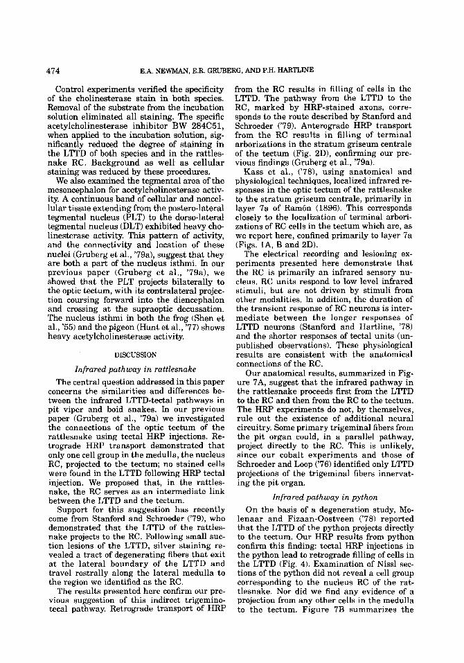
INFRARED PATHWAY IN RAl"AKJ3 AND PYTHON 475
n
RATTLESNAKE PYTHON
Fig. 7. The infrared pathway from the pit organ to the tectum. A) S y of pathway in the rattlesnake. B) Proposed pathway in the python. Unlike the rattlesnake, the python has multiple labial pits, only two of which are shown here. The interneuron within the python LlTD (open circle) is hypthtical and has not been demonstrated experimentally.
proposed infrared pathway in the python. Nissl-stained material reveals that the py-
thon LTTD is composed primarily of two types of cells: small neurons and large, multipolar neurons. Our observations confirm Molenaar's findings ('78a) that the large cells are confined to the main corpus of the LTTD. They do not extend into the caudal region of the nucleus. We find that the large LTTD cells that are stained following tectal HRP injections are also confined to the main body of the nucleus. This correspondence adds confidence to our identification of the large, multipolar neurons seen in Nissl sections as the ones that project to the tectum.
Comparison of infrared tectal-afferent cells Our results demonstrate that the trigemino-
tectal pathway of the rattlesnake and the python differ qualitatively in their connectiv- ity. The two species belong to families that are distantly related phylogenetically. As Bullock and Barrett ('68) suggest, the infrared systems of these two species may have evolved inde- pendently. However, the two species show many similarities in their infrared systems. Both families have a specialized nucleus of the spinal trigeminal system, the LTTD. This nucleus is not apparent in a snake species that does not have a specialized infrared sense (Schroeder and Loop, '76). In addition, pit vipers and boid snakes have an important infrared projection to the optic tectum which
is organized in a spatiotopic representation of the external world (Hartline et al., '78; Has- eltine et al., '77). Yet, within the medulla, the location of the cells that project to the tectum differs in the two families of snakes. In the rattlesnake, these cells lie in the RC. In the python they are located in the LTTD.
In spite of this difference, crotaline and boid infrared cells that project to the tectum share several morphological and histochemical fea- tures: 11, the cells have a similar multipolar appearance in both species; 2), both are large in size: approximately 25-45 pm in diameter in the rattlesnake and approximately 20-30 pm in diameter in the python; 31, the cells stain heavily for acetylcholinesterase activity in both species; 4), the large cells in both species share the same functional connectivi- ty, receiving infrared input and projecting to the contralateral optic tectum.
The similarities suggest that the infrared pathways in these two species may be closely related, differing only in that the tectal affer- ents form a distinct nucleus in the rattlesnake but not in the python. The tectal afferent cells in both species might have a common origin and share the same pattern of connectivity. In the python, the large cells of the LTTD would function as relay neurons equivalent to the RC cells of the rattlesnake. Under this hy- pothesis, these neurons should receive input solely from other cells in the LTTD. Figure 7B illustrates this proposed circuitry.

476 E.A. NEWMAN, E.R. GRUBERG. AND P.H. HARTLINE
Parallels of the infrared pathway in mammals
Thermosensitive single unit activity has been recorded from the marginal layer of the trigeminal subnucleus caudalis in several mammalian species, including cat (Dostrovsky and Hellon, '78; Burton et al., '79) and rat and rabbit (Dickenson et al., '79). The location of these mammalian neurons suggests that the LTTD of pit vipers and boids, which directly overlies the caudal portion of the spinal tri- geminal nucleus, may be derived from the marginal zone of the reptilian homologue of the nucleus caudalis. This suggestion is strengthened by the anatomical findings of Meszler ('751, which show that the ultrastruc- ture of the LTTD of the pit viper closely resembles that of the mammalian nucleus caudalis.
The nucleus caudalis has been shown to project to the ipsilateral lateral reticular for- mation in a number of mammalian species. (Carpenter and Hanna, '61; Stewart and King, '63; Dunn and Matzke, '68; Roberts and Matzke, '71; Tiwara and King, '74). In the cat, this projection originates from the marginal, thermosensitive zone of the nucleus (Burton et al., '79). In addition, Tiwara and King ('74) find that the lateral reticular formation in the squirrel monkey sends a small projection to the contralateral superior colliculus. Thus, in some mammals, there is an area in the lateral reticular formation that receives trigeminal input; a similar region, in other mammals, projects to the superior colliculus. This sug- gests that the LTTD-RC-tectal pathway of the pit viper may be homologous to a trigemino- reticular formation-collicular pathway i n mammals. A direct projection from the nucle- us caudalis to the superior colliculus has not been observed in mammals.
In summary, we have shown that the in- frared trigemino-tectal pathways of the rat- tlesnake and the python differ qualitatively in their organization. In the rattlesnake, the pathway proceeds from the LTTD to the RC to the tectum. In the python, a direct LTTD- tectal projection occurs. The cells that project to the tectum in these two species are of similar size and shape and both exhibit cho- linesterase activity. In spite of their disparate locations, they may have a common origin.
ACKNOWLEDGMENTS
We thank Cliff Ragsdale for helpful sugges- tions concerning cholinesterase staining and Janice Pelletier for reviewing the manuscript.
This research was supported in part by the National Institutes of Health (TO1 EY00090, "32 EY07028) and by a grant from the Bell Telephone Laboratories, Inc.
LITERATURE CITED Bullock, T.H., and R. Barrett (1968) Radiant heat recep-
tion in snakes. Commun. Behav. Bid. A. 2:19-29. Bullock, T.H., and F.D.J. Diecke (1956) Properties of an
infrared receptor. J . Physiol. 234:47-87. Burton, H., A.D. Craig, J r . , D.A. Poulos, and J.T.
Molt (1979) Efferent projections from temperature sensitive recording loci within the marginal mne of the nucleus caudalis of the spinal trigeminal complex in the cat. J. Comp. Neurol. 283:753-778.
Carpenter, M.B., and G.R. Hanna (1961) Fiber projec- tions from the spinal trigeminal nucleus in the cat. J. Comp. Neurol., 127rl17-132.
Dickenson, AH., R.F. Hellon, and D.C.M. Taylor (1979) Facial thermal input to the trigeminal spinal nucleus of rabbits and rats. J . Comp. Neurol. 185:203-210.
Dostrovsky, J.O., and R.F. Hellon (1978) The represen- tation of facial temperature in the caudal trigeminal nucleus of the cat. J . Physiol. 277329-47.
Dowben, R.M., and J.E. Rose (1953) A metal-filled mi- croelectrode. Science 218:2224.
Dunn, J., and H.A. Matzke (1968) Efferent fiber connec- tions of the marmoset (Oedipomidus oedipus) trigeminal nucleus caudalis. J . Comp. Neurol. 133:429-438.
Gesteland, R.C., B. Howland, J.Y. Lettvin, and W.H. Pitts (1959) Comments on microelectrcdes. Proc. Inst. Radio Eng. 47:1856-1862.
Gruberg, E.R., E. Kicliter, E.A. Newman, L. Kass, and P.H. Hartline (1979a) Connections of the tectum of the rat- tlesnake Crotalus viridrs: An HRP study. J . Comp. Neu- ral. 288: 31-42.
Grubrg, EX, E A Newman, and PH. Hadine ('a) The python and the rattlesnake: a comparison of the infrared trigemino-tectal pathways using horseradish peroxidase and cholinesterase techniques. SOC. Neurosci. Abstr. 5: 708.
Hartline, P.H. (1972) Responses of tectal units to in- frared stimuli in rattlesnakes. Soc. Neurosci. Abstr.. 2nd Annual Meeting, p. 74.
Hartline, P.H. (1974) Thennoreception in snakes. In A. Fessard (4.): Handbook of Sensory Physiology. Berlin: Springer-Verlag. Vol. IIU3 pp. 297-312.
Hartline, P.H., L. Kass, and M.S. Loop (1978) Merging of modalities in the optic tedum: infrared and visual interaction in rattlesnakes. Science 299: 1225-1229.
Haseltine, E.C. (1978) Infrared and Visual Organization of the Tectum of Boid Snakes. Doctoral Dissertation, Indiana University.
Haseline, E., L. Kass, and P.H. Hartline (1977) Infrared and visual organization of the tectum of h i d snakes. Soc. Neurosci. Abstr. 3:90.
Hunt, S.P., P. Streit, H. Kunzle, and M. Cfienod (1977) Characterization of the pigeon istbmo- tectal pathway by selective uptake and retrograde move- ment of radioactive compounds and by horseradish per- oxidase labeling. Brain Res. 229:197-212. b, L., M.S Lmp, a d PH. Hatthe (1978) Anatanical
and physiological localization of visual and infrared cell layers in tectum of pit vipers. J. Comp. Neurol., 182r811-820.
Lynn, W.G. (1931) Structure and function of the facial pit of the pit viper. Am. J. Anat. 49:97-139.
Mesulam, M.-M (1976) The blue reaction product in hor- seradish peroxidase neurochemistry. J . Histochem. Cy- &hem 24: 1273-1280.
Mesulam, M.-M., and G.W. Van Hoesen (1976) A c e t y l ~ b

INFRARED PATHWAY IN RA”LESNAKE AND PYTHON 477
linesteraserich projections from the basal forebrain of the Rhesus monkey to nfoo3ltex Brain Res. 1@:152-157.
Meszler, R.M. (1975) Fine structure of the lateral de- scending nucleus of the pit viper Agkistrodon p. pisciuo- rm. Anat. Rec. 181:426.
Mole-, G.J. (1974) An additional trigeminal system in certain snakes possessing infrared receptors. Brain Re. 78:34&344.
Mole-, G.J. (1978a) The sensory t r igemid system of a snake in the possession of infrared receptors. I. The sensory trigeminal nuclei. J. b p . Neurol. 179:1%136.
Mole-, G.J. (1978b) The sensory trigeminal system of a snake in the possession of infrared receptors. II. The central projection of the trigeminal nerve. J. Comp. Neurul., 179: 137-152.
Molenaar, G.J., and J.L.F.P. Fizaan-Oostveen (1978) Sec- ondary projections from the lateral descending and com- mon sensory triganid nuclei. In G.J. Mole- (ed.): J n f r d Sensitivity and the Organizaton of the Sensory Trigeminal System. Monograph Utrecht, pp. 129-155.
Newman, EA., E R Grubq, and PH. Hartline (1979) A central pathway of the infrared system of the rattlesnake Crotalus uiridis. Anat. Rec. 193:636637.
Newman, E.A., and J.Y. Lettvin (1978) Relation of the e-wave to ganglion cell activity and rod responses in the frog. Vision Res. 18:1181-1188.
Rambn, P. (1896) Estructura del encefalo del camaleon. Rev. Trimest. Micrograf. 1:4682.
Roberts, P.A., and HA. Matzke (19711 Projections of the subnucleus caudalis of the trigeminal nucleus in the sheep. J . Comp. Neurol., 141:273-282.
Schroeder, D.M., and M.S. Loop (1976) Trigeminal pro- jections in snakes possessing infrared sensitivity. J. Comp. Neurol. 1691.1-14.
Shen, S.C., P. Greenfield, and E.J. Boell (1955) The dis- tribution of cholinesterase in the frog brain. J . Comp. Neurol. 102;717-743.
Stanford, L.R., and P.H. Hartline (1978) Infrared re- sponses of neurons in the lateral descending nucleus of the trigeminal system in the rattlesnake, CrwhZus uiri- dis. Soc. Neurosci. Abstr. 4:102.
Stanford, L.R., and D.M. Schroeder (1979) An additional nucleus as part of the ascending infrared system between the lateral descending nucleus and the optic tectum of the rattlesnake, Crotalus. Anat. Rec. 193:692-693.
Stewart, W.A., and R.B. King (1963) Fiber projections from the nucleus caudalis of the spinal trigeminal nucle- us. J . Comp. Neurol. 121:271-282.
Terashima, S., and R.C. Goris (1975) Tectal organization of pit viper infrared reception. Brain Res. 83:490-494.
Tiwara, R.K., and R.B. King (1974) Fiber projections from trigeminal nucleus caudalis in primate (squirrel monkey and baboon). J . Comp. Neurol. 158:191-206.
Tyrer, N.M., and E.M. Bell (1974) The intensification of cobalt-filled neurone profiles using a modification of Timm’s sulphide-silver method. Brain Res. 73: 151-155.