the journal is index ries /abstract - international journal of life … · · 2015-04-16branches...
TRANSCRIPT

A E y
y
We publish rese
Life Sciences
Branches : Agricultural Science, Bioch
Ecology, Endocrinology, Entomology,
Proteomics, mmunobiology, Molecula
Physiology, Psychology, Veterinary Sc
Pharmaceutical Sciences
Branches : BioTechnology, Clinical an
International Regulatory Affairs, Med
Pharmaceutics, Pharmacology & Toxi
Pharmaceutical Engineering, Pharmac
Pharmacoinformatics, Pharmaceutica
The journal is index
Impact factor* 0.672
A E y
y
research/review article in the following subjects
iochemistry, Biology, Bioinformatics, Botany, Cytology, Cell b
ogy, Environmental Sciences, Food science and Technology, G
ular biology, Marine Science, Microbiology, Neurobiology, Pa
ry Science, Zoology
l and Hospital pharmacy, Herbal technology, Industrial Pharm
Medicine, Neuroscience, Novel drug delivery system, Nanotec
Toxicology, Pharmacognosy & Phytochemistry, Pharmacy pra
rmaceutical Management, Pharmaceutical Analysis, Pharmace
tical Technology, Quality Control & Assurance, Validation Tec
dexed in the following university /libraries
And Mo
A E y
y .
ell biology, Chemistry,
y, Genetics, Genomics &
y, Pathology, Physics,
harmacy, Immunology,
otechnology,
y practice,
maceutical Chemistry,
n Techniques
ries /abstract
More ......

IJLPR would take care in making your article published without
delay with your cooperation. IJLPR hopes that Researchers,
Research scholars, Academician, Industrialists, Consultancy etc.
would make use of this journal publication for the development of
science and technology.
*Kindly visit Instruction to authors available at www.ijlpr.com
for submission of manuscript for publication.
Any feed back / querry kindly email to
or you can call +91 9908947749 /+91 9676175127 / 9676175127

Research Article ISSN 2250-0480 VOL 5/ ISSUE 2/APRIL 2015
L - 21
Life Science Ecology
BIOCHEMICAL PROFILE AND ANTIMICROBIAL ACTIVITY OF A CYANOBACTERIUM,
SCYTONEMA TOLYPOTHRICHOIDES ISOLATED FROM ACIDIC RICE FIELD SOIL OF CACHAR
DISTRICT (ASSAM), INDIA
SHOUBHONIK DEB1, JAYASHREE ROUT 1*, MAHUYA SENGUPTA 2
AND BISWAJIT CHAKRABORTY2
1 Department of Ecology and Environmental Science, Assam University, Silchar-788011, Assam, India.
2 Department of Biotechnology, Assam University, Silchar, Silchar-788011, Assam, India
ABSTRACT
Scytonema tolypothrichoides, a filamentous cyanobacterium, has been isolated from acidic rice field soil from Dholai area of Cachar district in Assam, North-East India. The growth rate determination and biochemical analysis of chlorophyll a (Chl-a), total carotenoid content (TCC), phycobiliproteins, total proteins (PRT), total carbohydrates (CHO), and exopolysaccharides (EPS) were carried out. The algal strain had a higher phycocyanin content than those of other pigments. The nitrogen fixation potential of the alga has also been ascertained. Antimicrobial studies against some selected microbial strains revealed moderate activity. KEY WORDS: Scytonema tolypothrichoides, Rice field, Exopolysaccharides, Antimicrobial activity
1. INTRODUCTION
Cyanobacterial species occur in ecologically specific and geographically distinct habitats. Most species of cyanobacteria have specific ecological demands. Numerous species of the genera Scytonema are generally found to grow in ecologically distinct habitats of tropical ecosystem such as, lateritic soils, dripping rocks and aquatic bodies. The Scytonema group are the prominent components of microflora of tropical and subtropical soils, but their diversity and taxonomic classification are still less understood (Komárek et al., 2013). They studied the phenotypic characteristics of fourteen morphotypes of Scytonema
isolated from the microvegetation of lateritic, forest soils and stony substrates of South East Brazil. This heterocystous cyanobacterium has been found to enhance rice field soil fertility by fixing atmospheric nitrogen (Selvi and Sivakumar, 2012). Scytonema
javanicum played a vital role in crust formation and maintenance of productivity (Metting 1981). The species, Scytonema stuposum, in particular, forms bluish soil crusts in rice fields suggesting their role in
soil surface stabilization, moisture retention, providing a suitable habitat for growth of higher plants (Sethi et al., 2012). Diversity of cyanobacteria belonging to Scytonemataceae family growing in alkaline rice field soils of Tamilnadu state, India has been documented by Madhumathi et al., (2012). Hazarika et al., (2012) reported five different species of Scytonema from the acidic rice fields of the upper Brahmaputra Valley(Assam), India. Species like Scytonema bohneri, Scytonema hofmanni and Scytonema simplex were found to be common in the rice fields of Bongaigaon district of Assam (Das and Sarma, 2010). Cyanobacteria from some rice field soil of Cachar district (Assam) are also documented (Rout and Dey, 1999). Several reviews (Geitler, 1932; Desikachary, 1959; Starmach, 1966; Bourrelly, 1970; and Komárek and Anagnostidis 1989) addressed the traditional taxonomic description of the genus Scytonema while a modern approach based on polyphasic characterization has recently gained popularity. The approach deals with various aspects

Research Article ISSN 2250-0480 VOL 5/ ISSUE 2/APRIL 2015
L - 22
Life Science Ecology
of their biochemistry such as chlorophyll-a, phycocyanin, phycoerithrin, carotenoids, proteins, carbohydrates, exopolysaccharides, fatty acids and DNA profiling as more reliable tools for characterization of cyanobacteria (Sfriso et al., 2014). Besides diversity and their role in improving soil fertility, cyanobacteria, as a source of pharmacologically active compounds, has attracted immense interest (Kumar et al., 2010; Sethubathi and Prab , 2010; Battu et al., 2011 and Mhadhebi et al., 2012). Proteau et al., (1993) reported scytonemin, a predominantly UV-A-photoprotective pigment from Scytonema javanicum. Kreitlow et al., (1999) investigated the hydrophilic and lipophilic extracts of cyanobacterial strains for antibiotic activities. Cachar district located in the state of Assam, India is characterized by diverse habitats with potential for a wide range of cyanobacteria which remained virtually unexplored. Accordingly the present research focuses on the isolation and validation of one strain of Scytonema from acidic rice field soil of Dholai sub-division in the district through morphological and biochemical studies.
2. MATERIALS AND METHODS
2.1 Study site Cachar district in Southern Assam lies between latitude 90.44°E and longitude 20.04° N encompassing an area of 3786 sq. km. The soil samples were collected from rice field of Dholai area (N24˚ 35'22.2" E 92˚ 50'57.4") having an average pH of 6.4 with a clay loam texture. Details of the geographical location of study sites have been mentioned in Figure1.
2.2 Isolation of the strains from soil samples Dilution plate method (Lukešová, 1993) was followed for isolation of strain from 10g of rice field soil. 1mL aliquots of soil suspensions were spread on solidified (1.5%) BG-110 media (Rippka et al., 1979). Plates were incubated at 24±1 °C for a 16:8 (light: dark) photoperiod under white fluorescent light having intensity of 2000-3000 lx. Morphological observations such as cell shape, length and breadth of intercalary cells and heterocysts were taken into account and taxonomically identified on the basis of their cell or colony morphology (Desikachary, 1959). Repeated measurements were obtained from different cells and heterocysts to ensure accuracy (Singh et al., 2008).
Figure 1
Map showing the location of study sites in Cachar district.

Research Article ISSN 2250-0480 VOL 5/ ISSUE 2/APRIL 2015
L - 23
Life Science Ecology
2.3 Cyanobacterial growth measurement Cyanobacterial growth was monitored by the method of Sfriso et al., (2014). A total of 8 culture tubes, containing 9 mL aliquots of sterile BG-110 liquid medium were inoculated with 1mL stock culture of the isolated strain. Each tube was incubated for 8 days on the culture rack, with a light exposition of 2000-3000 lx, in light/dark cycles of 16:8 hours at an average room temperature of 24 °C, with proper shaking. At 5 days time interval during growth, specific amount of culture was withdrawn from each tube and were used for quantification of chlorophyll-a(Chl-a), total carotenoid contents(TCC) and exopolysaccharides (EPS), total proteins(PRT) and total carbohydrates(CHO) having three biological replicates for analyses, with eight points to observe. The biochemical parameters, Chl-a, TCC and EPS were estimated on the same day of sample withdrawal while the remaining amount of culture samples were kept frozen for total protein and total carbohydrate quantification and analyzed together at the end of 40th day. Chlorophyll a values were obtained according to Parsons and Strickland (1965). Specific growth rate and generation time were ascertained as per Levasseur et al., (1993). Total carotenoid contents (TCC) were estimated following Parsons et al., (1984). Phycobiliproteins of the dried biomass of S. tolypothrichoides were analyzed during the stationary growth phase and were extracted in 0.05M phosphate buffer (pH 6.8) by subjecting them to repeated freezing and thawing for 48h with a 24h interval (Moraes et al., 2011) and evaluated according to Bennet and Bogorad (1973). The EPS fractions of exopolymeric substances were extracted by the method of Underwood et al., (1995) and quantified as carbohydrate. Both total carbohydrates of the wet biomass and the carbohydrate (EPS fractions) were determined by phenol-sulfuric acid method (Dubois et al., 1956). Total soluble protein was estimated by a modified Lowry method (Herbert et al., 1971). 2.4 Acetylene reduction activity (ARA) The gas chromatographic quantification of ethylene formed (acetylene reduction activity, ARA) was utilized as an index of nitrogen fixation, which was estimated by the method given by Prasanna et al., (2003) in gas chromatograph (Bruker, model CDS 2.0). Commercially available standard ethylene was utilized for quantification and test tubes with an
equivalent volume of water served as controls. The ARA values presented is means of triplicate measurements and is expressed as nmoles of ethylene per mg chlorophyll per hour.
2.5 Antimicrobial activity A 100ml of cyanobacterial culture was harvested at stationary phase (after 35 days) by centrifugation at 5000 rpm. The aqueous supernatant was collected in vials and the pellet extracted with 10 ml of methanol. The culture supernatants and solvent extracts were stored at 4°C for further studies. Disc diffusion method was used to determine antibacterial activity (Nagi et al., 2010). Sterile discs (6 mm, Himedia) were soaked in 500µl methanolic extracts and supernatant solution of cyanobacteria. The microbial strains, Staphylococcus aureus (KT68-07036), Escherichia coli (KT68-21103), were obtained from Bangalore Genei, Bangalore, India. The suspension of bacteria culture was prepared according to the MacFarland standard 0.5 and layered onto the Nutrient agar plates to produce the bacteria field. The sterile discs containing the extract and supernatant were placed on the bacteria field by a sterile forceps. Distilled water and methanol were used as negative control while commercial antibiotic disc of ciprofloxacin was used as a positive control. Finally, the plates were incubated at 37°C and the zone of inhibitions was observed after 24-48 hours. The extract showing positive activity in disc diffusion experiment was chosen for determination of their MIC (minimum inhibitory concentration) by using a tube dilution technique. These tests were carried out to determine the lowest concentration of methanolic extracts of the cyanobacterium that showed bacteriostatic effect.
2.6 Statistical analysis Correlation coefficients between the biochemical attributes of the cyanobacterial strain was analyzed by determining their Spearman’s correlation coefficients using SPSS(version 19)software.
2. RESULTS AND DISCUSSION
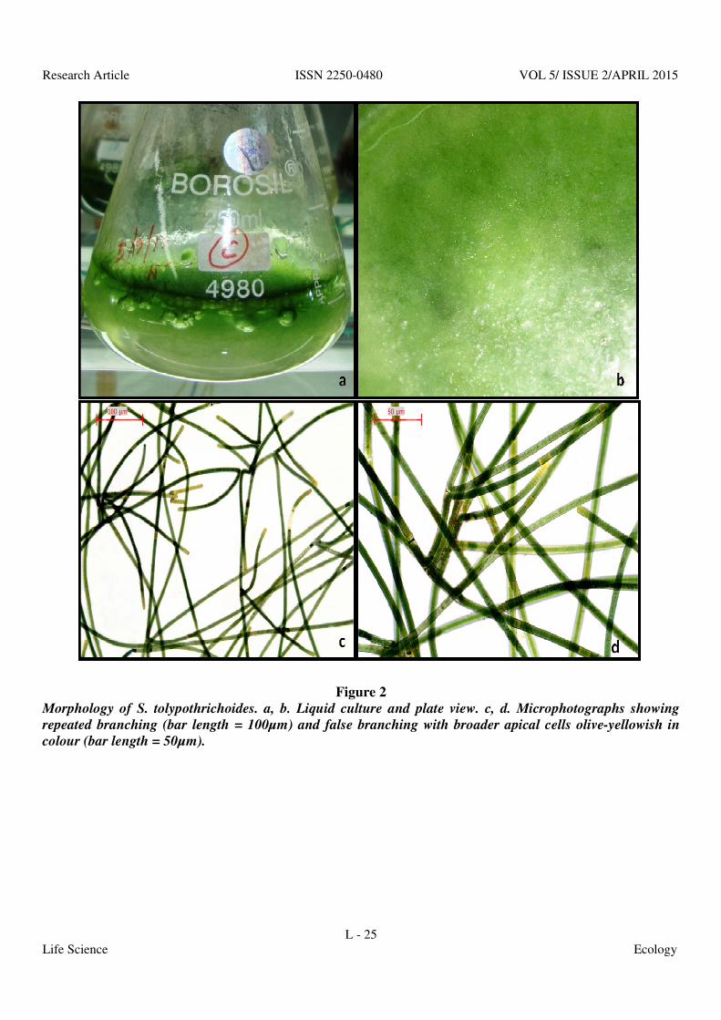
3.1 Morphology of Scytonema tolypothrichoides The morphology and distinctive features of Scytonema tolypothrichoides was observed with initial dark-green thallus development. The bunche

Research Article ISSN 2250-0480 VOL 5/ ISSUE 2/APRIL 2015
L - 24
Life Science Ecology
later formed a thick brownish green mat around 1.5cm high, floating and suspended in the liquid media (Figure 2- a, b, c, d). The thallus was dark green with 11–15 µm broad filaments. The trichome is 11–12µm broad with quadrate cells, densely granulated, mostly longer than width while those at the apices are broader than than its length exhibiting a olive-yellowish colour. Heterocysts are intercalary, rectangular and varied in length (15–25µm) with breadth ranging from 11–15µm. Sheath was hyaline and later orange brown in colour. Filaments are repeatedly branched with false branches resembling the main filament (Figure 2-c, d). The strain is thus identified as Scytonema tolypothrichoides Kǜtzing ex Born. et Flah. after Desikachary (1959).
3.2 Biochemical estimations and antimicrobial
activity The growth curves (Figure 3,4,5,6,7) of Scytonema
tolypothrichoides in terms of chlorophyll a (Chl-a), total carotenoid content (TCC), exopolysaccharides (EPS), total proteins (PRT) and total carbohydrate content (CHO) revealed that the strain had a longer
exponential phase with its stationary phase starting on the 30th day. It showed a specific growth rate of 0.19 d-1 with a lower generation time (122.2 h-1). During the mid stationary phase, highest content of chlorophyll a(4.94 mg L-1), TCC(1.6 mg L-1), exopolysaccharides (34.7 µg mL-1), total proteins (143.8 µg mL-1), total carbohydrate(275.2 µg mL-1) and phycobiliprotein (51.4 mg g-1) was recorded. The strain was found to produce relatively more phycocyanin (PC) than phycoerythrin (PE) and allophycocyanin (APC) (Figure 8, Table 1). The absorption spectrum of chlorophyll a during the stationary growth phase is presented in Fig.9. Similar results were obtained from the rice fields of Dima Hasao district in Assam, North-East India wherein two strains of Nostoc commune were characterized on the basis of their pigments and biochemical analysis in addition to molecular studies (Borah et al., 2014). A steady increase in biochemical attributes, chlorophyll-a (Chl-a), total carotenoid contents (TCC) and exopolysaccharides (EPS), total proteins (PRT) and total carbohydrates (CHO) during the growth phase has been noticed.
Research Article ISSN 2250-0480 VOL 5/ ISSUE 2/APRIL 2015
L - 25
Life Science Ecology
Figure 2
Morphology of S. tolypothrichoides. a, b. Liquid culture and plate view. c, d. Microphotographs showing
repeated branching (bar length = 100µm) and false branching with broader apical cells olive-yellowish in
colour (bar length = 50µm).

Research Article ISSN 2250-0480 VOL 5/ ISSUE 2/APRIL 2015
L - 26
Life Science Ecology
Figure 3
Cyanobacterial growth in terms of chlorophyll a (Chl-a).
Figure 4
Cyanobacterial growth in terms of total carotenoid content (TCC).
Figure 5
Cyanobacterial growth in terms of exopolysaccharides (EPS) production.

Research Article ISSN 2250-0480 VOL 5/ ISSUE 2/APRIL 2015
L - 27
Life Science Ecology
Figure 6
Cyanobacterial growth in terms of total proteins (PRT).
Figure 7
Cyanobacterial growth in terms of total carbohydrate content (CHO).
Table 1 Phycobiliprotein concentration (mean ± SD) of the Scytonema strain
SI.No. Phycobiliproteins (mg g-1)
1 Phycocyanin (PC)
30.89 ± 2.25
2 Allophycocyanins (APC)
14.98 ± 3.49
3 Phycoerythrin (PE) 5.49 ± 1.33

Research Article ISSN 2250-0480 VOL 5/ ISSUE 2/APRIL 2015
L - 28
Life Science Ecology
Figure 8
Absorption spectrum of Phycobiliproteins
Figure 9
Absorption spectrum of Chlorophyll a

Research Article ISSN 2250-0480 VOL 5/ ISSUE 2/APRIL 2015
L - 29
Life Science Ecology
Choudhary (2011) concluded that application of nitrogen-fixing cyanobacteria in the rice fields can be used for sustainable management of nitrogen fertilizer at different stages of paddy cultivation. S.
tolypothrichoides demonstrated an average nitrogen fixation potential (3.9 ± 0.12 nmoles C2H4/mg chl/h). Antimicrobial activity of some Scytonema sp. was studied by Bharat (2013) against six different clinical isolates of Staphylococcus aureus. S.
tolypothrichoides in the present work showed only
moderate antimicrobial activity with zone of inhibition of 10.1 ± 0.1mm (Figure 10-a) and a minimum inhibitory concentration of 19.2mg ml-1 against Staphylococcus aureus (KT68-07036). Correlation analysis of growth and biochemical characteristics such as chlorophyll-a (Chl-a), total carotenoid contents (TCC) and exopolysaccharides (EPS), total proteins (PRT) and total carbohydrates (CHO) revealed a significant positive correlation among the variables (Table 2).
Figure 10
Antimicrobial activity S. tolypothrichoides. a. Zone of inhibition against Staphylococcus aureus (KT68-
07036). b. Control : Zone of inhibition for methanol, distilled water and ciprofloxacin against
Staphylococcus aureus (KT68-07036)
Table 2
Correlation analysis of growth and biochemical parameters of S. tolypothrichoides
** Correlation is significant at the 0.01 level (2-tailed).

Research Article ISSN 2250-0480 VOL 5/ ISSUE 2/APRIL 2015
L - 30
Life Science Ecology
3. CONCLUSION
The paper highlights the importance of combined morphological and biochemical studies in characterization of cyanobacterial species as a part of polyphasic approach. The species isolated in the present work was found to possess a higher phycocyanin content compared to other phycobiliprotein pigments. High carbohydrate and protein content found in this alga contributes to the nutritional value of the species. A moderate nitrogen fixation potential and antimicrobial activity was noted for the species.
4. ACKNOWLEDGEMENT
Authors are thankful to Department of Biotechnology (DBT), and University Grants Commission (UGC), New Delhi, India for financial support.
5. REFERENCES
1. Battu G.R., Ethadi S., Murthy P., Praneeth V.S. and Rao M. In-vitro antibacterial activity and preliminary phytochemical screening of three algae from Visakhapatnam coast, Andhra pradesh, India. Int J Pharmacy Pharm Sci. 2011; 3(4): 399-401.
2. Bennet A. and Bogorad L. Complementary chromatic adaptation in a filamentous blue green alga. The J Cell Biol. 1973; 58: 419–435.
3. Bharat N. Irshad M., Rizvi M.M.H. and Fatma T. Antimicrobial and Cytotoxic Activities of Cyanobacteria. Int J Innov Res Sci Eng Technol. 2013; 2(9): 4328-4343.
4. Borah D., Rout J. and Thajuddin N. Polyphasic characterization of Nostoc
commune (Cyanobacteria, Nostocaceae) isolated from rice growing agro-ecosystems of Dima Hasao district of Assam, North-East India. Phytotaxa. 2014; 161 (2): 111–120.
5. Bourrelly P. Les algues d’eau douce. N., Boubee and Cie. Paris. 1970; III –p 512.
6. Choudhary K.K., Occurrence of nitrogen fixing cyanobacteria during different stages of paddy cultivation. Bangladesh J. Plant Taxon. 2011; 18 (1): 73-76.
7. Das A.K. and Sarma G.C. Rice Field Blue-green Algae of Bongaigaon District, Assam. Our Nat. 2010; 8: 357-359.
8. Desikachary T.V. Cyanophyta. Indian Council of Agricultural Research, New Delhi, India. 1959.
9. Dubois M., Gilles K.A., Hamilton J.K., Rebers P.A. and Smith F. Colorimetric method for determination of sugars and related substances. Anal Biochem. 1956; 28:350–356.
10. Geitler L., Cyanophyceae. – In Rabenhorst‘s Kryptogamenflora von Deutschland, Osterreich und der Schweiz. Akad. Verlagsges. Leipzig.1932; 14-1196.
11. Hazarika D., Duarah I. and Barukial J. An ecological assessment of algal growth with particular reference to blue-green algae from upper Brahmaputra valley of Assam. Ind J Fund Appl Life Sci. 2012; 2(3):29-35.
12. Herbert D., Phipps P.J., and Strange R.E. Chemical analysis of microbial cells. In: Norris J.R. and Ribbons D.W. (eds). Method Microbiol. 1971. Vol. VB. Academic Press, New York.
13. Kreitlow S., Mundt S. and Lindequist U. Cyanobacteria-a potential source of new biologically active substances. J Biotechnol. 1999; 70: 61-63.
14. Komarek, J. and Anagnostidis, K. Modern approaches to the classification system of cyanophytes –Nostocales. Algol Stud. 1989; 56: 247–345.
15. Komarek J., Anna Celia L.S., Bohunicka M., Mares J. Hentschke Guilherme S., Rigonato J. and Fiore Marli F. Phenotype diversity and phylogeny of selected Scytonema–species (Cyanoprokaryota) from SE Brazil. Fottea Olomouc. 2013; 13(2): 173–200.

Research Article ISSN 2250-0480 VOL 5/ ISSUE 2/APRIL 2015
L - 31
Life Science Ecology
16. Kumar R.S., Thajuddin N. and Venkateswari C. Antibacterial activity of cyanolichen and symbiotic cyanobacteria against some selected microorganisms. Afr J Microbiol Res. 2010; 4: 1408-1411.
17. Levasseur M., Thompson P.A. and Harrison P.J. Physiological acclimation of marine phytoplankton to different nitrogen sources. J Phycol. 1993; 29: 587-595.
18. Lukesova A. Soil algae in four secondary successional stages on abandoned fields. Algol Stud. 1993; 71:81-102.
19. Madhumathi V., Deepa P. and Vijakumar S. Agroecological survey of heterocystous cyanobacteria in Thanjavur District, Tamil Nadu, and India. Adv Appl Sci Resh. 2012; 3:530-534.
20. Metting B. The systematic and ecology of soil algae. Bot Rev. 1981; 47 (2):195–312.
21. Mhadhebi L., Chaieb K. and Bouraoui A. Evaluation of antimicrobial activity of organic extracts of six marine algae from tunisian mediterranean coasts. Int J Pharmacy Pharm Sci. 2012; 4(1): 534-537.
22. Moraes C.C., Sala L., Cerveira G.P. and Kalil S.J. C-phycocyanin extraction from Spirulina platensis wet biomass. Braz J Chem Eng. 2011; 28: 45–49.
23. Nagi A., AL-Haj, Nurmas I. M., Mariana N. S., Habsah M., Charles S. V. and Zamberi S. Antibacterial activity of marine source extracts against multidrug resistance organisms. American J Pharmacol Toxicol. 2010; 5(2):95-102.
24. Parsons T.R. and Strickland J.D.H. Particulate organic matter.III.I Pigment analysis. III.I.I Determination of phytoplankton pigments. J Fish Res Board Can. 1965; 18: 117-127.
25. Parsons T.R., Maita Y. and Lalli C.M. A manual of chemical and biological methods for seawater analysis. Pergamon press, Oxford .1984.; p. 73.
26. Proteau P.J., Gerwick W.H., Garcia-Pichel F. and Castenholtz R. The structure of scytonemin, an ultraviolet sunscreen pigment
from the sheaths of cyanobacteria. Experimentia. 1993; 49:825–829.
27. Prasanna R., Tripathi U., Dominic T.K., Singh A.K., Yadav A.K. and Singh P.K. An improvised technique for measurement of nitrogen fixation by blue-green algae and Azolla using intact soil cores. Exp Agr. 2003; 39:145–150.
28. Rippka R., Deruells J., Waterbury J.B., Herdman M. and Stanier R.Y. Generic assignments, strain histories and properties of pure cultures of cyanobacteria. J Gen Microbiol. 1979; 111: 1–61.
29. Rout J and Dey A. A study of algal flora from rice field of Irongmara (Barak Valley, Assam), Phykos.1999; 38 (1 and 2):19-25.
30. Selvi Thamizh K and Sivakumar K. Distribution of heterocystous cyanobacteria in rice fields of Cuddalore district, Tamilnadu. Int J Lf Sc Phar Res. 2012; 2(4): 30-39.
31. Sethi S. K., Samad L. K. and Adhikary S.P. Cyanobacteria and micro-algae in biological crusts on soil and sub-aerial habitats of eastern and north eastern region of India. Phykos. 2012; 42 (1): 1- 9.
32. Sethubathi G.V.B. and Prabu V.A. Antibacterial activity of cyanobacterial species from adirampattinam coast, southeast coast of palk bay. Cur Res J Biol Sci. 2010; 2: 24-26.
33. Starmach K., Cyanophyta–sinice. – In: Flora slodkowodna Polski, PAN, Panstw. Wyd. Nauk., Warszawa. 1966; 2: 807.
34. Sfriso A. A., Marchetto D., Gallo M. and Baldi F. Biochemical characterization of some cyanobacterial strains from salt marshes of the Venice Lagoon, J Appl Phenol. 2014; 26:273–278.
35. Singh, S.M., Singh, P. and Thajuddin, N. Biodiversity and Distribution of Cyanobacteria at Dronning Mod Land, East Antarctica. Acta Botanica Malacitana. 2008; 33: 17-28.
36. Underwood G.J.C., Paterson D.M. and Parkes R.J. The measurements of microbial carbohydrates exopolymers from intertidal sediments. Limnol Oceanogr. 1995; 40:1243–253.