the journal of biological chemistry vol. 264, no. 34...
TRANSCRIPT

THE JOURNAL OF BIOLOGICAL CHEMISTRY Vol. 264, No. 34, Issue of December 5, pp. 20216-20223,1989 Printed in U. S.A.
Biosynthesis of N-Glycolyneuraminic Acid THE PRIMARY SITE OF HYDROXYLATION OF N-ACETYLNEURAMINIC ACID IS THE CYTOSOLIC SUGAR NUCLEOTIDE POOL*
(Received for publication, May 2, 1989)
Elaine A. MuchmoreS, Monika Milewski, Ajit Varki, and Sandra Diaz From the Division of HematologylOncology, Department of Medicine, the Sun Diego Veterans Administration Medical Center, and the Cancer Biology Program, University of California Sun Diego Cancer Center, University of California, Sun Diego, California 92093
N-Glycolylneuraminic acid (Neu5Gc) is an oncofetal antigen in humans and is developmentally regulated in rodents. We have explored the biology of N-acetylneu- raminic acid hydroxylase, the enzyme responsible for conversion of the parent sialic acid, N-acetylneura- minic acid (Neu5Ac) to Neu5Gc. We show that the major sialic acid in all compartments of murine mye- loma cell lines is Neu5Gc. Pulse-chase analysis in these cells with the sialic acid precursor [6-SH]N-acetylman- nosamine demonstrates that most of the newly synthe- sized Neu5Gc appears initially in the cytosolic low- molecular weight pool bound to CMP. The percentage of Neu5Gc on membrane-bound sialic acids closely par- allels that in the CMP-bound pool at various times of chase, whereas that in the free sialic acid pool is very low initially, and rises only later during the chase. This implies that conversion from Neu5Ac to Neu5Gc occurs primarily while Neu5Ac is in its sugar nucleotide form. In support of this, the hydroxylase enzyme from a variety of tissues and cells converted CMP-Neu5Ac to CMP-Neu5Gc, but showed no activity towards free or a-glycosidically bound Neu5Ac. Furthermore, the ma- jority of the enzyme activity is found in the cytosol. Studies with isolated intact Golgi vesicles indicate that CMP-Neu5Gc can be transported and utilized for transfer of Neu5Gc to glycoconjugates. The general properties of the enzyme have also been investigated. The K,,, for CMP-Neu5Ac is in the range of 0.6-2.5 p ~ . No activity can be detected against the &methyl- glycoside of Neu5Ac. On the other hand, inhibition studies suggest that the enzyme recognizes both the 5’- phosphate group and the pyrimidine base of the sub- strate.
Taken together, the data allow us to propose path- ways for the biosynthesis and reutilization of NeuBGc, with initial conversion from Neu5Ac occurring pri- marily at the level of the sugar nucleotide. Subsequent release and reutilization of Neu5Gc could then account for the higher steady-state level of Neu5Gc found in all of the sialic acid pools of the cell.
Sialic acids, which are often the terminal carbohydrate
* This work was supported by Grants 1-K12-AM01408 (to E. A. M.) and GM32373 (to A. V.) from the United States Public Health Service, the Veterans Administration Service, and the American Cancer Society. The costs of publication of this article were defrayed in part by the payment of page charges. This article must therefore be hereby marked “aduertisenent” in accordance with 18 U.S.C. Section 1734 solely to indicate this fact.
$ To whom all correspondence should be addressed: Dept. of Med- icine, University of California, San Diego, V l l lE , La Jolla, CA 92093.
moieties on cell surface glycoproteins and glycolipids, are in a unique position to have effects on cell-cell recognition. These acidic sugars are a family of naturally occurring com- pounds which now has more than 25 members (I) , each presumed to arise from modification of the parent compound, N-acetylneuraminic acid (Neu5Ac).’ These modifications of the parent sialic acid can have important biological effects. For example, 9-0-acetylation of sialic acids has significant effects on enzymes of sialic acid metabolism, upon comple- ment activation, virus binding, and the antigenicity of gan- gliosides and bacterial polysaccharides (1-10).
Another common modified sialic acid is N-glycolylneura- minic acid (Neu5Gc). This sialic acid is an oncofetal antigen in humans. Extensive surveys have shown it to be expressed in fetal human tissue (11) and in certain tumors (12, 13), but not in normal adult human tissue (1). Some human tumor cell lines such as HeLa S3 (14), and the retinoblastoma lines Y-79, WERI-Rb 1, TOTL-1 (15), have also been reported to contain Neu5Gc. If a normal adult human is exposed to glycoconjugates containing Neu5Gc, an immunogenic re- sponse occurs. Such a phenomenon occurs when humans are exposed to horse serum; a major epitope recognized in this “serum sickness” reaction is Neu5Gc (16, 17). Spontaneously occurring antibodies to Neu5Gc also occur in patients with malignancies and with certain infectious diseases (18). These data suggest that postnatal suppression of Neu5Gc expression is complete prior to immune tolerization in humans, but that re-expression of this sialic acid can occur in certain disease states. In contrast, in murine species Neu5Gc is found in many adult tissues. However, the expression of Neu5Gc in these species shows clear developmental regulation, in tissues such as the colon and small intestine (19-21).
N-Acetylneuraminic acid is synthesized in the cytosol from the precursor N-acetylmannosamine (22-24). The free Neu5Ac is then activated to the CMP-sugar nucleotide (24-26), which is transported into the Golgi apparatus (27, 28) to act as the donor for sialyltransferase reactions. Currently available data indicate that Neu5Gc is ultimately derived from Neu5Ac by the action of N-acetylneuraminic acid hydroxylase, which requires NADPH or NADH, reducing agents and Fe’ ions for optimal activity (29, 30). Based upon [I4C]acetate labeling of surviving porcine submaxillary gland slices, the distribution of Neu5Ac and Neu5Gc in various compartments, and the assay of the hydroxylase in subcellular fractions, Buscher et al. (29) proposed a model in which two separate hydroxylases were involved, one in the cytosol and one bound to mem- branes. In this model, 40% of the Neu5Ac was converted to
The abbreviations used are: Neu5Ac, N-acetylneuraminic acid; Neu5Gc, N-glycolylneuraminic acid; Neu2en5Ac, 2,3,-dehydro-2,6- anhydro-N-acetylneuraminic acid ManGc, N-glycolylmannosamine.
20216

N-Glycolylneuraminic Acid Biosynthesis 20217
Neu5Gc on the free sugar in the cytosol, and 50% was con- verted on bound sialic acids, in the Golgi apparatus (29-31). However, Shaw and Schauer (32) recently reported that they found very little of the same enzyme activity in the membrane fraction from porcine submaxillary glands. They also reported that contrary to the prior findings, only GMP-Neu5Ac was hydroxylated by the porcine submandibular gland extracts. However, this finding was not reconciled with earlier data indicating that Neu5Gc was a major component of the free sialic acid pool in the same tissue (29) and that free Neu5Gc can be readily activated to GMP-Neu5Gc by CMP-sialic acid synthetase (25, 33).
In this paper, we have re-examined these issues using several independent approaches. First, we use pulse-chase analysis with [6-3H]mannosamine as a precursor of sialic acids to follow the biosynthesis of Neu5Gc in murine myeloma cells. Second, we demonstrate the hydroxylase enzyme in a variety of tissues and cells that contain Neu5Gc, and study its kinetics and substrate specificity. Third, we examine the subcellular distribution of the enzyme in the myeloma cells. Finally, we compare the transport and utilization of CMP- Neu5Ac and CMP-Neu5Gc by isolated intact Golgi vesicles. Taken together, the results permit us to propose pathways for the biosynthesis and turnover of Neu5Gc in intact cells that can account for almost all of the prior observations.
EXPERIMENTAL PROCEDURES’
RESULTS
The Major Sialic Acid in All Compartments of Murine Myeloma Cells Is Neu5Gc”We have previously shown that Neu5Gc is a major sialic acid in the glycoconjugates of the murine myeloma cell line P3X63Ag8 (38). The only other sialic acid found in these cells is the parent compound Neu5Ac. No evidence was found for 0-acetylation of either of these molecules. To study the distribution of Neu5Gc in these cells, they were subjected to “equilibrium labeling” in [6-3H] ManNAc for 72 h (about 4-5 doublings), washed, lysed, and fractionated as described under “Experimental Procedures.” Under these conditions, the percentage of the total labeled sialic acids which co-migrated with Neu5Gc was >99% in the membrane-bound fraction, 97% in the pellet obtained from precipitation with 90% ethanol, and 90% in the ethanol- soluble pool. Similar analysis of other murine myeloma cell lines such as NS-1 (42) and MOPC-11 (43), and murine monoclonal antibody producing hybridomas such as KS 1/4.3 (44) gave almost identical results (data not shown). Thus, almost all of the free and glycosidically bound sialic acids inside these cells are converted to Neu5Gc, and analysis of equilibrium labeled cells does not permit prediction of the subcellular site(s) of the hydroxylation reaction.
Since there is no known mechanism to convert Neu5Gc back to Neu5Ac, the results presented above could be inter- preted in several ways. First, it is possible that complete hydroxylation takes place predominantly in the cytosol, on the free or GMP-bound sialic acids, prior to the transfer to glycoconjugates in the Golgi apparatius. An alternate expla- nation is that the hydroxylation reaction actually takes place on membrane-bound sialic acids, and the Neu5Gc found in the cytosolic pool in the “equilibrium” labeling arises from degradation of glycoconjugates in the lysosomes, with re-
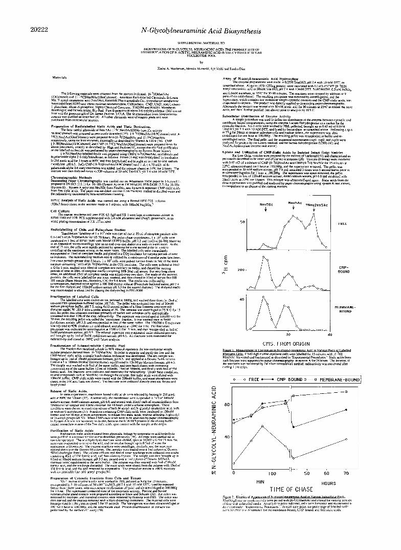
’ Portions of this paper (including “Experimental Procedures,” Figs. 1-6, and Table 1) are presented in miniprint at the end of this paper. Miniprint is easily read with the aid of a standard magnifying glass. Full size photocopies are included in the microfilm edition of the Journal that is available from Waverly Press.
utilization of the labeled Neu5Gc. Finally, as has been previ- ously suggested by others (29, 30) hydroxylation could take place on both the bound and the free pools. To help differ- entiate between these possibilities, we carried out pulse-chase experiments.
Pulse-Chase Studies Suggest That Hydroxylation Takes Place Predominantly on the CMP-Sialic Acid Fraction-Equal numbers of cells were pulsed for 15 min with [6-3H]ManNAc and chased for varying periods of time up to 135 min as described under “Experimental Procedures.” For longer chase periods (43 or 70 h) the initial pulse was increased to 2 h. At each timepoint, the cell suspension was chilled, harvested, washed in phosphate-buffered saline, and fractionated into membrane, ethanol pellet, and ethanol supernatant fractions. One millicurie was used for each pulse. After a 15-min pulse the incorporated radioactivity varied between 14,400 and 25,700 cpm, without relation to time of chase. To correct for differences in the total amount of label incorporated in each pulse, the radioactivity in each fraction was expressed as a percentage of the total radioactivity at that particular time point (see Table 1, Miniprint Section). As expected, the ethanol supernatant (representing the low-molecular cytosolic compounds) initially contained most of the radioactivity (73% at the end of a 15-min pulse). The label in the membrane fraction was initially low (13%) and increased with time to a maximum of 70% at 43 h of chase. Radioactivity was found in the ethanol supernatant in all the long chase periods. The fraction of the total label in the ethanol pellet (soluble pro- teins) was smaller than that in the membrane fraction at all time points.
Based upon prior knowledge of the biosynthesis of sialo- glyconjugates, the radioactivity from the N-acetylmannosa- mine precursor passes sequentially through the free and CMP- bound pools, and finally into the membrane-bound fraction (45-51). At each time point we therefore compared the degree of conversion of N-acetyl to N-glycolylneuraminic acid (hy- droxylation) in each of these compartments. An example of such an analysis is shown in Fig. 1 (15-min pulse and no chase). At this short time point, the percent hydroxylation of membrane-bound sialic acids (57%) and the CMP-sialic acid fraction (61%) were similar, but significantly lower than in the equilibrium label. However, there was only 7% hydroxyl- ation in the free sialic acid fraction, which is the precursor to the other two. The free sialic acid pool also contained an additional peak of radioactivity that migrated ahead of Neu5Ac. However, this peak did not interfere significantly with the estimation of the degree of hydroxylation in these fractions. The nature of this peak remains unknown, but preliminary evidence suggests that it is 2,3-dehydro-2,6-an- hydro-Neu5Ac (Neu2en5Ac), a product of breakdown of GMP-sialic acids (52).
A summary of the results from different chase times is shown in Fig. 2. The percent of hydroxylation of sialic acid in the CMP- and membrane-bound fractions increased progres- sively during the chase period, and reached a maximum of 87% after a 70-h chase. At all the time points studied, the extent of hydroxylation of the CMP-bound sialic acids was similar to that in the corresponding membrane-bound frac- tion. This suggested that most, if not all, of the hydroxylation took place in the low-molecular weight pool prior to the transfer of sialic acids to the membrane-bound glycoconju- gates. The percentage of hydroxylation in the free sialic acid pool was initially low, and although it increased gradually it was always lower than that in the corresponding GMP-sialic acid fraction. Taken together, these data suggested that most, if not all of the conversion took place on the CMP-bound
20218 N-Glycolylneuraminic Acid Biosynthesis
Neu5Ac. However, since Neu5Gc was always found in the free sialic acid pool, they did not rule out the possibility of con- version occurring on the free sugar at a slower rate. I t was also possible that once formed, free Neu5Gc is a preferred substrate for the CMP-sialic acid synthetase, or that the two reactions are coupled. A more likely explanation would be that the free Neu5Gc arose from the breakdown of pre-formed CMP-NeuSGc, and/or from “recycling” of sialic acids released in the lysosome from glycoconjugates. This could also explain the progressive increase in percentage of free Neu5Gc in the cytosolic pool with increasing time. To resolve some of these issues, we studied the substrate specificity of the enzyme reaction.
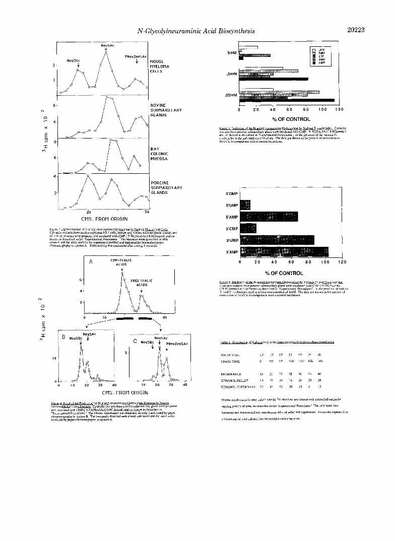
Demonstration of NeuSAc Hydroxylase Activity on CMP- Neu5Ac in Multiple Tissues and Cells Which Express NeuSGc-To test the hypothesis that the preferred substrate for the hydroxylase was CMP-NeutiAc, we set up an assay system using CMP-[3H]Neu5Ac as a substrate. Several differ- ent sources known to express Neu5Gc were used (murine myeloma cells, rat colonic mucosa, and porcine and bovine submaxillary gland tissue). Tissue preparation and assay con- ditions were as described under “Ex9erimental Procedures.” As seen in Fig. 3, each of these tissue and cell extracts caused conversion of the radioactive Neu5Ac in CMP-Neu5Ac to a labeled compound which upon acid cleavage generated Neu5Gc. Internal standards with [14C]Neu5Ac were included in each lane to confirm the location of Neu5Ac, and to correct for lane to lane variation (not shown). A third peak, probably Neu2en5Ac, was also produced in all preparations.
The Product of the Enzyme Reaction Is CMP-NeuSGc-In the preceding experiment, the CMP-sialic acids were degraded to free sialic acids prior to analysis. To directly demonstrate the synthesis of CMP-N~U~GC, and t o rule out the production of free Neu5Gc from free Neu5Ac, the ethanol supernatant from the reaction mixture was separated directly into CMP- and free-sialic acids by paper chromatography in system B. The two peaks were resolved, eluted in distilled water, and then studied for the amount of Neu5Gc present by paper chromatography in system A and by high performance liquid chromatography, as described under “Experimental Proce- dures.” As shown in Fig. 4, there is significant breakdown of CMP-sialic acids to free sialic acids (approximately 40%) during the reaction. When the two peaks were eluted and separately analyzed, it could be seen that the major sialic acid in the CMP-bound fraction was Neu5Gc (62%). On the other hand, the free sialic acids separated into equal amounts of Neu5Ac and the probable Neu2enAc, with only a very small amount of Neu5Gc. In separate experiments, we found that free Neu5Ac itself showed no significant conversion to Neu5Gc under any conditions used (data not shown). Thus, even the small amount of free Neu5Gc found at the end of the reaction with CMP-Neu5Ac apparently arose from break- down of the initial product, CMP-Neu5Gc.
Alternate proof of the product was obtained by analyzing the sialic acids released from the CMP-sialic acid pool by high performance liquid chromatography on a Bio-Rad HPX-72s column, with [‘4C]Neu5Ac as an internal control for the separation. External nonradioactive standards of Neu5Gc and Neu5Ac were also injected, and peak profiles monitored by UV absorbance at 210 nm. Again, the major sialic acid derived from the CMP-sialic acid product co-migrated with Neu5Gc (data not shown).
Direct Comparison of Free, CMP-bound, and a-Glycosidi- cally Bound Neu5Ac as Substrates for the Hydroxylase-The results presented above suggested that the CMP-sugar nu- cleotide might be the sole substrate for the enzyme. To directly
TABLE 2 Comparison of hydroxylase action against free, nucleotide-bound, and
a-glycosidically bound sialic acids Free-, CMP-bound, and a-glycosidically linked [9-3H]Neu5Ac of
identical specific activity were prepared as described under “Experi- mental Procedures.” Equal amounts of each substrate (200,000 cpm, 6.2 pmol) were incubated with cytosolic extracts from porcine sub- maxillary gland. The a-glycosidically linked sialic acids were released with neuraminidase, and the CMP-bound sialic acids were released with 25 m M acetic acid. Conversion of [9-3H]Neu5Ac to [9-3H] Neu5Gc was measured in each case by descending chromatography in system A.
Substrate Neu5Gc formed
% [9-3H]Neu5Ac <2 [9-3H]Neu5Ac-labeled fetuin <2 CMP-[9-3H]Neu5Ac 34
TABLE 3 Comparison of uptake of CMP-Neu5Ac and CMP-Neu5Gc by isolated
intact rat liver golgi vesicles A mixture of CMP-[9-3H]Neu5Ac and CMP-[9-3H]Neu5Gc was
incubated with isolated intact Golgi vesicles, which were then reiso- lated by centrifugation. The percentage of labeled Neu5Gc in each fraction was determined as described under “Experimental Proce- dures.”
Source Neu5Gc %
Starting mixture 48 Un-incorporated 51 Low-molecular weight, incorporated 47 Incorporated, neuraminidase released 40
compare the activity of the hydroxylase against free, nucleo- tide-bound, and a-glycosidically bound Neu5Ac, we prepared samples of [9-3H]Neu5Ac, CMP-[9-3H]Neu5Ac, and [9-3H] Neu5Ac-fetuin with identical specific activity (see “Experi- mental Procedures” for details). These three substrates at the same concentration were exposed to the hydroxylase under identical conditions, and the conversion of [9-3H]Neu5Ac to [9-3H]Neu5Gc monitored. As shown in Table 2, there was no detectable conversion of the free or glycosidically bound [9- 3H]Neu5Ac under conditions where more than one-third of the CMP-[9-3H]Neu5Ac was converted to CMP-[9-3H] Neu5Gc. These results strengthen the conclusion that the conversion of Neu5Ac to Neu5Gc takes place primarily at the sugar nucleotide level.
CMP-Neu5Gc Is Taken up by Golgi Vesicles and Transferred to Endogenous Glycoproteins-It has previously been shown that CMP-Neu5Ac can be taken up by isolated intact rat liver Golgi vesicles by a specific transporter (27, 28,53) and trans- ferred to N-linked oligosaccharides on endogenous glycopro- tein acceptors by luminally oriented sialyltransferases? If the primary site of hydroxylation of Neu5Ac is at the sugar nucleotide level, then CMP-Neu5Gc should be a substrate for this series of reactions. Since methods for the preparation of pure Golgi vesicles from the murine myeloma cells have not been worked out, we chose to use isolated intact Golgi vesicles from rat liver. The vesicles were incubated with a mixture of CMP-[9-3H]Neu5Ac and CMP-[9-3H]Neu5Gc, and the sialic acids incorporated into glycoproteins released and studied. AS shown in Table 3, CMP-Neu5Gc and CMP-Neu5Ac were taken up by the Golgi vesicles and incorporated into the endogenous glycoproteins at an approximately equal rate.
The Hydroxylase Enzyme Is Predominantly in the cytosol-
Diaz, s., Higa, H., Hayes, B. K., and Varki, A. (1989) J. Biol. Chem. in press.
N-Glycolylneuraminic Acid Biosynthesis 20219
Taken together, the pulse-chase analyses and enzymological data presented above indicate that the primary site of the hydroxylation reaction is at the sugar nucleotide level. If this were the case, one would expect the hydroxylase enzyme itself to have a primarily cytosolic location. To test this hypothesis, we compared the distribution of the hydroxylase with that of lactate dehydrogenase, an enzyme known to be localized to the cytosol. As shown in Table 4, the enzyme from NS-1 murine myeloma cells was predominantly in the cytosol, as defined by this criterion. However, a small amount of CMP- Neu5Gc could be formed by the membrane pellet that was essentially depleted of lactate dehydrogenase activity. These results show that while a fraction of the hydroxylase is mem- brane-associated, the majority is in the predicted location, in the cytosol.
Enzyme Cofactors and Properties-We also studied the general properties and cofactor requirements of the enzyme preparations from the murine myeloma cells and the porcine submaxillary gland. The pH optimum showed a broad range between 6.4 and 7.4, in keeping with proposed cytosolic loca- tion of the enzyme (see above). 10 mM dithiothreitol in the initial homogenization buffer was required for enzyme stabil- ity during the 60-90 min incubation period. The porcine enzyme was stable in the crude extract for several weeks at -70 “C. The stability of the enzyme from murine myeloma cell preparations was more variable than that derived from porcine submandibular gland; the reason for this is not clear. Optimal activity of both enzymes required the presence of other factors in the reaction mixture. For enzyme extracted from porcine gland, activity was greatest when 5 mM NADH, 0.5 mM FeS04, and 0.5 mM ascorbate were present. When only one of these cofactors was present, product formation was greatest if it was NADH (17% control) rather than FeS04 or ascorbate (1% control). For enzyme extracted from murine myeloma NS-1 cells, addition of 5 M NADH alone consistently resulted in greater product formation (111-151% control) compared to the combination. NADPH and NADH were equally effective as cofactors for both enzymes. Some of these data are similar to those reported by Schauer and others for the porcine submaxillary gland enzyme (29, 30, 32).
Enzyme Kinetics-Under the conditions of assay described under “Experimental Procedures,” the velocity of the reaction was almost linear for 1-2 h; typical reactions were therefore incubated for 90 min. The Michaelis-Menten kinetics of the crude enzyme preparations were studied. The VmaX of the reaction varied between different preparations from the NS- 1 murine myeloma cells (0.05-0.07 pmol/min/mg), and was higher (0.29 pmol/min/mg) in the pig submandibular gland extracts. From multiple experiments the apparent K, of the crude enzyme for CMP-NeuAc was calculated to be between
TABLE 4 Comparison of subcellular distribution of the Neu5Ac hydroxylase
and lactate dehydrogenase (LDH) from N S - 1 cells Murine myeloma NS-1 cells were washed in phosphate-buffered
saline, fractionated, and assayed for the Neu5Ac hydroxylase, lactate dehydrogenase, and protein as described under “Experimental Pro- cedures.”
Sample Specific activity Total activity
Hydroxylase LDH Hydroxylase LDH
unitslmg % sup 1 0.175 9.74 54 94 sup 2 0.332 1.64 28 4 sup 3 0.300 1.98 5 1 Pellet 0.067 0.045 13 1
0.6 and 0.9 PM (NS-1 cells) and 2.5 p~ (porcine submaxillary gland enzyme).
Enzyme Specificity-The low apparent K , value for CMP- Neu5Ac suggested that the enzyme might be specific for this substrate. No Neu5Gc was formed from free Neu5Ac under any conditions studied. Since the Neu5Ac in CMP-Neu5Ac is in the uncommon @-linkage (26), we studied the activity of the enzymes against the @-methylglycoside of Neu5Ac (Neu5AcPBOMe). The resulting material was subjected to mild acid hydrolysis to remove the @-methyl group, and then stud- ied by paper chromatography. However, no detectable hy- droxylation occurred, even with concentrations as high as 6.8 PM (data not shown). This indicated that the enzyme recog- nition of the sugar nucleotide was not solely directed toward the P-linkage of the Neu5Ac to CMP.
To explore if substrate recognition involved the CMP moiety, we studied the effects of various nucleotides on the enzyme activity. At 0.005-0.05 mM 5’-CMP there was a small enhancement of Neu5Gc formation (range 110-130% of con- trol). At higher concentrations, inhibition was seen with es- sentially no Neu5Gc formed when 5 mM 5’-CMP was present. The effects of other 5”nucleotides were also studied; some representative results are shown in Fig. 6. Some of the other nucleotides had comparable inhibitory effects to that of 5’- CMP at millimolar concentrations.
Since the phosphate group of CMP-sialic acids is in the 5’ position, we examined the effect of mononucleotides with phosphate groups at 3‘ or 5’ positions. As shown in Fig. 7, activity was markedly inhibited by 5 mM 5’-UMP, 5’-CMP and 3‘-CMP. However, the same concentrations of 3’-UMP, 3’-AMP, and 5’-AMP had minimal effect upon product for- mation. Thus, inhibition was caused by 5’-pyrimidine nucle- otides or by 3’-CMP. We therefore tested various concentra- tions of ribose 1-phosphate and ribose 5-phosphate. Neither sugar phosphate showed significant inhibition of product for- mation at concentrations as high as 20 mM (data not shown). Taken together, these results suggest that the low apparent K,,, of the enzyme for CMP Neu5Ac involves specific recog- nition of the intact sugar nucleotide, including the pyrimidine base and the phosphate moiety. Detailed studies of this rec- ognition must await the purification of the enzyme.
DISCUSSION
Previous studies of the biosynthesis of Neu5Gc have been carried out predominantly in the porcine submaxillary gland (29, 30, 47) and in a rat mammary carcinoma cell line that has a small proportion of Neu5Gc (54). We have used murine myeloma cell lines that convert almost all of their sialic acids to Neu5Gc. This has permitted us to take several different approaches towards identifying the subcellular site of the biosynthesis of Neu5Gc.
The hydroxylation of Neu5Ac to Neu5Gc could potentially take place on newly synthesized or recycled free Neu5Ac, on CMP-Neu5Ac, in the Golgi apparatus immediately after transfer of Neu5Ac to a glycoconjugate, or at some later point in the life of the glycoconjugate. We have presented several lines of evidence that indicate that most, if not all, of the hydroxylation takes place at the nucleotide sugar level. First, the pulse-chase experiments indicate that most of the hydrox- ylation reaction occurs in the cytosolic low-molecular weight pool. At early time points in the pulse-chase, very little Neu5Gc is found in the free sialic acid pool. In contrast, the CMP-sialic acid pool shows substantial conversion to Neu5Gc, identical to that in the membrane-bound fraction a t each time point. Second, enzymatic studies in a variety of cells and tissues that synthesize Neu5Gc indicate that CMP-

20220 N-Glycolylneuraminic Acid Biosynthesis
FIG. 7. Proposed pathways for the biosynthesis and reutilization of N-glycolylneuraminic acid. The pathways proposed are based upon the prior literature and the data presented in this study. See text for detailed dis- cussion.
. piziziq PLASMA MEMBRANE
S N e u S A c
a?
? @ I Neu5Ac NeuSGc
I
I I @ @
4 h
r\ ? w
ManNGc < @ Ne!!: @ > CMP-Neu5Gc- s-Neu5Gc UDP-GlcNAc
4 ManNAc + NeuSAc
0 + CMP-NeuSAc , s - N e u 5 A c A
@ = N-ACETYLNEURAMINATE HYDROXYLASE @ = CMP-ACYLNEURAMINATE SYNTHETASE
CMP-ACYLNEURAMINATE HYDROLASE 1 NEURAMINIDASE (SIALIDASE)
4 4
@ = SIALIC ACID TRANSPORTER . Neu5Ac is the preferred substrate for the hydroxylation re- action. Third, the great majority of the hydroxylase activity is found in the cytosolic fraction of myeloma cells, and the optimal reaction conditions mimic those that might be found in the cytosol of intact cells. Finally, both CMP-Neu5Ac and CMP-Neu5Gc are utilized equally well by isolated intact rat liver Golgi vesicles as donors for sialylation of endogenous acceptors.
Taken together with prior literature, the data we have presented permit us to propose the pathways for biosynthesis and reutilization of Neu5Gc shown in Fig. 7. In this model, we propose that CMP-Neu5Ac in the cytosol is hydroxylated by a specific enzyme, resulting in the formation of CMP- Neu5Gc. The CMP-Neu5Gc can then be transported into the lumen of the Golgi apparatus, where it can serve as a donor for the various specific sialyltransferases. Work by others has in fact shown that some sialyltransferases can utilize CMP- Neu5Gc as well as they can utilize CMP-Neu5Ac (33). We have shown that CMP-Neu5Gc is equally well utilized by isolated intact Golgi vesicles. In this model, the appearance of free Neu5Gc in the cytosolic pool could be explained by the release and export (55-57) of Neu5Gc from glycoconjugates in the lysosome. This could also explain the gradual increase in the percentage of free Neu5Gc in the cytosol during the pulse-chase study. An alternate source of free Neu5Gc could be the direct breakdown of CMP-Neu5Gc, which was actually seen in the in vitro reaction conditions. However, we currently know too little about the enzyme CMP-sialic acid hydrolase (58) to test this hypothesis in the intact cell. The model would also help to explain the relative differences in percentage of hydroxylation between the short pulse-chase and the equilib- rium-labeled experiments. Thus, in the murine myeloma cells, a single cohort of newly synthesized CMP-Neu5Ac molecules would be only partially hydroxylated, this being reflected in the lower percentage of labeled Neu5Gc in newly sialylated molecules. However, Neu5Gc once formed cannot be con- verted back to Neu5Ac by any currently known mechanism. Thus, recycling of Neu5Gc released in the lysosomes into the cytosolic pool could result in a progressive rise in the per-
centage of Neu5Gc in the overall cellular sialic acid, until a steady-state between new synthesis and degradation of NeuSGc is reached. In the case of the murine myeloma cells, this equilibrium is apparently reached when greater than 90% of the total cellular sialic acids are converted to Neu5Gc. The mechanisms by which Neu5Gc gets ultimately degraded re- main unknown. In the model presented in Fig. 7, it is pre- sumed that Neu5Gc is degraded to N-glycolylmannosamine (ManGc) (59) by the enzyme acylneuraminate-pyruvate-lyase (3, 60). However, if ManGc is actually produced in this manner in the intact cell, the ultimate fate of this sugar also needs to be determined.
Some characteristics of the hydroxylase enzyme are worthy of comment. The predominantly cytosolic location of the enzyme is unusual among enzymes with similar mechanisms and cofactors, which are usually membrane-bound. For in- stance, the cytochrome P-450 family of enzymes, which share many of the properties and cofactor requirements of this hydroxylase, tend to be tightly integrated into microsomal membranes and are rather unstable when liberated from that environment (61). Since a fraction of this hydroxylase enzyme is membrane-associated, we cannot rule out the possibility that it is all originally membrane-bound, and is artifactually released by proteolysis following homogenization. However, in either case, the active site must have access to the cytosol, where the CMP-Neu5Ac substrate is located. This model also does not attempt to incorporate the finding that CMP-sialic acid synthetase has a predominantly nuclear location (62,63).
The experiments aimed at understanding the requirements for enzyme recognition of the substrate are not conclusive. While the enzyme recognizes the native substrate with a K,,, in the nanomolar range, the unique @-glycosidic linkage of Neu5Ac in the sugar nucleotide is insufficient by itself to explain this recognition. On the other hand, the nucleotide inhibition studies suggest that the enzyme recognizes the pyrimidine base and the 5”phosphate group. Further char- acterization of this enzyme and its properties must await its purification.
Acknowledgment-We would like to thank Dr. Herman Higa for
N-Glycolylneuraminic Acid Biosynthesis 20221
his help with some of the experiments and for reviewing the manu- 29. Buscher, H. P., Casals Stenzel, J., Schauer, R., and Mestres script. Ventura, P. (1977) Eur. J. Biochem. 77,297-310
30. Schauer, R. (1978) Methods Enzymol50,374-386 31. Schauer, R., and Wember, M. (1971) Hoppe-Seyler’s Z. Physiol. Note Added in Proof-While this manuscript was under review a
paper appeared describing the Of CMP-Neu5Gc by mouse 32. Shaw, L., and Schauer, R. (1988) Bioi. Chern. Hoppe-Seyler 369 , Chem. 352,1282-1290
liver golgi vesicles (64). 477-486
1.
2.
3.
4. 5. 6.
7. 8.
9.
10.
11.
12.
13.
14.
15.
16.
17.
18.
19.
20.
21.
22.
23.
24.
25.
26.
27.
28.
REFERENCES
Schauer, R. (1982) Sialic Acids: Chemisty, Metabolism and Func- tion, Cell Biology Monographs, Volume 10, Springer-Verlag, New York
Sander, Weber, M., Schauer, R., and Corfield, A. P. (1982) Adu. Exp. Med. Biol. 152,215-222
Schauer, R., Wember, M., Wirtz Peitz, F., and Ferreira do Amaral, C. (1971) Hoppe-Seyler’sZ. Physiol. Chem. 352,1073- 1080
Varki, A., and Diaz, S. (1983) J. Biol. Chem. 258, 12465-12471 Muchmore, E., and Varki, A. (1987) Science 2 3 6 , 1293-1295 Cheresh, D. A., Reisfeld, R. A., and Varki, A. (1984) Science 2 2 5 ,
Varki, A., and Kornfeld, S. (1980) J. Exp. Med. 152,532-544 Orskov, F., Orskov, I., Sutton, A., Schneerson, R., Lin, W., Egan,
W., Hoff, G. E., and Robbins, J. B. (1979) J. Exp. Med. 149 ,
Rogers, G . N., Herrler, G., Paulson, J. C., and Klenk, H.-D. (1986) J. Biol. Chem. 261,5947-5951
Herrler, G., Rott, R., Klenk, H. D., Muller, H. P., Shukla, A. K., and Schauer, R. (1985) EMBO J. 4,1503-1506
Hirabayashi, Y., Kasakura, H,, Matsumoto, M., Higashi, H., Kato, S., Kasai, N., and Naiki, M. (1987) Jpn. J . Cancer Res.
Hirabayashi, Y., Higashi, H., Kato, S., Taniguchi, M., and Mat- sumoto, M. (1987) Jpn. J. Cancer Res. 78,614-620
Higashi, H., Hirabayashi, Y., Fukui, Y., Naiki, M., Matsumoto, M., Ueda, S., and Kato, S. (1985) Cancer Res. 45, 3796-3802
Carubelli, R., and Griffin, M. J. (1968) Biochim. Biophys. Acta
Ohashi, Y., Sasabe, T., Nishida, T., Nishi, Y., and Higashi, H.
Merrick, J. M., Zadarlik, K., and Milgrom, F. (1978) Int. Arch.
Fujii, Y., Higashi, H., Ikuta, K., Kato, S., and Naiki, M. (1982)
Nishimaki, T., Kano, K., and Milgrom, F. (1979) J. Immunol.
Bouhours, D., and Bouhours, J.-F. (1983) J. Biol. Chem. 258 ,
Muchmore, E., Varki, N., Fukuda, M., and Varki, A. (1987)
Bouhours, D., and Bouhours, J.-F. (1988) J. Biol. Chem. 263,
Kundig, W., Ghosh, S., and Roseman, S. (1966) J. Biol. Chem.
Watson, D. R., Jourdian, G. W., and Roseman, S. (1966) J. Bid.
Warren, L., and Felsenfeld, H. (1962) J. Biol. Chem. 237, 1421-
Kean, E. L., and Roseman, S. (1966) J. Biol. Chem. 241 , 5643-
Comb, D. G., Watson, D. R., and Roseman, S. (1966) J. Biol.
844-846
669-685
78,251-260
170,446-448
(1983) Am. J. Ophthalmol. 9 6 , 321-325
Allergy Appl. Immunol. 57,477-480
Mol. Immunol. 19,87-94
122,2314-2318
299-304
FASEB J. 1, 229-235
15540-15545
241,5619-5626
Chem. 241,5627-5636
1431
5650
Chem. 241,5637-5642 Capasso, J. M., and Hirschberg, C. B. (1984) Proc. Natl. Acad.
Sci. U. S. A. 81, 7051-7055 Carey, D. J., and Hirschberg, C. B. (1981) J. Biol. Chem. 256,
989-993
33. Higa, H. H., and Paulson, J. C. (1985) J. Biol. Chem. 260,8838- 8849
34.
35.
36.
37. 38. 39.
40.
41.
42. 43.
44.
45.
46.
47.
48.
49.
50.
51. 52.
53.
54.
55.
56.
57.
58.
59.
60. 61.
62. 63.
64.
Weinstein, J., de Souza-e-Silva, U., and Paulson, J. C. (1982) J.
Varki, A., Muchmore, E., and Diaz, S. (1986) Proc. Natl. Acad.
Warren, L., and Glick, M. C. (1966) Methods Enzymol. 8, 131-
Varki, A., and Diaz, S. (1984) Anal. Bwchem. 137,236-247 Diaz, S., and Varki, A. (1985) Anal. Biochem. 150,32-46 Lowry, 0. H., Rosebrough, N. J., Farr, A. L., and Randall, R. J .
Howell, B. F., McCune, S., and Schaffer, R. (1979) Clin. Chem.
Leelavathi, D. E., Estes, L. W., Feingold, D. S., and Lombardi,
Kohler, G., and Milstein, C. (1974) J. Mol. Bwl. 9 0 , 691-698 Laskov, R., and Scharff, M. D. (1970) J. Exp. Med. 131 , 515-
Varki, N. M., Reisfeld, R. A., and Walker, L. E. (1984) Cancer
Johnston, I. R., McGuire, E. J., Jourdian, G. W., and Roseman,
Glick, M. C., Comstock, C. A., Cohen, M. A., and Warren, L.
Yurchenco, P. D., Ceccarini, C., and Atkinson, P. H. (1978)
Brown, A. E., Schwartz, E. L., Dreyer, R. N., and Sartorelli, A.
Bennett, G., Kan, F. W., and O’Shaughnessy, D. (1981) J. Cell
Fenverda, W., Blok, C. M., and Van Rinsum, J. (1983) Biochem.
Bennett, G., and O’Shaughnessy, D. (1981) J. Cell Biol. 88, 1-15 Beau, J. M., Schauer, R., Haverkamp, J., Kamerling, J. P.,
Dorland, L., and Vliegenthart, J. F. (1984) Eur. J. Biochem.
Biol. Chem. 267,13835-13844
Sci. U. S. A. 83,882-886
133
(1951) J. Biol. Chem. 193,265-275
25 (2), 269-272
B. (1970) Biochem. Biophys. Acta 2 1 1 , 124-138
54 1
Res. 44,681-688
S. (1966) J. Biol. Chem. 241,5735-5737
(1971) Biochim. Biophys. Acta 233, 247-257
Methods Enzymol. 60,175-204
C. (1982) Biochim. Biophys. Acta 714,217-225
Biol. 88,16-28
J. 216,87-92
140,203-208 Milla, M. E., and Hirschberg, C. B. (1989) Proc. Natl. Acad. Sci.
U. S. A. 86, 1786-1790 Sherblom, A. P., and Dahlin, C. E. (1985) J. Biol. Chem. 260 ,
Hildreth, J., IV, Sacks, L., and Hancock, L. W. (1986) Biochem.
Renlund, M., Tietze, F., and Gahl, W. A. (1986) Science 2 3 2 ,
Jonas, A. J. (1986) Biochem. Biophys. Res. Commun. 137 , 175-
Kean, E. L., and Bighouse, K. J. (1974) J. Biol. Chem. 2 4 9 ,
Jourdian, G. W., and Roseman, S. (1962) J. Biol. Chem. 2 3 7 ,
Warren, L. (1986) Biochim. Biophys. Acta 888, 278-281 Ingelman-Sundberg, M. (1986) in Cytochrome P-450, Structure,
Mechanism, and Biochemistry (Ortiz de Montellano, P. R., ed) pp. 119-160, Plenum Press, New York
1484-1492
Biophys. Res. Commun. 139,838-844
759-762
181
7813-7823
2442-2446
Kean, E. L. (1970) J. Biol. Chem. 245, 2301-2308 Coates, S. W., Gurney, T., Jr., Sommers, L. W., Yeh, M., and
Lepers, A., Shaw, L., Cacan, R., Schauer, R., Montreuil, J., and Hirschberg, C. B. (1980) J. Biol. Chem. 255,9225-9229
Verbert, A. (1989) FEBS. Lett. 250, 245-250
Continued on next page.
20222 N-Glycolylneuraminic Acid Biosynthesis SUPPLEMENTAL MATERIAL To-
HYDROXYLAnON OF N-AlXTYLNEURAMIMC ACID IS THE CYTOSOLIC SUGAR B I O S Y ~ S I S O F N G L Y C O L n - N E L I R A ~ C A C I D : TW? PRIMARY STTEOF
NUCZEOTIDE POOL
by
Elamc A. Muchmne. Monrka M i l e w s ~ , A J I ~ Varki and Sandra Dim
M a I e r i a I ~
W
2 1
bp
J
sa
200
40
30
FREE
BOUND cnp-
MEMBRANE- BOUND
80
40
1 - FREE - CMP-BOUND - MEMBRANE-BOUND1
M l N HOURS
T I ME OF CHASE
N-Glycolylneuraminic Acid Biosynthesis 20223
NeuSAc
NeuSGc ? N e u Z e n s A ~
K"
A 20
1 c
MOUSE MYELOMA CELLS
b
-
BOVINE SUBMAXILLARY GLANDS
1 R A T COLONIC MUCOSA I PORCINE SUBMAXILLARY
*I GLANDS
2 30
CMS. FROM ORIGIN
10
n
CMP-SIALIC ACIDS
6
4
2
0 IO
5
B NeuSGc + NeuSAc
&
0 10 20 30 40
K" ?NeuPenSAc
NeuSAc
" K "
IO 20 30 40
5mM
.05mM
5'CMP
B'UMP
TAMP
B'CMP
B'UMP
B'AMP
0 2 0 4 0 6 0 8 0 1 0 0 1 2 0
PlJLSE TIME IS ' 15' Is' Is' Is' 2h Zh
CHASETIME 0 45 15' I05 135' 43h 7Oh
MEMBRANES 13 21 23 38 46 73 MI
ETHANOLPELLET 14 32 24 32 21 29 28
ETHANOLSUPERNATE 71 47 53 30 33 4 12