acidic ph increases airway surface liquid viscosity in...
TRANSCRIPT

The Journal of Clinical Investigation R e s e a R c h a R t i c l e
8 7 9jci.org Volume 126 Number 3 March 2016
IntroductionCystic fibrosis (CF) is a life-shortening disease caused by muta-tions in the gene encoding the cystic fibrosis transmembrane conductance regulator (CFTR) anion channel (1–3). Lung disease is the major cause of CF morbidity and mortality. The airways of individuals with advanced CF lung disease are infected, inflamed, and remodeled and contain mucus that obstructs airways. To bet-ter understand the factors that initiate CF airway disease, we devel-oped CFTR–/– pigs and CFTRΔF508/ΔF508 pigs (referred to herein as CF pigs) (4, 5). At birth, their airways lack infection and inflammation, and, within weeks to months, they develop the classical manifesta-tions of CF lung disease (5, 6). We discovered that newborn piglets manifest at least two host defense defects against bacteria (7). Loss of CFTR-mediated HCO3
– secretion generates an airway surface liquid (ASL) with an abnormally reduced pH, and the acidic envi-ronment inhibits the activity of ASL antimicrobials (8, 9). In addi-tion, loss of CFTR-mediated HCO3
– and Cl– secretion alters mucus, so that it fails to break free after secretion from submucosal glands and impairs mucociliary transport in vivo (10). Mucus produced by airway goblet cells in CF pigs may also be abnormal, as evidenced by histopathological analysis of older animals (6, 11).
Mucociliary transport defends airways by capturing patho-gens in mucus that is propelled out of the lung by cilia (12–14). Studies performed using sputum collected from people with advanced CF airway disease suggest that CF sputum has several abnormalities (15–18). However, chronic bacterial infection, neutrophil-dominated inflammation, and airway remodeling with submucosal gland hypertrophy and goblet cell hyperplasia could alter mucus and sputum. In addition, obtaining compara-ble sputum or mucus samples from “controls” or “normals” can be problematic. However, finding impaired mucociliary trans-port at birth in CF piglets indicated that mucus abnormalities are a primary CF defect (10, 19).
The goals of this study were to test the hypothesis that CF ASL has abnormal viscosity at the outset of disease and then to discover the basis of any abnormality. We used newborn piglets to avoid alterations in viscosity that might be caused by secondary CF manifestations, including bacterial products, DNA, proteases, cells and cellular debris, inflammation, altered neurohumoral sig-naling, and airway remodeling with goblet cell hyperplasia and submucosal gland hypertrophy. We studied ASL immediately after collection from piglets or while it covered cultured airway epithe-lia to avoid the effects of freezing or storage. Although mucins are the major protein and structural component of ASL and determine its viscoelastic properties (20–23), we chose to study native ASL, because purification and solubilization can alter the properties of mucins (24), and we wished to assess differences that might be of physiological and therapeutic significance.
Cystic fibrosis (CF) disrupts respiratory host defenses, allowing bacterial infection, inflammation, and mucus accumulation to progressively destroy the lungs. Our previous studies revealed that mucus with abnormal behavior impaired mucociliary transport in newborn CF piglets prior to the onset of secondary manifestations. To further investigate mucus abnormalities, here we studied airway surface liquid (ASL) collected from newborn piglets and ASL on cultured airway epithelia. Fluorescence recovery after photobleaching revealed that the viscosity of CF ASL was increased relative to that of non-CF ASL. CF ASL had a reduced pH, which was necessary and sufficient for genotype-dependent viscosity differences. The increased viscosity of CF ASL was not explained by pH-independent changes in HCO3
– concentration, altered glycosylation, additional pH-induced disulfide bond formation, increased percentage of nonvolatile material, or increased sulfation. Treating acidic ASL with hypertonic saline or heparin largely reversed the increased viscosity, suggesting that acidic pH influences mucin electrostatic interactions. These findings link loss of cystic fibrosis transmembrane conductance regulator–dependent alkalinization to abnormal CF ASL. In addition, we found that increasing Ca2+ concentrations elevated ASL viscosity, in part, independently of pH. The results suggest that increasing pH, reducing Ca2+ concentration, and/or altering electrostatic interactions in ASL might benefit early CF.
Acidic pH increases airway surface liquid viscosity in cystic fibrosisXiao Xiao Tang,1,2 Lynda S. Ostedgaard,1 Mark J. Hoegger,1 Thomas O. Moninger,3 Philip H. Karp,1 James D. McMenimen,1 Biswa Choudhury,4 Ajit Varki,4,5 David A. Stoltz,1,6,7 and Michael J. Welsh1,2,6
1Department of Internal Medicine, 2Howard Hughes Medical Institute, Pappajohn Biomedical Institute, and 3Central Microscopy Research Facilities, University of Iowa, Iowa City, Iowa, USA. 4Glycobiology Research and Training Center and 5Departments of Medicine and Cellular and Molecular Medicine, UCSD, La Jolla, California, USA. 6Department of Molecular Physiology and Biophysics
and 7Department of Biomedical Engineering, University of Iowa, Iowa City, Iowa, USA.
Conflict of interest: The University of Iowa Research Foundation has submitted patent applications for CF pigs and has licensed materials and technologies to Exemplar Genet-ics. M.J. Welsh holds equity in Intrexon Corp., which owns Exemplar Genetics.Submitted: July 27, 2015; Accepted: December 8, 2015.Reference information: J Clin Invest. 2016;126(3):879–891. doi:10.1172/JCI83922.
Downloaded from http://www.jci.org on March 7, 2016. http://dx.doi.org/10.1172/JCI83922
The Journal of Clinical Investigation R e s e a R c h a R t i c l e
8 8 0 jci.org Volume 126 Number 3 March 2016
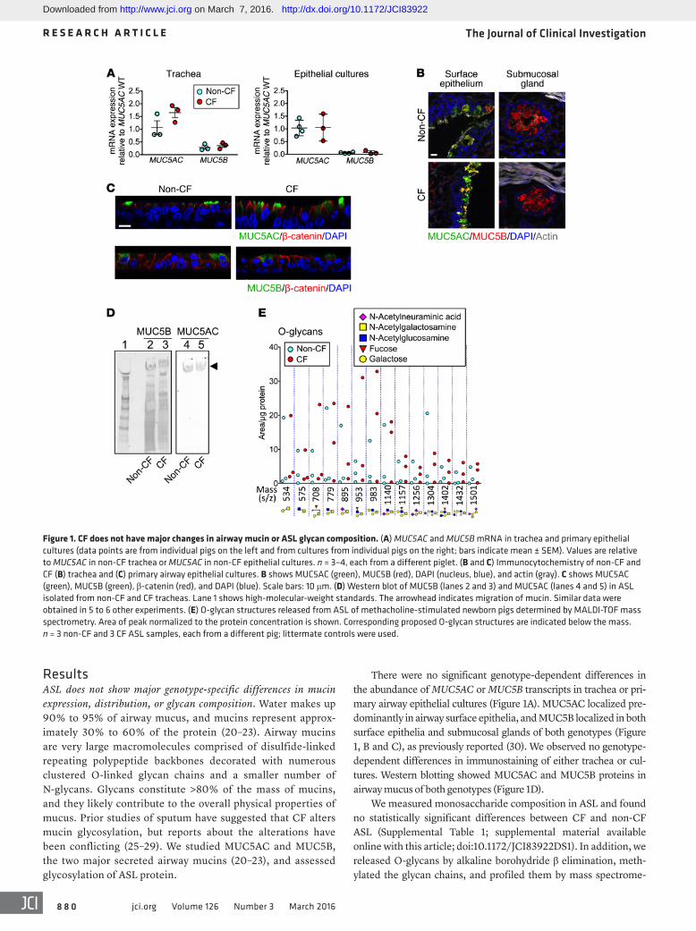
There were no significant genotype-dependent differences in the abundance of MUC5AC or MUC5B transcripts in trachea or pri-mary airway epithelial cultures (Figure 1A). MUC5AC localized pre-dominantly in airway surface epithelia, and MUC5B localized in both surface epithelia and submucosal glands of both genotypes (Figure 1, B and C), as previously reported (30). We observed no genotype- dependent differences in immunostaining of either trachea or cul-tures. Western blotting showed MUC5AC and MUC5B proteins in airway mucus of both genotypes (Figure 1D).
We measured monosaccharide composition in ASL and found no statistically significant differences between CF and non-CF ASL (Supplemental Table 1; supplemental material available online with this article; doi:10.1172/JCI83922DS1). In addition, we released O-glycans by alkaline borohydride β elimination, meth-ylated the glycan chains, and profiled them by mass spectrome-
ResultsASL does not show major genotype-specific differences in mucin expression, distribution, or glycan composition. Water makes up 90% to 95% of airway mucus, and mucins represent approx-imately 30% to 60% of the protein (20–23). Airway mucins are very large macromolecules comprised of disulfide-linked repeating polypeptide backbones decorated with numerous clustered O-linked glycan chains and a smaller number of N-glycans. Glycans constitute >80% of the mass of mucins, and they likely contribute to the overall physical properties of mucus. Prior studies of sputum have suggested that CF alters mucin glycosylation, but reports about the alterations have been conflicting (25–29). We studied MUC5AC and MUC5B, the two major secreted airway mucins (20–23), and assessed glycosylation of ASL protein.
Figure 1. CF does not have major changes in airway mucin or ASL glycan composition. (A) MUC5AC and MUC5B mRNA in trachea and primary epithelial cultures (data points are from individual pigs on the left and from cultures from individual pigs on the right; bars indicate mean ± SEM). Values are relative to MUC5AC in non-CF trachea or MUC5AC in non-CF epithelial cultures. n = 3–4, each from a different piglet. (B and C) Immunocytochemistry of non-CF and CF (B) trachea and (C) primary airway epithelial cultures. B shows MUC5AC (green), MUC5B (red), DAPI (nucleus, blue), and actin (gray). C shows MUC5AC (green), MUC5B (green), β-catenin (red), and DAPI (blue). Scale bars: 10 μm. (D) Western blot of MUC5B (lanes 2 and 3) and MUC5AC (lanes 4 and 5) in ASL isolated from non-CF and CF tracheas. Lane 1 shows high-molecular-weight standards. The arrowhead indicates migration of mucin. Similar data were obtained in 5 to 6 other experiments. (E) O-glycan structures released from ASL of methacholine-stimulated newborn pigs determined by MALDI-TOF mass spectrometry. Area of peak normalized to the protein concentration is shown. Corresponding proposed O-glycan structures are indicated below the mass. n = 3 non-CF and 3 CF ASL samples, each from a different pig; littermate controls were used.
Downloaded from http://www.jci.org on March 7, 2016. http://dx.doi.org/10.1172/JCI83922
The Journal of Clinical Investigation R e s e a R c h a R t i c l e
8 8 1jci.org Volume 126 Number 3 March 2016
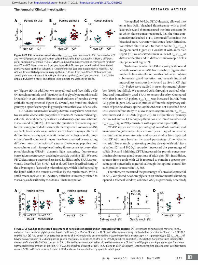
We applied 70-kDa FITC-dextran, allowed it to enter into ASL, bleached fluorescence with a brief laser pulse, and then measured the time constant (τ) at which fluorescence recovered, i.e., the time con-stant for unbleached FITC-dextran diffusion into the bleached area. A shorter τ indicates faster diffusion. We related the τ in ASL to that in saline (τASL/τsaline) (Supplemental Figure 2). Consistent with an earlier report (31), we observed similar values of τASL/τsaline at different depths and in different microscopic fields (Supplemental Figure 2).
To determine whether ASL viscosity is abnormal at birth, we obtained ASL from newborn piglets after methacholine stimulation; methacholine stimulates submucosal gland secretion and reveals impaired mucociliary transport in vivo and ex vivo in CF pigs (10). Piglets were studied in an environmental cham-
ber (100% humidity). We removed ASL through a tracheal win-dow and immediately used FRAP to assess viscosity. Compared with that in non-CF piglets, τASL/τsaline was increased in ASL from CF piglets (Figure 2A). We also studied differentiated primary cul-tures of porcine airway epithelia; the ASL was not disturbed for 2 to 4 weeks before study to allow mucus accumulation. τASL/τsaline was increased in CF ASL (Figure 2B). In differentiated primary cultures of human CF airway epithelia, we also found an increased τASL/τsaline (Figure 2C), consistent with a previous report (31).
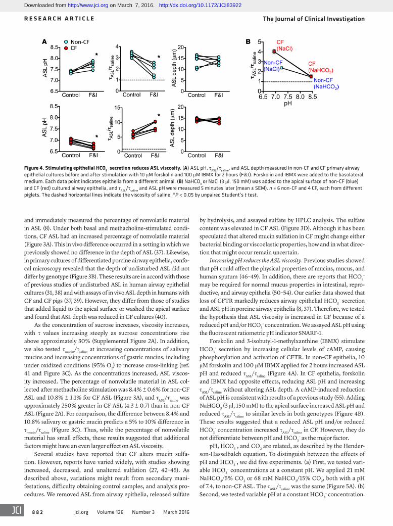
CF ASL has an increased percentage of nonvolatile material and an increased sulfate content. An increased percentage of nonvolatile material can increase viscosity, and several studies have reported that CF ASL may have an increased percentage of nonvolatile material. For example, pretreating porcine airways with inhibitors of anion (Cl– and HCO3
–) secretion increased the percentage of solids (34), and inhibiting CFTR increased the protein concentra-tion in submucosal gland secretions of adult pigs (35). In addition, sputum from people with CF is reported to contain a greater per-centage of nonvolatile material, although the optimal control for such studies is uncertain (16, 36).
Therefore, we measured the percentage of nonvolatile material in ASL. We placed newborn piglets in an environmental chamber; opened a tracheal window; collected ASL, as previously described;
try (Figure 1E). In addition, we assayed total and free sialic acids (N-acetyl neuraminic acid [Neu5Ac] and N-glycolylneuraminic acid [Neu5Gc]) in ASL from differentiated cultures of porcine airway epithelia (Supplemental Figure 1). Overall, we found no obvious genotype-specific changes in glycosylation at this level of analysis.
CF ASL has an increased viscosity. Several assays have been used to assess the viscoelastic properties of mucus. At the macrorheologi-cal scale, shear rheometry has been used to assay sputum elastic and viscous moduli (20–23). However, the quantities of mucus required for that assay precluded its use with the very small volumes of ASL available from newborn animals in vivo or from primary cultures of differentiated airway epithelia. At the microrheological scale, prop-erties of small volumes of mucus have been assessed by measuring diffusion rates or behavior of a tracer (molecules, peptides, and nanospheres and microspheres) using fluorescence recovery after photobleaching (FRAP), dynamic light scattering, fluorescence correlation spectroscopy, and single-particle tracking (23). We used FITC-dextran as a tracer and assessed its diffusion by FRAP, as pre-viously described (19, 31–33). Lai et al. (23) have described some of the advantages of assessing microrheology, which is influenced by the liquid within the mucus as well as by the mucin mesh. With a small tracer such as FITC-dextran, diffusion is inversely related to viscosity, as described by the Stokes-Einstein equation.
Figure 2. CF ASL has an increased viscosity. τASL/τsaline was measured in ASL from newborn CF and non-CF piglets or pig and human cultured epithelia. Each data point is from a different pig or human donor (mean ± SEM). (A) ASL removed from methacholine-stimulated newborn non-CF and CF littermates. n = 6 per genotype. (B) ASL on unperturbed, well-differentiated primary airway epithelial cultures. n = 21 per genotype (see also Supplemental Figure 3). (C) ASL on differentiated primary airway epithelial cultures from non-CF and CF humans (see also Supplemental Figure 4 for ASL pH of human epithelia). n = 7 per genotype. *P < 0.05 by unpaired Student’s t test. The dashed lines indicate the viscosity of saline.
Figure 3. CF ASL has an increased percentage of nonvolatile material and an increased sulfate content. (A) Percentage of nonvolatile material in ASL collected from newborn piglets under basal conditions (n = 17 non-CF and n = 12 CF) and after administering methacholine (n = 16 non-CF and n = 6 CF) (2.5 mg/kg, i.v.). (B) ASL depth in unperturbed cultures of airway epithelia determined by z-scanning confocal microscopy. n = 11 per genotype. (C) τmucin/τsaline of bovine salivary mucin (n = 6) and porcine gastric mucin (n = 4) exposed to 21% O2 or 95% O2 (oxidized condition). The dashed horizontal lines indicate the viscosity of saline. (D) Sulfate content in ASL collected from airway epithelia cultured from newborn CF and non-CF piglets. n = 6 per genotype. Data were normalized to the amount of protein. *P < 0.05 by unpaired Student’s t test. In A, B, and D, each data point is from a different pig, and error bars represent mean ± SEM. In C, data represent mean ± SEM and error bars are hidden by symbols in left graph.
Downloaded from http://www.jci.org on March 7, 2016. http://dx.doi.org/10.1172/JCI83922
The Journal of Clinical Investigation R e s e a R c h a R t i c l e
8 8 2 jci.org Volume 126 Number 3 March 2016
by hydrolysis, and assayed sulfate by HPLC analysis. The sulfate content was elevated in CF ASL (Figure 3D). Although it has been speculated that altered mucin sulfation in CF might change either bacterial binding or viscoelastic properties, how and in what direc-tion that might occur remain uncertain.
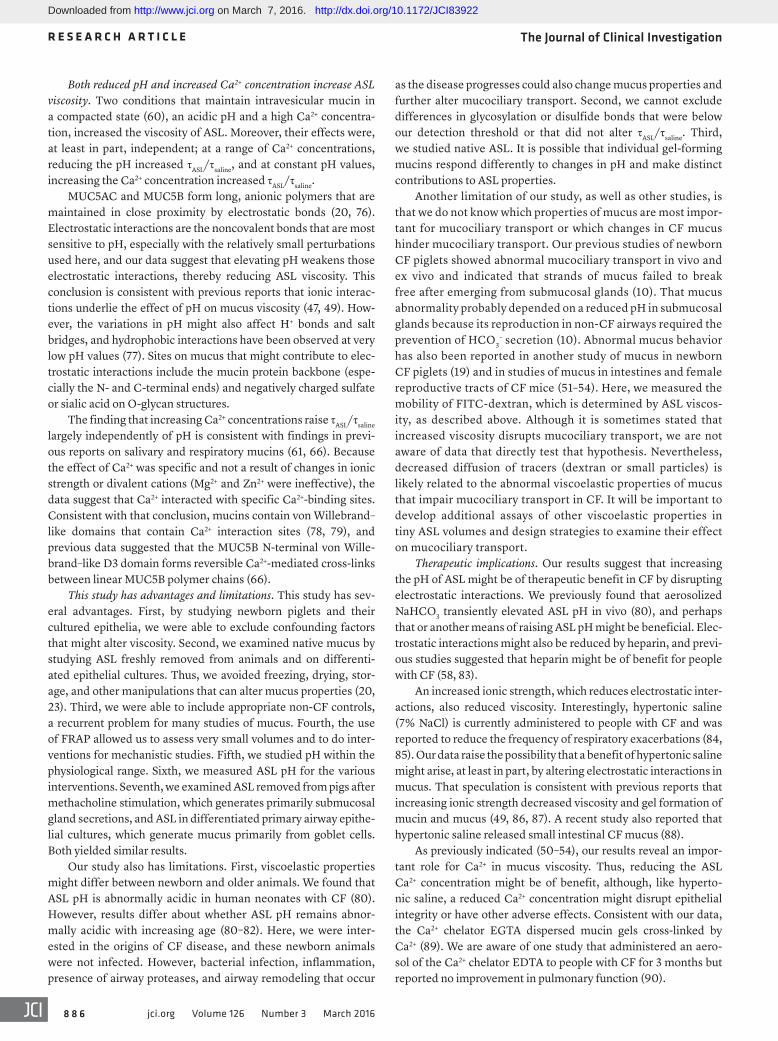
Increasing pH reduces the ASL viscosity. Previous studies showed that pH could affect the physical properties of mucins, mucus, and human sputum (46–49). In addition, there are reports that HCO3
– may be required for normal mucus properties in intestinal, repro-ductive, and airway epithelia (50–54). Our earlier data showed that loss of CFTR markedly reduces airway epithelial HCO3
– secretion and ASL pH in porcine airway epithelia (8, 37). Therefore, we tested the hypothesis that ASL viscosity is increased in CF because of a reduced pH and/or HCO3
– concentration. We assayed ASL pH using the fluorescent ratiometric pH indicator SNARF-1.
Forskolin and 3-isobutyl-1-methylxanthine (IBMX) stimulate HCO3
– secretion by increasing cellular levels of cAMP, causing phosphorylation and activation of CFTR. In non-CF epithelia, 10 μM forskolin and 100 μM IBMX applied for 2 hours increased ASL pH and reduced τASL/τsaline (Figure 4A). In CF epithelia, forskolin and IBMX had opposite effects, reducing ASL pH and increasing τASL/τsaline without altering ASL depth. A cAMP-induced reduction of ASL pH is consistent with results of a previous study (55). Adding NaHCO3 (3 μl, 150 mM) to the apical surface increased ASL pH and reduced τASL/τsaline to similar levels in both genotypes (Figure 4B). These results suggested that a reduced ASL pH and/or reduced HCO3
– concentration increased τASL/τsaline in CF. However, they do not differentiate between pH and HCO3
– as the major factor.pH, HCO3
–, and CO2 are related, as described by the Hender-son-Hasselbalch equation. To distinguish between the effects of pH and HCO3
–, we did five experiments. (a) First, we tested vari-able HCO3
– concentrations at a constant pH. We applied 21 mM NaHCO3/5% CO2 or 68 mM NaHCO3/15% CO2, both with a pH of 7.4, to non-CF ASL. The τASL/τsaline was the same (Figure 5A). (b) Second, we tested variable pH at a constant HCO3
– concentration.
and immediately measured the percentage of nonvolatile material in ASL (8). Under both basal and methacholine-stimulated condi-tions, CF ASL had an increased percentage of nonvolatile material (Figure 3A). This in vivo difference occurred in a setting in which we previously showed no difference in the depth of ASL (37). Likewise, in primary cultures of differentiated porcine airway epithelia, confo-cal microscopy revealed that the depth of undisturbed ASL did not differ by genotype (Figure 3B). These results are in accord with those of previous studies of undisturbed ASL in human airway epithelial cultures (31, 38) and with assays of in vivo ASL depth in humans with CF and CF pigs (37, 39). However, they differ from those of studies that added liquid to the apical surface or washed the apical surface and found that ASL depth was reduced in CF cultures (40).
As the concentration of sucrose increases, viscosity increases, with τ values increasing steeply as sucrose concentrations rise above approximately 30% (Supplemental Figure 2A). In addition, we also tested τmucin/τsaline at increasing concentrations of salivary mucins and increasing concentrations of gastric mucins, including under oxidized conditions (95% O2) to increase cross-linking (ref. 41 and Figure 3C). As the concentrations increased, ASL viscos-ity increased. The percentage of nonvolatile material in ASL col-lected after methacholine stimulation was 8.4% ± 0.6% for non-CF ASL and 10.8% ± 1.1% for CF ASL (Figure 3A), and τASL/τsaline was approximately 250% greater in CF ASL (4.3 ± 0.7) than in non-CF ASL (Figure 2A). For comparison, the difference between 8.4% and 10.8% salivary or gastric mucin predicts a 5% to 10% difference in τmucin/τsaline (Figure 3C). Thus, while the percentage of nonvolatile material has small effects, these results suggested that additional factors might have an even larger effect on ASL viscosity.
Several studies have reported that CF alters mucin sulfa-tion. However, reports have varied widely, with studies showing increased, decreased, and unaltered sulfation (27, 42–45). As described above, variations might result from secondary mani-festations, difficulty obtaining control samples, and analysis pro-cedures. We removed ASL from airway epithelia, released sulfate
Figure 4. Stimulating epithelial HCO3– secretion reduces ASL viscosity. (A) ASL pH, τASL/τsaline, and ASL depth measured in non-CF and CF primary airway
epithelial cultures before and after stimulation with 10 μM forskolin and 100 μM IBMX for 2 hours (F&I). Forskolin and IBMX were added to the basolateral medium. Each data point indicates epithelia from a different animal. (B) NaHCO3 or NaCl (3 μl, 150 mM) was added to the apical surface of non-CF (blue) and CF (red) cultured airway epithelia, and τASL/τsaline and ASL pH were measured 5 minutes later (mean ± SEM). n = 6 non-CF and 4 CF, each from different piglets. The dashed horizontal lines indicate the viscosity of saline. *P < 0.05 by unpaired Student’s t test.
Downloaded from http://www.jci.org on March 7, 2016. http://dx.doi.org/10.1172/JCI83922

The Journal of Clinical Investigation R e s e a R c h a R t i c l e
8 8 3jci.org Volume 126 Number 3 March 2016
pH-insensitive reducing agent tris(2-carboxyethyl)phosphine (10 mM, 1 hour) to ASL decreased τASL/τsaline in both non-CF and CF epithelia, abolished the difference between genotypes, and elim-inated the increased τASL/τsaline produced by 15% CO2 (Figure 6, A and B). Lack of a pH effect when mucin networks are broken into their subunits by disulfide reduction (20–23) suggests that pH alters viscosity by influencing mucin structure.
We considered the possibility that an acidic ASL pH might increase viscosity by inducing the formation of additional disulfide bonds. To test that possibility, we added iodoacet-amide (IAA, 25 mM, 15 minutes) to ASL; IAA caps free thiols of cysteines to prevent new disulfide bond formation. We then increased CO2 from 5% to 15%. Reducing pH increased τASL/τsaline, even in the presence of IAA (Figure 6C and Supplemental Figure 6A). As a control for the effectiveness of IAA, we applied 95% O2 to the cultured airway epithelia in a humidified cham-ber for 15 minutes to induce oxidation and disulfide bond for-mation (41). As expected, IAA prevented the oxidation-induced increase in τASL/τsaline (Figure 6D and Supplemental Figure 6B). These data suggest that an acidic pH does not increase viscosity by inducing formation of additional disulfide bonds.
pH affects ASL viscosity by altering electrostatic interactions. These findings and the reversible effects of ASL pH suggested that pH influences noncovalent interactions in ASL. The noncovalent inter-
We applied 3 μl of 24 mM NaHCO3, with either 5% CO2 or 15% CO2, to non-CF cultured airway epithelia. At the reduced pH, τASL/τsaline increased (Figure 5B). (c) Third, we tested an increased HCO3
– concentration and a reduced pH. Without adding liquid to the ASL, we increased CO2 from 5% to 15%, which reduced pH and, according to the Henderson-Hasselbalch equation, likely also increased HCO3
– concentration. τASL/τsaline increased (Figure 5C). (d) Fourth, we tested buffering pH with HEPES in HCO3
–/CO2-free solution. As pH fell, τASL/τsaline increased (Figure 5D and Supplemental Figure 5). (e) Last, in experiments a–d, we mea-sured τASL/τsaline on primary cultures of airway epithelia. We also tested ASL freshly removed from newborn piglets. We varied the CO2 concentration in an enclosed and humidified chamber and found that for both CF and non-CF ASL, as ASL pH fell, τASL/τsaline increased (Figure 5E).
Thus, although HCO3– is the major ASL pH buffer (56), and
HCO3– secretion is an important factor determining ASL pH, these
results indicate that it is pH and not the HCO3– concentration itself
that influences ASL viscosity.An acidic pH does not increase ASL viscosity by inducing disulfide
bond formation. Mucin structure depends on interchain disulfide bonds (20–23). Because mucins are the major structural protein in ASL, we predicted that disrupting disulfide bonds would reduce ASL viscosity and eliminate the effect of acidification. Adding the
Figure 5. Increasing ASL pH reduces ASL viscosity. (A) 21 mM NaHCO3 (3 μl, 5% CO2) or 68 mM NaHCO3 (3 μl, 15% CO2) was added to the apical surface of non-CF cultured airway epithelia; HCO3
– concentration and CO2 were balanced to achieve the same pH. τASL/τsaline and ASL pH were measured 5 minutes later. n = 9 epithelia per genotype, each from a different pig. (B) 24 mM NaHCO3 (3 μl with 5% or 15% CO2) was applied to the apical surface of non-CF cultured airway epithelia. Five minutes later, τASL/τsaline and ASL pH were measured. n = 6 epithelia per genotype, each from a different pig. (C) Non-CF cul-tured airway epithelia were exposed to 5% or 15% CO2 in a humidified chamber at 37°C. τASL/τsaline and ASL pH were measured 5 minutes later. n = 6 epithe-lia per genotype, each from a different pig. (D) To eliminate HCO3
–/CO2, HEPES buffer (3 μl, 20 mM in saline) at a pH of 6.8 or 7.8 was applied to the apical surface of non-CF cultured airway epithelia. τASL/τsaline and ASL pH were measured 5 minutes later (see also Supplemental Figure 5, which indicates that changes in viscosity were not due to HEPES per se). n = 6 epithelia per genotype, each from a different pig. (E) Methacholine-stimulated ASL was collected from newborn non-CF (blue) and CF (red) pigs after methacholine stimulation and immediately assayed for τASL/τsaline and pH in a humidified chamber containing either 5% or 15% CO2. The line is a linear regression. n = 5 pigs per genotype; littermate controls were used. The dashed horizontal lines indicate the viscosity of saline. *P < 0.05 by unpaired Student’s t test. Mean ± SEM.
Downloaded from http://www.jci.org on March 7, 2016. http://dx.doi.org/10.1172/JCI83922

The Journal of Clinical Investigation R e s e a R c h a R t i c l e
8 8 4 jci.org Volume 126 Number 3 March 2016
actions that are most sensitive to changes in pH are ionic interactions (electrostatic bonds). We used two strategies to test the hypothesis that pH affects ASL viscosity by altering ionic interactions.
The acidic glycosaminoglycan chains contained in hepa-rin can disrupt ionic interactions (57, 58). We found that heparin reduced the τASL/τsaline of CF and non-CF ASL, both in the presence of 5% CO2 and when pH was reduced by 15% CO2 (Figure 7A and Supplemental Figure 7).
An increase in ionic strength can destabilize electrostatic interactions between mucin polymers. We previously found that the salt concentration in porcine ASL did not differ by genotype (8). We removed ASL from newborn piglets and added either 0.9% or 7% NaCl. Compared with isotonic saline (0.9%), 7% saline reduced τASL/τsaline (Figure 7B and Supplemental Figure 8). Adding hypertonic saline also decreased τASL/τsaline under more acidic 15% CO2 conditions. Conversely, adding water instead of 0.9% NaCl to dilute the ASL salt concentration increased τASL/τsaline (Supplemental Figure 9).
Increasing the Ca2+ concentration increases ASL viscosity. Find-ing that noncovalent interactions influence τASL/τsaline and previous evidence that Ca2+ may cross-link and stabilize mucins (59–61) led us to test the effect of Ca2+ concentration. The Ca2+ concen-trations of human and porcine ASL are approximately 4 mM and approximately 1 to 2 mM, and they are similar in CF and non-CF ASL (62–65).
We removed ASL from newborn piglets and varied the Ca2+ concentration. We studied ASL ex vivo in order to avoid effects on tight junctions and airway cells. At a pH of approximately 7.3, increasing Ca2+ increased τASL/τsaline (Figure 8A). Reducing pH to approximately 6.8 further increased τASL/τsaline by approx-imately 50% when Ca2+ was very low (20 mM EGTA) and at each Ca2+ concentration tested. Solutions containing two other divalent cations, MgCl2 or ZnCl2, did not reproduce the effects of CaCl2 and did not differ from a solution that contained NaCl at the same ionic strength (Figure 8B and Supplemental Figure 10). Thus, the effects of divalent cations were specific to Ca2+. These results suggest that pH and Ca2+ affect τASL/τsaline at least in part via different mechanisms. To test whether electrostatic interactions influence the effect of Ca2+, we maintained pH at
a constant of approximately 6.8 and added 7% NaCl; the Ca2+- induced increase in ASL viscosity was attenuated but not elimi-nated (Figure 8C and Supplemental Figure 11).
Earlier studies examined the interaction of Ca2+ with mucin (61, 66). Ca2+ binding to MUC5B was specific to Ca2+ over other divalent cations, and Ca2+ binding showed a component that was lost in the presence of NaCl and a component that was resistant to high NaCl concentrations (2 M). The specific, high-affinity Ca2+-binding sites may involve cysteine-rich, von Willebrand–like domains. Our data are consistent with those earlier studies and suggest that Ca2+ may elevate ASL viscosity by more than one mechanism.
Figure 6. An acidic pH does not increase ASL viscosity by inducing disulfide bond formation. (A) Effect of tris(2-carboxyethyl)phosphine (TCEP; 10 mM, 3 μl in PBS for 1 hour) on τASL/τsaline of cultured non-CF and CF airway epithelia. n = 6 epithelia per condition, each from a different pig. (B) Effect of tris(2-carboxyethyl)phosphine on τASL/τsaline of cultured non-CF airway epithelia exposed to 5% or 15% CO2. n = 5 epithelia per condition, each from a different pig. (C) Effect of IAA (25 mM, 3 μl in PBS for 15 minutes) on τASL/τsaline of cultured non-CF airway epithelia exposed to 5% or 15% CO2 (see also Supplemental Figure 6A for ASL pH.). n = 6 epithelia per condition, each from a different pig. (D) Effect of 25 mM IAA on the τASL/τsaline response to an oxidizing environment (95% O2 for 15 minutes) (see also Supplemental Figure 6B for ASL pH). n = 6–7 epithelia per condition, each from a different pig. The dashed horizontal lines indicate the viscosity of saline. *P < 0.05 by unpaired Student’s t test. Mean ± SEM.
Figure 7. pH affects ASL viscosity by altering ionic interactions. (A) Effect of heparin (3 μl of 1 mg/ml in PBS for 2 hours) on τASL/τsaline of non-CF and CF cultured airway epithelia exposed to 5% or 15% CO2 (see Supplemental Figure 7 for ASL pH). n = 5–6 per condition, each from a different pig. (B) Effect of adding 0.9% or 7% NaCl (4 μl added to 10 μl ASL for 30 minutes) on τASL/τsaline. ASL was collected from non-CF and CF piglets that were stimulated with methacholine. ASL was exposed to 5% or 15% CO2 (see Supplemental Figure 8 for ASL pH). n = 6 per condition, each from a different pig. The dashed horizontal lines indicate the viscos-ity of saline. *P < 0.05 by unpaired Student’s t test. Mean ± SEM.
Downloaded from http://www.jci.org on March 7, 2016. http://dx.doi.org/10.1172/JCI83922

The Journal of Clinical Investigation R e s e a R c h a R t i c l e
8 8 5jci.org Volume 126 Number 3 March 2016
DiscussionOur results indicate that the viscosity of ASL is increased in CF. Although we cannot rule out subtle changes, the difference between CF and non-CF ASL is not attributable to a defect in mucin itself, but rather to the environment in which it resides, i.e., an abnormally acidic ASL. These findings link loss of CFTR to altered ASL viscosity; without CFTR-dependent HCO3
– secretion, ASL pH falls and increases viscosity. Thus, altered ASL viscosity is a primary defect that might contribute, at least in part, to the pathogenesis of CF. We also recognize that the subsequent conse-quences of infection, inflammation, and airway remodeling may further modify viscosity and fuel defective mucociliary transport.
The results address other potential hypotheses for an increased ASL viscosity in CF. First, studies of mouse intestine, reproductive tract, and A549 cells suggested that HCO3
– plays a role in deter-mining mucus viscoelastic properties (50–54). However, it has not been clear whether it is HCO3
– concentration or pH that is the key variable. Although HCO3
– is the major pH buffer in ASL (56) and although reduced HCO3
– secretion decreases the pH of CF ASL, our data indicate that it is pH, rather than the HCO3
– concen-tration itself, that affects viscosity. This result is consistent with previous data indicating that pH affects the physical properties of isolated porcine gastric mucin, bovine gallbladder mucin, human salivary mucin, mucus from primary cultures of human cervi-cal cells, and human sputum (47–49, 67–70). Some studies have emphasized a role for HCO3
– secretion in the release of mucus from tissue and noted that HCO3
– on the basolateral surface of intestine is required for normal mucus release (50–54). It might be possible that there are differential requirements for HCO3
– concentration versus pH in the restricted volume of the intestinal crypt or the airway submucosal gland that influence mucus prop-erties or interactions with cells.
Second, we did not find major CF-associated alterations in glycosylation. This result is consistent with that of a previous study that collected mucus secreted from human submucosal glands, thereby minimizing effects of airway infection (26), and a study that measured mucus secreted by cultures of human airway epithelia (71). Previous studies of CF sputum have reported both
increased and decreased sialylation (25, 28, 72, 73); perhaps dif-ferences between studies can be attributed, in part, to variations in the contribution and extent of primary versus secondary (infec-tion and inflammation) influences on ASL.
Third, we found increased sulfation of CF ASL. Earlier stud-ies have reported widely varying results, ranging from increased to decreased sulfation of CF mucus (27, 42–45). Secondary contribu-tions and difficulty in obtaining control samples might be respon-sible for the variation. A potential mechanism for altered sulfation is that loss of CFTR function may elevate pH in the Golgi complex, which could in turn alter the activity of enzymes performing post-translational modifications (74). Perhaps pH-dependent activity of terminal sulfotransferases changes mucin sulfation. However, other mechanisms might be involved, and some studies have not observed altered intracellular vesicle acidification (75). It has been speculated that increased sulfation might alter the biophysical properties of mucus or change the binding of bacteria or viruses (42). Our data do not exclude the possibility that increased sulfa-tion affects ASL viscosity, but they suggest that any such effects must be small relative to those induced by pH and Ca2+. The conclu-sion that altered sulfation does not have major effects on viscosity is also consistent with the findings of several earlier studies (51–54) and with the finding that acutely inhibiting HCO3
– and Cl– secretion in wild-type porcine airways reproduced the impaired mucociliary transport phenotype observed in CF airways (10, 34).
Fourth, we found that the percentage of nonvolatile material was increased in ASL from CF pigs. This result is consistent with those of studies of secretions from porcine submucosal gland ducts, porcine airways, and human sputum (34–36). An increased concentration of soluble or insoluble material can increase viscos-ity and may contribute to a greater viscosity of CF ASL. However, the data suggest that the increased viscosity in CF is not primarily attributable to the increased percentage of nonvolatile material. For example, at the same pH, ASL removed from CF and non-CF pigs had a similar τASL/τsaline (Figure 5E). The same was true with ASL on primary cultures of airway epithelia (Figure 4B). In addi-tion, both breaking disulfide bonds and disrupting electrostatic interactions equalized the viscosity of CF and non-CF ASL.
Figure 8. An increased Ca2+ concentration increases ASL viscosity. (A) Effect of Ca2+ concentration on τASL/τsaline at varying ASL pH values. ASL (10 μl) collected from newborn non-CF piglets was studied after addition of 20 mM EGTA, 10 mM NaCl, 10 mM CaCl2, or 100 mM CaCl2 in 4 μl saline containing 20 mM HEPES at a pH of 7.35 or 6.8. The final Ca2+ concentrations were calculated based on volume of addition, ASL volume, Ca2+ in added solution, and an ASL Ca2+ concentration of 3 mM. n = 7 for 20 mM EGTA, 10 mM NaCl, and 10 mM CaCl2 and n = 4 for 100 mM CaCl2; n refers to the number of pigs. (B) Effect on τASL/τsaline of adding 4 μl of saline (20 mM HEPES, pH 7.35) containing 10 mM CaCl2, 10 mM MgCl2, 10 mM ZnCl2, or 30 mM NaCl to 10 μl ASL for 30 minutes. ASL was collected from newborn non-CF pigs (see Supplemental Figure 10 for ASL pH). n = 6–7 per condition, each from a different pig. (C) Effect of 7% NaCl on Ca2+-induced increase in ASL viscosity. ASL was collected from newborn non-CF piglets and studied after addition of 20 mM EGTA (0 mM Ca2+) or 100 mM CaCl2 (30 mM Ca2+ calculated as described for A). Additions were in 4 μl of 0.9% or 7% NaCl containing 20 mM HEPES at pH 6.8 (see Supplemental Figure 11 for pH values). n = 6 per condition, each from a different pig. The dashed horizontal lines indicate the viscosity of saline. *P < 0.05 by 1-way ANOVA. Mean ± SEM.
Downloaded from http://www.jci.org on March 7, 2016. http://dx.doi.org/10.1172/JCI83922
The Journal of Clinical Investigation R e s e a R c h a R t i c l e
8 8 6 jci.org Volume 126 Number 3 March 2016
as the disease progresses could also change mucus properties and further alter mucociliary transport. Second, we cannot exclude differences in glycosylation or disulfide bonds that were below our detection threshold or that did not alter τASL/τsaline. Third, we studied native ASL. It is possible that individual gel-forming mucins respond differently to changes in pH and make distinct contributions to ASL properties.
Another limitation of our study, as well as other studies, is that we do not know which properties of mucus are most impor-tant for mucociliary transport or which changes in CF mucus hinder mucociliary transport. Our previous studies of newborn CF piglets showed abnormal mucociliary transport in vivo and ex vivo and indicated that strands of mucus failed to break free after emerging from submucosal glands (10). That mucus abnormality probably depended on a reduced pH in submucosal glands because its reproduction in non-CF airways required the prevention of HCO3
– secretion (10). Abnormal mucus behavior has also been reported in another study of mucus in newborn CF piglets (19) and in studies of mucus in intestines and female reproductive tracts of CF mice (51–54). Here, we measured the mobility of FITC-dextran, which is determined by ASL viscos-ity, as described above. Although it is sometimes stated that increased viscosity disrupts mucociliary transport, we are not aware of data that directly test that hypothesis. Nevertheless, decreased diffusion of tracers (dextran or small particles) is likely related to the abnormal viscoelastic properties of mucus that impair mucociliary transport in CF. It will be important to develop additional assays of other viscoelastic properties in tiny ASL volumes and design strategies to examine their effect on mucociliary transport.
Therapeutic implications. Our results suggest that increasing the pH of ASL might be of therapeutic benefit in CF by disrupting electrostatic interactions. We previously found that aerosolized NaHCO3 transiently elevated ASL pH in vivo (80), and perhaps that or another means of raising ASL pH might be beneficial. Elec-trostatic interactions might also be reduced by heparin, and previ-ous studies suggested that heparin might be of benefit for people with CF (58, 83).
An increased ionic strength, which reduces electrostatic inter-actions, also reduced viscosity. Interestingly, hypertonic saline (7% NaCl) is currently administered to people with CF and was reported to reduce the frequency of respiratory exacerbations (84, 85). Our data raise the possibility that a benefit of hypertonic saline might arise, at least in part, by altering electrostatic interactions in mucus. That speculation is consistent with previous reports that increasing ionic strength decreased viscosity and gel formation of mucin and mucus (49, 86, 87). A recent study also reported that hypertonic saline released small intestinal CF mucus (88).
As previously indicated (50–54), our results reveal an impor-tant role for Ca2+ in mucus viscosity. Thus, reducing the ASL Ca2+ concentration might be of benefit, although, like hyperto-nic saline, a reduced Ca2+ concentration might disrupt epithelial integrity or have other adverse effects. Consistent with our data, the Ca2+ chelator EGTA dispersed mucin gels cross-linked by Ca2+ (89). We are aware of one study that administered an aero-sol of the Ca2+ chelator EDTA to people with CF for 3 months but reported no improvement in pulmonary function (90).
Both reduced pH and increased Ca2+ concentration increase ASL viscosity. Two conditions that maintain intravesicular mucin in a compacted state (60), an acidic pH and a high Ca2+ concentra-tion, increased the viscosity of ASL. Moreover, their effects were, at least in part, independent; at a range of Ca2+ concentrations, reducing the pH increased τASL/τsaline, and at constant pH values, increasing the Ca2+ concentration increased τASL/τsaline.
MUC5AC and MUC5B form long, anionic polymers that are maintained in close proximity by electrostatic bonds (20, 76). Electrostatic interactions are the noncovalent bonds that are most sensitive to pH, especially with the relatively small perturbations used here, and our data suggest that elevating pH weakens those electrostatic interactions, thereby reducing ASL viscosity. This conclusion is consistent with previous reports that ionic interac-tions underlie the effect of pH on mucus viscosity (47, 49). How-ever, the variations in pH might also affect H+ bonds and salt bridges, and hydrophobic interactions have been observed at very low pH values (77). Sites on mucus that might contribute to elec-trostatic interactions include the mucin protein backbone (espe-cially the N- and C-terminal ends) and negatively charged sulfate or sialic acid on O-glycan structures.
The finding that increasing Ca2+ concentrations raise τASL/τsaline largely independently of pH is consistent with findings in previ-ous reports on salivary and respiratory mucins (61, 66). Because the effect of Ca2+ was specific and not a result of changes in ionic strength or divalent cations (Mg2+ and Zn2+ were ineffective), the data suggest that Ca2+ interacted with specific Ca2+-binding sites. Consistent with that conclusion, mucins contain von Willebrand–like domains that contain Ca2+ interaction sites (78, 79), and previous data suggested that the MUC5B N-terminal von Wille-brand–like D3 domain forms reversible Ca2+-mediated cross-links between linear MUC5B polymer chains (66).
This study has advantages and limitations. This study has sev-eral advantages. First, by studying newborn piglets and their cultured epithelia, we were able to exclude confounding factors that might alter viscosity. Second, we examined native mucus by studying ASL freshly removed from animals and on differenti-ated epithelial cultures. Thus, we avoided freezing, drying, stor-age, and other manipulations that can alter mucus properties (20, 23). Third, we were able to include appropriate non-CF controls, a recurrent problem for many studies of mucus. Fourth, the use of FRAP allowed us to assess very small volumes and to do inter-ventions for mechanistic studies. Fifth, we studied pH within the physiological range. Sixth, we measured ASL pH for the various interventions. Seventh, we examined ASL removed from pigs after methacholine stimulation, which generates primarily submucosal gland secretions, and ASL in differentiated primary airway epithe-lial cultures, which generate mucus primarily from goblet cells. Both yielded similar results.
Our study also has limitations. First, viscoelastic properties might differ between newborn and older animals. We found that ASL pH is abnormally acidic in human neonates with CF (80). However, results differ about whether ASL pH remains abnor-mally acidic with increasing age (80–82). Here, we were inter-ested in the origins of CF disease, and these newborn animals were not infected. However, bacterial infection, inflammation, presence of airway proteases, and airway remodeling that occur
Downloaded from http://www.jci.org on March 7, 2016. http://dx.doi.org/10.1172/JCI83922
The Journal of Clinical Investigation R e s e a R c h a R t i c l e
8 8 7jci.org Volume 126 Number 3 March 2016
liseconds. Time series images after photobleaching were collected at 400-millisecond intervals until maximal recovery was reached. Typ-ically, 5 to 6 recovery curves from different locations in each sample (2–4 μm below the surface) were obtained and averaged. The τ for fluorescence recovery were determined by regression analysis and are presented as τASL/τsaline.
ASL depth measurement. ASL depth measurements were done in unperturbed cell cultures (without prior washing) in a humidi-fied chamber at 37°C. ASL was stained with FITC-dextran (70 kDa, Sigma- Aldrich) added as a powder 2 hours before measurements. The cultured epithelia were covered with a high-boiling-point perfluoro-carbon (FC-70, 3M) to prevent evaporation. ASL depth was measured by z-scanning confocal microscopy (LSM 510 META, Zeiss, equipped with a ×40 water-immersion objective). ASL depth was determined from z-image stacks. Generally, at least 6 images from different loca-tions across the epithelial surface (away from the meniscus) were acquired and averaged for each epithelium.
Percentage nonvolatile material in ASL. Pigs were anesthetized as described above. The trachea was surgically exposed and accessed ventrally using heat cautery. We then made a small anterior incision through the tracheal rings using heat cautery to prevent bleeding. To ensure that air was completely humidified, animals were studied in a humidified chamber (100% relative humidity, 25°C–30°C). We col-lected ASL using a procedure designed to minimize the generation of excessive capillary forces during sampling. We fused thin lens paper (VWR Scientific Products) with Parafilm M (Pechiney Plastic Packag-ing) in an oven (205°C) for 70 to 90 seconds. This procedure reduced the volume of liquid that the paper would absorb and minimized evap-oration from the surface not touching ASL. We prepared 0.5- × 2-cm strips of this material, washed them 3 times in double-distilled water, and dried them overnight at 40°C. The Parafilm M–fused paper strips were weighed and then gently placed in contact with the luminal sur-face of the posterior trachea for 15 seconds. Immediately after removal from the trachea, the Parafilm M–fused paper strips were placed (paper side up with Parafilm M touching the weight apparatus) on a precision balance (Mettler Toledo XP26DR). A synchronized computer mea-sured mass 10 times per second for 200 seconds (BalanceLink; Met-tler Toledo) while evaporation occurred. The amount of ASL collected was determined by plotting mass versus time, fitting a 1-phase expo-nential decay to the data (GraphPad Prism 5; GraphPad Software Inc.), and extrapolating to mass at time 0, i.e., the time at which the strip was removed from the airway surface. We then dried the strips overnight at 40°C and determined the mass after evaporative removal of solvent. To determine the percentage of ASL that was nonvolatile, we divided the ASL solute mass by the total mass and multiplied by 100%. We measured the fraction of nonvolatile mass under basal conditions and 5 minutes after the delivery of methacholine (2.5 mg/kg, i.v.).
Measurement of sulfate in ASL. To measure the sulfate content in ASL, we collected ASL washed with PBS from airway epithelia cul-tured from newborn CF and non-CF piglets. Sulfate was released by acid hydrolysis. 50 μl of 200 mM HCl was added to ASL samples (15–50 μl) and incubated at 100°C overnight in screw-cap tubes. After cooling, the samples were neutralized by adding NaOH and then cen-trifuged at 12,000 g for 20 minutes to remove the insoluble material. The supernatants were diluted 1:10 in water, and 10 μl was injected into a Dionex high-performance anion-exchange chromatography (HPAEC) instrument for sulfate determination. The HPAEC system
Finally, these findings may also be relevant for other respira-tory diseases reported to have an acidic ASL pH, such as asthma (91), chronic obstructive pulmonary disease (92), and acute respi-ratory distress syndrome (93).
MethodsCFTR–/– and CFTR+/+ pigs. We previously reported the generation of CFTR–/– pigs (4). Animals were produced by mating CFTR+/– male and female domestic pigs. Newborn littermates were obtained from Exemplar Genetics. Animals were studied 8 to 15 hours after birth. Euthanasia was with intravenous Euthasol (Virbac).
Preparation of differentiated primary cultures of airway epithelia. Epithelial cells were isolated from the trachea and bronchi by enzy-matic digestion; seeded onto collagen-coated, semi-permeable mem-branes (1.13-cm2 polyester; Corning); and grown at the air-liquid inter-face, as previously described (94). Culture medium, a 1:1 mixture of DMEM/F12, was supplemented with 2% Ultroser G (PALL Corp.). Differentiated epithelia were used at least 14 days after seeding.
ASL pH measurement. ASL pH was measured as previously described (8). Briefly, to assess pH in primary cultures of airway epithe-lia, we used the fluorescent ratiometric pH indicator SNARF-1 conju-gated to dextran (Molecular Probes), which was prepared as a suspen-sion in perfluorocarbon, and 200 μl was added to the apical surface. Two hours later, epithelia were studied in a humidified, 5% CO2 atmo-sphere at 37°C on the stage of an inverted confocal microscope (Zeiss 510 Meta NLO). For assaying pH of ex vivo ASL, SNARF-1 powder was incubated with ASL and measurements were done in a humidified chamber. SNARF-1 was excited at 514 nm, and the ratio of fluorescence intensity measured at 580 nm and 640 nm was used to calculate pH.
ASL collection for viscosity and glycosylation studies. Collection of ASL from piglets was done in an enclosed humidified chamber (100% relative humidity, 25°C–30°C). Pigs were anesthetized with ketamine (20 mg/kg, intramuscularly) and xylazine (2 mg/kg, intramuscularly), and sedation was maintained with propofol (2 mg/kg, i.v.). The neck was dissected to expose the trachea. Tracheal secretion was stimu-lated with methacholine (2.5 mg/kg, i.v.). After approximately 5 min-utes, a small incision was made in the ventral tracheal wall, and sterile polyester-tipped applicators (Puritan Medical Products) were used to collect ASL as it traveled up the airway, thus minimizing contact with the tracheal wall. Applicators were inserted into microcentrifuge tubes, which were capped in the chamber to prevent evaporation. The tubes were briefly centrifuged to remove ASL from applicators, and the ASL was then pooled. We collected approximately 50 μl ASL from newborn CF piglets and up to approximately 100 μl ASL from non-CF piglets, which allowed for technical replicates and testing of interven-tions. Less volume from CF pigs is consistent with reduced volume secretion from CF submucosal glands (95). ASL was usually collected from non-CF and CF paired littermates studied on the same day and was used immediately after collection.
ASL viscosity measured by FRAP. Freshly collected ASL or ASL on the apical surface of cultured airway epithelia was stained by depos-iting FITC-dextran powder (70 kDa, Sigma-Aldrich). ASL viscosity was assessed by measuring FRAP of FITC-dextran using a confocal microscope (LSM 510 META, Zeiss). All imaging and bleaching were carried out in a single plane using the 488-nm laser line. Following acquisition of a baseline image, a 6- × 18-μm rectangular region was photobleached by increasing the laser intensity to 100% for 400 mil-
Downloaded from http://www.jci.org on March 7, 2016. http://dx.doi.org/10.1172/JCI83922
The Journal of Clinical Investigation R e s e a R c h a R t i c l e
8 8 8 jci.org Volume 126 Number 3 March 2016
MUC5B (1:5,000; LSBio) or MUC5AC (1:16,000; Novus Biologicals), detected with donkey anti-rabbit 680 and donkey anti-mouse 800 (1:10,000), and visualized on the Odyssey IR imager (LiCor).
MALDI-TOF mass spectra of O-glycans isolated from porcine air-way mucus. O-glycans were released from known amounts of pro-tein samples by the reductive β elimination method. Briefly, samples were taken in a known volume and an equal volume of 0.1 M NaOH containing 2 M NaBH4 to make the final concentration of 50 mM NaOH with 1 M NaBH4. The reaction was done at 45°C for 16 hours with continuous stirring. The samples were then cooled over an ice bath, and 30% aqueous HOAc was added drop wise to neutralize the solution. The reaction mixture was then passed over Dowex 50W cation exchange resin (Bio-Rad, 200 mesh) to remove sodium from the reaction mixture. Samples were then lyophilized, and the boric acid in the sample was removed by repeated evaporation with acidified methanol and absolute methanol, respectively. The O-glycans were further purified by passing the samples over Sep-Pak C18 1cc Vac Cartridges (Waters), and the flow through was col-lected and dried down by lyophilization prior to permethylation and MALDI mass spectrometry. The mass spectra were done exactly as described before, where the mass range was selected from 200 to 2,000 amu (96, 97). The identified masses were proposed accord-ing to published reports (96, 97).
Measurement of fucose, glucosamine, galactose, galactosamine, and mannose. Monosaccharides in ASL were determined by HPAEC with pulsed amperometric detection (HPAE-PAD) as previously described (98). Known volumes of sample were mixed with equal volumes of 4 N TFA to a final concentration of 2 N TFA and hydrolyzed at 100°C for 4 hours on a heating block (Reacti-Therm, Pierce). The unreacted acid was then removed by dry nitrogen flush, followed by coevapora-tion with 50% aqueous iso-propyl alcohol twice to ensure complete removal of acid. The samples were dissolved in a known volume of water and injected into an HPAEC-PAD instrument (ICS3000, Dionex). Monosaccharide profiling was performed using a Dionex CarboPac PA-1 column (4.0 × 250 mm, 10 μm), and quantification was performed by comparing the results with measured amounts of authentic standards injected as external calibrants. Separation of monosaccharides was performed using a combination of 3 solvents: Milli-Q water (Millipore), 100 mM NaOH containing 5 mM NaOAc, and 100 mM NaOH containing 250 mM NaOAc. An instrument-sup-plied standard quad waveform specific for monosaccharide analysis was used to obtain optimal detection.
Measurement of total and free Neu5Ac and Neu5Gc. Neu5Ac and Neu5Gc, present both as free and bound to N-/O-glycans, were quantified using 1,2-diamino-4,5-methylene dioxybenzene–tagged (DMB-tagged) sialic acid by RP-HPLC using a fluorescent detec-tor as previously described (99). For free sialic acid quantification, known amounts of sample were passed over the spin filter with a 3-kDa molecular weight cutoff (NANOSEP 3K OMEGA, Pall Life Sciences). The flow through was collected, dried, and tagged with DMB to form a fluorescent adduct prior to detection using HPLC-FL. Total sialic acid in the samples was analyzed following mild acid release using 2 N HOAc at 80°C for 3 hours. Acid from the reaction mixture was removed by vacuum evaporation. The reaction mixture was then dissolved in water and filtered over a 3-kDa molecular weight cutoff spin filtration unit (NANOSEP 3K OMEGA, Pall Life Sciences). The flow through–containing total sialic acid was then
used for ion chromatography consisted of a Dionex system, with an IonPac AS18 (4*250 mm) anion-exchange column and guard column. Separation was achieved using isocratic elution (38 mM NaOH for 15 minutes for 1 run) at a flow rate of 1.0 ml/min, and a conductivity detector was used for detection. Measurement of sulfate content was performed at the Complex Carbohydrate Research Center at the Uni-versity of Georgia.
Quantitative RT-PCR. We used PrimeTime chemistry (IDT) (for tracheal tissues) and SYBR Green (ABI) (for epithelial cultures) on an ABI 7500 Fast Real-Time PCR System to measure porcine MUC5B and MUC5AC mRNA. Briefly, tracheal tissue or epithelial cultures were col-lected in RNAlater (Ambion), and total RNA was isolated (RNeasy Lipid Tissue Mini Kit, Qiagen). First-strand cDNA was synthesized with ran-dom hexamers (SuperScript III, Invitrogen). Sequence-specific primers and probes for porcine MUC5B and MUC5AC and β-actin were from IDT. We used IDT PrimeTime and SYBR green primers (SYBR green forward: 5′-CTTCCACACAGCACAGCACT-3′; reverse: 5′-GGAAGTC-CAGGTCAAACCAC-3′; PrimeTime forward: 5′-CTCCCAAGTCGG-CATCAAG-3′; reverse: 5′-TTCTGGTCATTGGTGCAGG-3′; probe: 5′-56FAM/TCTCCCCCG/SEN/CCATCAGTCC/3IABkFQ-3′) to mea-sure MUC5AC. IDT PrimeTime primers and SYBR Green primers were used to measure MUC5B (SYBR green forward: 5′-GACTTTCATCC-CACCCCTCA-3′; reverse: 5′-ATGTGGGTGATGGCGGGCCT-3′; Prime-Time forward: 5′-CTTTCATCCCACCCCTCAC-3′; reverse: 5′-GTC-GAAGGTCTTGTAGTGGAAG-3′; probe: 5′-56FAM/TCTCCCCCG/ZEN/CCATCAGTCC/3IABkFQ-3′) and β-actin (Ss03376160_u1, ACTB) in separate reactions. For each tissue, amounts of mucin mRNA were nor-malized to β-actin mRNA. These normalized values were then expressed relative to that in non-CF MUC5AC.
Immunocytochemistry. Newborn pig airway epithelial cultures were rinsed briefly in PBS (Ca2+ and Mg2+ free), fixed in 4% para-formaldehyde (EMS), permeabilized with 0.3% TX-100 (Thermo Fisher), and blocked in Super-Block (Thermo Fisher) supplemented with 10% filtered normal goat serum (Jackson Immunologicals). Epi-thelia were immunostained with anti-MUC5B (1:2,500; Santa Cruz) or anti-MUC5AC (1:5,000; Novus Biologicals) and anti–β-catenin (1:100; Zymed). Subsequently, the filters were incubated in second-ary antibody, goat anti-mouse Alexa Fluor 488 (Molecular Probes) and goat anti-rabbit Alexa Fluor 568 and phalloidin 633 (Life Tech-nologies), mounted in Vectashield plus DAPI (Vector Labs), and visu-alized using an Olympus Fluoview FV1000 confocal microscope. Paired images were immunostained, visualized, and subsequently enhanced identically. Tracheas from newborns were excised and fixed in 2% paraformaldehyde at room temperature for 1 hour before freezing in OCT and storage at –80°C. Cryosections (7 μm) were permeabilized with 0.3% TX-100 (Thermo Fisher) and blocked in Super-Block (Thermo Fisher) supplemented with 10% filtered nor-mal goat serum (Jackson Immunologicals). Sections were immuno-stained and processed as above.
Western blotting. Airway mucus was collected from sedated new-born pig littermates after methacholine stimulation. An equal vol-ume of 2X sample buffer was added to 10 μl non-CF and CF mucus and incubated at 100°C for 20 to 30 minutes. Samples were run on 4% to 15% Tris-HCl gels (Thermo Fisher) with high-molecular-mass standards (Life Technologies). Electrophoresed gels were trans-ferred to PDF-FL (Millipore) overnight. Membranes were blocked in 0.01% casein buffer in PBS (Ca2+ and Mg2+ free), immunostained with
Downloaded from http://www.jci.org on March 7, 2016. http://dx.doi.org/10.1172/JCI83922

The Journal of Clinical Investigation R e s e a R c h a R t i c l e
8 8 9jci.org Volume 126 Number 3 March 2016
Author contributionsXXT, LSO, MJH, and MJW designed studies. XXT, LSO, MJH, TOM, PHK, JDM, and BC conducted experiments. XXT, LSO, MJH, AV, DAS, and MJW analyzed data. XXT, DAS, and MJW wrote the manuscript.
AcknowledgmentsThis work was funded by the NIH (HL091842, HL51670, HL11744, by R01GM32373 to A. Varki), by the Research Resource for Biomedical Glycomics (NIH P41GM10349010 to Parastoo Azadi at the Complex Carbohydrate Research Center), by the Cystic Fibrosis Foundation (Research Development Program, OSTEDG1410, STOLTZ14XX0), and by the Roy J. Carver Charitable Trust. D.A. Stoltz was funded by the Gilead Sciences Research Scholars Program in Cystic Fibrosis. M.J. Welsh is an investigator of the Howard Hughes Medical Institute.
Address correspondence to: Michael J. Welsh, Howard Hughes Medical Institute, University of Iowa Carver College of Medicine, 6332 PBDB, Iowa City, Iowa 52242, USA. Phone: 319.335.7619; E-mail: [email protected].
tagged with DMB as follows. Sialic acids were derivatized with DMB. Reactions consisted of 7 mM DMB, 18 mM sodium hydrosulfite, 1.4 M acetic acid, and 0.7 M 2-mercaptoethanol and were carried out for 2.5 hours at 50°C in the dark. DMB–sialic acid derivatives were then resolved by HPLC using a reverse-phase C18 column (Acclaim 120, 4.6 mm × 250 mm, 5 μm, Dionex). A mixture of 85% Milli-Q water, 7% methanol, and 8% acetonitrile was used as elution buffer at a flow rate of 0.9 ml/min. Detection of fluorescently labeled sialic acids was achieved at excitation and emission wavelengths of 373 nm and 448 nm, respectively.
Statistics. Data are generally presented as points from individual animals or epithelia from different animals, with mean ± SEM indi-cated by bars. For statistical analysis, we used an unpaired, 2-tailed Student’s t test to compare 2 groups and a 1-way ANOVA for multiple comparisons (Figure 8, B and C). Differences were considered statisti-cally significant at P < 0.05.
Study approval. The University of Iowa Animal Care and Use Committee approved all animal studies. Human samples for generat-ing airway epithelial cultures were collected with approval of the Uni-versity of Iowa Institutional Review Board.
1. Quinton P. Physiological basis of cystic fibro-sis: a historical perspective. Physiol Rev. 1999;79(suppl 1):S3–S22.
2. Cutting GR. Cystic fibrosis genetics: from molec-ular understanding to clinical application. Nat Rev Genet. 2015;16(1):45–56.
3. Ramsey BW. Management of pulmonary disease in patients with cystic fibrosis. N Engl J Med. 1996;335(3):179–188.
4. Rogers CS, et al. Disruption of the CFTR gene produces a model of cystic fibrosis in newborn pigs. Science. 2008;321(5897):1837–1841.
5. Ostedgaard LS, et al. The ΔF508 mutation causes CFTR misprocessing and cystic fibrosis-like dis-ease in pigs. Sci Transl Med. 2011;3(74):74ra24.
6. Stoltz DA, et al. Cystic fibrosis pigs develop lung disease and exhibit defective bacterial eradica-tion at birth. Sci Transl Med. 2010;2(29):29ra31.
7. Stoltz DA, Meyerholz DK, Welsh MJ. Origins of cystic fibrosis lung disease. N Engl J Med. 2015;372(4):351–362.
8. Pezzulo AA, et al. Reduced airway surface pH impairs bacterial killing in the porcine cystic fibrosis lung. Nature. 2012;487(7405):109–113.
9. Abou Alaiwa MH, et al. pH modulates the activ-ity and synergism of the airway surface liquid antimicrobials β-defensin-3 and LL-37. Proc Natl Acad Sci U S A. 2014;111(52):18703–18708.
10. Hoegger MJ, et al. Impaired mucus detachment disrupts mucociliary transport in a piglet model of cystic fibrosis. Science. 2014;345(698):818–822.
11. Chang EH, et al. Sinus hypoplasia precedes sinus infection in a porcine model of cystic fibrosis. Laryngoscope. 2012;122(9):1898–1905.
12. Wanner A, Salathe M, O’Riordan TG. Mucociliary clearance in the airways. Am J Respir Crit Care Med. 1996;154(6 pt 1):1868–1902.
13. Robinson M, Bye PT. Mucociliary clearance in cys-tic fibrosis. Pediatr Pulmonol. 2002;33(4):293–306.
14. Wine JJ, Joo NS. Submucosal glands and airway defense. Proc Am Thorac Soc. 2004;1(1):47–53.
15. Fahy JV, Dickey BF. Airway mucus function and dys-
function. N Engl J Med. 2010;363(23):2233–2247. 16. Lethem MI, James SL, Marriott C. The role of
mucous glycoproteins in the rheologic proper-ties of cystic fibrosis sputum. Am Rev Respir Dis. 1990;142(5):1053–1058.
17. Puchelle E, Bajolet O, Abely M. Airway mucus in cystic fibrosis. Paediatr Respir Rev. 2002;3(2):115–119.
18. Rubin BK. Secretion properties, clearance, and ther-apy in airway disease. Transl Respir Med. 2014;2:6.
19. Birket SE, et al. A functional anatomic defect of the cystic fibrosis airway. Am J Respir Crit Care Med. 2014;190(4):421–432.
20. Thornton DJ, Rousseau K, McGuckin MA. Struc-ture and function of the polymeric mucins in air-ways mucus. Annu Rev Physiol. 2008;70:459–486.
21. Bansil R, Turner BS. Mucin structure, aggrega-tion, physiological functions and biomedical applications. Curr Opin Colloid Interface Sci. 2006;11(2–3):164–170.
22. Ambort D, Johansson ME, Gustafsson JK, Ermund A, Hansson GC. Perspectives on mucus properties and formation — lessons from the bio-chemical world. Cold Spring Harb Perspect Med. 2012;2(11):a014159.
23. Lai SK, Wang YY, Wirtz D, Hanes J. Micro- and macrorheology of mucus. Adv Drug Deliv Rev. 2009;61(2):86–100.
24. Raynal BD, Hardingham TE, Thornton DJ, Shee-han JK. Concentrated solutions of salivary MUC5B mucin do not replicate the gel-forming properties of saliva. Biochem J. 2002;362(pt 2):289–296.
25. Xia B, Royall JA, Damera G, Sachdev GP, Cum-mings RD. Altered O-glycosylation and sulfation of airway mucins associated with cystic fibrosis. Glycobiology. 2005;15(8):747–775.
26. Schulz BL, et al. Mucin glycosylation changes in cystic fibrosis lung disease are not manifest in submucosal gland secretions. Biochem J. 2005;387(pt 3):911–919.
27. Schulz BL, et al. Glycosylation of sputum mucins is altered in cystic fibrosis patients. Glycobiology.
2007;17(7):698–712. 28. Shori DK, et al. Altered sialyl- and fucosyl-link-
age on mucins in cystic fibrosis patients pro-motes formation of the sialyl-Lewis X determi-nant on salivary MUC-5B and MUC-7. Pflugers Arch. 2001;443(suppl 1):S55–S61.
29. Thomsson KA, Carlstedt I, Karlsson NG, Karlsson H, Hansson GC. Different O-glycosylation of respiratory mucin glycopeptides from a patient with cystic fibrosis. Glycoconj J. 1998;15(8):823–833.
30. Meyerholz DK, et al. Loss of CFTR function produces abnormalities in tracheal development in neonatal pigs and young children. Am J Respir Crit Care Med. 2010;182(10):1251–1261.
31. Derichs N, Jin BJ, Song Y, Finkbeiner WE, Ver-kman AS. Hyperviscous airway periciliary and mucous liquid layers in cystic fibrosis measured by confocal fluorescence photobleaching. FASEB J. 2011;25(7):2325–2332.
32. Saltzman WM, Radomsky ML, Whaley KJ, Cone RA. Antibody diffusion in human cervical mucus. Biophys J. 1994;66(2 pt 1):508–515.
33. De Clercq B, Cleuren B, Deschout H, Braeck-mans K, Ameloot M. Distinguishing free and anomalous diffusion by rectangular fluores-cence recovery after photobleaching: a Monte Carlo study. J Biomed Opt. 2013;18(7):76012.
34. Trout L, King M, Feng W, Inglis SK, Ballard ST. Inhibition of airway liquid secretion and its effect on the physical properties of airway mucus. Am J Physiol Cell Physiol. 1998;274(2 pt 1):L258–L263.
35. Thiagarajah JR, Song Y, Haggie PM, Verkman AS. A small molecule CFTR inhibitor produces cystic fibrosis-like submucosal gland fluid secretions in normal airways. FASEB J. 2004;18(7):875–877.
36. Martens CJ, et al. Mucous solids and liquid secre-tion by airways: studies with normal pig, cystic fibrosis human, and non-cystic fibrosis human bronchi. Am J Physiol Lung Cell Mol Physiol. 2011;301(2):L236–L246.
37. Chen J-H, et al. Loss of anion transport without increased sodium absorption characterize new-
Downloaded from http://www.jci.org on March 7, 2016. http://dx.doi.org/10.1172/JCI83922

The Journal of Clinical Investigation R e s e a R c h a R t i c l e
8 9 0 jci.org Volume 126 Number 3 March 2016
born porcine cystic fibrosis airway epithelia. Cell. 2010;143(6):911–923.
38. Thiagarajah JR, Song Y, Derichs N, Verkman AS. Airway surface liquid depth imaged by surface laser reflectance microscopy. J Gen Physiol. 2010;136(3):353–362.
39. Griesenbach U, et al. Quantification of periciliary fluid (PCL) height in human airway biopsies is feasible, but not suitable as a biomarker. Am J Respir Cell Mol Biol. 2010;44(3):309–315.
40. Matsui H, et al. Evidence for periciliary liquid layer depletion, not abnormal ion composition, in the pathogenesis of cystic fibrosis airways dis-ease. Cell. 1998;95(7):1005–1015.
41. Yuan S, et al. Oxidation increases mucin polymer cross-links to stiffen airway mucus gels. Sci Transl Med. 2015;7(276):276ra227.
42. Rose MC, Voynow JA. Respiratory tract mucin genes and mucin glycoproteins in health and dis-ease. Physiol Rev. 2006;86(1):245–278.
43. Mohapatra NK, et al. Alteration of sulfation of glycoconjugates, but not sulfate transport and intracellular inorganic sulfate content in cys-tic fibrosis airway epithelial cells. Pediatr Res. 1995;38(1):42–48.
44. Scanlin TF, Glick MC. Terminal glycosyla-tion in cystic fibrosis. Biochim Biophys Acta. 1999;1455(2–3):241–253.
45. Zhang Y, Doranz B, Yankaskas JR, Engelhardt JF. Genotypic analysis of respiratory mucous sulfation defects in cystic fibrosis. J Clin Invest. 1995;96(6):2997–3004.
46. Lieleg O, Vladescu I, Ribbeck K. Characterization of particle translocation through mucin hydro-gels. Biophys J. 2010;98(9):1782–1789.
47. Bhaskar KR, et al. Profound increase in viscosity and aggregation of pig gastric mucin at low pH. Am J Physiol Cell Physiol. 1991;261(5 pt 1):G827–G832.
48. Smith BF, Peetermans JA, Tanaka T, LaMont JT. Subunit interactions and physical properties of bovine gallbladder mucin. Gastroenterology. 1989;97(1):179–187.
49. Veerman EC, Valentijn-Benz M, Nieuw Amerongen AV. Viscosity of human salivary mucins: effect of pH and ionic strength and role of sialic acid. J Biol Buccale. 1989;17(4):297–306.
50. Quinton PM. Cystic fibrosis: impaired bicar-bonate secretion and mucoviscidosis. Lancet. 2008;372(9636):415–417.
51. Chen EYT, Yang N, Quinton PM, Chin W-C. A new role for bicarbonate in mucus formation. Am J Phyisol Lung. 2010;299(4):L542–L549.
52. Garcia MA, Yang N, Quinton PM. Normal mouse intestinal mucus release requires cystic fibrosis transmembrane regulator-dependent bicarbonate secretion. J Clin Invest. 2009;119(9):2613–2622.
53. Gustafsson JK, et al. Bicarbonate and functional CFTR channel are required for proper mucin secretion and link cystic fibrosis with its mucus phenotype. J Exp Med. 2012;209(7):1263–1272.
54. Muchekehu RW, Quinton PM. A new role for bicarbonate secretion in cervico-uterine mucus release. J Physiol. 2010;588(pt 13):2329–2342.
55. Coakley RD, et al. Abnormal surface liquid pH regulation by cultured cystic fibrosis bron-chial epithelium. Proc Natl Acad Sci U S A. 2003;100(26):16083–16088.
56. Kim D, Liao J, Hanrahan JW. The buffer capacity
of airway epithelial secretions. Front Physiol. 2014;5:188.
57. Tang JX, et al. Anionic poly(amino acid)s dis-solve F-actin and DNA bundles, enhance DNase activity, and reduce the viscosity of cystic fibro-sis sputum. Am J Physiol Lung Cell Mol Physiol. 2005;289(4):L599–L605.
58. Broughton-Head VJ, Shur J, Carroll MP, Smith JR, Shute JK. Unfractionated heparin reduces the elasticity of sputum from patients with cys-tic fibrosis. Am J Physiol Lung Cell Mol Physiol. 2007;293(5):L1240–L1249.
59. Verdugo P, Aitken M, Langley L, Villalon MJ. Molecular mechanism of product storage and release in mucin secretion. II. The role of extra-cellular Ca++. Biorheology. 1987;24(6):625–633.
60. Verdugo P. Goblet cells secretion and mucogene-sis. Annu Rev Physiol. 1990;52:157–176.
61. Raynal BD, Hardingham TE, Sheehan JK, Thorn-ton DJ. Calcium-dependent protein interactions in MUC5B provide reversible cross-links in salivary mucus. J Biol Chem. 2003;278(31):28703–28710.
62. Joris L, Dab I, Quinton PM. Elemental compo-sition of human airway surface fluid in healthy and diseased airways. Am Rev Respir Dis. 1993;148(6 pt 1):1633–1637.
63. Kozlova I, Vanthanouvong V, Almgren B, Hog-man M, Roomans GM. Elemental composition of airway surface liquid in the pig determined by x-ray microanalysis. Am J Respir Cell Mol Biol. 2005;32(1):59–64.
64. Kozlova I, Nilsson H, Henriksnas J, Roomans GM. X-ray microanalysis of apical fluid in cystic fibrosis airway epithelial cell lines. Cell Physiol Biochem. 2006;17(1–2):13–20.
65. Potter JL, Matthews LW, Spector S, Lemm J. Stud-ies on pulmonary secretions. II. Osmolality and the ionic environment of pulmonary secretions from patients with cystic fibrosis, bronchiectasis, and laryngectomy. Am Rev Respir Dis. 1967;96(1):83–87.
66. Ridley C, et al. Assembly of the respiratory mucin MUC5B: a new model for a gel-forming mucin. J Biol Chem. 2014;289(23):16409–16420.
67. Espinosa M, Noe G, Troncoso C, Ho SB, Villalon M. Acidic pH and increasing [Ca(2+)] reduce the swelling of mucins in primary cultures of human cervical cells. Hum Reprod. 2002;17(8):1964–1972.
68. Celli JP, et al. Rheology of gastric mucin exhibits a pH-dependent sol-gel transition. Biomacromol-ecules. 2007;8(5):1580–1586.
69. Holma B. Influence of buffer capacity and pH- dependent rheological properties of respiratory mucus on health effects due to acidic pollution. Sci Total Environ. 1985;41(2):101–123.
70. Holma B, Hegg PO. pH- and protein-dependent buffer capacity and viscosity of respiratory mucus. Their interrelationships and influence on health. Sci Total Environ. 1989;84:71–82.
71. Holmen JM, et al. Mucins and their O-gly-cans from human bronchial epithelial cell cultures. Am J Physiol Lung Cell Mol Physiol. 2004;287(4):L824–L834.
72. Davril M, et al. The sialylation of bronchial mucins secreted by patients suffering from cystic fibrosis or from chronic bronchitis is related to the severity of airway infection. Glycobiology. 1999;9(3):311–321.
73. Dosanjh A, Lencer W, Brown D, Ausiello DA,
Stow JL. Heterologous expression of ΔF508 CFTR results in decreased sialylation of mem-brane glycoconjugates. Am J Physiol. 1994; 266(2 pt 1):C360–C366.
74. Barasch J, et al. Defective acidification of intra-cellular organelles in cystic fibrosis. Nature. 1991;352(6330):70–73.
75. Gibson GA, Hill WG, Weisz OA. Evidence against the acidification hypothesis in cystic fibrosis. Am J Physiol Cell Physiol. 2000;279(4):C1088–C1099.
76. Sheehan JK, et al. Identification of molec-ular intermediates in the assembly path-way of the MUC5AC mucin. J Biol Chem. 2004;279(15):15698–15705.
77. Barz B, Turner BS, Bansil R, Urbanc B. Folding of pig gastric mucin non-glycosylated domains: a discrete molecular dynamics study. J Biol Phys. 2012;38(4):681–703.
78. Xu AJ, Springer TA. Calcium stabilizes the von Willebrand factor A2 domain by pro-moting refolding. Proc Natl Acad Sci U S A. 2012;109(10):3742–3747.
79. Forstner JF, Forstner GG. Calcium binding to intestinal goblet cell mucin. Biochim Biophys Acta. 1975;386(1):283–292.
80. Abou Alaiwa MH, et al. Neonates with cystic fibrosis have a reduced nasal liquid pH: A small pilot study. J Cyst Fibros. 2014;13(4):373–377.
81. McShane D, et al. Airway surface pH in subjects with cystic fibrosis. Eur Respir J. 2003;21(1):37–42.
82. Garland AL, et al. Molecular basis for pH- dependent mucosal dehydration in cystic fibrosis airways. Proc Natl Acad Sci U S A. 2013;110(40):15973–15978.
83. Ledson M, Gallagher M, Hart CA, Walshaw M. Nebulized heparin in Burkholderia cepacia col-onized adult cystic fibrosis patients. Eur Respir J. 2001;17(1):36–38.
84. Elkins MR, et al. A controlled trial of long-term inhaled hypertonic saline in patients with cystic fibrosis. N Engl J Med. 2006;354(3):229–240.
85. Donaldson SH, et al. Mucus clearance and lung function in cystic fibrosis with hypertonic saline. N Engl J Med. 2006;354(3):241–250.
86. Crowther RS, Marriott C, James SL. Cation induced changes in the rheological properties of purified mucus glycoprotein gels. Biorheology. 1984;21(1–2):253–263.
87. Chace KV, Naziruddin B, Desai VC, Flux M, Sachdev GP. Physical properties of purified human respiratory mucus glycoproteins: effects of sodium chloride concentration on the aggre-gation properties and shape. Exp Lung Res. 1989;15(5):721–737.
88. Ermund A, Meiss LN, Scholte BJ, Hansson GC. Hypertonic saline releases the attached small intestinal cystic fibrosis mucus. Clin Exp Pharma-col Physiol. 2015;42(1):69–75.
89. Chin W-C, Orellana MV, Verdugo P. Spontaneous assembly of marine dissolved organic matter into polymer gels. Nature. 1998;391:568–572.
90. Brown J, Mellis CM, Wood RE. Edetate sodium aerosol in Pseudomonas lung infection in cystic fibrosis. Am J Dis Child. 1985;139(8):836–839.
91. Hunt JF, et al. Endogenous airway acidification. Implications for asthma pathophysiology. Am J Respir Crit Care Med. 2000;161(3 pt 1):694–699.
92. Kostikas K, et al. pH in expired breath condensate
Downloaded from http://www.jci.org on March 7, 2016. http://dx.doi.org/10.1172/JCI83922

The Journal of Clinical Investigation R e s e a R c h a R t i c l e
8 9 1jci.org Volume 126 Number 3 March 2016
of patients with inflammatory airway diseases. Am J Respir Crit Care Med. 2002;165(10):1364–1370.
93. Gessner C, et al. Exhaled breath condensate acidification in acute lung injury. Respir Med. 2003;97(11):1188–1194.
94. Karp PH, et al. An in vitro model of differentiated human airway epithelia: methods and evaluation of primary cultures. In: Wise C, ed. Epithelial Cell Culture Protocols. Vol. 188. Totowa, New Jersey, USA: Humana Press, Inc.; 2002:115–137.
95. Joo NS, Cho HJ, Khansaheb M, Wine JJ. Hypose-cretion of fluid from tracheal submucosal glands of CFTR-deficient pigs. J Clin Invest. 2010;120(9):3161–3166.
96. Carlson DM. Structures and immunochem-ical properties of oligosaccharides isolated from pig submaxillary mucins. J Biol Chem. 1968;243(3):616–626.
97. Morelle W, Faid V, Chirat F, Michalski JC. Analy-sis of N- and O-linked glycans from glycoproteins
using MALDI-TOF mass spectrometry. Methods Mol Biol. 2009;534:5–21.
98. Hardy MR, Townsend RR, Lee YC. Monosaccha-ride analysis of glycoconjugates by anion exchange chromatography with pulsed amperometric detec-tion. Anal Biochem. 1988;170(1):54–62.
99. Lewis AL, Nizet V, Varki A. Discovery and characterization of sialic acid O-acetylation in group B Streptococcus. Proc Natl Acad Sci U S A. 2004;101(30):11123–11128.
Downloaded from http://www.jci.org on March 7, 2016. http://dx.doi.org/10.1172/JCI83922