the journal of plastinationjournal.plastination.org/.../jp_vol.27.1/jp_vol.27.1_july15_full.pdf ·...
TRANSCRIPT
i
The official publication of the International Society for Plastination
The Journal of Plastination
I SSN 2 311 -77 61
Volume 27 (1); July 2015
IN THIS ISSUE:
Mathematically Quantifying
Learning Experience:
Correlating Magnetic Resonance
Imaging (MRI) and Plastinated
Brain Sections Using Utility
Analysis – p 7
Cleaning Excessive Cross-Linker
Crystallization on S10 Plastinated
Brain Slices - p 13
The Mannequins of
Dr Auzoux, an Industrial Success
in the Service of Veterinary
Medicine – p 18
Plastination of a Whole Horse for
Veterinary Education – p 29
Report on the 11th International
Interim Conference on
Plastination – p 33
i
The Journal of Plastination
ISSN 2311-7761 The official publication of the International Society for Plastination
Editorial Board:
Renu Dhingra New Delhi, India
Geoffrey D. Guttman Fort Worth, TX USA
Rafael Latorre Murcia, Spain
Scott Lozanoff Honolulu, HI USA
Ameed Raoof. Ann Arbor, MI USA
Mircea-Constantin Sora Vienna, Austria
Hong Jin Sui Dalian, China
Carlos Baptista Toledo, OH USA
Philip J. Adds Editor-in-Chief Institute of Medical and Biomedical Education (Anatomy) St. George’s, University of London London, UK
Robert W. Henry Associate Editor Department of Comparative Medicine College of Veterinary Medicine Knoxville, Tennessee, USA
Selcuk Tunali Assistant Editor Department of Anatomy Hacettepe University Faculty of Medicine Ankara, Turkey
Executive: Carlos Baptista, President Rafael Latorre, Vice-President Selcuk Tunali, Secretary Joshua Lopez, Treasurer
Instructions for Authors
Manuscripts and figures intended for publication in The Journal of Plastination should be sent via e-mail attachment to: [email protected]. Manuscript preparation guidelines are on the last two pages of this issue.
Cover: Latin American Flags courtesy of 3dflags.com

The Journal of Plastination 27(1):1 (2015)
Journal of Plastination Volume 27 (1); July 2015
Contents
Letter from the President, Carlos. A. C. Baptista 2
Letter from the Editor, Philip J. Adds 4
Mathematically Quantifying Learning Experience: Correlating Magnetic Resonance Imaging (MRI) and Plastinated Brain Sections Using Utility Analysis, Vijitashwa Pandey, Vipul Shukla, Carlos. A. C. Baptista
7
Cleaning Excessive Cross-Linker Crystallization on S10 Plastinated Brain Slices, Murad A. AlShehry, Maher M. AlObaysi, Nasser Al-Hamdan
13
The Mannequins of Dr Auzoux, an Industrial Success in the Service of Veterinary Medicine, Christophe Degueurce, Philip J Adds
18
Plastination of a Whole Horse for Veterinary Education, Sheng-Bo Yu, Jian-Fei Zhang, Yan-Yan Chi, Hai-Bin Gao, Jie Liu, Hong-Jin. Sui
29
11th International Interim Conference on Plastination, Carlos. A. C. Baptista, , Ana Paula S. V. Bittencourt, Yuri F. Monteiro, Laissa da S. Juvenato, Athelson S. Bittencourt
33
Abstracts from the 11th International Interim Conference on Plastination 37
Instructions for Authors 53

The Journal of Plastination 27(1):2 (2015)
LETTER FROM THE PRESIDENT
Carlos A. C. Baptista, MD, PhD
Dear Fellow Plastinators,
The 11th International Interim Conference on Plastination, held in Vitória, Brazil
was a new and exciting experience. This was the first Interim Meeting sponsored
by the ISP held in Latin America.
The origin of Plastination in Latin America goes back almost to the origin of
plastination itself. In 1983 Dr. Santiago Aja Guardiola, of Mexico, brought
plastination to his laboratory at the Facultad de Ciencias Veterinarias y
Zootecnia, Universidad Nacional Autónoma de México, México D. F. In 1984,
after a visit to the United States I set up the first laboratory of Plastination in
Brazil in the University of São Paulo. It was located in a small room at the school
of Medicine. It was improvised because I did not have a lot of resources at that
time. The silicone was given to me by a friend and colleague Dr. Philip Conran,
Professor at the Medical College of Ohio who bought S10/S3/S6 from Biodur and
shipped it to Brazil. I was joined by a colleague of the Department of Anatomy,
Dr. Esem Cerqueira and both of us produced our first plastinated specimens.
What an exciting time!
In 1990 during the 5th International Conference on Plastination held in
Heidelberg, Germany I was happy to encounter two colleagues from Brazil (I was
already living in the USA). They went to Heidelberg to learn the art of
plastination from Dr. Von Hagens: Professor Susanne Queiroz, Universidade
Federal do Rio de Janeiro, Brazil and Professor Aldo Junqueira Rodrigues Jr,
Faculdade de Medicina, Universidade de São Paulo. Both professors were very
active in plastination. Professor Queiroz retired recently (but continues
plastinating) and Professor Rodrigues unfortunately passed away in 2009.
The laboratory of Dr. Athelson Bittencourt (Universidade Federal do Espirito
Santo) is the first state-of-the-art plastination laboratory built in Brazil since Dr.
Queiroz and Rodrigues built their laboratories. In fact Dr. Bittencourt’s laboratory
is equipped to perform the three basic plastination techniques, that is, silicone,
epoxy and polyester.
For almost 30 years plastination was dormant in Brazil. I am delighted to see so
much interest for plastination in Brazil. The attendance at the Interim
Conference in Vitoria was superb and it was a testament to the renewed interest
in the technique. The success of the Interim Meeting was a tribute to Dr.
Bittencourt, his staff and students who organized an amazing meeting.
Plastination in Latin America is flourishing. Since 1983 and 1984 many
laboratories of plastination have been created. Here is a list (please forgive me if

The Journal of Plastination 27(1):3 (2015)
the list is not complete) of laboratories by country: (list courtesy of Dr. Nicolas
Ottone):
Argentina: Instituto de Morfología J. J. Naón, Facultad de Medicina, Universidad
de Buenos Aires ; Cátedra de Anatomía, Facultad de Veterinaria, Universidad de
Buenos Aires.
Brazil: Universidad Federal de Espirito Santo, Vitoria, Espirito Santo; Universidad
de Sao Paulo, Sao Paulo; Universidade Federal do Rio de Janeiro; Universidade
Federal Fluminense, Niteroi, Rio de Janeiro ; Universidade de Campinas, Sao
Paulo.
Chile: Laboratorio de Plastinación y Técnicas Anatómicas, Facultad de
Odontología ; Universidad de La Frontera, Temuco ; Facultad de Veterinaria,
Universidad Santo Tomás, Santiago de Chile ; Universidad de los Andes, Santiago
de Chile ; Universidad Austral, Valdivia.
Colombia: Fundación Universitaria Autónoma de las Américas, Pereira ;
Universidad de Antioquía, Medellín; Universidad del Cauca, Popayán.
Costa Rica: Instituto Nacional de Anatomía, San José.
México: Facultad de Ciencias Veterinarias y Zootecnia, Universidad Nacional
Autónoma de México, México D. F. ; Facultad de Medicina, Universidad Nacional
Autónoma de México, México D. F.; Universidad Autónoma de Aguascalientes,
Aguascalientes ; Instituto Politécnico Nacional, México D. F.
Facultad de Medicina Veterinaria y Zootecnia, Universidad Autónoma del Estado
de México, Mexico D. F. ; Escuela de Medicina Veterinaria y Zootecnia ;
Universidad Autónoma B. Juárez de Oaxaca. Oaxaca ; Escuela Nacional
Preparatoria Miguel E. Schulz Universidad Nacional Autónoma de México,
México D. F.
Perú: Universidad Peruana Cayetano Heredia, Lima; Universidad Nacional de San
Antonio Abad del Cusco, Cusco; Universidad Particular Andina del Cusco, Cusco.
Universidad Alas Peruanas, Lima.
I am looking forward to our next conference, the 18th International conference
on Plastination that will be held in the city of Toledo, Ohio, USA. Like the Interim
Meeting in Vitoria it will be an excellent opportunity for the exchange of ideas
and promotion of plastination.
The family of plastinators is growing! Isn’t that Great!
Warmest Regards
Carlos A. C. Baptista
President

The Journal of Plastination 27(1):4 (2015)
LETTER FROM THE EDITOR
Dear Readers,
The Problems of Specimen Preservation – a brief history
Two dates stand out as milestones in the recent history of anatomy: 1869 and
1977. It was in 1869 that the German chemist August Wilhelm von Hofmann
(1888-1892) formally identified formaldehyde (though its existence had been
reported earlier); and in 1977 Gunther von Hagens published his seminal paper on
the preservation of biological specimens by plastination (Bickley et al., 1981).
Prior to the discovery of formaldehyde, and its solution in water, formalin,
anatomical examination of the human
(or indeed any other) body, had to be
carried out speedily and preferably in
winter, so that the process of
putrefaction was slowed. Bodies sold to
the anatomy schools by the
“resurrection men” (grave robbers)
fetched higher prices in winter.
Dissections usually lasted three days,
with the abdominal and chest cavities
dissected on the first day, the head and
cranial cavity on the second day, and the
limbs on the third, following the body’s
own, pre-ordained order of decay. The most
celebrated depiction of a dissection, Rembrandt’s
“The Anatomy Lesson of Dr Nicolaes Tulp” (1532) is
remarkable for the fact that it shows the dissection
of the left arm, while the rest of the body remains
intact – clearly deviating from the accepted practice
of the time for artistic effect (Afek et al., 2009);
whereas “The Anatomy Lesson of Dr Deyman”,
painted much later, suggests that in this case, the
usual sequence has been followed (Fig 1).
The shortage of bodies for dissection and their
rapid decomposition inevitably led to other avenues
being explored in the quest for lasting anatomical
specimens. Small specimens could be preserved in
alcohol, suspended in glass jars (Fig 2.), though this
method was unsuitable for large specimens or
whole bodies (although in 1805, Admiral Nelson’s
body was brought back to London from the battle
of Trafalgar in a barrel of brandy) (Fig 3). There
were attempts to preserve bodies by dehydration in
alcohol, which met with varied success. The best-known exponent of this
Figure 1 - ‘Self-portrait with red brain’ (after Rembrandt), by Alex Rennie (original oil painting, based on Rembrandt’s ‘The Anatomy lesson of Dr Jan Deyman’), in the author’s collection
colle
Figure 2- Specimen of a child’s arm prepared by Frederik Ruysch (1638-1731), Kunstkamera, St Petersburg. http://www.kunstkamera.ru/en/museum_exhibitions/2floor/1st_collections/2_XIII_08/ (accessed 4/11/15)
Philip J. Adds, MSc, FIBMS

The Journal of Plastination 27(1):5
technique was the French anatomist Honoré Fragonard, cousin of the more
famous painter, Jean-Honoré. Fragonard injected the viscera and blood vessels of
his subjects with coloured wax before dehydration, and then applied a secret
varnish that greatly improved their preservation, to such an extent that
specimens prepared in the 1790s
can still be seen in the Fragonard
Museum near Paris (Degueurce et
al., 2010).
In the eighteenth and nineteenth
centuries, there was, notably in
Florence, a flourishing industry
producing models in wax.
Remarkable examples of the wax
model-makers’ art can be seen at
La Specola in Florence, the
Josephinum in Vienna, and in the
Gordon Museum at Guy’s Hospital
in London where the great model
maker Joseph Towne plied his trade
– or more accurately, his art – for
over 50 years (Fig 4). Attempts were
also made to reproduce anatomical
specimens in other materials such
as wood (Fig 5) and papier mâché.
With the discovery of formalin,
anatomical models became much less in demand, (though anatomical and clinical
models have enjoyed something of a renaissance over the last twenty years or
so). For nearly a century, nothing much changed in
anatomy until Gunther von Hagens burst on to the
scene in 1977. I think it would not be an
exaggeration to say that anatomy has been
transformed by these two events to a degree not
seen since the advent of Vesalius nearly five hundred
years ago.
In this issue of the Journal of Plastination, we publish
papers reflecting on the history – and future of
specimen preservation – from the days before
formalin, when an enterprising French doctor
starting mass-producing anatomical models of
horses made of papier mâché, to the cutting edge of
plastination technology today, with an account from
China on the production of a real, plastinated horse. The impact of plastination on
education, both medical and veterinary, has been immense, and this issue
includes a paper looking into quantifying the learning experience using
Figure 3 - The death of Admiral Lord Nelson at the battle of Trafalgar, 1805 (detail), by Daniel Maclise (1806-1870) https://www.pinterest.com/pin/9288742958375634/ (accessed 4/11/15)
Figure 4 - An example of the anatomical modelling of Joseph Towne (1808-1879), image courtesy of the Gordon Museum, Guy’s Campus, King’s College, London.
Figure 5 - Example of a skull carved from wood by John Hogan (1800-1858). Examples of Hogan’s work can be seen in the Crawford Art Gallery, Cork, Ireland http://www.crawfordartgallery.ie/index.html

The Journal of Plastination 27(1):6 (2015)
plastinated brain sections coupled with MRI images. Producing plastinated
specimens such as brain slices is not always without its pitfalls however, and this
issue contains an account from Egypt of problems encountered with long-term
storage of plastinated specimens.
With best wishes,
Phil Adds, Editor, the Journal of Plastination
References
Afek A, Friedman T, Kugel C, Barshack I, Lurie DJ. 2009: Dr. Tulp's Anatomy Lesson
by Rembrandt: the third day hypothesis. IMAJ 11: 389-92
Bickley HC, von Hagens G, Townsend FM. 1981: An improved method for
preserving of teaching specimens. Arch Pathol Lab Med 105:674-676.
Degueurce C, Duy SV, Bleton J, Hugon P, Cadot L, Tchapla A, Adds PJ. 2010: The
celebrated ecorchés of Honoré Fragonard. Part 2: The details of the technique
used by Fragonard. Clin Anat 23: 258-264

The Journal of Plastination 27(1):7-12(2015)
EDUCATIONAL ARTICLE
Mathematically Quantifying Learning Experience: Correlating Magnetic Resonance Imaging (MRI) and Plastinated Brain Sections Using Utility Analysis
Vijitashwa Pandey
1
Vipul Shukla2
Carlos A.C. Baptista3
1Department of
Industrial and Systems
Engineering
Oakland University
Rochester, MI 48309
USA 2,3
Department of
Neurosciences College
of Medicine, University
of Toledo, Ohio, USA
ABSTRACT: Objectives: Many researchers have shown that when used in conjunction, multiple pedagogic
approaches increase student learning. Diagnostic imaging is used extensively to complement
cadaveric dissection in courses such as neuroanatomy. This article provides a general framework
to analyze and quantify the learning utility from combining multiple teaching methods for a richer
learning experience. We present an example from neuroanatomy that combines the use of
Magnetic Resonance Imaging and plastinated specimens.
Materials and Methods: Two brains, from female cadavers aged between 70-90 years of age,
were removed from the body, fixed in 10% formalin (mixture of 10 pbv of 37% formalin with 90
pbv water) and stored for at least 6 months before use. After six months, each brain was washed
in tap-water overnight and sectioned coronally using a deli slicer. Slices measuring 10 mm in
thickness were produced which were then plastinated using the standard S10/S3 silicone method.
The plastinated brain slices were then used in conjunction with MRI images to analyze students’
preferences in neuroanatomy teaching.
Results: Our method first aims to understand the tradeoff preferences of the educators and the
students between multiple teaching methods. These preferences and tradeoff information can be
incorporated into a learning utility function - that brings a wealth of tools from decision analysis -
to analyze the proper allocation of teaching time between different methods. The synergistic
effect of using multiple teaching tools in anatomy classes is, therefore, formally quantified.
Conclusions: Using the example of MRI and plastinated specimens in neuroanatomy, we
showed how one can analyze tradeoff between two modalities. In other words, one can determine
how many hours of one modality can be traded off for another to have the same learning utility.
One can also deduce the best allocation of a fixed total number of hours to maximize learning
utility.
.
KEY WORDS: Neuroanatomy, learning, utility analysis, MRI, plastination, brain.
Vijitashwa Pandey, PhD, Department of Industrial and Systems Engineering, Address: 2200 N. Squirrel Road, Rochester MI 48309, Phone: (248) 370-4044, Fax: (248)-370-4625 Email: [email protected]
Introduction
Neuroanatomy is an essential course for healthcare
professional students that aim to impart knowledge
regarding the structure and development of the human
nervous system (Mateen and D'Eon, 2008). Alongside
diagnostic imaging (DI), current medical neuroanatomy
curricula utilize two-dimensional cross-sections of the
central nervous system to teach anatomy (Nolte and
Angevine, 2007). Students must integrate these two
dimensional images into a mental image, in order to
grasp the spatial relationships of neuroanatomical
structures within three dimensions. Due to time
constraints and the extent of knowledge required,
students find the task of visualizing three dimensional
structures from two dimensional cross sections arduous.
The complexity of the nervous system, including spatial
overlap of substructures, exacerbates this issue.
Emphasis is placed on the interpretation of diagnostic
images, which serves as another challenge for students
to master in a short period of time.
EDU
CA
TION
AL A
RTIC
LE

8 Pandey et al.
A recent study by Lujan and DiCarlo (2006)
demonstrated that pre-clinical students prefer multiple
styles (modalities) for learning and conceptualizing
anatomy. As a result, anatomy courses are constantly
supplemented with newer educational tools, including
plastinated specimens and diagnostic imaging, with
varying degrees of success. Plastination, a process
created by Gunther von Hagens in 1977, confers
durability to organs, which, in contrast to models, are
anatomically correct and non-toxic (Fig. 1) (Bickley et
al., 1981). A recent study by Hoffman et al. (2010) has
shown that the use of plastinated specimens as a sole
learning tool for anatomy even produces similar results
to traditional cadaveric dissection. As a result, there has
been increased utilization of plastinated specimens as
an aid to teach anatomy.
Figure 1 - Plastinated axial and coronal cross-sections of
the human brain.
Diagnostic imaging is another essential tool for bridging
the gap between structural and functional clinical
neuroanatomy. Medical students find the integration of
diagnostic imaging, such as MRI, into anatomy of great
importance towards gaining knowledge and preparing for
various clinical disciplines (Machado et al., 2013). In
particular, neural structures can be used to create a
simulated three-dimensional image for the region of
interest (Fig. 2). This facilitates crucial insight into
isolated images of pathology, as well as the normal
structure as a whole. It is a powerful and frequently used
diagnostic tool, making it extremely important to
understand in preclinical years.
Although numerous tools exist to help students learn
anatomy, some students are still fearful of the topic. This
anxiety stems from the difficulty of neuroanatomy and
the presence of numerous spatial orientations. The use
of plastinated specimens can aid in combating this issue.
The inability of students to conceptualize three-
dimensional neuroanatomical structures from a two-
dimensional image, such as an MRI, provides an added
level of difficulty when studying neuroanatomy. Although
neuroanatomical software helps to combat the
challenges faced by healthcare professional students, it
offers minimal aid to students with poor spatial skills
(Levinson et al., 2007). Plastinated specimens have the
potential to circumvent this limitation of neuroanatomical
software. In a typical curriculum, where time and
resources are limited, no study has as yet provided
insights into finding the optimal combination of the two
methods.
Figure 2 - MRIs of axial and coronal cross-sections of the
human brain.
In this paper, the practicable knowledge provided to the
students is termed learning utility, measured
quantitatively by a utility function. A utility function is a
representation of the preferences of the decision maker,
defined as the educator, in a mathematical form. It
assigns a numerical value to the outcomes, thus
measuring their desirability. Normative1 decision
analysis indicates that a rational decision maker
maximizes the expectation of this function when making
uncertain decisions. A utility function is scaled between 0
and 1, where 0 corresponds to the least acceptable
outcome and 1 corresponds to the best possible
outcome. A utility function can also be used in the
presence of uncertainty, i.e. it correctly ranks uncertain
alternatives. The framework, therefore, departs from the
ad-hoc techniques prevalent in medical education, by
using normative utility analysis. In this paper, we focus
primarily on MRIs and plastinated specimens, however
the methodology proposed is general enough to be
applied to other scenarios where the efficacy of multiple
teaching tools is to be evaluated. The purpose of this
1 Decision Analysis (DA) is termed normative as it prescribes
what a decision maker must do given his/her preferences. It is
not a descriptive field in that it does not try to understand how
people make decisions. The normativeness comes from DA
being founded in mathematics.

Mathematically Quantifying Learning Experience 9
study is to create a mathematical framework for
analyzing and quantifying the learning utility from
plastinated neuroanatomical specimens and diagnostic
imaging.
Materials and Methods
Specimen preparation
Fixation:
Two brains, from female cadavers aged between 70-90
years of age, were removed from the body and then
placed in a container of 10% formalin (mixture of 10 pbv
of 37% formalin with 90 pbv water) and stored for at
least 6 months before use. After six months, each brain
was washed in tap water overnight and sectioned
coronally using a deli slicer. Slices measuring 10 mm in
thickness were produced.
Dehydration by Freeze Substitution:
Brain slices were dehydrated using the freeze
substitution method (Schwab and von Hagens, 1981;
Tiedmann and Ivic ,1988; Henry, 2005). Freeze
substitution at -25° C in acetone is the recommended
dehydration procedure for minimal shrinkage of tissue
(Weber et al., 2007) and was utilized in this study. When
purity of acetone above 99.5% was achieved,
dehydration was considered complete and the
specimens were transferred to silicone for impregnation.
Forced Impregnation:
Brain sections were transferred quickly from the acetone
to the impregnation mixture and submersed in Silicone
S10/S3, (Biodur Products, Heidelberg) overnight. A grid
was used to keep the specimens submerged in the
resin. The following morning the vacuum pump was
turned on and the pressure in the vacuum chamber was
slowly decreased. Each day of impregnation, pressure
was decreased by 1/3 of the current pressure until the
pressure reached 220 mm (9 in) of mercury.. The
following day specimens were transferred to room
temperature, removed from the silicone bath and left to
drain the excess polymer.
Gas Curing/ Hardening:
Before curing, the specimens were blotted dry at room
temperature. Specimens were exposed to S6 (Biodur
Products, Heidelberg) vapor for three days, or until the
curing process was completed.
Results
Framework to measure interaction effects between
MRI and Plastination
Although multiple teaching methods may improve the
students’ learning utility understanding; an open
question remains – what is the optimal combination? A
general framework to quantify the combined utility from
multiple learning methods is needed. Every decision
maker has a tradeoff behavior between multiple
attributes they are considering for a given decision
situation. In our multiple teaching modalities example,
tradeoff behavior refers to how many hours of one
modality the educator is willing to sacrifice for another
(and vice versa), to keep the learning utility constant. For
example, when a curriculum recommends 10 hours each
of MRI and plastination modalities, an educator may be
indifferent towards sacrificing one hour of MRI for two
additional hours of plastination modality. Once this
information is encoded in a utility function, the tradeoff
decisions can be made relatively easily without constant
input from the educator.
To determine the mathematical expression for the
overall learning utility function, initial individual learning
utility curves which correspond to each modality must be
identified. Learning utility derived from MRI and that from
plastinated specimens is an increasing function of time
spent doing each. We utilize S-curves (Fig. 3) - a typical
learning curve used in literature. These functions can be
normalized between zero and one, where zero signifies
no learning utility to the student while one signifies the
maximum possible learning utility. An S-curve exhibits a
slow initial phase, an exponential growth in learning as a
function of time, followed by a leveling off of the curve,
as tmax is reached. Notice that tmax can be (and
generally would be) different for the two modalities. If the
curve levels off for one modality (e.g. MRI training) at a
certain time it signifies achievement of maximum benefit
from that methodology. The use of another modality, e.g.
plastinated specimens, is necessary for increased
learning and achievement. Of course, other functions are
possible such as exponential, linear or even stepwise
functions. Logistic function provides the advantage that it
can succinctly incorporate learning phases in a single
closed-form expression.
10 Pandey et al.
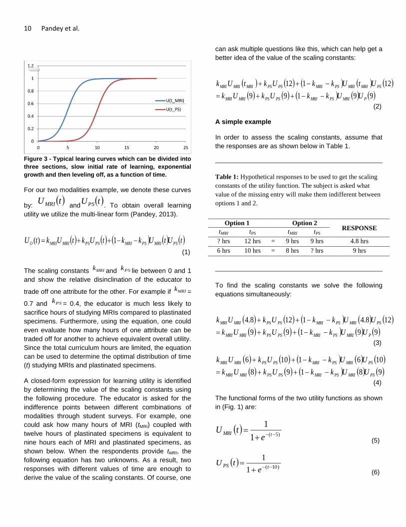
Figure 3 - Typical learing curves which can be divided into
three sections, slow initial rate of learning, exponential
growth and then leveling off, as a function of time.
For our two modalities example, we denote these curves
by: tU MRI and
tUPS . To obtain overall learning
utility we utilize the multi-linear form (Pandey, 2013).
tUtUkktUktUktU PSMRIPSMRIPSPSMRIMRIO 1)(
(1)
The scaling constants MRIk and PSk lie between 0 and 1
and show the relative disinclination of the educator to
trade off one attribute for the other. For example if MRIk =
0.7 and PSk = 0.4, the educator is much less likely to
sacrifice hours of studying MRIs compared to plastinated
specimens. Furthermore, using the equation, one could
even evaluate how many hours of one attribute can be
traded off for another to achieve equivalent overall utility.
Since the total curriculum hours are limited, the equation
can be used to determine the optimal distribution of time
(t) studying MRIs and plastinated specimens.
A closed-form expression for learning utility is identified
by determining the value of the scaling constants using
the following procedure. The educator is asked for the
indifference points between different combinations of
modalities through student surveys. For example, one
could ask how many hours of MRI (tMRI) coupled with
twelve hours of plastinated specimens is equivalent to
nine hours each of MRI and plastinated specimens, as
shown below. When the respondents provide tMRI, the
following equation has two unknowns. As a result, two
responses with different values of time are enough to
derive the value of the scaling constants. Of course, one
can ask multiple questions like this, which can help get a
better idea of the value of the scaling constants:
99199
12112
PMRIPSMRIPSPSMRIMRI
PSMRIMRIPSMRIPSPSMRIMRIMRI
UUkkUkUk
UtUkkUktUk
(2)
A simple example
In order to assess the scaling constants, assume that
the responses are as shown below in Table 1.
Table 1: Hypothetical responses to be used to get the scaling
constants of the utility function. The subject is asked what
value of the missing entry will make them indifferent between
options 1 and 2.
Option 1 Option 2 RESPONSE
tMRI tPS tMRI tPS
? hrs 12 hrs = 9 hrs 9 hrs 4.8 hrs
6 hrs 10 hrs = 8 hrs ? hrs 9 hrs
To find the scaling constants we solve the following
equations simultaneously:
99199
128.41128.4
PMRIPSMRIPSPSMRIMRI
PSMRIPSMRIPSPSMRIMRI
UUkkUkUk
UUkkUkUk
(3)
98198
1061106
PSMRIPSMRIPSPSMRIMRI
PSMRIPSMRIPSPSMRIMRI
UUkkUkUk
UUkkUkUk
(4)
The functional forms of the two utility functions as shown
in (Fig. 1) are:
)5(1
1
tMRI
etU
(5)
)10(1
1
tPS
etU
(6)

Mathematically Quantifying Learning Experience 11
1. Using these equations, one can find the
respective utility values and substitute them
into the simultaneous equations above to
obtain the values of the scaling constants.
These are MRIk = 0.5 and PSk =0.4. The
overall learning utility is given by:
tUtUtUtUtU PSMRIPSMRIO 1.04.05.0)(
(7)
Since1 PSMRI kk
, the two teaching methods are
complements, i.e. they provide more learning utility
together than the sum of their individual utilities. A sum
greater than one would have implied that they are
substitutes i.e., there would be a substantial overlap in
their contributions to the learning utility. We now look at
an isopreference curve (Fig. 4) corresponding to the
learning utility function in Equation 7. An isopreference
curve is such that a decision maker is indifferent
between the points on the curve i.e. as one moves along
the curve one attribute improves while the other worsens
just enough so that the net effect is cancelled. Using this
curve, one can make tradeoff decisions as to how many
hours of a modality can be substituted for another
without having any effect on the overall learning utility.
For example, one can see from the curve that 4 hours of
MRI coupled with 11.4 with plastinated specimens has
the same learning utility as that of 7 and 7.4 hours
respectively. In other words, if MRI time is increased by
3 hours, one could reduce the time spent studying
plastinated specimens by 4 hours.
Figure 4 - Isopreference curve for the overall learning
utility. The curve can be used to do tradeoff analysis
between two methods of learning.
One can also determine the optimal allocation of a fixed
number of study hours using the method described
above. In this case, we will maximize the overall utility
under the constraint of fixed number of hours. For
example, if the number of hours is fixed at 20 hours, the
optimal division is 7.6 hours for MRI and 12.4 hours for
plastinated specimens. Similarly, for a given learning
level, one could find the minimum total number of hours
required and the division between the two methods.
Discussion
The use of anatomical teaching tools or modalities,
including plastinated specimens and diagnostic imaging,
aids in teaching students structural and clinical
neuroanatomy. This article discussed the synergistic
effect of using multiple teaching tools in anatomy classes
and presented a method to formally quantify it. Many
researchers have shown that students’ learning
increases dramatically when many different modalities
are used in conjunction. Neuroanatomy can be
augmented by diagnostic imaging, just as MRI is used
extensively to complement cadaveric dissection.
Recently, plastinated specimens are also being used to
give students a better understanding of three-
dimensional structures. This immediately raises the
question of how much time should be spent doing each.
This paper provided a methodology for addressing this
issue by using utility analysis.
Our method first tries to understand the tradeoff between
multiple teaching modalities. These modalities can be
combined using a learning utility function, which provides
a wealth of tools, to analyze the proper allocation of
teaching time. The educators and students can be asked
for their preferences, which are then incorporated into a
learning utility function. Using the example of MRI and
plastinated specimens in neuroanatomy, we showed
how one can analyze tradeoff between two modalities. In
other words, one can determine how many hours of one
modality can be traded off for another to have the same
learning utility. One can also deduce the best allocation
of a fixed total number of hours to maximize learning
utility.
Normative utility theory is founded in mathematics and
the recommendations made by the proposed model will
best represent the educator and students’ preferences.
Although the approach presented demonstrates how
preferences can be modeled and incorporated into a
classroom, subsequent research will aim to validate the
approach utilized. Further research will consist of survey
data demonstrating the synergistic effects of using
plastinated specimens for teaching diagnostic imaging.

12 Pandey et al.
This will provide a formal way of allocating teaching
resources to maximize learning utility.
Acknowledgements
The authors would like to thank Dr. John Wall for help
with procuring the MRI images used in this paper.
References
Bickley HC, von Hagens G, Townsend FM. 1981: An
improved method for preserving of teaching
specimens. Arch Pathol Lab Med 105:674-676.
Henry RW. 2005: Silicone impregnation and curing. J Int
Soc Plastination 20:36-37.
Hoffmann D, May N, Thomsen, T, Holec M,
Andersen, K, Pizzimenti M. 2010: Medical students
using plastinated prosections as a sole learning tool
perform equally well on identification exams as
compared to those performing dissections over the same
regions. FASEB Journal 24:176.5.
Levinson AJ, Weaver B, Garside S, McGinn H, Norman
GR. 2007: Virtual reality and brain anatomy: a
randomized trial of e-learning instructional designs. Med
Educ 41:495-501.
Lujan HL, DiCarlo SE. 2006: First-year medical students
prefer multiple learning styles. Adv Physiol Educ 30:13-
16.
Machado JA, Barbosa JM, Ferreira MA. 2013: Student
perspectives of imaging anatomy in undergraduate
medical education. Anat Sci Educ 6:163-169.
Mateen, FJ, D'Eon MF. 2008: Neuroanatomy: a single
institution study of knowledge loss. Med Teach 30:537-
539.
Nolte J, Angevine, JB Jr. 2007: The Human Brain in
Photographs and Diagrams. 3rd
ed. St. Louis, MO:
Mosby, Inc. p. 272.
Pandey V. 2013: Decision based design. Taylor and
Francis, Boca Raton, FL USA, pp. 41-106.
Schwab K, von Hagens G. 1981: Freeze substitution of
macroscopic specimens for plastination. Acta Anat 111:
139-140.
Tiedemann K, Ivic-Matijas D. 1988: Dehydration of
macroscopic specimens by freeze substitution in
acetone. J Int Soc Plastination 2:2-12.
Weber W, Latorre R, Henry RW. 2007: Polyester
plastination of biological tissue: P35 technique. J Int Soc
Plastination 22: 50-58.

The Journal of Plastination 27(1):13-17 (2015)
TECHNICAL REPORT
Cleaning Excessive Cross-Linker Crystallization on S10 Plastinated Brain Slices
Murad A. AlShehry*
Maher M. AlObaysi
Nasser Al-Hamdan
King Fahad Medical City
(KFMC), Riyadh,
Kingdom of Saudi
Arabia
ABSTRACT: Tissue plastination is known to be an excellent method for preserving anatomical specimens. The
products are generally durable and usable for academic and clinical education. However,
prolonged periods of storage in changing temperature and humidity parameters can lead to
certain biological changes if not stored properly, which may include growth of opportunistic
organisms. This study reports a case of what seemed like a fungal growth on silicone plastinated
brain slices in our facility. In order to study the causative organisms we carried out macroscopic
as well as microscopic examinations of the isolated specimens. Characteristic feathery
crystallizations were largely seen on the white matter. After incubation of surface scrapings and
obtaining cultures on growth media, mycological analysis identified Aspergillus fumigates as the
causative organism, a common airborne fungi. Most of our collections of contaminated brain
slices have been tested, cleaned and finally disinfected using two methods; one was a method
published by Prinz et. al.(1999) the second was an idea to use an industrial laboratory surface
disinfectant (Virkon ®) commonly used in our hospital laboratories. After further investigations and
expert consultations, the crystals were confirmed to be a procedural error of adding extra cross-
linker from the source plastination laboratory and not in fact a fungal contamination.
.
KEY WORDS: S10 plastination; brain; fungal contamination; crystallizations; repair.
*Correspondence to: Faculty of Medicine, King Fahad Medical City, Riyadh, Saudi Arabia. Tel: +966562628669. Email: [email protected]
Introduction
Tissue plastination has been used for many years to
preserve anatomical and biological specimens. The
technique was developed in 1978 by Gunther von
Hagens of Heidelberg, Germany. The process takes the
form of replacing water and lipids in biological tissues
with polymers (silicone, epoxy, polyester) that are
impregnated and then hardened. The outcome is hard,
dry, odorless and durable specimens (von Hagens et al.,
1987).
In the high temperature climates and dry conditions of
Riyadh city in Saudi Arabia, our institution imported
readymade plastinated anatomical specimens for the
purpose of teaching medical students. Some of these
specimens were more than 5 years old and are stored at
room temperature (21-25° C) with central air conditioning
and dehumidification systems. However, the storage
facility encountered a long period of a malfunctioning air
conditioning system during summer vacation. In the
room in which our plastinated specimens were stored
the temperature reached 30° C to 35°
C over this period.
Additionally, the technologists reported that at one time a
broken fire sprinkler had flooded the laboratory floor over
the weekend during the same summer break, and this is
likely to have increased humidity levels in our storage
facility, although no direct damping or damage has been
reported. However, after several observations by our
staff regarding the change in appearance of some
plastinated items, we decided to isolate these specimens
and investigate the cause of these changes. Six S10
silicone plastinated specimens showing morphological
changes were initially examined: 2 brain slices (Figure
1), 1 lower limb, 1 thigh cross-section, 1 lung and 1
sagittal head and neck section (Figure 2).
TECH
NIC
AL R
EPO
RT

14 AlShehry et al.
Figure 1 - Visible crystallizations on silicone plastinated
brain slice
Figure 2 - Different anatomical specimens from the same
storage facility. Changes were seen as darkened areas. 1)
lower limb, 2) thigh cross-section, 3) lung 4) sagittal head
and neck section.
Some of the anatomical specimens showed minor wear
and tear marks with no deep penetration; these were not
included. Others had simply a layer of dust that was
easily brushed off and cleaned with a damp cloth. Out of
the 6 specimens collected, the most striking feature was
the accumulation of dry, white crystallization on the 2
brain slices. These were seen largely on the white
matter of the brain slices. It was of our own interest,
therefore, to harvest some of this crystallization for an
initial microbiological investigation. After the
investigation, the rest of our collection of S10 brain slices
was subjected to a planned treatment.
Materials and Methods
Testing For Infection
After consultation with our microbiology laboratory staff,
a specific protocol for obtaining cultures was followed.
Materials used for this analysis were:
Sabouraud dextrose agar Petri dishes and corn
meal dextrose agar Petri dishes for culture
media for fungi.
Sterile cotton swaps.
Microscope slides for analysis.
Lactophenol blue dye as a general mycological
dye.
Clear cellophane tape.
70% alcohol as disinfectant
Scrapings of suspected areas of growth on the initial six
specimens were inoculated onto labelled Sabouraud
dextrose agar and corn meal dextrose agar Petri dishes.
In addition, multiple swabs of the benches in the storage
area were taken as a control, and each swab was
inoculated onto similar media. These Petri dishes were
then placed in an incubator at 30°
C for 7 days. The
fungal colonies that grew were taken to a mycologist for
analysis in the main hospital's Microbiology laboratory,
where fungi isolates were identified. The traditional test
of adhesive tape preparation was initially carried out, this
helps to obtain a sample by using the adherent side of a
transparent cellophane tape. The carrier transparent
tape is then placed on top of a microscope slide on
which two drops of lactophenol cotton blue stain have
been placed; two more drops are then placed on top of
the tape, which is then covered with a cover slip. The
slides are then examined under a light microscope to
identify the fungus species (Forbes et al., 2007, pp.653,
657).
Disinfection
Disinfection was carried out using two methods:
A. The Prinz et al. (1999) chlorine
alcohol/formalin treatment
1. Manually brush the plastinated brain slices
while the sample is held under running cold tap
water, to remove the crystals (Figure 3). This
Cleaning Excessive Cross-Linker Crystallization 15
2. method helps to reduce the likelihood of the
crystals becoming airborne, and thus reduces
the risk of inhalation, which could be hazardous
to health.
3. Submerge the specimen in formalin for 5 min.
4. Immerse the specimen in an alcohol chlorine
solution (100mg of chlorine per 100 ml of
absolute alcohol) for 20mins.
5. Rinse with tap water.
6. Dry the specimen in a well-ventilated area.
B. The Virkon Method
1. Follow step 1 above.
2. Immerse the specimen in a freshly prepared
solution of 3% Virkon a few times.
3. Dry the specimen in a well-ventilated area.
The Virkon used is in powder form. The desired
concentration is 3% (i.e. 3 grams in every 100 ml of
water). The solution has a bright pink color when active.
However, the solution will lose this color within 7 days,
and consequently may lose its active properties.
Therefore, it is advised to prepare the solution on the
same day. When handling the powder form of Virkon it
is highly recommended to use gloves and a face mask,
as it may cause irritation to the skin or eyes, or to lungs
if inhaled.
Figure 3 - Brushing the crystallization while the brain slice
is submerged in water.
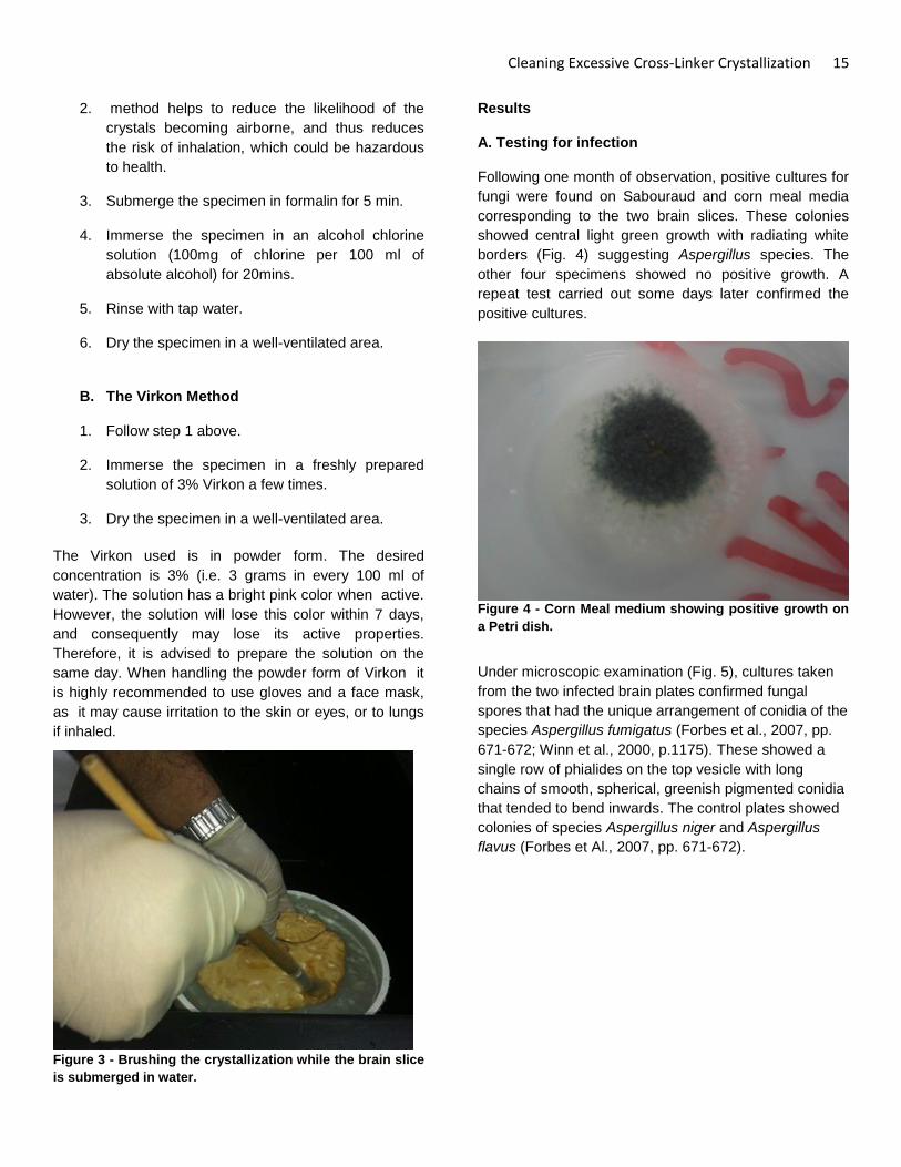
Results
A. Testing for infection
Following one month of observation, positive cultures for
fungi were found on Sabouraud and corn meal media
corresponding to the two brain slices. These colonies
showed central light green growth with radiating white
borders (Fig. 4) suggesting Aspergillus species. The
other four specimens showed no positive growth. A
repeat test carried out some days later confirmed the
positive cultures.
Figure 4 - Corn Meal medium showing positive growth on
a Petri dish.
Under microscopic examination (Fig. 5), cultures taken
from the two infected brain plates confirmed fungal
spores that had the unique arrangement of conidia of the
species Aspergillus fumigatus (Forbes et al., 2007, pp.
671-672; Winn et al., 2000, p.1175). These showed a
single row of phialides on the top vesicle with long
chains of smooth, spherical, greenish pigmented conidia
that tended to bend inwards. The control plates showed
colonies of species Aspergillus niger and Aspergillus
flavus (Forbes et Al., 2007, pp. 671-672).

16 AlShehry et al.
Figure 5 - Aspergillus fumigatus (x400)
The majority of the swabs taken from damaged brain
slices showed growth on Sabouraud media of common
airborne fungal species, such as Aspergillus fumigatus,
Penicillium expansum and Aspergillus niger. However,
the transparent tape impression on the crystallization did
not show clear signs of fungi, which made the diagnosis
of fungal infection inconclusive.
B. Disinfection
Both treatment methods showed excellent results;
neither of the brain slices was damaged by the solution
nor did they show any evidence of microbiological
growth after they had been dried. However, there were a
few crystal remnants in some fissures that were colored
with the Virkon solution's pink color (Fig. 6). This minor
pink discoloration was the only reported disadvantage.
Figure 6 - A plastinated brain slice after the Virkon
treatment showing some minor discoloration.
Disinfection Effectiveness test:
After drying the specimens, the brain slices were tested
for any microbiological contamination that may damage
the specimen in the future. The tests were done by
surface swabs on separate labelled Sabouraud media.
Discussion
It is not uncommonly reported that anatomical
specimens can host opportunistic organisms, especially
fungi, but not the plastinated ones. In the literature, a few
articles report cases of fungal contamination; Prinz and
colleagues (1999) reported fungal growth on plastinated
specimens from their institution in Brazil, and suggested
performing mycological test on suspected specimens.
They found that plastinated abdominal sections with
visible contamination had grown Aspergillus fumigatus,
whereas infested plastinated brain specimens showed
Penicillium janthinellum (Prinz et. al., 1999). This is
different to our finding of Aspergillus fumigatus with its
apparent preference to brain slices, as other specimens
tested negative for fungal growth. In another paper that
was published by Hammad and colleagues (2002),
fungal growth of Aspergillus species was reported under
similar conditions, but in formalin-fixed human cadavers.
However, they reported that it may be possible for
certain types of fungi to adapt and develop resistance to
low formalin concentrations. Plastinated tissue is
different in the sense that it lacks moisture, and almost
never requires closed-chamber storage when compared
to formalin-fixed specimens (Hammad et al., 2002).
According to Winn et al. (2000, p. 1178), some
Aspergillus species have a tendency to produce local
birefringent crystals in the form of deposits of calcium
oxalate in blood vessels in living humans. In addition,
cadavers lack vitality and immune defense mechanisms
to fight infections. Moreover, Forbes et al. (2007, p.669-
670) report clinical cases of Aspergillus fumigatus
infection in the central nervous system of
immunocompromised patients). These crystallization
processes in living beings are not the same as the
feathery crystallizations that were found on our isolated
plastinated brain specimens as seen in Figure 1.
The plastination process is considered to be complete
when all water molecules are completely substituted by
a polymer through the four steps of the plastination
process (von Hagens et al., 1987). Any area that is not

Cleaning Excessive Cross-Linker Crystallization 17
plastinated well enough could be considered as a target
for invasive fungi or bacteria, which may then start the
process of tissue decay. In this instance our brain slices
seem to be free from any decay. Some of our staff
technicians mistook an excess of cross-linker for fungal
growth due to their lack of knowledge of the plastination
process.
In summary, opportunistic fungal growth may occur on
silicone-plastinated brain slices. In cases of fungal
contamination it is advisable to follow the steps of
disinfection described above. Both the Prinz et al. (1999)
chlorine alcohol/formalin and Virkon treatments would
disinfect and remove any crystallization of the above-
mentioned fungal species on contaminated brain slices.
However, in the case reported here, we have concluded
that the crystals that we observed were caused by a
laboratory technical error of using excessive cross-linker,
and were in fact not a result of fungal colonisation.
Although the disinfection processes described here
caused a minor discoloration, this effect is barely
noticeable and insignificant (Fig. 6). In addition, it is
possible to use other disinfectants that are equal to
Virkon, however it must be borne in mind that the
solution should not be aggressive as this may damage
the specimen.
Recommendations
Attendance at the plastination meetings and workshops
is highly recommended. These meetings allow specific
problems to be addressed and guidance can be
obtained from the pioneers of this technology.
Furthermore, supervision of technical staff is highly
advised to assure mixing the correct amounts of solution
at the right concentrations.
Conclusion
Based on these findings, when white feathery
crystallizations are seen on S10 plastinated brain slices
they should not to be confused with fungal contamination
or decay. If these crystals are seen on plastinated
specimens they can be removed by using the
submerged brush method. On wet specimens it is
advised to follow one of the disinfection methods as a
precaution, to prevent any fungal growth.
Acknowledgments
This study was supported by King Fahad Medical City
KFMC, Faculty of Medicine Research Laboratories.
The Authors would like to thank Mr. Daniel Corcoran for
his expert consultation. Secondly, Mrs Nada M. Abutaleb
from department of Microbiology in KFMC for her
collaboration in producing this article. Finally, thanks to
Prof. Omar H K Kasule Sr., Dr. Abdulhakim Alshehri, Mr.
Radwan BaAbbad and Mr. Bader Alanazi for their
support.
References
Forbes B., Sahm D., and Weissfeld A. 2007: Bailey &
Scott’s Diagnostic Microbiology. 12th edition Mosby
Elsevier, St. Louis, Missouri, USA.
Hammad FE, Al-Janabi AA, Mohamed SA. 2002: Fungi
that grow on formalin-fixed cadavers. Saudi Med J 23(7):
871-872.
Prinz R., Correia J., Moraes A., da Silva A., Queiroz S.
And Pezzi L. 1999: Fungal Contamination of Plastinated
Specimens. J Plast, 14(2): 20-24.
Von Hagens G., Tiedemann K., Kriz W. 1987: The
current potential of plastination. Anat Embryol, 175(4):
411-421.
Winn, W Jr., Allen S., Janda W., Koneman E., Procop
G., Schreckenberger P. and Woods P. 2000: Koneman’s
Color Atlas and Textbook of Diagnostic Microbiology, 6th
Edition. Lippincott Williams & Wilkins, USA, p 1175,
1178.

The Journal of Plastination 27(1):18-28 (2015)
LEGACY
The Mannequins of Dr. Auzoux, An Industrial Success In The Service of Veterinary Medicine
Christophe Degueurce1
Philip J Adds2
1Conservateur du
Musée Fragonard
École Nationale
Vétérinaire d’Alfort
Maisons-Alfort, France
2 Division of Biomedical
Sciences (Anatomy)
St. George’s,
University of London
London, UK
ABSTRACT: Dr. Louis Auzoux (1797-1880) is well known for the anatomical models of papier mâché that he
produced and exported all over the world. Although the human models are more widely known,
they are by no means the only ones that the famous medical industrialist designed and marketed:
animals, plants and especially flowers are another facet of his art. Models of the horse were
especially important for Auzoux’s business. The paper horses, the sets of bone defects and jaws
that he created were purchased in great quantities by the French government of the day to
provide the materials needed for training recruits in a time of war. There was also a programme to
improve horse breeding throughout France through these fascinating objects. These magnificent
creations that were distributed all round the world, and which once were the pride of France, are
now damaged, ignored and dispersed. Sadly, they are now in great danger of being lost forever.
This historical review is an extensively revised translation of an article that was originally
published in French (Degueurce, 2013).
KEY WORDS: Auzoux, anatomical model, papier mâché, horse, honeybee, silkworm
Correspondence to: PJ Adds, Institute of Medical and Biomedical Education (Anatomy), St George’s University of London, Cranmer Terrace, London SW17 0RE UK. Tel: +44 (0) 208 725 5208, email [email protected]
Introduction
The mannequins of Dr Auzoux, an industrial success
in the service of veterinary medicine
The discovery in the National Archives of France of a
hitherto unexploited archive (Arch. Nat.) followed by
several visits to Saint-Aubin-d’Ecrosville, where
Auzoux’s factory was established, and conversations
with the curator of the Neubourg Museum2, have shed
new light on the life and legacy of this most industrious,
industrial doctor. This article summarises the authors’
research into Auzoux’s achievements with the domestic
animals (Degueurce, 2013).
Preservation and decay, the bane of the anatomist’s
life
Ever since the first anatomists attempted to explore the
body to reveal its structure, they despaired as their
careful dissections withered and, inevitably, decayed.
2 Musée de l’écorché d’anatomie, 54 Avenue de la
Liberation, 27110 Le Neubourg, France
Transforming the ephemeral into the durable became an
urgent priority, particularly in the second half of the 18th
century, when the demand for anatomical education
became much greater.
Many museum collections from this period display the
attempts that were made, which mainly fall into two
categories. The most common consisted of preserving
the whole body, or its parts, by dehydration or
immersion. Dehydration, or mummification, had the
advantage of eliminating cellular fluids, and thereby
preventing putrefaction. While Honoré Fragonard3
remains the best known practitioner of this technique,
there were many others who also used it to enrich the
museum collections of Faculties of Medicine and
‘cabinets of curiosities’ throughout Europe. This method,
however, had the drawback of reducing even the most
3 Honoré Fragonard (1732-1799) French doctor,
anatomist and veterinarian. Cousin of the painter Jean-Honoré Fragonard. Taught at the first Veterinary School in France (at Maison-Alfort). Some of his preserved specimens can still be seen in the Fragonard Museum near Paris.
LEGA
CY

The Mannequins of Dr. Auzoux 19
bulky muscles to thin, desiccated strips. Smaller
specimens could be submerged in a preserving bath,
which preserved their bulk, but it was difficult to use this
technique for whole-body specimens.
The second approach was to make a model – an
artificial representation – of the specimen before it
decayed, and this provided a challenge to the modellers,
artists and craftsmen of the day. Many different materials
were tried, including coloured wax, plaster and even
glass (Degueurce 2012a, p. 80). But the material that
achieved a global success in the 19th century – then only
to fall into total oblivion – was papier mâché. And papier
mâché will always be associated with the name of Louis
Auzoux (1797-1880), who exploited its possibilities so
well that he created a flourishing industrial enterprise,
producing anatomical models that, to this day, can still
be seen in the museums of five continents.
Louis Auzoux and his papier mâché anatomical
models
Louis Auzoux, doctor of medicine and brilliant inventor,
was born in 1797 into an affluent family of cultivators in
the village of Saint-Aubin-d’Ecrosville, about 100 km
West of Paris (Degueurce 2102b, p. 23-34). His
outstanding academic achievements, which led to a
doctorate of medicine in Paris, gave him access to the
medical celebrities of the day. The exact circumstances
which pushed him into launching himself into “the
anatomical industry” remain unclear, though what is
certain was his talent for self-publicity.
We know that he started his researches very early on,
and we know that his work cost him a great deal -
financially as well as intellectually. Originally, he was
inspired by Jean-François Ameline, Professor of
Anatomy at Caen, who enjoyed a modest success with a
novel type of anatomical mannequin, made from pieces
of card fixed on to a real skeleton. These mannequins
could be dismantled layer by layer, tracking the nerves
and vessels and revealing the anatomical relations of the
abdominal and thoracic viscera (Ameline, 1825 p. 5).
This ingenious idea was, however, rather limited, and
the small number of parts meant that it was a long way
from being an adequate substitute for a real dissection.
Auzoux, then a young student in Paris, became aware of
the process, and even travelled to Caen to visit
Ameline’s workshop. A short while after, in September
1822, he presented to the Academie royale de médecine
his own version, a “membre abdominal” with real bones
for its base, just like Ameline’s, making them direct
competitors (Collectif, 1825, 1). A new piece – a head,
neck and superior part of the trunk – soon attracted the
attention of the Government (Collectif, 1825, 2), which
then placed an order for a whole body mannequin.
Delivered in 1825, this piece contained an innovation
that would revolutionise its production: he used artificial
bones instead of the real skeleton. So began for the
doctor a fruitful career that led him to create several
hundred models, produced quasi-industrially, at an
affordable price which assured their wide distribution.
This prototype was revised, and led to the grand modèle
of 1830, which would be marketed, incredibly, right up to
the 1970s. There followed an anatomised female in the
position of the Venus of the Medicis, then numerous
models of organs, singly or assembled to form a region
of the body. In 1834, Auzoux dubbed his invention
‘anatomie clastique’, (clastic anatomy) from the Greek
klaeïn, to break up/separate, an allusion to the
educational dismantling of his various specimens (Anon.
1834, p. 453).
Nor did he stop at Man. As an eclectic naturalist and
keen zoologist, Auzoux also produced models of ’type
specimens’ of a variety of animals, including the turkey
“as the type specimen of the fowls”, a shark “as the type
specimen of the cartilaginous fishes”, and the cockchafer
“as the type specimen of the adult insects”. The series
was completed with the sea perch (fishes) the leech
(annelid worms), and the boa constrictor (reptiles).
Particular species of animals that were important for the
economy would become the object of even more
detailed models. For the bee, Auzoux created a virtuoso
production, showing, on a honeycomb, the stages of
development and the internal structure of the adults.
There followed the silkworm, with its butterflies of both
sexes and their caterpillars. As for the horse, it was to
hold a place of prime importance in the life and work of
Louis Auzoux, just as it did in the lives of his
contemporaries: draught animal, pivot of industry and
agriculture, animal of luxury for the haut monde, and
indispensable auxiliary of the army. It is easy to forget
how central the horse was to life in the 19th century, and
the interest that it sustained was universal, so it is not
surprising that the idea of an equine model followed
soon after his first human models.

20 Degueurce and Adds
The creation of the first equine model
The anatomical horse, which, as we shall see, was
extremely complex, occupied Louis Auzoux almost from
the beginning. Documents in the archive track the
progress of the project from its beginnings in 1842 in the
form of an exchange of letters with a young relative, an
officer cadet at the Royal Cavalry School of Saumur4 in
Western France. This young cadet informed Auzoux that
his Professor of Hippology, the Marquis de Saint-Ange5,
thought that a great advantage could be gained by using
an equine mannequin based on the human papier
mâché model that was already in use at the School. The
anatomical specimens they were using, dehydrated and
tarred6, were in poor condition, and could give only a
basic idea of the anatomy of the horse. Two years later,
Louis Auzoux wrote back, describing in detail the famous
horse, apparently now finalised; in his letter he explains
the considerable difficulties he had to overcome, in
particular from the lack of accurate anatomical
illustrations (Arch. Nat). He envisaged collaborating with
the Professor to produce a simplified, less expensive
version adapted for a wider distribution among soldiers
in the ranks.
To make his horse model, Auzoux first had to carry out a
completely original anatomical study, just as he had
done earlier for the human model. The file on the equine
model preserved in the National Archives confirms the
paucity of information that was available at that time, and
includes just one plate from the celebrated Cours
d’hippiatrique by Philippe-Étienne Lafosse (Lafosse,
1772) (Fig. 1). Animal anatomy was still a developing
science, and there was still much to learn about equine
anatomy. Auzoux describes his model thus:
4 The teenage cadets of the Cavalry School
distinguished themselves again in 1940, defending the town during the Battle of Saumur. 5 Charles Casimir Beucher, marquis de Saint-Ange
(1789-1879) 6 A tarred horse écorché can still be seen at the Centre
Sportif d’Équitation Militaire de Fontainbleau. As far as we know, it is the last remaining specimen of this type.
Figure 1 - Plate showing three engravings taken from the
Cours d’Hippiatrique by Phillippe-Étienne Lafosse (1772).
“The horse is at rest. I have taken the pose and the
balance from the training manual of M. Lecoq, professor
at the veterinary school of Lyon. For the anatomical
details, I have had to reproduce them after nature for
there no longer exists a complete anatomy of the horse:
the treatise of M. Rigot7 was helpful, but it covers only
the bones and muscles”.
Auzoux had to wait two more years before he could
submit his model to the Royal Academy of Medicine
(Renault, 1845). He also sought the judgment of his
fellow professionals: the archives contain several notes,
jotted down while veterinary teachers examined his
specimen, as well as numerous letters agreeing to
meetings for these evaluations. Auzoux greatly valued
their contribution:
“While occupied with the horse, I have had of necessity
to make numerous loans to the veterinary schools, either
for the classic design of the horse, or for the changes
introduced in certain organs” (Auzoux, 1858, p. XIV).
In the field of agriculture and breeding, Auzoux
benefitted greatly from the friendship of Antoine Richard
(Richard du Cantal)8, an extraordinary character: a
veterinarian but also doctor, farmer, agronomist, one
time teacher and Director of the School of Stud farms,
before following a political career. Richard published
7 Félix Rigot (1803-1847) appointed Professor of
Anatomy at the École royale vétérinaire d’Alfort in 1838. Published a series of papers on the anatomy of the horse. 8 Antoine Richard du Cantal (1802 – 1891) French
doctor, veterinarian, agronomist and politician. In 1854 he and Geoffroy Saint-Hilaire founded the Zoological Society of Acclimatization.

The Mannequins of Dr. Auzoux 21
several important works on the improvement of the
horse, in which he stressed the importance of the
structure and mechanics of the animal’s body (Richard,
1847).
Auzoux was introduced to the military world by Colonel
Maxime Jacquemin, Second in Command of the School
of Cavalry9. As a young man during the last military
campaigns of the Empire, Jacquemin had been struck by
the lack of training the recruits received in caring for their
horses, with deplorable consequences for these
unfortunate animals. For him, hippology was an exact
“mathematical” science, based on the structure and
function of the body of the horse, hence his high opinion
of Auzoux. Jacquemin carried on an extensive
correspondence with the doctor-industrialist, on the
subject of horse anatomy, the hoof, and the models
showing limb defects (Arch. Nat.)
The different horse models
Auzoux’s first specimen10
, presented to the Royal
Academy of Medicine in April 1844, was described as
“an equine of 1.1 metres in height (to the withers)”.
Following the same principle as his human écorchés, the
right part displayed superficial structures while the left
half could be taken apart to reveal deeper structures.
The trunk was split horizontally. Having first removed the
limbs, the dorsal portion, complete with head, neck and
viscera could be raised in one piece via a hinge placed
under the tail (Figs. 2a-e) (Dumont et al., 2008). The
inspectors of the Academy of Medicine noted several
imperfections: the mannequin was clumsy: “too wide in
the chest”, the legs were “too bulky”; the muscles of the
posterior regions were “too massive”. But all were in
agreement in praising both the initiative and the result.
Auzoux had succeeded.
9 Maxime Jacquemin (1795-1863) soldier, scholar and
author of works on horse husbandry. Commandant of the Cavalry School of Saumur from 1848. 10
The method of fabrication was the same as for the other models. Molds were used to cast the different constituent parts, which were then adjusted, assembled, painted and labelled.
Figure 2a - Right side of the horse, showing the vessels
and the superficial muscles; one can clearly see the plane
of cleavage between the dorsal and ventral parts allowing
the horse to be opened.
Figure 2b - Left cranio-lateral view of the horse, showing
the vessels and deep muscles; the plane of separation of
the parts of the trunk can also be seen.
22 Degueurce and Adds
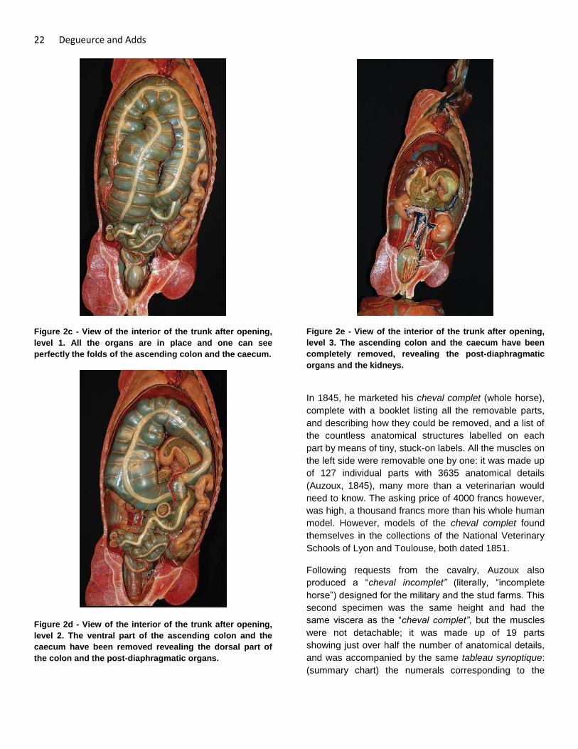
Figure 2c - View of the interior of the trunk after opening,
level 1. All the organs are in place and one can see
perfectly the folds of the ascending colon and the caecum.
Figure 2d - View of the interior of the trunk after opening,
level 2. The ventral part of the ascending colon and the
caecum have been removed revealing the dorsal part of
the colon and the post-diaphragmatic organs.
Figure 2e - View of the interior of the trunk after opening,
level 3. The ascending colon and the caecum have been
completely removed, revealing the post-diaphragmatic
organs and the kidneys.
In 1845, he marketed his cheval complet (whole horse),
complete with a booklet listing all the removable parts,
and describing how they could be removed, and a list of
the countless anatomical structures labelled on each
part by means of tiny, stuck-on labels. All the muscles on
the left side were removable one by one: it was made up
of 127 individual parts with 3635 anatomical details
(Auzoux, 1845), many more than a veterinarian would
need to know. The asking price of 4000 francs however,
was high, a thousand francs more than his whole human
model. However, models of the cheval complet found
themselves in the collections of the National Veterinary
Schools of Lyon and Toulouse, both dated 1851.
Following requests from the cavalry, Auzoux also
produced a “cheval incomplet” (literally, “incomplete
horse”) designed for the military and the stud farms. This
second specimen was the same height and had the
same viscera as the “cheval complet”, but the muscles
were not detachable; it was made up of 19 parts
showing just over half the number of anatomical details,
and was accompanied by the same tableau synoptique:
(summary chart) the numerals corresponding to the

The Mannequins of Dr. Auzoux 23
absent anatomical details were simply missing on the
specimen (Auzoux, 1855). One of these models, dating
from 1846, can be seen at the Fragonard Museum in
Maisons-Alfort (just outside Paris), and another in the
Science Museum in London. Auzoux even planned to
produce three much smaller models, 65 cm in height:
one complete, one incomplete and the last simply an
écorché (Lequime, 1844), a project of which there is now
unfortunately no trace except in the catalogues.
The criticisms of the Royal Academy of Medicine in 1844
were echoed in 1847 by Colonel Jacquemin (Jacquemin,
1847). According to him, the mannequin was
“improvable”, a criticism that stung Auzoux, and which
spurred him on to produce a new horse, 1.30 m in height
and of irreproachable quality, with the profile of a pure-
bred Arab steed (Fig. 3). This model was marketed at
the beginning of the 1850s11
, in two versions, cheval
complet and incomplet each accompanied by its own
summary chart (Auzoux, 1855).
Figure 3 - A worker poses beside a cheval clastique, type
Arab, in the 20th century.
The other equine models
Auzoux’s next project was to create a series of
pathological equine legs complete with various
pathologies. Detection of such lesions was of course of
the highest importance to horse buyers, whether military
or not, but especially to officers of the remonte12
. He
11
Auzoux seems not to have publicized this new version very much; perhaps he didn’t want to draw attention to the perceived imperfections of the earlier one. It is therefore difficult to put an exact date on the creation of the new model, which is mentioned only once in the Auzoux archive. An example of the second model can be seen today in the collection of the University of Halle-Wittenberg, Germany. 12
Remonte: part of the army responsible for supplying the troops and military establishments with horses.
started with fifty limb models, and added to them over
the years (Fig. 4a, b). In this, he was helped and
encouraged by his friend Jacquemin who studied and
commented on the specimens with great zeal, as is
shown by his many letters from the beginning of the
1850s.
Auzoux added a brilliant refinement. As the lesions can
be palpated through the skin, he designed some leg
models, cut above the hock that were covered with
natural skin, placing the student in as realistic a situation
as possible. A ‘triptych’ was even successfully marketed,
consisting of one dissected limb, one limb affected with
bony defects, and a third with soft tissue defects.
Figure 4 - An example of an osseous lesion (hock) a)
anterior; b) lateral view.
Around the same time, Auzoux produced a series of
thirty “jaws” (incisor arches) of the horse, copied from
natural specimens (Auzoux, 1850 p. 1), and
corresponding to the animal’s age13
(Figs. 5a, b).
Diagnosing the age of a horse was obviously an
important skill for the cavalry officers responsible for
procuring horses for the army. Auzoux’s jaws also
revealed the tricks that unscrupulous horse dealers
would use, such as digging a cavity in the tooth and
colouring it with Indian ink make to the animal appear
younger.
13
Note that only the incisor arches are used to tell the age of a horse.

24 Degueurce and Adds
Figure 5a - The set of thirty jaws created by Auzoux.
Figure 5b - Detailed views of one of the jaw models.
The hoof is obviously of paramount importance to the
well-being and usefulness of the horse. Auzoux made
two foot models, one showing the complete anatomy of
the region with tendons, ligaments, bones, synovial
sheaths, vessels and nerves (Fig. 6), and the other
showing the detailed structure of the hoof (Fig. 7), with
the intention of illustrating the theory of the English
veterinarian Bracy Clark14
, who proposed that the
horse’s foot was as deformable as that of other species
(Clark, 1817), though this view was ridiculed at the time.
14
Bracy Clark (1771 – 16 December 1860) was an English veterinary surgeon specialising in the horse, who wrote extensively on the structure and functioning of the hoof.
Auzoux also produced some isolated horse organs to
illustrate his lectures on comparative anatomy and
physiology, as well as windowed stomach models (Fig.
8) and the genital organs of the mare.
Figure 6 - Model of the foot of the horse.
Figure 7 - The separated parts of the hoof, illustrating
Bracy Clark’s theory of hoof mechanics.

The Mannequins of Dr. Auzoux 25
Figure 8 - Windowed stomach of the horse.
Marketing and distribution of the models
The complexity of these equine mannequins raised the
price considerably, and Auzoux feared that they would
suffer a similar fate to that of the Traité de l’anatomie de
l’homme by Jean-Baptiste Bourgery15
, magnificently –
and expensively – illustrated by Nicolas Jacob, but which
failed to sell. Auzoux had encountered difficulties trying
to sell his homme complet. He had to fight to get the
Faculties of Medicine to purchase them, and only
managed to achieve his sales targets after persistent
reminders and approaches to the public authorities.
Not surprisingly, only the Government departments were
able to afford specimens as expensive as Auzoux’s
horses. As for the military, the training of the cavalry and
artillery was changing, with more emphasis being placed
on horsemanship. In 1845, the Minister of War decided
to place a cheval clastique at the Royal School of
Cavalry in Saumur (Picard, 1890) and in several other
depots round the country (Arch. Nat.). At the same time,
the Secretary of State for Commerce and Agriculture
started to equip the Veterinary Schools and stud farms
with Auzoux’s models. Two years later the Schools of
Artillery started to receive the cheval clastique as well as
models of the jaws and lesions (Arch. Nat.).
15
Jean-Baptiste Marc Bourgery (May 19, 1797 – June 1849) French physician and anatomist. The Traité de l’anatomie de l’homme, published in 8 volumes, is considered to be one of the most comprehensive and beautifully illustrated anatomical works ever published.
Additional publications: animal husbandry or just
advertising?
These successes, however, were not enough to satisfy
Auzoux. In a booklet published in 1847 entitled “Of the
utility of the anatomie clastique from the point of view of
the choice, the employment, and the care of the horse”
(Auzoux 1847), he showed how his creations could be
used for training the officer cadets, but also – an
astonishing generalisation – how they could also be
used for the improvement of horse breeding in France.
His friend Jacquemin completed the booklet in glowing
terms with “An account of the Anatomie clastique of Dr
Auzoux and its influence on the training of the cavalry”
(Jacquemin, 1847). Other publications with the aim of
publicising his creations would soon follow. The bulky
and fragile nature of his creations made it difficult to
transport them for demonstrations, so these booklets
were an important way of reaching his target audience.
Auzoux stressed that in perfecting the training of a
cavalry officer, it was necessary to improve not only the
quality of the available horses, but also their hygiene and
husbandry, although it is not immediately obvious how a
cardboard anatomical horse could shed light on the
management of contagious diseases! Auzoux had no
training in this field, so he made use of his friends and
his reading. The archives show that he cut out journal
articles, and also borrowed passages from the military
training manuals to show how much harm the lack of
care had caused during the wars of the Empire (Auzoux,
1847, p. 6). Neglecting the real reason for these
problems – the absence of a well-organised system of
resupply on top of local shortages – he went on to
suggest that with better training, these misfortunes could
be avoided.
The issue of improving the national equine herd through
improved breeding generated a good deal of controversy
in the mid-19th century. Auzoux, (echoing the words of
Richard du Cantal) suggested that poor breeding was
the result of the ignorance of the stockbreeders
(Richard, 1847, p. 404). The introduction of the merino
sheep into France in the 18th century was used as an
example; this scheme had originally failed due to the
lack of understanding about the care that the merino
needed. According to Richard du Cantal, to produce
horses of good quality, it was necessary first of all to

26 Degueurce and Adds
train the breeders in the science of horse husbandry
They would then be able to select good stock and to rear
them, aware of their needs and ailments.
The dissemination of knowledge in the equestrian world
was to become the leitmotiv of Auzoux’s advertising
campaign. In 1854, he published the booklet
“Insufficiency in France, of the horse for war and for
pleasure. Possibility of obtaining them by creating in the
Cavalry Regiments, Schools of stock breeders by means
of the clastique horse of Doctor Auzoux” (Auzoux, 1854).
The idea was that the trained cavalry soldiers would go
on to become the apostles of rural horse husbandry…
“The 7 or 8000 released16
each year will go to the centre
of horse production that is to say to the farms, taking
with them the art of improving the stock.”
The regiments of cavalry and artillery were a captive
audience from whom Auzoux hoped to profit. Indeed, it
was for them that he had created his cheval incomplet.
He envisaged that the military administration would
introduce an overall training in horse husbandry for a
moderate cost and for the greater good of the French
economy. All that was needed was to develop some
basic ideas of anatomy, physiology and hygiene that
were already being taught as part of the course of
military horsemanship, relying on (to make the lessons
more expressive)….his cheval clastique (Auzoux, 1847,
p. 9). Auzoux expanded on this theme in further booklets
dedicated to his collections of lesions and jawbones
(Auzoux, 1848, 1853).
Official orders
The results lived up to his expectations. The School of
Cavalry of Saumur, flagship of the French Cavalry,
started taking his models from July 1845 (Arch. Nat.). In
October 1851, they ordered the collection of osseous
lesions, the anatomy of the foot, and the ‘Bracy Clark’
hoof (ibid.). On top of that, in 1853 the army purchased
“sixty-eight complete copies of the artificial horse”, to be
delivered at the rate of twelve per year, as well as a
number of jaws and lesions; the regiments of artillery
followed suit (ibid.). In military circles, Auzoux had
arrived.
16
from military service.
In agriculture, the results were, however, less
convincing. Auzoux tried hard to get the government
departments to adopt the ideas which had been so
successful with the military administration. In 1860, he
invited the conseils généraux (General Councils) to
create a school of agriculture in each département
(administrative divisions of France) with the aim of
educating all those who were involved in equine
husbandry, enticing them by offering a diploma (Auzoux,
1860). The Minister of Agriculture supported his
initiative, and in August 1860, sent out a circular letter
requesting the Prefects to encourage their Departmental
councils to purchase the cheval clastique (Arch. Nat). It
was a failure. Only a few models were purchased: the
School of Horse-Breeding received one in 184617
; the
Veterinary School of Alfort, a partner in the project, very
soon got its own, which was exchanged for the newer
version in June 1856 (Arch. Nat.); this model has since
disappeared. They purchased the set of jaws in January
1851 (ibid.). The Veterinary School of Lyon received the
jaws at the beginning of 1851, then the cheval complet in
December, where it remains to this day.
The equine models, a global success
In the fullness of time, these models went on to become
a global success and were distributed all round the
world, where they can still be seen today in museums as
far afield as New Delhi and Sydney18
, proof indeed of
their global impact.
Auzoux’s models were such an outstanding success that
in 1862, the good doctor was awarded the honour of
‘Officer of the Legion of Honour’, as “inventor of the
17
This School, founded by Royal decree in 1840, was notable for its courses in Anatomy, which were supported by the cheval clastique. Auzoux could be forgiven for hoping that each depot and stud farm would be similarly equipped, since teaching had been established in each institution, by ministerial order of June 7
th, 1837; one may doubt that this ever happened.
18 In the Albert Hall Museum in New Delhi, the
anatomical horse “as used by the Cavalry regiments of France” presents, according to the description, “3000 structures on 97 parts”. It is accompanied by a human mannequin, 116 cm in height, and a number of other pieces (flowers, etc.) The Powerhouse in Sydney, Australia, also houses an important collection of Auzoux’s models.

The Mannequins of Dr. Auzoux 27
clastic anatomical appliances used by the army and the
military academies” (Le Moniteur, 1862).
Apart from a model of a ruminant stomach, a series of
fourteen bovine jaws and two cows’ uteri, (one in a
resting state and the other at the end of gestation, with
its fetus), Auzoux ventured no further into the anatomy of
the domestic animals.
The papier mâché models were quite fragile, and
constant use - disassembly and reassembly, inevitably
led to damage. There were also considerable losses due
to plunder by the Prussian army during the Franco-
Prussian war of 1870-1871. So great were the losses
that the Minister of War requested from Auzoux in 1874
an estimate for the delivery of “around a hundred and
ten cases”, each containing a set of three legs complete
with lesions, and the anatomy of the hoof (Arch. Nat.).
Figure 9. The complete range of Auzoux’s products, c.
1910. On the left is the horse, on the floor just to the right
of it are two horse’s legs, one écorché and one complete
with skin, the horse’s foot and the Bracy Clark hoof; under
the elephant’s head in the centre is the set of horse jaws
in their presentation case; on the shelves to the right are
the windowed stomach and, above it, another pair of
horse’s legs.
It is difficult to put an accurate figure on the scale of
Auzoux’s output. While the number of different models
was probably between a hundred and a hundred and
fifty, the total number of individual parts is probably
greater than 300, as certain sets, although indicated by a
single catalogue number, were made up of 30-50 parts
(Fig. 9). These pieces are today dispersed in many
different collections, in very varied conditions. Although
well cared-for by collectors and museums, they are
unfortunately often neglected in commercial, military or
educational collections. In the 19th century, the majority
of regiments, colleges and high schools held significant
collections of Auzoux’s models. Today, most of these no
longer exist or are badly damaged. Conservation of this
national treasure should be an urgent priority for
France’s cultural institutions (Fig. 10).
Figure 10. Molds for making the horse models, preserved
in Neubourg.
Picture credits
Fig. 1. Archives nationales
Fig. 2a-e. École nationale vétérinaire de Maisons-
Alfort.
Figs. 3, 9, 10. Musée de l’écorché d’anatomie du
Neubourg.
Figs. 4a, b, 5a, b, 6, 7, 8. Musée de l’École nationale
vétérinaire de Maisons-Alfort.
Photography: Figs 1, 2a-e, 3, 7, 8, 9, 10, Degueurce,
Christophe. Figs 4, 5, 6, Ruiz, Guillaume.
References
Archive sources
Archives Nationales, 242API (Montandon collection)
Ameline J-F. 1825: Observations sur les pièces
d’anatomie de M. le docteur auzoux. Caen, Bonneserre.
Anon. 1843: Article “clastique” in: Anonymous,
Dictionnaire de la conversation et de la lecture, t. 14, P.,
Belin.
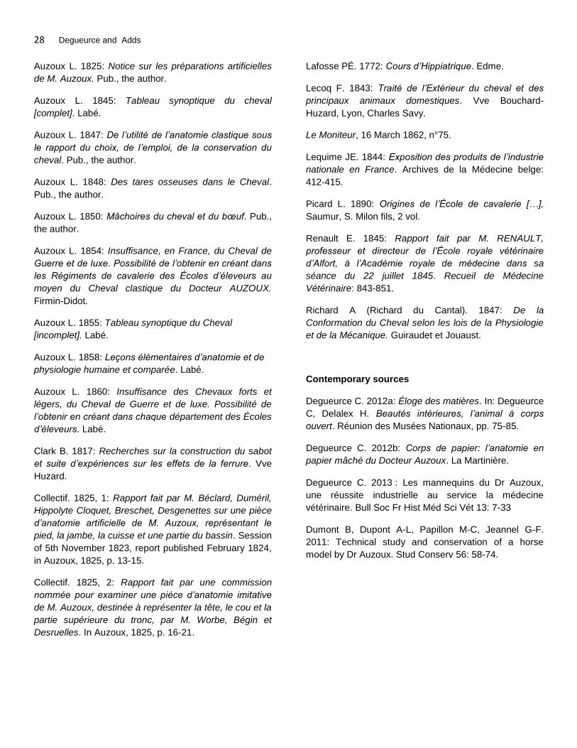
28 Degueurce and Adds
Auzoux L. 1825: Notice sur les préparations artificielles
de M. Auzoux. Pub., the author.
Auzoux L. 1845: Tableau synoptique du cheval
[complet]. Labé.
Auzoux L. 1847: De l’utilité de l’anatomie clastique sous
le rapport du choix, de l’emploi, de la conservation du
cheval. Pub., the author.
Auzoux L. 1848: Des tares osseuses dans le Cheval.
Pub., the author.
Auzoux L. 1850: Mâchoires du cheval et du bœuf. Pub.,
the author.
Auzoux L. 1854: Insuffisance, en France, du Cheval de
Guerre et de luxe. Possibilité de l’obtenir en créant dans
les Régiments de cavalerie des Écoles d’éleveurs au
moyen du Cheval clastique du Docteur AUZOUX.
Firmin-Didot.
Auzoux L. 1855: Tableau synoptique du Cheval
[incomplet]. Labé.
Auzoux L. 1858: Leçons élémentaires d’anatomie et de
physiologie humaine et comparée. Labé.
Auzoux L. 1860: Insuffisance des Chevaux forts et
légers, du Cheval de Guerre et de luxe. Possibilité de
l’obtenir en créant dans chaque département des Écoles
d’éleveurs. Labé.
Clark B. 1817: Recherches sur la construction du sabot
et suite d’expériences sur les effets de la ferrure. Vve
Huzard.
Collectif. 1825, 1: Rapport fait par M. Béclard, Duméril,
Hippolyte Cloquet, Breschet, Desgenettes sur une pièce
d’anatomie artificielle de M. Auzoux, représentant le
pied, la jambe, la cuisse et une partie du bassin. Session
of 5th November 1823, report published February 1824,
in Auzoux, 1825, p. 13-15.
Collectif. 1825, 2: Rapport fait par une commission
nommée pour examiner une pièce d’anatomie imitative
de M. Auzoux, destinée à représenter la tête, le cou et la
partie supérieure du tronc, par M. Worbe, Bégin et
Desruelles. In Auzoux, 1825, p. 16-21.
Lafosse PÉ. 1772: Cours d’Hippiatrique. Edme.
Lecoq F. 1843: Traité de l’Extérieur du cheval et des
principaux animaux domestiques. Vve Bouchard-
Huzard, Lyon, Charles Savy.
Le Moniteur, 16 March 1862, n°75.
Lequime JE. 1844: Exposition des produits de l’industrie
nationale en France. Archives de la Médecine belge:
412-415.
Picard L. 1890: Origines de l’École de cavalerie […],
Saumur, S. Milon fils, 2 vol.
Renault E. 1845: Rapport fait par M. RENAULT,
professeur et directeur de l’École royale vétérinaire
d’Alfort, à l’Académie royale de médecine dans sa
séance du 22 juillet 1845. Recueil de Médecine
Vétérinaire: 843-851.
Richard A (Richard du Cantal). 1847: De la
Conformation du Cheval selon les lois de la Physiologie
et de la Mécanique. Guiraudet et Jouaust.
Contemporary sources
Degueurce C. 2012a: Éloge des matières. In: Degueurce
C, Delalex H. Beautés intérieures, l’animal à corps
ouvert. Réunion des Musées Nationaux, pp. 75-85.
Degueurce C. 2012b: Corps de papier: l’anatomie en
papier mâché du Docteur Auzoux. La Martinière.
Degueurce C. 2013 : Les mannequins du Dr Auzoux,
une réussite industrielle au service la médecine
vétérinaire. Bull Soc Fr Hist Méd Sci Vét 13: 7-33
Dumont B, Dupont A-L, Papillon M-C, Jeannel G-F.
2011: Technical study and conservation of a horse
model by Dr Auzoux. Stud Conserv 56: 58-74.

The Journal of Plastination 27(1):29-32(2015)
TECHNICAL REPORT
Plastination of a Whole Horse for Veterinary Education
Sheng-Bo Yu
1
Jian-Fei Zhang1
Yan-Yan Chi1
Hai-Bin Gao2
Jie Liu2
Hong-Jin. Sui1,2
1
Department of
Anatomy, Dalian
Medical University.
No.9 west section,
Lushun South Road,
Dalian 116044, China
ABSTRACT: Objective: To explore the procedure of preparation of a whole plastinated equine
specimen to be used in veterinary education. Methods: A formalin-preserved horse was dissected
to display the brain, spinal cord and the superficial muscles complete with their innervation. The
specimen then underwent silicone impregnation. Results: The flexibility of the nerves and muscle
tissues after plastination was maintained, and muscles as well as nerve structures were easily
discriminated. The horse was positioned in a stance of a lively spring which facilitated exhibition
of both dorsal and ventral structures. Conclusion: The silicone plastination technique produced a
dry, odorless and durable specimen that is suitable for handling that will serve as an ideal whole
equine specimen for veterinary anatomical education.
2 Dalian Hoffen Bio-
technique Co., Ltd.
No.36, Guangyuan
Street, Lushunkou
Economic
Development Zone,
Dalian 116052, China
KEY WORDS: equine; nervous system; plastination; silicone impregnation; veterinary anatomy
Correspondence to: Prof. Hong-Jin Sui, Department of Anatomy, Dalian Medical University, Dalian 116044, P.R. China. Fax.: +86 411 86110324; Email: [email protected]
Introduction
Plastination is the most important technique to have
emerged in recent years for the preservation of
biological specimens. Since its introduction in 1979, it
has gained wide acceptance throughout the world (von
Hagens, 1979). The most widespread application of this
technique is in the preparation of a wide range of
anatomical specimens for teaching, and it has been
considered an important tool in recent proposals for the
teaching of anatomy (Reidenberg et al., 2002, Latorre et
al., 2007b, Valdecasas et al., 2009). Careful dissection
of an embalmed animal can be a challenging, time-
consuming process, and maintaining a dissected animal
for a long period of time without deterioration due to
fungal and microbial growth or desiccation is extremely
difficult. Moreover, large animals that are immersed in
fixative solution are not ideal for teaching and learning
anatomy. The procedure described here employs
silicone impregnation to preserve the original delicate
tissues, and to furnish a dry, odorless and durable
specimen of a whole large animal, in this case a horse.
Materials and methods
The experiment using the horse was approved by Dalian
Agriculture Bureau on Ethics in the Care & Use of
Laboratory Animals. A freshly dead horse was collected
from farmland. The right common carotid artery and
jugular vein of the cadaver were cannulated and blood
was washed out by introducing saline by gravity feed
from a height of approximately 2.5m. This was followed
by 10% formalin to fix the whole body. The animal was
then stored in a tank containing the same solution for a
period of over 6 months until dissection began.
Dissection
The superficial structures of the horse, including the
skin, subcutaneous fat, fascia and blood vessels were
removed to expose the muscles and their innervating
nerves. In the head, the soft tissues covering the
calvaria were removed, except for the ears. Craniotomy
was performed to expose the brain. The branches of the
facial nerve were carefully dissected out as they
TECH
NIC
AL R
EPO
RT
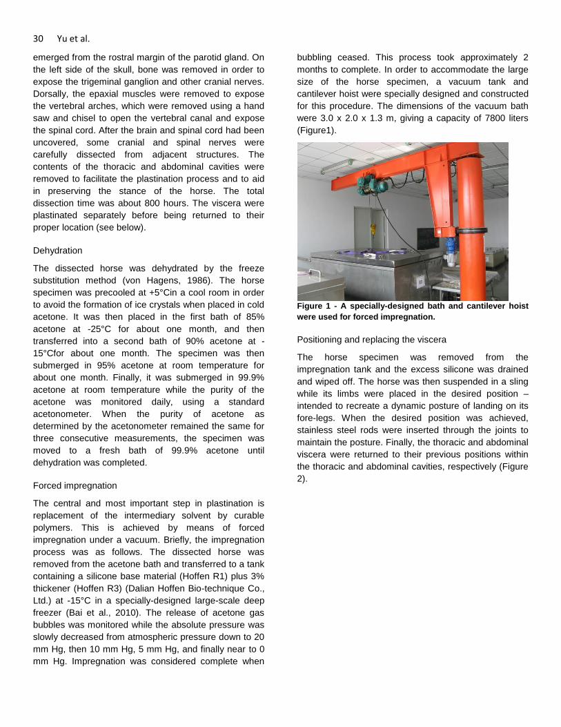
30 Yu et al.
emerged from the rostral margin of the parotid gland. On
the left side of the skull, bone was removed in order to
expose the trigeminal ganglion and other cranial nerves.
Dorsally, the epaxial muscles were removed to expose
the vertebral arches, which were removed using a hand
saw and chisel to open the vertebral canal and expose
the spinal cord. After the brain and spinal cord had been
uncovered, some cranial and spinal nerves were
carefully dissected from adjacent structures. The
contents of the thoracic and abdominal cavities were
removed to facilitate the plastination process and to aid
in preserving the stance of the horse. The total
dissection time was about 800 hours. The viscera were
plastinated separately before being returned to their
proper location (see below).
Dehydration
The dissected horse was dehydrated by the freeze
substitution method (von Hagens, 1986). The horse
specimen was precooled at +5°Cin a cool room in order
to avoid the formation of ice crystals when placed in cold
acetone. It was then placed in the first bath of 85%
acetone at -25°C for about one month, and then
transferred into a second bath of 90% acetone at -
15°Cfor about one month. The specimen was then
submerged in 95% acetone at room temperature for
about one month. Finally, it was submerged in 99.9%
acetone at room temperature while the purity of the
acetone was monitored daily, using a standard
acetonometer. When the purity of acetone as
determined by the acetonometer remained the same for
three consecutive measurements, the specimen was
moved to a fresh bath of 99.9% acetone until
dehydration was completed.
Forced impregnation
The central and most important step in plastination is
replacement of the intermediary solvent by curable
polymers. This is achieved by means of forced
impregnation under a vacuum. Briefly, the impregnation
process was as follows. The dissected horse was
removed from the acetone bath and transferred to a tank
containing a silicone base material (Hoffen R1) plus 3%
thickener (Hoffen R3) (Dalian Hoffen Bio-technique Co.,
Ltd.) at -15°C in a specially-designed large-scale deep
freezer (Bai et al., 2010). The release of acetone gas
bubbles was monitored while the absolute pressure was
slowly decreased from atmospheric pressure down to 20
mm Hg, then 10 mm Hg, 5 mm Hg, and finally near to 0
mm Hg. Impregnation was considered complete when
bubbling ceased. This process took approximately 2
months to complete. In order to accommodate the large
size of the horse specimen, a vacuum tank and
cantilever hoist were specially designed and constructed
for this procedure. The dimensions of the vacuum bath
were 3.0 x 2.0 x 1.3 m, giving a capacity of 7800 liters
(Figure1).
Figure 1 - A specially-designed bath and cantilever hoist
were used for forced impregnation.
Positioning and replacing the viscera
The horse specimen was removed from the
impregnation tank and the excess silicone was drained
and wiped off. The horse was then suspended in a sling
while its limbs were placed in the desired position –
intended to recreate a dynamic posture of landing on its
fore-legs. When the desired position was achieved,
stainless steel rods were inserted through the joints to
maintain the posture. Finally, the thoracic and abdominal
viscera were returned to their previous positions within
the thoracic and abdominal cavities, respectively (Figure
2).
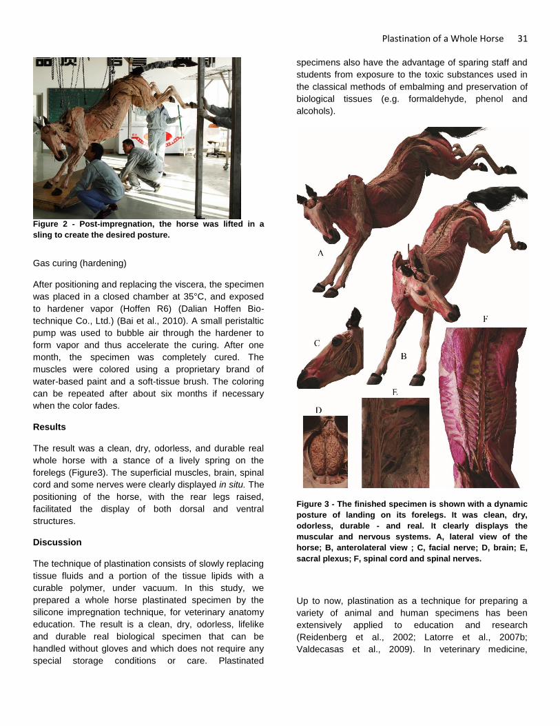
Plastination of a Whole Horse 31
Figure 2 - Post-impregnation, the horse was lifted in a
sling to create the desired posture.
Gas curing (hardening)
After positioning and replacing the viscera, the specimen
was placed in a closed chamber at 35°C, and exposed
to hardener vapor (Hoffen R6) (Dalian Hoffen Bio-
technique Co., Ltd.) (Bai et al., 2010). A small peristaltic
pump was used to bubble air through the hardener to
form vapor and thus accelerate the curing. After one
month, the specimen was completely cured. The
muscles were colored using a proprietary brand of
water-based paint and a soft-tissue brush. The coloring
can be repeated after about six months if necessary
when the color fades.
Results
The result was a clean, dry, odorless, and durable real
whole horse with a stance of a lively spring on the
forelegs (Figure3). The superficial muscles, brain, spinal
cord and some nerves were clearly displayed in situ. The
positioning of the horse, with the rear legs raised,
facilitated the display of both dorsal and ventral
structures.
Discussion
The technique of plastination consists of slowly replacing
tissue fluids and a portion of the tissue lipids with a
curable polymer, under vacuum. In this study, we
prepared a whole horse plastinated specimen by the
silicone impregnation technique, for veterinary anatomy
education. The result is a clean, dry, odorless, lifelike
and durable real biological specimen that can be
handled without gloves and which does not require any
special storage conditions or care. Plastinated
specimens also have the advantage of sparing staff and
students from exposure to the toxic substances used in
the classical methods of embalming and preservation of
biological tissues (e.g. formaldehyde, phenol and
alcohols).
Figure 3 - The finished specimen is shown with a dynamic
posture of landing on its forelegs. It was clean, dry,
odorless, durable - and real. It clearly displays the
muscular and nervous systems. A, lateral view of the
horse; B, anterolateral view ; C, facial nerve; D, brain; E,
sacral plexus; F, spinal cord and spinal nerves.
Up to now, plastination as a technique for preparing a
variety of animal and human specimens has been
extensively applied to education and research
(Reidenberg et al., 2002; Latorre et al., 2007b;
Valdecasas et al., 2009). In veterinary medicine,
32 Yu et al.
however, application of this technique has only just
begun to develop (Latorre et al., 2007a, Latorre et al.,
2007b). The importance of correlating anatomical
studies with diagnostic and therapeutic approaches in
practice has long been recognized. Such studies in the
horse have, until recently, lagged behind this discipline
in human medicine and surgery (Latorre et al., 2007a).
Additionally, in the case of large animals such as the
horse, only plastinated regional specimens or individual
organs, such as the cephalic block, heart and transverse
sections, have been available for use in evaluation of
their effectiveness in anatomical education (Latorre et
al., 2007b). A lack of whole large animal specimens may
be detrimental to the students’ comprehension of whole-
body animal anatomy. To address this lack, we
endeavored to plastinate a whole dissected horse by the
silicone-impregnation technique, in order to stimulate
further correlations of anatomical structures and equine
medical and surgical procedures, and thereby to
advance knowledge and understanding in practice and
teaching of equine anatomy.
Additionally, this work demonstrates that plastination
with the silicone technique is applicable to the
preparation of a whole horse specimen. In this study,
Hoffen silicone R1/R3 was used for forced impregnation.
The result was a clean, dry, odorless and durable real
whole horse.
Acknowledgements
The authors would like to thank the technicians in Dalian
Hoffen Bio-technique Co., Ltd. for their skillful technical
assistance in silicone impregnation.
References
Bai J,Gao HB,Jie L,Luan BY,Meng WJ,Zhang JF
Yu SB,Gong J,Zhang CH,Sui HJ. 2010: The
application of Hoffen P45 plastination technique on
preparation of sectional specimen. Chinese J Clin Anat
28(1):107-108.
Latorre R, Rodríguez MJ. 2007a: In search of clinical
truths: equine and comparative studies of anatomy.
Equine Vet J 39(3):263-268.
Latorre RM, García-Sanz MP, Moreno M, Hernández F,
Gil F, López O, Ayala MD, Ramírez G, Vázquez JM,
Arencibia A, Henry RW. 2007b: How useful is
plastination in learning anatomy? J Vet Med Educ
34(2):172-176.
Reidenberg JS, Laitman JT. 2002: The new face of
gross anatomy. Anat Rec 269(2):81-88.
Valdecasas AG, Correas AM, Guerrero CR, Juez J.
2009: Understanding complex systems: lessons from
Auzoux's and von Hagens's anatomical models. J Biosci
34(6):835-843.
Von Hagens G. 1979: Impregnation of soft biological
specimens with thermosetting resins and elastomers.
Anat Rec 194(2):247–255.
Von Hagens G. 1985: Heidelberg Plastination Folder:
Collection of technical leaflets for plastination.
Heidelberg: Anatomiches Institut 1, Universität
Heidelberg, p 16-33.

The Journal of Plastination 27(1):33 (2015)
11th International Interim Conference on Plastination
Vitória, Espírito Santo, Brazil
July 13-16, 2015
1Carlos A C Baptista,
2Ana Paula S. V. Bittencourt,
2Yuri F. Monteiro,
2Laissa da S. Juvenato,
2Athelson S. Bittencourt,
1College of Medicine and Life Sciences, University of Toledo, Ohio USA.
2Universidade Federal
do Espirito Santo, Vitória, Brazil
Before the Interim!
For many years plastination was a desire for the Brazilian anatomists. They had a lot of lectures on the subject for many
years so they were familiar with the process, at least theoretically speaking. But, you can only learn plastination by doing
it! That has been our thought for many years, and I am sure it is the thought of many who practice the art of plastination.
The opportunity to hold the 11th Interim meeting on Plastination at this moment in Brazil was unique. The first step was to
find a laboratory in Brazil capable of housing the material and equipment necessary to demonstrate the fundamental
principles of the silicone, epoxy and polyester techniques. The creation of the Plastination Laboratory of the University of
Espirito Santo in Vitoria, Brazil by Prof. Athelson Bittencourt was a milestone to propel plastination in Brazil to new
heights. The laboratory was completed in January 2015, material and equipment were in place, and soon after the first
specimens were produced.
We Seized the Moment!
The time arrived for the first hands-on workshop on plastination sponsored by the ISP to be held in Latin America. There
were hundreds of interested anatomists, biologists, physicians, dentists who could finally see and learn the technique of
plastination in action. The 11th Interim Conference on Plastination was held in Vitoria, Brazil in the summer of 2015, based
at the laboratory in the campus of the University of Espirito Santo.
The welcome message of Dr. Athelson Bittencourt was followed by Dr. Reinaldo Centoducatte, President of the
University, Dr. Glaucia Rodrigues de Abreu, Director of the Health Science Center, and the President of ISP, Dr. Carlos A
Baptista.
The keynote address was delivered by Dr. Vladimir Chereminskiy on “New Dissection Techniques: Reasons and Details
of New Dissection Techniques for the Manufacturing of Plastinates.”
Participants:
More than 67 participants (representing 7 countries and 17 States of Brazil) attended the four-day conference. The
meeting comprised morning lectures and afternoon hands-on activities. The hands-on activities were directed by a cast of
distinguished experts on plastination: Dr. Robert W. Henry (USA), Dr. Kees H. de Jong (Netherlands), Dr. Rafael Latorre
(Spain), Dr. Dmitry Starchik (Russia), Dr. Vladimir Chereminskiy (Germany), Dr. Carlos A. Baptista (USA) and Dr.
Athelson S. Bittencourt (Brazil). Lectures on the topic of plastination were presented by the experts above and also by a
distinguished group of scientist from Brazil on the topic of plastination and related fields: Dr. Marco Sampaio (Rio de
Janeiro), Dr. Carlos Rueff Barroso (Vitoria), Dr. Andrea Oxley da Rocha (Porto Alegre) and Dr. Richard Cabral (Sao
Paulo).
Program:
The oral sessions of the first day were dedicated to the basic principles of plastination. The session began with an
overview of plastination (Dr. Baptista) followed by dehydration (Dr. Henry), impregnation (Dr. Latorre), curing (Dr.
MEETIN
G R
EPO
RT

The Journal of Plastination 27(1):34 (2015)
Baptista) , room temperature plastination (Dr. Henry) and principles of polyester (Dr. de Jong). This session provided a
technical basis for the afternoon workshop. The day ended with a tour of the Government Palace (built in the XVI century)
and a Reception and visit to the Human Body Exhibition: ’From Cell to Man’ at the Life Science Museum of the Federal
University of Espirito Santo.
Presentations on the second day focused on technical aspects of the epoxy technique (E12) sheet plastination (Dr.
Latorre) and polyester technique (P40) (Dr. de Jong). Plastination of fetus and brain specimens without shrinkage was
presented by Dr. Chereminskiy and the pros and cons of the room temperature technique were presented by Dr. Starchik.
On the third day, presentations focused on strategies to obtain specimens for plastination (Dr. Oxley da Rocha), how to do
sheet plastination without acetone and using Brazilian Polymer (Dr. Sampaio) and a discussion on the plastination
science exhibits by Dr. Bittencourt. The day ended with a Gala Dinner and a visit to the Penha Convent, founded in 1558
and located on the top of a high mountain overlooking the city of Vitória.
The last day of the conference was devoted to issues of Health and Safety and how to set up a plastination lab. A
presentation on “Safety and Hazardous Issues in Plastination” was provided by Dr. Baptista. Following the presentation,
Dr. Bittencourt shared with the audience his experience on how to set up a laboratory in Brazil, discussing several aspects
of safety, regulations and importation. There were several other oral presentations, posters and specimens exhibited by
attendees of the conference
Workshop
The hands-on workshop was held in the afternoons and complemented the didactic sessions presented in the mornings.
To facilitate learning each international instructor was paired with a Portuguese-speaking presenter (who had participated
in international plastination workshops previously).
Day 1 - dehydration (freeze substitution in -25° C acetone), and impregnation (cold- and room-temperature).
Day 2 - polyester technique. Slicing a fixed brain on a meat slicer and an unfixed leg on a band saw for polyester
impregnation. Constructing a glass chamber and casting a brain slice with P40.
Day 3 - dismantling and sawing P40 slices and curing of silicone (cold and room temperature).
Day 4 - final review of room-temperature and cold-curing manipulation, dismantling epoxy ‘sandwich method’ chamber
and wrapping specimens for transportation.
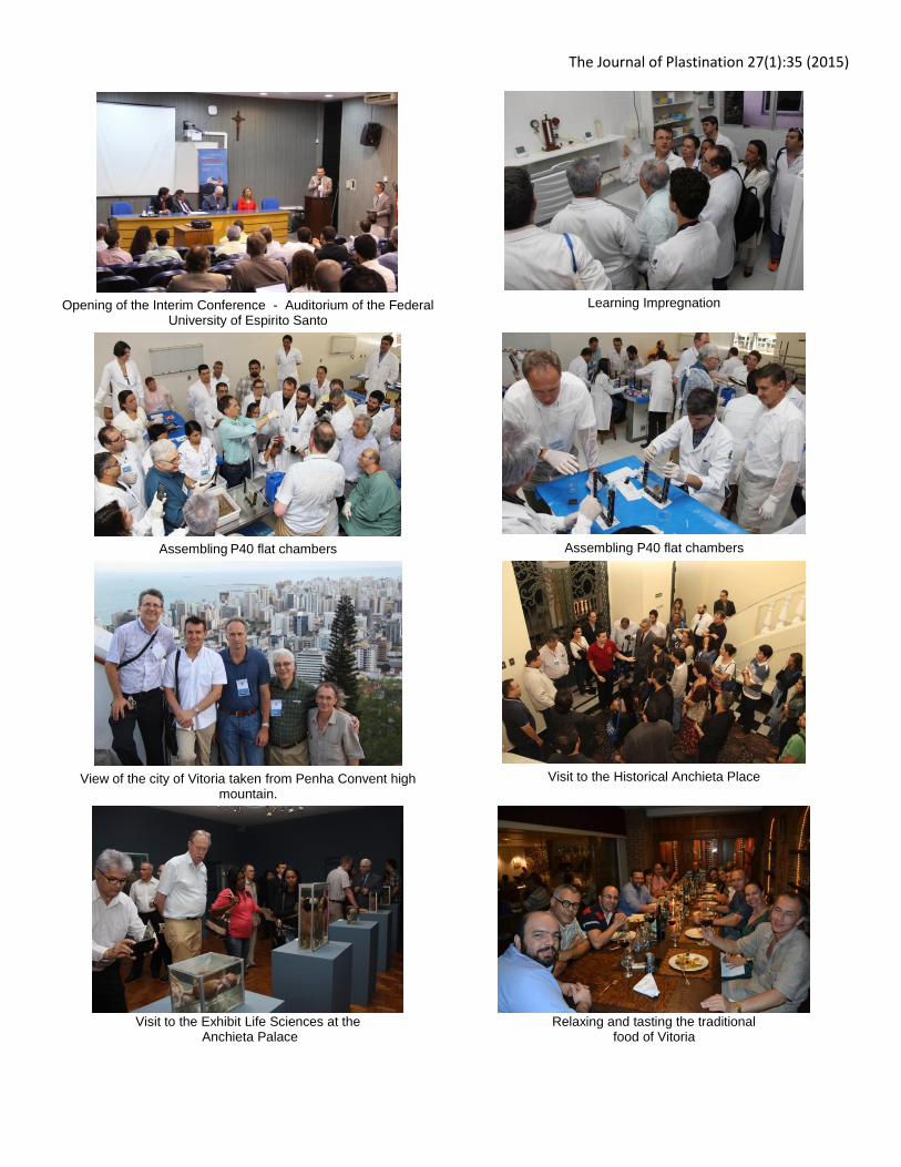
The Journal of Plastination 27(1):35 (2015)
Opening of the Interim Conference - Auditorium of the Federal
University of Espirito Santo
Learning Impregnation
Assembling P40 flat chambers
Assembling P40 flat chambers
View of the city of Vitoria taken from Penha Convent high
mountain.
Visit to the Historical Anchieta Place
Visit to the Exhibit Life Sciences at the
Anchieta Palace
Relaxing and tasting the traditional
food of Vitoria

The Journal of Plastination 27(1):36 (2015)

The Journal of Plastination 27(1):37 (2015)
Abstracts from 11th International Interim Conference on Plastination Vitória, Espírito Santo, Brazil
July 13-16, 2015
Bilateral Anatomical Variations of Musculature of the First Dorsal Fibro-Osseous
Compartment of the Wrist.
Lima FS1, Leo JA4, Oliveira KM3, Silva FS2, Rosa RC2.
1. Departamento de Ciências Biológicas; 2. Departamento de Biologia Estrutural, UFTM, Uberaba/MG, Brazil;
3. Departamento de Anatomia, UFJF, Governador Valadares/MG, Brazil; 4. Curso de Medicina, UNIUBE,
Uberaba/MG, Brazil. UFPB, João Pessoa/PB, Brazil.
Introduction: The tendons of the abductor pollicis longus (APL) and extensor pollicis brevis (EPB) are located in the
dorsal carpal region. The knowledge of anatomical variations of the first dorsal fibro-osseous compartments muscle
wrist is clinically relevant to De Quervain’s stenosing tenosynovitis and reconstructive surgeries. A variety of reports of
multiple insertion tendons in the first dorsal fibro-osseous compartment of the wrist is found in literature, but among
these are few reports describing the occurrence of fusion. Objective: Report an unusual anomalous bilateral fusion of
muscle bellies of the first dorsal compartment of the wrist.
Methods: The upper limbs of 32 cadavers were analyzed. The description of the characteristic morphology was
performed taking into account the origin and insertion pattern of muscle fibers. Anthropometric measurements of
muscle were carried out using string over the muscle belly or tendon being measured using a universal digital calliper
(Mitutoyo®).
Results: The presence of fusion of the muscle belly of the APL and EPB was found in five limbs and bilaterally observed in
one cadaver. The abductor pollicis longus of the right upper limb (ALP_R) was 9.0 cm long, with the presence of two
insertion fascicles, one for the abductor pollicis brevis and the other to the opponent pollicis. The abductor pollicis
longus of the left upper limb (ALP_L) was trifurcated into: intermediate tendon (I), lateral tendon (L) and medial tendon
(M). The intermediate tendon was 7.5 cm long and lateral tendon 7.0 cm. The tendons I and L were inserted in the base
of the first metacarpal, while tendon M had three fascicles inserted: abductor pollicis brevis, opponens pollicis, and
anteromedial region of the base of the first metacarpal. The extensor pollicis brevis of the right upper limb (EPB_R) was
7.2 cm in length, 1.2 cm in width, while the extensor pollicis brevis of the left upper limb (EPB_L) was 8.5 cm in length
and 1.1 cm in width. Bilaterally ECP origin was observed in the dorsal radial region and insertion of the dorsal
aponeurosis at the metacarpophalangeal joint of the thumb level.
Conclusion: An unusual fusion of the APL and EPB, concomitantly with a variant insertion pattern, is the highlight of the
current case report. Our case study shows that these additional tendons may prove to be biomechanically
advantageous. Moreover, these tendons may be effectively used for reconstructive surgery.
AB
STRA
CTS

The Journal of Plastination 27(1):38 (2015)
Aberrant Contribution of Extensor Pollicis Longus of the First Dorsal Compartment
of the Wrist.
Santos PR1, Ferreira FS1, Elias BAB3, Leo JA3, Oliveira KM2, Rosa RC1.
1. Departamento de Biologia Estrutural, UFTM, Uberaba/MG, Brasil; 2. Departamento de Anatomia, UFJF,
Governador Valadares/MG, Brasil; 3. Curso de Medicina, UNIUBE, Uberaba/MG, Brasil. dade Federal da
Paraíba, João Pessoa-PB-Brasil.
Introduction: Knowledge of the anatomical variations of the muscles of the first dorsal fibro-osseous compartment of
the wrist is clinically relevant to De Quervain’s stenosing tenosynovitis and reconstructive surgery. In the literature are
found a variety of reports of multiple tendons of insertion in the first dorsal fibro-osseous compartment of the wrist, but
among these are few reports describing the occurrence of fusion and muscle contributions.
Objective: This report describes an unusual bilateral aberrant contribution of the extensor pollicis longus (EPL).
Methods: The description of the characteristic morphology was performed taking into account the origin and insertion
pattern of the muscle fibers. Anthropometric measurements of muscle were carried out using string over the muscle
belly or tendon being measured using a universal digital calliper (Mitutoyo®).
Results: In the same cadaver was found the presence of this contribution and an anomalous muscle function of the
abductor pollicis longus (APL) and extensor pollicis brevis (EPB). The APL of the right upper limb (ALP_R) was 11.5 cm in
length and 2.7 cm in width. The ALP_R had a single tendon of 9.0 cm length, with the presence of two insertion fascicles,
one for the abductor pollicis brevis (APB) and the other to the opponent pollicis (OP). The APL of the left upper limb
(ALP_L) was 15.7 cm in length and 2.5 cm in width. The tendon of ALP_L was trifurcated into intermediate tendon (I),
lateral tendon (L) and medial tendon (M). The I tendon was 7.5 cm long, whereas the M and L tendons were 7.0 cm in
length. The L and I tendons inserted in the base of the first metacarpal, while M tendon had three fascicles inserting:
APB, OP and anteromedial region of the base of the first metacarpal. Both the EPBs received an unusual donation from
the EPLs by a slender auxiliary tendon, with an average length of 9.2 cm, which intersected obliquely and laterally under
the extensor retinaculum, entering within the first dorsal compartment of the wrist, merging with the tendon of EPB.
The EPB of the right upper limb (EPB_R) was 7.2 cm in length, while the EPB of the left upper limb (EPB_L) had a length
of 8.5 cm. The innervation of these fused muscle bellies was as usual, by the posterior interosseous nerve. In the wrist
and hand region no neurovascular variation was found.
Conclusion: Atypical contribution over the EPL and unusual fusion of APL and EPB, concomitant with a variant insertion
pattern, is the highlight of the current case report. This case report shows that these additional tendons may prove to be
biomechanically advantageous. Furthermore, these tendons may be effectively used for reconstructive surgery.

The Journal of Plastination 27(1):39 (2015)
Morphology, Development and Heterochrony of the Carapace of Podocnemis expansa
(Testudines, Podocnemididae).
Pereira KF1, Vieira LG2, Santos ALQ2, Lima FC1.
1. Human and Comparative Anatomy Laboratory, Federal University of Goiás – Regional Jataí, Jataí – GO,
Brazil; 2. Wild Animal Teaching and Researching Laboratory, Federal University of Uberlândia, Uberlândia –
MG, Brazil.
Introduction: The Testudines present a particular morphological structure formed by the shell that comprises a ventral
portion, the plastron, and a second, dorsal portion, the carapace. We discuss the possible intra-specific alterations that
occur during all of the embryonic period, due to the importance of ontogenic data in the interpretation of new fossils
which document the evolution of the lineage of the turtles, as well as for the understanding of the anatomy of the
current living groups.
Objective: Description of the morphology, formation sequence and development of the carapace bones of P. expansa.
Methods: Embryos (62) and nestlings (43) of Podocnemis expansa were acquired in the reproduction field in the River
Araguaia – GO. Each specimen was fixed in 10% formaldehyde solution, cleared, and the bones and cartilages stained
with Alcian blue and Alizarin red S, respectively. Some embryos were also dehydrated and embedded in paraffin
following the basic histology protocol for H & E staining.
Results: The carapace has mixed osseous structure of endo and exoskeleton.
This structure begins its formation in the beginning of stage 16 with the ossification of the periosteal collar of the ribs.
With the exception of the peripheral bones, the other ones begin their ossification during the embryonic period. On
histological investigation it was found that the costal bones and neural bones have a close relation to the endoskeleton
components, originating themselves as intramembranous expansions of the periosteal collar of the ribs and neural
arches, respectively. The condensation of the mesenchyme adjacent to the periosteal collar induces the formation of
spikes that grow in trabeculae permeated by fibroblasts below the skin. The bone from the nuchal region also ossifies in
an intramembranous way, but does not show direct relation to the endoskeleton.
Such information confirms those related to the other Pleurodira, mainly with Podocnemis unifilis, sometimes with
conspicuous variations in the chronology of the ossification events.
Conclusion: Costals and neurals are plates derived from ribs and neural arches, respectively, in continuity with the
periostea of the endoskeleton. There were chronological differences in the ossification of the carapace of P. expansa in
comparison to the other Testudines. The first element to form was the ribs, which presented uniformity among the
reported species. The Podocnemididae P. expansa and P. unifilis share many similarities during their carapace ontogeny.
The main differences are in the chronology, and they may express variations because of abiotic variations that influence
the incubation period. The phylogenetic proximity of these two species may also explain such similarity.

The Journal of Plastination 27(1):40 (2015)
Epoxy Resin Embedding Technique: Removal of the Propylene Oxide Step Before
Impregnation.
Sassoli Fazan VP, School of Medicine of Ribeirão Preto, University of São Paulo, Brazil.
Introduction: Propylene oxide (PO) is an organic compound with the molecular formula CH3CHCH2O. This colorless
volatile liquid is produced on a large scale industrially and its major application is for the production of polyurethane
plastics. PO is commonly used in the preparation of biological samples for electron microscopy, to remove residual
ethanol previously used for dehydration. In a typical procedure, the sample is first immersed in a mixture of equal
volumes of ethanol (ETH) and PO for 5 minutes, and then four times in pure PO, 10 minutes each. PO was once used as a
racing fuel, but that usage is now prohibited for safety reasons. It is also used in thermobaric weapons, a type of
explosive that utilizes oxygen from the surrounding air to generate an intense, high-temperature explosion. Due to its
explosive characteristics, the Brazilian army ministry imposed several restrictions on the importation and use of PO even
for research purposes.
Objective: We aimed to develop an epoxy resin embedding technique for electron microscopy samples, removing the
PO step between ethanol dehydration and epoxy resin impregnation.
Methods: The epoxy resin used in this study was the EMbed 812® from Electron Microscopy Sciences Inc. (Catalog # RT
14120). Biological samples consisted of sural nerves from male Wistar rats, with ages from 30 days to 720 days. Nerves
were fixed in 2.5% glutaraldehyde (Merck) and dehydrated in graded ETH (Merck), from 25% to 100%, for 5 minutes
each. After the dehydration, impregnation was performed in a mixture of 100% ETH and resin, first in a proportion of 2:1
and then in a proportion of 1:2, for 2 hours each. Afterwards, the nerves were left overnight (~18 hours) in pure resin,
before embedding. The infiltration steps were performed under orbital agitation at room temperature the entire time,
including the overnight step. Samples of all experimental groups were histologically processed at once so that they were
submitted to absolutely the same experimental conditions throughout the experiments.
Results: Semi-thin (0.5 μm thick) transverse sections of the fascicles were stained with 1% toluidine blue and examined
with the aid of an Axiophot II photomicroscope (Carl Zeiss, Jena, Germany). The images were sent via a digital camera to
an IBM/PC where they were digitized. The study of nerve fascicles, myelinated fibers and endoneural space were
performed following the methods developed in our laboratory. All nerves showed good preservation of structures and
general morphological characteristics of the sural nerve fascicles were similar to those previously described. Very few
artefacts were present in some images, not necessarily related to the embedding technique.
Conclusion: The described technique proved to be reliable, reproducible and efficient for epoxy resin embedding of
nerve samples, without the use of PO. The image quality of samples was high enough to allow morphometric studies.
Support: FAPESP and CNPq.

The Journal of Plastination 27(1):41 (2015)
Craniometric Data of the Buff-Necked Ibis (Theristicus caudatus) (Boddaert 1783).
Werner LC, Silva LCS, Souza RAM.
Department of Veterinary Medicine, UNICENTRO, Guarapuava/PR, Brazil
Introduction: The buff-necked ibis is a bird of the Order Ciconiiformes, Threskiornithidae Family, which has long legs and
wide light-colored wings with particular black marks on the periophthalmic region. It has an exclusively South American
distribution and even though it is a species with great adaptability to anthropogenic environments, its anatomical
features have not yet been described.
Objective: The aim of this study was to obtain measurements of the skulls of the buff-necked ibis to contribute to the
anatomy of the species, as well as for veterinary comparative anatomy.
Methods: Craniometric aspects of six buff-necked ibis adults were analyzed. The birds, which died of various causes,
came from the SAAS of UNICENTRO, and were donated to the Animal Anatomy Laboratory of the same institution. The
measurements of the skulls of the animals were performed after previous removal of the skin, fascia and superficial
musculature with surgical instruments and subsequent maceration. Clarification was performed with H2O2 and a final
cleaning of the skulls finished the preparation. With the use of an analogue caliper, the following measures were taken:
maximum skull length, the free end of the maxillary rostrum to the most caudal point of the supraocciopital bone;
maximum width of the skull measured between the post-orbital processes right and left; maximum height measured
from the basilar portion of the rostrum parasphenoid to the highest region of the skull (common point between
supraoccipitoparietal, interfrontal and frontoparietal sutures); maximum width measured between suprameatic
processes right and left; distance between the jaw and post orbital process; distance between the maxillary rostrum and
the highest region of the skull; distance between the maxillary rostrum and the basilar portion of the parasphenoid;
distance of paraoccipitalis processes from each other; and length and width of the foramen magnum.
Results: The average value for maximum skull length was 19.44 cm (SD: 0.79 cm); the average value for the maximum
width of the skull was 3.45 cm (SD: 0.09 cm); average maximum height of the skull was 2.84 cm (SD: 0.14 cm); average
maximum hind width was 2.91 cm (SD: 0.06 cm); the average distance between the jaw and the post-orbital process was
17.59 cm (SD: 0.72 cm); average value of the distance between the jaw to the highest region of skull was 18.34 cm (SD:
0.93 cm); the distance between the maxillary rostrum and the basilar portion of the rostrum parasphenoid was 15.90 cm
(SD: 1.78 cm); average value of distance between each process paraoccipitalis was 2.49 cm (SD: 0.06 cm); and the
average length and width of the foramen magnum was respectively 0.79 cm (SD: 0.10 cm) and 0.79 cm (SD: 0.02 cm).
Conclusion: The data obtained in this study elucidate the craniometry of the buff-necked ibis (Theristicus caudatus) and
can be used as a basis for comparative anatomy studies as well as providing knowledge about the morphology of this
avian species.

The Journal of Plastination 27(1):42 (2015)
Applicability of an Educational Game for Heart Anatomy: A Pilot Study
Santos CO1, 2, Silva SS3, Malheiros CD1, 2, Santos CLC4, Nascimento DGR3, Silva ML5.
1. Departamento de Ciências da Vida - UNEB, Salvador/BA, Brasil; 2. Escola Bahiana de Medicina e Saúde
Pública, Salvador/BA, Brasil; 3. Fisioterapeuta, Salvador/BA, Brasil; 4. Secretaria de Educação da Bahia,
Salvador/BA, Brasil; 5. Departamento de Exatas -UNINASSAU, Salvador/BA, Brasil.
Introduction: The study of human anatomy covers different teaching methodologies that provide the best construction
of learning and hence better understanding of the human body. Among these educational strategies, games can be
effective instructional tools.
Objective: To assess the applicability of an educational game as a motivator for the learning process of heart anatomy.
Methods: A pilot study was undertaken with undergraduate students from an educational institution in the city of
Salvador/BA who have had practical lessons of heart anatomy. A board game was devised consisting of two overlapping
circles of different sizes and a roulette-type pointer. The tray was divided into 32 areas (places) in which letters or the
numbers 1 to 6 were placed, simulating a die. The letters have content related to the subject matter through pictures or
questions. After the game, an evaluation questionnaire was provided to the players. The questionnaire consisted of 9
questions based on Nielsen instructions and Grassioulet, related to perceptions of the educational character, fun and
play structure. Of the 9 questions, 7 were objective and related to perception of the game’s educational character; these
were scored according to the Likert scale of 1 to 5 points, with the total sum ranging from 7 to 35 points (representing
respectively the least and the greatest educational character). The last two questions that reported the fun and the play
structure were analyzed using percentages.
Results: The game was applied to 30 graduate students of the same class. Regarding the perception of the educational
character of the game, the scores ranged from 24 to 35. As for their feelings about playing, 54.3% found it enjoyable,
28.6% felt joy, 5.7% considered it boring, 2.9% felt fatigue; other sensations amounted to 8.6% (learning, anxiety or no
sense). When asked about the structure of the game, 56.7% would not change any features and 43.3% suggested
changes. Among the changes, increasing the letters was the most common suggestion found, followed by increase
houses and, in the letters of images, make the questions more specific.
Conclusion: The educational game helped in the process of learning the anatomy of the heart, according to results from
‘good’ to ‘great’ perception of the educational character provided by this study. In addition, most students considered
the game fun and felt joy, thus making learning enjoyable in human anatomy and more efficient, suggesting greater
success in the achievement of knowledge by students. Modifications in the play structure, as pointed out in this
research, will be incorporated, and it is intended to expand the use of this tool with other graduate students, as well as
through the inclusion of other body systems.

The Journal of Plastination 27(1):43 (2015)
The Dissection Workshop as a Tool in the Production of High Quality Anatomical Specimens.
Rocha AO1, De Campos D1, Simoneti LEL2, Pedron J2, Girotto MC2, Pacini GS2, Santos GC2,
Bonatto-Costa JA1.
1. DCBS; 2. Curso Medicina, UFCSPA, Porto Alegre/RS, Brasil.
Introduction: Due to the lack of human bodies for Anatomy teaching in Brazilian universities and the difficulty in
obtaining them, body preservation methods have been improved with the emergence of new techniques such as
plastination, which offers better resistance and durability to the tissues. However, dissection remains an indispensable
method for the production of high-quality specimens capable of being preserved through the process of plastination.
Therefore, specific spaces for the practice of dissection can optimize the use of human bodies, in addition to providing a
broader learning experience in Anatomy and promoting the production of anatomical specimens to be plastinated.
Objective: To demonstrate the relevance of a space for dissection, such as the Dissection Workshop at UFCSPA, in the
promotion of teaching and in providing specimens to undergo the process of plastination.
Methods: The Dissection Workshop is an extension course for undergraduate students who have completed the
discipline of Human Anatomy. On average, 30 positions are available for students in each course. Each course is
composed of 16 sessions of 2.5 hours each, totaling 40 hours. The sessions consist of lessons of different dissection
techniques and practical activities supervised by professors. The dissections are performed in groups of up to 4 students,
using selected material and following previously stated objectives.
Results: The course first took place in 2010, when only 20 positions were available for monitors and scholars from the
discipline of Anatomy. Given the interest, more positions were offered over the years. In 2014, there were 30 positions
available and over 100 students enrolled in the course. An average of 8 specimens are produced every year. They are
used as teaching material in gross anatomy classes to undergraduate courses and the highest quality specimens are
displayed in an annual exhibition held by the discipline in the Anatomy Museum, which received more than 6,500
visitors in its last edition. The Dissection Workshop also allows the development of research related to the produced
material and the analysis of the impact of its activities on the participants’ academic training. On average, the program is
presented at 4 different scientific events in the field every year.
Conclusion: The Dissection Workshop at UFCSPA provides undergraduate students the opportunity to consolidate their
knowledge in Human Anatomy and to learn surgical techniques through dissection. Moreover, high quality materials are
produced to be used in practical anatomy classes and to be exhibited in the Anatomy Museum. As specimens are
required to be dissected prior to being plastinated, the Workshop, in addition to being a teaching environment, allows
the production of an impressive number of high technical quality anatomical specimens capable of undergoing
plastination.

The Journal of Plastination 27(1):44 (2015)
Heart Rate Variability Comparison between Healthy Men and Women under Musical
Stimulus.
Seiji FS1, Silva LDN2, Andrade JA3, Gonçalves AC1, Rosa RC1, Smith RL4.
1. Biologia Estrutural/UFTM; 2. Fisioterapia Aplicada /UFTM; 3. Medicina/UFTM; 4. Morfologia e
Genética/UNIFESP
Introduction: Music is used as a therapeutic resource because it is considered to have the ability to reduce stress,
anxiety, blood pressure and heart rate. The primary auditory cortex is more active in noise or musical stimulus in both
men and women, but women have greater stimulation while men show the de-activation of the right prefrontal cortex.
Objective: The aim of this study was to compare the heart rate variability (HRV) between healthy men and women
under different musical stimuli.
Methods: Twenty-two men and thirty women were studied, who had never previously studied music or any musical
instrument. Four different pieces were performed (Third Symphony by Beethoven, Day Light of Konoha, Bad Romance
and Drum Solos) for 20 minutes (5 minutes each piece) preceded by a rest period of five minutes in silence. Throughout
this period the volunteers were instructed to remain at rest, with quiet breathing and avoiding talking to the evaluators.
Heart rate (HR) data were collected by a HR monitor (Polar brand, model no. RS800CX). Data analysis was done in the
time domain, through RMSSD and pNN50 indexes and in the frequency domain using the low attendance figures - BF
(sympathetic activity), high frequency - AF (parasympathetic activity) and LF / AF (sympathovagal modulation). The t-test
was used for unpaired comparison between groups of men and women.
Results: Statistically significant differences were found (p <0.05) in the AF index while running music 4 (Drum Solos).
Conclusion: Women showed greater parasympathetic predominance compared to men.

The Journal of Plastination 27(1):45 (2015)
Silicone Plastination Technique of the Human Brain
Starchik D.
International Morphological Centre, Saint-Petersburg, Russian Federation
Objective: Silicone plastinated specimens have a number of advantages compared to conventional specimens and are
widely used as visual aids in teaching the central nervous system. However, because of the special features of nervous
tissues, plastination of the whole brain often leads to considerable specimen shrinkage and deformation. The objective
of this research is to develop a silicone plastination technique for the whole human brain with minimal shrinkage.
Methods: After cephalotomy has been done the brain is mobilized with the cerebellar tentorium incised from the upper
edges of the petrous part of the temporal bones. The cranial nerves are then cut, as well as the internal carotid arteries
and vertebral arteries close to the cranial base. The spinal cord is cut as low as possible in the vertebral canal with long
curved scissors, then the brain together with cranial dura mater is removed carefully and put into an oval container.
Special cannulas are placed into the vertebral and internal carotid arteries, and 250-300 ml of 20% formalin solution is
injected for the preliminary fixation. Further fixation is done by suspending the brain in a container filled with 1%
formalin for 2 weeks. The process is then repeated while increasing the concentration of the fixing solution to 3%, 5%,
7% and leaving the specimen for 1 week respectively in each solution. The last stage of fixation is keeping the brain in
10% formalin solution for at least 3 weeks. After fixation has been completed, cranial arachnoid mater and dura mater
are removed. The cerebellum is then fixed to the lower surface of the occipital lobes with thin wooden sticks to prevent
deformation and preservation of the natural brain shape. Dehydration is done with pure acetone at -25° C for 6-7 weeks
changing the solution once a week. The degreasing stage is normally omitted, and the brain is placed directly into a
silicone composition consisting of low-molecular silicone and cross-linker. Impregnation is done in a vacuum chamber at
room temperature for 10 days, decreasing the pressure slowly and closely controlling acetone bubbles rising.
Impregnation is completed at 1-3 mm mercury and considerable decrease in the number of acetone bubbles. The object
is then removed from the silicone bath and exposed for 10-12 hours to allow excess polymer to drain. The surface of the
specimen is sprayed with catalyst for polymerization and it is left in an air-tight wrapping for several days. Shrinkage rate
is evaluated by specimen volume changes during plastination. The method of evaluation involves submerging the brain
in water before dehydration and after silicone curing.
Results: More than 250 specimens of the human brain have been plastinated using this technique. It has been
established with morphometric measurements that the shrinkage rate of the whole brain does not exceed 12 % of the
original volume and is even less for smaller brain specimens.
Conclusion: This technique is more time- and labor-intensive compared to conventional methods but allows the
production of plastinated brain specimens of natural size and shape.

The Journal of Plastination 27(1):46 (2015)
Plastination in Combination with Classical Anatomy Learning Tools: an Experience at
Cambridge University
Latorre R1, 2, Bainbridge D2, Tavernor A2, López-Albors O1.
1. Department of Anatomy and Comparative Pathology, University of Murcia, Spain
2. Department of Physiology, Development and Neuroscience, University of Cambridge, UK
Introduction: The University of Cambridge (UK) has a wide experience in education; its system is recognized as one of
the best in the world. A careful selection process, based on academic background, in addition to a teaching methodology
which combines University facilities with small groups teaching by the Colleges (supervisions) ensures the success of this
higher education system. However, the Department of Physiology, Development and Neuroscience have never used
plastinated specimens to teach anatomy. In contrast, the University of Murcia has wide experience in the design,
development and use of plastinated organs. An educational innovation project over a complete academic year would
make it possible to share, and learn from, the teaching experiences of these two universities. The aim of this study was
to survey the views of veterinary students regarding the use of plastinated prosections alongside wet cadaver dissection
within practical sessions and their use in the small-group supervisions that are an important feature of a Cambridge
undergraduate education.
Methods: This study was conducted in the context of the undergraduate curriculum at the Cambridge Veterinary School
(UK), in the academic year 2014/2015 following the courses taught in first year (67 students): Veterinary Anatomy and
Physiology (VAP); and in second year (64 students): Veterinary Reproductive Biology (VRB) and Comparative Vertebrate
Biology (CVB). A collection of 135 plastinated specimens processed by the standard S10 method in the Plastination
Laboratory of the School of Veterinary Medicine, (University of Murcia, Spain) were selected in response to the content
of the programs. Specimens were accessible during practical sessions alongside wet cadavers in the dissection room.
After the practicals, students had free access to the plastinated specimens in the Veterinary Anatomy Museum where
they were also available for their supervision meetings. An anonymous closed questionnaire, using a five point
numerical estimation Likert scale (1-5 grades), was completed by the students to gather information relating to the
effectiveness of the plastinated specimens as a learning resource.
Results: The level of student satisfaction with the combined use of wet dissections and plastinated prosections in the
dissection room was high (4.48), although it was higher (p<0.05) for the second year students (98.4%) than for the first
year students (95.5%). They felt the specimens allowed them to see details which were often more difficult to identify in
their dissections, for instance nerves. The handling of prosections by the students made it faster and easier for them to
understand and learn anatomy. The use of plastinated organs combined with wet cadaver dissection was more
important for those students who have previous experience of learning anatomy only with cadaver dissection. 87% of
students would like to have access to plastinated specimens during wet cadaver dissection in the practicals. This
proportion rose significantly (p<0.01) to 96.9% for the second year students compared to 77.7% for the first year
students. The time spent by Cambridge students in small group supervisions is very high compared with other
universities. All supervisors who used the plastinated organs during this experience felt they facilitated their teaching
and would like to have specimens available in future.

The Journal of Plastination 27(1):47 (2015)
97.7% of students thought that the plastinated specimens helped them in general to understand and learn anatomy.
This proportion was significantly higher for second year students (100%) than for first year students (95.8%). All students
surveyed (100%) agreed or strongly agreed to the recommendation of the use of plastinated specimens next year.
Conclusion: In the opinion of students the use of plastinated specimens in the dissection room combined with wet
cadaver dissection benefited the learning of anatomy. The students felt the use of plastinated prosections, as a tool for
learning anatomy, was highly effective when their use in the practical program was combined with their use during small
group supervisions.
Support: This work was supported by the following Project 19597/EE/14 (Fundacion Seneca, Comunidad Autonoma de la
Region de Murcia, Spain)
Waterlogged Archaeological Ivory Conservation. Elephant Tusks From Bajo de la Camapana
Phoenician Shipwreck Site, at the Museo Nacional de Arqueología Subacuática.
Buendía M1, Latorre R2, Lopez-albors2.
1. National Museum of Underwater Archaeology, Cartagena, Spain; 2. Department of Anatomy and
Comparative Pathological Anatomy, Regional Campus of International Excellence “Campus Mare Nostrum”,
University of Murcia, Spain.
Objective: Between 2007 and 2011, the systematic archaeological excavation on the underwater site Bajo de la
Campana, San Javier, Murcia, was developed under a cooperation agreement signed between the Ministry of Culture of
Spain and the Institute of Developed Nautical Archaeology, Texas A & M University (TAMU). Recovered materials from a
Phoenician wreck with the same name, dating VII to VI B.C. are an important testimony of the maritime trade in
Phoenician times in southeast Spain, one of the rare and unique findings now known, three Phoenician shipwrecks in
Spain (Mazarrón I, II and Bajo de la Campana) and two on the coast of Israel (Tanit and Elissa). The wreck was carrying a
cargo of raw materials, with manufactured and luxury goods. Among more than 1000 artefacts recorded, 53 elephant
tusks stand out, some with preserved inscriptions. A research project on waterlogged archaeological ivory conservation
is being developed by the National Museum of Underwater Archaeology. The main goal is to study ivory, the altering
factors of an underwater environment and degradation processes during burial. All this information allows us to find a
conservation treatment able to dry tusks, ensuring dimensional stability. The plastination laboratory of The Veterinary
Medicine Faculty, University of Murcia, has participated actively in the application of the plastination procedure to
preserve ivory from Bajo de la Campana. First tests were applied on two samples with two different procedures, S15 +
S3 mixture (Biodur®) and PR10 + CR20 mixture (Corcoran®), at room temperature. The successful results encouraged us
to apply the first one, Biodur®, on a tusk fragment and then on complete tusk, with excellent results.
Methods: The use of plastination as a conservation method for underwater archaeological materials began with tests on
two samples of waterlogged archaeological ivory of 5 cm. diameter and 2 cm thick; two different procedures were
The Journal of Plastination 27(1):48 (2015)
compared, S15 + S3 mixture (Biodur®) and PR10 + CR20 mixture (Corcoran®) at room temperature. A 3D-CT scan
studymonitoring and baseline weight pictures were obtained from the samples before and after the plastination
procedure in order to determine the degree of change caused by this procedure. Success in the first results, based on
the criteria of dimensional stability and final appearance, were especially in the sample treated with mixture S15 + S3
(Biodur®), made us apply this procedure on a tusk fragment, nº inv. SJBC_11_2980, 24 cm length and maximum
diameter of 4.6 cm. Initial wet weight: 373.61 g, final weight: 370.70 g. After 27 days of dehydration with acetone at -25
° C, samples were impregnated with a mixture S15 + S3 (Biodur®) for 14 days, 8 days at room temperature. Curing with
cross-linker S6 took 12 days in the gas-curing chamber. This time, we did not remove the excess before curing the
silicone, to study the feasibility of removal. Finally, we applied the Biodur® procedure on a tusk, nº inv. SJBC_11_1926,
104cm length and 8.4 cm maximum diameter. Initial wet weight: 4,850 kg Final weight: 4,445 Kg. After 27 days of
dehydration with acetone at -25 ° C, samples were impregnated with mixture S15 + S3 (Biodur®) for 30 days, 16 days at
room temperature. Curing with cross-linker S6 took 12 days in the gas-curing chamber. Removing polymer excess before
curing, we observed little detachment of cementum, the outer layer of tusk, and a small fragment of distal end due to
underwater environment damage on the tusks. All these small fragments will be re-attached, once mechanical removal
of polymer on the surface is finished.
Results: With the S15 + S3 mixture (Biodur®) procedure applied on Bajo de la Campana’s samples and two tusks, we
have had very satisfactory results regarding dimensional stability and final appearance. The polymer selected, S15, due
its low viscosity, shows optimal incorporation capability and stability. The removal of surface polymer, before or after
curing, is possible mechanically and we can get a very natural appearance. Dimensional studies using 3D scanning and
aging tests are underway to evaluate the effectiveness of the treatment.
Conclusion: The method of plastination S15 + S3 mixture (Biodur®) used in the conservation of archaeological heritage,
elephant tusks of the underwater site Bajo de la Campana, seems to be a viable methodology that could preserve our
cultural heritage and the scientific and historical content they own, and may be invaluable for study and exhibition.
Grant support: Institute of Nautical Archaeology - I.N.A- de Texas A&M University, Museo Nacional de Arqueología
Subacuática, Cartagena, Dirección General de Bienes Culturales, Comunidad Autónoma de la Región de Murcia,
Autoridad administrativa CITES España, Dr. Rafael Latorre, Dr. Octavio López-Albors, Laboratorio de plastinación de la
Facultad de Veterinaria, Universidad de Murcia y Dr. Ian Godfrey of the Western Australian Museum, Fremantle.

The Journal of Plastination 27(1):49 (2015)
Museum of Anatomy: an Environment for the Democratization of Knowledge.
Rocha AO, Campos D, Costa JAB, Silva AS, Junior MAF, Kauling IE, Watanabe RT, Moraes MPO.
Departamento de Ciências Básicas da Saúde, UFCSPA, Porto Alegre/RS, Brasil.
Introduction: The UFCSPA Museum of Anatomy is a temporary exhibition, held annually since 2008, which aims to
demystify the use of human bodies for teaching. Every year, it has been improved by the expansion of its infrastructure,
resources, accessibility and number of visitors. In 2015, along with the combination of art, culture and technical
information, the Museum offered interactive applications that provided a unique knowledge environment. Thus, public
and private schools, universities and the general public had the opportunity to observe the way that bodies donated to
UFCSPA’s Body Donation Program for Education and Research in Anatomy (BDP) are used for learning anatomy.
Objectives: To evaluate the impact of the Museum as a learning tool and its function in the socialization of knowledge
for internal community (teachers, students and UFCSPA employees), external community and for volunteer tutors.
Methods: The authors analyzed data collected from the satisfaction survey questionnaires (containing the ways the
event was publicized) applied to visitors and guides in order to assess the importance of the exhibition in their
professional training and social development. Record books containing information on the number of visitors and their
institution of origin were also analyzed.
Results: The exhibition, in 2015, lasted 7 days and occupied two floors of UFCSPA’s Building 2 (total area of 680m2) and
had a total of 6,597 visitors in 2015, of which 647 (9.8%) were members of the internal community. In relation to the
5,950 (90.2%) external visitors, 24.06% were members from the community in general, 23.2% were from colleges, 17%
from public schools, 11.56% from private schools, 9.35% from technical schools and 4.93% were from pre-university
courses. The total number of questionnaire respondents was 2,936 (44% of visitors) and, of these, 36% answered that
they became aware of the Museum by school trips, 28% Facebook, 23% Friends, 6% Site of the university, 3% Poster and
Others, and 0.7% email. When referring to the quantity and quality of available audio-visual resources, approximately
97% rated them as either ‘Very Good’ or ‘Good’. As for infrastructure, 95.64% rated it as ‘Very Good’ or ‘Good’ and
91.79% approved of the organization and dynamics of the activities. Regarding the language used by the guides during
the visit, 94.58% considered it as ‘Very Good’ or ‘Good’. When asked if they would recommend the Museum of Anatomy
to someone else, 98.88% answered ‘Yes’. The Museum had 62 undergraduate volunteer guides, of which 91.93%
considered that the event contributed to their education and all of them (100%) agreed that the Museum is a tool in the
democratization of knowledge.
Conclusion: The 2015 UFCSPA Museum of Anatomy reached a record number of participants in comparison with
previous editions and reached different audiences, proving to be an important tool for democratizing knowledge. The
visitors’ and guides’ positive evaluation showed how relevant the Museum’s role is in teaching the population. Thus, the
Museum proved itself as an accessible way to add culture and science in the same space.

The Journal of Plastination 27(1):50 (2015)
Anatomical Specimen Plastination with an Alternative Silicone (PolisilTM)
Juvenato LS1, Monteiro YF1, Fernandes AA1, Bittencourt APSV2, Baptista CA3, Bittencourt AS1.
1. Department of Morphology; 2. Department of Physiological Sciences – Federal University of Espírito Santo,
Vitória/Brazil; 3. University of Toledo, Ohio/USA.
Introduction: The technique of plastination was created in 1977 by Gunther von Hagens, and since then it has become
an important procedure for preparing anatomical specimens. One of the most widely used polymers in this technique is
BiodurTM S10 silicone, which was specially developed and tested by von Hagens in Germany. As a consequence of the
high costs of S10 importation, it is necessary to find an alternative polymer to enable the application of this technique in
Brazil. Objective: The aim of this study was to compare the time course and the final results of room temperature
plastination using two distinct polymers: Poliplasti 10 (P10), PolisilTM, and Silicone S10 from BiodurTM.
Methodology: Four bovine kidneys, donated by Mafrical Inc. were used for experimental procedures. After 30 days of
fixation with 10% formalin, the specimens were dehydrated through baths of pure acetone, changed once a week for
three weeks. Next, forced impregnation at room temperature (20-25 °C) was performed. For this step, at first the
specimens were submerged in the test polymers – P10 and S10 – which were already mixed with their cross-linking
agents TES and S6, respectively. After that, they were submitted to a progressively lowered pressure by using
independent, but similar, vacuum pumps (Busch-KB0010E, 12 m3/h). After impregnation, the specimens were treated,
with the use of a pencil, with the specific catalysts for curing and hardening: DBTL (PolisilTM) for the kidneys
impregnated with P10, and S3 (BiodurTM) for the kidneys impregnated with S10.
We obtained the following results: 1) the specimens prepared with the P10 silicone suffered from shrinkage, which
occurred after the beginning of the forced impregnation, generating a more wrinkled appearance to them when
compared to those impregnated with S10; 2) comparing to S10, the process using P10 was significantly longer, in the
order of 30% longer for impregnation and 300% for catalysis. Besides, it was necessary to volatilize the cross-linking
agent TES after catalyst application. With this study we suggest that the higher shrinkage of the specimens impregnated
with P10 is probably due to the three-fold higher viscosity of this polymer when compared to S10. The higher time for
catalysis of P10 may be due to the use of a lower concentration than is necessary for this process.
Conclusion: In spite of being not so efficient, impregnation with the Polisil silicone proved to be possible, but more tests
are necessary to adjust some variables in the technique and/or in the chemicals, in order to obtain a more satisfactory
result.
Financial support: CNPq, MEC, FAPES, ProEx-UFES

The Journal of Plastination 27(1):51 (2015)
Evaluation of Professors’ Knowledge about Plastination Technique at the Health Sciences
Center of Federal University of Espírito Santo
Lizardo JHF, Veiga LC, Rueff-Barroso CR.
Department of Morphology, Federal University of Espírito Santo, Vitória/ES, Brazil
Introduction: Plastination is a revolutionary conservation method of biological specimens created by Gunther von
Hagens (Germany, 1977). This technique presents several advantages, such as the long duration of anatomic specimens
and absence of toxic substances. Thus it can be largely used in research, teaching and anatomy-related activities at the
University and the community.
Objectives: To evaluate professors’ perception and knowledge about plastination technique at the Health Sciences
Center.
Methods: Professors of the Health Sciences Center were randomly invited to answer a questionnaire about plastination
methods and its presence at this Center, to evaluate their knowledge about this matter.
Results: Twenty professors participated of the present study, 18 women and two men. The first question was whether
the interviewee had ever heard of plastination, and 65% (n=13) of them were not aware of the technique. Only two
(10%) participants had heard of Gunther von Hagens. Four (20%) volunteers had visited exhibitions with plastinated
specimens. Most participants (n=11; 55%) did not know whether they wanted to study in plastinated specimens, 15%
(n=3) did not respond and 30% (n=6) said it would be nice to study in plastinated specimens. Moreover, 35% (n=7) of
interviewees believe that plastinated specimens can replace those preserved in formaldehyde and glycerine. Lastly, the
majority of professors (n=13; 65%) were not aware that there is a Laboratory of Plastination at Federal University of
Espírito Santo and that an international meeting will be hosted at the Center.
Conclusion: The present study demonstrated that, despite plastination having several advantages, this technique is not
well known by professors at the Health Sciences Center of the Federal University of Espírito Santo. Thus, activities such
as the organization of events aiming to publicize and to spread knowledge of plastination are of extreme importance.

The Journal of Plastination 27(1):52 (2015)
Evaluation of Undergraduate Students’ Knowledge about Plastination Technique at the
Health Sciences Center of Federal University of Espírito Santo
Rueff-Barroso CR, Veiga LC, Macedo SM, Lizardo JHF.
Department of Morphology, Federal University of Espírito Santo, Vitória/ES, Brazil.
Introduction: In 1977 Professor Gunther von Hagens created an extraordinary new technique of body preservation to
teach macroscopic anatomy that was called plastination. Since then, this method has been refined and popularized and
is used as a vehicle to bring the knowledge about the human body to students, health professionals and the general
public.
Objective: This work aims to evaluate the students’ knowledge about plastination in the Health Sciences Center (HSC) of
Federal University of Espírito Santo (UFES).
Methods: Students of Audiology and Speech Therapy, Dentistry, Medicine, Nursing, Nutrition, Occupational Therapy,
Pharmacy and Physical Therapy programs that are currently studying anatomy were invited to answer a questionnaire
about plastination. It was composed of ten questions, with personal information (gender and undergraduate school)
followed by specific questions about plastination, including knowledge about the technique and the creator of the
method, visits to exhibitions, personal experiences with this technique, desire to study in plastinated specimens and
whether they knew that there is a plastination lab at our University.
Results: Three hundred and sixty-six students answered the questions. Only 111 (30.3%) students mentioned that they
were aware of the plastination technique and 40 (10.9%) of them knew who was the creator of this method. Those who
declared themselves to be aware of plastination were mostly Physical Therapy students (n=36; 32.4%). Only 34 (9.3%)
students had been to a plastination exhibition, most of them were studying medicine (n=11; 32%) and they had visited
exhibitions located in Espírito Santo. Many students would like to study in plastinated specimens (n=124; 33.9%), mainly
those enrolled in Physiotherapy (n=39; 54.2%) and Audiology and Speech Therapy (n=22; 47%). Only 61 (16.7%) of the
students believed that plastinated specimens are able to replace those preserved in formaldehyde and glycerine. Eighty-
one (22.1%) students were aware that there is a plastination lab at UFES, mostly Physiotherapy students (n=40; 55.6%).
The technique was described as ‘interesting’ (n=11; 15.9%), ‘very interesting’ (n=4; 5.8%), ‘innovative’ (n=4; 5.8%),
‘amazing’, ‘incredible’ and ‘fantastic’ (n=3; 4.3% each).
Conclusion: Plastination needs to be better publicized among our students. However, when analyzing those who had a
chance to get to know this technique, there was an evident admiration along with the desire to study in plastinated
specimens, mostly shown by Physical Therapy students.

The Journal of Plastination 27(1):53 (2015)
Journal of Plastination Instructions for Authors (Revised January 2013)
JOURNAL OF PLASTINATION is owned and controlled by the International Society for Plastination (ISP). Goals - The Journal of Plastination (ISSN 1090-2171) is to provide a medium for the publication of scientific papers dealing with all aspects of plastination and preservation of biological specimens. Submission Guidelines All manuscripts must be submitted to the Editorial Office via the e-mail: [email protected]. If you experience any problems or need further information, please contact Philip J. Adds, [email protected].
Authors must have an e-mail address at which they may be reached.
Necessary Files for Submission Include:
Cover letter
Manuscript (including references and figure legends)
Table(s) (when appropriate)
Figure(s) (when appropriate)
Copyright Release Form (after acceptance)
Note: The above items should be prepared as separate files. Each file must contain a file extension (.doc, tif, jpg, eps).
File formats appropriate for text and table submissions: Microsoft Word
File formats appropriate for figure submissions: TIFF, JPEG (JPG) and EPS
Categories of submissions: Articles published in Journal of Plastination are grouped into general article types (listed below). Final designation of a manuscript’s article type is determined by the EDITOR.
Original Research – Plastination
Original Research – preservation
Education
Case reports
Technical brief notes
Review - by invitation only
Legacy – institutions and people
Correspondence
Editorial
Acceptance of a submission implies the transfer of copyright from the authors to the publisher. It is the author's responsibility to obtain permission to reproduce illustrations, tables and figures from other publications. Copyright Transfer Form may be downloaded from http://www.journal.plastination.org/downloads/copyright.pdf. After the form is completed and signed by all the authors, it should be submitted to the Editorial Office ([email protected]) as a pdf or jpeg file via an e-mail attachment. Manuscript preparation
Cover Letter The cover letter should include a statement of authorship, notification of conflicts of interest, ethical adherence, and any financial disclosures. Cover letters may be addressed to the Editor-in-Chief, Journal of Plastination.
Manuscript The manuscript should consist of subdivisions in the following sequence:
Title Page Abstract with keywords Text Introduction Materials and methods Results Discussion References Figure Legends
Title Page The first page of the manuscript should include:
Title of paper
Each author’s name
Institution from which paper emanated, with city, state, and postal code. Each affiliation should be listed as a separate entity, with a superscript number that links it to the individual author.

The Journal of Plastination 27(1):54 (2015)
For example: S. D. HOLLADAY
1*, B. L. BLAYLOCK2 and B. J. SMITH
1 1Department of Biomedical Sciences and Pathobiology, Virginia Maryland Regional College of Veterinary Medicine, Virginia Polytechnic Institute and State University, Blacksburg, VA 24061-0442, USA. 2College of Pharmacy and Health Sciences, University of Louisiana at Monroe, Monroe, LA 71209, USA.
Corresponding Author’s name, address, telephone and telefax numbers, and e-mail address.
For example: *Correspondence to: Dr Shane D. HOLLADAY, Department of Biomedical Sciences and Pathobiology, Virginia Maryland Regional College of Veterinary Medicine, Virginia Polytechnic Institute and State University, Blacksburg, VA 24061-0442, USA. Tel.: +001 404 739 6403; Fax: +001 404 739 6492; E-mail: [email protected] It is the corresponding author’s responsibility to notify the Editorial Office of changes of address. Only the corresponding author should communicate with the Editorial office for matters regarding each manuscript. Abstract & Key Words: The abstract should be no longer than 250 words. It should contain a description of the objectives, materials and methods, results, and conclusions. The abstract should include a section on technique/technical development if the paper is significantly technical in nature. The abstract must be written in complete sentences and be intelligible without reference to the rest of the paper. No references should be used in the abstract. On the same page, list, in alphabetical order, five Key Words that reflect the content of the manuscript. Consult the Medical Subject Headings for appropriate key words. Key words should be set in lower case (except for essential capitals), separated by a semicolon and bolded. Text The body of the text should be written using American English spelling. Where quantities are specified, S.I. units should be used. Equivalent Imperial or U.S. units, if desired, should follow in parentheses e.g. 1 Kg (2.2 pounds).
References:
References to published works, abstracts and books must include all that are relevant and necessary to the manuscript.
Citations in the text should be in parentheses and listed chronologically; e.g. (Bickley et al., 1981; von Hagens, 1985; Henry and Haynes, 1989) except when the authors name is part of a sentence; e.g. "…von Hagens (1985) reported that…" When references are made to more than one paper by the same author published in the same year, designate each citation as 1999 a, b, c, etc.
Literature cited may only include the publications, which are cited in the text. References are to be listed alphabetically using abbreviated journal names according to Index Medicus. Page numbers of the citation must be included.
Examples of the reference style are as follows:
For a journal article: Bickley HC, von Hagens G, Townsend FM. 1981: An improved method for preserving of teaching specimens. Arch Pathol Lab Med 105:674-676.
For a book section: Henry R, Haynes C. 1989: The urinary system. In: Henry R, editor. An atlas and guide to the dissection of the pony, 4th ed. Edina, MN: Alpha Editions, p 8-17.
For other publications: Von Hagens G. 1985: Heidelberg plastination folder: Collection of technical leaflets for plastination. Heidelberg: Anatomiches Institut 1, Universität Heidelberg, p 16-33.
Figure legends
Legends for all figures should be brief, specific and not be a substitute listing for the result section, and appear on a separate page at the end of the manuscript, following the list of references.
Legends must be numbered consecutively as they first appear in the text. All symbols or abbreviations appearing in any figure must be defined in the legend.
Tables
All tables must be cited in the text and have titles. Table titles should be complete but brief. Information other than that defining the data should be presented as footnotes.

The Journal of Plastination 27(1):55 (2015)
Create tables using the table creating and editing feature of Microsoft Word. Do not use Excel or comparable spreadsheet programs.
Each table should be simple and uncomplicated, with NO vertical and as few horizontal lines as possible.
Each table is to appear on a separate page and must include the table title and appropriate column heads.
Save each table in a separate word document file and upload individually, like figures.
Do not embed tables within the body of the manuscript.
Figures
All figures must be cited in the text and must have legends.
Each figure should be attached as a separate file and labeled with the appropriate number.
Figures should be created, saved and submitted as either a TIFF, JPEG (JPG) or an EPS file.
Line drawings must have a resolution of at least 1200 dpi, and electronic photographs, scanned images, radiographs, CT and MRI scans must have a resolution of at least 300 dpi.
The size of each figure should be at least 8.25 cm / 3.25 inches (one-column width) or 16 cm / 6 inches (two-column width).
Magnification must be recorded and have a “scale bar” in the photo. Since reproduction of illustrations is costly, authors should limit the number of figures to those which adequately present the findings, and add to the understanding of the manuscript.
Figures that are submitted in color must be published in color. Authors are responsible for the costs of any color reproductions. Contact the editor for details.
