the m1 and m2 paradigm of macrophage activation: time for ... · the m1 and m2 paradigm of...
TRANSCRIPT

The M1 and M2 paradigm of macrophage activation: timefor reassessmentFernando O. Martinez1* and Siamon Gordon2*
Addresses: 1Botnar Research Center, Nuffield Department of Orthopaedics, Rheumatology and Musculoskeletal Sciences, University of Oxford,Windmill Road, OX3 7LD, Oxford, UK; 2Sir William Dunn School of Pathology, University of Oxford, South Parks Road, Oxford, OX1 3RE, UK
*Corresponding authors: Fernando O. Martinez ([email protected]); Siamon Gordon ([email protected])
F1000Prime Reports 2014, 6:13 (doi:10.12703/P6-13)
All F1000Prime Reports articles are distributed under the terms of the Creative Commons Attribution-Non Commercial License(http://creativecommons.org/licenses/by-nc/3.0/legalcode), which permits non-commercial use, distribution, and reproduction in any medium,provided the original work is properly cited.
The electronic version of this article is the complete one and can be found at: http://f1000.com/prime/reports/b/6/13
Abstract
Macrophages are endowed with a variety of receptors for lineage-determining growth factors, T helper(Th) cell cytokines, and B cell, host, and microbial products. In tissues, macrophages mature and areactivated in a dynamic response to combinations of these stimuli to acquire specialized functionalphenotypes. As for the lymphocyte system, a dichotomy has been proposed for macrophage activation:classic vs. alternative, also M1 and M2, respectively. In view of recent research about macrophagefunctions and the increasing number of immune-relevant ligands, a revision of the model is needed.Here, we assess how cytokines and pathogen signals influence their functional phenotypes and theevidence for M1 and M2 functions and revisit a paradigm initially based on the role of a restricted set ofselected ligands in the immune response.
IntroductionThe concept of classic and alternative activation, alsotermed M1 and M2 to mimic Th cell nomenclature, hasbecome increasingly broad and overinterpreted, hinder-ing the understanding of pathogenesis and possiblemanipulation. Although there is evidence that manystimuli combine to determine the phenotype of macro-phages, our view of this complex process has become toobipolar.
Macrophages evolved in simple multicellular organismsto perform phagocytic clearance of dying cells indevelopment and adult life, and to protect the hostthrough innate immunity, both as resident tissuemacrophages and monocyte-derived recruited cells dur-ing inflammation. The development of acquired immu-nity with reciprocal interactions between macrophagesand activated T and B lymphocytes provided novel levelsof regulation and acquisition of enhanced antimicrobialresistance. The role of Th1-derived interferon-gamma(IFN-g) in cell-mediated immunity to intracellular
infection and of interleukin-4 (IL-4) (Th2) in extracel-lular parasitic infection gave rise to the concept ofanalogous M1 and M2 macrophages, now extended to awider range of immunomodulatory agents and trophicfunctions.
In this review, we discuss signaling and genetic andfunctional signatures acquired during maturation andactivation and consider how they fit the current M1/M2model of macrophage polarization. Growing informa-tion indicates that recognition receptors, cytokines,and the signaling and genetic programs behind themcontrol every aspect of cell activation, pointing to theneed to recognize a broader functional repertoire formacrophages.
M1-M2 concept: backgroundBecause macrophages are key modulator and effectorcells in the immune response, their activation influencesand responds to other arms of the immune system.In 1986, Mosmann, Coffman and colleagues put forward
Page 1 of 13(page number not for citation purposes)
Published: 03 March 2014© 2014 Faculty of 1000 Ltd

the hypothesis that two subsets of helper T cells could bedistinguished by the cytokines secreted after T lympho-cyte activation, mediating distinct regulatory and effectorfunctions [1]. Coffman recounts that the hypothesisderived from separate studies to answer the followingquestions: “are there T helper cells analogous to theclasses of antibody made by B cells?” and “how areallergic responses, especially the immunoglobulinE (IgE) class of antibody, regulated?” [2]. These questionsare implicitly relevant for infective diseases, in whichintracellular and extracellular pathogens induce IgG vs.IgE responses, respectively, and macrophages deal withthe infection, but also in type I and type II immunediseases in which macrophages contribute to tissuedamage and pathology.
The term macrophage activation (classical activation)was introduced byMackaness in the 1960s in an infectioncontext to describe the antigen-dependent, but non-specific enhanced, microbicidal activity of macrophagestoward BCG (bacillus Calmette-Guerin) and Listeriaupon secondary exposure to the pathogens [3]. Theenhancement was later linked with Th1 responses andIFN-g production by antigen-activated immune cells[4] and extended to cytotoxic and antitumoral properties[5,6]. At the time, the effect on the macrophages of theTh2 arm of immunity leading to IgE and extracellularparasite protection and allergic responses remainedunclear. The discovery that the mannose receptor wasselectively enhanced by the Th2 IL-4 and IL-13 in murinemacrophages, and induced high endocytic clearance ofmannosylated ligands, increased major histocompatibil-ity complex (MHC) class II antigen expression, andreduced pro-inflammatory cytokine secretion, led Stein,Doyle, and colleagues to propose that IL-4 and IL-13induced an alternative activation phenotype, a statealtogether different from IFN-g activation but far fromdeactivation [7,8].
While investigating the factors that regulate macrophagearginine metabolism, Mills and colleagues found thatmacrophages activated in mouse strains with Th1 and Th2backgrounds differed qualitatively in their ability torespond to the classic stimuli IFN-g or lipopolysaccharide(LPS) or both and defined an important metabolicdifference in the pathway: M1 macrophages made thetoxic nitric oxide (NO), whereas M2 macrophages madethe trophic polyamines [9]. They proposed that these betermed M1 and M2 macrophage responses, although thismodel dealt more with the predisposition ofmacrophagesto develop specific phenotypes, it relied on the transform-ing growth factor-beta (TGF-b)-mediated inhibition ofinducible nitric oxide synthase (iNOS) and was indepen-dent of T and B cells.
Bona fide evidence of in vivo macrophage alternativeactivation, equivalent to the observation of Mackanessfor intracellular pathogens, came from work done byAllen, de Baetselier, Brombacher, and colleagues inparasite infection, which elicits a strong IgE and Th2response; a recent review provides amore comprehensivefunctional perspective revealing heterogeneity in theresponse, depending on the nematode, the tissue, andtype of macrophage [10].
Until here, the questions were in tune with the Th1 andTh2 context. However, other cytokines and factors, such asIL-10, TGF-b (today recognized regulatory T [Treg]products), and glucocorticoids did not fit clearly in thecontext of the Th1 Th2 response and nonetheless seemedto elicit similar phenotypes in macrophages, with reportsshowing upregulation of mannose receptor, induction ofIL-10 itself, and apparent antagonism to classic stimuliwith downregulation of inflammatory cytokines anddampening of reactive nitrogen intermediate and reactiveoxygen intermediate killing mechanisms. To integrate thephenotypic similarities and differences, Mantovani andcolleagues grouped the stimuli in a continuum betweentwo functionally polarized states, based on their effects onselected macrophage markers, termed M1 (IFN-g com-bined with LPS or tumor necrosis factor [TNF]) and M2(IL-4 [M2a], IL-10, and GCs [M2c]) (Figure 1A) [11];activation induced by Fc receptors and immune-complexes, described by Mosser, was termed M2b. Thiscareful categorization also made distinctions between M2groups, such as “product of Th2 activation”, “pro-Th2activation”, and “immunoregulation”. Later, findingsregarding granulocyte macrophage colony-stimulating fac-tor (GM-CSF) and macrophage colony-stimulating factor(M-CSF) effects in macrophages led to the independentinclusion of these as M1 and M2 stimuli, respectively [12].
The current classification of macrophage immune activa-tion is challenging because two very distinct aspects areconsidered: the in vitro effects of selected immune-relatedligands on the phenotype of macrophages and in vivoevidence for distinct subsets of macrophages in disease,comparable to polarized B- and T-cell responses. The mainlimitations of the current view are, first, it ignores the sourceand context of the stimuli; second, the M1 and M2 stimulido not exist alone in tissues; and, third, macrophages maynot form clear-cut activation subsets nor expand clonally.Next, we discuss these and other aspects of themacrophageactivation paradigm based on published evidence.
Grouping of M1 and M2 stimuli: need forimmunological contextualizationThe definition of M1 and M2 macrophage polarityderives from the pre-genomic era, when a few markers
Page 2 of 13(page number not for citation purposes)
F1000Prime Reports 2014, 6:13 http://f1000.com/prime/reports/b/6/13
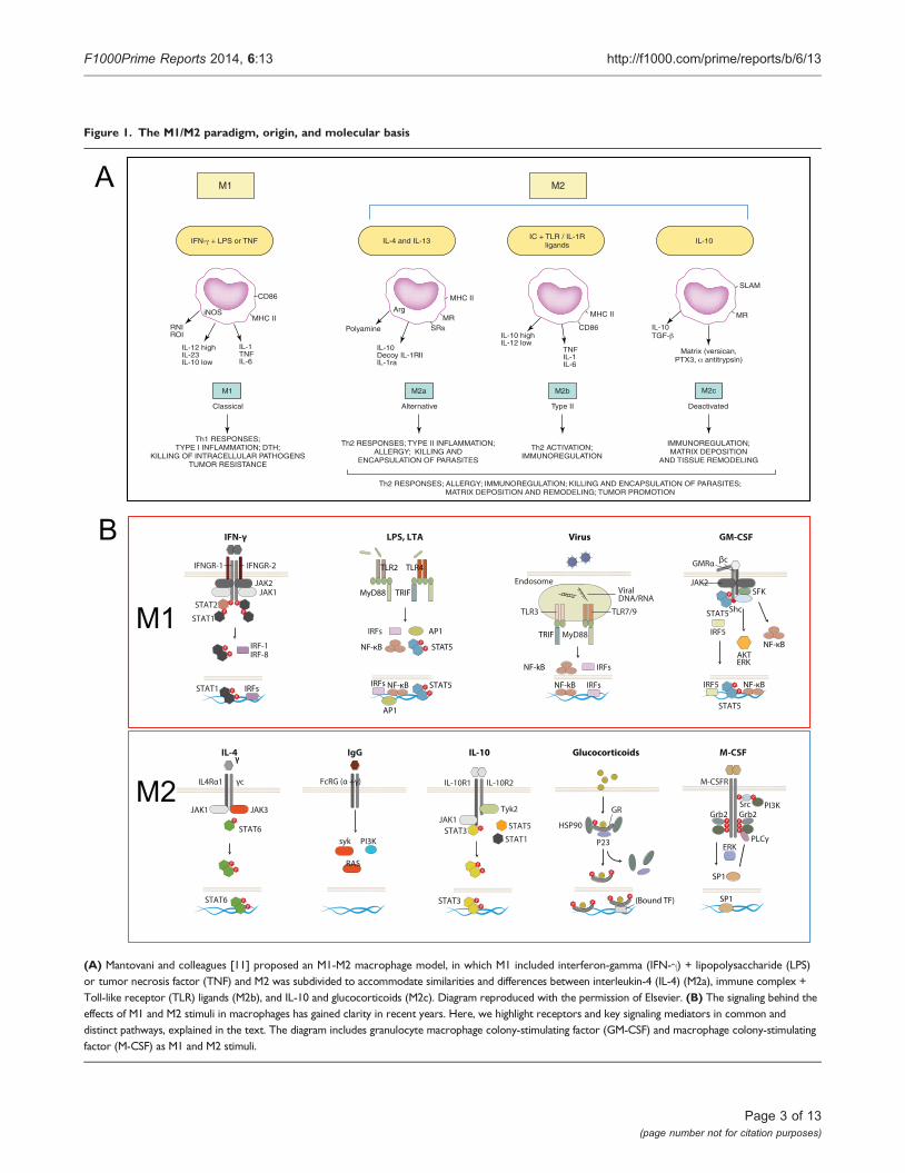
Figure 1. The M1/M2 paradigm, origin, and molecular basis
iNOS
RNIROI
IL-12 highIL-23IL-10 low
IL-1TNFIL-6
M1
Classical
IL-4 and IL-13
Arg
Polyamine
IL-10Decoy IL-1RIIIL-1ra
SRsMR
M2a
Alternative
TNFIL-1IL-6
CD86
IL-10
IL-10
MR
M2c
IL-10 highIL-12 low
CD86
M2b
Deactivated
SLAM
M2M1
IFN- + LPS or TNFIC + TLR / IL-1R
ligands
MHC II
MHC II
MHC II
TGF-
Matrix (versican,PTX3, antitrypsin)
Type II
Th1 RESPONSES;TYPE I INFLAMMATION; DTH;
KILLING OF INTRACELLULAR PATHOGENSTUMOR RESISTANCE
Th2 RESPONSES; TYPE II INFLAMMATION;ALLERGY; KILLING AND
ENCAPSULATION OF PARASITES
Th2 ACTIVATION;IMMUNOREGULATION
IMMUNOREGULATION;MATRIX DEPOSITION
AND TISSUE REMODELING
Th2 RESPONSES; ALLERGY; IMMUNOREGULATION; KILLING AND ENCAPSULATION OF PARASITES;MATRIX DEPOSITION AND REMODELING; TUMOR PROMOTION
A
B
JAK1JAK2
P
P
P
P
PP
PP
STAT1
STAT2
STAT1
IFNGR-2IFNGR-1
IFN-γ
IRF-1IRF-8
IRFs
IL-4
γc
STAT6
JAK1 JAK3
STAT6P
PP
P
PP
IL4Rα1
STAT5
AP1
NF-κBIRFs
IRFs
NF-κBP
P
AP1
PP
STAT5
TLR2
MyD88
TLR4
TRIF
LPS, LTA
SFK
Shc
AKT
JAK2
GM-CSF
IRF5
P
PSTAT5
PP
NF-κB
NF-κB
STAT5
IRF5
GMRα βc
ERK
PI3K
ERK
SrcP
PPP
PPP
M-CSFR
P
Grb2Grb2
SP1
SP1
PLCγ
M-CSF
IL-10R1 IL-10R2
JAK1Tyk2
HSP90
P23
GR
(Bound TF)
IL-10 Glucocorticoids
STAT3 STAT5P
PPP
STAT3
P
P P
P P
P P
STAT1
PPP
TLR7/9
MyD88
IRFs
ViralEndosome
Virus
IRFsNF-kB
NF-kB
TRIF
TLR3
DNA/RNA
IgG
P
sykP
FcRG (α +γ)
PI3K
RAS
γγ
M1
M2
(A) Mantovani and colleagues [11] proposed an M1-M2 macrophage model, in which M1 included interferon-gamma (IFN-g) + lipopolysaccharide (LPS)or tumor necrosis factor (TNF) and M2 was subdivided to accommodate similarities and differences between interleukin-4 (IL-4) (M2a), immune complex +Toll-like receptor (TLR) ligands (M2b), and IL-10 and glucocorticoids (M2c). Diagram reproduced with the permission of Elsevier. (B) The signaling behind theeffects of M1 and M2 stimuli in macrophages has gained clarity in recent years. Here, we highlight receptors and key signaling mediators in common anddistinct pathways, explained in the text. The diagram includes granulocyte macrophage colony-stimulating factor (GM-CSF) and macrophage colony-stimulatingfactor (M-CSF) as M1 and M2 stimuli.
Page 3 of 13(page number not for citation purposes)
F1000Prime Reports 2014, 6:13 http://f1000.com/prime/reports/b/6/13

were considered to establish differences and similaritiesin macrophage responses to stimuli. The originaldefinition took into account the possible context of thestimuli, but current generalized views lack this perspec-tive. Furthermore, updated knowledge of cytokinesignaling, the role of cytokines in the development ofthe hematopoietic system and in disease models withgenetically modified mice and transcriptomic andproteomic analysis reveal a far more complex pictureand challenge the current grouping. In the next section,we discuss the different role of stimuli considered in theM1 and M2 paradigm and highlight signaling pathwaysand gene expression singularities between the M1 andM2 stimuli.
M1 stimuliThe M1 stimuli are grouped according to their ability toinduce prototypic inflammatory responses and markers,but their source, role, receptors, and signaling pathwaysdiffer substantially. We discuss, as examples, three of themain M1 stimuli recognized today. IFN-g is the maincytokine associated with M1 activation and the main Th1cell product. Other cells, such as natural killer (NK) cellsand macrophages, themselves have been shown toproduce the cytokine. The IFNGR-1 and IFNGR-2 chainsform IFN-g receptor (Figure 1B). The receptor recruitsJanus kinase (Jak)1 and Jak2 adaptors that activateSTAT1 (signal transducers and activators of transcrip-tion1) and interferon regulatory factors (IRF), such asIRF-1 and IRF-8; for a recent comprehensive review, see[13]. IFN-g controls specific gene expression programsinvolving cytokine receptors (CSF2RB, IL15 receptoralpha [RA], IL2RA, and IL6R), cell activation markers(CD36, CD38, CD69, and CD97), and a number of celladhesion molecules (intercellular adhesion molecule 1[ICAM1], integrin alpha L [ITGAL], ITGA4, ITGbeta-7[B7], mucin 1 [MUC1], and ST6 beta-galactosamidealpha-2,6-sialyltranferase 1 [SIAT1]). The major media-tors of IFN-g-induced signaling, STAT1, JAK2, and IRF1,and regulators cytokine inducible SH2-containing pro-tein (CISH), N-myc-interactor (NMI), protein tyrosinephosphatase, receptor type, C PTPRC, protein tyrosinephosphatase, receptor type, O (PTPRO), and suppressorof cytokine signaling 1 (SOCS1) are also under thecontrol of the cytokine [14]. IFN-g is included incombination with LPS in the M1/M2 paradigm, andgene expression profiles of the combination are differentfrom LPS or IFN-g profiles alone [15,16]. Mice lackingIFN-g or its receptors are viable and fertile and theirsteady-state macrophage numbers are normal [17,18].Macrophages, however, show impaired production ofantimicrobial products, and mice are susceptible toMycobacterium bovis and Listeria monocytogenes. This defectis not important for prototypical Th1/M1 responses only:
knockout (KO) mice are susceptible to protozoa, such asTrypanosoma cruzi [19], Leishmania amazonensis andLeishmania major [20], and Cryptosporidium parvum [21],as well as defense against some nematodes (e.g.Litomosoides sigmodontis [22], Schistosoma mansoni [23],and Schistosoma japonicum [24]). In humans, mutationsresulting in the lack of expression of the receptordrive severe immunodeficiency (e.g. susceptibility tomycobacteria M. avium; M. kansasii; M. chelonei,Salmonella typhimurium and S. paratyphi) in patientswith familial disseminated atypical mycobacterialdisease [25].
Pathogens are recognized by pattern recognition recep-tors. The activation induced is part of the M1 group andalso defined as “innate” activation [26]. Full bacteriainduce gene programs similar to those of isolated Toll-like receptors (TLRs), and major parts of the pathogenprofiles can be ascribed to TLR ligands, such as LPS,muramyl dipeptide, and lipoteichoic acid [16]. LPS is thebest-studied M1 macrophage signal and is recognized byTLR4 (Figure 1B), although recent evidence shows thatLPS can also be recognized by TLR4-independentmechanisms leading to inflammasome activation[27,28]. Conventionally, TLR4 activation inducesMyD88 and MaL/Tirap (Toll-interleukin 1 receptordomain containing adaptor protein)-dependent path-ways that lead to strong pro-inflammatory cytokineprofiles (e.g. IFN-b, IL-12, TNF, IL-6, and IL-1b),chemokines (e.g. chemokine [C-C motif] ligand 2CCL2, chemokine [C-X-C motif] ligand 10 [CXCL10],and CXCL11), and antigen presentation molecules, suchas MHC members, co-stimulatory molecules, and anti-gen-processing peptidases. The profiles are controlled bynuclear factor of kappa light polypeptide gene enhancer(NF-kB), activator protein 1 (AP-1), IRFs, STAT1, andEGR (early growth response) family members, many ofwhich participate in the IFN response [13]. Althoughthere is a degree of overlap between LPS and IFN-g geneprofiles, similarities are not enough to consider thestimuli to be homologous. As for IFN-g, the numbers ofmacrophages in TLR KO animals are normal, but theiractivation is defective and therefore survival to infectionis severely impaired; for seminal and recent views, see[29-32]. In humans, genetic mutations in the TLR familyhave gained clarity, and as for mice, there is evidence forsusceptibility to infection with mycobacteria, pneumo-cocci, meningococci, malaria, and susceptibility todevelop bacteremia [33].
Granulocyte macrophage colony-stimulating factor(GM-CSF) is the latest addition to the M1 category ofstimuli. GM-CSF is produced by a variety of cells, includingmacrophages and parenchyma cells. The GM-CSF receptor
Page 4 of 13(page number not for citation purposes)
F1000Prime Reports 2014, 6:13 http://f1000.com/prime/reports/b/6/13

forms a dodecamer structure [34] and recruits JAK2, leadingto the activation of STAT5, extracellular signal-regulatedkinase (ERK), and V-Akt murine thymoma viral oncogenehomolog 1 (AKT) as well as the nuclear translocationof NF-kB and IRF5 upon binding (Figure 1B) [35]. Manyof these regulators are part of the IFN-g and TLR signalingpathways. GM-CSF enhances antigen presentation,complement- and antibody-mediated phagocytosis, micro-bicidal capacity, leukocyte chemotaxis, and adhesion. GM-CSF induces monocyte and macrophage cytokine produc-tion of IL-6, IL-8, G-CSF, M-CSF, TNF, and IL-1b, but lessthan, for example, LPS. GM-CSF transcriptome analysisshows that GM-CSF regulates several known cell surfacemolecules (e.g. CD14, Fc fragment of IgG, high affinity Ia(FCgR1A), CD163, and nuclear receptor subfamily 1,group H, member 3 [NR1H3]) [36]. The GM-CSF KO hasnormal numbers of macrophages in some tissues but hasdefects in the maturation of alveolar macrophages anddevelops pulmonary alveolar proteinosis [37]. In humans,mutations in the GM-CSF receptor, especially in thecommon beta chain, lead to alveolar macrophage defectsandproteinosis but also tomalignancy [38,39]. As such, themain functions proposed for GM-CSF include regulation ofhematopoietic cell proliferation and differentiation, andmodulation of the function of mature hematopoietic cells.Other stimuli that share pro-inflammatory properties havebeen termed M1 (e.g. TNF, IL-1ß, and IL-6). This addsfurther heterogeneity to a group that already comprises Tcells, bacterial products, and a lineage-determiningcytokine.
M2 stimuliThe M2 group of stimuli arose from the initial IL-4observations, and they are grouped mainly due to theirability to antagonize prototypic inflammatory responsesandmarkers; however, as forM1 stimuli, their source, role,receptors, and signaling pathways differ. We discuss, asexamples, five of themainM2 stimuli. IL-4 is produced bythe Th2 cells, eosinophils, basophils, or macrophagesthemselves and is recognized by three different receptorpairs. IL-4Ra1 can pair with the common gamma chain(gc), enabling IL-4 binding, and with the IL13Ra1 chain,enabling IL-4 or IL-13 binding (Figure 1B). In addition,IL-13 binds to the IL13Ra2 chain, a controversial signalingreceptor. Receptor binding of IL-4 activates JAK1 and JAK3.JAK activation leads to STAT6 activation and translocation.Other transcription factors involved include c-Myc andIRF4. IL-4 induces macrophage fusion and decreasesphagocytosis. The IL-4 multispecies transcriptomeincludes transglutaminase 2 (TGM2), mannose receptor(MRC1), cholesterol hydroxylase CH25H, and theprostaglandin-endoperoxide synthase PTGS1 (prostaglan-din G/H synthase 1), the transcription factors IRF4,Krüppel-like factor 4 (KLF4), and the signalingmodulators
CISH and SOCS1 [40]. IL-13 signatures are similar to IL-4signatures but are not totally overlapping [41]. In IL-4 KOanimals, the numbers of macrophages andmaturation arenormal, and defects appear in the immune responseagainst nematodes and some viral infections; for recentreviews, see [42,43]. In humans, polymorphisms in theIL-4R have been associated with the development ofasthma and atopy [44,45].
Another M2 category is the type II-activated macrophagedefined by Mosser and classified M2b by Mantovani andcolleagues. This represents the only example of crosstalkwith the B cell. This is another combined state, similar tothe M1 combination of IFN-g + LPS, in which ligation ofFcgRs on LPS-activated macrophages turns off IL-12 andinduces IL-10 secretion in addition to upregulatingantigen presentation and, importantly, promoting Th2responses [46,47]. IgGs are recognized by the Fc gammareceptor family that includes the activatory FcgRI (CD64),the inhibitory FcgRIIA (CD32), FcgRIIB (CD32), and theactivatory FcgRIIIA (CD16a) and FcgRIIIB (CD16b).CD32 seems to be crucial for the type II activation inhuman monocytes and macrophages [48]. FcR signalinginvolves spleen tyrosine kinase (Syk) and phosphoino-sitide 3-kinase (PI3K) activation [49], but details inmacrophages and its interplay with Myd88 pathwaysneed further study. Type II-activated macrophages aredistinct from IL-4-activated macrophages and their geneexpression profiles overlap only partially. FcR KO animalshave normal macrophage numbers, but their opsonicphagocytic capacity is highly impaired [50]. In humans,genetic differences in FcRs contribute to autoimmunediseases, such as systemic lupus erythematosus, rheuma-toid arthritis, and multiple sclerosis [50]. Curiously, IgE,more relevant for Th2 and antiparasitic responses, has notbeen implicated in this phenotype.
Glucocorticoids and IL-10 are included in the current M2category, although they represent a very different type ofstimulus. Glucocorticoid hormones secreted by the adrenalglands are metabolized by cellular enzymes in macro-phages. Active glucocorticoids are lipophilic and diffusethrough the membrane to bind the glucocorticoid receptor(GCR) alpha, leading to nuclear translocation of thecomplex (Figure 1B). The GCR complex binds DNAdirectly to promote/repress gene transcription or indirectlyby interacting with transcription factors, such as NF-kB orAP-1. Expression analysis of glucocorticoid-stimulatedmonocytes showed induction of complement component1 subunit A (C1QA), TSC22 domain family, member3 (DSIPI), MRC1, thrombospondin 1 (THBS1), IL-10,IL1R2, and CD163 [51]. Long-term exposure drivesdifferent gene expression programs that interact with LPSand IFN-g pathways in a complex and non-exclusively
Page 5 of 13(page number not for citation purposes)
F1000Prime Reports 2014, 6:13 http://f1000.com/prime/reports/b/6/13

antagonistic manner [52]. The profiles are altogetherdifferent from those induced by IL-4. Glucocorticoids affectmonocyte adherence, spreading, phagocytosis, andapoptosis. Mice with GCR deficiency do not survive longafter birth, because of respiratory failure, and alterations inreceptor dimerization induce susceptibility to sepsis [53].In humans, GCR polymorphisms in the genes are pleio-tropic and have been involved in a variety of malignanciesand inflammatory and autoimmune disorders [54].
IL-10 binds the IL-10 receptor, a dimer of IL10R1 andIL10R2 (Figure 1B). Receptor autophosphorylation leadsto the activation of the transcription factor STAT3 andits binding mediates inhibition of pro-inflammatorycytokine expression. IL-10 is a Th2 product and potentinhibitor of Th1 cells [55]. IL-10 is produced by virtuallyall leukocytes. Inmacrophages, IL-10 is elicited in responseto TLR activation, glucocorticoids, and C-type lectinsignaling (e.g. DC-SIGN [CD209 molecule] and dectin1 ligation). The macrophage transcriptome induced byIL-10 includes selected Fc receptors, the chemoattractantsCXCL13 and CXCL4, and the recognition receptors formylpeptide receptor 1 (FPR1), TLR1, TLR8, and macrophagereceptor with collagenous domain (MARCO) [56]. IL-10-deficient mice have normal macrophage numbers butdevelop inflammatory bowel disease following coloniza-tion of the gut with resident enteric bacteria [57] and showexaggerated inflammatory responses to parasites [58].In humans, defects in the cytokine receptors are similarand involve colitis and exacerbated inflammation [59].
M-CSF, like GM-CSF, is a late addition to the paradigmand has been classified as an M2 stimulus. The M-CSFreceptor is a tyrosine kinase transmembrane receptor(Figure 1B). M-CSF binding leads to receptor dimeriza-tion, autophosphorylation, activation of ERK, phospha-tidylinositol 3-kinase, phospholipase C, and eventuallySp1 transcription factor nuclear localization. The tran-scriptional response to M-CSF includes transient geneclusters with overrepresentation of cell cycle genes (e.g.cyclins A2, B1, D1, and E1) and downregulation ofhuman leukocyte antigen (HLA) members and stablegene clusters, including TLR7 and the complementC1QA/B/C subunits [15].
Reports generally focus on the differences betweenM-CSF and GM-CSF; a recent and comprehensivecomparison of the response to GM-CSF and M-CSF byhuman and mouse macrophages shows 530 genesregulated in the same direction in both human andmurine models [60,61]. M-CSF mutant mice showreduced levels of monocytes and selected macrophagesand osteopetrosis [62]. Mutations in the M-CSF receptorin humans lead to myelodysplastic syndromes or acute
myeloid leukemia [63], and the mutations have beenassociated with hereditary diffuse leukoencephalopathy[64], but no human patients with osteopetrosis second-ary to M-CSF deficiency have been identified.
The M2 group includes very different stimuli that spanfour levels of recognition/response: a level in which themacrophage acquires matured phenotypes, a level inwhich the macrophage interacts with immune cells(eosinophils, basophils, and Th2 cells), a level in whichthe macrophage actually deals with the pathogen, anda resolution level.
We discussed signaling cascades elicited by current M1andM2 stimuli, which are complex and include transientand stable gene signatures. Importantly, activation ofmacrophages is controlled not only by intracellularkinases and transcription factors. Other mechanisms,such as microRNAs (miRNAs) [65-69], enhancer RNAs[70-72], and epigenetic enzymes, control the activationlandscape [73-78]. These are beginning to be elucidatedin macrophages, and more information exists for themouse. The research done on epigenetics does not coverthe full spectrum of macrophage activation but doesprovide messages similar to the signaling pathwaysdiscussed above (i.e. specific mechanisms controldifferent forms of activation, and these mechanisms arenot fixed; for every acetylase or methylase, there is acounterpart ready to be activated). Before discussing ourview of macrophage activation, we briefly discuss theevidence for M1/M2 activation in disease.
M1 and M2 in disease: lack of defined subsetsIn vitro studies have contributed to our understanding ofmacrophage activation, and as discussed, KO animals forkey cytokines and receptors have established a role forsome of these in the development and maturation ofmacrophages, whereas others regulate activation and thetuning of the response. The activation signatures defined invitro are highly influenced by factors that are oftenoverlooked but important in vivo (e.g. maturation of thecell, adhesion, extracellular matrix composition, andchemoattractants). Translating in vitro results to diseaseposes a major problem because of the complexity of in vivosystems and the failure to mimic these conditions in vitro[79].Defining specificM1 andM2 functionswith cytokinesor receptor KO is difficult because the genes are pleiotropicand expressed at different stages of macrophage develop-ment or in other cell types; development of conditional,macrophage-specific KOs will help to illuminate thesefunctions. Whole genome studies have shown substantialdifferences between M1 and M2 activation programs inhumans and mice, indicative of evolutionary plasticityamong macrophages, yet adding difficulty to translation.
Page 6 of 13(page number not for citation purposes)
F1000Prime Reports 2014, 6:13 http://f1000.com/prime/reports/b/6/13

When it comes to infection, it is clear that macrophageresponses to different pathogens are affected by virulenceand evasion mechanisms. Non-infectious diseases inhumans are not as homogeneous as in mouse models,and thus we are often looking at a collection of tissue andsystemic conditions that lead to a common syndrome butwith different macrophage phenotypes.
Because of the relationship with the Th1 and Th2paradigm, macrophage M1 and M2 markers have beeninvestigated in prototypic diseases (e.g. Th2 Asthma [80]and Th1 chronic obstructive pulmonary disease (COPD)[81]). Another area where macrophage profiles havebeen investigated is atherosclerosis [82] and tumors [83].For a recent review, see [84]. The emphasis has alwaysbeen to fit the profile of tissue macrophages in diseasesto in vitro predictions, but the message is clear: in vitromodels are unable to mimic the complex profilesobserved in disease and, as such, the numbers of genesthat can be confirmed is limited. Our feeling is that whenit comes to tissue macrophages, we need to start with afresh view. Importantly, because macrophages candevelop mixed M1/M2 phenotypes in pathologicalconditions, we need to focus not only on populationsbut also at the single-cell level [85,86].
From a functional view, the main properties of macro-phages are phagocytosis, endocytosis, secretion, and
microbial killing, but chemotaxis, adhesion, and trophicfunctions are an integral part of their activation [11].Because macrophages are able to perform all theseactivities in the steady state, M1 and M2 contribution todisease is, for the most part, modulation and tuning. Asbefore with markers, functions are complex (e.g.phagocytosis involves a collaboration of multiplereceptors as well as interactions with different particles).The discrimination and killing of microbes and hosttarget cells are also incompletely understood. To date,M1- or M2-specific functions beyond M1-enhancedmicrobicidal and M2 antiparasitic defense haveexpanded to encompass metabolic, thermoregulatory,healing, and antiviral effects [10,42,43,87-90]. Recentevidence shows that M1 and M2 activation displaydifferences but also overlapping effects that needclarification and a more dynamic appreciation of theactivation process. In Table 1, we have summarized a fewfindings that exemplify the increasing complexity of theM1/M2 landscape.
Detailing M1 and M2 marker studies in particularhuman disease processes is beyond the scope of thisreview; we highlight some considerations:
(a) Rather than distinct macrophage populations, M1and M2 signatures do not necessarily exclude each otherand often coexist; the resultant mixed phenotype then
Table 1. Selection of M1 and M2 effects in macrophages
M1 (IFN-g) M2 (IL-4/IL-13)
Functions elicited in macrophagesPhagocytosis / endocytosis -Increases phagocytosis of C. albicans [95]
-Decreases Fc-mediated phagocytosis [96]-Decreases complement-mediatedphagocytosis [97]
-Decreases phagocytosis of particles while increasinginflammatory cytokine production [98]
Autophagy -Induces autophagy in TB infection [99] -Decreases autophagy in TB infection [100]Macrophage Fusion -Increases fusion in combination with concanavalin
A [101]-Induces fusion in alveolar macrophages [102]
-Induces fusion [103]-Inhibits IFN-g-induced fusion [101]
Nitric Oxide -Induces Mycobacteria killing via NO [104] -Favours Arginase-1 vs. i-NOS, Arg1+ macrophagessuppress Th2 inflammation and fibrosis [105]
Parasite killing and expulsion -Mediates parasite killing via NO [106, 107] -Although the cytokine is important for wormexpulsion, the effect does not depend onmacrophages [108]
Virus replication -Inhibits replication of HIV at early pre-integrationsteps [109]
-Inhibits HIV replication at post-integration level [109]
MarkersHuman CD64, IDO, SOCS1, CXCL10 MRC1, TGM2, CD23, CCL22Mouse CXCL9, CXCL10, CXCL11, NOS2 Mrc1, tgm2, Fizz1, Ym1/2, Arg1
The current M1 and M2 paradigm includes a variety of stimuli of different natures. This complicates the understanding of the contribution of adaptiveimmunity to the innate response, and the specialized functions that arise with activation. Here, we focus on IFN-g and IL-4 effects. This table is notcomprehensive, nor does it include other stimuli currently part of the M1-M2 paradigm. With the table we wish to highlight the distinction between M1 andM2 functions, whose features are not polar but are reflections of a more subtle process.C. albicans, Candida albicans; CCL, chemokine (C-C motif) ligand; CXCL, chemokine (C-X-C motif) ligand ;iNOS, inducible nitric oxide synthase; IFN-b,interferon beta; IFN-g, interferon gamma; MRC1, mannose receptor; NO, nitric oxide; NOS2, nitric oxide synthase 2; SOCS1, suppressor of cytokinesignaling 1; TB, tuberculosis; TGM2, transglutaminase 2.
Page 7 of 13(page number not for citation purposes)
F1000Prime Reports 2014, 6:13 http://f1000.com/prime/reports/b/6/13
depends on the balance of activatory and inhibitoryactivities and the tissue environment.
(b) Pleiotropism of stimuli and lack of cell specificity ofmarkers indicate that macrophage specialization rests inpart on their ability to migrate and deliver specificfunctions where barriers have failed to stop infection.
(c) The role of M1 or M2 stimuli needs to be consideredin their dynamic complexity, beyond the current bipolardogma of IFN-g as exclusively important for intracellularpathogens or IL-4 for allergy and extracellular parasiticdefense. The multipolar interplay between theseand many other signals requires further studies in vivoand in vitro.
Figure 2. A multipolar view of the macrophage activation paradigm from an immunological perspective
Growthand
survivalfactors
Lymphoid
andmyeloid
Cytokines
Interac�on
withPathogens
Resolu�on
Lineagedeterminingcytokines
(CONVENTIONALMATURATION)
M-CSFR GM-CSFR
IFN-γ
ClassicalandAlterna�ve
ac�va�on
IL-4, IL-13
Proandan�nflammatory
TNF, IL-6, IL-1ß IL-10, TGF-ß,
Survival,recruitment
andreten�on
Adhesion moleculesChemokines
Other
VitD3, Retinoic acid,PPRgamma ligands
Directinterac�on
TLRs, NODs, NLRs,RLRs, Nucleic acid sensors
IgG, IgE, IgA Complement,Lectins, Ficolins
Humoral
Systemicmechanisms
Glucocorticoids
Localmechanisms
ECM Proteoglycans, ATP and sugar nucleoCdes,Resolvins, Maresins, etc
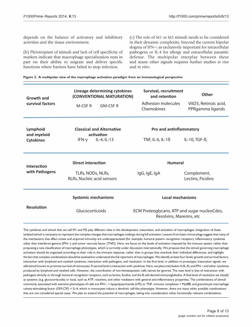
The cytokines and stimuli that we call M1 and M2 play different roles in the development, maturation, and activation of macrophages. Integration of theseisolated stimuli is necessary to represent the complex changes thatmacrophages undergo during full activation. Lessons from basic immunology suggest thatmany ofthe mechanisms that affect innate and acquired immunity are underappreciated (for example, humoral pattern recognition receptors, inflammatory cytokinesother than interferon-gamma (IFN-g) and tumor necrosis factor (TNF)). Here, we focus on the levels of activation imposed by the immune system rather thanproposing a new classification of macrophage phenotypes, which is currently under discussion internationally. We propose that the stimuli governing macrophageactivation should be organized according to their role in the immune response, rather than in groups that overlook their individual differences, and highlightthe fact that complex combinations should be assessed to understand the full repertoire of macrophages.We identify at least four levels: growth and survival factors,interaction with lymphoid and myeloid cytokines, interaction with pathogens, and resolution. In the first level, in addition to prototypic maturation signals, weadd stimuli known to promote survival ofmonocytes. A second level is interactionwith cytokines. Here, we place interleukin-4 (IL-4), and IFN-g and other cytokinesproduced by lymphoid and myeloid cells. However, the contribution of non-hematopoietic cells cannot be ignored. The next level is that of interaction withpathogens directly or through humoral recognition receptors, such as lectins, ficolins, and the B cell-derived immunoglobulins. A final level of resolution we classifyas systemic (e.g. glucocorticoids) or local, such as ATP, resolvins, and other mediators with general anti-inflammatory properties. The combinations of stimulicommonly associated with extreme phenotypes of cells are IFN-g + lipopolysaccharide (LPS) or TNF, immune complexes + Myd88, and granulocyte macrophagecolony-stimulating factor (GM-CSF) + IL-4, which in monocytes induce a dendritic cell-like phenotype. However, there are many other possible combinationsthat are not considered special cases. We plan to extend the potential of macrophages, taking into consideration other functionally relevant combinations.
Page 8 of 13(page number not for citation purposes)
F1000Prime Reports 2014, 6:13 http://f1000.com/prime/reports/b/6/13
Disclosures
ConclusionsToday, we know that, in addition to Th1 and Th2 cells,Treg cells and Th17 cells participate in the pathogenesisand resolution of disease and that these cells are able todisplay shades of activation within the general categories[91]. The Th1/Th2 paradigm has remained valuablein T lymphocyte heterogeneity and, to an extent, in thefield of macrophage activation, but a reassessment isrequired to accommodate current findings.
We do not here propose a revisedmodel or nomenclaturefor macrophage activation but present a view on whatneeds to be taken into account (Figure 2). Our opinion isthat macrophages do not form stable subsets but respondto a combination of factors present in the tissue; wehave, rather than subsets of macrophages, pathways thatinteract to form a complex, even mixed, phenotypes.A satisfactory paradigm needs to take into account at leastfour levels of recognition/response: first, one in whichthe monocyte survives and acquires matured pheno-types; second, a level in which the macrophage interactswith immune cells (NK and Th cells, eosinophils, andbasophils); third, one in which the macrophage dealswith the pathogen, and a final level, of resolution. Also,the M1-M2 paradigm is commonly associated withproperties of mature macrophages, but activation takesplace in the extended macrophage family, includingmonocytes [41,48,51,92], myeloid-derived dendriticcells [93,94] and multinucleated giant cells. In tissues,all these events combine to produce a resultantphenotype, and, though useful for the sake of under-standing, any sort of hierarchy or order does notrepresent the biology of the cells. We require a dynamicview of this process to take into account the multipleelements in their systemic and local milieu and todefine the kinetics, plasticity, reversibility, and memoryof their responses in order to encompass the fullfunctional range of activated macrophages. The processis highly complex, and for an improved understanding,considerably more information is required aboutmacrophages in vivo and at the population level aswell as at the single-cell level. Epigenetic, gene expres-sion, and functional studies will help to elucidate thesematters.
At present, the M1/M2 paradigm has provided a usefulframework, especially for selected immune responses,but a more comprehensive classification is clearlyrequired. This should guide an iterative research strategyfrom in vitro to in vivo studies and back to disease modelsin genetically defined mice, for example, to establishmechanisms and possible therapeutic targets for manip-ulation in human disease.
AbbreviationsAP-1, activator protein 1; C1QA/B/C, complementcomponent 1 subunit A/B/C; CISH, cytokine inducibleSH2-containing protein; c-Myc, V-Myc avian myelocyto-matosis viral oncogene homolog; CXCL, chemokine(C-X-C motif) ligand; EGR, early growth response; ERK,extracellular signal-regulated kinase; FCgR, Fc fragmentof IgG, high affinity; GCR, glucocorticoid receptor;GM-CSF, granulocyte macrophage colony-stimulatingfactor; IFN, interferon; IFNGR, interferon gamma recep-tor; Ig, immunoglobulin; IL, interleukin; IL-4Ra1, inter-leukin 4 receptor alpha 1; IRF, interferon regulatoryfactor; ITGB7, integrin beta-7; Jak, Janus kinase; KO,knockout; LPS, lipopolysaccharide; M-CSF, macrophagecolony-stimulating factor; MHC,major histocompatibilitycomplex; MRC1, mannose receptor; MyD88, myeloiddifferentiation primary response 88; NF-kB, nuclear factorof kappa light polypeptide gene enhancer; NK, naturalkiller; SOCS1, suppressor of cytokine signaling 1; STATsignal transducers and activators of transcription; TGF-b,transforming growth factor-beta; Th, T-helper; TLR,Toll-like receptor; TNF, tumor necrosis factor; Treg,T regulatory.
The authors declare that they have no disclosures.
AcknowledgmentsThe authors acknowledge Ronny Milde for their helpwith Figure 1B and Elsevier for Figure 1A.
References1. Mosmann TR, Cherwinski H, Bond MW, Giedlin MA, Coffman RL:
Two types of murine helper T cell clone. I. Definitionaccording to profiles of lymphokine activities and secretedproteins. J Immunol 1986, 136:2348-57.
2. Coffman RL: Origins of the T(H)1-T(H)2 model: a personalperspective. Nat Immunol 2006, 7:539-41.
3. Mackaness GB: Cellular resistance to infection. J Exp Med 1962,116:381-406.
4. Nathan CF, Murray HW, Wiebe ME, Rubin BY: Identification ofinterferon-gamma as the lymphokine that activates humanmacrophage oxidative metabolism and antimicrobial activ-ity. J Exp Med 1983, 158:670-89.
5. Pace JL, Russell SW, Schreiber RD, Altman A, Katz DH:Macrophageactivation: priming activity from a T-cell hybridoma isattributable to interferon-gamma. Proc Natl Acad Sci USA 1983,80:3782-6.
6. Celada A, Gray PW, Rinderknecht E, Schreiber RD: Evidence for agamma-interferon receptor that regulates macrophagetumoricidal activity. J Exp Med 1984, 160:55-74.
7. Stein M, Keshav S, Harris N, Gordon S: Interleukin 4 potentlyenhances murine macrophage mannose receptor activity: amarker of alternative immunologic macrophage activation.J Exp Med 1992, 176:287-92.
Page 9 of 13(page number not for citation purposes)
F1000Prime Reports 2014, 6:13 http://f1000.com/prime/reports/b/6/13
8. Doyle AG, Herbein G, Montaner LJ, Minty AJ, Caput D, Ferrara P,Gordon S: Interleukin-13 alters the activation state of murinemacrophages in vitro: comparison with interleukin-4 andinterferon-gamma. Eur J Immunol 1994, 24:1441-5.
9. Mills CD, Kincaid K, Alt JM, Heilman MJ, Hill AM: M-1/M-2macrophages and the Th1/Th2 paradigm. J Immunol 2000,164:6166-73.
10. Jenkins SJ, Allen JE: Similarity and diversity in macrophageactivation by nematodes, trematodes, and cestodes. J BiomedBiotechnol 2010, 2010:262609.
11. Mantovani A, Sica A, Sozzani S, Allavena P, Vecchi A, Locati M: Thechemokine system in diverse forms of macrophage activationand polarization. Trends Immunol 2004, 25:677-86.
12. Verreck FAW, Boer T de, Langenberg DML, Hoeve MA, Kramer M,Vaisberg E, Kastelein R, Kolk A, Waal-Malefyt R de, Ottenhoff THM:Human IL-23-producing type 1 macrophages promote butIL-10-producing type 2 macrophages subvert immunity to(myco)bacteria. Proc Natl Acad Sci USA 2004, 101:4560-5.
13. Hu X, Ivashkiv LB: Cross-regulation of signaling pathways byinterferon-gamma: implications for immune responses andautoimmune diseases. Immunity 2009, 31:539-50.
14. Waddell SJ, Popper SJ, Rubins KH, Griffiths MJ, Brown PO, Levin M,Relman DA: Dissecting interferon-induced transcriptional pro-grams in human peripheral blood cells. PLoS ONE 2010, 5:e9753.
15. Martinez FO, Gordon S, Locati M, Mantovani A: Transcriptionalprofiling of the human monocyte-to-macrophage differentia-tion and polarization: new molecules and patterns of geneexpression. J Immunol 2006, 177:7303-11.
16. Nau GJ, Richmond JFL, Schlesinger A, Jennings EG, Lander ES,Young RA: Human macrophage activation programs inducedby bacterial pathogens. Proc Natl Acad Sci USA 2002, 99:1503-8.
17. Huang S, Hendriks W, Althage A, Hemmi S, Bluethmann H, Kamijo R,Vilcek J, Zinkernagel RM, Aguet M: Immune response in mice thatlack the interferon-gamma receptor. Science 1993, 259:1742-5.
18. Dalton DK, Pitts-Meek S, Keshav S, Figari IS, Bradley A, Stewart TA:Multiple defects of immune cell function in mice withdisrupted interferon-gamma genes. Science 1993, 259:1739-42.
19. Marinho CRF, Nuñez-Apaza LN, Martins-Santos R, Bastos KRB,Bombeiro AL, Bucci DZ, Sardinha LR, Lima MRD, Alvarez JM: IFN-gamma, but not nitric oxide or specific IgG, is essential forthe in vivo control of low-virulence Sylvio X10/4 Trypano-soma cruzi parasites. Scand J Immunol 2007, 66:297-308.
20. Pinheiro RO, Rossi-Bergmann B: Interferon-gamma is requiredfor the late but not early control of Leishmania amazonensisinfection in C57Bl/6 mice. Mem Inst Oswaldo Cruz 2007,102:79-82.
21. Jakobi V, Petry F: Humoral immune response in IL-12 and IFN-gamma deficient mice after infection with Cryptosporidiumparvum. Parasite Immunol 2008, 30:151-61.
22. Saeftel M, Arndt M, Specht S, Volkmann L, Hoerauf A: Synergism ofgamma interferon and interleukin-5 in the control of murinefilariasis. Infect Immun 2003, 71:6978-85.
23. Rezende SA, Oliveira VR, Silva AM, Alves JB, Goes AM, Reis LF: Micelacking the gamma interferon receptor have an impairedgranulomatous reaction to Schistosoma mansoni infection.Infect Immun 1997, 65:3457-61.
24. Du X, Wu J, Zhang M, Gao Y, Zhang D, Hou M, Ji M, Wu G:Upregulated expression of cytotoxicity-related genes in IFN-gknockoutmice with Schistosoma japonicum infection. J BiomedBiotechnol 2011, 2011:864945.
25. Dorman SE, Holland SM: Interferon-gamma and interleukin-12pathway defects and human disease. Cytokine Growth Factor Rev2000, 11:321-33.
26. Mukhopadhyay S, Chen Y, Sankala M, Peiser L, Pikkarainen T, Kraal G,Tryggvason K, Gordon S: MARCO, an innate activation markerof macrophages, is a class A scavenger receptor for Neisseriameningitidis. Eur J Immunol 2006, 36:940-9.
27. Kayagaki N, Wong MT, Stowe IB, Ramani SR, Gonzalez LC, Akashi-Takamura S, Miyake K, Zhang J, Lee WP, Muszynski A, Forsberg LS,Carlson RW, Dixit VM: Noncanonical inflammasome activationby intracellular LPS independent of TLR4. Science 2013,341:1246-9.
28. Hagar JA, Powell DA, Aachoui Y, Ernst RK, Miao EA: CytoplasmicLPS activates caspase-11: implications in TLR4-independentendotoxic shock. Science 2013, 341:1250-3.
29. Takeda K, Akira S: TLR signaling pathways. Semin Immunol 2004,16:3-9.
30. Yamamoto M, Takeda K: Current views of toll-like receptorsignaling pathways. Gastroenterol Res Pract 2010, 2010:240365.
31. Kawai T, Akira S: Toll-like receptors and their crosstalk withother innate receptors in infection and immunity. Immunity2011, 34:637-50.
32. Netea MG, Wijmenga C, O’Neill LAJ: Genetic variation in Toll-like receptors and disease susceptibility. Nat Immunol 2012,13:535-42.
33. Casanova J, Abel L, Quintana-Murci L: Human TLRs and IL-1Rs inhost defense: natural insights from evolutionary, epidemio-logical, and clinical genetics. Annu Rev Immunol 2011, 29:447-91.
34. Hansen G, Hercus TR, McClure BJ, Stomski FC, Dottore M, Powell J,Ramshaw H, Woodcock JM, Xu Y, Guthridge M, McKinstry WJ,Lopez AF, Parker MW: The structure of the GM-CSF receptorcomplex reveals a distinct mode of cytokine receptoractivation. Cell 2008, 134:496-507.
35. Krausgruber T, Blazek K, Smallie T, Alzabin S, Lockstone H, Sahgal N,Hussell T, Feldmann M, Udalova IA: IRF5 promotes inflammatorymacrophage polarization and TH1-TH17 responses. NatImmunol 2011, 12:231-8.
36. Lehtonen A, Ahlfors H, Veckman V, Miettinen M, Lahesmaa R,Julkunen I: Gene expression profiling during differentiation ofhuman monocytes to macrophages or dendritic cells. J LeukocBiol 2007, 82:710-20.
37. Dranoff G, Mulligan RC: Activities of granulocyte-macrophagecolony-stimulating factor revealed by gene transfer and geneknockout studies. Stem Cells 1994, 12 (Suppl 1):173-82; discussion182-4.
38. Dirksen U, Nishinakamura R, Groneck P, Hattenhorst U, Nogee L,Murray R, Burdach S: Human pulmonary alveolar proteinosisassociated with a defect in GM-CSF/IL-3/IL-5 receptorcommon beta chain expression. J Clin Invest 1997, 100:2211-7.
39. Dirksen U, Hattenhorst U, Schneider P, Schroten H, Göbel U,Böcking A, Müller KM, Murray R, Burdach S: Defective expressionof granulocyte-macrophage colony-stimulating factor/inter-leukin-3/interleukin-5 receptor common beta chain in chil-dren with acute myeloid leukemia associated withrespiratory failure. Blood 1998, 92:1097-103.
40. Martinez FO, Helming L, Milde R, Varin A, Melgert BN, Draijer C,Thomas B, Fabbri M, Crawshaw A, Ho LP, Hacken NH ten,Cobos Jiménez V, Kootstra NA, Hamann J, Greaves DR, Locati M,Mantovani A, Gordon S: Genetic programs expressed in resting
Page 10 of 13(page number not for citation purposes)
F1000Prime Reports 2014, 6:13 http://f1000.com/prime/reports/b/6/13

and IL-4 alternatively activated mouse and human macro-phages: similarities and differences. Blood 2013, 121:e57-69.
41. Scotton CJ, Martinez FO, Smelt MJ, Sironi M, Locati M, Mantovani A,Sozzani S: Transcriptional profiling reveals complex regulationof the monocyte IL-1 beta system by IL-13. J Immunol 2005,174:834-45.
42. Martinez FO, Helming L, Gordon S: Alternative activation ofmacrophages: an immunologic functional perspective. AnnuRev Immunol 2009, 27:451-83.
43. van Dyken SJ, Locksley RM: Interleukin-4- and interleukin-13-mediated alternatively activated macrophages: roles inhomeostasis and disease. Annu Rev Immunol 2013, 31:317-43.
44. Ford AQ, Heller NM, Stephenson L, Boothby MR, Keegan AD: Anatopy-associated polymorphism in the ectodomain of theIL-4R(alpha) chain (V50) regulates the persistence of STAT6phosphorylation. J Immunol 2009, 183:1607-16.
45. Beghé B, Barton S, Rorke S, Peng Q, Sayers I, Gaunt T, Keith TP,Clough JB, Holgate ST, Holloway JW: Polymorphisms in theinterleukin-4 and interleukin-4 receptor alpha chain genesconfer susceptibility to asthma and atopy in a Caucasianpopulation. Clin Exp Allergy 2003, 33:1111-7.
46. Edwards JP, Zhang X, Frauwirth KA, Mosser DM: Biochemical andfunctional characterization of three activated macrophagepopulations. J Leukoc Biol 2006, 80:1298-307.
47. Anderson CF, Mosser DM: A novel phenotype for an activatedmacrophage: the type 2 activated macrophage. J Leukoc Biol2002, 72:101-6.
48. Sironi M, Martinez FO, D’Ambrosio D, Gattorno M, Polentarutti N,Locati M, Gregorio A, Iellem A, Cassatella MA, van Damme J,Sozzani S, Martini A, Sinigaglia F, Vecchi A, Mantovani A: Differentialregulation of chemokine production by Fcgamma receptorengagement in human monocytes: association of CCL1 witha distinct form of M2 monocyte activation (M2b, Type 2).J Leukoc Biol 2006, 80:342-9.
49. Sánchez-Mejorada G, Rosales C: Signal transduction by immu-noglobulin Fc receptors. J Leukoc Biol 1998, 63:521-33.
50. Takai T: Roles of Fc receptors in autoimmunity. Nat Rev Immunol2002, 2:580-92.
51. Ehrchen J, Steinmüller L, Barczyk K, Tenbrock K, Nacken W,Eisenacher M, Nordhues U, Sorg C, Sunderkötter C, Roth J:Glucocorticoids induce differentiation of a specifically acti-vated, anti-inflammatory subtype of human monocytes. Blood2007, 109:1265-74.
52. van de Garde MDB, Martinez FO, Melgert BN, Hylkema MN,Jonkers RE, Hamann J: Chronic exposure to glucocorticoidsshapes gene expression and modulates innate and adaptiveactivation pathways in macrophages with distinct changes inleukocyte attraction. J Immunol 2014, 192:1196-208.
53. Kleiman A, Hübner S, Rodriguez Parkitna JM, Neumann A, Hofer S,Weigand MA, Bauer M, Schmid W, Schütz G, Libert C, Reichardt HM,Tuckermann JP: Glucocorticoid receptor dimerization isrequired for survival in septic shock via suppression ofinterleukin-1 in macrophages. FASEB J 2012, 26:722-9.
54. Bray PJ, Cotton RGH: Variations of the human glucocorticoidreceptor gene (NR3C1): pathological and in vitro mutationsand polymorphisms. Hum Mutat 2003, 21:557-68.
55. Fiorentino DF, Bond MW, Mosmann TR: Two types of mouseT helper cell. IV. Th2 clones secrete a factor that inhibitscytokine production by Th1 clones. J Exp Med 1989,170:2081-95.
56. Park-Min K, Antoniv TT, Ivashkiv LB: Regulation of macrophagephenotype by long-term exposure to IL-10. Immunobiology 2005,210:77-86.
57. Sellon RK, Tonkonogy S, Schultz M, Dieleman LA, Grenther W,Balish E, Rennick DM, Sartor RB: Resident enteric bacteria arenecessary for development of spontaneous colitis andimmune system activation in interleukin-10-deficient mice.Infect Immun 1998, 66:5224-31.
58. Gazzinelli RT, Wysocka M, Hieny S, Scharton-Kersten T, Cheever A,Kühn R, Müller W, Trinchieri G, Sher A: In the absence ofendogenous IL-10, mice acutely infected with Toxoplasmagondii succumb to a lethal immune response dependent onCD4+ T cells and accompanied by overproduction of IL-12,IFN-gamma and TNF-alpha. J Immunol 1996, 157:798-805.
59. Glocker E, Kotlarz D, Boztug K, Gertz EM, Schäffer AA, Noyan F,Perro M, Diestelhorst J, Allroth A, Murugan D, Hätscher N, Pfeifer D,Sykora K, Sauer M, Kreipe H, Lacher M, Nustede R, Woellner C,Baumann U, Salzer U, Koletzko S, Shah N, Segal AW, Sauerbrey A,Buderus S, Snapper SB, Grimbacher B, Klein C: Inflammatorybowel disease and mutations affecting the interleukin-10receptor. N Engl J Med 2009, 361:2033-45.
60. Hashimoto S, Suzuki T, Dong HY, Yamazaki N, Matsushima K: Serialanalysis of gene expression in human monocytes and macro-phages. Blood 1999, 94:837-44.
61. Lacey DC, Achuthan A, Fleetwood AJ, Dinh H, Roiniotis J, Scholz GM,Chang MW, Beckman SK, Cook AD, Hamilton JA: DefiningGM-CSF- and macrophage-CSF-dependent macrophageresponses by in vitro models. J Immunol 2012, 188:5752-65.
62. Wiktor-Jedrzejczak W, Bartocci A, Ferrante AW, Ahmed-Ansari A,Sell KW, Pollard JW, Stanley ER: Total absence of colony-stimulating factor 1 in the macrophage-deficient osteope-trotic (op/op) mouse. Proc Natl Acad Sci USA 1990, 87:4828-32.
63. Tobal K, Pagliuca A, Bhatt B, Bailey N, Layton DM, Mufti GJ:Mutation of the human FMS gene (M-CSF receptor) inmyelodysplastic syndromes and acute myeloid leukemia.Leukemia 1990, 4:486-9.
64. Rademakers R, Baker M, Nicholson AM, Rutherford NJ, Finch N,Soto-Ortolaza A, Lash J, Wider C, Wojtas A, DeJesus-Hernandez M,Adamson J, Kouri N, Sundal C, Shuster EA, Aasly J, MacKenzie J,Roeber S, Kretzschmar HA, Boeve BF, Knopman DS, Petersen RC,Cairns NJ, Ghetti B, Spina S, Garbern J, Tselis AC, Uitti R, Das P,van Gerpen JA, Meschia JF, et al.: Mutations in the colonystimulating factor 1 receptor (CSF1R) gene cause hereditarydiffuse leukoencephalopathy with spheroids. Nat Genet 2012,44:200-5.
65. Etzrodt M, Cortez-Retamozo V, Newton A, Zhao J, Ng A,Wildgruber M, Romero P, Wurdinger T, Xavier R, Geissmann F,Meylan E, Nahrendorf M, Swirski FK, Baltimore D, Weissleder R,Pittet MJ: Regulation of monocyte functional heterogeneity bymiR-146a and Relb. Cell Rep 2012, 1:317-24.
66. Graff JW, Dickson AM, Clay G, McCaffrey AP, Wilson ME:Identifying functional microRNAs in macrophages withpolarized phenotypes. J Biol Chem 2012, 287:21816-25.
67. Lagrange B, Martin RZ, Droin N, Aucagne R, Paggetti J, Largeot A,Itzykson R, Solary E, Delva L, Bastie J: A role for miR-142-3p incolony-stimulating factor 1-induced monocyte differentiationinto macrophages. Biochim Biophys Acta 2013, 1833:1936-46.
68. Mildner A, Chapnik E, Manor O, Yona S, Kim K, Aychek T, Varol D,Beck G, Itzhaki ZB, Feldmesser E, Amit I, Hornstein E, Jung S:Mononuclear phagocytemiRNome analysis identifiesmiR-142
Page 11 of 13(page number not for citation purposes)
F1000Prime Reports 2014, 6:13 http://f1000.com/prime/reports/b/6/13
as critical regulator ofmurine dendritic cell homeostasis. Blood2013, 121:1016-27.
69. Swaminathan S, Hu X, Zheng X, Kriga Y, Shetty J, Zhao Y, Stephens R,Tran B, Baseler MW, Yang J, Lempicki RA, Huang D, Lane HC,Imamichi T: Interleukin-27 treated humanmacrophages inducethe expression of novel microRNAs which may mediate anti-viral properties. Biochem Biophys Res Commun 2013, 434:228-34.
70. Santa F de, Barozzi I, Mietton F, Ghisletti S, Polletti S, Tusi BK, Muller H,Ragoussis J, Wei C, Natoli G: A large fraction of extragenic RNApol II transcription sites overlap enhancers. PLoS Biol 2010, 8:e1000384.
71. Kaikkonen MU, Lam MTY, Glass CK: Non-coding RNAs asregulators of gene expression and epigenetics. Cardiovasc Res2011, 90:430-40.
72. Lam MTY, Cho H, Lesch HP, Gosselin D, Heinz S, Tanaka-Oishi Y,Benner C, Kaikkonen MU, Kim AS, Kosaka M, Lee CY, Watt A,Grossman TR, Rosenfeld MG, Evans RM, Glass CK: Rev-Erbs repressmacrophage gene expression by inhibiting enhancer-directedtranscription. Nature 2013, 498:511-5.
73. Bowdridge S, Gause WC: Regulation of alternative macrophageactivation by chromatin remodeling.Nat Immunol 2010, 11:879-81.
74. Lawrence T, Natoli G: Transcriptional regulation of macro-phage polarization: enabling diversity with identity. Nat RevImmunol 2011, 11:750-61.
75. Ivashkiv LB: Epigenetic regulation of macrophage polarizationand function. Trends Immunol 2013, 34:216-23.
76. Mukhopadhyay S, Ramadass AS, Akoulitchev A, Gordon S: Forma-tion of distinct chromatin conformation signatures epigen-etically regulate macrophage activation. Int Immunopharmacol2013, 18:7-11.
77. Satoh T, Takeuchi O, Vandenbon A, Yasuda K, Tanaka Y, Kumagai Y,Miyake T, Matsushita K, Okazaki T, Saitoh T, Honma K, Matsuyama T,Yui K, Tsujimura T, Standley DM, Nakanishi K, Nakai K, Akira S: TheJmjd3-Irf4 axis regulates M2macrophage polarization and hostresponses against helminth infection. Nat Immunol 2010,11:936-44.
78. Kruidenier L, Chung C, Cheng Z, Liddle J, Che K, Joberty G,Bantscheff M, Bountra C, Bridges A, Diallo H, Eberhard D,Hutchinson S, Jones E, Katso R, Leveridge M, Mander PK, Mosley J,Ramirez-Molina C, Rowland P, Schofield CJ, Sheppard RJ, Smith JE,Swales C, Tanner R, Thomas P, Tumber A, Drewes G, Oppermann U,Patel DJ, Lee K, et al.: A selective jumonji H3K27 demethylaseinhibitor modulates the proinflammatory macrophageresponse. Nature 2012, 488:404-8.
79. Davies LC, Jenkins SJ, Allen JE, Taylor PR: Tissue-residentmacrophages. Nat Immunol 2013, 14:986-95.
80. Madore A, Perron S, Turmel V, Laviolette M, Bissonnette EY,Laprise C: Alveolar macrophages in allergic asthma: an
expression signature characterized by heat shock proteinpathways. Hum Immunol 2010, 71:144-50.
81. Shaykhiev R, Krause A, Salit J, Strulovici-Barel Y, Harvey B,O’Connor TP, Crystal RG: Smoking-dependent reprogrammingof alveolar macrophage polarization: implication for patho-genesis of chronic obstructive pulmonary disease. J Immunol2009, 183:2867-83.
82. Feig JE, Vengrenyuk Y, Reiser V, Wu C, Statnikov A, Aliferis CF,Garabedian MJ, Fisher EA, Puig O: Regression of atherosclerosis ischaracterized by broad changes in the plaque macrophagetranscriptome. PLoS ONE 2012, 7:e39790.
83. Ojalvo LS, Whittaker CA, Condeelis JS, Pollard JW: Gene expres-sion analysis of macrophages that facilitate tumor invasionsupports a role for Wnt-signaling in mediating their activityin primary mammary tumors. J Immunol 2010, 184:702-12.
84. Murray PJ, Wynn TA: Protective and pathogenic functions ofmacrophage subsets. Nat Rev Immunol 2011, 11:723-37.
85. Pettersen JS, Fuentes-Duculan J, Suárez-Fariñas M, Pierson KC,Pitts-Kiefer A, Fan L, Belkin DA, Wang CQF, Bhuvanendran S,Johnson-Huang LM, Bluth MJ, Krueger JG, Lowes MA, Carucci JA:Tumor-associated macrophages in the cutaneous SCCmicroenvironment are heterogeneously activated. J InvestDermatol 2011, 131:1322-30.
86. Vogel DYS, Vereyken EJF, Glim JE, Heijnen PDAM, Moeton M, van derValk P, Amor S, Teunissen CE, van Horssen J, Dijkstra CD:Macrophages in inflammatory multiple sclerosis lesions havean intermediate activation status. J Neuroinflammation 2013, 10:35.
87. Odegaard JI, Chawla A: Alternative macrophage activation andmetabolism. Annu Rev Pathol 2011, 6:275-97.
88. Ricote M, Valledor AF, Glass CK: Decoding transcriptionalprograms regulated by PPARs and LXRs in the macrophage:effects on lipid homeostasis, inflammation, and atherosclero-sis. Arterioscler Thromb Vasc Biol 2004, 24:230-9.
89. Cunard R, Ricote M, DiCampli D, Archer DC, Kahn DA, Glass CK,Kelly CJ: Regulation of cytokine expression by ligands ofperoxisome proliferator activated receptors. J Immunol 2002,168:2795-802.
90. Palsson-McDermott EM, O’Neill LAJ: The Warburg effect thenand now: from cancer to inflammatory diseases. Bioessays2013, 35:965-73.
91. Zhu J, Paul WE: Heterogeneity and plasticity of T helper cells.Cell Res 2010, 20:4-12.
92. Tamassia N, Le Moigne V, Calzetti F, Donini M, Gasperini S, Ear T,Cloutier A, Martinez FO, Fabbri M, Locati M, Mantovani A,McDonald PP, Cassatella MA: The MyD88-independent pathwayis not mobilized in human neutrophils stimulated via TLR4.J Immunol 2007, 178:7344-56.
93. Mazzoni A, Segal DM: Controlling the Toll road to dendritic cellpolarization. J Leukoc Biol 2004, 75:721-30.
94. Kapsenberg ML: Dendritic-cell control of pathogen-drivenT-cell polarization. Nat Rev Immunol 2003, 3:984-93.
95. Maródi L, Schreiber S, Anderson DC, MacDermott RP, Korchak HM,Johnston RB: Enhancement of macrophage candidacidalactivity by interferon-gamma. Increased phagocytosis, killing,and calcium signal mediated by a decreased number ofmannose receptors. J Clin Invest 1993, 91:2596-601.
96. Frausto-Del-Río D, Soto-Cruz I, Garay-Canales C, Ambriz X,Soldevila G, Carretero-Ortega J, Vázquez-Prado J, Ortega E: Inter-feron gamma induces actin polymerization, Rac1 activationand down regulates phagocytosis in human monocytic cells.Cytokine 2012, 57:158-68.
97. Schlesinger LS, Horwitz MA: Phagocytosis of Mycobacteriumleprae by human monocyte-derived macrophages ismediated by complement receptors CR1 (CD35), CR3(CD11b/CD18), and CR4 (CD11c/CD18) and IFN-gammaactivation inhibits complement receptor function and pha-gocytosis of this bacterium. J Immunol 1991, 147:1983-94.
Page 12 of 13(page number not for citation purposes)
F1000Prime Reports 2014, 6:13 http://f1000.com/prime/reports/b/6/13

98. Varin A, Mukhopadhyay S, Herbein G, Gordon S: Alternativeactivation of macrophages by IL-4 impairs phagocytosis ofpathogens but potentiates microbial-induced signalling andcytokine secretion. Blood 2010, 115:353-62.
99. Matsuzawa T, Kim B, Shenoy AR, Kamitani S, Miyake M,Macmicking JD: IFN-g elicits macrophage autophagy via thep38 MAPK signaling pathway. J Immunol 2012, 189:813-8.
100. Harris J, Haro SA de, Master SS, Keane J, Roberts EA, Delgado M,Deretic V: T helper 2 cytokines inhibit autophagic control ofintracellular Mycobacterium tuberculosis. Immunity 2007,27:505-17.
101. Takashima T, Ohnishi K, Tsuyuguchi I, Kishimoto S: Differentialregulation of formation of multinucleated giant cells fromconcanavalin A-stimulated human blood monocytes by IFN-gamma and IL-4. J Immunol 1993, 150:3002-10.
102. Nagasawa H, Miyaura C, Abe E, Suda T, Horiguchi M: Fusion andactivation of human alveolar macrophages induced by recom-binant interferon-gamma and their suppression by dexametha-sone. Am Rev Respir Dis 1987, 136:916-21.
103. Helming L, Gordon S: Macrophage fusion induced by IL-4alternative activation is a multistage process involvingmultiple target molecules. Eur J Immunol 2007, 37:33-42.
104. Herbst S, Schaible UE, Schneider BE: Interferon gamma activatedmacrophages kill mycobacteria by nitric oxide inducedapoptosis. PLoS ONE 2011, 6:e19105.
105. Pesce JT, Ramalingam TR, Mentink-Kane MM, Wilson MS, El KasmiKC, Smith AM, Thompson RW, Cheever AW, Murray PJ, Wynn TA:Arginase-1-expressing macrophages suppress Th2 cytokine-driven inflammation and fibrosis. PLoS Pathog 2009, 5:e1000371.
106. Thomas GR, McCrossan M, Selkirk ME: Cytostatic and cytotoxiceffects of activated macrophages and nitric oxide donors onBrugia malayi. Infect Immun 1997, 65:2732-9.
107. Piedrafita D, Parsons JC, Sandeman RM, Wood PR, Estuningsih SE,Partoutomo S, Spithill TW: Antibody-dependent cell-mediatedcytotoxicity to newly excysted juvenile Fasciola hepatica invitro is mediated by reactive nitrogen intermediates. ParasiteImmunol 2001, 23:473-82.
108. Urban JF, Noben-Trauth N, Schopf L, Madden KB, Finkelman FD:Cutting edge: IL-4 receptor expression by non-bone marrow-derived cells is required to expel gastrointestinal nematodeparasites. J Immunol 2001, 167:6078-81.
109. Cassol E, Cassetta L, Rizzi C, Alfano M, Poli G: M1 and M2apolarization of human monocyte-derived macrophages inhi-bits HIV-1 replication by distinct mechanisms. J Immunol 2009,182:6237-46.
Page 13 of 13(page number not for citation purposes)
F1000Prime Reports 2014, 6:13 http://f1000.com/prime/reports/b/6/13