the management of respiratory motion in radiation oncology ... · the management of respiratory...
TRANSCRIPT

The management of respiratory motion in radiation oncology reportof AAPM Task Group 76a…
Paul J. Keallb�
Virginia Commonwealth University (Chair)
Gig S. MagerasMemorial Sloan-Kettering Cancer Center (Co-Chair)
James M. BalterUniversity of Michigan
Richard S. EmerySaint Vincent’s Cancer Center
Kenneth M. ForsterUT Southwestern Medical Center
Steve B. JiangMassachusetts General Hospital
Jeffrey M. KapatoesTomoTherapy, Inc.
Daniel A. LowWashington University
Martin J. MurphyVirginia Commonwealth University
Brad R. MurrayCross Cancer Institute
Chester R. RamseyThompson Cancer Survival Center
Marcel B. Van HerkNetherlands Cancer Institute
S. Sastry VedamUT MD Anderson Cancer Center
John W. WongJohns Hopkins University
Ellen YorkeMemorial Sloan-Kettering Cancer Center
�Received 3 May 2006; revised 27 July 2006; accepted for publication 4 August 2006;published 26 September 2006�
This document is the report of a task group of the AAPM and has been prepared primarily to advisemedical physicists involved in the external-beam radiation therapy of patients with thoracic, ab-dominal, and pelvic tumors affected by respiratory motion. This report describes the magnitude ofrespiratory motion, discusses radiotherapy specific problems caused by respiratory motion, explainstechniques that explicitly manage respiratory motion during radiotherapy and gives recommenda-tions in the application of these techniques for patient care, including quality assurance �QA�guidelines for these devices and their use with conformal and intensity modulated radiotherapy. Thetechnologies covered by this report are motion-encompassing methods, respiratory gated tech-niques, breath-hold techniques, forced shallow-breathing methods, and respiration-synchronizedtechniques. The main outcome of this report is a clinical process guide for managing respiratorymotion. Included in this guide is the recommendation that tumor motion should be measured �whenpossible� for each patient for whom respiratory motion is a concern. If target motion is greater than5 mm, a method of respiratory motion management is available, and if the patient can tolerate theprocedure, respiratory motion management technology is appropriate. Respiratory motion manage-ment is also appropriate when the procedure will increase normal tissue sparing. Respiratory motionmanagement involves further resources, education and the development of and adherence to QA
procedures. © 2006 American Association of Physicists in Medicine. �DOI: 10.1118/1.2349696�3874 3874Med. Phys. 33 „10…, October 2006 0094-2405/2006/33„10…/3874/27/$23.00 © 2006 Am. Assoc. Phys. Med.

3875 Keall et al.: Respiratory motion in radiation oncology 3875
The members of this Task Group wish to dedicate this work to Dr. Dale Kubo, a pioneer in the development and clinicalimplementation of respiratory motion management technology. Sadly, Dr. Kubo passed away during the formulation of thisreport.
TABLE OF CONTENTS
I. INTRODUCTION AND SCOPE. . . . . . . . . . . . . . . . 3875A. How to read this document. . . . . . . . . . . . . . . 3875B. Introduction. . . . . . . . . . . . . . . . . . . . . . . . . . . . 3875C. Scope. . . . . . . . . . . . . . . . . . . . . . . . . . . . . . . . . 3876
II. GLOSSARY AND ABBREVIATIONS. . . . . . . . . . . 3876III. PROBLEMS OF RESPIRATORY MOTION
DURING RADIOTHERAPY. . . . . . . . . . . . . . . . . . 3877A. Image-acquisition limitations. . . . . . . . . . . . . . 3877B. Treatment-planning limitations. . . . . . . . . . . . . 3877C. Radiation-delivery limitations. . . . . . . . . . . . . 3877
IV. MAGNITUDE AND MEASUREMENT OFRESPIRATORY MOTION. . . . . . . . . . . . . . . . . . . . 3877
A. The mechanics of breathing. . . . . . . . . . . . . . . 3877B. Measuring respiratory motion. . . . . . . . . . . . . 3878C. Motion observables and observations. . . . . . . 3878D. Summary of motion observations. . . . . . . . . . 3879
V. COMMON ISSUES FOR RESPIRATORYMOTION MANAGEMENT. . . . . . . . . . . . . . . . . . . 3881
A. Treatment planning. . . . . . . . . . . . . . . . . . . . . . 3881B. Quality assurance. . . . . . . . . . . . . . . . . . . . . . . 3881C. Intensity modulated radiation therapy. . . . . . . 3882D. Workload. . . . . . . . . . . . . . . . . . . . . . . . . . . . . . 3883
VI. METHODS TO ACCOUNT FORRESPIRATORY MOTION INRADIOTHERAPY. . . . . . . . . . . . . . . . . . . . . . . . . . 3883A. Motion-encompassing methods. . . . . . . . . . . . 3883
1. Introduction. . . . . . . . . . . . . . . . . . . . . . . . . 38832. Slow CT scanning. . . . . . . . . . . . . . . . . . . . 38843. Inhalation and exhalation breath-hold CT.. 38844. Four-dimensional CT/
respiration-correlated CT. . . . . . . . . . . . . . . 3884B. Respiratory gating methods. . . . . . . . . . . . . . . 3884
1. Introduction. . . . . . . . . . . . . . . . . . . . . . . . . 38842. Gating using an external respiration
signal. . . . . . . . . . . . . . . . . . . . . . . . . . . . . . 38853. Gating using internal fiducial markers. . . . 38864. Gated IMRT. . . . . . . . . . . . . . . . . . . . . . . . . 3886
C. Breath-hold methods. . . . . . . . . . . . . . . . . . . . . 38861. Introduction. . . . . . . . . . . . . . . . . . . . . . . . . 38862. Deep-inspiration breath hold. . . . . . . . . . . . 38873. Active-breathing control. . . . . . . . . . . . . . . 38874. Self-held breath hold without respiratory
monitoring. . . . . . . . . . . . . . . . . . . . . . . . . . 38885. Self-held breath hold with respiratory
monitoring. . . . . . . . . . . . . . . . . . . . . . . . . . 38896. Breath hold in combination with IMRT... 3889
D. Forced shallow breathing with abdominalcompression. . . . . . . . . . . . . . . . . . . . . . . . . . . 3890
E. Real-time tumor-tracking methods. . . . . . . . . . 3890
Medical Physics, Vol. 33, No. 10, October 2006
1. Determining the tumor position. . . . . . . . . 38902. Compensating for time delays in the
beam-positioning response. . . . . . . . . . . . . 38913. Repositioning the beam. . . . . . . . . . . . . . . . 38914. Correcting the dosimetry for breathing
effects. . . . . . . . . . . . . . . . . . . . . . . . . . . . . . 38915. Recommendations for the
implementation of a real-time trackingresponse to respiratory motion. . . . . . . . . . 3892
6. Quality assurance. . . . . . . . . . . . . . . . . . . . . 38927. Synchronization of IMRT with motion.... 3892
VII. SUMMARY AND RECOMMENDATIONS. . . . . 3892A. Clinical process recommendations. . . . . . . . . . 3892B. Treatment-planning recommendations. . . . . . . 3894C. Personnel recommendations. . . . . . . . . . . . . . . 3894D. Quality assurance recommendations. . . . . . . . 3894E. Recommendations for further investigations.. 3894
I. INTRODUCTION AND SCOPE
A. How to read this document
Readers are urged to review the general respiratory mo-tion issues described in Secs. I–V D. Those interested in spe-cific respiratory motion management techniques will findthose described in subsections of Sec. VI, which comprisesthe bulk of the report. Readers interested in process-specificissues, such as patient selection, treatment or QA issues, willfind those described in further subsections under each of thetechnique-specific subsections. The summary and recom-mendations are given in Sec. VII.
B. Introduction
Intrafraction motion is an issue that is becoming increas-ingly important in the era of image-guided radiotherapy. In-trafraction motion can be caused by the respiratory, skeletalmuscular, cardiac, and gastrointestinal systems. Of these foursystems, much research and development to date has beendirected towards accounting for respiratory motion. Themanagement of respiratory motion in radiation oncology isthe subject of this task group report.
Respiratory motion affects all tumor sites in the thoraxand abdomen though the disease of most prevalence and rel-evance for radiotherapy is lung cancer. Lung cancer accountsfor 28% of all cancer deaths in the U.S. �American CancerSociety Cancer Facts and Figures 2004�. An estimated173,770 new cases were diagnosed in 2004, with an esti-mated 160,440 deaths. �American Cancer Society Cancer
Facts and Figures 2004�. The five-year survival rate for all
3876 Keall et al.: Respiratory motion in radiation oncology 3876
stages combined is 15%. �American Cancer Society CancerFacts and Figures 2004�. However, there is clinical evidenceof a local control and survival advantage for higher doselevels.1–9 Lung complications have been shown to correlatewith mean lung dose �or similar surrogate, such as V20�.
10–15
The need for normal tissue sparing is of increasing impor-tance due to the growing use of concomitant chemotherapy.Thus, there is clinical evidence that technologies that allowan increased dose to the tumor while sparing healthy tissuewill improve the balance between complications and cure.
It is important to note that respiratory motion is just onepotential source of error in radiotherapy. Other important er-rors such as large inter-physician GTV variations for lungcancer16–19 and CTV variations for breast cancer20,21 havebeen published. The dosimetric consequences of these varia-tions are almost an order of magnitude larger than thosecaused by respiration-induced motion �see Sec. IV�. Also,setup errors for lung18,22–28 and breast29–36 cancer are of thesame or of a higher order than those of respiratory motion.Respiratory motion varies from day to day, and tumor andnormal tissues can shrink, grow, and shift in response toradiation therapy and potentially to other concomitanttherapies.
C. Scope
Methods that are used in the management of respiratorymotion in radiation oncology and that are covered by thisreport include:
• Motion-encompassing methods;• respiratory gated techniques;• breath-hold techniques;• forced shallow-breathing methods;• respiration-synchronized techniques.
It is recognized that most facilities currently do not haveaccess to methods that explicitly account for respiratory mo-tion, and, thus, guidelines for treatments at these facilities arealso included in the “Motion-encompassing methods” sec-tion. Note that respiratory management methods are not re-quired for all patients.
The emphasis of this task group is on techniques that havebeen clinically implemented and used to treat patients. Lessemphasis is placed on techniques that have been publishedand are under development, but have yet to be implementedin patient treatments. While there has been work on jet ven-tilation techniques37–40 and other emerging technology, thesemethods will not be discussed further here.
Some of the imaging methods involved in the manage-ment of respiratory motion involve the application of addi-tional ionizing radiation. The benefit of the additional imag-ing information should be weighed against the potential risksassociated with the extra patient dose. Readers are referred tothe report �currently being compiled� of AAPM Task Group
75 “Radiographic imaging doses in radiation therapy.”Medical Physics, Vol. 33, No. 10, October 2006
Charged-particle therapy delivery is not explicitly ad-dressed, although many of the procedures are applicable tocharged particle therapy, given the additional concern of thevariation in particle range caused by respiratory motion.
II. GLOSSARY AND ABBREVIATIONS
This section will contain abbreviations of commonly usedterms from the report as well as suggested terminology forinstances when multiple words or phrases are used to de-scribe the same object or function, such as:
4D Four-dimensional
ABC Active-breathing controlCTV Clinical target volume41,42
Deep exhalation Maximum expiratory level43
Deep inhalation Maximum inspiratory level43
DIBH Deep-inspiration breath holdDRR Digitally reconstructed
radiographDuty cycle The fraction of time a radiation
beam is active during the delivery of arespiratory gated treatment field
Exhalation Resting expiratory level43
FB Free breathingGate A device that �for this application�
restricts image acquisition or treatmentdelivery to a particular part of therespiratory cycle
GTV Gross tumor volume41,42
Hysteresis The lagging of an effect �e.g., tumormotion� behind its cause �e.g., muscularcontractions� resulting in the tumortaking a different path during inhalationand exhalation
Inhalation Resting inspiratory level43
Interfraction Occurring between treatment sessionsIntrafraction Occurring within a treatment sessionPhase A particular stage in a periodic process
�e.g., regular respiratory motion�.Physicist A qualified medical physicist as defined
by the AAPM �www.aapm.org/medical_physicist/fields.asp�
PTV Planning target volume41,42
Range of motion Displacement between inhalation andexhalation
RC Respiratory correlatedRespiratory gated The synchronization of imaging and
radiation delivery with respiration, suchthat image acquisition/radiation deliveryonly occurs during a certain part of therespiratory cycle
Respiratorysynchronized
The synchronization of radiation deliverywith respiration via movement of thelinear accelerator or the patient suchthat the radiation beam is following thetumor during treatment
Spirometer A device that measures the volume of airentering and exiting the lungs

3877 Keall et al.: Respiratory motion in radiation oncology 3877
III. PROBLEMS OF RESPIRATORY MOTIONDURING RADIOTHERAPY
A. Image-acquisition limitations
If respiratory motion is not accounted for, as is the casewhen conventional radiotherapy techniques are applied inthoracic and abdominal sites, it causes artifacts during imageacquisition. Motion artifacts are commonly seen with tho-racic CT images. An example of the difference between arespiratory gated and a nongated CT scan for a patient isshown in Fig. 1. Artifacts from CT scans manifest them-selves as target/normal tissue delineation errors and ad-versely affect dose-calculation accuracy. It is important tonote that respiratory motion can generate artifacts for all im-aging modalities, including positron emission tomography�PET� scanning.44–47
B. Treatment-planning limitations
During treatment planning, margins need to be large
FIG. 1. Coronal views of CT scans of the same patient taken �a� during freebreathing �FB� and �b� with respiratory gated scanning at exhalation. FromRef. 140.
enough to ensure coverage of the target for most of the treat-
Medical Physics, Vol. 33, No. 10, October 2006
ment delivery. Generally, for CT-planned lung cancer treat-ments, the gross tumor volume �GTV�41,42 is outlined, and amargin is added to include the suspected microscopicspread48 �which when added to the GTV creates the clinicaltarget volume �CTV��. Thus, using ICRU 6242 nomenclature,to obtain the planning target volume �PTV� from the CTVinvolves margins to account for intrafraction motion, inter-fraction motion, and setup error. Accounting for respiratorymotion by adding treatment margins to cover the limits ofmotion of the tumor is suboptimal, because this increases theradiation field size and consequently the volume of healthytissues exposed to high doses. However, if the margins arenot sufficiently large, part of the CTV will not receive ad-equate dose coverage. Because of the artifacts observed inCT images in which respiratory motion has not been ac-counted for, the magnitude of margin to allow for respiratorymotion is difficult to quantify, particularly for individual pa-tients in whom a wide range of tumor motion isobserved.49,50
C. Radiation-delivery limitations
Radiation delivery in the presence of intrafraction organmotion causes an averaging or blurring of the static dosedistribution over the path of the motion while inter-fractionmotion causes a shift of the dose distribution. This displace-ment results in a deviation between the intended and deliv-ered dose distributions. Assuming a static beam, the totalpositional error affecting the dose is the composite vector ofinternal �e.g., tumor-bone� and external �bone-treatmentroom� displacements. Thus, for conventional �non-intensitymodulated radiation therapy� �IMRT� treatments, in whichthe dose gradient in the center of each field can be assumedto be fairly small, the effect is manifested by a blurring of thedose distribution by the anatomy moving near the beamedges, in effect increasing the beam penumbra. This effect isthought to be exacerbated during IMRT delivery, causingmotion artifacts in dose distribution due to the interplay be-tween motion of the leaves of a multileaf collimator �MLC�and the component of target motion perpendicular to thebeam. Further discussion of IMRT effects is given in Sec.V C.
IV. MAGNITUDE AND MEASUREMENT OFRESPIRATORY MOTION
A. The mechanics of breathing
The primary function of the lung is to facilitate gas �O2
and CO2� exchange between blood and air, thus maintainingnormal levels of gas pressure �partial pressure of oxygen,PO2
, and partial pressure of carbon dioxide, PCO2� in the
arterial blood. Respiration is an “involuntary” action; i.e., aperson would continue to breathe despite being unconscious.However, within limits, individuals are capable of control-ling the frequency and displacement magnitude of their res-
piration as well as breath holds. Unlike cardiac motion, the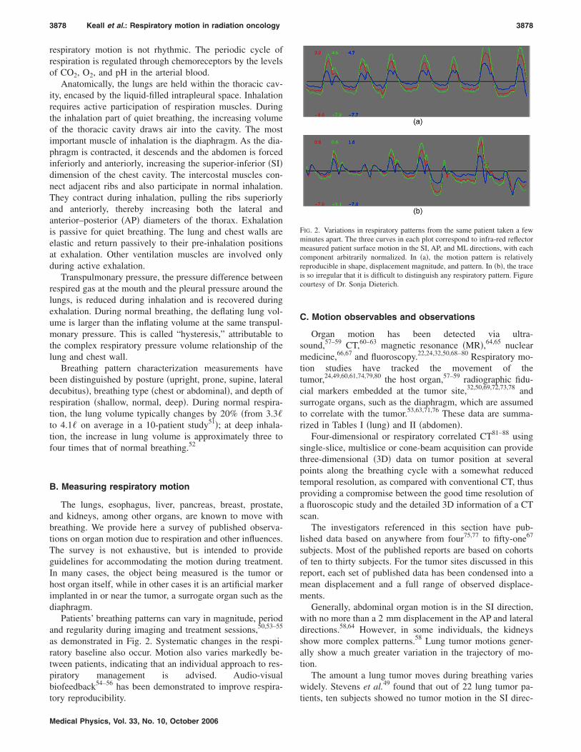
3878 Keall et al.: Respiratory motion in radiation oncology 3878
respiratory motion is not rhythmic. The periodic cycle ofrespiration is regulated through chemoreceptors by the levelsof CO2, O2, and pH in the arterial blood.
Anatomically, the lungs are held within the thoracic cav-ity, encased by the liquid-filled intrapleural space. Inhalationrequires active participation of respiration muscles. Duringthe inhalation part of quiet breathing, the increasing volumeof the thoracic cavity draws air into the cavity. The mostimportant muscle of inhalation is the diaphragm. As the dia-phragm is contracted, it descends and the abdomen is forcedinferiorly and anteriorly, increasing the superior-inferior �SI�dimension of the chest cavity. The intercostal muscles con-nect adjacent ribs and also participate in normal inhalation.They contract during inhalation, pulling the ribs superiorlyand anteriorly, thereby increasing both the lateral andanterior–posterior �AP� diameters of the thorax. Exhalationis passive for quiet breathing. The lung and chest walls areelastic and return passively to their pre-inhalation positionsat exhalation. Other ventilation muscles are involved onlyduring active exhalation.
Transpulmonary pressure, the pressure difference betweenrespired gas at the mouth and the pleural pressure around thelungs, is reduced during inhalation and is recovered duringexhalation. During normal breathing, the deflating lung vol-ume is larger than the inflating volume at the same transpul-monary pressure. This is called “hysteresis,” attributable tothe complex respiratory pressure volume relationship of thelung and chest wall.
Breathing pattern characterization measurements havebeen distinguished by posture �upright, prone, supine, lateraldecubitus�, breathing type �chest or abdominal�, and depth ofrespiration �shallow, normal, deep�. During normal respira-tion, the lung volume typically changes by 20% �from 3.3�to 4.1� on average in a 10-patient study51�; at deep inhala-tion, the increase in lung volume is approximately three tofour times that of normal breathing.52
B. Measuring respiratory motion
The lungs, esophagus, liver, pancreas, breast, prostate,and kidneys, among other organs, are known to move withbreathing. We provide here a survey of published observa-tions on organ motion due to respiration and other influences.The survey is not exhaustive, but is intended to provideguidelines for accommodating the motion during treatment.In many cases, the object being measured is the tumor orhost organ itself, while in other cases it is an artificial markerimplanted in or near the tumor, a surrogate organ such as thediaphragm.
Patients’ breathing patterns can vary in magnitude, periodand regularity during imaging and treatment sessions,50,53–55
as demonstrated in Fig. 2. Systematic changes in the respi-ratory baseline also occur. Motion also varies markedly be-tween patients, indicating that an individual approach to res-piratory management is advised. Audio-visualbiofeedback54–56 has been demonstrated to improve respira-
tory reproducibility.Medical Physics, Vol. 33, No. 10, October 2006
C. Motion observables and observations
Organ motion has been detected via ultra-sound,57–59 CT,60–63 magnetic resonance �MR�,64,65 nuclearmedicine,66,67 and fluoroscopy.22,24,32,50,68–80 Respiratory mo-tion studies have tracked the movement of thetumor,24,49,60,61,74,79,80 the host organ,57–59 radiographic fidu-cial markers embedded at the tumor site,32,50,69,72,73,78 andsurrogate organs, such as the diaphragm, which are assumedto correlate with the tumor.53,63,71,76 These data are summa-rized in Tables I �lung� and II �abdomen�.
Four-dimensional or respiratory correlated CT81–88 usingsingle-slice, multislice or cone-beam acquisition can providethree-dimensional �3D� data on tumor position at severalpoints along the breathing cycle with a somewhat reducedtemporal resolution, as compared with conventional CT, thusproviding a compromise between the good time resolution ofa fluoroscopic study and the detailed 3D information of a CTscan.
The investigators referenced in this section have pub-lished data based on anywhere from four75,77 to fifty-one67
subjects. Most of the published reports are based on cohortsof ten to thirty subjects. For the tumor sites discussed in thisreport, each set of published data has been condensed into amean displacement and a full range of observed displace-ments.
Generally, abdominal organ motion is in the SI direction,with no more than a 2 mm displacement in the AP and lateraldirections.58,64 However, in some individuals, the kidneysshow more complex patterns.58 Lung tumor motions gener-ally show a much greater variation in the trajectory of mo-tion.
The amount a lung tumor moves during breathing varieswidely. Stevens et al.49 found that out of 22 lung tumor pa-
FIG. 2. Variations in respiratory patterns from the same patient taken a fewminutes apart. The three curves in each plot correspond to infra-red reflectormeasured patient surface motion in the SI, AP, and ML directions, with eachcomponent arbitrarily normalized. In �a�, the motion pattern is relativelyreproducible in shape, displacement magnitude, and pattern. In �b�, the traceis so irregular that it is difficult to distinguish any respiratory pattern. Figurecourtesy of Dr. Sonja Dieterich.
tients, ten subjects showed no tumor motion in the SI direc-

3879 Keall et al.: Respiratory motion in radiation oncology 3879
tion. Of the remaining 12 subjects, the average SI displace-ment was anywhere from 3 to 22 mm �mean 8±4 mm�. Theyfound no correlation between the occurrence or magnitude oftumor motion and tumor size, location, or pulmonary func-tion, suggesting that tumor motion should be assessed indi-vidually. Seppenwoolde et al.50 measured 3D trajectories for20 patients via dual real-time fluoroscopic imaging of a fi-ducial marker implanted in or near the tumor. They observed
TABLE I. Lung tumor-motion data. The mean rangemeters for each cohort of subjects. The motion is in tleft-right; SI: superior-inferior.
Observer S
Barnes: �Ref. 74� Lower lobe 18.5 �Middle, upper lobe 7.5 �2Chen �Ref. 73� �0–5Ekberg �Ref. 22� 3.9 �0Engelsman: �Ref. 24�Middle/upper lobe �2–Lower lobe �2–Erridge �Ref. 104� 12.5 �Ross: �Ref. 60� Upper lobe -Middle lobe -Lower lobe -Grills �Ref. 80� �2–3Hanley �Ref. 61� 12 �1Murphy �Ref. 77� 7 �2–Plathow: �Ref. 65� Lower lobe 9.5 �4.5Middle lobe 7.2 �4.3Upper lobe 4.3 �2.Seppenwoolde �Ref. 50� 5.8 �0Shimizu �Ref. 75� -Sixel �Ref. 79� �0–1Stevens �Ref. 49� 4.5 �0
TABLE II. Abdominal motion data. The mean range of motion and the�minimum-maximum� ranges in millimeters for each site and each cohort ofsubjects. The motion is in the superior-inferior �SI� direction.
Site Observer
Breathing mode
Shallow Deep
Pancreas Suramo �Ref. 57� 20 �10–30� 43 �20–80�Bryan �Ref. 59� 20 �0–35� -
Liver Weiss �Ref. 66� 13±5 -Harauz �Ref. 67� 14 -Suramo �Ref. 57� 25 �10–40� 55 �30–80�Davies �Ref. 58� 10 �5–17� 37 �21–57�
Kidney Suramo �Ref. 57� 19 �10–40� 40 �20–70�Davies �Ref. 58� 11 �5–16� -
Diaphragm Wade �Ref. 68� 17 101Korin �Ref. 64� 13 39Davies �Ref. 58� 12 �7–28� 43 �25–57�Weiss �Ref. 66� 13±5 -Giraud �Ref. 63� - 35 �3–95�Ford �Ref. 76� 20 �13–31� -
Medical Physics, Vol. 33, No. 10, October 2006
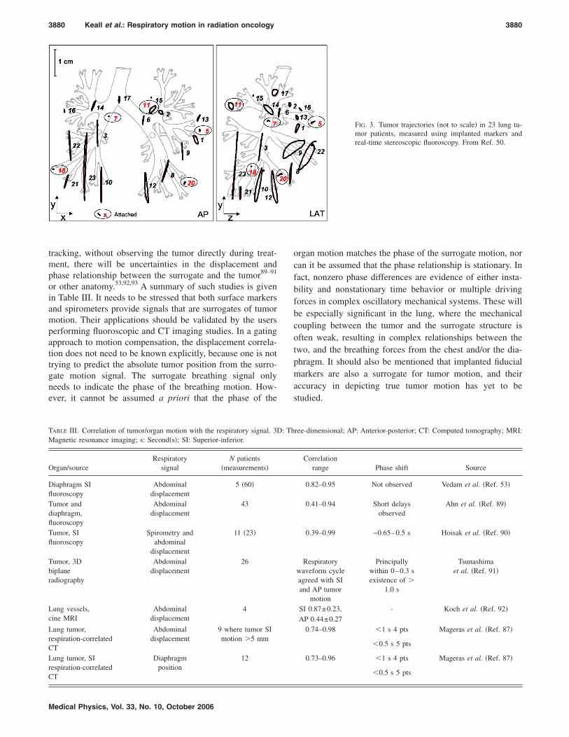
hysteresis in the trajectories of half the patients, amountingto a 1 to 5 mm separation of the trajectories during inhala-tion and exhalation, with four out of 20 patients exceeding a2 mm separation. This indicates that in cases where highaccuracy is required in dose alignment, a real-time trackingor gating process based on surrogate breathing signals shouldnot only correlate with the tumor’s motion along each axiswith the breathing signal, but should have knowledge of therespiratory phase, because the phase difference is what leadsto the hysteresis effect. In Fig. 3, motion trajectories duringradiotherapy of lung tumors, measured using implanted goldmarkers, are depicted.50
D. Summary of motion observations
A review of the respiratory motion literature leads to thefollowing conclusion: There are no general patterns of respi-ratory behavior that can be assumed for a particular patientprior to observation and treatment. The many individualcharacteristics of breathing—quiet versus deep, chest versusabdominal, healthy versus compromised, etc.—and the manymotion variations associated with tumor location and pathol-ogy lead to distinct individual patterns in displacement, di-rection, and phase of tumor motion.
In many cases, it is difficult or impossible to observe thetumor directly during treatment delivery prompting research-ers to observe surrogate structure motion expected to have aclose relationship with the tumor motion. If a surrogatestructure, such as the chest wall or diaphragm, is used to
tion and the �minimum-maximum� ranges in milli-dimensions �SI, AP, LR�. AP: anterior-posterior; LR:
Direction
AP LR
- -- -- -
2.4 �0–5� 2.4 �0–5�
- -- -
9.4 �5–22� 7.3 �3–12�1 �0–5� 1 �0–3�
0 9 �0–16�1 �0–4� 10.5 �0–13��0–10� �0–6�
5 �0–13� 1 �0–1�- -
� 6.1 �2.5–9.8� 6.0 �2.9–9.8�� 4.3 �1.9–7.5� 4.3 �1.5–7.1�� 2.8 �1.2–5.1� 3.4 �1.3–5.3�
2.5 �0–8� 1.5 �0–3�6.4 �2–24� -
�0–5� �0–4�- -
of mohree
I
9–32�–11�0�–12�
6�9�6–34�
0�–20�15�–16.4–10.2
6–7.1–25�
3�–22�
signal tumor position for the purpose of beam gating or
3880 Keall et al.: Respiratory motion in radiation oncology 3880
tracking, without observing the tumor directly during treat-ment, there will be uncertainties in the displacement andphase relationship between the surrogate and the tumor89–91
or other anatomy.53,92,93 A summary of such studies is givenin Table III. It needs to be stressed that both surface markersand spirometers provide signals that are surrogates of tumormotion. Their applications should be validated by the usersperforming fluoroscopic and CT imaging studies. In a gatingapproach to motion compensation, the displacement correla-tion does not need to be known explicitly, because one is nottrying to predict the absolute tumor position from the surro-gate motion signal. The surrogate breathing signal onlyneeds to indicate the phase of the breathing motion. How-ever, it cannot be assumed a priori that the phase of the
TABLE III. Correlation of tumor/organ motion with the respiratory signal. 3DMagnetic resonance imaging; s: Second�s�; SI: Superior-inferior.
Organ/sourceRespiratory
signalN patients
�measurements�
Diaphragm SIfluoroscopy
Abdominaldisplacement
5 �60�
Tumor anddiaphragm,fluoroscopy
Abdominaldisplacement
43
Tumor, SIfluoroscopy
Spirometry andabdominal
displacement
11 �23�
Tumor, 3Dbiplaneradiography
Abdominaldisplacement
26
Lung vessels,cine MRI
Abdominaldisplacement
4
Lung tumor,respiration-correlatedCT
Abdominaldisplacement
9 where tumor SImotion �5 mm
Lung tumor, SIrespiration-correlatedCT
Diaphragmposition
12
Medical Physics, Vol. 33, No. 10, October 2006
organ motion matches the phase of the surrogate motion, norcan it be assumed that the phase relationship is stationary. Infact, nonzero phase differences are evidence of either insta-bility and nonstationary time behavior or multiple drivingforces in complex oscillatory mechanical systems. These willbe especially significant in the lung, where the mechanicalcoupling between the tumor and the surrogate structure isoften weak, resulting in complex relationships between thetwo, and the breathing forces from the chest and/or the dia-phragm. It should also be mentioned that implanted fiducialmarkers are also a surrogate for tumor motion, and theiraccuracy in depicting true tumor motion has yet to bestudied.
FIG. 3. Tumor trajectories �not to scale� in 23 lung tu-mor patients, measured using implanted markers andreal-time stereoscopic fluoroscopy. From Ref. 50.
ree-dimensional; AP: Anterior-posterior; CT: Computed tomography; MRI:
Correlationrange Phase shift Source
0.82–0.95 Not observed Vedam et al. �Ref. 53�
0.41–0.94 Short delaysobserved
Ahn et al. �Ref. 89�
0.39–0.99 −0.65–0.5 s Hoisak et al. �Ref. 90�
Respiratorywaveform cycleagreed with SIand AP tumor
motion
Principallywithin 0–0.3 sexistence of �
1.0 s
Tsunashimaet al. �Ref. 91�
SI 0.87±0.23, - Koch et al. �Ref. 92�AP 0.44±0.27
0.74–0.98 �1 s 4 pts Mageras et al. �Ref. 87�
�0.5 s 5 pts
0.73–0.96 �1 s 4 pts Mageras et al. �Ref. 87�
�0.5 s 5 pts
: Th

3881 Keall et al.: Respiratory motion in radiation oncology 3881
V. COMMON ISSUES FOR RESPIRATORY MOTIONMANAGEMENT
Issues that are common to all methods of respiratory mo-tion management are discussed in this section, includingtreatment planning, QA, IMRT, and workload.
A. Treatment planning
Two useful articles that discuss important principles andprovide guidelines for treatment planning for lung cancerradiotherapy have been published by Senan et al.94,95 Themain geometric consideration for treatment planning oncethe GTV and CTV have been defined is the CTV-PTV mar-gin, which accounts for estimated geometric errors. In termsof target motion, the effect of all geometrical uncertainties isa displacement of the target during treatment relative to thedose distribution determined from the treatment plan. Con-sidering the target as a static structure and the dose distribu-tion as mobile allows the dose delivered to be summed overthe time period of all fractions. When there are many frac-tions, the random errors can be accurately described as ablurring of the dose distribution.96 The blurring is approxi-mated as a convolution of the dose distribution with theprobability distribution function of the total displacementvector of the target versus the treatment machine.97,98 A con-volution is not completely correct to describe the dosechanges �see, for example, Refs. 99–101�, but is quite accu-rate in practice.102 Systematic errors cannot be accounted forby this approach, which makes pretreatment imaging proce-dures �as described above� and frequent monitoring duringtreatment particularly important to reduce them. The follow-ing components contribute to the overall geometric error andshould be considered when designing CTV-PTV margins:
• Inter- and intraobserver variations in GTV16–19 andCTV20,21 delineation;
• motion artifacts in the CT scan, which cause target de-lineation errors;
• respiratory motion and heartbeat during delivery,50
which are periodic functions of time;• daily variations of respiratory motion;50,54,55,103
• variations caused by changing organ volumes;• tumor growth and shrinkage;• treatment-related anatomic changes, such as reductions
in bronchiole obstructions and changes in atelectasis�collapsed lung� regions;
• patient setup error: Typical 3–5 mm �1 standarddeviation�.18,22–28,104
Note that respiration-motion management techniques notonly affect the accuracy of target localization, but can alsoplay a role in normal tissue sparing.61,105 It is also importantto note that fast tumor shrinkage occurs quite often in lungradiotherapy, which may give rise to systematic deliveryerrors.104 The combined effect of random and systematic er-rors, including respiration, can be quantified in a dose-probability computation106,107 or through biological
108,109
modeling.Medical Physics, Vol. 33, No. 10, October 2006
B. Quality assurance
Quality assurance has a crucial role in all aspects of ra-diation oncology, as outlined in the report of AAPM TaskGroup 40.110 This section describes QA techniques used inthe management of respiratory motion. This section is di-vided into general descriptions and recommendations com-mon to different methods of accounting for respiratory mo-tion. QA procedures specific to each technique are describedseparately later.
A key issue in gated or breath-hold treatments using ex-ternal respiratory monitors is the accuracy of such monitorsin predicting internal target-organ position. As described ear-lier, internal/external correlation can be disturbed or lostcompletely by transient changes in breathing. For these rea-sons, patient training is important in allowing the patient tofamiliarize him- or herself with the breathing technique andfor evaluating his or her ability to achieve reproducible res-piratory signals. Breath-hold methods in particular requireactive patient participation. They also call for special staffeffort, as therapists must be trained to coach and advise thepatients. The limitations of equipment should also be under-stood �for example spirometer drifts� so that when issuesoccur during simulation or treatment the diagnosis and cor-rection of the issue is timely.
Some respiratory motion management techniques involveadditional devices that come into contact with the patient,thus hygiene practices for the safety of the patient and thestaff need to be established. Generally, devices that comeinto contact with patient mucosal surfaces should be dis-carded after use; devices that come into contact with thepatient skin can be reused provided appropriate proceduresare followed.
Frequency: As with all QA procedures, the appropriatetests should be performed after any hardware or softwarechanges or after service or changes to the respiratory motionmanagement device itself or the equipment �CT scanner,fluoroscope or linear accelerator� interfacing with the respi-ratory motion management device. Furthermore, until famil-iarity with the system is sound, QA may be performed morefrequently as determined by the physicist and the nature ofthe test.
Patient training: The ability to achieve reproduciblebreathing or breath-hold patterns is a requirement for allow-ing the patient to proceed to simulation and treatment. Inparticular, this affects the self-consistency of a CT scan thatspans multiple respiratory cycles or breath holds and the re-producibility of patient anatomy between simulation andtreatment. Prior to the start of simulation, the patient shouldbe made familiar with the equipment and its purpose. Aphysicist or trained designee should perform the coachingand evaluation at least in the initial clinical implementation.For breath-hold techniques, the training session, consistingof a series of breath holds in the treatment position, estab-lishes the patient’s respiratory level for treatment and breath-hold duration.
Simulation: By viewing the patient with fluoroscopy or
ciné CT, the magnitude of respiratory motion and the corre-
3882 Keall et al.: Respiratory motion in radiation oncology 3882
lation between the tumor motion and the respiratory signalcan be evaluated. For breath-hold techniques, one shouldverify that the tumor position �or other anatomic surrogates ifthe tumor is not visible� is stable within each breath hold andreproducible between breath holds. Patients who cannot holdtheir breath for the entire duration of the CT scan will requiresegmentation of the scan region �ideally not through the tar-get� to permit shorter breath holds. If the potential exists thatthe patient will be unable to comply with breathing orbreath-hold techniques for treatment, a backup CT scan with-out such a requirement is recommended during simulation.
Treatment: At the start of the first treatment fraction, thepatient should be reacquainted with the equipment, includingpracticed controlled breathing or breath holds. For breath-hold techniques, it is preferable to deliver a treatment field ina single breath hold. If the duration of this breath hold is toolong for patient comfort, careful documentation in the chartshould be made about break points for individual beams. Thetherapists will need to monitor the treatment machine, thepatient, and the gating or breath-hold system display.
Radiographs to check internal constancy: Although exter-nal monitors may correlate well with the respiratory organswithin a single session, thus reducing intrafractional varia-tions, the relationship between external monitor and internalorgan positions may change between sessions, which canadversely affect organ reproducibility and produce interfrac-tional variations. A program of frequent radiographs of thesurrogate organ �or target, if visible� throughout treatment isessential to measure interfractional variations and should beacquired during the respiratory cycle part or breath hold usedfor simulation and treatment. Sometimes, lung tumors aresufficiently discernible in the radiographs to allow directconfirmation of their position. Daily verification is recom-mended for the first few treatments, followed by �at least�weekly verification to ensure that the anatomy at the respi-ratory position used for treatment remains constant. If theradiographs indicate differences from simulation, the dosim-etric consequences and remedies are evaluated by the physi-cist and the physician. For treatment machines with an exitdetector, more advanced verification techniques are possible.For example, ciné-mode acquisition, by which several im-ages are acquired during each field delivery, may be utilized.
As with all radiotherapy procedures, constant vigilance bythe treatment staff is important. Training and education forall staff involved with respiratory management, as well asperiodic retraining, is recommended. A physicist should beavailable to solve any hardware-related problems.
C. Intensity modulated radiation therapy
IMRT has seen widespread application owing to its abilityto conform the spatial distribution of the dose deposited in apatient more effectively. The implications for targets in thethoracic and abdominal regions have been particularly im-portant due to the many organs at risk in these regions. How-ever, respiratory motion intuitively presents considerable is-sues for IMRT delivery, since beam-intensity gradients are
no longer confined solely to the edges of the beams. Rather,Medical Physics, Vol. 33, No. 10, October 2006
such gradients can be inside the field defined by the primarycollimators. Thus, if a target is also moving inside this samefield with its own period unique from the MLC leaves andpossibly deforming, it is easy to understand why there areconcerns over the use of IMRT with targets affected by res-piratory motion. Yu et al.111 �see also Kissick et al.112� dem-onstrated this effect using theoretical models that yieldeddose variations for “clinically relevant parameters” of up to100%. In a dynamic wedge simulation, Pemler et al.113
showed that the magnitude of dosimetric errors may ap-proach 15% for a single dynamic wedge treatment. Bortfeldet al.114 demonstrated dosimetric errors on the order of ±8%for a single point in the middle of the treatment field �low-dose gradient region� in the simulation of a single IMRTtreatment. Kubo and Wang115 and Keall et al.116 analyzed thedosimetric error for a single MLC-based IMRT treatmentusing film. In each study, films of treatments delivered withand without motion were compared. To simulate motion, filmwas moved a distance and at a rate consistent with respira-tory motion. Errors of up to 20% were reported within thefield �low-dose gradient region�, with even larger errors onthe edges of the field �high-dose gradient regions�.
Based on these findings, it would seem that the concernover potential dosimetric error introduced by respiratory mo-tion for IMRT treatments is justified; however, Yu et al.111
showed that fluence variations within a moving target tend toaverage out over the typical course of 30 fractions, when oneassumes that the breathing phase or frequency is randomfrom day to day. Along similar lines, Bortfeld et al.114
showed that dosimetric errors introduced by respiratory mo-tion also tend to average out with fractionation; this wasfurther supported in MLC-based IMRT studies by George etal.117 and Chui et al.118
In a follow-up study, Jiang et al.119 experimentally veri-fied the findings of Bortfeld et al.114 for a single point in alow-dose gradient region using MLC-based IMRT; however,these studies assumed or applied simplistic, one-dimensional�1D� motion, which can be quite different from the real,complex phenomenon of breathing.50,78 Furthermore, targetdeformation may be present, although this deformation hasyet to be quantified. They, therefore, cautioned that fraction-ation alone should not be relied on, at least in cases of large��1 cm� motion, until their findings could be verified undermore realistic conditions.
To summarize, the above studies indicate that caution iswarranted when considering IMRT for targets subject to res-piratory motion, particularly for single or few-fraction treat-ments common for stereotactic body radiotherapy. For indi-viduals who still intend on using IMRT without any directmotion-correction strategy, it needs to be emphasized that thefull extent of breathing motion should be assessed and con-sidered when assessing margins for the treatment plan. Evenwith correction strategies, there can still be residual targetmotion with respect to the beam, for example, with respira-tory gated treatment, which may exhibit similar, albeit
smaller, effects.
3883 Keall et al.: Respiratory motion in radiation oncology 3883
D. Workload
Respiratory motion management techniques utilize spe-cific technology that requires increased medical supervisionand longer treatment times for the delivery of this precisetreatment. Additional physics, physician and therapist sup-port is required during the simulation, planning and treat-ment processes, which are described in more detail below. Ifimaging procedures are performed, further resources are in-volved. When acquiring a respiratory management device forclinical use, there are capital costs, staff training costs andtime, acceptance testing and commissioning procedures to beperformed as well as the development and execution of on-going QA and staff education and training programs.
Before simulation, the scheduling of patients that areidentified by physicians includes relaying the informationabout potential patients to the physics group. Depending onthe respiratory management technique, the physics groupmay need to schedule a training session with the patient,which can take up to one hour with the patient and an addi-tional half-hour to full hour to assemble the equipment forthis training session. A physicist �or designated staff memberwho is appropriately trained to manage the procedure� thenneeds to be present for the CT imaging session. The physicistmay need to evaluate the quality of the imaging study and, ifnecessary, repeat the imaging study. Some respiratory man-agement devices have patient-specific disposable accessoriesthat need to be ordered, purchased and stored. The treatmentplanning may require special instructions and physics over-sight which can take several hours in some cases.
At many institutions, a physicist is required to be presentfor the first treatment with respiratory management proce-dures. Coaching the patient at simulation and on the first dayof treatment is fairly common and recommended. For sometechniques and patients, further coaching is needed. Finally,
TABLE IV. Summary of intra- and inter-fractional variations for different metbreathing control, SD—standard deviation, LR—left-right, AP—anterior-psetup error, 3D—3-dimensional error, mDIBH moderately deep inspiration
Reference Technique O
Cheung �Ref. 201� BH at inspirationwith ABC
Lun
Dawson �Ref. 202� BH at exhalationwith ABC
Dia
Ford �Ref. 76� Gating at exhalationwith RPM
Dia
Hanley �Ref. 61� DIBH DiaMah �Ref. 160� DIBH DiaNegoro �Ref. 170� Abdominal
compression withstereotactic body
frame
Lun
Remouchamps �Ref. 203� mDIBH with ABC Dia
Wagman �Ref. 133� Gating at exhalationwith RPM
Abdom
a review and QA of the respiratory traces or images acquired
Medical Physics, Vol. 33, No. 10, October 2006
at the time of treatment needs to be done. Currently, thisrequires approximately two hours of work per patient. Thereare also material and machine time considerations. Time re-quired at the CT scanner is longer, treatment times arelonger, and a room may be required for an hour-long trainingsession. The extra time at an accelerator has the cost of de-creased patient throughput. There is also the capital invest-ment, use and depreciation costs of the equipment used forthese treatments.
VI. METHODS TO ACCOUNT FOR RESPIRATORYMOTION IN RADIOTHERAPY
Methods to reduce the impact of respiratory motion inradiotherapy can be broadly separated into five major catego-ries: Motion-encompassing methods, respiratory gating tech-niques, breath-hold techniques, forced shallow-breathingtechniques, and respiration-synchronized techniques. Thesemethods are discussed in detail in this section. A summary ofpublished intra- and inter-fractional variations for the differ-ent methods is given in Table IV.
A. Motion-encompassing methods
1. Introduction
This section discusses imaging and treatment-planningguidelines for tumor sites affected by respiratory motion.Since respiratory induced tumor motion will be present dur-ing radiation delivery, it is important to estimate the meanposition and range of motion during CT imaging.
The three techniques possible for CT imaging that caninclude the entire range of tumor motion for respiration �atthe time of CT acquisition� are slow CT, inhalation and ex-halation breath-hold CT, and four-dimensional �4D� orrespiration-correlated CT. These are listed in order of in-
of respiratory management. Abbreviations: BH—breath-hold, ABC—activeor, SI—superior-inferior, DIBH—deep inspiration breath-hold, * includes-hold. �Ref. 200�.
Intra-fractionvariation �cm�
Inter-fractionvariation �cm�
or - SD: 0.18 LR, 0.23 AP, 0.35 SI
m SD: 0.25 SD: 0.44
m Mean: 0.26 Mean: 0.0SD: 0.17 SD: 0.39
m SD: 0.25 -m - 0.4*
or Mean 3D: 0.7 Mean 3D: 0 .5*
Range: 0.2–1.1 Range: 0.4–0.8*
m Mean: 0.14 Mean: 0.19SD: 0.17 SD: 0.22
organs Mean: 0.20 -
hodsosteribreath
rgan
g tum
phrag
phrag
phragphragg tum
phrag
inal
creasing workload. It is important to understand that the

3884 Keall et al.: Respiratory motion in radiation oncology 3884
breathing patterns and, hence, tumor motion will change be-tween simulation sessions and treatment sessions. Further-more, the radiation dose to the patient from these imagingprocedures can be greater than standard CT simulation pro-cedures by a factor of 2–15 if no efforts are made to reduceCT dose.
2. Slow CT scanning
One solution for obtaining representative CT scans forperipheral lung tumors is slow scanning.120–122 The CT scan-ner is operated very slowly, and/or multiple CT scans areaveraged such that, on average, multiple respiration phasesare recorded per slice. Hence, the image of the tumor �atleast in the high-contrast areas� should show the full extentof respiratory motion, provided that the scanner operates at aparticular couch position for longer than the respiratorycycle. This technique yields a tumor-encompassing volume,with the limitation that the respiratory motion will changebetween imaging and treatment, and, thus, additional mar-gins are required to account for these variations. In additionto anatomic delineation, slow scanning is more advantageousthan standard scanning, because the dose calculation is per-formed on a geometry that is more representative of thatduring the entire respiratory cycle, as occurs during treat-ment. The disadvantage is the loss of resolution due to mo-tion blurring, which potentially leads to larger observer er-rors in tumor and normal organ delineation. Due to motionblurring, this method is only recommended for lung tumorsthat are not involved with either the mediastinum or the chestwall. This method is also not recommended for other tumorsites �e.g., the liver, pancreas, kidney, etc�. It has been sug-gested that PET, with its inherently long acquisition times, isalso a good solution for estimating the motion path of atumor;44–47 however, motion can also blur the object in thePET image such that a suspicious lesion may not even beapparent, in which case respiration-gated PET or 4D PETmay be a better option.
3. Inhalation and exhalation breath-hold CT
A solution to obtaining a tumor-encompassing volumethat can be implemented in most clinics is to acquire bothinhalation and exhalation gated or breath-hold CT scans ofthe patient during the CT simulation session. Taking bothinhalation and exhalation CT scans will more than double theCT scanning time and relies on the patient’s ability to holdhis or her breath reproducibly. The two scans require imagefusion and extra contouring. For lung tumors, the maximumintensity projection123 �MIP� tool �the MIP image in this con-text for a set of CT images is the maximum CT numberfound in a given voxel in the set� available in most visual-ization systems can be used to obtain the tumor-motion-encompassing volume, provided there is no mediastinal tu-mor involvement. The advantage of this approach over theslow scanning method mentioned above is that the blurringcaused by the motion present during FB is significantly re-duced. Dose calculation should be performed on the CT data
set that is most appropriate for the particular patient, e.g.,Medical Physics, Vol. 33, No. 10, October 2006
exhalation CT for patients generally spending more time atexhalation than inhalation. The exhalation scan will tend tounderestimate the lung volumes and, hence, overestimate thepercentage of lung volume receiving a specific dose. To savetime, a free-breathing CT could be used for the entire scanregion �typically including the entire thoracic cavity�, witheither breath-hold or gated CT scans at inhalation and exha-lation of a scan length sufficient to cover the tumor volumeto determine the range of motion of the GTV. Some form ofrespiratory monitoring is necessary to verify gating orbreath-hold constancy and to ensure that the scans are repre-sentative of the patient’s normal breathing range.
Breath-hold scans can also potentially be used for respi-ratory gated delivery, however, it should be noted that a res-piratory gated CT scan is preferred over a breath-hold scan atthe same respiratory position, because the predominant res-piratory muscles can be different for breath hold and FB�e.g., intercostal vs. diaphragm�, and any tumor lag �relativeto the external monitor� occurring during FB will be absentduring breath hold.
4. Four-dimensional CT/respiration-correlated CT
A promising solution for obtaining high quality CT datain the presence of respiratory motion is 4D CT or respira-tion-correlated CT �conventional and cone-beamapproaches�.81–88,124,125 Four-dimensional data can be ana-lyzed to determine the mean tumor position, tumor range ofmotion for treatment planning,123,126–128 and the relation oftumor trajectory to other organs and to a respirationmonitor.87 A limitation of 4D CT is that it is affected byvariations in respiratory patterns during acquisition.Breathing-training techniques have been developed,56 how-ever, even with these techniques artifacts can be observed.86
A 4D CT scan can be obtained in approximately a minuteof scanning time with a 16-slice CT scanner. Generally 8–25complete CT data sets are reconstructed, the optimal use ofwhich has yet to be determined. Four-dimensional CT can beused to reconstruct inhalation, exhalation, and slow CTscans.86 The MIP tool, as mentioned above, may be useful inobtaining the tumor-motion-encompassing target volume.Another motion-encompassing method is to derive a singleset out of the 4D CT scan where the tumor is close to itstime-averaged position. In that case, the expected dose blur-ring effect of respiration can be accounted for in the CTV-PTV margin.129
B. Respiratory gating methods
1. Introduction
Respiratory gating involves the administration of radia-tion �during both imaging and treatment delivery� within aparticular portion of the patient’s breathing cycle, commonlyreferred to as the “gate.” The position and width of the gatewithin a respiratory cycle are determined by monitoring thepatient’s respiratory motion, using either an external respira-tion signal or internal fiducial markers. As the beam is notcontinuously delivered, gated procedures are longer than
nongated procedures.
3885 Keall et al.: Respiratory motion in radiation oncology 3885
Respiratory gating is currently under study by severalcenters to account for respiratory motion during radiotherapyof thoracic and abdominal tumors.53,56,76,130–134 Respiratorymotion can be characterized by two variables that are re-corded as part of the respiration signal or the motion of theinternal anatomy. These variables are �a� displacement and�b� phase. Accordingly, the method of gating is referred to aseither displacement gating or phase gating. The displacementof the respiration signal measures its relative position be-tween two extremes of breathing motion, namely, inhalationand exhalation. In displacement-based gating, the radiationbeam is activated whenever the respiration signal is within apre-set window of relative positions. The second variable,phase, is calculated by an algorithm from the respiration sig-nal that must satisfy periodicity criteria; the radiation beam isactivated when the phase of the respiration signal is within apre-set phase window. Further details of displacement-basedand phased-based gating can be found in Vedam et al.145
Typically, a gate extends over a region of the breathing cyclewhere the motion of the tumor is estimated to be less, �gen-erally end exhalation�, or where the lung volume is maximal�end inhalation�. The ratio of the beam-on time within thegate to the overall treatment time is referred to as the dutycycle and is a measure of the efficiency of the method. Sometumor motion still occurs within the gate and is referred to as“residual motion.”135 The choice of gate width is a tradeoffbetween the amount of residual motion and duty cycle.
2. Gating using an external respirationsignal
Currently, the commercially available respiratory gatingsystem using an external respiration signal most widely dis-cussed in publications is the Real-time Position Management�RPM� system �Varian Medical Systems, Palo Alto, CA�;thus, the procedures described here are applicable to thisdevice, although they can be generalized to other implemen-tations. BrainLab �Westchester, IL� has an FDA-cleared res-piratory gating device, called “ExacTrac Gating/Novalis Gat-ing.” This device uses external markers for gating theradiation beam, however, it has x-ray imaging capabilitiesfor determining the internal anatomy position and for verify-ing the reproducibility of the internal anatomy during treat-ment. Siemens Medical Systems �Concord, CA� also has anFDA-approved linear accelerator gating interface and an An-zai belt �used for CT�, also approved for use in therapy.Three-dimensional video camera surveillance has also beenstudied for respiratory motion management.136
Owing to its noninvasive nature, gating using an externalrespiration signal can be applied to almost all ��90% � pa-tients. Breathing training may be beneficial in many casesand can improve the likelihood of the patient completing thesimulation session. With the Varian RPM system, an infraredreflective plastic box serving as the external fiducial markeris placed on the patient’s anterior abdominal surface, typi-cally midway between the xyphoid process and the umbili-cus, and chosen to maximize the AP respiratory induced mo-
tion. The marker box should be placed nearly horizontally, toMedical Physics, Vol. 33, No. 10, October 2006
permit the in-room camera to accurately detect the reflectivemarkers. If used during treatment, a durable skin mark at thebox location should be made at the time of imaging to ensurereproducible positioning during treatment.
Prior to a gated CT scan, determination of gating param-eters �displacement or phase, exhalation or inhalation, dutycycle� are based on observation of the external respirationsignal and, if possible, tumor motion. In prospective gatedCT, a respiration gating system sends a trigger to the CTscanner to acquire a CT slice. CT scan parameters �slicethickness, scanner rotation time, index, etc.� remain the sameas those used for standard CT scans. Gate width and CT scanrotation time should be similar. Shorter gate width results inanatomic positions outside the intended gate to be includedin the image, while longer gate width results in more ana-tomic motion occurring during the gate than is captured inthe CT image. Either situation is a potential source of error.Note that not all CT scanners can perform prospective gat-ing. The time required to acquire a prospective gated CTscan depends on the patient’s respiratory period, since thereis one slice triggered per cycle. Irregular breathing can fur-ther prolong the CT acquisition and/or lead to acquisition ofslices at the wrong part of the breathing cycle.
At treatment, marker block position and patient breathinginstructions are the same as during simulation. Once a stablerespiration trace has been established and gating thresholdsare verified, gated radiation delivery is initiated. The positionof the patient’s internal anatomy is verified using in-roomimaging. During treatment the therapist should be alert to thegraphical cues on the gating system monitor and be preparedto intervene if the patient’s breathing is very irregular ordifferent from simulation. In-room images that show the tu-mor, if possible, or an internal anatomic surrogate �often thediaphragm� are helpful in assessing the performance of thegating system over the course of treatment.81,137
For internal and external tracking systems, a possiblesource of error is that the surrogate for tumor motion �e.g.,tracking blocks, strain gauges, etc.� tracked by the gatingsystem does not accurately correspond with the time-dependent target position �Fig. 4�. This can cause the timingof the beam-on pulse to shift relative to the actual respiratorycycle of the target. Where available, a minimum of 30 s ofimaging data �fluoroscopy or CT ciné mode� should be digi-tally recorded in conjunction with the measured respirationtrace. The motion of the GTV—or anatomic surrogate suchas the diaphragm, if the GTV is not discernible—should becompared with the external respiratory signal; a time delaylarger than 0.5 s between the two, if consistently observed,should be corrected or accounted for when setting the gateinterval. An EPID-based approach for position verification inthis manner has been proposed by Berbeco et al.138
Dynamic test phantoms that simulate respiration areneeded, in order to test in vivo dosimetry and target localiza-tion. There are several important factors: �1� The phantomshould produce cyclical motion similar to human respiration;�2� the gating feedback mechanism must detect phantom mo-tion in a manner similar to the clinical process; �3� the device
should allow attachment of dose measuring detectors, such
3886 Keall et al.: Respiratory motion in radiation oncology 3886
as ion chambers or diodes; �4� the phantom should be reli-able and have a reasonable cost. Several custom-built phan-toms have been made to meet these criteria,86,116,119,139–143
and commercial systems are available. Ramsey et al. de-scribe further equipment QA tests for the Varian RPM respi-ratory gating system.139
3. Gating using internal fiducial markers
This section focuses on the real-time tumor-tracking ra-diotherapy system, developed jointly by Hokkaido Univer-sity and Mitsubishi and based on radiographic detection ofimplanted fiducials to gate radiation delivery.50,70,75,144–150
The fiducials �2 mm diameter gold spheres� are implanted inor near the tumor using a percutaneous or bronchoscopicimplanting technique. Fiducial position is tracked in all threedimensions several times a second using a pair of stereotactickilovoltage x-ray imaging systems in combination with au-tomatic detection software. The linear accelerator deliversradiation when each fiducial is within an acceptable range ofthe desired �simulation� position for both stereotactic x-raycameras.
Patient selection begins with assessment of tumor motionprior to fiducial implant, to ensure maximum benefit to thepatient with this invasive procedure. The patient must be ableto tolerate the implant procedure and remain motionless onthe treatment couch for an extended treatment �up to45 min�. For patients with lung cancer, pulmonary functioncriteria are set, based on the recommendation of the pulmo-nologist performing the implant. Because this technique hasbeen primarily used for stereotactic radiotherapy, most of thepatients have had relatively small lesions �4 cm in diameteror less�.
Treatment simulation uses a series of CT scans: a normal,free breathing scan; a breath-hold scan at inhalation, and abreath-hold scan at exhalation. Treatment plans are designedon both the inhalation and exhalation set of CT images, andthe radiation oncologist selects the best plan based on thedose distribution, assessing if increased lung sparing is foundon the inhalation plan. Six to ten static fields are used todeliver 48 Gy in four fractions. The implanted fiducials areidentified in the planning system, and DRRs are generated toreplicate the images to be acquired in the treatment room.
At the beginning of each treatment, the fiducial path is
monitored for several breathing cycles and the patient repo-Medical Physics, Vol. 33, No. 10, October 2006
sitioned, if needed, so that at the appropriate point in thebreathing cycle, the fiducial marker passes near the predictedlocation. Two gates, one from each imaging system, must bein coincidence to enable the beam. Treatment times are typi-cally longer than 30 min, and the duty cycle varies by patientand by the choice of respiratory cycle part to be used fortreatment.
QA procedures include confirmation that the coordinatesystems of the fluoroscopy unit and linear accelerator areproperly aligned. The coordinate system alignment should bechecked regularly, since there is a potential for drift withboth systems. The magnitude of marker motion detected bythe system needs to be verified, and it must also be assuredthat the automated tracking of the internal fiducial markers isrobust.
4. Gated IMRT
Kubo and Wang115 demonstrated the feasibility of gatingthe linear accelerator during a dynamic MLC delivery. Theyshowed that the dosimetry for gated IMRT delivery that in-cluded motion �1D mechanical device� was essentially thesame as that for delivery without motion. Target deformationwas not considered.
Respiratory gating techniques increase the treatment time.This is more pronounced for gated IMRT in which the prod-uct of the IMRT efficiency, typically 20%–50%, and the gat-ing duty cycle, 30%–50%,76,133 leads to a 4- to 15-fold in-crease in delivery time. Increasing dose rate from 300 to600 MU/min can reduce the clock time by approximately40%.134 Gated treatment session times are increased relativeto standard treatments by 2–10 min depending on patientcompliance.134 Considerations with increased delivery timeare patient comfort, increased likelihood of patient move-ment and decreased patient throughput. During substantiallylonger treatments, it has been suggested that tumor controlmay be reduced due to the increased intrafraction repair ofsublethally damaged tumor cells.151,152
C. Breath-hold methods
1. Introduction
Breath hold methods have been predominantly applied tolung cancer radiotherapy. Breast cancer radiotherapy may
FIG. 4. Comparison of external markerblock motion with internal motion ofthe clinical target volume �CTV� for apatient with �a� no phase shift and �b�a patient with significant phase shift.The respiratory gating thresholds areset using the external marker blockmotion. The beam-on pulses are high-lighted in red over the internal CTVposition. From Ref. 204.
also potentially benefit: although intrafraction motion is
3887 Keall et al.: Respiratory motion in radiation oncology 3887
small for normal respiration,153 during inhalation the dia-phragm pulls the heart posteriorly and inferiorly away fromthe breast, and thus may reduce both cardiac and lungtoxicity.154–159
2. Deep-inspiration breath hold
A reproducible state of maximum breath hold �deep-inspiration breath hold or DIBH� is advantageous for treatingthoracic tumors, because it significantly reduces respiratorytumor motion and changes internal anatomy in a way thatoften protects critical normal tissues. This section describes aspirometer-monitored technique that was developed andclinically implemented primarily for conformal radiationtreatments of NSCLC at the Memorial Sloan-Kettering Can-cer Center �MSKCC�.61,160,161 There are at least two com-mercial spirometry products that are compatible with theDIBH technique: The VMAX Spectra 20C �VIASYS Health-care Inc., Yorba Linda, CA� and the SpiroDyn’RX �Dyn’R,Muret, France�. Forty-five patients have been treated withDIBH at MSKCC �44 with NSCLC� between 1998 and2004; of these, eight were treated with DIBH in combinationwith IMRT.
The DIBH technique involves verbally coaching the pa-tient to a reproducible deep inhalation breath hold duringsimulation and treatment. The patient breathes through amouthpiece connected via flexible tubing to a spirometer.The naris is held closed with a nose clip. A computer pro-gram displays and records the volume of air breathed in andout as a function of time. The therapist coaches the patientthrough a modified version of the slow vital capacity maneu-ver, consisting of a deep inhalation, deep exhalation, seconddeep inhalation and breath hold. The maneuver yields highlyreproducible lung inflation at approximately 100% capacity,which can be maintained for 10–20 s �patient specific�.
Applicability of DIBH is limited by patient compliance:Approximately 60% of the lung cancer patients at MSKCCcannot perform the maneuver reproducibly enough to permitits use; thus it is used only for compliant patients in whomthe significant lung inflation allows treatment to a highertotal dose �10% or more with acceptable normal tissue dose-volume histograms and calculated lung complicationprobability161� than is possible with free breathing treatment.
Following a brief DIBH practice session, the patient re-ceives three helical CT scans in the treatment position: �1�With FB; �2� with spirometer-monitored deep inhalation�DI�; and �3� with spirometer-monitored inhalation. The FBand inhalation scans are for QA purposes, described below.The FB scan also serves as the alternative treatment plan CTif the patient cannot be completely treated with DIBH. Thesimulation process—including immobilization, isocenter se-lection, practice, 3 CT scans and resting between scans—takes approximately 2 hr. The treatment plan and DRRs usethe DI breath-hold CT scan.
During treatment, the therapists are instructed to turn onthe beam only when the target breath-hold level has beenachieved and to stop treatment if the level has fallen below a
pre-set tolerance. For static conformal treatments atMedical Physics, Vol. 33, No. 10, October 2006
2 Gy/fraction on linear accelerators operated at500–600 MU/min, a single breath hold is usually sufficientfor each field. More recently, IMRT in combination withDIBH has been introduced for patients able to hold theirbreath long enough to complete a field, approximately 20 sfor a typical beam-on time of 200 MU delivered at600 MU/min with the sliding window technique.159 Treat-ment sessions usually take 5–10 min longer than a similarbeam arrangement for an FB patient.
Patient-specific QA includes a check of the FB scan thatthe patient’s state of respiration does not alter the position ofthe spine, thus allowing positioning of the patient for treat-ment while breathing normally. The inhalation scan is usedto set breath-hold tolerance levels by determining the motionextent of the GTV for a known change in breath-holdvolume.160 In all imaging and treatment sessions, the thera-pist is instructed to wait 1 s following breath hold beforeturning on the beam, to allow for transient diaphragmrelaxation.160
The spirometer is calibrated with a 3� syringe for flowrates between approximately 0.5 and 3 � / s. The linearity ofspirometer integrated airflow versus actual �syringe� volumeis checked over a range of 0–3� in either flow direction;typical linearity is within 2%. The calibration is checkedwhenever the spirometer is gas sterilized, approximately ev-ery 2 to 3 mo. Occasionally, drift of the spirometer is ob-served following sterilization, which is usually correctableby reassembling the device.
3. Active-breathing control
Active-breathing control �ABC� is a method to facilitatereproducible breath hold.157,162 The ABC method was devel-oped at William Beaumont Hospital and is currently com-mercialized by Elekta Inc. �Norcross, GA� as the ActiveBreathing Coordinator. A device with similar capabilities,called the Vmax Spectra 20C, is available from VIASYSHealthcare Inc. �Yorba Linda, CA�. The ABC apparatus cansuspend breathing at any predetermined position and is oftenused at moderate or deep inhalation. It consists of a digitalspirometer to measure the respiratory trace, which is in turnconnected to a balloon valve. In an ABC procedure, the pa-tient breathes normally through the apparatus. The operatorspecifies the lung volume and stage of breathing cycle stageto “activate” the system, at which the balloon valve is closed.The patient is instructed to reach the specified lung volume,typically after taking two preparatory breaths. The valve isinflated with an air compressor for a pre-defined duration oftime, thereby “holding” the patient’s breath. The breath-holdduration is patient dependent, typically 15–30 s, and shouldbe well tolerated to allow for repeated �after a brief restperiod� breath holds.
The Beaumont experience157,158,163 shows that a moderate�deep� inhalation breath-hold �mDIBH� level set at 75% ofdeep inspiratory capacity achieves substantial and reproduc-ible internal organ displacement while maintaining patientcomfort. The intended mDIBH position is calculated from
the exhalation baseline and set during an initial training ses-
3888 Keall et al.: Respiratory motion in radiation oncology 3888
sion for each patient. Verbal instructions are always given tohelp a patient achieve a steady breathing pattern.
Prior to the start of simulation a series of baseline mea-surements should be made. Depending on the system, a pul-monary function test �PFT� may be needed at this time toprovide reference data on the individual patient’s lung capac-ity. Practice breath holds should be performed, and the pa-tient instructed of various means of indicating discomfortand signaling cessation of breath hold to the operators. TheCT scan should be optimized according to the maximumreproducible length of breath hold in an immobilized posi-tion; the timing of contrast should coincide with the appro-priate breath-hold scan of the region of interest. The breath-hold state, as well as duration of comfortable breath hold,should be documented for use during treatment.
Treatment plans include a margin dependent on the in-tended treatment verification strategy. If the patient is to betreated daily without image guidance, the margin should con-sider setup variation along with the long-term reproducibilityof ABC. The magnitudes of these margins for the patientpopulation in each clinic should be established for routineapplication of the ABC procedure.
Treatment at each beam angle should be delivered in asingle breath hold, when possible. If a single breath-hold istoo long, then one can “break up” the single breath-hold intotwo or more smaller breath-holds. These smaller breath-holds should be recorded particularly if they are coordinatedwith the delivery of IMRT segments on earlier acceleratorswhich require the “breakup” segments as individual beams.Each beam needs to be delivered before releasing the patientfrom breath-hold.
An important concern of patient-related QA is reproduc-ibility of breath hold. It is essential that all operating person-nel understand system functions and that the patient receiveand understand appropriate instruction. The process for es-tablishing a breath hold at a given state �e.g., exhalation,inhalation, deep inhalation� should be documented andtested. A standard set of patient instructions for communica-tion with the ABC operator and for emergency actions toreestablish breathing is recommended. It is important to es-tablish a hygienic procedure for cleaning reusable items�e.g., rubber mouthpiece�.
The key functions that should be maintained and checkedfrequently for safe use of an ABC system are the calibrationof airflow and volume, the ability to stop and restart air flow,and the safety release mechanisms. It is important to under-stand how the ABC unit establishes a breathing trace. Cur-rent systems use mechanical spirometers or temperature sen-sors. The calibration for the temperature sensor is absolute,whereas the spirometer-based system operates by establish-ing a baseline at each exhalation. Both systems are typicallycalibrated using a 3.0� syringe. Apart from the vendor’s rec-ommended calibration, the volume calibration should bechecked at different flow rates similar to those seen in pa-tients. The minimum flow rate below which the mechanicalspirometer will not respond accurately should be established.
The equipment needed to provide ABC may affect the
processes of simulation and treatment. The air tube exitingMedical Physics, Vol. 33, No. 10, October 2006
the mouth, the chamber for breathing monitoring and con-trol, and ancillary hardware may occupy significant space,possibly restricting the geometry of the CT scanner or treat-ment unit. Prior to implementing ABC for a given body site,the processes of immobilization, simulation and deliveryshould be evaluated to determine an efficient means of inte-grating the ABC unit and support equipment.
4. Self-held breath hold without respiratorymonitoring
In this technique the patient voluntarily holds his/herbreath at some point in the breathing cycle, during whichdose delivery occurs. A control system for its implementationhas been developed74,164 for the Varian C Series accelerators,which makes use of the “Customer Minor �CMNR�” Inter-lock. The patient depresses a hand-held switch to clear theCMNR interlock, allowing the therapist to activate the beam.Releasing the switch asserts the CMNR interlock, turning thebeam off and disabling any further delivery until the switchis depressed again. Although only the therapist can turn thebeam on, both the therapist and the patient can turn the beamoff. The potential dosimetric advantages of increasing thelung volume61,157,158,161,163 makes deep inhalation the pre-ferred point for breath hold. Therefore, the earlier discussionof DIBH and ABC would be similar to the advantages withthis method. The self-held breath-hold system is not com-mercially available.
This mode of treatment relies heavily on the patient’sability to understand and perform a reproducible breath hold,maintain it for at least 10 s, and simultaneously operate thehand-held switch. Another selection criterion is the stabilityof internal anatomy during breath hold: Some patients havebeen observed to have continuous diaphragm motion duringbreath hold, even though they believe they are holding theirbreath. Following evaluation under fluoroscopy on a conven-tional simulator, patients receive a breath-hold CT scan, inwhich the scan sequence is segmented into 10 s acquisitions.Patients are given a switch attached to a buzzer, which theydepress to indicate to the CT therapist when they are holdingtheir breath.
Determination of PTV margins should take into accountbreath-hold reproducibility, as well as patient setup reproduc-ibility and internal motion. Setup reproducibility will dependon a department’s patient-positioning procedures and immo-bilization devices and has been shown to have one standarddeviation of about 5 mm for typical techniques.18 Barnes etal.74 showed that on average the margin for internal motionin the SI direction was reduced from 12.9 to 2.8 mm usingthe held-breath self-gating technique. Until sufficient statis-tical data are available, it is recommended that the margin betailored to the individual patient by measuring the reproduc-ibility during the simulator session, remembering that inter-fractional variations do occur and should be considered. Thechoice of breath-hold position will affect the volume of lungand hence the dose distributions that are potentially achiev-
able.
3889 Keall et al.: Respiratory motion in radiation oncology 3889
Treatment with self-held breath-hold gating is relativelystraightforward and efficient. When the therapist is ready toswitch the beam on, he/she instructs the patient over theintercom to perform the breath-hold maneuver and depressthe switch. If the patient needs to breathe prior to the fieldbeing completed, he/she simply releases the button to turnoff the beam, then repeats the breath-hold maneuver andpresses the button, allowing the therapist to resume treat-ment.
The “held-breath self-gating” technique74 has been stud-ied in 28 patients �up to 2004� with eight of them treatedwith the technique. As of this writing, the technique has beenused almost exclusively with an IMRT step-and-shoot deliv-ery technique, typically five fields with approximately 10segments per field, for a prescribed dose of 2.4 Gy/fraction.Each field requires about 150–200 MU, corresponding with15–20 s at a dose rate of 600 MU/min, and usually deliv-ered in 2 or 3 breath holds. The increased time may becomeburdensome if the patient can maintain the breath hold foronly the minimum 10 s; however, the majority of patients arecapable of significantly longer intervals, making it easier totolerate the procedure.
Important patient-related QA issues are ensuring accuratesetup, breath-hold stability and reproducibility. The amountof anatomic motion seen during a breath hold and reproduc-ibility in position between breath holds should be within5 mm. If the tumor cannot be visualized with fluoroscopy, ananatomic surrogate is used. This information is used to de-termine the patient suitability and margin requirements.There is minimal QA required for the equipment itself. Everytime it is used, there is visual confirmation on the treatmentconsole that the CMNR interlock is operational.
5. Self-held breath hold with respiratory monitoring
This technique uses a commercially available device�Varian RPM system�, to monitor patient respiration and con-trol dose delivery, but requires patients to voluntarily holdtheir breath during a specific part of the respiratory cycle.One advantage of this technique is that the simulation andtreatments can be delivered more efficiently than with FBrespiratory gated techniques, because the radiation is deliv-ered continuously during the breath hold. An additional ad-vantage is that patient respiration is constantly monitored,and a beam-hold condition automatically occurs if thebreath-hold level deviates from the intended one.
At the time of consultation, patients are tested for theirability to hold their breath for periods of 10 s. Patients mustalso be able and willing to follow verbal breathing instruc-tions and actively participate in their treatments. Pro-grammed audio instructions such as “breathe in, breathe out,hold your breath” are used to synchronize the helical CTscan with breath hold. The patient holds his/her breath atexhalation for periods of 7–15 s depending on ability. At theend of a scan segment, the CT scanner is programmed toissue a “breathe” command followed by a 20 s break. Typi-
cally multiple breath holds are required to scan the thorax.Medical Physics, Vol. 33, No. 10, October 2006
The CT therapist monitors the respiration trace on the RPMsystem during the breath hold to verify that the trace iswithin the threshold window.
When choosing PTV margins, the treatment plannershould take into account the patient setup uncertainty, breath-hold reproducibility, treatment goals, frequency of portal im-aging, and the presence or absence of implanted fiducialmarkers. A means of reducing the number of MUs requiredto deliver treatment, and thereby the number of breath holdsneeded, is to eliminate the use of wedges and replace themwith forward planning techniques that utilize the MLC.165
For QA purposes, the dome of the diaphragm is delineatedand displayed on both the AP and lateral DRR referenceimages for later comparison with portal images.
Prior to treatment, portal image verification of patient po-sition and gating interval is performed. Dose is deliveredonly when the marker position is within the gated interval.The patient should be instructed to take a break at any timeby simply inhaling, which will trigger a beam-hold condi-tion. In this event, the therapist depresses the “Beam-off”button, allows the patient to take a 20 s break, and theninstructs the patient to “exhale and hold your breath whenready,” for resumption of treatment.
Berson et al.166 have reported on 108 patients treated witheither an FB respiratory gating technique or the breath-holdtechnique described in this section. They found several ad-vantages to the breath-hold technique, including the elimina-tion of a possible time lag between the tumor and the exter-nal fiducial, efficiency gains in CT simulation and treatment,and improved diaphragm positional reproducibility. Time todeliver a treatment with the FB respiratory gating techniquewas approximately twice that with the breath-hold technique.Similarly, for a single-slice CT, scan time was approximatelyone-half with breath-hold, relative to FB gating. The breath-hold technique has the additional advantage of not requiringspecialized hardware or software to synchronize the CTscanner with the respiratory gating system.
6. Breath hold in combination with IMRT
As indicated in the above sections, breath-hold methodsare applicable to IMRT. The technological requirements aresimilar to those for respiratory gating: an accurate signal isneeded to enable and disable dose delivery. For dynamicMLC, this signal controls the interruption and resumption ofleaf motion, whereas for helical tomotherapy, the signal inaddition would enable and disable couch motion. Anotherpossible approach is to incorporate breath holds into theIMRT delivery sequence, that is, to segment the leaf-motionsequence into active �dose delivery� and inactive �no dose�periods, corresponding with the breath-hold and rest periods,respectively. The duration of these periods would be set bythe planner. For helical tomotherapy, the gantry would con-tinue to move during the rest period between breath holds;when the treatment delivery was about to resume, the patientcould be made aware with audio and/or visual cues. Anotheroption specific to helical tomotherapy is delivery of a low,
but relatively uniform dose, to the entire longitudinal extent
3890 Keall et al.: Respiratory motion in radiation oncology 3890
of the tumor with each breath hold, essentially giving thepatient a partial fraction. This partial fraction would be re-peated until the prescribed dose was delivered. The tech-nique avoids problems with inner-field abutment betweenbreath holds to which other techniques are susceptible.
D. Forced shallow breathing with abdominalcompression
Forced shallow breathing �FSB� was originally developedfor stereotactic irradiation of small lung and liver lesions byLax and Blomgren at Karolinska Hospital inStockholm.167–169 and has been used elsewhere.8,170–176 Thetechnique employs a stereotactic body frame with an at-tached plate that is pressed against the abdomen. The appliedpressure to the abdomen reduces diaphragmatic excursions,while still permitting limited normal respiration. The accu-racy and reproducibility of both the body frame and the pres-sure plate have been evaluated by several groups, with themost comprehensive assessment reported by Negoro et al.170
FSB has predominantly been applied to early stage lung andliver tumors without mediastinal involvement or nodal dis-ease. Typically, FSB has been used for stereotactic treat-ments, although the technology is also applicable to conven-tional lung treatments.
At treatment simulation, the patient is immobilized andpositioned using the stereotactic body frame �SBF�, consist-ing of a rigid frame with an attached “vacuum pillow” that iscustom fitted to each patient. Tumor motion in the cranial—caudal direction is assessed using a fluoroscopic simulator. Ifthe motion exceeds 5 mm, a small pressure plate is appliedto the abdomen such that the two superior, angled sides ofthe plate are positioned 2 to 3 cm below the triangular ribcage. The position of the bar that is attached to the SBF andsupports the plate is read from a scale on the side of theframe and is reproduced at each treatment setup. The posi-tion of the plate is controlled by a screw mechanism and ismeasured on a scale marked on the screw, in order to repro-duce the amount of compression at each treatment. Measure-ments of diaphragm motion �under fluoroscopy� on differentdays can be made to verify reproducibility.
Negoro et al. reported on 18 patients treated in 4 fractionsto a total dose of either 40 or 48 Gy. Daily orthogonal-viewportal imaging was used for patient alignment. Setup toler-ance was a 3 mm total deviation from the planned positionusing the SBF, requiring repositioning in 25% of the dailysetups. Ten of eighteen patients required abdominal compres-sion: the range of motion before compression was 8–20 mm�12.3 mm mean�, reduced to 2–11 mm �7.0 mm mean� withcompression.
Patient-related QA at simulation involves evaluation oftumor excursion under fluoroscopy from orthogonal direc-tions, and application of abdominal compression when tumorexcursion exceeds clinical goals. Usually, the maximumpressure that the patient can comfortably tolerate for thetreatment session duration is used. Because of difficulty in
reproducibly positioning the abdominal compression device,Medical Physics, Vol. 33, No. 10, October 2006
imaging is essential at each treatment fraction to verify tu-mor position, either via CT or by means of implanted fidu-cials visible in radiographs.
E. Real-time tumor-tracking methods
Another means of accommodating respiratory motion isto reposition the radiation beam dynamically so as to followthe tumor’s changing position, referred to as real-time tumortracking. Under ideal conditions, tracking can eliminate theneed for a tumor-motion margin in the dose distribution,while maintaining a 100% duty cycle for dose delivery. Tosucceed, this method should be able to do four things: �1�Identify the tumor position in real time; �2� anticipate thetumor motion to allow for time delays in the response of thebeam-positioning system; �3� reposition the beam; and �4�adapt the dosimetry to allow for changing lung volume andcritical structure locations during the breathing cycle.
1. Determining the tumor position
Detecting the tumor position is the most important andchallenging task in real-time tracking. Currently, there arefour possible means of locating the tumor during treatment:�1� Imaging of the tumor itself via, e.g., fluoroscopy; �2�imaging of fiducial markers implanted in the tumor; �3� in-ference of the tumor position from surrogate breathing mo-tion signals; and �4� nonradiographic tracking of an active orpassive signaling device implanted in the tumor. All of thesemethods are currently under development or used clinically.
Direct tumor imaging: In certain situations, it can be pos-sible to detect a lung tumor directly in radiographic/
FIG. 5. A lung tumor observed with a flat-panel amorphous silicon detectorforming part of the CyberKnife image-guided radiosurgery system. The tu-mor has four gold fiducial seeds implanted in it to enhance its positionmeasurement. From Ref. 177.
fluoroscopic images acquired during treatment. Figure 5

3891 Keall et al.: Respiratory motion in radiation oncology 3891
shows a lung tumor image from an amorphous silicon x-raydetector at an exposure level of approximately 0.50 mGy.177
Most lung tumors will not present a well-defined, high-contrast object suitable for automatic segmentation and im-age registration, nor will tumors in the pancreas and liver.Therefore, it is usually necessary to use an artificial markeras a surrogate for tumor position.
Tumor location using implanted fiducial markers: One ormore high-Z metal markers implanted in lung, pancreas, orliver tumors can be readily observed in x-ray images �Fig. 5�.If only one fiducial marker is used it is not possible to deter-mine from the images whether the fiducial has moved withrespect to the tumor. Three or more fiducial markers allowmeasurement of tumor translation and rotation, and markermigration can be inferred by monitoring the distance be-tween markers. Murphy et al.72 have used 2 mm diameterspherical gold balls sewn into the pancreas during explor-atory laparotomy. Chen,73 Murphy,178 and Shirato70 haveused 0.8 by 4 mm cylindrical gold seeds implanted into ornear lung tumors percutaneously or bronchoscopically. Goldfiducial markers are detectable in fluoroscopic images of theabdomen and pelvis at exposures as low as 0.18 mGy perimage,70 allowing continuous monitoring in the treatmentroom. Additional radiation dose from imaging should be con-sidered; the report of AAPM Task Group 75 provides a de-tailed review and guidelines for implementation of thesetechniques. To reduce radiographic imaging exposure, hybridtumor-tracking techniques combine episodic radiographicimaging and continuous monitoring of external breathingsignals, based on the premise that external motion surrogatescan accurately predict the internal tumor position in the timeinterval between image acquisitions.73,78,177–181
Tumor position prediction based on surrogate breathingsignals: In situations when continuous fluoroscopic imagingof the tumor is not feasible, it is necessary to infer the tumorposition from external respiration signals. If the correlation issimple and stationary, it can be sufficient to measure it beforetreatment with a fluoroscope, and used to predict tumor mo-tion during treatment. However, the physiology of breathingmotion suggests that stationary correlation is not necessarilya safe assumption.78,89–91,182,183 Nonstationary correlationshould be monitored and updated continually during treat-ment by acquiring images of the tumor position synchro-nously with the respiratory signal.179 This can be accom-plished with adaptive filter algorithms, which are designed topredict nonstationary signals by periodically updating theempirical relationship between the input �e.g., breathing� andthe output �e.g., tumor position� signals.178
Nonradiographic tumor tracking: Seiler et al.184 have de-scribed a miniature, implantable powered radiofrequency �rf�coil that can be tracked electromagnetically in three dimen-sions from outside the patient. Balter et al.185 have reportedon the performance of a wireless rf seed-tracking system fortumor localization. The electromagnetic approach could pro-vide an alternative to the use of radiological imaging to track
the tumor position.Medical Physics, Vol. 33, No. 10, October 2006
2. Compensating for time delays in the beam-positioning response
The response of a beam-positioning system to a tumorsignal cannot occur instantaneously. Seppenwoolde et al.50
report a delay of 90 ms between the recognition of a fiducialmarker in a fluoroscopic image and the onset of irradiation intheir gated beam-delivery system. This includes computa-tional time to locate the marker in the image as well as de-lays in triggering the beam onset. Mechanical systems torealign the beam have longer delays. The CyberKnife �Ac-curay, Sunnyvale, CA� system �described below� has a200 ms delay between acquisition of tumor coordinates andrepositioning of the linear accelerator. This delay is in addi-tion to image acquisition, read-out, and processing times.Repositioning an MLC aperture will likewise involve a timedelay of 100–200 ms or more.
The presence of a time delay requires that the tumor po-sition be predicted in advance, so that the beam can be syn-chronized to arrive at the actual position of the tumor oncethe adjustment has been made. The problem is further com-plicated in that a typical human breathing cycle, while nomi-nally periodic, has significant cycle-to-cycle fluctuations indisplacement, as well as longer-term fluctuations in both dis-placement and frequency.78,182 However, these fluctuationsare not purely random,186 suggesting the possibility to pre-dict a particular breath cycle from the observed characteris-tics of its predecessors. This is the basis for time series pre-diction by an adaptive filter. Murphy et al.178 have analyzedbreathing prediction using a variety of adaptive filters andhave found that the tumor position can be predicted with upto 80% accuracy �i.e., 20% residual uncertainty� in the pres-ence of a 200 ms system delay, but accuracy degrades rap-idly with longer delay intervals, which is consistent withfindings by Sharp et al.180 and Vedam et al.187
3. Repositioning the beam
There are presently two real-time beam-positioning meth-ods. The first one is MLC repositioning.116,188–195 The secondmethod uses a robotic manipulator to move the entire linearaccelerator with six degrees of freedom. In this approach, therobot �CyberKnife image-guided radiosurgery system� iscoupled through a real-time control loop to an imaging sys-tem that monitors the tumor position and directs the reposi-tioning of the linear accelerator.72,73,78,179 It has the advan-tage of adapting to the full 3D motion of the tumor. Bothmethods can use the same algorithms to satisfy the require-ments for tumor position identification, time delay compen-sation, and dosimetric corrections for breathing. It should benoted that cardiac motion can also cause tumor motion onthe order of 2 mm.50,72 In principle, couch,196 block,197 orjaw motion can also be used for beam repositioning.
4. Correcting the dosimetry for breathing effects
The effect of breathing on dosimetry was recently dis-cussed by Bortfeld et al.198 The treatment-planning imagingstudy used to calculate the dosimetry necessarily captures the
anatomy in one static configuration, whereas during breath-
3892 Keall et al.: Respiratory motion in radiation oncology 3892
ing, the anatomy and the air volume in the lung are continu-ally changing. This perturbs the attenuation of the treatmentbeam and changes the relative positions of tumor, normaltissue, and critical structures. Compared with the alternativeof treating with a motion margin, or missing the target com-pletely, these issues are second-order effects, but their impactneeds to be studied.
5. Recommendations for the implementation of areal-time tracking response to respiratorymotion
Observations of lung tumor motion show that it can fol-low a complex 3D trajectory.50 Therefore, a tracking methodshould preferably provide 3D coordinates of the tumor, al-though 2D motion in the plane perpendicular to the beamdirection is also acceptable. Three-dimensional informationrequires simultaneous acquisition from dual fluoroscopes.Breathing irregularity makes it difficult to predict more than0.5 s with sufficient accuracy to give real-time tracking aclear advantage over other respiratory compensation meth-ods. Therefore, the total time delay of a real-time trackingsystem should be kept as short as possible and, in any case,not more than 0.5 s.
6. Quality assurance
These procedures must address two fundamental sourcesof potential error in dose delivery: �1� Determination of thetumor position as a function of time and �2� calibration of thespatial relationship between the tracking coordinate systemand the beam-delivery coordinate system.
Sources of tumor-position uncertainties during trackingare essentially the same as for gating, and QA for both meth-ods will follow a similar methodology. The accurate transla-tion of tumor coordinates from the tracking device to thebeam-alignment system is of extreme importance. If the tu-mor is tracked directly via radiographic or fluoroscopic im-aging, the imaging system should either have a mechanicallyrigid relationship with the beam delivery system or be local-izable with an in-room tracking system, which itself willintroduce imprecision to the tumor/beam alignment. In hy-brid tracking that involves imaging coordinated with externalrespiratory signals, the imaging and the external monitoringsystems should maintain a calibrated relationship with eachother and with the beam-delivery system. The CyberKnifesystem uses a specially designed composite imaging/dosimetry phantom to check the geometrical relationship be-tween tracking system and beam-delivery systems. Thephantom is localized with the imaging/tracking system andirradiated with the planned dose. The position of the deliv-ered dose, relative to the plan, reveals any systematic co-alignment errors. This test takes approximately 1.5 h andshould be performed monthly.
7. Synchronization of IMRT with motion
The most sophisticated and yet challenging methods in-volve those that attempt to synchronize IMRT delivery with
116
respiratory motion. Keall et al. demonstrated the feasibil-Medical Physics, Vol. 33, No. 10, October 2006
ity of such an approach. In this study, the respiratory motion�as simulated by a 1D mechanical device� was superimposedon the original planned intensity pattern. They showed thatthe dosimetric results obtained with the motion-synchronizedapproach were very similar �within a few percent� to thosefor the static IMRT delivery that did not include motion.Target deformation was not considered.
One of the key dependencies of respiratory synchronizedapproaches is the derivation of a stable input trace that accu-rately reflects the target’s motion during respiration. In astudy of this topic, Neicu et al.188 termed this referencebreathing trace the “average tumor trajectory �ATT�.” Using11 lung data sets obtained from a real-time tracking system,they found that an ATT could be derived from patient dataand applied successfully. However, coaching was recom-mended as a means to make the ATT more reliable. Deliveryefficiency is driven by the accuracy of the ATT, since thesystem turns off radiation whenever the input trace deviatesfrom the ATT and waits until agreement is reestablished.Other dynamic MLC-based approaches to respiration-synchronized radiotherapy have also been proposed.189–195
Zhang et al. describe an approach in which an ATT is derivedand used during planning in conjunction with a 4D CT dataset and applied this method to helical tomotherapyplanning.199 The transition of one breathing stage to the nextis anticipated in the planning stages using the CT data asopposed to being superimposed after planning.
VII. SUMMARY AND RECOMMENDATIONS
This section summarizes the Task Group report and givesrecommendations for both the clinical and, particularly, thetechnical management of patients for whom respiratory mo-tion may be a concern and areas requiring further study. It isimportant to restate here that respiratory motion is just one ofthe many geometric errors in thoracic and abdominal radio-therapy and that respiratory patterns change from cycle tocycle and day to day.
Unless imaging the entire treatment volume continuously,respiratory surrogates are used to infer tumor motion. Inter-nal markers implanted in the tumor offer the most accurateinformation regarding target position during treatment; how-ever, the benefits of accuracy need to be weighed against thecost and invasive procedure of implanting markers in tumorsas well as against possible marker migration. If externalmarkers are used as the respiratory surrogate, the relationshipwith the internal target should be established, for example,by sampling the target position fluoroscopically for brief pe-riods of time at a number of intervals.
A. Clinical process recommendations
The Task Group recommends that for patients in whomrespiratory motion may be a concern that the flowchart inFig. 6 be followed. Box 1 of Fig. 6 asks if a method ofmeasuring motion is available. EORTC guidelines95 recom-mend that “An assessment of 3D tumor mobility is essentialfor treatment planning and delivery in lung cancer.” When
measuring tumor motion, the motion should be observed
3893 Keall et al.: Respiratory motion in radiation oncology 3893
over several breathing cycles if possible. It is important tonote that respiratory patterns change over time. If no methodexists for measuring motion, for example, with a standardrespiratory gated CT procedure, the prudent approach is toassume that motion is significant and treat with respiratorymanagement �box 6�. If a method of measuring motion, suchas fluoroscopy, is readily available �box 1�, it can be worth-while to measure the motion for three reasons:
�1� If the magnitude of the motion is significantly small��5 mm of range of motion in any direction�, relative toother errors in radiotherapy, the extra effort of usingrespiratory management techniques is unwarranted �box2�, unless significant normal tissue sparing �as deter-mined by your clinic� can be gained with the respiration-management technique. The 5 mm motion-limit crite-rion value was chosen, because this level of motion cancause significant artifacts and systematic errors duringimaging procedures. Note that due to respiratory varia-tions the motion magnitude may increase or decreaseduring the treatment course, and that if practical the mo-tion can be re-evaluated during treatment.
�2� If a patient-specific tumor-motion measurement is made,
FIG. 6. Recommended clinical process for patients with whom respiratorymotion during the radiotherapy process is a concern.
this information can and should be used in the CTV-to-
Medical Physics, Vol. 33, No. 10, October 2006
PTV margin used for treatment planning. If a respiratorymanagement device is not used, the dosimetric effect ofthe motion should be considered and an appropriatemargin should be derived. The most simple way is toconsider the entire range of motion when establishingthe internal margin.42 If respiratory management devicesare used, only the motion expected during the radiationtreatment delivery should be considered when establish-ing the respiratory motion component of the internalmargin.
�3� If the motion measurement and respiratory signal to beused for treatment are acquired simultaneously, phaseshifts or time lags between the internal and external mo-tion can be calculated and corrected.
The Task Group recommends that respiratory managementtechniques be considered if either of the following conditionsoccur:
• A greater than 5 mm range of motion is observed in anydirection; or
• significant normal tissue sparing �as determined by yourclinic� can be gained through the use of a respirationmanagement technique �box 2 of Fig. 6�.
The recommended 5 mm motion-limit criterion value maybe reduced for special procedures, such as stereotactic bodyradiotherapy. This value may be reduced in the future asother errors in radiotherapy, such as target delineation andsetup error, are reduced, with respiratory motion thereby be-coming the accuracy limiting factor. Furthermore, dependingon practicality, the motion may be re-evaluated during thetreatment course.
If a method of respiratory management is not available�box 3 of Fig. 6�, as is the case with most facilities, theguidelines in Section VI A should be followed. If a methodof respiratory management is available, the next question tobe answered �box 4 of Fig. 6� is whether the clinical goalscan be achieved without explicit respiratory management.This question is very complex and difficult to assess a priori.An example could include palliative cases in which thetreatment-related toxicity is expected to be low. Another ex-ample is the irradiation of very small metastases where evenwith a substantial margin the irradiated volumes may still besmall enough that no significant risk of treatment-related tox-icity exists. A confounding factor is that the patient’s futureneed for radiation therapy is unknown, and patients with me-tastases are often treated multiple times, which may causethe extra dose to become a concern.
The next important question to be answered is whether anindividual patient can tolerate the respiratory managementtechnique �box 5�. As outlined previously in the report, thereare many factors that may cause patients to be unsuitable fora particular respiratory management technique, and, in mostcases, there are few predictive factors to determine who willor will not be able to tolerate the procedure. The prudentapproach is to try respiratory management and, if unsuccess-
ful, to treat without explicit respiratory management.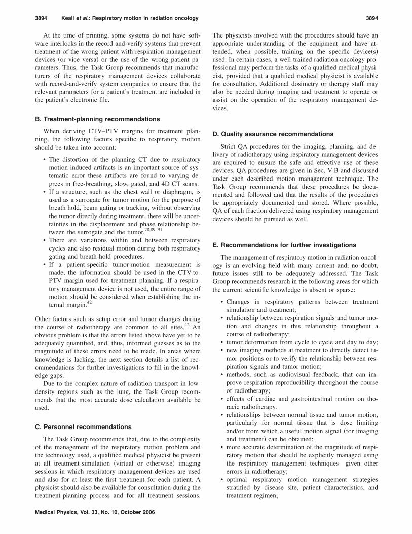
3894 Keall et al.: Respiratory motion in radiation oncology 3894
At the time of printing, some systems do not have soft-ware interlocks in the record-and-verify systems that preventtreatment of the wrong patient with respiration managementdevices �or vice versa� or the use of the wrong patient pa-rameters. Thus, the Task Group recommends that manufac-turers of the respiratory management devices collaboratewith record-and-verify system companies to ensure that therelevant parameters for a patient’s treatment are included inthe patient’s electronic file.
B. Treatment-planning recommendations
When deriving CTV–PTV margins for treatment plan-ning, the following factors specific to respiratory motionshould be taken into account:
• The distortion of the planning CT due to respiratorymotion-induced artifacts is an important source of sys-tematic error these artifacts are found to varying de-grees in free-breathing, slow, gated, and 4D CT scans.
• If a structure, such as the chest wall or diaphragm, isused as a surrogate for tumor motion for the purpose ofbreath hold, beam gating or tracking, without observingthe tumor directly during treatment, there will be uncer-tainties in the displacement and phase relationship be-tween the surrogate and the tumor.78,89–91
• There are variations within and between respiratorycycles and also residual motion during both respiratorygating and breath-hold procedures.
• If a patient-specific tumor-motion measurement ismade, the information should be used in the CTV-to-PTV margin used for treatment planning. If a respira-tory management device is not used, the entire range ofmotion should be considered when establishing the in-ternal margin.42
Other factors such as setup error and tumor changes duringthe course of radiotherapy are common to all sites.42 Anobvious problem is that the errors listed above have yet to beadequately quantified, and, thus, informed guesses as to themagnitude of these errors need to be made. In areas whereknowledge is lacking, the next section details a list of rec-ommendations for further investigations to fill in the knowl-edge gaps.
Due to the complex nature of radiation transport in low-density regions such as the lung, the Task Group recom-mends that the most accurate dose calculation available beused.
C. Personnel recommendations
The Task Group recommends that, due to the complexityof the management of the respiratory motion problem andthe technology used, a qualified medical physicist be presentat all treatment-simulation �virtual or otherwise� imagingsessions in which respiratory management devices are usedand also for at least the first treatment for each patient. Aphysicist should also be available for consultation during the
treatment-planning process and for all treatment sessions.Medical Physics, Vol. 33, No. 10, October 2006
The physicists involved with the procedures should have anappropriate understanding of the equipment and have at-tended, when possible, training on the specific device�s�used. In certain cases, a well-trained radiation oncology pro-fessional may perform the tasks of a qualified medical physi-cist, provided that a qualified medical physicist is availablefor consultation. Additional dosimetry or therapy staff mayalso be needed during imaging and treatment to operate orassist on the operation of the respiratory management de-vices.
D. Quality assurance recommendations
Strict QA procedures for the imaging, planning, and de-livery of radiotherapy using respiratory management devicesare required to ensure the safe and effective use of thesedevices. QA procedures are given in Sec. V B and discussedunder each described motion management technique. TheTask Group recommends that these procedures be docu-mented and followed and that the results of the proceduresbe appropriately documented and stored. Where possible,QA of each fraction delivered using respiratory managementdevices should be pursued as well.
E. Recommendations for further investigations
The management of respiratory motion in radiation oncol-ogy is an evolving field with many current and, no doubt,future issues still to be adequately addressed. The TaskGroup recommends research in the following areas for whichthe current scientific knowledge is absent or sparse:
• Changes in respiratory patterns between treatmentsimulation and treatment;
• relationship between respiration signals and tumor mo-tion and changes in this relationship throughout acourse of radiotherapy;
• tumor deformation from cycle to cycle and day to day;• new imaging methods at treatment to directly detect tu-
mor positions or to verify the relationship between res-piration signals and tumor motion;
• methods, such as audiovisual feedback, that can im-prove respiration reproducibility throughout the courseof radiotherapy;
• effects of cardiac and gastrointestinal motion on tho-racic radiotherapy.
• relationships between normal tissue and tumor motion,particularly for normal tissue that is dose limitingand/or from which a useful motion signal �for imagingand treatment� can be obtained;
• more accurate determination of the magnitude of respi-ratory motion that should be explicitly managed usingthe respiratory management techniques—given othererrors in radiotherapy;
• optimal respiratory motion management strategiesstratified by disease site, patient characteristics, and
treatment regimen;
3895 Keall et al.: Respiratory motion in radiation oncology 3895
• respiratory motion patterns and treatment implicationsin children and young adults treated with radiotherapyfor lymphoma and other pediatric diseases involvingthoracic radiotherapy;
• robust deformable image-registration algorithms to fa-cilitate dose accumulation due to anatomy deformation;
• treatment-planning solutions that can be integrated intocommercial treatment-planning systems;
• appropriate margin formalisms including respiratorymotion for the various respiratory motion managementstrategies;
• deformable phantoms to which anatomically accuraterespiratory motion can be applied;
• analysis of clinical outcome data in the presence of res-piratory motion and other errors.
ACKNOWLEDGMENTS
The members of this Task Group wish to thank the AAPMTreatment Delivery Subcommittee members Dr. Marc Son-tag, Dr. Chee-Wai Cheng, Dr. Janelle Molloy, Dr. MatthewPodgorsak, and Dr. Ron Zhu for their careful review andhelpful suggestions of this report. Members of the AAPMTherapy Committee and Professional Council also had usefulcomments. We would also like to thank Dr. Peter Balter, Dr.Sonja Dieterich, Dr. Stephan Erbel, Dr. Rohini George, Dr.Emily Heath, Dr. David Jaffray, Dr. Lech Papiez, Dr. AnilSethi, Dr. Jan Seuntjens, Dr. Craig Stevens, Dr. GabrielaStroian, Dr. Michelle Svatos, and Dr. Elisabeth Weiss forproviding information and input into the report. Finally,thanks to Devon Murphy Stein who carefully reviewed andimproved the clarity of the manuscript.
a�This manuscript is a moderately condensed version of the full report ofthe Task Group. The full report contains further background material andis available through the AAPM.
b�Author to whom correspondence should be addressed. Corresponding au-thor. Current address: Paul Keall, Ph.D., Radiation Physics Division, De-partment of Radiation Oncology, Stanford University, 875 Blake WilburDrive, Rm G-233, Stanford, CA 94305-5847. Telephone: �650� 723 5549;Fax: �650� 498 5008. Electronic mail: [email protected]
1C. A. Perez, M. Bauer, S. Edelstein, B. W. Gillespie, and R. Birch, “Im-pact of tumor control on survival in carcinoma of the lung treated withirradiation,” Int. J. Radiat. Oncol., Biol., Phys. 12�4�, 539–547 �1986�.
2N. C. Choi and J. A. Doucette, “Improved survival of patients with un-resectable non-small-cell bronchogenic carcinoma by an innovated high-dose en-bloc radiotherapeutic approach,” Cancer 48�1�, 101–109 �1981�.
3M. K. Martel, R. K. Ten Haken, M. B. Hazuka, M. L. Kessler, M. Straw-derman, A. T. Turrisi, T. S. Lawrence, B. A. Fraass, and A. S. Lichter,“Estimation of tumor control probability model parameters from 3-D dosedistributions of non-small cell lung cancer patients,” Lung Cancer 24�1�,31–37 �1999�.
4P. Okunieff, D. Morgan, A. Niemierko, and H. D. Suit, “Radiation dose-response of human tumors,” Int. J. Radiat. Oncol., Biol., Phys. 32�4�,1227–1237 �1995�.
5C. A. Perez, K. Stanley, P. Rubin, S. Kramer, L. Brady, R. Perez-Tamayo,G. S. Brown, J. Concannon, M. Rotman, and H. G. Seydel, “A prospec-tive randomized study of various irradiation doses and fractionationschedules in the treatment of inoperable non-oat-cell carcinoma of thelung. Preliminary report by the Radiation Therapy Oncology Group,”Cancer 45�11�, 2744–2753 �1980�.
6C. A. Perez, T. F. Pajak, P. Rubin, J. R. Simpson, M. Mohiuddin, L. W.Brady, R. Perez-Tamayo, and M. Rotman, “Long-term observations of the
patterns of failure in patients with unresectable non-oat cell carcinoma ofMedical Physics, Vol. 33, No. 10, October 2006
the lung treated with definitive radiotherapy. Report by the RadiationTherapy Oncology Group,” Cancer 59�11�, 1874–1881 �1987�.
7M. Machtay, “Higher BED is associated with improved local-regionalcontrol and survival for NSCLC treated with chemoradiotherapy: AnRTOG analysis,” Int. J. Radiat. Oncol., Biol., Phys. 63�2�, S66 �2005�.
8R. C. McGarry, L. Papiez, M. Williams, T. Whitford, and R. D. Timmer-man, “Stereotactic body radiation therapy of early-stage non-small-celllung carcinoma: Phase I study,” Int. J. Radiat. Oncol., Biol., Phys. 63�4�,1010-1015 �2005�.
9J. Wulf, K. Baier, G. Mueller, and M. P. Flentje, “Dose-response in ster-eotactic irradiation of lung tumors,” Radiother. Oncol. 77�1�, 83–87�2005�.
10S. L. Kwa, J. V. Lebesque, J. C. Theuws, L. B. Marks, M. T. Munley, G.Bentel, D. Oetzel, U. Spahn, M. V. Graham, R. E. Drzymala, J. A. Purdy,A. S. Lichter, M. K. Martel, and R. K. Ten Haken, “Radiation pneumoni-tis as a function of mean lung dose: An analysis of pooled data of 540patients,” Int. J. Radiat. Oncol., Biol., Phys. 42�1�, 1–9 �1998�.
11M. V. Graham, J. A. Purdy, B. Emami, W. Harms, W. Bosch, M. A.Lockett, and C. A. Perez, “Clinical dose-volume histogram analysis forpneumonitis after 3D treatment for non-small cell lung cancer �NSCLC�,”Int. J. Radiat. Oncol., Biol., Phys. 45�2�, 323–329 �1999�.
12M. L. Hernando, L. B. Marks, G. C. Bentel, S. M. Zhou, D. Hollis, S. K.Das, M. Fan, M. T. Munley, T. D. Shafman, M. S. Anscher, and P. A.Lind, “Radiation-induced pulmonary toxicity: A dose-volume histogramanalysis in 201 patients with lung cancer,” Int. J. Radiat. Oncol., Biol.,Phys. 51�3�, 650–659 �2001�.
13D. Oetzel, P. Schraube, F. Hensley, G. Sroka-Perez, M. Menke, and M.Flentje, “Estimation of pneumonitis risk in three-dimensional treatmentplanning using dose-volume histogram analysis,” Int. J. Radiat. Oncol.,Biol., Phys. 33�2�, 455–460 �1995�.
14Y. Seppenwoolde, J. V. Lebesque, K. de Jaeger, J. S. Belderbos, L. J.Boersma, C. Schilstra, G. T. Henning, J. A. Hayman, M. K. Martel, andR. K. Ten Haken, “Comparing different NTCP models that predict theincidence of radiation pneumonitis,” Int. J. Radiat. Oncol., Biol., Phys.55�3�, 724–735 �2003�.
15E. D. Yorke, A. Jackson, K. E. Rosenzweig, S. A. Merrick, D. Gabrys, E.S. Venkatraman, C. M. Burman, S. A. Leibel, and C. C. Ling, “Dose-volume factors contributing to the incidence of radiation pneumonitis innon-small-cell lung cancer patients treated with three-dimensional confor-mal radiation therapy,” Int. J. Radiat. Oncol., Biol., Phys. 54�2�, 329–339�2002�.
16J. Van de Steene, N. Linthout, J. de Mey, V. Vinh-Hung, C. Claassens, M.Noppen, A. Bel, and G. Storme, “Definition of gross tumor volume inlung cancer: Inter-observer variability,” Radiother. Oncol. 62�1�, 37–49�2002�.
17P. Giraud, S. Elles, S. Helfre, Y. De Rycke, V. Servois, M. F. Carette, C.Alzieu, P. Y. Bondiau, B. Dubray, E. Touboul, M. Housset, J. C. Rosen-wald, and J. M. Cosset, “Conformal radiotherapy for lung cancer: Differ-ent delineation of the gross tumor volume �GTV� by radiologists andradiation oncologists,” Radiother. Oncol. 62�1�, 27–36 �2002�.
18P. Bowden, R. Fisher, M. Mac Manus, A. Wirth, G. Duchesne, M. Mill-ward, A. McKenzie, J. Andrews, and D. Ball, “Measurement of lungtumor volumes using three-dimensional computer planning software,” Int.J. Radiat. Oncol., Biol., Phys. 53�3�, 566–573 �2002�.
19S. Senan, J. van Sornsen de Koste, M. Samson, H. Tankink, P. Jansen, P.J. Nowak, A. D. Krol, P. Schmitz, and F. J. Lagerwaard, “Evaluation of atarget contouring protocol for 3D conformal radiotherapy in non-smallcell lung cancer,” Radiother. Oncol. 53�3�, 247–255 �1999�.
20C. W. Hurkmans, J. H. Borger, B. R. Pieters, N. S. Russell, E. P. Jansen,and B. J. Mijnheer, “Variability in target volume delineation on CT scansof the breast,” Int. J. Radiat. Oncol., Biol., Phys. 50�5�, 1366–1372�2001�.
21R. Valdagni, C. Italia, P. Montanaro, M. Ciocca, G. Morandi, and B.Salvadori, “Clinical target volume localization using conventional meth-ods �anatomy and palpation� and ultrasonography in early breast cancerpost-operative external irradiation,” Radiother. Oncol. 42�3�, 231–237�1997�.
22L. Ekberg, O. Holmberg, L. Wittgren, G. Bjelkengren, and T. Landberg,“What margins should be added to the clinical target volume in radio-therapy treatment planning for lung cancer?,” Radiother. Oncol. 48,71–77 �1998�.
23J. T. Booth and S. F. Zavgorodni, “Set-up error and organ motion uncer-
tainty: A review,” Australas. Phys. Eng. Sci. Med. 22�2�, 29–47 �1999�.
3896 Keall et al.: Respiratory motion in radiation oncology 3896
24M. Engelsman, E. M. Damen, K. De Jaeger, K. M. van Ingen, and B. J.Mijnheer, “The effect of breathing and set-up errors on the cumulativedose to a lung tumor,” Radiother. Oncol. 60�1�, 95–105 �2001�.
25S. Essapen, C. Knowles, A. Norman, and D. Tait, “Accuracy of set-up ofthoracic radiotherapy: prospective analysis of 24 patients treated withradiotherapy for lung cancer,” Br. J. Radiol. 75�890�, 162–169 �2002�.
26C. W. Hurkmans, P. Remeijer, J. V. Lebesque, and B. J. Mijnheer, “Set-upverification using portal imaging; review of current clinical practice,”Radiother. Oncol. 58�2�, 105–120 �2001�.
27R. Halperin, W. Roa, M. Field, J. Hanson, and B. Murray, “Setup repro-ducibility in radiation therapy for lung cancer: a comparison betweenT-bar and expanded foam immobilization devices,” Int. J. Radiat. Oncol.,Biol., Phys. 43�1�, 211–216 �1999�.
28P. Rodrigus, D. Van den Weyngaert, and W. Van den Bogaert, “The valueof treatment portal films in radiotherapy for bronchial carcinoma,” Radio-ther. Oncol. 9�1�, 27–31 �1987�.
29D. Bohmer, P. Feyer, C. Harder, M. Korner, M. Sternemann, S. Dinges,and V. Budach, “Verification of set-up deviations in patients with breastcancer using portal imaging in clinical practice,” Strahlenther. Onkol.174 Suppl 2, 36–39 �1998�.
30K. M. Langen and D. T. Jones, “Organ motion and its management,” Int.J. Radiat. Oncol., Biol., Phys. 50�1�, 265–278 �2001�.
31G. van Tienhoven, J. H. Lanson, D. Crabeels, S. Heukelom, and B. J.Mijnheer, “Accuracy in tangential breast treatment set-up: A portal imag-ing study,” Radiother. Oncol. 22�4�, 317–322 �1991�.
32H. D. Kubo and B. C. Hill, “Respiration gated radiotherapy treatment: Atechnical study,” Phys. Med. Biol. 41�1�, 83–91 �1996�.
33C. Hector, S. Webb, and P. M. Evans, “A simulation of the effects ofset-up error and changes in breast volume on conventional and intensity-modulated treatments in breast radiotherapy,” Phys. Med. Biol. 46�5�,1451–1471 �2001�.
34C. L. Hector, S. Webb, and P. M. Evans, “The dosimetric consequences ofinter-fractional patient movement on conventional and intensity-modulated breast radiotherapy treatments,” Radiother. Oncol. 54�1�,57–64 �2000�.
35O. Pradier, H. Schmidberger, E. Weiss, H. Bouscayrol, A. Daban, and C.F. Hess, “Accuracy of alignment in breast irradiation: A retrospectiveanalysis of clinical practice,” Br. J. Radiol. 72�859�, 685–690 �1999�.
36C. L. Creutzberg, V. G. Althof, H. Huizenga, A. G. Visser, and P. C.Levendag, “Quality assurance using portal imaging: The accuracy of pa-tient positioning in irradiation of breast cancer,” Int. J. Radiat. Oncol.,Biol., Phys. 25�3�, 529–539 �1993�.
37N. R. MacIntyre, “High-frequency ventilation,” Crit. Care Med. 26�12�,1955–1956 �1998�.
38J. A. Krishnan and R. G. Brower, “High-frequency ventilation for acutelung injury and ARDS,” Chest 118�3�, 795–807 �2000�.
39E. C. Eichenwald and A. R. Stark, “High-frequency ventilation: Currentstatus,” Pediatr. Rev. 20�12�, e127–e133 �1999�.
40F. Yin, J. G. Kim, C. Haughton, S. L. Brown, M. Ajlouni, M. Stronati, N.Pamukov, and J. H. Kim, “Extracranial radiosurgery: Immobilizing livermotion in dogs using high-frequency jet ventilation and total intravenousanesthesia,” Int. J. Radiat. Oncol., Biol., Phys. 49�1�, 211–216 �2001�.
41ICRU, ICRU Report 50. Prescribing, recording and reporting photonbeam therapy �International Commission on Radiation Units and Mea-surements, Bethesda, MD, 1993�.
42ICRU, ICRU Report 62. Prescribing, Recording and Reporting PhotonBeam Therapy (Supplement to ICRU Report 50) �International Commis-sion on Radiation Units and Measurements, Bethesda, MD, 1999�.
43J. Wanger, Pulmonary Function Testing �Williams and Wilkins, Balti-more, 1996�.
44S. A. Nehmeh, Y. E. Erdi, K. E. Rosenzweig, H. Schoder, S. M. Larson,O. D. Squire, and J. L. Humm, “Reduction of respiratory motion artifactsin PET imaging of lung cancer by respiratory correlated dynamic PET:Methodology and comparison with respiratory gated PET,” J. Nucl. Med.44�10�, 1644–1648 �2003�.
45S. A. Nehmeh, Y. E. Erdi, C. C. Ling, K. E. Rosenzweig, H. Schoder, S.M. Larson, H. A. Macapinlac, O. D. Squire, and J. L. Humm, “Effect ofrespiratory gating on quantifying PET images of lung cancer,” J. Nucl.Med. 43�7�, 876–881 �2002�.
46S. A. Nehmeh, Y. E. Erdi, C. C. Ling, K. E. Rosenzweig, O. D. Squire, L.E. Braban, E. Ford, K. Sidhu, G. S. Mageras, S. M. Larson, and J. L.Humm, “Effect of respiratory gating on reducing lung motion artifacts in
PET imaging of lung cancer,” Med. Phys. 29�3�, 366–371 �2002�.Medical Physics, Vol. 33, No. 10, October 2006
47C. B. Caldwell, K. Mah, M. Skinner, and C. E. Danjoux, “Can PETprovide the 3D extent of tumor motion for individualized internal targetvolumes? A phantom study of the limitations of CT and the promise ofPET,” Int. J. Radiat. Oncol., Biol., Phys. 55�5�, 1381–1393 �2003�.
48P. Giraud, M. Antoine, A. Larrouy, B. Milleron, P. Callard, Y. De Rycke,M. F. Carette, J. C. Rosenwald, J. M. Cosset, M. Housset, and E. Touboul,“Evaluation of microscopic tumor extension in non-small-cell lung cancerfor three-dimensional conformal radiotherapy planning,” Int. J. Radiat.Oncol., Biol., Phys. 48�4�, 1015–1024 �2000�.
49C. W. Stevens, R. F. Munden, K. M. Forster, J. F. Kelly, Z. Liao, G.Starkschall, S. Tucker, and R. Komaki, “Respiratory-driven lung tumormotion is independent of tumor size, tumor location, and pulmonary func-tion,” Int. J. Radiat. Oncol., Biol., Phys. 51�1�, 62–68 �2001�.
50Y. Seppenwoolde, H. Shirato, K. Kitamura, S. Shimizu, M. van Herk, J.V. Lebesque, and K. Miyasaka, “Precise and real-time measurement of3D tumor motion in lung due to breathing and heartbeat, measured duringradiotherapy,” Int. J. Radiat. Oncol., Biol., Phys. 53�4�, 822–834 �2002�.
51C. D. Biancia, E. Yorke, C. S. Chui, P. Giraud, K. Rosenzweig, H. Amols,C. Ling, and G. S. Mageras, “Comparison of end normal inspiration andexpiration for gated intensity modulated radiation therapy �IMRT� of lungcancer,” Radiother. Oncol. 75�2�, 149–156 �2005�.
52R. M. Peters, The mechanical basis of respiration �Little, Brown, and Co.,Boston, 1969�.
53S. S. Vedam, V. R. Kini, P. J. Keall, V. Ramakrishnan, H. Mostafavi, andR. Mohan, “Quantifying the predictability of diaphragm motion duringrespiration with a noninvasive external marker,” Med. Phys. 30�4�, 505–513 �2003�.
54T. Neicu, R. Berbeco, J. Wolfgang, and S. B. Jiang, “Synchronized mov-ing aperture radiation therapy �SMART�: Improvement of breathing pat-tern reproducibility using respiratory coaching,” Phys. Med. Biol. 51�3�,617–636 �2006�.
55R. George, S. S. Vedam, T. D. Chung, V. Ramakrishnan, and P. J. Keall,“The application of the sinusoidal model to lung cancer patient respira-tory motion,” Med. Phys. 32�9�, 2850–2861 �2005�.
56V. R. Kini, S. S. Vedam, P. J. Keall, S. Patil, C. Chen, and R. Mohan,“Patient training in respiratory-gated radiotherapy,” Med. Dosim 28�1�,7–11 �2003�.
57I. Suramo, M. Paivansalo, and V. Myllyla, “Cranio-caudal movements ofthe liver, pancreas and kidneys in respiration,” Acta Radiol. Diagn.�Stockh� 25�2�, 129–131 �1984�.
58S. C. Davies, A. L. Hill, R. B. Holmes, M. Halliwell, and P. C. Jackson,“Ultrasound quantitation of respiratory organ motion in the upper abdo-men,” Br. J. Radiol. 67�803�, 1096–1102 �1994�.
59P. J. Bryan, S. Custar, J. R. Haaga, and V. Balsara, “Respiratory move-ment of the pancreas: An ultrasonic study,” J. Ultrasound Med. 3�7�,317–320 �1984�.
60C. S. Ross, D. H. Hussey, E. C. Pennington, W. Stanford, and J. F.Doornbos, “Analysis of movement of intrathoracic neoplasms using ul-trafast computerized tomography,” Int. J. Radiat. Oncol., Biol., Phys.18�3�, 671–677 �1990�.
61J. Hanley, M. M. Debois, D. Mah, G. S. Mageras, A. Raben, K. Rosen-zweig, B. Mychalczak, L. H. Schwartz, P. J. Gloeggler, W. Lutz, C. C.Ling, S. A. Leibel, Z. Fuks, and G. J. Kutcher, “Deep inspiration breath-hold technique for lung tumors: The potential value of target immobili-zation and reduced lung density in dose escalation,” Int. J. Radiat. Oncol.,Biol., Phys. 45�3�, 603–611 �1999�.
62S. Shimizu, H. Shirato, K. Kagei, T. Nishioka, X. Bo, H. Dosaka-Akita, S.Hashimoto, H. Aoyama, K. Tsuchiya, and K. Miyasaka, “Impact of res-piratory movement on the computed tomographic images of small lungtumors in three-dimensional �3D� radiotherapy,” Int. J. Radiat. Oncol.,Biol., Phys. 46�5�, 1127–1133 �2000�.
63P. Giraud, Y. De Rycke, B. Dubray, S. Helfre, D. Voican, L. Guo, J. C.Rosenwald, K. Keraudy, M. Housset, E. Touboul, and J. M. Cosset,“Conformal radiotherapy �CRT� planning for lung cancer: Analysis ofintrathoracic organ motion during extreme phases of breathing,” Int. J.Radiat. Oncol., Biol., Phys. 51�4�, 1081–1092 �2001�.
64H. W. Korin, R. L. Ehman, S. J. Riederer, J. P. Felmlee, and R. C. Grimm,“Respiratory kinematics of the upper abdominal organs: A quantitativestudy,” Magn. Reson. Med. 23�1�, 172–178 �1992�.
65C. Plathow, S. Ley, C. Fink, M. Puderbach, W. Hosch, A. Schmahl, J.Debus, and H. U. Kauczor, “Analysis of intrathoracic tumor mobilityduring whole breathing cycle by dynamic MRI,” Int. J. Radiat. Oncol.,
Biol., Phys. 59�4�, 952–959 �2004�.
3897 Keall et al.: Respiratory motion in radiation oncology 3897
66P. H. Weiss, J. M. Baker, and E. J. Potchen, “Assessment of hepaticrespiratory excursion,” J. Nucl. Med. 13�10�, 758–759 �1972�.
67G. Harauz and M. J. Bronskill, “Comparison of the liver’s respiratorymotion in the supine and upright positions: Concise communication,” J.Nucl. Med. 20�7�, 733–735 �1979�.
68O. Wade, “Movement of the thoracic cage and diaphragm in respiration,”J. Physiol. �London� 124, 193–212 �1954�.
69S. Malone, J. M. Crook, W. S. Kendal, and J. Szanto, “Respiratory-induced prostate motion: Quantification and characterization,” Int. J. Ra-diat. Oncol., Biol., Phys. 48�1�, 105–109 �2000�.
70H. Shirato, S. Shimizu, T. Kunieda, K. Kitamura, M. van Herk, K. Kagei,T. Nishioka, S. Hashimoto, K. Fujita, H. Aoyama, K. Tsuchiya, K. Kudo,and K. Miyasaka, “Physical aspects of a real-time tumor-tracking systemfor gated radiotherapy,” Int. J. Radiat. Oncol., Biol., Phys. 48�4�, 1187–1195 �2000�.
71S. Minohara, T. Kanai, M. Endo, K. Noda, and M. Kanazawa, “Respira-tory gated irradiation system for heavy-ion radiotherapy,” Int. J. Radiat.Oncol., Biol., Phys. 47�4�, 1097–1103 �2000�.
72M. J. Murphy, J. R. Adler, Jr., M. Bodduluri, J. Dooley, K. Forster, J. Hai,Q. Le, G. Luxton, D. Martin, and J. Poen, “Image-guided radiosurgery forthe spine and pancreas,” Comput. Aided Surg. 5�4�, 278–288 �2000�.
73Q. S. Chen, M. S. Weinhous, F. C. Deibel, J. P. Ciezki, and R. M. Mack-lis, “Fluoroscopic study of tumor motion due to breathing: Facilitatingprecise radiation therapy for lung cancer patients,” Med. Phys. 28�9�,1850–1856 �2001�.
74E. A. Barnes, B. R. Murray, D. M. Robinson, L. J. Underwood, J. Han-son, and W. H. Roa, “Dosimetric evaluation of lung tumor immobilizationusing breath hold at deep inspiration,” Int. J. Radiat. Oncol., Biol., Phys.50�4�, 1091–1098 �2001�.
75S. Shimizu, H. Shirato, S. Ogura, H. Akita-Dosaka, K. Kitamura, T. Nish-ioka, K. Kagei, M. Nishimura, and K. Miyasaka, “Detection of lung tu-mor movement in real-time tumor-tracking radiotherapy,” Int. J. Radiat.Oncol., Biol., Phys. 51�2�, 304–310 �2001�.
76E. C. Ford, G. S. Mageras, E. Yorke, K. E. Rosenzweig, R. Wagman, andC. C. Ling, “Evaluation of respiratory movement during gated radio-therapy using film and electronic portal imaging,” Int. J. Radiat. Oncol.,Biol., Phys. 52�2�, 522–531 �2002�.
77M. J. Murphy, D. Martin, R. Whyte, J. Hai, C. Ozhasoglu, and Q. T. Le,“The effectiveness of breath-holding to stabilize lung and pancreas tu-mors during radiosurgery,” Int. J. Radiat. Oncol., Biol., Phys. 53�2�, 475–482 �2002�.
78C. Ozhasoglu and M. J. Murphy, “Issues in respiratory motion compen-sation during external-beam radiotherapy,” Int. J. Radiat. Oncol., Biol.,Phys. 52�5�, 1389–1399 �2002�.
79K. E. Sixel, M. Ruschin, R. Tirona, and P. C. Cheung, “Digital fluoros-copy to quantify lung tumor motion: potential for patient-specific plan-ning target volumes,” Int. J. Radiat. Oncol., Biol., Phys. 57�3�, 717–723�2003�.
80I. S. Grills, D. Yan, A. A. Martinez, F. A. Vicini, J. W. Wong, and L. L.Kestin, “Potential for reduced toxicity and dose escalation in the treat-ment of inoperable non-small-cell lung cancer: A comparison of intensity-modulated radiation therapy �IMRT�, 3D conformal radiation, and elec-tive nodal irradiation,” Int. J. Radiat. Oncol., Biol., Phys. 57�3�, 875–890�2003�.
81E. C. Ford, G. S. Mageras, E. Yorke, and C. C. Ling, “Respiration-correlated spiral CT: A method of measuring respiratory-induced ana-tomic motion for radiation treatment planning,” Med. Phys. 30�1�, 88–97�2003�.
82S. S. Vedam, P. J. Keall, V. R. Kini, H. Mostafavi, H. P. Shukla, and R.Mohan, “Acquiring a four-dimensional computed tomography dataset us-ing an external respiratory signal,” Phys. Med. Biol. 48�1�, 45–62 �2003�.
83D. A. Low, M. Nystrom, E. Kalinin, P. Parikh, J. F. Dempsey, J. D.Bradley, S. Mutic, S. H. Wahab, T. Islam, G. Christensen, D. G. Politte,and B. R. Whiting, “A method for the reconstruction of four-dimensionalsynchronized CT scans acquired during free breathing,” Med. Phys.30�6�, 1254–1263 �2003�.
84K. Taguchi, “Temporal resolution and the evaluation of candidate algo-rithms for four-dimensional CT,” Med. Phys. 30�4�, 640–650 �2003�.
85J. Sonke, L. Zijp, P. Remeijer, and M. Van Herk, “Respiratory correlatedcone beam CT,” Med. Phys. 32�4�, 1176–1186 �2005�.
86P. J. Keall, G. Starkschall, H. Shukla, K. M. Forster, V. Ortiz, C. W.Stevens, S. S. Vedam, R. George, T. Guerrero, and R. Mohan, “Acquiring
4D thoracic CT scans using a multislice helical method,” Phys. Med.Medical Physics, Vol. 33, No. 10, October 2006
Biol. 49�10�, 2053–2067 �2004�.87G. S. Mageras, A. Pevsner, E. D. Yorke, K. E. Rosenzweig, E. C. Ford, A.
Hertanto, S. M. Larson, D. M. Lovelock, Y. E. Erdi, S. A. Nehmeh, J. L.Humm, and C. C. Ling, “Measurement of lung tumor motion usingrespiration-correlated CT,” Int. J. Radiat. Oncol., Biol., Phys. 60�3�, 933–941 �2004�.
88E. Rietzel, G. T. Chen, N. C. Choi, and C. G. Willet, “Four-dimensionalimage-based treatment planning: Target volume segmentation and dosecalculation in the presence of respiratory motion,” Int. J. Radiat. Oncol.,Biol., Phys. 61�5�, 1535–1550 �2005�.
89S. Ahn, B. Yi, Y. Suh, J. Kim, S. Lee, S. Shin, S. Shin, and E. Choi, “Afeasibility study on the prediction of tumor location in the lung from skinmotion,” Br. J. Radiol. 77�919�, 588–596 �2004�.
90J. D. Hoisak, K. E. Sixel, R. Tirona, P. C. Cheung, and J. P. Pignol,“Correlation of lung tumor motion with external surrogate indicators ofrespiration,” Int. J. Radiat. Oncol., Biol., Phys. 60�4�, 1298–1306 �2004�.
91Y. Tsunashima, T. Sakae, Y. Shioyama, K. Kagei, T. Terunuma, A.Nohtomi, and Y. Akine, “Correlation between the respiratory waveformmeasured using a respiratory sensor and 3D tumor motion in gated radio-therapy,” Int. J. Radiat. Oncol., Biol., Phys. 60�3�, 951–958 �2004�.
92N. Koch, H. H. Liu, G. Starkschall, M. Jacobson, K. Forster, Z. Liao, R.Komaki, and C. W. Stevens, “Evaluation of internal lung motion forrespiratory-gated radiotherapy using MRI: Part I–correlating internal lungmotion with skin fiducial motion,” Int. J. Radiat. Oncol., Biol., Phys.60�5�, 1459–1472 �2004�.
93H. H. Liu, N. Koch, G. Starkschall, M. Jacobson, K. Forster, Z. Liao, R.Komaki, and C. W. Stevens, “Evaluation of internal lung motion forrespiratory-gated radiotherapy using MRI: Part II–margin reduction ofinternal target volume,” Int. J. Radiat. Oncol., Biol., Phys. 60�5�, 1473–1483 �2004�.
94S. Senan, O. Chapet, F. J. Lagerwaard, and R. K. Ten Haken, “Definingtarget volumes for non-small cell lung carcinoma,” Semin. Radiat. Oncol.14�4�, 308–314 �2004�.
95S. Senan, D. De Ruysscher, P. Giraud, R. Mirimanoff, V. Budach, R. OnBehalf Of The Radiotherapy Group Of The European Organization For,and C. Treatment Of, “Literature-based recommendations for treatmentplanning and execution in high-dose radiotherapy for lung cancer,” Ra-diother. Oncol. 71�2�, 139–146 �2004�.
96J. Leong, “Implementation of random positioning error in computerisedradiation treatment planning systems as a result of fractionation,” Phys.Med. Biol. 32�3�, 327–334 �1987�.
97A. E. Lujan, E. W. Larsen, J. M. Balter, and R. K. Ten Haken, “A methodfor incorporating organ motion due to breathing into 3D dose calcula-tions,” Med. Phys. 26�5�, 715–720 �1999�.
98A. L. McKenzie, “How should breathing motion be combined with othererrors when drawing margins around clinical target volumes?,” Br. J.Radiol. 73�873�, 973–977 �2000�.
99W. A. Beckham, P. J. Keall, and J. V. Siebers, “A fluence-convolutionmethod to calculate radiation therapy dose distributions that incorporaterandom set-up error,” Phys. Med. Biol. 47�19�, 3465–3473 �2002�.
100I. J. Chetty, M. Rosu, N. Tyagi, L. H. Marsh, D. L. McShan, J. M. Balter,B. A. Fraass, and R. K. Ten Haken, “A fluence convolution method toaccount for respiratory motion in three-dimensional dose calculations ofthe liver: A Monte Carlo study,” Med. Phys. 30�7�, 1776–1780 �2003�.
101R. George, V. Kini, S. S. Vedam, V. Ramakrishnan, R. Mohan, and P. J.Keall, “Is the diaphragm motion probability density function normallydistributed?,” Med. Phys. 32, 396–404 �2005�.
102B. C. Cho, M. van Herk, B. J. Mijnheer, and H. Bartelink, “The effect ofset-up uncertainties, contour changes, and tissue inhomogeneities on tar-get dose-volume histograms,” Med. Phys. 29�10�, 2305–2318 �2002�.
103R. George, T. D. Chung, S. S. Vedam, V. Ramakrishnan, R. Mohan, E.Weiss, and P. J. Keall, “Audio-visual biofeedback for respiratory-gatedradiotherapy: Impact of audio instruction and audio-visual biofeedback onrespiratory-gated radiotherapy,” Int. J. Radiat. Oncol., Biol., Phys. 65�3�,924–933 �2006�.
104S. C. Erridge, Y. Seppenwoolde, S. H. Muller, M. van Herk, K. De Jaeger,J. S. Belderbos, L. J. Boersma, and J. V. Lebesque, “Portal imaging toassess set-up errors, tumor motion and tumor shrinkage during conformalradiotherapy of non-small cell lung cancer,” Radiother. Oncol. 66�1�,75–85 �2003�.
105E. D. Yorke, L. Wang, K. E. Rosenzweig, D. Mah, J. B. Paoli, and C. S.Chui, “Evaluation of deep inspiration breath-hold lung treatment plans
with Monte Carlo dose calculation,” Int. J. Radiat. Oncol., Biol., Phys.
3898 Keall et al.: Respiratory motion in radiation oncology 3898
53�4�, 1058–1070 �2002�.106M. van Herk, P. Remeijer, C. Rasch, and J. V. Lebesque, “The probability
of correct target dosage: Dose-population histograms for deriving treat-ment margins in radiotherapy,” Int. J. Radiat. Oncol., Biol., Phys. 47�4�,1121–1135 �2000�.
107J. C. Stroom, H. C. de Boer, H. Huizenga, and A. G. Visser, “Inclusion ofgeometrical uncertainties in radiotherapy treatment planning by means ofcoverage probability,” Int. J. Radiat. Oncol., Biol., Phys. 43�4�, 905–919�1999�.
108M. van Herk, P. Remeijer, and J. V. Lebesque, “Inclusion of geometricuncertainties in treatment plan evaluation,” Int. J. Radiat. Oncol., Biol.,Phys. 52�5�, 1407–1422 �2002�.
109M. van Herk, M. Witte, J. van der Geer, C. Schneider, and J. V. Lebesque,“Biologic and physical fractionation effects of random geometric errors,”Int. J. Radiat. Oncol., Biol., Phys. 57�5�, 1460–1471 �2003�.
110G. J. Kutcher et al., “Comprehensive QA for radiation oncology: Reportof AAPM Radiation Therapy Committee Task Group 40,” Med. Phys.21�4�, 581–618 �1994�.
111C. X. Yu, D. A. Jaffray, and J. W. Wong, “The effects of intra-fractionorgan motion on the delivery of dynamic intensity modulation,” Phys.Med. Biol. 43�1�, 91–104 �1998�.
112M. W. Kissick, S. A. Boswell, R. Jeraj, and T. R. Mackie, “Confirmation,refinement, and extension of a study in intrafraction motion interplay withsliding jaw motion,” Med. Phys. 32�7�, 2346–2350 �2005�.
113P. Pemler, J. Besserer, N. Lombriser, R. Pescia, and U. Schneider, “Influ-ence of respiration-induced organ motion on dose distributions in treat-ments using enhanced dynamic wedges,” Med. Phys. 28�11�, 2234–2240�2001�.
114T. Bortfeld, K. Jokivarsi, M. Goitein, J. Kung, and S. B. Jiang, “Effects ofintra-fraction motion on IMRT dose delivery: Statistical analysis andsimulation,” Phys. Med. Biol. 47�13�, 2203–2220 �2002�.
115H. D. Kubo and L. Wang, “Compatibility of Varian 2100C gated opera-tions with enhanced dynamic wedge and IMRT dose delivery,” Med.Phys. 27�8�, 1732–1738 �2000�.
116P. J. Keall, V. Kini, S. S. Vedam, and R. Mohan, “Motion adaptive x-raytherapy: A feasibility study,” Phys. Med. Biol. 46�1�, 1–10 �2001�.
117R. George, P. J. Keall, V. R. Kini, S. S. Vedam, J. V. Siebers, Q. Wu, M.H. Lauterbach, D. W. Arthur, and R. Mohan, “Quantifying the effect ofintrafraction motion during breast IMRT planning and dose delivery,”Med. Phys. 30�4�, 552–562 �2003�.
118C. S. Chui, E. Yorke, and L. Hong, “The effects of intra-fraction organmotion on the delivery of intensity-modulated field with a multileaf col-limator,” Med. Phys. 30�7�, 1736–1746 �2003�.
119S. B. Jiang, C. Pope, K. M. Al Jarrah, J. H. Kung, T. Bortfeld, and G. T.Chen, “An experimental investigation on intra-fractional organ motioneffects in lung IMRT treatments,” Phys. Med. Biol. 48�12�, 1773–1784�2003�.
120F. J. Lagerwaard, J. R. Van Sornsen de Koste, M. R. Nijssen-Visser, R. H.Schuchhard-Schipper, S. S. Oei, A. Munne, and S. Senan, “Multiple“slow” CT scans for incorporating lung tumor mobility in radiotherapyplanning,” Int. J. Radiat. Oncol., Biol., Phys. 51�4�, 932–937 �2001�.
121J. R. van Sornsen de Koste, F. J. Lagerwaard, R. H. Schuchhard-Schipper,M. R. Nijssen-Visser, P. W. Voet, S. S. Oei, and S. Senan, “Dosimetricconsequences of tumor mobility in radiotherapy of stage I non-small celllung cancer–an analysis of data generated using ‘slow’ CT scans,” Radio-ther. Oncol. 61�1�, 93–99 �2001�.
122J. R. de Koste, F. J. Lagerwaard, H. C. de Boer, M. R. Nijssen-Visser, andS. Senan, “Are multiple CT scans required for planning curative radio-therapy in lung tumors of the lower lobe?,” Int. J. Radiat. Oncol., Biol.,Phys. 55�5�, 1394–1399 �2003�.
123R. W. Underberg, F. J. Lagerwaard, B. J. Slotman, J. P. Cuijpers, and S.Senan, “Use of maximum intensity projections �MIP� for target volumegeneration in 4DCT scans for lung cancer,” Int. J. Radiat. Oncol., Biol.,Phys. 63�1�, 253–260 �2005�.
124W. Lu, P. J. Parikh, I. M. El Naqa, M. M. Nystrom, J. P. Hubenschmidt,S. H. Wahab, S. Mutic, A. K. Singh, G. E. Christensen, J. D. Bradley, andD. A. Low, “Quantitation of the reconstruction quality of a four-dimensional computed tomography process for lung cancer patients,”Med. Phys. 32�4�, 890–901 �2005�.
125T. Pan, T. Y. Lee, E. Rietzel, and G. T. Chen, “4D-CT imaging of avolume influenced by respiratory motion on multi-slice CT,” Med. Phys.31�2�, 333–340 �2004�.
126
G. Starkschall, K. M. Forster, K. Kitamura, A. Cardenas, S. L. Tucker,Medical Physics, Vol. 33, No. 10, October 2006
and C. W. Stevens, “Correlation of gross tumor volume excursion withpotential benefits of respiratory gating,” Int. J. Radiat. Oncol., Biol., Phys.60�4�, 1291–1297 �2004�.
127H. A. Shih, S. B. Jiang, K. M. Aljarrah, K. P. Doppke, and N. C. Choi,“Internal target volume determined with expansion margins beyond com-posite gross tumor volume in three-dimensional conformal radiotherapyfor lung cancer,” Int. J. Radiat. Oncol., Biol., Phys. 60�2�, 613–622�2004�.
128R. W. Underberg, F. J. Lagerwaard, J. P. Cuijpers, B. J. Slotman, J. R. vanSornsen de Koste, and S. Senan, “Four-dimensional CT scans for treat-ment planning in stereotactic radiotherapy for stage I lung cancer,” Int. J.Radiat. Oncol., Biol., Phys. 60�4�, 1283–1290 �2004�.
129J. W. Wolthaus, C. Schneider, J. J. Sonke, M. van Herk, J. S. Belderbos,M. M. Rossi, J. V. Lebesque, and E. M. Damen, “Mid-ventilation CT scanconstruction from four-dimensional respiration-correlated CT scans forradiotherapy planning of lung cancer patients,” Int. J. Radiat. Oncol.,Biol., Phys. 65�5�, 1560–1571 �2006�.
130H. D. Kubo, P. M. Len, S. Minohara, and H. Mostafavi, “Breathing-synchronized radiotherapy program at the University of California DavisCancer Center,” Med. Phys. 27�2�, 346–353 �2000�.
131S. S. Vedam, P. J. Keall, V. R. Kini, and R. Mohan, “Determining param-eters for respiration-gated radiotherapy,” Med. Phys. 28�10�, 2139–2146�2001�.
132G. S. Mageras, E. Yorke, K. Rosenzweig, L. Braban, E. Keatley, E. Ford,S. A. Leibel, and C. C. Ling, “Fluoroscopic evaluation of diaphragmaticmotion reduction with a respiratory gated radiotherapy system,” J. Appl.Clin. Med. Phys. 2, 191–200 �2001�.
133R. Wagman, E. Yorke, P. Giraud, E. Ford, K. Sidhu, G. Mageras, B.Minsky, and K. Rosenzweig, “Reproducibility of organ position with res-piratory gating for liver tumors: Use in dose-escalation,” Int. J. Radiat.Oncol., Biol., Phys. 55, 659–668 �2003�.
134G. S. Mageras and E. Yorke, “Deep inspiration breath hold and respira-tory gating strategies for reducing organ motion in radiation treatment,”Semin. Radiat. Oncol. 14�1�, 65–75 �2004�.
135R. I. Berbeco, S. Nishioka, H. Shirato, G. T. Chen, and S. B. Jiang,“Residual motion of lung tumors in gated radiotherapy with external res-piratory surrogates,” Phys. Med. Biol. 50�16�, 3655–3667 �2005�.
136C. Bert, K. G. Metheany, K. Doppke, and G. T. Chen, “A phantom evalu-ation of a stereo-vision surface imaging system for radiotherapy patientsetup,” Med. Phys. 32�9�, 2753–2762 �2005�.
137E. Yorke, K. E. Rosenzweig, R. Wagman, and G. S. Mageras, “Interfrac-tional anatomic variation in patients treated with respiration-gated radio-therapy,” J. Appl. Clin. Med. Phys. 6�2�, 19–32 �2005�.
138R. I. Berbeco, T. Neicu, E. Rietzel, G. T. Chen, and S. B. Jiang, “Atechnique for respiratory-gated radiotherapy treatment verification withan EPID in cine mode,” Phys. Med. Biol. 50�16�, 3669–3679 �2005�.
139C. R. Ramsey, D. Scaperoth, D. Arwood, and A. L. Oliver, “Clinicalefficacy of respiratory gated conformal radiation therapy,” Med. Dosim24�2�, 115–119 �1999�.
140P. J. Keall, V. R. Kini, S. S. Vedam, and R. Mohan, “Potential radio-therapy improvements with respiratory gating,” Australas. Phys. Eng. Sci.Med. 25�1�, 1–6 �2002�.
141S. Shen, J. Duan, J. B. Fiveash, I. A. Brezovich, B. A. Plant, S. A.Spencer, R. A. Popple, P. N. Pareek, and J. A. Bonner, “Validation oftarget volume and position in respiratory gated CT planning and treat-ment,” Med. Phys. 30�12�, 3196–3205 �2003�.
142J. Duan, S. Shen, J. B. Fiveash, I. A. Brezovich, R. A. Popple, and P. N.Pareek, “Dosimetric effect of respiration-gated beam on IMRT delivery,”Med. Phys. 30�8�, 2241–2252 �2003�.
143E. Nioutsikou, N. S.-T. J. Richard, J. L. Bedford, and S. Webb, “Quanti-fying the effect of respiratory motion on lung tumor dosimetry with theaid of a breathing phantom with deforming lungs,” Phys. Med. Biol.51�14�, 3359–3374 �2006�.
144K. Kitamura, H. Shirato, R. Onimaru, T. Shimizu, Y. Kodama, H. Endo,S. Shimizu, and K. Miyasaka, “Feasibility study of hypofractionatedgated irradiation using a real-time tumor-tracking radiation therapy sys-tem for malignant liver tumors,” Int. J. Radiat. Oncol., Biol., Phys. 54�2�,125–126 �2002�.
145K. Kitamura, H. Shirato, Y. Seppenwoolde, R. Onimaru, M. Oda, K.Fujita, S. Shimizu, N. Shinohara, T. Harabayashi, and K. Miyasaka,“Three-dimensional intrafractional movement of prostate measured dur-ing real-time tumor-tracking radiotherapy in supine and prone treatment
positions,” Int. J. Radiat. Oncol., Biol., Phys. 53�5�, 1117–1123 �2002�.
3899 Keall et al.: Respiratory motion in radiation oncology 3899
146K. Kitamura, H. Shirato, Y. Seppenwoolde, T. Shimizu, Y. Kodama, H.Endo, R. Onimaru, M. Oda, K. Fujita, S. Shimizu, and K. Miyasaka,“Tumor location, cirrhosis, and surgical history contribute to tumor move-ment in the liver, as measured during stereotactic irradiation using a real-time tumor-tracking radiotherapy system,” Int. J. Radiat. Oncol., Biol.,Phys. 56�1�, 221–228 �2003�.
147K. Kitamura, H. Shirato, S. Shimizu, N. Shinohara, T. Harabayashi, T.Shimizu, Y. Kodama, H. Endo, R. Onimaru, S. Nishioka, H. Aoyama, K.Tsuchiya, and K. Miyasaka, “Registration accuracy and possible migra-tion of internal fiducial gold marker implanted in prostate and livertreated with real-time tumor-tracking radiation therapy �RTRT�,” Radio-ther. Oncol. 62�3�, 275–281 �2002�.
148S. Shimizu, H. Shirato, K. Kitamura, N. Shinohara, T. Harabayashi, T.Tsukamoto, T. Koyanagi, and K. Miyasaka, “Use of an implanted markerand real-time tracking of the marker for the positioning of prostate andbladder cancers,” Int. J. Radiat. Oncol., Biol., Phys. 48�5�, 1591–1597�2000�.
149H. Shirato, T. Harada, T. Harabayashi, K. Hida, H. Endo, K. Kitamura, R.Onimaru, K. Yamazaki, N. Kurauchi, T. Shimizu, N. Shinohara, M. Mat-sushita, H. Dosaka-Akita, and K. Miyasaka, “Feasibility of insertion/implantation of 2.0 mm diameter gold internal fiducial markers for pre-cise setup and real-time tumor tracking in radiotherapy,” Int. J. Radiat.Oncol., Biol., Phys. 56�1�, 240–247 �2003�.
150H. Shirato, S. Shimizu, T. Shimizu, T. Nishioka, and K. Miyasaka, “Real-time tumor-tracking radiotherapy,” Lancet 353�9161�, 1331–1332�1999�.
151Y. Shibamoto, M. Ito, C. Sugie, H. Ogino, and M. Hara, “Recovery fromsublethal damage during intermittent exposures in cultured tumor cells:Implications for dose modification in radiosurgery and IMRT,” Int. J.Radiat. Oncol., Biol., Phys. 59�5�, 1484–1490 �2004�.
152J. F. Fowler, W. A. Tome, J. D. Fenwick, and M. P. Mehta, “A challengeto traditional radiation oncology,” Int. J. Radiat. Oncol., Biol., Phys.60�4�, 1241–1256 �2004�.
153R. P. Smith, P. Bloch, E. E. Harris, J. McDonough, A. Sarkar, A. Kassaee,S. Avery, and L. J. Solin, “Analysis of interfraction and intrafractionvariation during tangential breast irradiation with an electronic portal im-aging device,” Int. J. Radiat. Oncol., Biol., Phys. 62�2�, 373–378 �2005�.
154S. S. Korreman, A. N. Pedersen, T. J. Nottrup, L. Specht, and H.Nystrom, “Breathing adapted radiotherapy for breast cancer: Comparisonof free breathing gating with the breath-hold technique,” Radiother. On-col. 76�3�, 311–318 �2005�.
155H. M. Lu, E. Cash, M. H. Chen, L. Chin, W. J. Manning, J. Harris, and B.Bornstein, “Reduction of cardiac volume in left-breast treatment fields byrespiratory maneuvers: A CT study,” Int. J. Radiat. Oncol., Biol., Phys.47�4�, 895–904 �2000�.
156A. N. Pedersen, S. Korreman, H. Nystrom, and L. Specht, “Breathingadapted radiotherapy of breast cancer: Reduction of cardiac and pulmo-nary doses using voluntary inspiration breath-hold,” Radiother. Oncol.72�1�, 53–60 �2004�.
157V. M. Remouchamps, N. Letts, F. A. Vicini, M. B. Sharpe, L. L. Kestin,P. Y. Chen, A. A. Martinez, and J. W. Wong, “Initial clinical experiencewith moderate deep-inspiration breath hold using an active breathing con-trol device in the treatment of patients with left-sided breast cancer usingexternal beam radiation therapy,” Int. J. Radiat. Oncol., Biol., Phys.56�3�, 704–715 �2003�.
158V. M. Remouchamps, F. A. Vicini, M. B. Sharpe, L. L. Kestin, A. A.Martinez, and J. W. Wong, “Significant reductions in heart and lung dosesusing deep inspiration breath hold with active breathing control andintensity-modulated radiation therapy for patients treated with locore-gional breast irradiation,” Int. J. Radiat. Oncol., Biol., Phys. 55�2�, 392–406 �2003�.
159K. E. Sixel, M. C. Aznar, and Y. C. Ung, “Deep inspiration breath hold toreduce irradiated heart volume in breast cancer patients,” Int. J. Radiat.Oncol., Biol., Phys. 49�1�, 199–204 �2001�.
160D. Mah, J. Hanley, K. E. Rosenzweig, E. Yorke, L. Braban, C. C. Ling,and G. Mageras, “Technical aspects of the deep inspiration breath holdtechnique in the treatment of thoracic cancer,” Int. J. Radiat. Oncol.,Biol., Phys. 48, 1175–1185 �2000�.
161K. E. Rosenzweig, J. Hanley, D. Mah, G. Mageras, M. Hunt, S. Toner, C.Burman, C. C. Ling, B. Mychalczak, Z. Fuks, and S. A. Leibel, “The deepinspiration breath-hold technique in the treatment of inoperable non-small-cell lung cancer,” Int. J. Radiat. Oncol., Biol., Phys. 48�1�, 81–87
�2000�.Medical Physics, Vol. 33, No. 10, October 2006
162J. W. Wong, M. B. Sharpe, D. A. Jaffray, V. R. Kini, J. M. Robertson, J.S. Stromberg, and A. A. Martinez, “The use of active breathing control�ABC� to reduce margin for breathing motion,” Int. J. Radiat. Oncol.,Biol., Phys. 44�4�, 911–919 �1999�.
163J. S. Stromberg, M. B. Sharpe, L. H. Kim, V. R. Kini, D. A. Jaffray, A. A.Martinez, and J. W. Wong, “Active breathing control �ABC� forHodgkin’s disease: Reduction in normal tissue irradiation with deep in-spiration and implications for treatment,” Int. J. Radiat. Oncol., Biol.,Phys. 48�3�, 797–806 �2000�.
164D. J. Kim, B. R. Murray, R. Halperin, and W. H. Roa, “Held-breathself-gating technique for radiotherapy of non-small-cell lung cancer: Afeasibility study,” Int. J. Radiat. Oncol., Biol., Phys. 49�1�, 43–49 �2001�.
165Y. Xiao, J. Galvin, M. Hossain, and R. Valicenti, “An optimized forward-planning technique for intensity modulated radiation therapy,” Med.Phys. 27�9�, 2093–2099 �2000�.
166A. M. Berson, R. Emery, L. Rodriguez, G. M. Richards, T. Ng, S. Sang-havi, and J. Barsa, “Clinical experience using respiratory gated radiationtherapy: Comparison of free breathing and breath-hold techniques,” Int. J.Radiat. Oncol., Biol., Phys. 60�2�, 419–426 �2004�.
167I. Lax, H. Blomgren, I. Naslund, and R. Svanstrom, “Stereotactic radio-therapy of malignancies in the abdomen. Methodological aspects,” ActaOncol. 33�6�, 677–683 �1994�.
168H. Blomgren, I. Lax, I. Naslund, and R. Svanstrom, “Stereotactic highdose fraction radiation therapy of extracranial tumors using an accelera-tor. Clinical experience of the first thirty-one patients,” Acta Oncol.34�6�, 861–870 �1995�.
169I. Lax, “Target dose versus extratarget dose in stereotactic radiosurgery,”Acta Oncol. 32�4�, 453–457 �1993�.
170Y. Negoro, Y. Nagata, T. Aoki, T. Mizowaki, N. Araki, K. Takayama, M.Kokubo, S. Yano, S. Koga, K. Sasai, Y. Shibamoto, and M. Hiraoka, “Theeffectiveness of an immobilization device in conformal radiotherapy forlung tumor: Reduction of respiratory tumor movement and evaluation ofthe daily setup accuracy,” Int. J. Radiat. Oncol., Biol., Phys. 50�4�, 889–898 �2001�.
171J. Wulf, U. Hadinger, U. Oppitz, B. Olshausen, and M. Flentje, “Stereo-tactic radiotherapy of extracranial targets: CT-simulation and accuracy oftreatment in the stereotactic body frame,” Radiother. Oncol. 57�2�, 225–236 �2000�.
172R. Timmerman, L. Papiez, R. McGarry, L. Likes, C. DesRosiers, S. Frost,and M. Williams, “Extracranial stereotactic radioablation: Results of aphase I study in medically inoperable stage I nonsmall cell lung cancer,”Chest 124�5�, 1946–1955 �2003�.
173L. Papiez, R. Timmerman, C. DesRosiers, and M. Randall, “Extracranialstereotactic radioablation: Physical principles,” Acta Oncol. 42�8�, 882–894 �2003�.
174K. K. Herfarth, J. Debus, F. Lohr, M. L. Bahner, P. Fritz, A. Hoss, W.Schlegel, and M. F. Wannenmacher, “Extracranial stereotactic radiationtherapy: Set-up accuracy of patients treated for liver metastases,” Int. J.Radiat. Oncol., Biol., Phys. 46�2�, 329–335 �2000�.
175F. Lohr, J. Debus, C. Frank, K. Herfarth, O. Pastyr, B. Rhein, M. L.Bahner, W. Schlegel, and M. Wannenmacher, “Noninvasive patient fixa-tion for extracranial stereotactic radiotherapy,” Int. J. Radiat. Oncol.,Biol., Phys. 45�2�, 521–527 �1999�.
176R. Timmerman, L. Papiez, and M. Suntharalingam, “Extracranial stereo-tactic radiation delivery: Expansion of technology beyond the brain,”Technol. Cancer Res. Treat. 2�2�, 153–160 �2003�.
177M. J. Murphy, “Tracking moving organs in real time,” Semin. Radiat.Oncol. 14�1�, 91–100 �2004�.
178M. J. Murphy, J. Jalden, and M. Isaksson, “Adaptive filtering to predictlung tumor breathing motion during image-guided radiation therapy,”Proceedings of the 16th International Congress on Computer-assistedRadiology and Surgery, pp. 539–544 �2002�.
179A. Schweikard, G. Glosser, M. Bodduluri, M. J. Murphy, and J. R. Adler,“Robotic motion compensation for respiratory movement during radiosur-gery,” Comput. Aided Surg. 5�4�, 263–277 �2000�.
180G. C. Sharp, S. B. Jiang, S. Shimizu, and H. Shirato, “Prediction ofrespiratory tumor motion for real-time image-guided radiotherapy,” Phys.Med. Biol. 49�3�, 425–440 �2004�.
181A. Schweikard, H. Shiomi, and J. Adler, “Respiration tracking in radio-surgery,” Med. Phys. 31�10�, 2738–2741 �2004�.
182P. Liang, J. J. Pandit, and P. A. Robbins, “Nonstationarity of breath-by-breath ventilation and approaches to modeling the phenomenon,” in Mod-
eling and control of ventilation, edited by S. J. G. Semple, L. Adams, and
3900 Keall et al.: Respiratory motion in radiation oncology 3900
B. J. Whipp �Plenum, New York, 1995�, pp. 117–121.183E. N. Bruce, “Temporal variations in the pattern of breathing,” J. Appl.
Physiol. 80�4�, 1079–1087 �1996�.184P. G. Seiler, H. Blattmann, S. Kirsch, R. K. Muench, and C. Schilling, “A
novel tracking technique for the continuous precise measurement of tu-mor positions in conformal radiotherapy,” Phys. Med. Biol. 45�9�, N103–N110 �2000�.
185J. M. Balter, J. N. Wright, L. J. Newell, B. Friemel, S. Dimmer, Y. Cheng,J. Wong, E. Vertatschitsch, and T. P. Mate, “Accuracy of a wireless lo-calization system for radiotherapy,” Int. J. Radiat. Oncol., Biol., Phys.61�3�, 933–937 �2005�.
186G. Benchetrit, “Breathing pattern in humans: Diversity and individuality,”Respir. Physiol. 122�2–3�, 123–129 �2000�.
187S. S. Vedam, P. J. Keall, A. Docef, D. A. Todor, V. R. Kini, and R.Mohan, “Predicting respiratory motion for four-dimensional radio-therapy,” Med. Phys. 31�8�, 2274–2283 �2004�.
188T. Neicu, H. Shirato, Y. Seppenwoolde, and S. B. Jiang, “Synchronizedmoving aperture radiation therapy �SMART�: Average tumor trajectoryfor lung patients,” Phys. Med. Biol. 48�5�, 587–598 �2003�.
189L. Papiez, “The leaf sweep algorithm for an immobile and moving targetas an optimal control problem in radiotherapy delivery,” Math. Comput.Modell. 37, 735–745 �2003�.
190L. Papiez, “DMLC leaf-pair optimal control of IMRT delivery for a mov-ing rigid target,” Med. Phys. 31�10�, 2742–2754 �2004�.
191Y. Suh, B. Yi, S. Ahn, J. Kim, S. Lee, S. Shin, S. Shin, and E. Choi,“Aperture maneuver with compelled breath �AMC� for moving tumors: Afeasibility study with a moving phantom,” Med. Phys. 31�4�, 760–766�2004�.
192S. Webb, “Limitations of a simple technique for movement compensationvia movement-modified fluence profiles,” Phys. Med. Biol. 50�14�,N155–N161 �2005�.
193S. Webb, “The effect on IMRT conformality of elastic tissue movementand a practical suggestion for movement compensation via the modifieddynamic multileaf collimator �dMLC� technique,” Phys. Med. Biol.50�6�, 1163–1190 �2005�.
194S. Webb, “Does elastic tissue intrafraction motion with density changes
Medical Physics, Vol. 33, No. 10, October 2006
forbid motion-compensated radiotherapy?,” Phys. Med. Biol. 51�6�,1449–1462 �2006�.
195S. Webb, “Quantification of the fluence error in the motion-compensateddynamic MLC �DMLC� technique for delivering intensity-modulated ra-diotherapy �IMRT�,” Phys. Med. Biol. 51, L17–L21 �2006�.
196W. D. D’Souza, S. A. Naqvi, and C. X. Yu, “Real-time intra-fraction-motion tracking using the treatment couch: A feasibility study,” Phys.Med. Biol. 50�17�, 4021–4033 �2005�.
197M. Uematsu, “CT-guided focal high dose radiotherapy,” presented at the4th S Takahashi International Workshop on Three Dimensional Confor-mal Radiotherapy, Nagoya, Japan, 2004.
198T. Bortfeld, S. B. Jiang, and E. Rietzel, “Effects of motion on the totaldose distribution,” Semin. Radiat. Oncol. 14, 41–50 �2004�.
199T. Zhang, R. Jeraj, H. Keller, W. Lu, G. H. Olivera, T. R. McNutt, T. R.Mackie, and B. Paliwal, “Treatment plan optimization incorporating res-piratory motion,” Med. Phys. 31�6�, 1576–1586 �2004�.
200G. S. Mageras, E. Yorke, and S. Jiang, “4D IMRT Delivery,” in Image-guided IMRT, edited by T. Bortfeld, R. K. Schmidt-Ullrich, W. DeNeve etal. �Springer-Verlag, Heidelberg, 2005�, pp. 269–285.
201P. C. Cheung, K. E. Sixel, R. Tirona, and Y. C. Ung, “Reproducibility oflung tumor position and reduction of lung mass within the planning targetvolume using active breathing control �ABC�,” Int. J. Radiat. Oncol.,Biol., Phys. 57�5�, 1437–1442 �2003�.
202L. A. Dawson, K. K. Brock, S. Kazanjian, D. Fitch, C. J. McGinn, T. S.Lawrence, R. K. Ten Haken, and J. Balter, “The reproducibility of organposition using active breathing control �ABC� during liver radiotherapy,”Int. J. Radiat. Oncol., Biol., Phys. 51�5�, 1410–1421 �2001�.
203V. M. Remouchamps, N. Letts, D. Yan, F. A. Vicini, M. Moreau, J. A.Zielinski, J. Liang, L. L. Kestin, A. A. Martinez, and J. W. Wong, “Three-dimensional evaluation of intra- and interfraction immobilization of lungand chest wall using active breathing control: A reproducibility study withbreast cancer patients,” Int. J. Radiat. Oncol., Biol., Phys. 57�4�, 968–978�2003�.
204C. R. Ramsey, D. Scaperoth, and D. Arwood, “Clinical experience with acommercial respiratory gating system,” Int. J. Radiat. Oncol., Biol., Phys.48�3�, 164–165 �2000�.