the non-functional stomata on the leaf margin of...
TRANSCRIPT
245
Philippine Journal of Science142: 245-248, Special IssueISSN 0031 - 7683Date Received: ?? ???????? 2013
Yi Youguang and Benito C. Tan
Key Words: Selaginella, stomata, leaf border, SE Asia, guard cells
Department of Biological sciences, National University of SingaporeSingapore 119267
Corresponding Author: [email protected] [email protected]
The Non-Functional Stomata on the Leaf Margin of Selaginella
Stomata are structures on the surface of leaves known for their function in gaseous exchange in plants. After examining 26 species of Selaginella collected from Peninsular Malaysia and Singapore, we discover and report the presence of stomatal structures that are located right on the border of adaxial surface of lateral and axillary leaves of four species of Selaginella (S. roxburghii, S. ridleyi, S. rivalis and S.ciliaris). The marginal stomata appear to be non-functional due to their position on the leaf border which is one to two cells thick.
INTRODUCTIONStomata are small apertures found in the above ground parts of all terrestrial plants, especially on the leaf surfaces. They function to account for approximately 95% of gaseous exchange and regulate water loss via transpiration and CO2 uptake during photosynthesis (Taiz & Zeiger 2010, Camargo & Marenco 2011).
Selaginella are a group of Lycophyta whose diversity is still relatively not well studied in E and SE Asia. A few published keys based on morphological characters are available for the species identification in SE Asia (Alston 1934, 1935, Dahlen 1988, Wong 1983). Additionally, there were three recently conducted, albeit incomplete, phylogenetic studies consisting of European and American species of Selaginella (Manhart 1995, Korall et al. 1999, Korall & Kenrick 2002). Much of the study of basic biology of this group of basal plant taxa in the Malesian region are still waiting and needed investigation by the scientific community.
The examination and documentation of epidermal morphology of the leaf of spermatophytes and ferns in relation to the ecophysiology of plants and to discover new
diagnostic characters have been rather extensively conducted (Van Cotthem 1970, Nadeau & Sack 2002, Casson & Gray 2008, Gabriel y Galan et al. 2011, Nurit-Silva & Agra 2011, Wang et al. 2011, Giuliano & Gregorio 2012, Nurit-Silva et al. 2012). For example, stomata are observed to be distributed mainly on the abaxial surface of leaf of 35 species of tropical trees, while the adaxial surface of the leaf possess few or no stomata (Camargo & Marenco 2011).
However, not many studies have been done to examine the surface anatomy of the leaves of the different species of Selaginella (Harvey-Gindson 1897, Cosico 1972, Buck 1986, Dahlen 1988). This is especially true for the species of Selaginella in tropical SE Asia, which is the focus of our on-going research study of the leaf epidermal anatomy of species of Selaginella in Peninsula Malaysia and Singapore. The aim is to document, assess and evaluate the usefulness of epidermal elements in the natural grouping of species of Selaginella and in the inference of their homology and phylogeny.
Indeed, our preliminary finding on the study showed that the different species of Selaginella in the Peninsular Malaysia and Singapore exhibit different patterns of distribution of various types of epidermal cells, including the stomata, on the median, lateral and axillary leaves,
246
which, when taken together, can form a set of diagnostic characters to aid in identifying the species or species group. Overall, leaves of Selaginella spp. in Peninsula Malaya and Singapore can be both amphistomatus or hypostomatus. However, the abaxial surface of the vegetative leaves is always much higher in stomatal density than the adaxial surface by our observation.
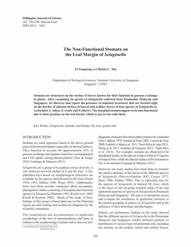
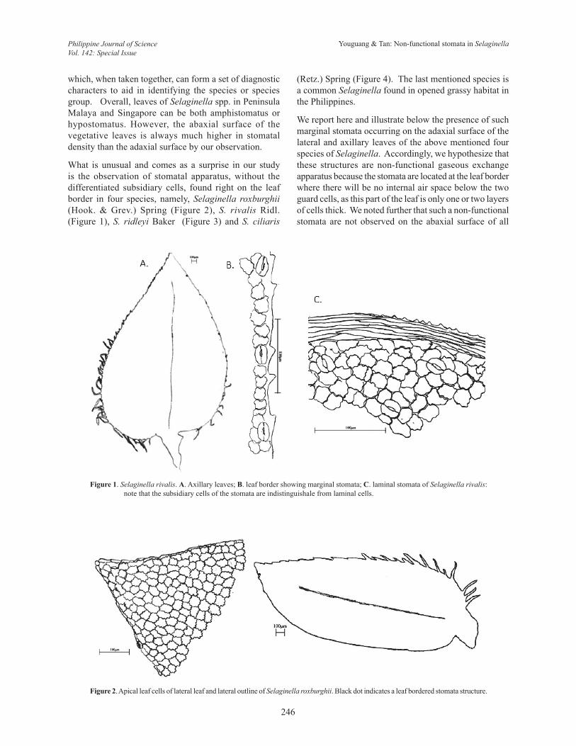
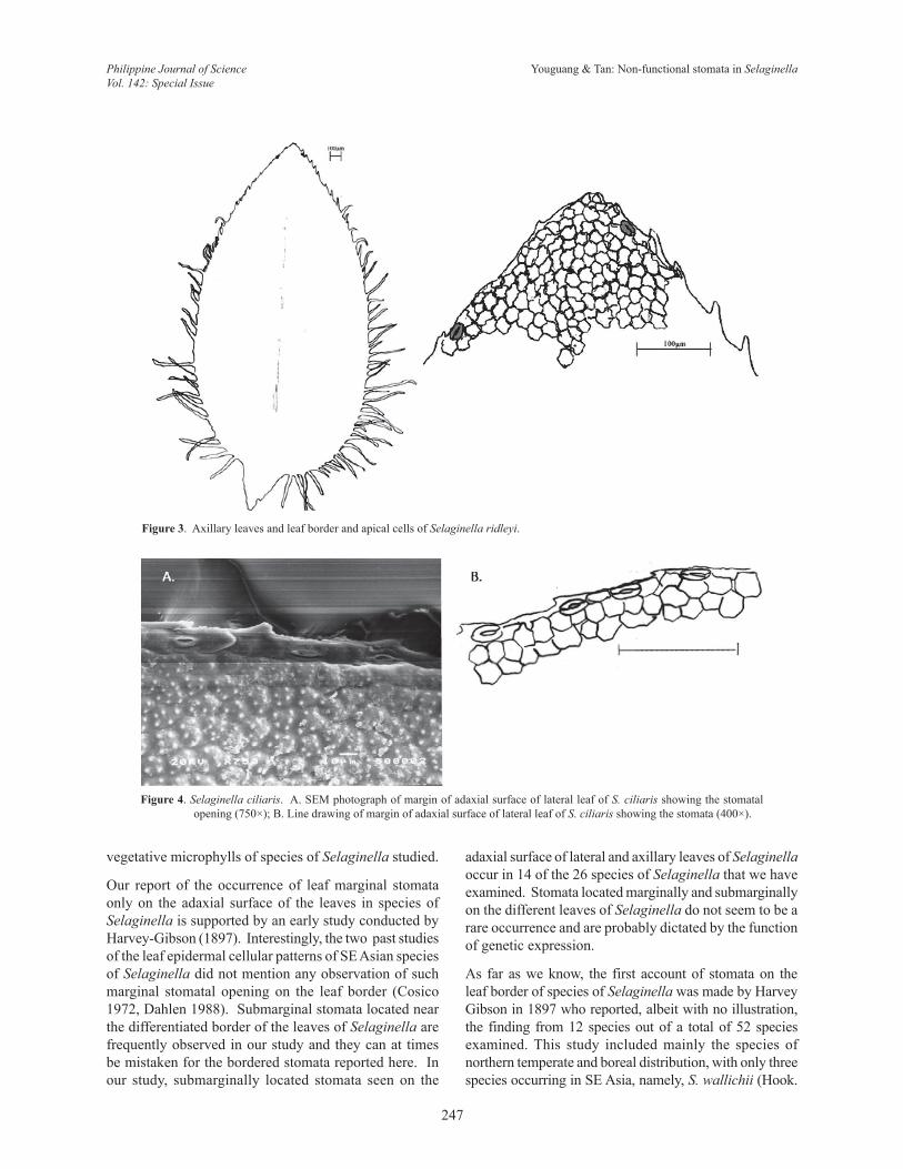
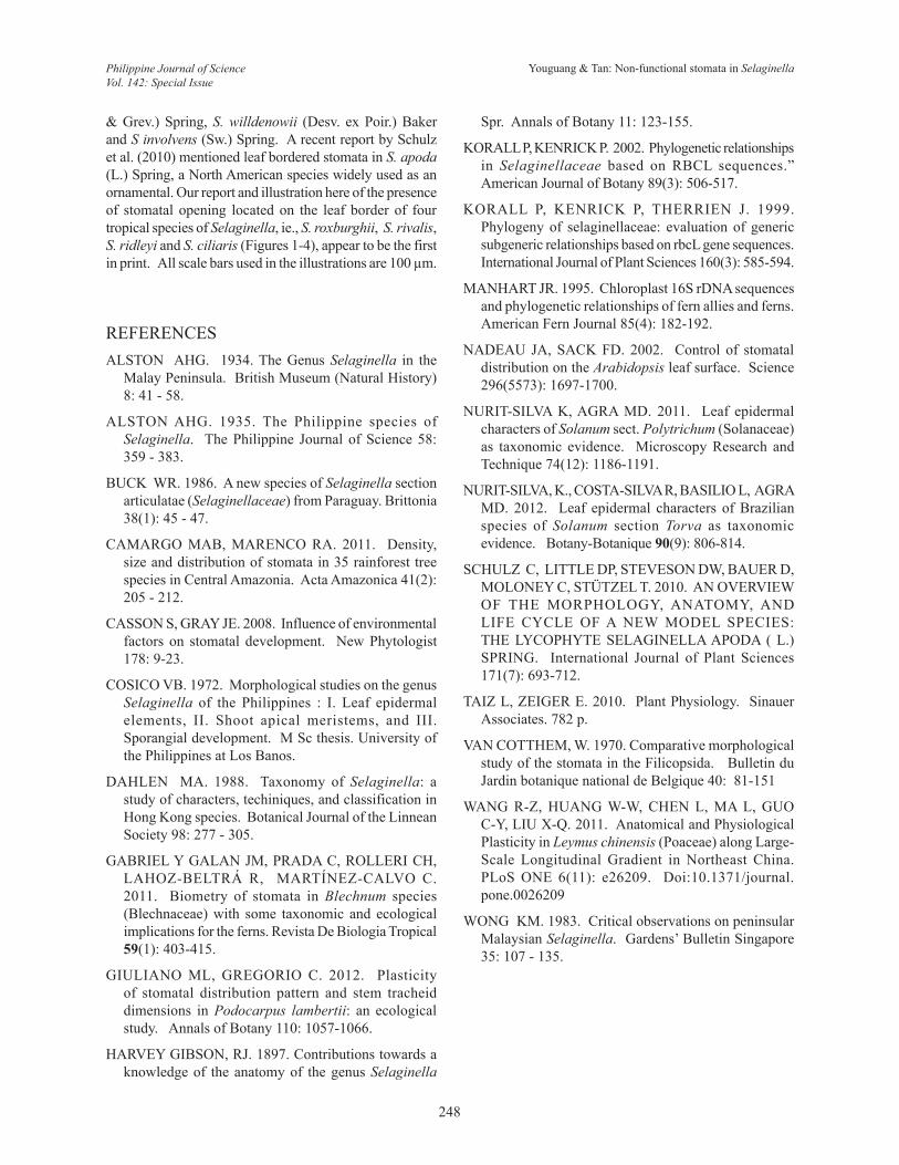
What is unusual and comes as a surprise in our study is the observation of stomatal apparatus, without the differentiated subsidiary cells, found right on the leaf border in four species, namely, Selaginella roxburghii (Hook. & Grev.) Spring (Figure 2), S. rivalis Ridl. (Figure 1), S. ridleyi Baker (Figure 3) and S. ciliaris
(Retz.) Spring (Figure 4). The last mentioned species is a common Selaginella found in opened grassy habitat in the Philippines.
We report here and illustrate below the presence of such marginal stomata occurring on the adaxial surface of the lateral and axillary leaves of the above mentioned four species of Selaginella. Accordingly, we hypothesize that these structures are non-functional gaseous exchange apparatus because the stomata are located at the leaf border where there will be no internal air space below the two guard cells, as this part of the leaf is only one or two layers of cells thick. We noted further that such a non-functional stomata are not observed on the abaxial surface of all
Figure 1. Selaginella rivalis. A. Axillary leaves; B. leaf border showing marginal stomata; C. laminal stomata of Selaginella rivalis: note that the subsidiary cells of the stomata are indistinguishale from laminal cells.
Figure 2. Apical leaf cells of lateral leaf and lateral outline of Selaginella roxburghii. Black dot indicates a leaf bordered stomata structure.
Youguang & Tan: Non-functional stomata in SelaginellaPhilippine Journal of ScienceVol. 142: Special Issue
247
vegetative microphylls of species of Selaginella studied.
Our report of the occurrence of leaf marginal stomata only on the adaxial surface of the leaves in species of Selaginella is supported by an early study conducted by Harvey-Gibson (1897). Interestingly, the two past studies of the leaf epidermal cellular patterns of SE Asian species of Selaginella did not mention any observation of such marginal stomatal opening on the leaf border (Cosico 1972, Dahlen 1988). Submarginal stomata located near the differentiated border of the leaves of Selaginella are frequently observed in our study and they can at times be mistaken for the bordered stomata reported here. In our study, submarginally located stomata seen on the
Figure 3. Axillary leaves and leaf border and apical cells of Selaginella ridleyi.
Figure 4. Selaginella ciliaris. A. SEM photograph of margin of adaxial surface of lateral leaf of S. ciliaris showing the stomatal opening (750×); B. Line drawing of margin of adaxial surface of lateral leaf of S. ciliaris showing the stomata (400×).
adaxial surface of lateral and axillary leaves of Selaginella occur in 14 of the 26 species of Selaginella that we have examined. Stomata located marginally and submarginally on the different leaves of Selaginella do not seem to be a rare occurrence and are probably dictated by the function of genetic expression.
As far as we know, the first account of stomata on the leaf border of species of Selaginella was made by Harvey Gibson in 1897 who reported, albeit with no illustration, the finding from 12 species out of a total of 52 species examined. This study included mainly the species of northern temperate and boreal distribution, with only three species occurring in SE Asia, namely, S. wallichii (Hook.
Youguang & Tan: Non-functional stomata in SelaginellaPhilippine Journal of ScienceVol. 142: Special Issue
248
& Grev.) Spring, S. willdenowii (Desv. ex Poir.) Baker and S involvens (Sw.) Spring. A recent report by Schulz et al. (2010) mentioned leaf bordered stomata in S. apoda (L.) Spring, a North American species widely used as an ornamental. Our report and illustration here of the presence of stomatal opening located on the leaf border of four tropical species of Selaginella, ie., S. roxburghii, S. rivalis, S. ridleyi and S. ciliaris (Figures 1-4), appear to be the first in print. All scale bars used in the illustrations are 100 µm.
REFERENCES ALSTON AHG. 1934. The Genus Selaginella in the
Malay Peninsula. British Museum (Natural History) 8: 41 - 58.
ALSTON AHG. 1935. The Philippine species of Selaginella. The Philippine Journal of Science 58: 359 - 383.
BUCK WR. 1986. A new species of Selaginella section articulatae (Selaginellaceae) from Paraguay. Brittonia 38(1): 45 - 47.
CAMARGO MAB, MARENCO RA. 2011. Density, size and distribution of stomata in 35 rainforest tree species in Central Amazonia. Acta Amazonica 41(2): 205 - 212.
CASSON S, GRAY JE. 2008. Influence of environmental factors on stomatal development. New Phytologist 178: 9-23.
COSICO VB. 1972. Morphological studies on the genus Selaginella of the Philippines : I. Leaf epidermal elements, II. Shoot apical meristems, and III. Sporangial development. M Sc thesis. University of the Philippines at Los Banos.
DAHLEN MA. 1988. Taxonomy of Selaginella: a study of characters, techiniques, and classification in Hong Kong species. Botanical Journal of the Linnean Society 98: 277 - 305.
GABRIEL Y GALAN JM, PRADA C, ROLLERI CH, LAHOZ-BELTRÁ R, MARTÍNEZ-CALVO C. 2011. Biometry of stomata in Blechnum species (Blechnaceae) with some taxonomic and ecological implications for the ferns. Revista De Biologia Tropical 59(1): 403-415.
GIULIANO ML, GREGORIO C. 2012. Plasticity of stomatal distribution pattern and stem tracheid dimensions in Podocarpus lambertii: an ecological study. Annals of Botany 110: 1057-1066.
HARVEY GIBSON, RJ. 1897. Contributions towards a knowledge of the anatomy of the genus Selaginella
Spr. Annals of Botany 11: 123-155.
KORALL P, KENRICK P. 2002. Phylogenetic relationships in Selaginellaceae based on RBCL sequences.” American Journal of Botany 89(3): 506-517.
KORALL P, KENRICK P, THERRIEN J. 1999. Phylogeny of selaginellaceae: evaluation of generic subgeneric relationships based on rbcL gene sequences. International Journal of Plant Sciences 160(3): 585-594.
MANHART JR. 1995. Chloroplast 16S rDNA sequences and phylogenetic relationships of fern allies and ferns. American Fern Journal 85(4): 182-192.
NADEAU JA, SACK FD. 2002. Control of stomatal distribution on the Arabidopsis leaf surface. Science 296(5573): 1697-1700.
NURIT-SILVA K, AGRA MD. 2011. Leaf epidermal characters of Solanum sect. Polytrichum (Solanaceae) as taxonomic evidence. Microscopy Research and Technique 74(12): 1186-1191.
NURIT-SILVA, K., COSTA-SILVA R, BASILIO L, AGRA MD. 2012. Leaf epidermal characters of Brazilian species of Solanum section Torva as taxonomic evidence. Botany-Botanique 90(9): 806-814.
SCHULZ C, LITTLE DP, STEVESON DW, BAUER D, MOLONEY C, STÜTZEL T. 2010. AN OVERVIEW OF THE MORPHOLOGY, ANATOMY, AND LIFE CYCLE OF A NEW MODEL SPECIES: THE LYCOPHYTE SELAGINELLA APODA ( L.) SPRING. International Journal of Plant Sciences 171(7): 693-712.
TAIZ L, ZEIGER E. 2010. Plant Physiology. Sinauer Associates. 782 p.
VAN COTTHEM, W. 1970. Comparative morphological study of the stomata in the Filicopsida. Bulletin du Jardin botanique national de Belgique 40: 81-151
WANG R-Z, HUANG W-W, CHEN L, MA L, GUO C-Y, LIU X-Q. 2011. Anatomical and Physiological Plasticity in Leymus chinensis (Poaceae) along Large-Scale Longitudinal Gradient in Northeast China. PLoS ONE 6(11): e26209. Doi:10.1371/journal.pone.0026209
WONG KM. 1983. Critical observations on peninsular Malaysian Selaginella. Gardens’ Bulletin Singapore 35: 107 - 135.
Youguang & Tan: Non-functional stomata in SelaginellaPhilippine Journal of ScienceVol. 142: Special Issue