the right chemistry - howard hughes medical … the right chemistry ... james a. baker, iii, esq....
TRANSCRIPT

Kinesins || “Good” Bacteria || Lab Security W I N T E R 2 0 0 5w
ww
.hhm
i.org
/bul
leti
n
T H E R I G H T
C H E M I S T R YHHMIinvestigatorCarolynBertozzi
isabrightlightinanewfield.

F E A T U R E S
C O N T E N T S Winter 2005 || Volume 17 Number 4
8 The Right Chemistry[COVER STORY] hhmi Investigator Carolyn Bertozzi found her call-ing in a college course in organic chemistry. “I loved solving the problems,” she says. She still does. By Mary Beth Gardiner
14 We Get a Kick From KinesinsUnder the hood of the cell, researchers get their hands dirty explor-ing the motors that propel molecular cargo along cellular super-highways. By Paul Muhlrad
20 Rules, Regs, and Red TapeAgainst the threat of bioterrorism, the government cracks down on lab security. But repercussions from the new laws could change the very culture of science. By Marlene Cimons
26 The Friendly Bacteria Within UsWhile we tend to think of bacteria as harmful, we all carry plenty of microbes that work to the good. Can we use them to prevent or treat diseases? By Maya Pines
31 Flying GlassAt Janelia Farm, the walls of windows have a structure and sociology all their own.
D E P A R T M E N T S
2 I N S T I T U T E N E W S
Science and Medicine: Bridging the Gap
3 PRESIDENT’S LETTER
Women in Science
U P F R O N T
4 The End of Our Genome
6 Peaceful Revolution
13 Q & A
Aging and Brain Function
19 I N S I D E H H M I
The Eye of the Beholder
25 R E S E A R C H N E W S
Student Contributes Big to Anti-Cancer Research
31
26
43
20
N E W S & N O T E S
34 Helping the Brain to Make Connections
35 Speed Reader
36 Brain Work at the
Worm Shack
38 New Directions
40 Nerve Verve
41 Hughes on the Big Screen
42 Can Cancer Kill Itself?
43 Cranial Exploration
in the Splash Class
45 H H M I L A B B O O K
48 N O TA B E N E ON THE COVER: Carolyn Bertozzi is director of the Biological Nanostructures program at the Lawrence Berkeley National Laboratory. Photograph by Barbara Ries.

HHMI TRUSTEES
James A. Baker, III, Esq. Senior Partner, Baker & Botts
Alexander G. Bearn, M.D. Former Executive Officer, American Philosophical Society; Professor Emeritus of Medicine, Cornell University Medical College
Frank William Gay Former President and Chief Executive Officer, summa Corporation
Joseph L Goldstein, M.D. Professor and Chairman, Department of Molecular Genetics, University of Texas Southwestern Medical Center at Dallas
Hanna H. Gray, Ph.D., CHAIRMAN President Emeritus and Harry Pratt Judson Distinguished Service Professor of History, The University of Chicago
Garnett L. Keith Chairman, SeaBridge Investment Advisors, L.L.C. Former Vice Chairman and Chief Investment Officer, The Prudential Insurance Company of America
Jeremy R. Knowles, D.Phil. Dean Emeritus and Amory Houghton Professor of Chemistry and Biochemistry, Harvard University
William R. Lummis, Esq. Former Chairman of the Board of Directors and Chief Executive Officer, The Howard Hughes Corporation
Kurt L. Schmoke Dean, Howard University School of Law
Anne M. TatlockChairman and Chief Executive OfficerFiduciary Trust Company International
HHMI OFF ICERS
Thomas R. Cech, Ph.D., President
Peter J. Bruns, Ph.D., Vice President for Grants and Special Programs
David A. Clayton, Ph.D., Vice President and Chief Scientific Officer
Stephen M. Cohen, Vice President and Chief Financial Officer
Joan S. Leonard, Esq., Vice President and General Counsel
Avice A. Meehan, Vice President for Communications and Public Affairs
Gerald M. Rubin, Ph.D., Vice President and Director, Janelia Farm Research Campus
Landis Zimmerman, Vice President and Chief Investment Officer
HHMI BULLET IN STAFF
Stephen G. Pelletier, Editor
Jim Keeley, Science Editor
Jennifer Donovan, Education Editor
Patricia Foster, Manager of Publishing
Mary Beth Gardiner, Assistant Editor
Maya Pines, Contributing Editor
Laura Bonetta, Katherine A. Wood, fact checking
Steven Marcus, story editing
Cay Butler, Kathy Savory, copy editing
David Herbick Design, publication design
Telephone (301) 215 8855 ■ Fax (301) 215 8863 ■ www.hhmi.org The Bulletin is published by the HHMI Office of Communications and Public Affairs.
© 2005 Howard Hughes Medical Institute
The opinions, beliefs and viewpoints expressed by
authors in the HHMI Bulletin do not necessarily
reflect the opinions, beliefs and viewpoints or official
policies of the Howard Hughes Medical Institute.
FROM TOP LEFT, CLOCKWISE: LOUIS PSIHOYOS; PAUL FETTERS; ASIA KEPKA; SCIMAT/PHOTO RESEARCHERS, INC.

The gap between basic biology and med-ical practice is growing. As knowledge inmolecular genetics and cell biology
accelerates, the biomedical community is find-ing it increasingly difficult to harness the explo-sion of new information and translate it intomedical practice. Bridging the Bed-Bench Gap,a National Research Council report publishedearlier this year, called training of Ph.D.researchers to translate science to clinical med-icine a “critical need.”
To address this problem, hhmi will award
up to $10 million to stimulate the integration ofmedical knowledge into Ph.D. training. The goalis to prepare biomedical scientists to apply newbiological knowledge to human health. A betterunderstanding of medicine also can guide scien-tists in research directions that are most likely tobenefit the diagnosis and treatment or preven-tion of human disease.
“We envision a new cadre of Ph.D.researchers who understand pathobiology andknow the language and processes of medicine,”said hhmi President Thomas R. Cech. “Our
goal is to increase the pool of people who aredoing medically oriented research.”
On December 1, 2004, the Institute openeda competition for grants for training programsthat bring the knowledge and skills of medicineand pathobiology into biomedical graduatestudy. Awards will range from $400,000 to $1million over 4 years. Smaller grants will supportmodification of existing programs. Innovativenew graduate programs that incorporate signif-icant pathobiological and medical knowledgeand skills can receive up to $250,000 a year.
“We seek creative, innovative, and cost-effective solutions to this training challenge,”said Peter J. Bruns, hhmi vice president forgrants and special programs. “We also are look-ing for approaches that can serve as models forthe biomedical research training community.”
Any university in the United States thatoffers Ph.D. training in a biomedical science iseligible to apply. The grants can be used to sup-port planning of new curricula, development ofnew courses, and release of clinical faculty toparticipate in graduate training activities.Student-related expenses can also be covered,including stipend support and health insur-ance, travel to medical meetings, expenses ofclinical training experiences, and tuition.
“hhmi already supports two programs thatgive medical students insight into the world ofbasic science research: Research Fellowships forMedical Students and the hhmi-NIH ResearchScholar Program,” said William Galey, hhmi’sdirector of graduate science education. “Basicscientists need a similar understanding of clini-cal medicine.”
For details on the new hhmi Medicine intoGraduate Training Initiative, see www.hhmi.org/grants/ inst i tut ions/medintograd.html.Applicant registration and proposal submis-sion are via hhmi’s Web-based competitionsystem at www.hhmi.org/grants/gcs. Applicantsmust register their intent to submit proposalsby April 20, 2005.
In another new graduate training initiativeannounced late last year, hhmi is partneringwith the NIH National Institute of BiomedicalImaging and Bioengineering (NIBIB) to sup-port biological science Ph.D. programs thatincorporate the physical and computationalscience or engineering disciplines to fosterinterdisciplinary training. (See hhmi Bulletin,Fall 2004, page 2, and www.hhmi.org/news/092704.html.)
—JENNIFER BOETH DONOVAN
I N S T I T U T E N E W S
Science and Medicine:Bridging the GapNew grants will help integrate medical knowledge into Ph.D. training.
2 h h m i b u l l e t i n | w i n t e r 2 0 0 5
2004 Holiday Lectures Monica Joshi, 16, a student at WashingtonInternational School, is one of 95 high school students and 50 HHMI staff who volunteered to com-plete a survey of their attitudes about obesity and weight control and to have their body fat and leanmass measured in this air displacement capsule, the Bod Pod. Gregg Wintering from LifeMeasurements, Inc., is recording Joshi’s data. HHMI investigators Ronald M. Evans and Jeffrey M.Friedman discussed the pooled results of the body density experiment and the survey during HHMI’s2004 Holiday Lectures on Science, “The Science of Fat,” in December 2004. Friedman and Evanstalked about their obesity research, examining why some people are overweight and others are lean,what science can tell us about how human bodies control weight, and the future of drugs to treatobesity. In the next Holiday Lectures, to be delivered in December 2005, HHMI investigators DavidM. Kingsley and Sean B. Carroll will speak on evolution. The Holiday Lectures are presented beforean audience of 200 Washington, D.C.-area high school students and are Webcast around the world.They also are produced as DVDs, which HHMI makes available at no charge to students and teach-ers. For more information, see www.hhmi.org/lectures/.
PA
UL
FE
TT
ER
S

Institute history. Going forward, we can augment that initial pool of appli-cants by convening temporary nominating groups, an approach borrowed from that used by the National Academy of Sciences. A temporary nomi-nating group might be asked to introduce greater diversity into the pool of nominees by identifying excellent candidates on the basis of their research area, career stage, and other factors, including gender and ethnicity.
We’ll continue to look at a host of factors that affect the success of women and other underrepresented groups within the hhmi com-munity. For example, my colleagues and I are committed to ensuring diversity within the membership of the review boards that help guide our decisions, both at the time candidates are selected as hhmi investigators and when they are reviewed for reappointment. Right now, between 20 and 33 percent of the scientific leaders who serve on our various review panels are female, but it’s an area we’ll continue to work on.
We will also improve the communication of our current poli-cies that offer greater flexibility to investigators with significant fam-ily responsibilities. Currently, our investigators have the option of postponing their review for a year because of the birth or adoption of a child, and we need to make sure that this option is clearly understood.
In addition, we will modify a long-standing policy that barred hhmi investigators from serving as permanent department chairs. On the surface, this may seem like an unusual approach to supporting the careers of our women investigators, because many scientists would hap-pily avoid the administrative responsibility that comes with such a post! Yet it appears that our rule may have had a disproportionate impact on women, who are increasingly sought out for leadership roles in their host institutions. We’ll still require hhmi investigators to devote at least 75 percent of their time to research—and to pass a rigorous review of their research accomplishments every five years—but the title of “chairman” will no longer force an investigator to resign from hhmi.
Finally—and this is the major challenge for the future—hhmi and other organizations need to think of new ways to encourage young women scientists to seek careers as professors. Nearly half of the Ph.D.s in the biological sciences are awarded to women, and yet many decide not to choose careers in academic research. We need to ask ourselves why, and then to make sure that our educational programs are working to pave the way for a more equitable future.
arolyn Bertozzi, who is profiled in this issue of the Bulletin, thrives on difficult scientific problems and, it turns out, large dollops of peanut butter. Intellectual drive fueled by complex carbohydrates makes perfect sense for a scientist interested in the myriad roles played by sugar molecules on the cell’s surface. Bertozzi’s research straddles departments
and disciplines and puts her in rarefied company: Nationwide, women account for only 5 percent of full professors in the chemical sciences.
That particular statistic—among others—has received considerable attention in recent months. Like the leaders of other research organi-zations and universities, I am well aware that women now comprise almost half of recent Ph.D.s in the biological sciences but continue to be underrepresented in the leadership ranks. In the biological sciences, only about 14 percent of full professors are women; although this situation is somewhat better than in chemistry, clearly many more women than men leave academia after earning the Ph.D. or after obtaining their first independent faculty position.
The Howard Hughes Medical Institute has reason to be proud of the exceptional quality of the women scientists whose careers it has helped foster. Approximately 20 percent of our investigators are women. The excellence of their work has been recognized in a variety of ways, from election to the National Academy to Linda Buck’s receiving the 2004 Nobel Prize in Physiology or Medicine. And more than a few women have left hhmi to assume leadership roles at the nation’s top universi-ties and research institutes, among them Shirley Tilghman (president of Princeton University), Susan Lindquist (director of the Whitehead Institute from 2001 to 2004), Sharon Long (dean of the School of Humanities and Sciences at Stanford University), and Carla Shatz (chair of the Department of Neurobiology at Harvard Medical School).
Yet this is no time for hhmi to be complacent. As the largest private funder of biomedical research in the nation, we’re obligated to ask if the Institute is doing enough to support the careers of women scientists. Over the past several months, we’ve had a number of lively conversa-tions on these issues with current hhmi investigators, our distinguished alumnae, and members of our Medical Advisory Board. As a result, the Institute is taking a variety of steps that will, we hope, better support our women investigators and the broader goals of enhancing diversity within the scientific community.
First, we are reviewing our nomination process for future hhmi investigator competitions. Currently, we ask nearly 200 research universi-ties, medical schools, and research institutes to nominate candidates to be considered for these appointments. This mechanism has served to identify superb candidates, and the proportion of women investigators selected in the most recent competition (25 percent) is the highest percentage in
h h m i b u l l e t i n | w i n t e r 2 0 0 5 3
P R E S I D E N T ’ S L E T T E R
Women in Science
KA
Y C
HE
RN
US
H
Thomas R. CechPresident
Howard Hughes Medical Institute
C

characteristic sign of cellular aging. But cellsalso possess a unique enzyme, known as telom-erase, which can lengthen telomeres by addingDNA to the ends of chromosomes through useof its own rna template. Regulation of telom-erase is critical, however, because too muchtelomerase activity after embryonic develop-ment can promote tumors.
In 2001, Peter Baumann in Cech’s laborato-ry (he’s now an assistant investigator at theStowers Institute for Medical Research inKansas City), discovered a protein called POT1(for “protection of telomeres”), which plays animportant role in capping the ends of chromo-somes and in regulating telomere length. POT1is the only protein known to bind to humantelomeric DNA tails. “Before that discovery,”says Baumann, “people weren’t even in agree-ment that there was a protein at the very endsof human chromosomes.” At the same time,
4 h h m i b u l l e t i n | w i n t e r 2 0 0 5
U p F r o n tThe End of Our GenomeThomas Cech’s lab takes a closer look at the protein that protects the tips of human chromosomes—and ensures survival.
Hhmi scientists have visualizedthe three-dimensional structureof a protein that surrounds theends of human chromosomes.Among other insights, the scien-
tists have learned how the protein homes in on aspecific DNA sequence and acts like a protectivecap to prevent erosion of the chro-mosome ends, which are critical tonormal cell division and survival.
The researchers—hhmiPresident Thomas R. Cech and hislaboratory colleagues Ming Lei,now assistant professor at theUniversity of Michigan, andElaine R. Podell at the Universityof Colorado at Boulder—pub-lished their findings in theDecember 2004 issue of NatureStructural and Molecular Biology.According to Cech, his team’sfindings not only provide newinsights into essential cellularfunctions taking place at the endof the chromosome but also raiseimportant new questions.
protecting the protectorsDuring normal DNA replication,the very ends of a DNA moleculeare lost. To prevent what other-wise would be erosion of chromo-somes, they are capped with aspecialized region of DNA knownas a telomere—a short, repetitiousDNA sequence that does not code
for any protein. In humans, an entire telomereis thousands of base pairs long—made up of arepeating sequence of six nucleotides—with the100–300 base pairs at the very end extendingbeyond the double helix as a single-strandedDNA “tail.” The telomeres of normal cells grad-ually become shorter with each cell division, a
Ming Lei (left), Elaine Podell, and
Thomas Cech found new insights into
essential cellular functions—raising
new questions for future study.
h h m i b u l l e t i n | w i n t e r 2 0 0 5 5
Cech’s team found a ver-sion of the POT1 proteinin fission yeast. Otherversions of POT1 havesince been found in plantsand mice—each recogniz-ing a telomeric sequencethat is specific to its respec-tive organism.
POT1 is critical tonormal cell division andsurvival; experiments infission yeast have shownthat without it most cellsdie immediately. Cells thatdo manage to survivequickly lose their telom-eres, which interferes withnormal cell division andeventually leads to massiveDNA errors and abnor-mal, circular chromo-somes. In human cellsgrown in the laboratory,too much POT1 can bedisruptive, causing abnor-mal lengthening or short-ening of telomeres.
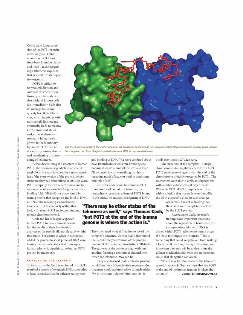
Before determining the structure of humanPOT1, the researchers’ prediction of what itmight look like was based on their understand-ing of the yeast version of the protein, whosestructure they had determined in 2003. In yeast,POT1 wraps up the end of a chromosome bymeans of an oligonucleotide/oligosaccharide-binding fold (OB-fold)—a shape found inmany proteins that recognize and bind to DNAor RNA. The repeating six-nucleotidetelomeric unit fits precisely within thisfold, with many POT1 molecules bindingto each chromosome end.
Cech and his colleagues expectedhuman POT1 to have a similar design,but the results of their biochemicalanalyses of the protein did not fit easily withinthis model. For example, when the scientistsadded the protein to short pieces of DNA con-taining the six nucleotides that make up ahuman telomeric repetition, the human POT1protein bound poorly.
consulting the crystalsTo its surprise, the Cech team found that POT1required a stretch of telomeric DNA containingat least 10 nucleotides for efficient recognition
and binding of DNA. “We were confused abouthow 10 nucleotides was even a binding site,because it wasn’t a multiple of six,” says Cech.“If you need to coat something that has arepeating motif of six, you need to bind somemultiple of six.”
To better understand how human POT1recognized and bound to a telomere, theresearchers crystallized a form of POT1 boundto the critical 10-nucleotide segment of DNA.
They then used x-ray diffraction to reveal thecomplex’s structure. Unexpectedly, they foundthat, unlike the yeast version of the protein,human POT1 contained two distinct OB-folds.The grooves of the two folds align with oneanother, forming a continuous channel intowhich the telomeric DNA can fit.
They also learned that, while the proteinwould bind to a 10-nucleotide sequence, thestructure could accommodate 12 nucleotides.“So it turns out it doesn’t bind one six, it
binds two times six,” Cech says.The structure of the complex—a single
chromosome’s tail might be coated with 8–24POT1 molecules—suggests that the end of thechromosome is tightly protected by POT1. Theresearchers were able to verify this hypothesiswith additional biochemical experiments:When the POT1-DNA complex was treatedwith a solution that normally would modifythe DNA at specific sites, no such changes
occurred—a result indicating thatthose sites were completely enclosed by the POT1 protein.
According to Cech, the team’sfindings raise important questionsabout the regulation of telomerase. Forexample, when telomeric DNA is
buried within POT1, telomerase cannot accessthe DNA to elongate the telomere. “This issomething that could keep the cell from makingtelomeres all day long,” he says. Therefore, animportant next step will be to determine thecellular mechanism that switches on the telom-ere so that elongation can occur.
“There may be other states of the telomereas well,” says Cech, “but we think that the POT1at the end of the human genome is where theaction is.” —JENNIFER MICHALOWSKI
“There may be other states of thetelomere as well,” says Thomas Cech,“but POT1 at the end of the humangenome is where the action is.”
MIN
G L
EI,
EL
AIN
E R
. P
OD
EL
L,
TH
OM
AS
R.
CE
CH
. IM
AG
E C
RE
AT
ED
WIT
H T
HE
PY
MO
L M
OL
EC
UL
AR
GR
AP
HIC
S S
YS
TE
M.
OP
PO
SIT
E P
AG
E:
LO
UIS
PS
IHO
YO
S
The POT1 protein binds to the end of a human chromosome by means of two oligonucleotide/oligosaccharide-binding folds, shown
here in green and blue. Single-stranded telomeric DNA is represented in red.

U p F r o n t
Peaceful Revolution Agents of change plot reforms in undergraduate biology education.
6 h h m i b u l l e t i n | w i n t e r 2 0 0 5
Looking a little like revolutionar-ies—which in some sense theyare—a band of university profes-sors huddles around a computerterminal in a dark corner of the
university conference center. The clock edgespast midnight as they wrangle about tactics.
Their mission? Nothing less thanrevolutionizing the way biology istaught to undergraduates.
“Most introductory courses relyon lectures and ‘cookbook’ labs, eventhough research shows that thosetechniques are not highly effective infostering conceptual understandingor scientific reasoning,” says Jo Han-delsman, professor of plant patholo-gy at the University of Wisconsin–Madison and one of 20 hhmiProfessors who received $1 millioneach to support reform of under-graduate science at research univer-sities. Handelsman believes thereason for this lack of innovation isthat “scientists actively resist chang-ing their teaching.” To move them,she asserts, is going to take nothingless than a revolution.
To help plant the seeds of thatuprising, Handelsman, who directshhmi ’s New Generation Programfor Scientific Teaching at the Univer-sity of Wisconsin–Madison, teamedup with William B. Wood—a professor ofmolecular, cellular, and developmental biologyat the University of Colorado at Boulder—andothers on a planning committee commissionedby the National Academies. They invited lead-ing life-sciences faculty to come with a junior-level colleague to spend a week during August2004 at the University of Wisconsin, working inteams and in plenary sessions to inform, plot,and inspire the insurrection.
More than 100 applied for the SummerInstitute on Undergraduate Education in Biolo-gy, which grew from a recommendation in the
National Research Council report Bio2010:Transforming Undergraduate Education forFuture Research Biologists. (Both the summerinstitute and the report were projects of theNational Academies and funded in part byhhmi.) Ultimately, 42 faculty members from 20research universities met in Madison to learn
from some of the pioneers in undergraduatebiology education and to create their own“teachable units”—a cohesive collection ofmaterials and activities on a topic in biology,designed to be the equivalent of three lectures—for conveying scientific thinking and biologicalconcepts at the introductory college level.
“Can students learn to think in Bio 101?”speaker Randall W. Phillis, an associate professorof biology at the University of Massachusetts–Amherst, asked rhetorically. Phillis, who won agrant from the Pew Charitable Trusts’ Centerfor Academic Transformation to revamp the
introductory biology courses on his campus,said the answer is an emphatic yes. Through“active learning,” which he defined as problem-solving using questions and activities, studentsbecome “active participants in learning insteadof passive recipients of knowledge,” said Phillis.
Active learning does not replace the contentof the course itself. “Content is important,” henoted, “but it is learned best if it is used in thecontext of doing science.”
Another speaker, Robin Wright, an admin-istrator and professor of genetics, cell biology,and development at the University of Minneso-ta, called active learning “hands-on, minds-on,dynamic, engaging, and uncomfortable.” Sheadded: “We are not teaching students biology;we are teaching them how to be humanbeings—to think, to be curious, to make diffi-cult decisions, to apply what they’ve learned.”
Whether they consciously realize it ornot, students desire just these kinds of out-comes. When they ask “Do I have to memo-rize this for the exam?” said Lydia Daniels,director of undergraduate programs in thedepartment of biological sciences at the Uni-versity of Pittsburgh, “what they really wantto know is ‘How am I going to use this? Whydo I need to know it?’” For example, Danielssuggests, instead of teaching math as a stand-alone subject, integrate it into biology such asby teaching equations and mathematicaltechniques that students need to solve biolog-ical problems, an approach consistent withthe recommendations for interdisciplinaryeducation in Bio2010.
Presentations by the institute’s speakers, abattle-tested and inspiring lot, were but one partof the intense week in Wisconsin last summer.Participants worked virtually around the clock,attending roundtable discussions at 7 a.m. androlling up their sleeves at midnight meetings tofine-tune their teams’ teachable units. And onFriday it was show-and-tell time, when eachteam presented its product and received feedbackfrom other participants and reviewers.
One team had tackled the question “Areyou my mother?”—in response to its assign-ment to develop a teachable unit on heredity.Challenging students to solve a case of twonewborns who may have been switched atbirth, team members Victoria Finnerty andRachelle Spell of Emory University, Martin L.Tracey and Ophelia I. Weeks of Florida Interna-tional University, Jennifer K. Knight andWilliam B. Wood of the University of Colorado
Jo Handelsman wants to transform science teaching.
JA
ME
S S
CH
NE
PF
(2
)

h h m i b u l l e t i n | w i n t e r 2 0 0 5 7
at Boulder, and William Segraves and David G.Wells of Yale University would teach how tomeasure the degree of genetic variabilitybetween individuals and how a genetic markersegregates. In the process, the students learn theprinciples of genetic linkage, DNA sequenceanalysis, and the importance of incorporatingappropriate experimental controls. They alsolearn a larger lesson: the use of scientific data toanswer real-world questions.
Another team took on evolution. Almostall introductory-biology students have mis-conceptions about evolution and the relation-ship of genotypes to phenotypes, Phillip G.Sokolove and Jeff W. Leips of the University ofMaryland, Baltimore County, pointed out.And many do not have the quantitative skillsto evaluate hypotheses related to evolution. Ina unit called “Are Humans Evolving? HowWould You Know?” they and fellow teammembers Elizabeth Torres of California State
University, Los Angeles; William F. Collinsand Joan M. Miyazaki of Stony Brook Univer-sity; and Mark D. Decker, Sue Wick, andRobin Wright of the University of Minnesotawould use a case study of a genetically based
human disease to teach students theHardy–Weinberg equilibrium—an equationfor predicting allele and genotype frequencyin a population. After the class analyzes realdata from rock pocket mice to verify theformula’s predictions, it examines fossil evi-dence and current disease data on humans toanswer the unit’s title questions.
The development of these and the otherteachable units is not meant to be a mere exer-cise; each participant pledged to implement atleast one of the units on his or her campus this
academic year. Each participant also acceptedthe honor and responsibility of being named anEducation Fellow in the Life Sciences by theNational Academies.
For added motivation, Handelsman sentthem off with a battle cry: “You’regoing home to begin staging a revolu-tion. Find sympathetic colleagues oncampus and nationally. Share yourideas. Combat misconceptions.Remember, we are doing this basedon scientific evidence.”
“We are the change agents,” says Sokolove.But he harbors no illusions about its pace. “Willteaching ever be rewarded in a research univer-sity the way research is? Probably not. But aparadigm shift in science takes 35 years. Whyshould we expect a change in teaching to hap-pen overnight?”
Nevertheless, progress is now discernible.At his own campus, Sokolove says, hiscolleagues are starting to talk to each otherabout teaching. “They never used to do that.”
—JENNIFER BOETH DONOVAN
Workshop participants shared ideas on interdiscipli-
nary teaching, undergraduate research, and curricula.
“Content is…learned best if it is used in the context of doingscience.” —RANDALL PHILLIS

8 h h m i b u l l e t i n | w i n t e r 2 0 0 5
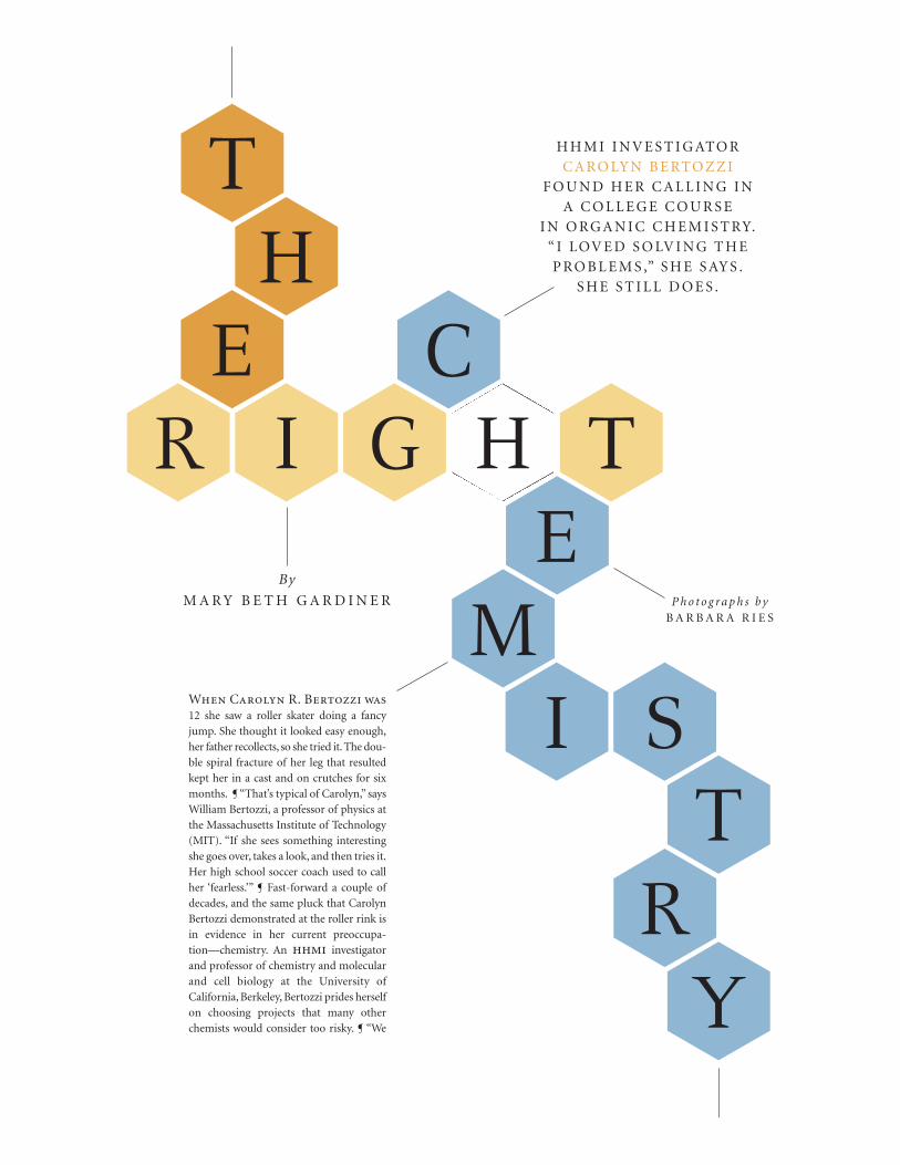
H H M I I N V E S T I G ATO R
C A RO LY N B E RTO Z Z I
F O U N D H E R C A L L I N G I N
A C O L L E G E C O U R S E
I N O R G A N I C C H E M I S T RY.
“ I LOV E D S O LV I N G T H E
P RO B L E M S ,” S H E S AY S .
S H E S T I L L D O E S .
By
M A R Y B E T H G A R D I N E R
When Carolyn R. Bertozzi was12 she saw a roller skater doing a fancyjump. She thought it looked easy enough,her father recollects, so she tried it. The dou-ble spiral fracture of her leg that resultedkept her in a cast and on crutches for sixmonths. ¶ “That’s typical of Carolyn,” saysWilliam Bertozzi, a professor of physics atthe Massachusetts Institute of Technology(MIT). “If she sees something interestingshe goes over, takes a look, and then tries it.Her high school soccer coach used to callher ‘fearless.’” ¶ Fast-forward a couple ofdecades, and the same pluck that CarolynBertozzi demonstrated at the roller rink isin evidence in her current preoccupa-tion—chemistry. An hhmi investigatorand professor of chemistry and molecularand cell biology at the University ofCalifornia, Berkeley, Bertozzi prides herselfon choosing projects that many otherchemists would consider too risky. ¶ “We
P h o t o g r a p h s b y B A R B A R A R I E S
TH
ER I G H T
C
EM
IT
RY
S

10 h h m i b u l l e t i n | w i n t e r 2 0 0 5
like to do things that some people might say are really ‘out there,’” saysBertozzi with a laugh. Her predilection for heading down untrod paths isvery much at home in the progressive culture at Berkeley, and it attractsadventurous students with bright ideas of their own, which she makes apoint of encouraging. Typical is her recent response to a visiting postdoc-toral candidate who reported having a research proposal dinged by review-ers for being “premature.”“All the more reason to do it,” she urged.
Bertozzi’s lab group,whose size has swelled past 50 this year, including 30Ph.D.students and postdoctoral fellows,occupies most of the cramped eighthfloor of the main campus’s chemistry building and has spilled over to a satel-lite location at the Lawrence Berkeley National Laboratory (LBNL),nestled inthe surrounding hills nearby. Most members of this tight-knit group have abackground in chemistry but have come to Bertozzi’s lab specifically becauseit applies the tools of chemistry to help answer biological questions related tohuman health and disease.Their raft of projects includes efforts to investigatecell-surface interactions involved in cancer, inflammation,and bacterial infec-tion; to develop biomimetic materials, such as bone substitutes; and to definesome of the basic elements of glycobiology, the study of carbohydrates.
Bertozzi, a founder and co-director of Berkeley’s graduate program inchemical biology, now in its fourth year, is a leader in this burgeoning newfield. She is also one of five directors of the Molecular Foundry—an inter-disciplinary institute, now under construction at LBNL, that will focus onnanoscience and nanotechnology.
How does she find the energy to keep so many plates spinning at thesame time? Nick Agard, a third-year graduate student working in her lab,has a two-word explanation.
Peanut butter.
C A R B O - L O A D I N GBy Agard’s estimate, which others in the lab corroborate, Bertozzi goesthrough “at least two or three jars a week.”
There is, in fact, a certain symmetry to Bertozzi’s reliance on a carbo-hydrate-rich food source like peanut butter as her energy mainstay. Theheart of her research is a focus on the carbohydrates that dot the landscapeof cell surfaces. Also called sugars or polysaccharides, these branched andvariously sized molecules hang from most of the proteins (“glycoproteins”)and many of the fats (“glycolipids”) lodged in the cell’s membranes.Glycoproteins and glycolipids serve as beacons for communicating withother cells in the vicinity. The message might be that things are fine or itmight be a call for help if the cell is damaged or under attack by a pathogen.
Bertozzi has long been fascinated by what polysaccharides do.As far backas 20 years ago, scientists observed that as tumors develop there are changesin glycosylation (the process by which proteins or other molecules are mod-ified by the addition of sugars) that are characteristic of those tumors.Similarly,sugars change in distinctive ways during embryonic development.Bertozzi real-ized that if it were possible to correlate polysaccharide structure with diseasestate, this could provide a diagnostic or even prognostic marker.
She had been thinking for years that if she could develop a way to mon-itor glycosylation and measure it quickly, simply, and noninvasively in liv-ing animals, “that would be a really transforming modality.” Such anapproach might help researchers to gain fundamental and practical knowl-edge about how cell-surface sugars contribute to both health and illness.
It now appears that Bertozzi and her group have begun meeting whatshe calls this “major challenge of my professional life.” Details of their gly-cosylation-reporting technique, which involves remodeling the cell-surface
sugars in mice, were published in the August 19, 2004, issue of Nature.This method had its genesis back in 1996, when Bertozzi joined the
Berkeley faculty. “One of the ideas I wanted to pursue was that you couldtap into the metabolic pathways that produce polysaccharides,” she says.Polysaccharides are polymers of monosaccharides, which come from thesimple sugars that we eat, such as glucose and galactose. From these dietarysugars we generate a number of building-block monosaccharides, which getassembled into polysaccharides attached to proteins or lipids. Finally, thoseglycoconjugates go through a secretory process and are ultimately presentedon the cell membrane.
“So I was thinking, what if you modified those simple dietary sugarswith a chemical-reporter group, something that you can visualize?” saysBertozzi. “If you could get that sugar metabolized and integrated as a cell-surface glycoconjugate, now the reporter group would be resident on thecell surface and would provide a read-out for the presence of that sugar.”
Bertozzi’s group was the first to accomplish this feat, publishing theirresults in 1997 in the journal Science. Essentially, they figured out a way tofeed cells a sugar decorated with a small functional group, the ketone, whichthen could then be tagged with probes for visualization on the surfaces ofliving cells. Later, the technique was refined for applications to living animals.In a subsequent publication in Science in 2000,Bertozzi’s group demonstratedthat another small functional group called an azide, made up of only threeatoms of nitrogen, also could be delivered to cell surface glycoconjugates by
the metabolism of simple sugars. The azide takes up a tiny volume of space,says Bertozzi, but it has a huge amount of chemical potential. Once implant-ed in a cell-surface sugar, it is available to form a very strong covalent bondwith another reagent, called a phosphine, without interfering with the sugar’sability to carry out its normal signaling function.
Bertozzi’s group developed a key reaction by which the azide and phos-phine can be linked together, which they termed the Staudinger ligation.Named after a German chemist and Nobelist, this chemical reaction wascalled “a gift to chemical biology” in a review published in 2004 inAngewandte Chemie, a highly respected chemistry journal, because of its ele-gance and general usefulness in the field. Bertozzi’s group has now modi-fied this reaction to create a reporter system in living animals. Aside fromits scientific merit, this project has been a rare career-building opportuni-ty as well, according to Jenn Prescher, a graduate student who was firstauthor on the Nature paper.“Not too many graduate students ever have theexperience of being able to master some aspect of organic chemistry andthen work with it all the way into animals,” she says.
Currently, the group is working with physician-scientists at Stanfordand Johns Hopkins medical schools to test how well different imaging sys-tems can monitor various reporter molecules in mice. They are also devis-ing other chemical modifications of the Staudinger ligation and are look-
I T N O W A P P E A R S T H ATB E R T O Z Z I A N D H E R
G R O U P H AV E B E G U NM E E T I N G W H AT S H E
C A L L S T H E “ M A J O RC H A L L E N G E O F M Y
P R O F E S S I O N A L L I F E .”

h h m i b u l l e t i n | w i n t e r 2 0 0 5 11
ing at ways to label more than one sugar on a cell to capture even more bio-logical information. “That’s the Holy Grail,” says Bertozzi.
B O R E D O F E D U C AT I O NNow 38, Bertozzi was born in Lexington, Massachusetts, a suburb of Boston,the middle daughter between older sister Andrea and younger sister Diana.Having a father who was a nuclear physicist at MIT, the girls were accus-tomed to seeing interesting gadgets like magnets and gyroscopes migratefrom his lab to their home. And being “MIT kids,” the sisters went to sum-mer day camp and, later on, had summer jobs at MIT. So it was no big sur-prise when they showed leanings toward math and science, inclinations thateventually took root. Andrea is now a math professor at UCLA, and Dianais an occupational therapist practicing in New Jersey.
“It was clear from very early on that my older sister was a math genius,”says Bertozzi. “For me, it wasn’t clear until later what I would be. I was nota kid who was brilliant at one thing. I was just kind of a normal kid, but Icould be pretty good at something if I worked hard at it.”
Bertozzi managed to distinguish herself in other ways. “Music becamemy thing,” she says, “and I was very athletic in high school—I played soccerand softball.”And she played very well, according to her mother, Norma, whosays Bertozzi’s performance as defenseman in soccer garnered her the honorof being named a Middlesex County League All-Star.At the piano, her fatherreports, Bertozzi showed unusual talent. “She took some lessons, but she didn’t want to practice because it was boring,” he says. “She preferred to fig-ure out how to play the songs she knew, playing by ear, two-handed, which tome was sort of astounding.”For a time, Bertozzi seriously considered a careerin music.Today,her keyboard is her refuge.“After a long,difficult day, if I needto unwind I’ll plug in my headset and just bang on the piano,” she says.
Though math was not her strongest suit in school, academically Bertozzifell into step behind Andrea, who was only 14 months older, faithfully trip-ping along in her shadow—taking the same classes, joining the math team—until college, when their paths diverged. Just before accepting an offer from
Princeton, where Andrea was enrolled, Bertozzi made a last-minute deci-sion to apply to Harvard. She got in and quickly settled into her new inde-pendence, starting toward a major in biology and playing keyboards andsinging in a heavy metal “hair” band called Bored of Education.
But organic chemistry, which Bertozzi took during her sophomore year,proved life-changing. “I loved solving the problems,” she says. “I wouldn’tgo out on weekends because I just wanted to read the book and see if I couldwork the problems.”Realizing her calling, she switched her major from biol-ogy to chemistry and ended up graduating summa cum laude and winningthe award for best senior thesis—which documented her design and con-struction of a laser-based photoacoustic calorimeter.
After college, Bertozzi did a summer internship at AT&T’s BellLaboratories in chemical physics. But she really wanted to work at the inter-face of chemistry and biology. So she chose Berkeley for her graduate stud-ies, launching her career in carbohydrate chemistry by working with MarkBednarski—she was one of his first graduate students—on the synthesis andbiological activity of C-glycosides. Midway through her dissertationresearch, Bednarski was diagnosed with cancer and, in an epiphany, he leftresearch to pursue a medical degree. Bertozzi turned what could have beena disastrous situation into an opportunity, rallying to finish her own the-sis and advising his other students on theirs.
“In retrospect, it was actually good training,”she says.“It was good expe-rience in mentoring and in writing grants and papers, and I learned howto set up a lab and initiate projects from scratch. This accelerated thingswhen I started my first faculty position.” Because these lessons proved sovaluable to her, Bertozzi says she now steers some of her own students intosimilar situations, encouraging them to initiate new projects and work withnew professors if they get the chance.
Another important step toward Bertozzi’s career in glycobiology was herpostdoctoral work in the laboratory of Steven D. Rosen at the University ofCalifornia, San Francisco. She had become interested in the selectin familyof adhesion molecules,which had just been discovered at that time (late 1980sand early 1990s). It was clear that selectins bind to certain carbohydrates,“and
her students bringenormous knowledgeand expertise, saysbertozzi. “i get to bea perpetual student,and i live for thatkind of enrichment.”

12 h h m i b u l l e t i n | w i n t e r 2 0 0 5
that the binding was important in inflammation andin the immune response generally,” she says.
Rosen had cloned and characterized L-selectin, amolecule involved in the adhesion of blood-borne lym-phocytes to endothelial cells within lymph tissue.WhenBertozzi called Rosen to, in her words, “sell myself tohim as an amateur biologist,”it did not take much per-suading. “We were taking on a structural problem atthat time and I really needed someone who could helpus with the standards,” says Rosen. “She knew ourwork—knew the field—from having read about it,andit was clear there would be no deficit whatsoever in hergetting on board. She fit in perfectly.”
The research that Bertozzi did with Rosen—identifying the sulfated carbohydrates on endothelialcells that facilitate binding of L-selectin—continuesto this day, he says. “That first project laid the foun-dation for a long and continuing interest in biologi-cal sulfation. It set the stage for a lot of other work inmy lab, in her lab, and in many other labs.”Rosen citesas particularly significant the work Bertozzi’s groupis doing on Mycobacterium tuberculosis, the causativeagent in tuberculosis.
Collaborative interaction, considered by many tobe the élan vital of research, provides the spark and inspiration to head innew or unexpected directions. Bertozzi believes fervently in this principle,as her numerous collaborations with Rosen attest. Another of her collabo-rations, this one at LBNL with two other Berkeley researchers, fuses mate-rials science with molecular biology and carbohydrate chemistry. The pro-ject’s aim is to attach a small piece of DNA to the surface of living cells usingBertozzi’s method for cell-surface engineering, explains Matthew B. Francis,a fellow chemistry professor and one of the collaborators. Then, a second,complementary piece of DNA is attached to the surface of a microchip.When a solution containing DNA-tagged cells is streamed across themicrochip,“the cells go right to where the complementary DNA is bound,”says Francis.“Ultimately, the idea is to build biosensors using this concept.”
G I M M E A “ B ”Francis and Bertozzi collaborate on a grant, work in the same building, andserve on many of the same committees. About his colleague, Francis says,“You don’t see too many people who work that hard and are that energeticabout it. She truly loves what she does, and that’s infectious. It’s sort of likehaving a cheerleader in the department, although she probably wouldn’t likeme to make that comparison.”
Regardless of metaphor, it is clear that people are drawn in by Bertozzi’spalpable enthusiasm for her field and by her remarkable gift for explainingit simply. When she gives a presentation, she “makes it feel like she’s talk-ing just to you, as if it’s a conversation across a table,” says Jenny Czlapinski,a third-year postdoctoral fellow in the lab. Her first introduction to Bertozziwas a talk to an audience of synthetic chemists at Northwestern University.“It was amazing, the most well-attended organic seminar I attended dur-ing graduate school,” says Czlapinski. “She exudes so much energy you getcaught up in it. Even those people who hadn’t even a smidgen of interestin biology were coming out of there saying it was just fantastic.”
Bertozzi’s talent for communicating science in the classroom has been
recognized several times over by Berkeley administrators. Framed teachingaward certificates line one wall of her office. The chemistry dean’s officereceives frequent requests for her as a speaker. She also makes time for peri-odic lectures to Berkeley undergrads and at Bay-area public schools. Oneof her commitments, for example, is to Nano*High, LBNL’s once-a-monthSaturday program for teaching high school students about nanoscience.
Mentoring the next generation of scientists is something that comesnaturally to Bertozzi. As busy as she is, she maintains an open-door policyin her office and encourages drop-ins. She also clearly enjoys the cama-raderie of the lab, to the point where the line between mentor and studentoften blurs. Recently, for instance, reluctant to accept the onset of age-relat-ed presbyopia, Bertozzi agreed to be fitted for glasses only if a posse fromthe lab went with her to help pick out frames. “I prefer being treated as apeer rather than Herr Professor,” she says.
She has recruited her students into other adventures as well, includinggiving tennis lessons—until an inflamed foot tendon sidelined her. Stillnursing the injury, these days she stays fit by cycling the hills between cam-pus and her nearby house and working out in her home gym. She keeps hertennis elbow oiled, though, by late night practice batting the ball against thewall in the hallway outside her office. “She likes to fidget when she’s writ-ing,” says grad student Jenn Prescher. “It helps her think.”
Bertozzi, who in 1999 was one of the youngest scientists ever to receivea MacArthur “genius”award, remains humble. Characteristically, for exam-ple, she’s quick to point out that her lab group is plenty sharp enough tokeep her on her toes. “These people are phenomenal,” she says. “I was aBerkeley student myself, but if I were a student now in my own group, I don’tthink I could keep up.”
But Bertozzi’s modesty, though sincere, belies the facts, says Steve Rosen.“Carolyn is really a great citizen on her campus, nationally, and interna-tionally. She’s a terrific scientist and teacher, and students flock to her becauseof her great work and her ability to convey excitement in the work that’s goingon,” he says. “She’s a star on an incredibly exciting trajectory.”
CELL INTERIOR
protein or lipid
Simple sugar"building blocks"
(monosaccharides)
C E L L
M E M B R A N E
CELL EXTERIOR
X
X
Unnatural sugarwith attached
"functional group"
Cell surfaceglycoprotein
orglycolipid
XX
M E TA B O L I C E N G I N E E R I N G O F C E L L S U R FA C E S U G A R SSugars modified to bear reactive functional groups (circled in red), such as ketones or azides, canbe “fed” to cells and incorporated along with natural sugars into glycoproteins or glycolipidslodged in the cell membrane. As shown in the inset, introduction of a reporter molecule (circled inyellow) — for example, a fluorescent imaging agent — that binds to the functional groupprovides a way of visualizing the pattern of sugars on that particular cell type.
X
Y
Reportermolecule
Y
H DA
VID
HE
RB
ICK

h h m i b u l l e t i n | w i n t e r 2 0 0 5 13
HHMI investigator Randy L. Buckner isalways surprised when his studies onaging and brain function get media
attention. He shouldn’t be. Baby boomers arepushing 60, approaching the stage of lifewhen their risk of Alzheimer’s disease dou-bles every 5 years. So when Buckner and histeam at Washington University in St. Louistalk about how the brain compensates forcognitive loss, people listen.According to histwo most recent papers, changes in the brainthat occur with normal aging and that canimpede high-level thinking are separate fromthose of Alzheimer’s disease.
Why do you argue that Alzheimer’s disease isnot accelerated aging? Buckner: Data from structural studies, func-tional studies, even research on rare geneticmutations all strongly support a separationhypothesis—that aging and Alzheimer’s dis-ease affect different regions of the brain. Innormal aging, sections of the frontal lobeshrink, but in Alzheimer’s the main areaaffected is the medial temporal lobe, whichcontains the hippocampus. The effects are dif-ferent too. The cognitive loss from normalaging involves executive function—our abilityto plan and do complex tasks. Simple remem-bering is usually retained. But patients withAlzheimer’s disease experience profound,often rapid, memory loss. They forget recent-ly learned information, for example, and askthe same questions over and over.
Exciting research by William E. Klunk at theUniversity of Pittsburgh School of Medicine,using a new compound with PET [position emis-sion tomography] to image amyloid plaques[fibrous-protein deposits characteristic ofAlzheimer’s] in the brain, lets us see rather direct-ly what we think is the pathology in Alzheimer’s.Helped by our Washington University colleagueMark Mintun, we’ve been integrating amyloidimaging with structural changes and can see theprogression of atrophy in the brain.
The world is focused on changes inAlzheimer’s. Meanwhile, what do we know
about the physical changes of normal agingand their effects on cognitive function? Buckner: Clinicians focus on Alzheimer’s dis-ease because it is a big problem. Half of the peo-
ple over age 85 have some form of demen-tia, most often Alzheimer’s.
With nondemented aging, we seechanges in white matter in anterior parts ofthe brain, and we take hypertension as atleast a likely cause. We also see declines in
the levels of neurotransmitters, such asdopamine, which have beenlinked to declines in executivefunction. If it turned out thatneurochemical modulationswere closely related to cogni-tive changes in aging, Iwouldn’t be surprised. Theremay be a shared mechanismor the changes may be dis-tinct. We want to disentanglethose influences and find out.
If hypertension is treated,does executive functionimprove?Buckner: We don’t know,though there are hints thatmore hypertension meansmore damage. Arthur F.Kramer at the University ofIllinois at Urbana-Champaign looked at elderlypeople with exceptional cardiovascular fitness,and they had what looked like healthier whitematter than that of normal folks.
Why worry about these changes if they don’tlead to Alzheimer’s?Buckner: Let’s assume for a moment that thefield cures Alzheimer’s disease. Then we’ll be leftwith this other class of change, typically consid-ered normal aging, that may suddenly become thefocus, and we don’t have as much research on it.
People in their 80s are slower than theiryounger selves, in every cognitive way. We aretrying to understand these ubiquitous changes ata mechanistic level in order to get a better under-standing of the complex constellation of factors
I N T E R V I E Wthat change with aging, and to see if some folksare more at risk. If we identify the mechanisms,maybe we can identify molecular cascades [thepropagation of neurodegenerative changes] andslow them, or prevent them, so that an 80-year-old will act more like a 50-year-old.
Does cognitive training help?Buckner: A lot of people are working on cogni-tive training, myself included, and our studiesshow that frontal resources are much moreavailable given the right guidance. With the useof simple task helpers during memory exercises,older adults show increased activity in thesefrontal regions, and their memory performanceimproves. The challenge is in developing strate-gies that are generalizable. Individuals in studiescan get better at a set of tasks they are trained
on, but it doesn’t always work for other situa-tions. The challenge of finding ways that helpcognition and generalize to many situations isan important future topic for the field.
What made you focus your research onAlzheimer’s disease and the cognitive effectsof aging? Buckner: A lot of us choose to do research inareas that apply to our families. Longevity runsin my family, and several members have hadAlzheimer’s disease. Two of my grandparentshad Alzheimer’s in their early 80s. When I cameto Washington University, there was strongcommunity interest in aging, so I had wonder-ful colleagues and scientific accessibility as wellas personal interest. —CORI VANCHIERI
QA&
Aging and Brain Function A conversation with Randy Buckner.
Randy Buckner studies factors that contribute to cognitive loss in aging.
PE
TE
R N
EW
CO
MB

1
3 4
2

h h m i b u l l e t i n | w i n t e r 2 0 0 5 15
Under the hood of the cell, researchers get theirhands dirty exploring the motors that propelmolecular cargo along cellular superhighways.
« Take a walk with the two-motor domain kinesin protein as itmoves stepwise along the cellularroadway known as a microtubule.Microtubule binding energy and asecond energy source, ATP, fuelkinesin as it repeatedly slings itsrear “foot” around to take the leadposition, moving the moleculealong in a ratcheted manner.
By Paul Muhlrad Illustrations by Graham Johnson
At a recent seminar, hhmi investigator LarryGoldstein flashed a slide of Godzilla, the monster of Japanese sci-fi, towering over a cityscape, devouringa string of railroad cars.The next slide showed Arnold Schwarzenegger as Conan the Barbarian,bedeckedin fur loincloth and sword, muscles bulging. Goldstein’s point was to remind his audience that size mat-ters: An organism’s size can impose some daunting challenges on the cells it contains. ¶ Conan’s massivelegs, Goldstein said, contain individual axons—wiry projections from nerve cells, or neurons—thatfrom the tips of his toes to the base of his Barbarian spine span more than 1 meter. At one end of that
FROMKINESINS
KickWEGETA

16 h h m i b u l l e t i n | w i n t e r 2 0 0 5
axon are nerve endings that would alert Conan if, say, a bad guy stepped onhis foot. But those nerve endings are assembled from proteins manufacturedin the cell body, at the other end of the long cell. How, Goldstein asked, dothose molecules get from one end of the neuron to the other? And if Conan’snerve endings get injured, how can the cell notify its central command cen-ter, back in the cell body, so that Conan can respond appropriately?
One short answer is “kinesins.”Along with their kin from the dynein and myosin families, kinesins
are motor proteins that the cell uses to propel molecular cargo. In recentyears Goldstein, who is at the University of California, San Diego, andother investigators have developed high-tech methods for watching howthese molecular motors move. They have produced a dazzling galleryof photographs and videos revealing the inner world of cells in motion.And their discoveries have uncovered links between malfunctioningmolecular motors and some destructive human diseases.
GIANT AXONS OF THE SQUID
Ronald D. Vale entered the molecular-motor field in the early 1980s at theMarine Biological Laboratory (MBL) in Woods Hole, Massachusetts, where,as a graduate student, he studied squid giant axons. These nerve wires, whichtrigger squids’ rapid escape from danger, are close to a millimeter in diam-eter, about 100 times thicker than mammalian axons. Under an ordinarylight microscope,Vale and his Woods Hole collaborators Mike Sheetz, BruceSchnapp, and Tom Reese could see individual filaments running down thelength of the axon.Video-enhancement methods, developed independentlyby Robert Allen, of Dartmouth College, and Shinya Inoue, of the MBL,enabled the researchers to follow the smallest visible features, tiny organelles(cellular components) traveling along the filament tracks.
“Our prejudice was that actin and myosin were the major motile system,”as they are in muscles, recalls Vale,now an hhmi investigator at the Universityof California, San Francisco (UCSF). But electron-microscope examinationshowed that the filaments were microtubules—hollow fibers best known forforming the spindle that chromosomes traverse during cell division—and
myosin motors do not ride on microtubule tracks.So Vale began isolating the proteins from squid axons in search of the
organelle-transporting mechanism. Assuming the motor protein probablywas bound to the organelles, he mixed various protein combinations fromsquid axons with organelles and microtubules and then viewed the mixturesunder a microscope, hoping to find one that would cause the organelles toglide along the microtubules. One late night in the lab, Vale ran a set ofexperiments that left out the organelles.“We just wanted to make sure thatnothing was happening if we didn’t have the organelles there,” he explains.But something was happening—one of the protein mixtures stuck to theglass microscope slide and sent the microtubules gliding along the surface,
Conducting the Choir“There’s a whole universe of other kinds of motor proteins out there,” says Anna Marie Pyle, an
HHMI investigator at Yale University’s school of medicine. Pyle’s lab studies RNA helicases, which
traverse RNA strands rather than protein cables. Pyle’s lab recently measured the movements of
the NS3 helicase, which hepatitis C virus uses to smooth out its RNA genome as part of its
replication cycle. Instead of observing individual helicase molecules under the microscope, Pyle and
postdocs Victor Serebrov and Jane Kawaoka devised innovative enzyme-mixing experiments to
demonstrate that the helicase operates just like those pliers you use to separate speaker wires.
“You attach that little tool onto one of the wires and pull it through the hole, and then the other
strand gets stripped off. And just like your hand has to let go and then come closer to the pliers as
you pull the wires through, that’s how these proteins appear to behave,” Pyle says.
Pyle’s analysis showed that the helicase plows through exactly 18 base pairs with every rip,
and then pauses to regain leverage. The researchers credit the unprecedented accuracy of their
measurements to the fact that they were able to synchronize the helicase molecules with extreme
precision, allowing them to time the motions of many motors simultaneously.
“We think single-molecule experiments are great, and we are doing them, too. But bulk enzyme
experiments are often discounted by people who say, ‘Oh well, you can’t hear the notes if everybody’s
singing together,’” Pyle jokes, defending her different approach. “But that’s not true if you have a good
choir. You can hear them perfectly well, and you can often hear them louder.” —PAUL MUHLRADAnna Marie Pyle
Larry Goldstein

h h m i b u l l e t i n | w i n t e r 2 0 0 5 17
even without any organelles. “That was a complete, after-midnight, ‘can’t-believe-I’m-seeing-this’ result!” Vale recalls.
With more experiments, he isolated the motor protein from the mix-ture and named it kinesin. Scientists now recognize kinesin as one of themost prevalent proteins in cells—having found it in just about every organ-ism and cell type in which they have looked.
From that summer at Woods Hole,Vale was hooked on motors.“The wholefield is so captivating,” he says.“Watching movement created by protein mol-ecules under a microscope—it doesn’t get any more interesting than that.”
POKING UNDER THE HOOD
Since those pioneering experiments, cell biologists have become even bold-er in their quest to understand the effects of motor proteins. Once satisfiedmerely to see organelles and microtubules in motion, now they want toobserve the machinations of the proteins themselves—and of their indi-vidual parts.
In 1996,Vale and Robert Fletterick, a colleague at UCSF, probed the verydepths of kinesin. Using x-ray crystallography—a technique for studyingprotein structures—they mapped the three-dimensional structure of theprotein’s motor domain, the part that contacts microtubules. Before solv-ing the structure, Vale and Fletterick had assumed that kinesin must movein a fundamentally different way than the better-characterized myosinmotor. After all, myosin “hops” along actin filaments, falling off after everyjump, while kinesin takes many steps along microtubules before falling off.Also, the central core of the kinesin “engine”—the motor domain—is lessthan half the size of myosin’s, and the sequence of amino acids in the twoproteins is completely different. But surprisingly, the x-ray pictures showedthat the motor domains of myosin and kinesin had practically identicalshapes.“That really changed our thinking,”Vale recalls.“These are not twocompletely unrelated [proteins], but they’re actually variations, in manyways, of a similar basic machine.”That realization,Vale says, led his researchteam to develop new experiments to understand the next part of the puz-zle: how the motor works.
As much as the x-ray crystallography advanced scientists’ understand-ing of kinesin, it still could not explain how the motor moves. The x-rayimages were static snapshots of the protein, posed in only one of its manycontortions made during its travels. So Vale and his colleague Ronald A.Milligan, of the Scripps Research Institute in La Jolla, California, turned toelectron microscopy to collect a set of action shots. That technology wouldnot let them directly watch the proteins in motion either. Instead, they tookfreeze-frame pictures of individual kinesin motors walking along micro-tubules. To achieve that goal, they combined kinesin molecules with vari-ous chemical analogs, or look-alikes, of ATP (adenosine triphosphate), the
molecule that provides the energy kinesinneeds to move. The researchers knew thatkinesin underwent a shape change whenit bound ATP, another change when the
ATP converted to ADP, and still another alteration when it released the ener-gy-spent ADP.With structures and chemical properties close to but not quitethe same as ATP, the analogs served as monkey wrenches tossed into the gearworks, locking the motor in one or another of those positions.
The complete kinesin protein, it turns out, is composed of two ball-shaped motor domains—the “feet”—tethered by short strands to a rod-shaped torso.Vale and Milligan suspected that the tethers, called the “neck-linker regions,”were the critical hinges that controlled kinesin’s movement.By attaching minuscule gold beads to parts of the neck linkers, theresearchers flagged those positions of the molecule to make them clearly vis-ible under the electron microscope.
The microscope images confirmed Vale’s suspicions that kinesin’sneck linker makes a series of swinging motions as it cycles between ATPbinding, breakdown, and release. When ATP binds the motor domain,the neck linker momentarily snaps down toward the microtubule andthrows its partner motor domain to the next step along the microtubule.As the ATP changes to ADP, the neck linker and motor domain relax andrelease their footing, poised to take the next step. The cycles for each link-er and motor domain are coordinated, so that when one foot steps down,the other steps up.
PROTEINS WITH HEADLIGHTS
If crystallography and electron microscopy gave molecular gearheads likeVale a chance to get their hands greasy tinkering with kinesin’s engine, thenfluorescence microscopy offered a broader view of the vehicles in motion.
Researchers in Goldstein’s lab monitor traffic patterns inside nerve axonsby essentially equipping motor proteins with headlights. They have devisedJ
EN
NIF
ER
ALT
MA
N (
OP
PO
SIT
E),
AN
NE
HA
MM
ER
SK
Y (
TO
P L
EF
T),
LA
BO
RA
TO
RY
OF
JO
HN
E.
HE
US
ER
, W
AS
HIN
GT
ON
UN
IVE
RS
ITY
, S
T.
LO
UIS
(T
OP
RIG
HT
).
« Taken nearly two decades ago,this classic image of kinesin motorproteins in repose was capturedwith an electron microscope.
Scientists nowrecognize kinesin asone of the mostprevalent proteins incells—having foundit in just about everyorganism and celltype in which theyhave looked.
«

18 h h m i b u l l e t i n | w i n t e r 2 0 0 5
ways to attach fluorescent molecules to the proteins or their cargoes, whichilluminate them as they travel through the cell, looking like cars cruising alonga dark highway.And the scientists have seen some cellular freeway snarls rival-ing the rush-hour traffic outside their La Jolla lab.
Goldstein first became interested in cellular traffic flow after his lab cloneda number of kinesin genes from fruit flies and began studying mutants.Examining the neurons of mutant flies with dysfunctional kinesin motors,his group saw a striking effect: They accumulated clogs of organelles and vesi-cles throughout their axons. This paralleled earlier work by Daryl D. Hurdand William M.Saxton,who reported similar effects in other kinesin mutants.Goldstein recognized that such clogs represented a fairly general defect asso-ciated with cellular transport problems.
It came as no surprise that defective kinesin could slow traffic, butGoldstein had not quite appreciated the significance of his observation untilhe read up on Alzheimer’s disease at the university library. Coming acrosssome electron micrographs of brain tissue that illustrated markers ofAlzheimer’s-disease called dystrophic neurites, he realized that the diseasedbrain cells looked exactly like the clogged nerves in his fly mutants. It dawnedon Goldstein that motor-driven cell congestion might be at the root of thisdevastating neurodegenerative disease.
Thinking back to Conan’s meter-long leg neuron,Goldstein put the prob-lem into perspective.“If you convert microns to feet,you have a 30- to 50-footroom (the cell body) where all the synthesis happens; and this long tube (theaxon) that’s 200 miles long that you have to move all these things you builtdown to the synapse.”And some of those cargoes are not much narrower thanthe axon itself.“It looks like the Achilles heel of the cell,”Goldstein says. So heand his colleagues immediately began searching for a link between kinesin andAlzheimer’s disease,and before long they found one.They discovered that amy-loid precursor protein (APP),which leads to the “amyloid plaque”deposits thatlitter the brains of Alzheimer’s patients,appears to work like a tow hitch,help-ing to latch kinesin motors to many of the cargoes they haul across the cell.When researchers in Goldstein’s lab illuminated the APP in fruit flies by fus-ing it with a fluorescent protein, they saw tiny yellow spots cruising down theaxons in the fly. But when Shermali Gunawardena, a postdoc in the Goldstein
lab, introduced excessive levels of APP in the fly, she saw the same type of axon-al traffic jams as occurred in the kinesin mutants, reinforcing the connectionbetween kinesin-driven nerve traffic and Alzheimer’s disease.
Goldstein’s lab has also been looking into the role of motor proteins inother human diseases and has uncovered some tantalizing leads.Gunawardena recently discovered that pathogenic forms of huntingtin, theprotein associated with Huntington’s disease, another genetic neurologic dis-order, also causes axon traffic jams.
TAKING A HIKE
Vale, together with collaborators in Paul R. Selvin’s lab at the University ofIllinois, recently took fluorescence-imaging technology back to kinesin’sindividual moving parts, directly watching the motor taking steps alongmicrotubules by mounting a single fluorescent molecule onto one of its feet.
A debate had been simmering about the protein’s “stride.”Vale’s group hadproposed a normal gait,each foot moving past the other with every step.Othershad envisioned a model more like an inchworm or a wedding march: one footalways advancing first, with the other following and then meeting it in place.
To settle the argument,Vale and Selvin took the approach of a track coachaffixing reflective dots to a runner’s feet to analyze the stride. They attached anindividual fluorescent molecule to one of the two motor domains on kinesinmolecules and then watched the motors walk along microtubules.To detect thefaint light and discern the incredibly small steps made by the motor domains—strides of only a few nanometers (nm,or millionths of a millimeter)—the teamdeveloped a sophisticated microscope that could track a single dyed domaintraveling a fraction of a kinesin step.And to slow down the motors so they couldcarefully capture every step, the scientists starved the molecules by supplyingprecious little ATP fuel.
If the motor used the inchworm walk, its illuminated foot would havemoved 8.3 nm with every stride. But after measuring hundreds of kinesinsteps, the team found that each foot moved about 17 nm per step—the dis-tance predicted by the normal gait model, in which the foot travels from8.3 nm behind the “torso” to 8.3 nm in front of it.
Now, as part of his quest to understand kinesin’s motions on a morebasic level,Vale has experiments in the works to revisit the moves of the crit-ical neck linker that he initially outlined with electron microscopy. “We’dlike to look directly at what the neck linker is doing as the molecule is walk-ing, and we’re trying to put little fluorescent sensors into the molecule thatare sensitive enough so that we can measure those motions.”
The online version of this story contains links to sites that visualize the liveliness of
kinesins. Visit www.hhmi.org/bulletin to connect to kinesin-related animations,
movies, and still images.
WEBEXTRA
Researchers havedevised ways to attachfluorescent molecules to the proteins or theircargoes, whichilluminate them as theytravel through the cell,looking like cars cruisingalong a dark highway.
H
AN
NE
HA
MM
ER
SK
Y
Ronald Vale

h h m i b u l l e t i n | w i n t e r 2 0 0 5 19
Can the insides of a tiny blind worm thatlives in rotting vegetation and carrion bebeautiful?
In the hands of Erika Hartwieg, who“paints” with an electron microscope on black-and-white film, the anatomy of the roundwormdoes indeed yield a finely detailed, luminousimage with an appeal beyond the purely scien-tific. Hang one of her photographs on a wall,and it could pass for a piece of abstract art.
Day in and day out, Hartwieg prepares andstudies unimaginably thin cross-sectional slicesof Caenorhabditis elegans, the workhorse wormof geneticists, in the laboratory of H. RobertHorvitz at the Massachusetts Institute ofTechnology (MIT). Horvitz, an hhmi investiga-tor, received the Nobel Prize in Physiology orMedicine in 2002 for discovering genes in C. ele-gans that control apoptosis—naturally occur-ring, or programmed, cell death.
“Erika is indispensable,” says Horvitz.“Her technical knowledge and skills are excep-tional. Few people in the world can match herability at serial-section electron microscopy.”Serial-section refers to making a series of thincross-sections, each of which must be keptintact and unwrinkled to form an unbrokenchain of slices.
When Hartwieg photographs these wormsections with the electron microscope, theyappear as highly magnified ovals filled with cellsand organelles, membranes and cytoplasm, voidsand channels, and fibers. The textures rangefrom lumpy to faintly stippled, the tones fromdarkest black to the most feathery of grays. Bothsymmetry and apparent chaos are revealed.
Everything Hartwieg does bears her carefulimprint, from her microscopy to the colorfulgeometric etchings she makes at home—sever-al of which adorn the walls of the MIT lab—tothe pottery she throws on her own wheel. “I aman artist, and my work in the lab and outside ofit is so visual,” she explains. “And all of itrequires discipline and precision.”
Precision is what Hartwieg credits for hersuccess in a varied career, which began in hernative Germany and took her to top biologylabs both there and in the United States. For thepast 14 years, she has been the electron micro-
scopist for Horvitz and his band of postdocsand graduate students.
All this from a woman who, as a girl, kepther bedroom in such disarray that “my mothersaid I would never amount to anything in life,”Hartwieg recalls with a laugh.
After earning a master’s degree in biologicalresearch, “I fell into electron microscopy in the1960s when it was the newthing,” says Hartwieg.Invented in the 1930s, theelectron microscope (EM)was the gee-whiz instru-ment of the 1950s andbeyond for its ability to seestructures not detectable bythe standard light micro-scope. In today’s biologylab, it seems almost passébeside newer glamour tech-nologies—gene microar-rays and high-throughputsequencing machines—butelectron microscopy is stilla key player in research thatprobes the fundamentals ofanimal development andbehavior.
For example, a muta-tion may result in a wormthat can’t wiggle in its usualS-shaped pattern. Searching for the responsibleanatomical defect within the nerve and musclecells requires the powerful magnification of theEM. With her serial cross-sections, Hartwieg canlocate a particular cell of interest with extremeaccuracy, enabling the scientists to preciselycharacterize the mutation-caused abnormality.
The image obtained with the EM is only asgood as the quality of a specimen’s preparation.Hartwieg says that the process takes four or fivedays, working on five worms at a time andgoing from freshly killed worm to viewing-ready sections. The average adult worm is 1 millimeter long, and lining up five of them inparallel within a drop of quick-jelling agar “isthe most difficult step of all,” she says.
After infusing the agar with a plastic resinto create a hard block, Hartwieg uses a micro-
tome—a machine akin in principle to yourneighborhood deli’s meat slicer—to cut a por-tion of each worm into cross-sections, whichshe likens to “pieces of salami.” But these wormcold cuts are sliced by the microtome’s dia-mond knife to a thickness of only 50 nanome-ters, or as much as 2,000 times thinner than thewidth of a human hair.
Speaking of which, Hartwieg uses a smalltool tipped with an eyelash (her own) to holdthe ribbons of sections steady on a water surfacefor placement in a tiny copper grid. Then shewashes the grid in succession with three types ofstains, each containing different heavy metals
that interact directly with the beam of electronsin the EM, resulting in scattering of the electronswith different energies, which form the imageon the fluorescent screen. In addition to theseries of cross-sections, Hartwieg also makeslongitudinal slices: The finished photographscan be assembled in a mosaic to create a table-top-sized portrait of, say, the worm’s nose.
So what transformed the girl with the messybedroom into a paragon of organization andprecision? “What changed me,” she says, “is thatI saw when you do this kind of science, you justhave to do a good job, and that if I was good atit there would be many opportunities for me.”
And so it has proved to be. Says Horvitz: “Ialways know that she will do her best, and Iknow that Erika’s best is the best that can bedone by anyone.” — RICHARD SALTUS
I N S I D E H H M I
Outside the lab, Erica Hartwieg applies her artistry to etchings and pottery.
JA
SO
N G
RO
W
The Eye of the Beholder


h h m i b u l l e t i n | w i n t e r 2 0 0 5 21
Against the threat of bioterrorism, the government cracks down on lab security.But repercussions from the new laws could change the very culture of science. BY MARLENE CIMONS ILLUSTRATIONS BY DAN PAGE
Rules,Regs,
and RedTape
The laboratory of Philippa Marrack and John W. Kappler is known among colleagues as a mecca of collaborative research, and its physical space reflects this open spirit:There are no locked doors—no doors at all, in fact—and the walls do not even reach theceiling. People walk in and out of the lab at the National Jewish Medical and ResearchCenter in Denver all day long. ¶ The two hhmi investigators, a husband-and-wife teamthat studies T cell biology, have long nurtured this collegial atmosphere because theybelieve there are incalculable rewards in working cooperatively. “If we stopped,” saysMarrack,“it would destroy the whole ethos of the lab.” ¶ Recently, however, their researchapproach has come up against the realities of a world increasingly fearful of bioterrorism.Because Marrack and Kappler’s experiments often require the use of toxins made bythe bacterium Staphylococcus aureus, they must adhere to new federal restrictions,
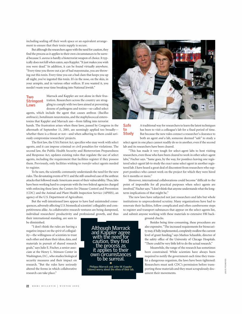
22 h h m i b u l l e t i n | w i n t e r 2 0 0 5
including sealing off their work space or an equivalent arrange-ment to ensure that their toxin supply is secure.
But although the researchers agree with the need for caution, theyfind the process as it applies to their own circumstances to be surre-al because S. aureus is hardly a bioterrorist weapon of choice. It typ-ically does not kill when eaten, says Kappler.“It just makes you wishyou were dead.” In addition, it can be found virtually anywhere.“Every time you throw out a jar of bad mayonnaise, you are throw-ing out this toxin. Every time you eat a bad clam that keeps you upall night, you’ve ingested this toxin. It’s in the nose, on the skin, inyour armpits, and in various other orifices. If you wanted it, youneedn’t waste your time breaking into National Jewish.”
Marrack and Kappler are not alone in their frus-tration. Researchers across the country are strug-gling to comply with two laws aimed at preventingdozens of pathogens and toxins—so-called select
agents, which include the agent that causes anthrax (Bacillusanthracis), botulinum neurotoxins, and the staphylococcal entero-toxins that Kappler and Marrack use—from falling into terroristhands. The frustration arises when these laws, passed by Congress in theaftermath of September 11, 2001, are seemingly applied too broadly—whether there is a threat or not—and when adhering to them could seri-ously compromise researchers’ projects.
The first law, the USA Patriot Act, specifies who may work with selectagents, and it can impose criminal or civil penalties for violations. Thesecond law, the Public Health Security and Bioterrorism Preparednessand Response Act, updates existing rules that regulate the use of selectagents, including the requirement that facilities register if they possessthem. Previously, only facilities wishing to transfer select agents neededto register.
To be sure, the scientific community understands the need for the newrules. The devastating events of 9/11 and the still-unsolved case of the anthraxattacks that followed made Americans aware of their vulnerability. Thus, labshave been working hard to cooperate with the two federal agencies chargedwith enforcing these laws: the Centers for Disease Control and Prevention(CDC) and the Animal and Plant Health Inspection Service (APHIS), anagency of the U.S. Department of Agriculture.
But the well-intentioned laws appear to have had unintended conse-quences, adversely affecting U.S. biomedical scientists’ collegiality and com-petitiveness alike. As collaborative research ventures are being dampened,individual researchers’ productivity and professional growth, and thus their international standing, are seen to be diminished.
“I don’t think the rules are having anegative impact on the spirit of collegial-ity—the willingness of scientists to trusteach other and share their ideas, data, andmaterials in pursuit of shared researchgoals,” says Julie E. Fischer, a senior asso-ciate at the Henry L. Stimson Center inWashington, D.C., who studies biologicalsecurity measures and their impact onresearch. “But the rules have seriouslyaltered the forms in which collaborativeresearch can take place.”
A traditional way for researchers to learn the latest techniqueshas been to visit a colleague’s lab for a fixed period of time.But because the new rules connect a researcher’s clearance toboth an agent and a lab, someone deemed “safe” to study a
select agent in one place cannot readily do so in another, even if the secondlab and its researchers have been cleared.
“This has made it very tough for select-agent labs to host visitingresearchers, even those who have been cleared to work in other select-agentlabs,” Fischer says. “Same goes, by the way, for postdocs leaving one regis-tered select-agent lab to study the exact same select agent in another regis-tered lab. I have heard a great deal of discontent from researchers who sup-port postdocs who cannot work on the project for which they were hiredfor 6 months or more.”
Moreover, international collaborations could become “difficult to thepoint of impossible for all practical purposes when select agents areinvolved,”Fischer says.“I don’t think that anyone understands what the long-term implications of that might be.”
The new laws have subjected not just researchers and labs but wholeinstitutions to unprecedented scrutiny. Many organizations have had torenovate their facilities, follow complicated and often cumbersome stepsto register and transport substances that appear on the select-agents list,and submit anyone working with these materials to extensive FBI back-
ground checks.Besides being time-consuming, these procedures are
also expensive.“The increased requirements for biosecuri-ty may, if fully implemented,completely swallow the currentlevel of grant funding,” says Markus Schaufele, director ofthe safety office of the University of Chicago Hospitals.“There could be very little left to do the actual research.”
Meanwhile, the range of the research has sometimesbeen constrained. While scientists have always beenrequired to notify the government each time they trans-fer a dangerous organism, the laws have been tightened:Researchers must seek CDC’s permission before trans-porting these materials and they must scrupulously doc-ument their movements.
TwoStringentLaws
Although Marrackand Kappler agree with the need forcaution, they find
the process as it applies to their
own circumstances to be surreal.
■
Philippa Marrack and John W. Kappler (above) worry about the ethos of their lab.
Safe to Study
LO
UIS
PS
IHO
YO
S

h h m i b u l l e t i n | w i n t e r 2 0 0 5 23
These requirements already have proved problematic for scientists work-ing on unexpected pathogen outbreaks. Last year, for example, researchers atthe Wisconsin State Laboratory of Hygiene found themselves stalled in theirability to quickly investigate an outbreak of monkeypox because the virus wasnot among those they listed when registering with the federal government.Clinical specimens legally could not be transferred to the lab from the placeswhere they were isolated from patients. Instead, virus samples first had to besent to the CDC, circumventing—and delaying—the state’s response.
“Such an incident leaves us wondering how a serious health crisisinvolving a select agent such as anthrax might evolve in the current regu-latory environment,” wrote R. Timothy Mulcahy, associate vice chancellorfor research policy at the University of Wisconsin–Madison, in a November2003 editorial in Science magazine.
Another constraint is researchers’need to undergo FBI screening beforebeing allowed to work with agents on the select list. This process can takeconsiderable time, as the agency is currently working its way through thou-sands of people seeking clearance, and it can wind up reducing the pool ofparticipating scientists.
Foreign students trying to come to the United States to conduct researchare having an especially tough time. “It takes our researchers longer to getinto the country because of the backlog and the increased scrutiny, and obvi-ously this slows up the research,” says Amy Wilkerson, associate vice pres-ident of research support at the Rockefeller University.
To be registered and certified, labs must undergo inspection byeither the CDC or APHIS, depending on the agents involved.Often, this is not an easy process for the researchers, who seemuch of it as nit-picking and intrusive.“If you change the floor
plan or move a piece of equipment,” says David W. Drummond, director ofthe safety department at the University of Wisconsin–Madison, “you haveto record this with the controlling agency.”
This can be especially maddening when the institution leaders believethey have already done their homework. Drummond insists that the uni-versity’s security experts scoured the facilities looking for vulnerable areasand putting new measures into place, including high-security locks and keycontrols where select agents are stored. Nevertheless, federal regulators con-tinue to review Wisconsin’s registration application, submitted more thana year ago, in March 2003. In November 2004, the university received a listof questions with a 10-day response deadline. And until that registration isapproved, he says, “there’s a cloud of uncertainty hanging over our heads.It’s difficult for researchers to do business—they have grants with deadlines,they need to show progress, and they need to hire personnel. Meanwhile,new staff can’t start work until they are cleared by the FBI as required bythe USA Patriot Act. Our researchers estimate that current procedures near-ly double the time needed to conduct their research.We believe we are com-plying with the rules and conducting research in a safe and secure manner,but we won’t know whether our procedures are adequate until we receivedecisions on our registration. We hope and trust that the federal agencieswill work together to streamline processes and reduce delays because somuch of this research is critical to national security.” [In February 2005,Wisconsin received word that its registration had been finalized.]
For its part, the CDC, unaccustomed to its new enforcement role,acknowledges that it still is finding its way.“This is new ground for us,” saysVon Roebuck, a CDC spokesman. The CDC has fully registered 313 facilitiessince November 12,2004,according to Roebuck,but there are hundreds moreto go.“We are trying to keep pace,” Roebuck says.
Is ItWorthIt?
Exempt SARS?Fearing a slowdown in whatthey described as the “aston-ishing pace” of advances, aninternational group of 13researchers met in SanFrancisco last spring at thePositive Strand RNA Virussymposium and drafted a let-ter urging the U.S.governmentnot to add the coronavirus thatcauses SARS (severe acute res-piratory syndrome) to the listof select agents. They wereconcerned that drug and vac-cine development would behurt if scientists in the UnitedStates were burdened withrequirements that could stallinternational scientific collab-oration.
“The huge success in theidentification and characteri-zation of coronavirus as the
etiologic agent of SARSrequired unprecedented glob-al cooperation,” says KathrynV. Holmes, a coronavirusexpert at the University ofColorado Health SciencesCenter.“If select-agent status isgiven to this virus, it willrestrict the action of U.S. sci-entists with others and delaythe free and easy collaborationthat has characterized thislandmark research so far.Vaccine and drug therapy arecoming very quickly now—wethink it would be a bad time toinhibit research.”
Officials at the U.S. feder-al agencies responsible for theselect-agents list, including theCDC, have been discussingthe possibility of adding theSARS coronavirus to the list;at this time, however, no finaldecision has been made,according to CDC spokesmanVon Roebuck.

24 h h m i b u l l e t i n | w i n t e r 2 0 0 5
are seriously considering it, just to get outfrom under these regulations.”
Similarly, other scientists are wonderingwhether the bureaucratic hassle is worth it.“I’ve heard numerous stories of facilities sim-ply getting rid of the select agent rather thango through the extensive registration andinspection process,” says Jay T. Skarda, man-ager of safety at National Jewish.
Karen VanDusen, director of environ-mental health and safety at the University ofWashington in Seattle, has seen a similar reac-tion among researchers. “We sit down withresearchers and ask, Are you aware of the
requirements you will have to meet in order to do this kind of research? Wetalk to them about security additions to their labs, hiring practices, back-ground checks—and some have said, ‘I don’t think I’m going to bother.’”
Of course, the scientific community does not wantto compromise national security, but what is par-ticularly troubling to many is that their research isbeing compromised without necessarily improving
national security. They argue that the regulations fail to distinguish amongagents as to the degree of risk. Instead of allowing institutions to make per-formance-based decisions, scientists must live with a one-size-fits-all setof regulations.
“I’m not saying that rules aren’t needed,” notes W. Emmett Barkley,hhmi’s director of the office of laboratory safety.“I am saying there is roomfor developing different levels of control, depending on the risk.”Not all lab-oratory procedures merit the extraordinary security measures beingimposed at this time. And in some cases, these measures are not only dis-ruptive for the researchers, but could constitute, in effect, a Maginot Linefor the country. “There is a great deal of fear that the source of materials[used in a future bioterrorism attack] will be obtained from laboratories,”says Barkley, “when in fact they are virtually anywhere you’d want to findthem. We are in a situation where the fear and the security concerns are
themselves controlling the debate on what is risky and what is notrisky,” as opposed to a scientific assessment of risk.
Some researchers are concerned that the current intense andinflexible regulatory climate will lead to disturbing changes in thevery nature of biological research and to its core values of conduct.“We are in the beginning of a massive cultural shift,”says VanDusen.“We have to tell researchers, ‘Don’t leave your lab doors open. It’snot OK to come and go.’ It just can’t be that way anymore.”
Barkley, too, sees a cultural shift.“The most successful labs havethis wonderful desire to be open, to be collaborative, to share mate-rials and stimulate new directions of research,” he says. “And nowthey are being constrained.”
Yet this system need not change, at least not by much. TheStimson Center’s Fischer, for one, hopes the federal governmentwill engage with the research and safety communities in a frankdialogue about the real costs and benefits of the select-agent rules.“This is critical if we are to achieve something more than a falsesense of security,” she says, “and avoiding hampering those whostrive to better understand diseases, whether they occur naturallyor deliberately.”
Not everybody in the scientific community, how-ever, subscribes to the view that the rules are havinga chilling impact on the conduct of scientific research.
“Concerns that the select agent rule would pre-vent laboratories and researchers from performingselect-agents research have proved unfounded—utter-ly unfounded,” says Richard H. Ebright, an hhmiinvestigator at the Waksman Institute at Rutgers, theState University of New Jersey. “The numbers speakfor themselves. The number of select-agents grants,laboratories, and researchers has increased by morethan a factor of 10.”
“As of November 2004, more than 300 laborato-ries and more than 12,000 persons had been registeredfor access to select agents. These numbers are disturbingly high. Most cit-izens would be astonished—and alarmed—to learn that more than 12,000persons had access to—and ability to release, distribute, or sell—fully vir-ulent live bioweapons agents.”
For clearance, much depends on the results of a rigorous facility inspec-tion and the kind of changes needed if some systems are deemed insecure.Thisprocess at Rockefeller, according to Wilkerson, went relatively smoothly. TheCDC site-visit team was “very professional and collegial,”she says.“They werehere for a day and a half, and things went pretty much as expected. We weretold to expect a report in 4 to 6 weeks, and there were no surprises.”
Marrack and Kappler at National Jewish, however, had a very different experience. Told they could use no more than 5 milligrams ofstaph-produced toxins per investigator at any one time unless they rebuilttheir labs to be highly secured, the two researchers decided instead toreduce the amount of toxin they store to below federal limits. To com-ply, they had to destroy 27 milligrams of toxin worth $5,000. One sideeffect, Marrack says, is that the lab will have to time its experiments care-fully to be sure it does not run out of toxins.
Ultimately, however, Kappler says, “we probably will look for anotherway to do these experiments. There are other proteins around that aren’ton the list that work similarly. But it means having to retool our experiments,and sometimes start from the beginning. That would be a big deal, but we
Room forImprovement
H
“Concerns that theselect agent rulewould prevent
researchers fromperforming select-
agents research haveproved unfounded—utterly unfounded.”
■
Richard Ebright (below) doesn’t believe new regulations have had a chilling impact.
JE
NN
IFE
R A
LTM
AN

R E S E A R C H N E W S
Student Contributes Bigto Anti-Cancer ResearchThe HHMI/NIH Cloister Program gives another young physician-scientist his “defining opportunity.”
DO
UG
KN
UT
SO
N
Wade Alleman steeled himself for agrilling. He had just presented hisresearch findings to members of the
urologic oncology branch at the National Can-cer Institute (NCI) at the National Institutes ofHealth (NIH). But the traditional battery oftough questions never came.
Instead, W. Marston Linehan, the branchchief, leaned back in his chair during that 2003meeting and declared: “How did you get all thiswork done in one year? And how can we bringit to clinical trial?”
It is rare for a young and relatively inexperi-enced scientist, let alone a medical student, tohave a scant year’s worth of research translate intoa clinical trial. But that could be the result of Alle-man’s work. He spent his year as an hhmi/ NIHresearch scholar at NCI studying a new use—treating kidney cancer—for an old drug.
“Wade worked very hard and was deter-mined to see this project through,” says Line-han. “He started with a project that we thoughthad promise; however, it was his determinationand single-minded focus that brought it tocompletion.”
Now back at the Mayo Clinic College ofMedicine and expecting to graduate this May,Alleman, 28, regards his time at the hhmi/NIHResearch Scholars Program, also known as theCloister Program, as “the defining opportunityof my medical school experience.” The program,established in 1985, gives outstanding U.S. med-ical students an opportunity to receive researchtraining at NIH under the direct mentoring ofsenior NIH scientists.
aiming for the right targetsAlleman tested the drug in six hypermethylat-ed clear cell kidney cancer cell lines, with dra-matic results: the tumor-suppressor-geneexpression was restored. And in mice, the treat-ment made the tumors shrink or stop growing.The work was published in the October 15,
2004, issue of Clinical Cancer Research.“It was tested 20 years ago in kidney cancer
and declared ineffective—because they tested it inall kidney cancers, and it was only effective inabout 19 percent of patients,” says Alle-man.“Yet this was exactly what you wouldhave suspected if you’d isolated that par-ticular subpopulation of kidney cancers.”
“This is about as pure an anti-cancerapproach as I could imagine,” says Line-han. “You could argue that 19 percent is‘only’ 19 percent, but if you were one ofthose 19 percent and this approachworked…well, miracle!” It could even beconsidered miraculous, or at least theaccomplishment of a scientist’s career, bythe research community itself. “If duringthis lifetime I am part of something thatcures 19 percent of people with a certaindisease,” Linehan adds, “my life will havebeen worthwhile.”
down a different pathAlleman was raised in Elko, Nevada. Hisdad was the local optometrist, known byall; his mother was a homemaker. “It wasin the middle of nowhere, but a goodplace to grow up,” he recalls.
He had long thought about becoming a doc-tor. “In Elko, doctors were very much revered—and there was a shortage of them,” Alleman says.“At one time there was a ‘bounty’ of $1,000 forany family that could bring a doctor to town. SoI grew up knowing there was a need.”
He became hooked on medicine after he metFrederick Walker, a surgeon from Forest Hill,Maryland, who was a guest lecturer at the Nation-al Youth Science Camp in Bartow, West Virginia,where Alleman worked for several summers. Theytalked at length, and Alleman eventually spent asummer working directly for Walker, thrilled that“I got to scrub on a couple of cases.”
Some of the following years were marked
h h m i b u l l e t i n | w i n t e r 2 0 0 5 25
by “character growth” experiences—workingnights in a slaughterhouse to earn money formedical school, for example, and two yearstraipsing through the rainy jungles ofGuatemala as a missionary of the Church ofJesus Christ of Latter-day Saints.
Earlier in his medical training, Allemanleaned toward a specialty in urology. But expe-riences at NIH helped him clarify that anotherfield, radiology, would be “a better fit for me asa physician.” During his final months in theNIH lab, Alleman became fascinated by thefield of molecular imaging. “The ability to drawconclusions based on the visualization of geneexpression, rather than merely gross tumor size,was exhilarating, and I wanted to learn more,”
Alleman says. When he returned to medicalschool after his year in the Cloister Programand learned more about the role of medicalimaging in clinical practice, he became evenmore interested in the field. “By the time I com-pleted my radiology elective,” he says, “I knewthis was the specialty for me.” He plans to com-bine bench science with clinical practice, spe-cializing in radiology.
Regardless of Alleman’s ultimate choice offield, however, impressed colleagues at NIH pre-dict success. He worked “extremely well withboth our clinical as well as basic research staff,”says Linehan. “He has a great career ahead as aphysician-scientist.” —MARLENE CIMONS
Medical student Wade Alleman’s research led a senior
scientist to ask: "How can we bring it to clinical trial?"

26 h h m i b u l l e t i n | w i n t e r 2 0 0 5
the night before having dental surgery in 1998,a71-year-old Canadian woman wasgiven antibiotics to prevent infection.The operation on her teeth went well,but a fewdays later she developed diarrhea so severe that she wentinto shock and was rushed to the hospital.Tests showed she hadbeen hit with toxins produced by Clostridium difficile, a gen-erally mild bug that resides naturally in the intestinal tract.Usually kept in check by the body’s “good”bacteria, C. difficileposes little threat unless something—like a course of antibi-otics—kills off some of those protective bacteria.
After two months of intensive treatment and physicalrehabilitation, the dental patient survived. Other people havenot been so lucky. In a single Quebec hospital over the last 18months, 100 patients died of C. difficile infection. Fatalities ofthis sort have been increasing rapidly not only in the provinceof Quebec, whose health minister suggested that “enthusias-
tic” prescribing of antibi-otics might have caused the
outbreaks, but in other parts ofCanada and the United States as well.
We each carry two to five pounds of livebacteria in our bodies. Some, like C. difficile, are poten-
tially harmful. Many bacteria, however, are quite useful—souseful, in fact, that we could not live without them.
Until recently, scientific research has focused on fighting“bad” bacteria—the ones that cause cholera, scarlet fever,typhoid, tuberculosis, and other major infectious diseases.Scientists pretty much ignored the good bacteria, which oftenoutnumber the bad ones.
In the past few years, however, researchers have begun torecognize the enormous contributions made by this friend-ly “bacterial nation,” as Jeffrey I. Gordon, director of theCenter for Genome Sciences at Washington University Schoolof Medicine in St. Louis, calls it. Trillions upon trillions of
TheFriendly Bacteria Within
Us
While we tend to think of bacteria as harmful, we all carry plenty of microbes that work to the good. Can we
use them to prevent or treat diseases? By Maya Pines
Beneficial bacteria include (top)Lactococcus lactis, as well as
(right) Lactobacillus bulgaricus(blue), Streptococcus
thermophilus (orange), and amember of the Bifidobacteriumfamily (magenta), all found inyogurt and cheese. Normally
benign, Bacteroides fragilis (bot-tom) is an intestinal microbe
that can wreak havoc under cer-tain conditions, such as post-
surgery. (The bacteria in thesescanning electron micrographs
have been color enhanced.)
DR
. G
AR
Y G
AU
GL
ER
/PH
OT
O R
ES
EA
RC
HE
RS
, IN
C.
(TO
P),
SC
IMA
T/P
HO
TO
RE
SE
AR
CH
ER
S,
INC
. (R
IGH
T),
S
CIE
NC
E P
HO
TO
LIB
RA
RY
, LT
D./
PH
OT
OT
AK
E (
BO
TT
OM
).

28 h h m i b u l l e t i n | w i n t e r 2 0 0 5
microbes, representing some 1,000 species, are packed within us, especial-ly in our guts. A single milliliter of the colon’s contents might harbor 100billion of them. This “nation” functions like a kind of internal organ, saysGordon, affecting our well-being.
T H E P R O M I S E O F P R O B I O T I C S ?
Using good bacteria to promote health—a practice sometimes called pro-biotics—has a long history, but it never quite became an accepted therapy.About a century ago Elie Metchnikoff, director of the Pasteur Institute inParis, France, started a yogurt craze when he announced that Bulgarian peas-ants who ate yogurt regularly tended to live to ripe old ages; yogurt containslive cultures of lactobacilli, one of the better-known strains of good bacte-ria. Several other substances that supposedly contained beneficial bacteriawere also used to treat infections in the gut or vagina. This practice endedaround the time of World War II, when the newly discovered, often life-sav-ing, antibiotics proved to work more rapidly and effectively.
Now, however,“We are being forced to look at alternatives to antibioticsto combat the ever-increasing number of infections that occur because of exces-sive use of antibiotics,” Christopher J. Bulpitt and his colleagues at London’sImperial College School of Medicine wrote in the British Medical Journal in2002. Severe diarrhea, for instance, often results from treatment with antibi-otics, which wipe out good bacteria along with the bad. Could this side effectbe prevented by maintaining enough good bacteria in the patients’ intestinesto act as guardians?
To find out, Bulpitt’s team analyzed nine randomized, double-blind,placebo-controlled trials in which all patients had been treated with antibi-otics, but some also received various combinations of microbes that werebelieved to be good, while others received placebos. The team concluded that“probiotics are a possible solution,” but only for preventing antibiotic-asso-
ciated diarrhea. They found little support for using probiotics as a cure.One way to learn which bacteria are most useful in particular cir-
cumstances is to let nature be your guide, as a group of Swedish researchersdid when they concocted a highly unusual nasal spray. In 2001, KristianRoos and his associates at the Lundby Hospital in Gothenburg were seek-ing new treatments for infants and toddlers who have repeated bouts ofear infections (otitis media). They targeted the one child in 20 who comesdown with these painful infections frequently—up to six times a year—despite repeated treatment with antibiotics, to which the child becomesincreasingly resistant. So they started out by “harvesting”the microbes theyfound living in the eustachian tubes (which connect the nose and middleear) of healthy children at a daycare center. Among these microorganismsthey identified some 800 different strains of α-hemolytic streptococci. Nextthey tested each strain’s ability to stop the growth of otitis-causing bacte-ria in the lab. Finally, they chose the five most active strains, which they putinto the nasal spray.
At the end of three months, 42 percent of the children who had beengiven this bacterial spray remained free from otitis, while only 22 percentof those who received a placebo escaped new ear infections. The scientistsconcluded that “recolonization” with selected bacteria does protect againstrecurrent attacks of otitis, at least to some extent.
T H I N L I N E
The British and Swedish efforts, and other clinical studies of this sort, gen-erally paint an optimistic picture of probiotics. But many scientists remainskeptical, and few such treatments are currently in use. The real hurdle isstill our lack of precise knowledge. What proportion of the bacteria in ourbodies is good? How many are pathogenic? How many good bacteriasometimes become bad, and vice versa? Are many of them simply strad-dling the fence?
Nobody knows. An adult human has about 10 times more microbialcells than human cells, so “based on cell number, each of us is 90 percentmicrobial and 10 percent human,”says Gordon.“The genomes of our gutmicrobes probably contain 100 times more genes than our own genome,providing us with traits we haven’t needed to develop on our own.”Yet atleast half of these bacteria cannot be grown outside the gut because “wehaven’t learned how to reproduce their normal conditions in the lab,” hesays, “so we don’t have an accurate view of them.” Together with someacquired viruses,yeast cells,archaea (single-celled microorganisms that livein geysers and other extreme environments),and occasional parasites, thebacteria form “a constantly open ecosystem,” Gordon says.
Some of the good bacteria have a symbiotic relationship with our intes-tines (they help us and we help them, usually by providing nutrients). Othershave a commensal relationship (one partner benefits without harming theother). But in our guts, nothing is permanent. Bacteria take on shifting rolesas they encounter changing circumstances. “If the formidable barrier pro-
Good bacteria can break down certain foods, such as plant starches, that wecannot digest on our own. “This enables us to extract more energy from what weconsume,” says Jeffrey I. Gordon, director of the Center for Genome Sciences atWashington University School of Medicine in St. Louis. (Similarly, cows candigest cellulose thanks to the good bacteria that live in their rumens.)
Good bacteria promote the storage of energy as fat. According to Gordon,this raises the possibility that “an individual’s predisposition to obesity orleanness may be partly determined by the composition of the microbes livingin the gut.”
Good bacteria help shape our postnatal development. For example, they helpto form our intestinal blood vessels, through which we absorb nutrients.
Good bacteria synthesize vitamin K and other vitamins that we cannotgenerate on our own. They break down carcinogens. They also may influencethe metabolism of drugs.
Good bacteria increase the rate at which the cells of the intestinal lining renewthemselves, ridding us of damaged cells that could bring on gastrointestinal cancer.
The good bacteria that infants acquire from their mothers and from the generalenvironment at birth “educate the newborns’ immune systems,” says Gordon. “Thisappears to reduce allergic responses.”
Each human carries a different set of bacteria, and its composition varies alongthe length of the gut. Some of these bacteria are permanent residents; othersare transient “tourists,” just passing through.
What Good Bacteria Do Some things we know about good bacteria, besides the generalization thatthey help to counteract pathogens:
“The genomes of our gut microbesprobably contain 100 times more
genes than our own genome,providing us with traits we haven’t
needed to develop on our own.”—JEFFREY I. GORDON

h h m i b u l l e t i n | w i n t e r 2 0 0 5 29
duced by symbiotic bacteria is destroyed,”notes Gordon,“somepreviously minor bacteria can expand and produce disease.There’s also a lot of horizontal gene transfer (from one bac-terium to another), creating new strains and spreading antibi-otic resistance. It’s very dynamic!”
“Take Bacteroides fragilis, for instance. Usually it’s fairlyinnocent,” he says. “But after stomach surgery or some otherinsults, it can cause abscesses. A researcher at Harvard, Laurie F. Comstock,recently discovered that this happens when the bacterium’s outer capsulechanges, making it more dangerous.”
Similarly, up to two-thirds of the world’s population carries Helicobacterpylori, and in most people it does no harm. In 10 percent of infected peo-ple, however, it leads to stomach ulcers or cancer (which may be either gas-tric lymphoma or adenocarcinoma of the stomach).
“The question one should ask is not how many bacteria in our guts arepathogenic,”says Gordon,“but how many of them have pathogenic potential.”
O R I G I N S O F V I R U L E N C E
Bacterial virulence seems to involve what B. Brett Finlay, an hhmi inter-national research scholar at the University of British Columbia inVancouver, calls a kind of “cross-talk” between bacteria and their hosts.
Bacteria appeared in the world long before humans did. After we cameon the scene, some bacteria “co-evolved”with us so they could take advantageof what is for them a wonderful environment—the human gut,where so manynutrients are concentrated. This meant the bacteria had to learn how to over-come the many physical,cellular,and molecular barriers the human body pre-sented, wrote Howard Ochman, a biochemist at the University of Arizona, ina recent issue of Science. They may have added or subtracted certain genes.
Natural selection favored those bacteria that made the most effectivechanges. This happened “regardless of whether the ultimate outcome of theinteraction is harmful, benign, or beneficial to the host,”said Ochman.“Onlyfrom the host’s perspective are these distinctions crucial.”
The first job of infectious bacteria is to attach themselves to specificreceptors on human cells, says Finlay. And sometimes the host cell collab-orates.As his team discovered while studying diarrhea-causing enteropath-
ogenic Escherichia coli (EPEC), these bacteria use two differ-ent kinds of adhesive molecules to latch onto human cells.The first molecule somehow “rings a doorbell” on the hostcell, telling it to produce a sort of pedestal, which almostimmediately grows out of the cell surface. This pedestal thenenables EPEC to attach itself securely to the cell with its sec-ond adhesive molecule.
Although most diseases are caused by the initial adherence of bacteriato cells, says Finlay, no drugs are yet available to derail this process. If sci-entists learn to block bacterial attachment, they may be able to prevent orstop infections, he suggests.
But why these particular bacteria tried to adhere to human cells in thefirst place remains a mystery.Were they previously good bacteria that some-how turned bad?
Eduardo A.Groisman,an hhmi investigator at Washington University’sSchool of Medicine in St. Louis, first tackled this problem 10 years ago in anarticle,“How to become a pathogen,”that he and Howard Ochman publishedin Trends in Microbiology. The idea was to find the genes responsible for pro-ducing virulence, focusing on differences between the activities of nonpath-ogenic E. coli bacteria and a strain of Salmonella that can cause typhoid fever.With the tools available at the time, and more recently with the help ofsequenced microbe genomes, the two scientists worked to identify “patho-genicity islands”—groups of genes that are found only in pathogens and thatcontribute to disease.
In some cases, however, pathogenic and benign bacteria have almostthe same genes, Groisman observes. One possible explanation for their dif-ferent behavior is that these genes are regulated in alternative ways.“We’renow studying how this differential regulation may affect virulence,”he says.
A N I M A L M O D E L S
The complexity of the interactions between the huge bacterial nation andthe human gut “defies imagination,”says Gordon. So his lab created animalmodels that could be analyzed more easily. All the animals—mice orzebrafish—were raised under germ-free conditions; for comparison, halfwere later “colonized” with specific strains of bacteria.
Jeffrey I. Gordon (left) andEduardo A. Groisman, both atWashington University School
of Medicine in St. Louis, areamong investigators whostudy bacteria—both the“good” and the bad—that
comprise what Gordon callsthe “bacterial nation.”
PE
TE
R N
EW
CO
MB
(2
)

30 h h m i b u l l e t i n | w i n t e r 2 0 0 5
For the mouse experiments,Gordon chose a strain of Bacteroides thetaio-taomicron, good bacteria frequently found in the guts of both mice andhumans; its main job is to provide the enzymes needed to process certain car-bohydrates in plants.The B. thetaiotaomicron genome was sequenced in 2003and the proteins it produces have been sorted out,making it possible to exam-ine the microbe’s activities with some of the newest tools of genetics. TheGordon team has shown, for instance, that B. thetaiotaomicron stimulates pro-duction of an antibiotic protein that can kill infectious microbes such asListeria monocytogenes, which causes food-borne gastroenteritis. Another ofthe researchers’ findings is that B. thetaiotaomicron promotes the develop-ment of small “networks of branched, interconnected blood vessels” in new-borns, the scientists reported in the Proceedings of the National Academy ofSciences. This capillary network does not grow properly in germ-free mice.
Zebrafish offer several advantages to researchers. Because these smallfish develop rapidly (the larvae hatch within three days of fertilization) andremain transparent through early adulthood, it is possible to observe theembryos’ growing digestive tracts and their resident bacteria. Using DNAmicroarrays, Gordon’s team recently examined the genes that were activatedin zebrafish intestines after exposure to specific good bacteria. They foundthat 59 zebrafish genes responded to these bacteria in exactly the same wayas do the corresponding genes in mice, even though the two species divergedmillions of years ago. This implies that responses to these bacteria go backvery far in evolution; most likely they were critical not only to mice andzebrafish but also to many other species.
O V E R S O L D T H E R A P I E S ?
While researchers work step by step, accumulating information with the aidof animal models, the marketplace apparently is not waiting for final results.Various brands of probiotic food supplements are already being sold aroundthe world with promises of fabulous benefits for cats, dogs, birds, horses,and farm animals: “improved growth,” “better health,” “establishment ofbeneficial gut microflora,” “better utilization of food,” “reduced intestinalupsets,” and “increased resistance to infections,” which should reduce the
need to treat livestock with antibiotics.Chickens in particular are frequently treated with Preempt, a product
developed with help from U.S. Department of Agriculture scientists, whichcontains 29 kinds of good bacteria found in healthy chickens.At least 10 per-cent of chickens are infected with Salmonella bacteria, a leading cause offood-borne illnesses in humans. The idea is to spray newly hatched chickswith Preempt so that when they peck at their wet feathers they will swal-low its bacteria. The reasoning is that the product’s good bacteria will growin the chicks’ intestines, forming a protective barrier that cannot bebreached.As a result, any ingested Salmonella will be unable to attach them-selves to the chicks’ intestines and will be forced out of the animals’ bod-ies. This model seems to work to some extent.
When it comes to humans, however, the use of probiotics remains morecontroversial. There are strong commercial interests in its favor—yogurtmarketed as “probiotic” is one example—but inconsistent experimentalresults. Much of the published research consists of reports on only a fewpatients, and many of these reports are contradictory. Skeptical scientistshave called probiotics everything from “conbiotics” to “snake oil.” Even themanufacturers of probiotic compounds agree that more precise informa-tion is needed.
According to the Harvard Health Letter of March 2002,“Probiotics havebeen oversold. The claims are seductive: pills, powders, and solutions con-taining ‘friendly’bacteria will boost the immune system, prevent cancer, andperform assorted other health miracles. … But that doesn’t mean it’s basedupon total fiction. … The evidence suggests that probiotic therapy couldbe useful someday as a form of preventive medicine—and not just for dis-eases affecting the gut.”
As Gordon puts it,“Bacteria have learned to manipulate our biology inmany ways that benefit themselves and us. We now have the tools to identi-fy the pathways through which they operate, as well as the chemicals theysynthesize.” This information could lead to new ways of diagnosing, treat-ing, and ultimately preventing a variety of diseases. “Bacteria are fabulousteachers,” says Gordon.“They are pointing the way.” H
HHMI investigator Ruslan Medzhitov, an immunolo-
gist at Yale University School of Medicine, recently
discovered an entirely new role for our resident bac-
teria: They help protect us from radiation and poisons.
As he explained in a paper in Cell published July 23,
2004, he came to this conclusion indirectly while
studying the cells of the innate immune system, our
first line of defense against pathogens. Medzhitov
expected that the receptors on these cells would
detect only the bad microbes. “We thought these
receptors would simply ignore any components of
good bacteria,” he says. “But on the contrary, they
recognized both good and bad.” As he later found, it
was absolutely essential for these immune-system
cells to recognize good bacteria to stimulate the
intestinal tissue’s systems of maintenance and repair.
In one experiment, Medzhitov’s team worked with
three groups of mice that were exposed to fairly strong
radiation, as much as might be used to kill tumor cells.
One group, which had normal bacterial “flora” in their
guts, survived the radiation with relative ease. When
the team irradiated a second group of mice, whose
colons had been deprived of normal bacteria by antibi-
otics, they all died. Then the researchers tried to pre-
vent such deaths by giving a third group of mice—sim-
ilarly deprived of normal bacteria—a chance to drink
water that had been laced with some components of
good bacteria. Although they received the same dose
of radiation as the second group during that time (one
week), they all survived. “The cells of the mice’s innate
immune systems recognized the bacterial components
they had swallowed,” says Medzhitov. “That activated
their immune systems and protected the mice from
some of the damage they would otherwise have suf-
fered.” The team obtained similar results in mice
exposed to toxic chemicals.
This finding raises the hope that human cancer
patients may be protected in similar ways. Restor-
ing the normal supply of good bacteria in their intes-
tines might help them avoid a possible side effect of
radiation or chemotherapy: an injured and bleeding
gut, which can be fatal.
A Palliative for Chemo?
Ruslan Medzhitov
AM
Y E
TR
A

h h m i b u l l e t i n | w i n t e r 2 0 0 5 31
Come to the northern virginia construction site at hhmi’s Janelia FarmResearch Campus on any clear morning andyou’ll see an aerial ballet. Enormous sheets ofglass glint in the sun as they rise from the ground and sweep through the air. Grasped by a dozen hugesuction cups, the flying glass swings gracefully on thick cables suspended from the arm of a toweringred crane. Gliding up and then gently down, the panes float into the outstretched hands of the hard-hatted men waiting on a deck of the massive structure,who situate each window into its prescribed place.
This dance will be repeated many times over in the coming months as hundreds of pieces of glassare set into place, bringing alive the ingenious architectural features of the Janelia Farm landscape build-ing that will house the offices, labs, and support facilities at the heart of the biomedical research cen-ter. Beyond the expected benefit of providing natural light and visibility, the glass will play an inte-gral, load-bearing role in the structure. And the glass will also serve a higher function. Metaphorically,it embodies the philosophy of transparent, collaborative science that will take place at Janelia Farm.
■ Janelia Farm | Update
■ A piece of roof glass is flown in by crane andcable to become part of this gradually mate-rializing glass corridor. The corridor’s wallpanels, which weigh on average 1,400pounds apiece, are 10.6 feet high and vary inwidth from 10 to 12 feet, depending onwhere they fit within the building’s curve.
At Janelia Farm,the walls ofwindows have a structure andsociology alltheir own.
Flying GlassP H OTO G RA P H S B Y PA U L F E T T E R S

32 h h m i b u l l e t i n | w i n t e r 2 0 0 5
The structural glass will be put to use in a number ofways, most notably the transparent corridor that runs thelength of the building along the undulant face of both thesecond and third levels.Though certainly attractive, the col-umn-free corridor is not intended to be merely ornamen-tal. It is a conscious design element critical to the socialdynamic planned for Janelia Farm,says Robert H.McGhee,institute architect and senior facilities officer at hhmi.“Theglass corridor lies between the labs and the offices, so it helpsconnect them.” Traveling the corridors, McGhee says,researchers will get “a real sense of what everyone is doing,which is part of the collaborative notion of this building.”
Moreover, while the offices and labs of most researchbuildings are not situated with outside views in mind, andoften have none whatsoever, the building will allow every-one working there not only an unbroken vista of the out-doors but also the ability to go outdoors. The corridors andoffice pods will open onto grassy, meadow-like expanses—roof gardens that top the levels below.
“The building is designed to be different,”says McGhee.“The office groups, or pods, are arranged like small housesthat people work in.And you walk from those into a pantry-like interaction area to get into the labs.So you can’t help butrun into people, and you can’t help but see people as youwork. That’s really the key to the glass corridors, in the soci-ology and in the way the building is going to function.”
In addition to constituting the corridors, the glass will beused, together with stainless steel, to create two arching fea-ture stairways that connect all three floors.The glass will alsosurround recessed interior gardens on the ground floor, aswell as the entryway,lobby,and pergola-inspired dining room.
The unusual curvature of the building,along with otherarchitectural features, has necessitated customizedapproaches in engineering and materials design.The fittingsthat hold the corridors’glass slabs together, for example,andthe hollow aluminum rafters that stealthily accommodatethe sprinkler system, electrical wiring, and heating and airconditioning needs for the glass-roofed dining area werecustom designed and built to specification.Such innovativedesign solutions are causing a stir in the local world of com-mercial builders.“We haven’t seen anything like this beforein Loudoun County, in terms of the complex integration oflife safety and other systems,”says Brian Knode, senior proj-ect director with the Mark Winkler Company,which is over-seeing the Janelia Farm project. Knode is working closelywith county officials to ensure that the engineering anddesign meet all regulatory codes.
Going to such great lengths to bring architect RafaelViñoly’s design to reality is important, says McGhee.“Wewouldn’t go through this if it didn’t reinforce a significantprogram element of the building,”which is to facilitate col-laborative research broadly across disciplines. In otherwords, the delicate ballet of flying glass contributes to abold vision of what research can and should be, a visionplayed out at the intersection of form and function.
—MARY BETH GARDINER
■ A computer rendering (right) gives an indication of how the Janelia Farm ResearchCampus would appear from an aerialvantage point. The curvature of the building’sface dictates a radial design for the laborato-ries, where one wall is part of a curving glasscorridor. Because of the unusual, non-angulardesign, the architects and contractors collab-orated to create 3-dimensional models toensure that all geometries were correct.
■ Roof panels, seen below, are solar-coated tomodulate temperature extremes and reduceglare, and will be outfitted with a firesprinkler system as well as gaskets anddrains to handle rain and condensation. Uponcompletion, each glass corridor (far right)will approach 1,000 feet in length. Should aglass slab need to be replaced, a motorizedcart has been adapted to trundle the heavypanels up and down the corridor.

h h m i b u l l e t i n | w i n t e r 2 0 0 5 33
Manufactured in Belgium by Saint-Gobain Glass, the strong, resilient, and unusu-
ally clear glass used in the Janelia Farm landscape building was fabricated in one
large factory run. Because structural glass is rarely called for, Saint-Gobain makes
production runs only twice a year. In this case, the request was for a lot of glass.
“As volume of load-bearing structural glass goes, this will be the largest such
glass installation in the world, as far as I know,” says Charles Blomberg, the archi-
tect in building-designer Rafael Viñoly’s firm who is responsible for the building’s
“skin.” Blomberg has worked with Viñoly on a number of glass-dense architectural
projects around the world.
Manufacturers began adding iron to glass in the 1950s to give windows a
green tint (too many people were walking into them) and to make the glass flow
better during production. Iron is taken out of the formula for glass that needs to
be very clear, such as that used in museums and jewelry display cases—and in
the glass used at Janelia Farm. Laminating together multiple panels of low-iron
glass creates exceptional strength without a loss of clarity, says Blomberg.
The lamination, solar coating, and other assembly processes required to
ready the glass for its particular use in the Janelia Farm landscape building took
place at multiple sites in Europe, Canada, and the United States. Nine months of
exhaustive performance testing at a site in Pennsylvania assured that the glass
is suitable for load-bearing use and that the laminated slabs are able to withstand
extremes of factors such as wind, temperature, moisture, air pressure, and impact.
In addition to weight-bearing tests designed to mimic heavy loads from snow
and wind, reports Blomberg, there was a series of movement tests, where the
mock-up corridor assembly was strongly rocked to simulate severe building
movement. Thermal cycling tests evaluated the capacity of the glass to withstand
extreme differences between outdoor and indoor temperatures. To test how the
glass and joints might stand up to varying wind and air pressures, the mock-up
was placed inside a hydrostatic chamber equipped with devices to either push air
in or suck it out. “The pressure inward was equivalent to a 110-mph wind,” says
Blomberg. “And the suction test was actually more onerous than anything the build-
ing would experience in real life.”
In devising the tests, the team of designers and contractors imagined all man-
ner of worst-case scenarios. One concern was the possibility of a heavy lab cart rolling
into the glass. So they created a 100-pound concrete-filled steel cylinder six inches
in diameter and hung it from a pendulum. They drew the chunk back as far as pos-
sible and let it fly at the glass corridor mock-up. “The force of the blow was some-
where around 400 ft/lbs,” says Blomberg, “The whole corridor assembly shook, but
the glass wasn’t fazed.”
To ensure absolute confidence, the team pushed the glass beyond specified
requirements in most of their tests. They even broke the glass, to see what the
overall impact would be when different layers were broken and under different
loads. “We took it to the next level, what is called the ‘failsafe mode,’” says Chris
Fiato of Enclos Corporation, the specialty glass subcontractor working on the proj-
ect. “We wanted to know if we could ever get a catastrophic or progressive fail-
ure. We never did.” — M.B.G.
Glass Passes Muster

34 h h m i b u l l e t i n | w i n t e r 2 0 0 5
N E W S & N O T E S
Researchers have discovered a critical pro-tein that regulates the growth and activa-tion of neural connections in the brain.
The protein—called dendrite arborization andsynapse maturation 1, or Dasm1—functions inthe developing brain, where it controls thesprouting of new dendritic connections andstimulates existing synaptic connections amongimmature neurons. Dasm1 is potentially active
in the mature brain as well, where it may play arole in memory formation.
The researchers—hhmi investigators YuhNung Jan and Lily Jan, lead author Song-HaiShi, and colleagues, all at the University of Cal-ifornia, San Francisco—published their find-ings in two papers in the September 7, 2004,issue of the Proceedings of the National Acade-my of Sciences.
Dendrites are tree-like filaments that deliv-er “input” information to the nerve cell body.Dendritic spines are mushroom-shaped protu-berances that extend from the surface of den-drites and receive chemical signals emitted byneighboring neurons. In response, the dendritestrigger an electrical impulse from the cell bodyand down the cable-like axon, which then pass-es its message to the dendrites of other neuronsin the form of neurotransmitters. Growth ofnew dendrites therefore can increase the num-ber of connections between neurons, andchanges in the strength of the signals allow thebrain to create memories.
In exploring the growth and developmentof dendritic spines, the Jan team first identifieda gene in the fruit fly Drosophila that appears toplay a role in “arborization” (dendrite growth).Then, by comparing the fruit fly gene withdatabases of vertebrate genomes, they identi-fied a homologous gene in mice, which theynamed Dasm1.
Initial studies revealed that the gene washighly expressed in the brains of embryonicmice. “When we used antibody markers to lookat the distribution of the protein, we saw it pri-marily in the dendrites, with very little in theaxons,” says Yuh Nung Jan. “If you look at areasof the hippocampus rich in dendrites, they justlight up, whereas in axonal areas there is very lit-tle evidence for the presence of this protein.”
Consequently, when the researchers blockedthe activity of the version of the Dasm1 genefound in rats, they found dendrite growth to bedrastically reduced in cultured brain cells.
They also studied the effects of the Dasm1protein on the maturation of neuronal connec-tions, or synapses. Newly formed, immaturesynapses are silent, meaning they lack a type ofcellular structure, known as AMPA (α-amino-3-hydroxy-5-methyl-4-isoxazolepropionicacid) receptors, which receive neurotransmittermolecules. However, these neurons have otherreceptors, called NMDA (N-methyl-d-aspar-tate) receptors, which are associated with long-term changes in the strength of neuronal sig-naling. Although dendrites (which influencethe development of neuronal connections) doacquire active AMPA receptors during matura-tion, it was not known whether this processdepends on Dasm1.
Helping the Brain toMake ConnectionsNewly discovered protein awakens, maintains neural connections.
Yuh Nung Jan and his wife and collaborator, Lily Y. Jan, have discovered a protein that is critical to the
growth and activation of neural connections in the brain and acts in ways that are “quite surprising.”
FR
ED
ME
RT
Z

h h m i b u l l e t i n | w i n t e r 2 0 0 5 35
Shi, the Jans, and their colleaguesfound that interfering with Dasm1function drastically decreased AMPAreceptor function. “While we expectedthat Dasm1 would contribute to den-drite arborization, the finding thatreducing its activity caused a dysfunc-tion in synaptic maturation was quitesurprising,” says Yuh Nung Jan.
“It’s known that the ratio ofAMPA to NMDA receptors increasesduring development, and it alsoincreases during long-term potentia-tion,” says Lily Jan, but Shi’s “identifi-cation of a molecule that [controlsboth] dendrite arborization andsynapse maturation is quite impor-tant.” The team’s experiments alsorevealed that Dasm1 was responsible for “awak-ening” silent synapses.
The mechanism of Dasm1 is not yetknown, she adds, but the protein’s structurehints that it is a receptor molecule. “The Dasm1
molecule has a large extracellular domain, a sin-gle transmembrane domain, and a large cyto-plasmic domain,” all of which are “characteristicof receptor molecules.” This suggests that, likeother receptors, Dasm1 nestles in the cell mem-
brane, receiving chemical signals thatactivate cellular processes.
Further evidence that Dasm1 is areceptor comes from an experiment inwhich Shi treated neurons with a mol-ecule that mimicked Dasm1, but inwhich the portion of the molecule thatextends into the cell had been replacedby a segment from another protein.This treatment rendered the proteinunable to interact with Dasm1’s usualpartners inside the cell, and dendritegrowth was impaired. The result “givesus hints that there is a signaling path-way within the cell activated by Dasm1that we need to explore,” says Lily Jan.
The next step, according to theresearchers, is to knock out the
Dasm1 gene in mice. In that way, they may seewhether their observations in isolated brain tis-sue and cultured cells can be extended to neu-ral development in vivo.
—DENNIS MEREDITH
A neuron from the brain in which DNA has been stained red and the
Dasm1 protein, which controls mammalian dendrite development, has
been stained green.
When Paul W. Sternberg first studied thegenome of the worm Caenorhabditiselegans as an MIT graduate student in
1979, there were perhaps three papers he felt hehad to read that year to stay current. Twenty-fiveyears later, the literature has mushroomed to morethan 9,000 papers and 20,000 scientific abstractson just the tiny nematode alone—and that doesn’tcount the vast library of research on the genes itshares with other organisms, which expands theuniverse of papers by orders of magnitude.
“I don’t know if I have 30,000 or 300,000papers I’m responsible for,but it’s more than I cando,” says Sternberg, anhhmi investigator at theCalifornia Institute ofTechnology.
Scientists, of course,don’t want to miss criticalinformation—perhapspublished by a researcherthousands of miles away—that could influence theirthinking, lead them to
design their experiments differently, and perhapssave many hours of labor. But how can one sortthrough reams of text quickly and efficiently toretrieve such key documents?
Enter Textpresso, a new text-mining systemfor scientific literature that is said to be more pre-cise than Google or even Medline and is almost asaccurate as human curators in identifying relevantinformation. Developed by Sternberg and two col-leagues, Hans-Michael Müller and Eimear E.Kenny, the search engine is designed to specificallyretrieve information about C. elegans from papers.
And it assists in supplementing Wormbase, thegenetic database for C. elegans and more than 20other nematode species, which is curated in Stern-berg’s laboratory. Importantly, the basic softwarecan serve as the scaffolding of search engines forgenomic information on other organisms, such asyeast or fruit flies.
Textpresso, whose apt name evokes the stimu-lation of caffeine in the context of appealing flavor,has two essential elements: the full texts of scientif-ic articles, which are split into sentences that can beindividually accessed; and entire categories, ratherthan just key words, that contain the terms to beused in searches, making retrieval more precise.
Sternberg, Müller, and Kenny got started byreading through some 500 papers to cull a list ofkey words, concepts, and relationships, whichwere then organized into the categories that nowform the backbone of the search engine. They
Speed Reader A search engine called Textpresso mines the scientific literature to give researchers faster access to critical information.
TO
P:
CO
UR
TE
SY
OF
JA
N L
AB
; A
LS
O P
UB
LIS
HE
D I
N P
NA
S,
SE
PT
EM
BE
R 7
, 2
00
4.
CO
PY
RIG
HT
20
04
,N
AT
ION
AL
AC
AD
EM
Y O
F S
CIE
NC
ES
, U
.S.A
. B
OT
TO
M:
TIM
OT
HY
CO
OK

36 h h m i b u l l e t i n | w i n t e r 2 0 0 5
N E W S & N O T E S
wanted the engine to be capable of high recall, sothat nothing crucial would be missed, and to beprecise, to avoid wasting time wading throughirrelevant material.
“Initially, we were going to have just a cou-ple of categories,” says Sternberg, “but then werealized it would be easier to do ‘everything.’Even if it wasn’t perfect, we’d just learn fromthat.” He adds, only half in jest, that they eventhought of having categories for “speculation”(so that users could determine whether some-one had formed a hypothesis possibly worthexploring) and “gratitude” (reflecting acknowl-edgment in a paper by peers).
After nearly two years of hard work, Text-presso was launched in February 2003. Thesearch engine, which currently receives an aver-age of about 1,200 hits a day, covers more than19,000 publications, including 4,420 full-textpapers, over 11,000 paper and meetingabstracts, and nearly 3,000 abstracts from theWorm Breeders Gazette alone.
Sternberg says he can now do in seconds whatused to take hours. Type in the name, say, of thegene let-60, and voilà: Textpresso delivers 3,741matches in 572 publications. To refine the search,
he adds the categories “pathway” and “regulation,”which whittles the hits down to a more manage-able 479 matches in 196 publications. In the inter-est of relevance to the user, papers are listed indescending order based on the number of matches.
“Textpresso is a significant step forward indoing searches that are both specific and sensi-tive for a model-organism database,” says LynetteHirschman, a computer scientist at the MITRECorporation, a government-funded think tank inBedford, Massachusetts.
One measure of Textpresso’s effectiveness isthat other geneticists are adapting the software fortheir own databases.“The software’s response timeis great and it really helps focus the search,” says J.Michael Cherry, whose research team curates ayeast-genome database at Stanford University.Cherry’s lab recently launched Tetrahymena Text-presso—a database of information about ciliateprotozoa, which are used extensively in geneticsstudies—and a similar resource for yeast, Saccha-romyces Textpresso. Reporting that his applicationsof Textpresso are already receiving lots of positivefeedback, Cherry says that “it’s definitely a usefultool for accessing biological knowledge.”
— LINDA MARSA
DA
NN
Y T
UR
NE
R
How to sift through thousands of scientific papers? Paul Sternberg thought there must be a better way.
The lab-worker population in Guy A. andKim A. Caldwell’s laboratory tends todouble each weekday afternoon as under-
graduates file in after classes to take their placesat the bench. Although their daily chores mayinclude feeding, counting, or observing tinyCaenorhabditis elegans roundworms, these stu-dents are not on hand merely to do the gruntwork. They are bona fide researchers—advanc-ing the lab’s agenda, cultivating their own scien-tific futures, and more than happy to explore theouter limits.
“You can take a crazy idea—like ‘Can wormshave seizures?’—and take a chance on thatresearch with an undergraduate without riskingthe career of a postdoc or graduate student,” saysGuy Caldwell, coordinator of the hhmi Under-graduate Research Intern Program at the Univer-sity of Alabama and assistant professor of bio-logical sciences there. When you have a group ofstudents eager to work hard in the lab even with-out getting published, they are willing to pursueriskier projects, he says. This can lead to greaterrewards. For the past four years, for example, aCaldwell lab undergraduate researcher has beennamed to the elite list of the USA Today All-USAAcademic Team.
Recently, the lab discovered a worm modelof epilepsy, through the efforts of former under-graduate turned-Ph.D student Shelli N. Williams,who was aided by two hhmi undergraduateresearch interns, Andrea L. Braden and Cody J.Locke. The worms carry a mutation in the LIS1gene, which is linked to the human-brain birthdefect called lissencephaly. Children with thisrare (1 in 30,000) condition, in which the nor-mally wrinkled surface of the brain’s cortex issmooth, have mental retardation and severeepilepsy. Similarly, the mutant worms are moresusceptible than normal worms to havingepilepsy-like convulsions—owing, the teamfound, to the mutation’s effect on specific
Brain Workat the WormShackResearchers at the University ofAlabama push undergraduates totake creative risks.

h h m i b u l l e t i n | w i n t e r 2 0 0 5 37
N E W S & N O T E S
inhibitory neurons.Because the number,
placement, and organizationof these neurons in themutant worms appearednormal, the researcherslooked at the nerve cells’ abil-ity to release the inhibitoryneurotransmitter γ-aminobutyric acid (GABA),a chemical that normallyprevents motor neuronsfrom becoming “overexcit-ed”; when it is not present inthe correct amounts, convul-sions can result.
When the Caldwellteam examined the nervecells of mutant worms, theynoticed gaps in the lineupof the synaptic vesicles, tinyintercellular sacs, that storeGABA and release it whenthe nerve fires. The work-ing hypothesis now is thatthe mutated LIS1 misdi-rects the movement of thesynaptic vesicles at the endsof nerve cells, resulting inreduced release of theinhibitory neurotransmit-ter and a lowered convul-sion threshold.
Caldwell hopes this C. elegans convulsionmodel will help decode mysteries of epilepsy andlead to better treatments for the seizures thataffect some 2 percent of human beings. There is,of course, a long way to go. “Epilepsy—one ofthe worst parts of lissencephaly—is still a black
box on a genetic level,” he acknowledges. “Butthis work shows we can dissect out that part ofthe disease.”
The team published its findings in the Sep-tember 15, 2004, issue of Human MolecularGenetics.
Caldwell expects undergraduate researchersto assist with another major project—a large-scale worm-genome screen to find genes thatmay affect the aggregation and degradation ofproteins associated with Parkinson’s disease. The
lab’s work in this area began with a freshman’sresearch project to model another relatedhuman-movement disorder, called dystonia, inworms. In fact, current hhmi intern AmberClark is performing the first funded screen fordrugs that may affect dystonia protein activity.
Studyingbrain disordersin “brainless”worms mightseem like, well,a no-brainer—
unlikely to yield results applicable to humanbeings. But Caldwell is quick to explain why hebelieves worms are fitting for both neuro-science and undergraduate research. “Thehuman brain has some 100 billion neurons,whereas the [adult] worm has only 302. Weknow each type of neuron and how they con-nect to each other,” he says. Also, the lab’sgenetic screens involve a simple assay of feed-ing worms specially engineered bacteria, whichinhibit specific genes, and then inspecting the
transparent worms under a microscope fordefects. “In this way, a multitude of undergradscan carry out a sophisticated project where theday-to-day work is simple,” notes Caldwell.This contemporary version of a mutant hunt iswell suited to undergraduate work.
Incentives for students’ participation run thegamut from eminently practical rewards toattractive outlets for their idealism. Locke pointsout that his experiences in the Worm Shack, asthe lab is affectionately known, have “put me in agreat position” for graduate school. And as Cald-well and his co-investigator wife, Kim, assistantprofessor of biological sciences and director ofthe university’s hhmi-sponsored Rural ScienceScholars Program, stress to their undergraduatestudents, they are contributing to the bigger pic-ture of deciphering a major medical problem.
Meanwhile, practical and idealistic incen-tives apply as well to the Caldwells and their fullycredentialed colleagues. The undergrads, saysGuy Caldwell, “keep the lab’s research agendayoung at heart.” —KENDALL POWELL
From left, colleague scientists at the University of Alabama: Shelli Williams, Cody Locke, Guy Caldwell, and Kim Caldwell, collabo-
rators in the "Worm Shack." The work links scientists at various stages in their careers.
Having undergraduates in the lab keepsthe lab’s research agenda “young at heart."
—Guy Caldwell
MIK
E C
LE
MM
ER

38 h h m i b u l l e t i n | w i n t e r 2 0 0 5
N E W S & N O T E S
GALLSTONES: A GENETIC LINKCholesterol gallstone disease (CGD) afflictsabout 20 million people a year, causing themconsiderable pain and often necessitating sur-gery. But new studies may point the way tosome relief.
Researchers have found an experimental
compound that prevents CGD in mice by acti-vating the biochemical pathway known to stimu-late the liver’s bile-acid secretion. In addition toindicating new approaches for the developmentof preventive drugs, their results suggest novelstrategies for identifying people with a genetical-ly increased risk for forming gallstones.
hhmi investigatorDavid J. Mangelsdorfand his colleagueshhmi research associ-ate Antonio Moschettaand Angie L. Bookoutat the University ofTexas SouthwesternMedical Center at Dal-las published theirfindings in the Decem-ber 2004 issue ofNature Medicine.“What we saw wasremarkable,” says Man-gelsdorf. “After just 5–7days of treatment, theseanimals, which were ona diet that would nor-mally produce choles-terol gallstone disease,showed no trace of thedisease.”
Gallstones areformed when the nor-mal balance of bileacids and phospho-lipids that are pumpedfrom the liver into thegallbladder is disrupt-ed. If bile becomessupersaturated withcholesterol, some ofthe cholesterol precipi-tates out as crystals,which, under condi-tions created by thechemical imbalance,can form gallstones.These entities then
trigger inflammation, which is the majorsymptom of patients with CGD.
In their studies, Mangelsdorf, Moschetta,and Bookout sought to determine the role of aprotein known as farnesoid X receptor (FXR),which controls genes whose proteins regulate thetransport of the liver’s bile acids and phospho-lipids. The researchers were prompted by theresults of previous studies, which had indicatedthat FXR’s level of activity is low in strains ofmice susceptible to gallstone disease.
Mangelsdorf says mice are good models forCGD because they have the same genetic regula-tory pathways to control the components of bileas humans. Also, the mouse version of CGDphysiologically mimics the disease in humans.
The Mangelsdorf team used knockoutmice that lacked the FXR gene and fed them a“lithogenic” diet (high in cholesterol and othercomponents of bile) designed to induce gall-stone formation.
The knockout mice subsequently exhibitedcholesterol saturation and lower levels of biliarylipids, resulting in cholesterol crystals—condi-tions that closely matched those seen in humanswith CGD. The researchers also found that thebile acids created the same hydrophobic (waterrepellant) conditions and inflammation that arehallmarks of the disease in humans. And whenthey measured the activity of genes known to beinvolved in transporting lipid components ofbile, they found low activity.
“Once we had established that the FXR-defi-cient animals were much more susceptible thannormal animals to getting all the sequelae ofCGD, we decided to explore the effects ofenhancing FXR activity in a strain of mouse thatwas known to have FXR but that was also sus-ceptible to the disease,” says Mangelsdorf. “Wewanted to determine whether a drug couldreestablish the proper equilibrium of the bilecomponents.”
Thus, in addition to feeding CGD-suscepti-ble mice a lithogenic diet, the researchers alsogave them a synthetic compound (code-namedGW4064 and owned by GlaxoSmithKline)known to mimic the natural chemical thatswitches on FXR. The compound’s effects onthe mice were dramatic, says Mangelsdorf.“Their cholesterol saturation, bile lipids, andbile hydrophobicity were normal. And theyshowed no cholesterol-crystal precipitation or
New DirectionsHHMI investigators find promising avenues for treating two diseases.
David Mangelsdorf, left, found a way to block cholesterol gallstone disease in mice.
Polarizing-light microscopy of gallbladder bile shows the deposition of cholesterol
crystals, which can form gallstones under the right biochemical conditions. PH
OT
O:
CH
AR
LE
S F
OR
D;
IMA
GE
: C
OU
RT
ES
Y O
F M
AN
GE
LS
DO
RF
LA
B;
AL
SO
PU
BL
ISH
ED
AS
CO
VE
R,
NA
TU
RE
ME
DIC
INE
,V
OL
. 10
, N
O.
12.

h h m i b u l l e t i n | w i n t e r 2 0 0 5 39
N E W S & N O T E S
TIM
AR
CH
IBA
LD
inflammation.” By contrast, susceptible micethat did not receive GW4064 showed evidenceof gallstone formation.
“While we have not shown in this study thatthe drug that activates FXR cures the diseaseonce it starts, it does prevent gallstones fromoccurring,” says Mangelsdorf. Although furtherstudies will be needed to determine whether theFXR-activating drug can dissolve gallstones thathave already formed, the team’s findings haveclinical implications for the prevention and diag-nosis of CGD, he says. “The lack of FXR mightwell be a diagnostic marker for genetic predispo-sition to CGD.”
Also promising, he says, is the potential forsuch a drug to prevent pancreatic inflammationand “microlithiasis” in people whose gallbladdershave been removed because of gallstones. In thisdisorder, a sludge of cholesterol-supersaturatedbile inflames the bile duct. By restoring the nor-mal properties of bile, the drug would render itless viscous and inflammatory.
Meanwhile, although the synthetic com-pound used in the research was an expensiveexperimental substance, says Mangelsdorf, “Ihave no doubt that the pharmaceutical industrywill use these findings as a basis for commercialdrug development, provided there are no seriousside effects in humans.”
ALZHEIMER’S DISEASE:TROJAN-HORSE THERAPY
The plaques, or amyloid protein chains, thatclog the brains of people with Alzheimer’s dis-ease are built up from individual units called ß-amyloid (Aß) peptides. Pharmaceutical compa-nies have tried to develop effective Aß peptideinhibitors, so far without success.
But a new approach pioneered by hhmiresearchers protects brain cells in culture bydispatching a small molecule into the cell toenlist the aid of a larger “chaperone” protein,which blocks accumulation of the brain-clog-ging peptides. This “Trojan horse” techniqueovercomes a major challenge in drug design—the limited ability of molecules small enough toenter a cell to interfere with interactionsbetween much larger proteins. The researcherssay it also might be possible to use the approachto sabotage proteins central to pathogenicorganisms, such as human immunodeficiencyvirus (HIV), and they hope it will be useful forproducing drugs to rapidly mutate targets ofcancer chemotherapeutics.
hhmi investigator Gerald R. Crabtree andhis colleagues Jason E. Gestwicki and Isabella
A. Graef at Stanford University School of Med-icine reported their findings in the October 29,2004, issue of the journal Science.
“The insurmountable problem [thus far]has been that protein interactions represent thebinding of two large, perfectly matched sur-faces, and small-molecule drugs are only a tinyfraction of the size of those surfaces,” says
Crabtree. “So even if small molecules are con-structed to bind selectively at a site betweentwo such proteins, they either squirt out or theplastic surfaces of the proteins just bend toaccommodate them.”
In early experiments, Roger Briesewitz, aformer member of the Crabtree laboratory andhhmi fellow who is now on the faculty ofOhio State University, developed the Trojan-horse approach with Crabtree to interfere withprotein-protein interactions by designing smallmolecules with two binding sites. The first sitewould selectively bind to the protein whose
interaction was to be blocked, and the secondsite would bind to another, comparably sizedprotein called a chaperone. Such proteins areubiquitous in cells and normally serve as“helper” molecules that guide proteins to theirproper functional locations. Chaperone mole-cules are so plentiful in the cell, in fact, thatrecruiting a fraction of them for use in a treat-
ment approach would not compromise theirnormal function, notes Crabtree.
It was Graef ’s insight, Crabtree says, thatthe Trojan-horse technique might be ideal forstopping the formation of toxic amyloid aggre-gates and thereby preventing Alzheimer’s dis-ease. “Isabella suggested that we try Aß peptideas a target because it is small enough that abulky chaperone protein could possibly inter-fere with [its] amyloid formation.”
Responding to this idea, Gestwicki con-structed a series of small “linker” moleculesthat would attach to a class of molecules called
Gerald Crabtree: pairing a small molecule with a large one to make a whole greater than the sum of its parts.

40 h h m i b u l l e t i n | w i n t e r 2 0 0 5
N E W S & N O T E S
TIM
AR
CH
IBA
LD
FKBP—a family of chaperone proteins foundnaturally at high concentrations in the cell. Theother end of the linker attached to a dye mole-cule called Congo red, which selectively stainsamyloid in muscle and nerves.
In test-tube studies, the researchers foundthat their Trojan-horse molecules did block thegrowth of amyloid aggregates from their Aßpeptide components. In particular, they foundthat the molecules inhibited growth of theshorter amyloid chains, which are believed tobe more toxic to neurons. They also found that,by varying the linker molecules, they couldoptimize certain pharmaceutical properties ofthe Trojan-horse assemblage—regarding, forexample, its ability to penetrate the cell mem-brane to enter the cell.
A second round of optimization with theirlinkers enabled the scientists to achieve evenbetter results. “In fact,” says Crabtree, “weachieved much better protective effects at lowconcentrations than have been achieved bypharmaceutical companies and by other aca-demic groups using other approaches toinhibiting Aß aggregation.”
The next step will be to test the Trojan-horse molecules on mouse models ofAlzheimer’s disease and determine whetherthey impede disease progression.
Crabtree says that if it is successful in thesetests, the Trojan-horse approach ultimatelymight complement other therapies now beingtested for Alzheimer’s disease, including anti-inflammatory treatments to prevent neuronalcell death from toxic aggregates and inhibitorsof aberrant molecular signaling pathways inAlzheimer’s disease.
Crabtree also speculates that his group’sapproach could be applied widely. For example,it might be used to interfere with other clini-cally important protein-protein interactions,such as those involving enzymes critical to thereplication of HIV.
“HIV proteins are difficult drug targetsbecause they can mutate rapidly to rendersmall-molecule inhibitors inefficient,” he says.“Such drugs typically bind only to a fewamino acids in the protein, which the viruscan easily alter by mutation. But in ourapproach, we could distribute the binding overa large protein-protein interaction surface,which would be far more difficult for the virusto block. A similar approach could also betaken with rapidly mutating targets of cancerchemotherapeutics.”
—DENNIS MEREDITH
Nerve VerveBridging physics and biology,researchers use laser-assistednanosurgery to explore nerveregeneration in the roundwormCaenorhabditis elegans.
When several scientists from Turkeygot together in northern Californiafor Thanksgiving 2003, they shared
more than just an American tradition and a tastydinner. Hulusi Cinar, his wife Nese, and MehmetFatih Yanik talked about how they might collab-orate on an interesting experiment. Yanik, basedat Stanford University’s department of appliedphysics, was building a femtosecond lasernanosurgery system—which shoots pulses ofintense laser light that can cut or vaporize astructure precisely within a few hundrednanometers—and he wanted to test it on organ-isms. The Cinars, both biologists at the Universi-
ty of California, Santa Cruz (UCSC), had beenstudying the roundworm Caenorhabditis elegans,and they were intrigued by the prospect ofobserving behavioral effects in the animal thatresulted from precisely cut nerves.
Eager to proceed with the experiment—andhelp bridge the fields of physics and biologyalong the way—the three enlisted the aid ofUCSC biologists Yishi Jin and Andrew D.Chisholm as well as engineering physicist AdelaBen-Yakar (then at Stanford, now at the Univer-sity of Texas at Austin). By the following Thanks-giving, the group had obtained its experimentalresults, since published in the December 16,2004, issue of Nature. “This is why it’s good tohave social hours,” says Jin, an hhmi investiga-tor. “Although many times ideas are not followedup, this time they were.”
Best of all, the team developed a new modelfor studying nerve regeneration that might shedlight on how to treat neurodegenerative diseases,nerve damage, and spinal-cord injury.
The two groups, it turned out, made for anexcellent collaboration. The Santa Cruz
The germination of Yishi Jin’s recent research on nerve regeneration came in a serendipitous social hour.

h h m i b u l l e t i n | w i n t e r 2 0 0 5 41
N E W S & N O T E S
AN
DR
EW
CO
OP
ER
/MIR
AM
AX
FIL
MS
researchers knew C. elegans intimately as a modelfor studying nervous-system development. Usinggreen-fluorescence markers, they could observeunder a microscope the worm’s physical struc-tures—such as its nerves. And they knew exactlywhich nerves controlled an easy-to-observe behavior: the ability to movebackward. If, for instance, the genesfor those particular motor neuronswere mutated or knocked out, theworm could not contract the musclesthat shift it into reverse gear.
For its part, the Stanford group had developedthe femtosecond laser into a tool that in principlecould cut such nerves without damaging sur-rounding tissues, although Ben-Yakar and Yanikhad to spend several months adapting the behe-moth machine to focus its power on a tiny worm.By summer, they were ready to operate.“Fatih[Yanik] was calling us on the phone,” Jin recalls,“saying ‘I have the worm right here now—wheredo you want me to cut?’” After getting directions,he would sever the axons—the long part of theneuron—and send photos for confirmation.
Mastering the surgery and making it repeat-able was a technical coup, but the real surprisecame when the team observed the worms, over
3-day periods, as they recovered. After surgery,Yanik would wake up a worm and touch it onthe head every few hours. As expected, the crea-ture did not behave normally—it could not backup. But then Yanik and Hulusi Cinar saw one
that, after 12 hours, regained the ability to movein reverse, and they jumped for joy. “It was a veryexciting moment—like a miracle,” Yanik recalls.More operations on more worms revealed that,in about half the cases, the severed axons recon-nected and once more became functional.
“It’s pretty amazing that the two ends canfuse and work again as a nerve fiber,” says OliverHobert, a neuroscientist at Columbia UniversityMedical Center in New York City. Consequently,“this experiment is a beautiful entry point intofurther studies on nerve regeneration.” Forstarters, researchers could test whether certain fac-tors involved in the normal development of thenervous system might also spur repair. They need
only repeat the experiment on mutant wormsmissing a particular factor and see if their nerve-regeneration process is diminished or enhanced.
Indeed, Hulusi Cinar has already embarked onsuch an investigation, driven by the possibility of
discovering new drugs. For example, hesays, studies might quickly point theway to new treatments for diabeticpatients with damage to peripheralnerves, which—like those in C.elegans—lack a protective sheath ofmyelin tissue.“What’s going wrong
with those nerves? Can we slow the process? Canwe regenerate them? Our model could be used tostudy those questions,” says Cinar.
Meanwhile, Jin hopes to explore other ques-tions, such as why half the worms do not regainfunction, whether different types of nerves havedifferent regenerative properties, and whetheryoung worms have a higher rate of recoverythan do elderly worms. If the femtosecond laserbecomes part of biologists’ toolkits in otherlabs, new investigations using nanosurgeryshould flourish. “It’s just the beginning and wehave no predictions about what to expect,” shesays. For now, “every single observation is goingto be exciting.” —KAREN F. SCHMIDT
Hughes on the Big ScreenLeonardo DiCaprio plays the youngHoward Hughes in the hit movie TheAviator, released in December byMiramax. Through the vision ofdirector Martin Scorsese, The Avia-tor explores Hughes’ life, work, andloves from the 1920s through the1940s, when the industrialist was atthe height of his powers as an inven-tor, businessman, and movie produc-er. HHMI President Thomas R. Cechsays that Hughes’ drive to “push backfrontiers, take risks, and succeed atthe very highest level” parallels thedetermination of HHMI investigatorsand grantees. The movie’s time peri-od does not include the formation ofHHMI, leading Cech to suggest asequel that connects Hughes withwhat Cech calls his most enduringlegacy—the medical institute thatbears his name.
“This experiment is a beautiful entrypoint into further studies on nerveregeneration.” —Oliver Hobert

42 h h m i b u l l e t i n | w i n t e r 2 0 0 5
N E W S & N O T E S
DA
VID
NE
VA
LA
All cells are subject to programmeddeath, or apoptosis, for the overall goodof the organism. Apoptosis provides a
way to remove damaged or unwanted cells, andit plays a key role in development and in pre-venting or fighting disease.
Cell death is a mechanism that developingorganisms use, for example, to prune brain cellsand to remove the webbing between fingers andtoes. It is also used to destroy cells with dam-aged DNA and to help the body recover fromimmune responses. “It’s like sculpture,” saysXiaodong Wang, an hhmi investigator at theUniversity of Texas (UT) Southwestern MedicalCenter at Dallas. “The body uses apoptosis tohelp shape what we are and to perform theseimportant functions.”
But cancer cells are often immune, in effect,to such self-destruction—they can prevent theirdeaths by making and stockpiling specific anti-apoptotic proteins. Scientists are now findingways to harness this machinery to put cancercells back on the pathway to self-destruction.
Working independently, Wang and anotherhhmi investigator, Stanley J. Korsmeyer of theDana-Farber Cancer Institute and Harvard Med-ical School, are directing efforts to develop newtypes of therapeutic agents that can mimic theaction of proteins involved in cell death.
inhibiting the inhibitorOne such agent, aptly named inhibitor of apop-tosis protein, or IAP, works by interfering with afamily of enzymes called caspases, which are keyagents of apoptosis; when activated, caspasesdigest the cell from the inside out.
Molecular antagonists exist to counteractthe IAPs, sending the cell back down a self-destructive pathway. Wang has discovered thefirst of several of these antagonistic proteinsexisting in human cells.
Four years ago, his group discovered a pro-tein that triggers cell death by antagonizing IAPs.Wang named this protein Smac, for second mito-chondria-derived activator. Smac, along withanother protein called cytochrome c, promotes
apoptosis in human cells by activating caspases,which can then bind to IAPs and remove theirinhibitory activity.
Further studies showed that only a small partof the Smac protein was necessary for it to func-tion. Just four amino acid residues could producea Smac-like effect. Wang teamed up with PatrickHarran, a biochemist at UT Southwestern Med-ical Center at Dallas, to see if they could developa molecule to mimic Smac’s activity.
Harran learned how to make synthetic chem-icals that interact with IAP much the way Smacdoes and, in vitro, he and his colleagues showed aSmac-like effect. But none of the chemicalsworked as well as the Smac protein did. “We hadatomic-level information on the structure, andthat gave us some ideas,” Harran says. “But it did-n’t tell us precisely what to do. We were gettingfrustrated.”
Then an unexpected discovery helped pointthe way. The process of making the syntheticchemicals often resulted in by-products that wereroutinely discarded. One day, one of Harran’s stu-dents decided to purify the by-product from areaction and run it through the assay. To his sur-prise, the compound showed unusually high levelsof Smac-like activity. The researchers then back-tracked to determine the compound’s chemicalstructure.
“We didn’t recognize immediately what itwas, but once that was known, it became clear
why it performed better,” Harransays. The by-product turned out tobe a dimer, a single synthetic peptidemolecule formed by two smaller,identical molecules. Harran saysthere are several reasons why thedimer may work better as a Smac-like molecule.
“The simplest explanation—andwe’re testing this idea now—is that itbinds to its target at two pointsrather than just one, making theoverall interaction stronger,” he says.Moreover, studies show the Smacprotein itself functions as a dimer.
“With this information, wecould get a lot more sophisticated,”Harran says. “Once we knew thatthis two-point binding was essen-tial, we started to think about waysin which we could keep that ele-ment while making the compoundmore drug-like.”
The group now has developedsynthetic compounds that perform
Can Cancer Kill Itself? Searching for ways to target and destroy cancer cells, scientistsexplore a new approach—using chemical agents that prompt the cells to commit suicide.
Xiaodong Wang says the process of cell death is like sculpture: "The body uses apoptosis to help shape what we are."

h h m i b u l l e t i n | w i n t e r 2 0 0 5 43
N E W S & N O T E S
ST
EV
E G
ILB
ER
T
in cell culture just like the Smac protein.What’s more, studies of laboratory-grown cellsshowed that the compound readily passesthrough the cell membrane and targets onlycancer cells, leaving normal cells healthy. That’sprobably because normal cells don’t rely onthese anti-apoptotic proteins to survive, thescientists say.
to do the “honorable” thingAccording to Korsmeyer, normal cells have avariety of anti-death mechanisms at their dispos-al. Cancer cells, on the other hand, tend to bespecialized at making excessive amounts of aspecific anti-death protein and therefore becomehighly vulnerable in its absence. His group isdeveloping agents, different from those studiedby Wang and colleagues, that can target the BCL-2 family of apoptotic proteins.
Several years ago, Korsmeyer discovered thatcertain lymphoma cells specialize in makingBCL-2, the founding member of a large family ofproteins now known to orchestrate the cell-deathmachinery. “It turns out to be a family in whichthere are good guys and bad guys,” saysKorsmeyer, noting that some members activate
death while others work to block it.Through his studies, Korsmeyer has untan-
gled the complex signaling system that familymembers use to communicate among themselvesand with mitochondria. For example, a series ofmolecules—which go by the names of BID, BAD,and BIM—function as antennae for death sig-nals or cellular damage, and spur cell death bycommunicating the pro-death message to mito-chondria, which can respond by releasing theirpro-death agents. This subclass of BCL-2 familyproteins, referred to as BH3-only, contain animportant peptide subunit, called BH3, thatallows them to block inhibitory proteins such asBCL-2 or, in some cases, directly activate addi-tional pro-death proteins, named BAX or BAK.Based on this mechanism, he and his colleagueshave developed a modified BH3 peptide that ini-tiates apoptosis in leukemia cells.
“The problem with simply using the naturalBH3 peptide is that it has a critical three-dimen-sional spring-like structure that unfolds whentaken out of context from the whole protein,”says Loren D. Walensky, a biochemist and pedi-atric oncologist at Dana-Farber who helpeddevelop the agent. To create a stable, biologically-active compound, the researchers used a chemi-cal technique called hydrocarbon stapling, devel-oped by Gregory L. Verdine of HarvardUniversity, that allowed them to replace some ofthe natural amino acids within the BH3 peptidesequence with artificial ones that react chemical-ly to form a sturdy linkage. Applying this strate-gy, the group was able to brace the peptide fromwithin, restoring its helical structure andenhancing its killing activity, Walensky says.
Experiments with cultured leukemia cellsshowed that the modified peptides hit their tar-get at the mitochondria, causing the cells to self-destruct. Furthermore, leukemic mice treatedwith the new compounds showed regression oftheir malignant cells and survived longer thanuntreated control animals.
In a separate study, Korsmeyer and AnthonyLetai of Dana-Farber demonstrated that whenmice with leukemia were stripped of their BCL-2armor, their cancer cells receded and the micelived longer than their BCL-2-making counter-parts. This experiment, according to Korsmeyer,is the first to show that simply removing the bar-rier to cell suicide can kill cancer in animals.
“These cancer cells may be somewhat pre-disposed to doing the ‘honorable’ thing if wecould roust some of those BH3-only proteins outof the BCL-2 pockets,” he says.
—SUSAN GAIDOS
Among proteins that control cell death "there are
good guys and bad guys," says Stanley Korsmeyer.
Cranial Explorationsin the Splash!Class On a weekend afternoon they’reunlikely to forget, teenagers practice brain surgery at MIT.
On the Saturday before Thanksgiving, twodozen grade-school students, scalpels inhand, line up at laboratory countertops
at the Massachusetts Institute of Technology(MIT) for the highlight of their three-hour neu-roanatomy workshop—dissecting sheep brains.Fifty chilled, preserved brains were shipped in awhite bucket from the biological specimen com-pany and are shared in the lab to encouragegroup discovery and collaboration.
The work is unfamiliar and potentially gross,and the students show a range of responses asthey try to muster some professionalism. “It’s likea butcher house,” says Tyler Quinn, who carefullyorganizes pieces of gray and white matter to pre-serve “a bit of dignity” for the formerly vitalorgan. Quinn, 12, attends Elm Street MiddleSchool in Nashua, New Hampshire.
“I feel like I should be remorseful becauseI’m cutting through a brain,” says Sarah Gulick,14, sitting at the next lab bench over. “But it’s sobeautiful.” She slices the bumpy cortex with sur-gical precision, revealing the smooth contours ofthe curved hippocampus underneath. “It’s likebutter,” Gulick says. Across the room, anotherteam struggles to find the small almond-shapedamygdala located at one end of the hippocampus.
The course is part of a program calledSplash!, designed for students in grades sixthough twelve and run by an MIT student club.Every fall, some 1,000 young people—many ofthem home-schooled or gifted—attend morethan 200 weekend classes ranging from interna-tional finance to the art of comics.
Splash! classes appeal to students who want tosample an MIT education or are simply seekingnew and stimulating learning environments, saysprogram director Michael Shaw, an MIT sopho-more with a double major in math and physics.

44 h h m i b u l l e t i n | w i n t e r 2 0 0 5
N E W S & N O T E S
AS
IA K
EP
KA
Neville Sanjana and Emily
Hueske help grade-school
students understand the
brain—"the amazing com-
puters inside our heads."
Most of the participants live in the New Englandarea; a few come from as far away as Texas andOregon. Some of them not only return year afteryear but also enjoy its benefits during the timein-between. “Last year, I pretty much made halfof my friends here,” says Gulick, who attends theCommonwealth School in Boston.
The neuroanatomy class begins in a semi-nar room with a slideshow—a whirlwind tourof the central nervous system. “We teach thisclass over three months to MIT undergradu-ates,” says instructor Neville Sanjana, whosegraduate education is supported by a hhmipredoctoral fellowship. “We’ll teach you inthree hours.” Sanjana works in the lab ofhhmi investigator H. Sebastian Seung, a pro-fessor in the departments of brain and cogni-tive sciences and physics at MIT, who has cov-ered most of the workshop costs.
Sanjana and co-instructor Emily Hueske, afellow graduate student, focus on three majorparts of the human brain—the cerebral cortex,the limbic structure, and the brainstem. “Thesmart stuff is divided into four lobes,” says San-jana, who then points out the sections of thecortex that handle vision, hearing, touch, smell,motor skills, personality, and social behavior.
A lot of what scientists know about the
brain, say the instructors, comes from studiesof people whose brains were damaged. Hueskerelates the gruesome but fascinating tale ofPhineas Gage, foreman of a railway construc-tion gang in Cavendish, Vermont, who surviveda 13-pound iron rod being shot through hisfrontal lobe in a blasting accident on September14, 1848—though his dependable and congen-ial personality changed for the worse.
Discussing the hippocampus, which plays acrucial role in the storage and retrieval ofmemories, Hueske observes that much of whatis known about that seahorse-shaped structurecomes from studies of a patient known as H.M.In 1953, when doctors attempted to cure hisepileptic seizures by removing about one halfof his hippocampus, they inadvertentlydestroyed his ability to encode new memories.Every day he wakes up and does not know whatday, month, or year it is, says Sanjana. Now 78,H.M. has lived in New England all his life and isstill followed closely by MIT researchers.
After the slideshow, Sanjana and Hueskepick up two trays of expertly dissected sheepbrains in the back of the room. The studentsdon lab coats and latex gloves to preview thestructures they will be discovering in the lab.Down the hall, after the brains and scalpels
have been distributed, the instructors preparetheir students for the main event, including areminder to be respectful of the specimens asparts of former living creatures. Sanjanademonstrates each maneuver on an overheadprojector, while Hueske circulates to provideencouragement and advice. “A good rule,” shesays, “is to make as few cuts as possible. Longstraight cuts are better than a lot of little ones.”Some of the students follow this and relatedguidance with cooler heads and steadier handsthan others, but virtually all agree that theworkshop and its pièce de résistance are noth-ing short of “awesome,” to use a favorite wordof their contemporaries.
It’s an awesome experience for the instruc-tors as well, both of whom note that this is theirsecond year teaching this Splash! workshop.Sanjana had in fact participated in the programwhen he was an undergraduate at Stanford.
“The most important thing about this classis that it be fun,” he says. “It’s certainly fun forEmily and me. We go to work every day andhave a great time in lab—we think this is themost interesting problem of our time. So wewant to share with [the students] how much wehave figured out about the amazing computersinside our heads.”— CAROL CRUZAN MORTON

A Guiding Force for Blood Vessels A new finding—that a specific pair of
proteins may work in tandem to help
guide blood-vessel growth and position-
ing—ultimately could help in the quest
for new drugs to prevent blood vessels
from nourishing tumors.
The two molecules are Semaphorin
3E (Sema3E), a member of a family of
protein signals that guides the growth
of nerve cells; and plexin-D1, a receptor
protein that nestles in the membranes
of growing cells. Plexin-D1 was known
to be important for vascular develop-
ment, but the specific signal to which it
responded was a mystery.
HHMI investigator David D. Ginty at
the Johns Hopkins University School of
Medicine, working with Thomas M. Jes-
sell, an HHMI investigator at Columbia
University College of Physicians and Sur-
geons, and other colleagues found
Sema3E in regions of the developing
mouse embryo that suggested it should
have a role in the patterning of blood
vessels. They also found a strikingly sim-
ilar pattern of expression in the blood-
vessel-cell receptor plexin-D1, leading
them to hypothesize that Sema3E might
be the signaling molecule that interacts
with plexin-D1. If this were true,
Sema3E could exert a repulsive force
that channels the blood vessels to grow
along their proper course.
This hypothesis was strengthened by
another of the researchers’ discover-
ies—unlike other members of the same
protein family, Sema3E binds selectively
to plexin-D1—a strong hint that the
two signals work together to control
vascular patterning.
Ginty and colleagues published their
findings January 14, 2005, in Science.
Genes and the Polyandrous FemaleIn what could be termed a truly semi-
nal discovery, researchers have shown
that when females are more promiscu-
ous, males have to work harder—not
only at behavioral and physiological lev-
els but at the genetic level.
A team led by HHMI investigator
Bruce T. Lahn of the University of Chica-
go compared the promiscuity of females
with the evolution rate of semenogelin, a
protein in the seminal fluid that controls
semen viscosity in a variety of primates
such as humans, chimpanzees, orang-
utans, and gorillas. The scientists tested
species that represent diverse mating
systems—characterized by highly
promiscuous females, monogamous
females, and sexual behavior in between.
“When we plotted data on the evolution
rate of the semenogelin protein against
the level of female promiscuity, we saw
a clear correlation whereby species with
more promiscuous females showed
much higher rates of protein evolution,”
says Lahn.
In some extreme cases,
semenogelin’s effects on viscosity are so
strong that the semen becomes a solid
plug in the vagina. According to Lahn,
such plugs might serve as a sort of
molecular “chastity belt” to prevent fer-
tilization by subsequent suitors.
“The idea is that in species with
promiscuous females, there’s more
selective pressure for the male to make
his semen more competitive,” says
Lahn. “It’s similar to the pressures of a
competitive marketplace.”
The team reported its findings in the
I N B R I E F
h h m i b u l l e t i n | j u l y 2 0 0 1 45
H H M I L A B B O O KR E S E A R C H N E W S F R O M H H M I S C I E N T I S T S | B y S T E V E N I . B E N O W I T Z
A Key Protein for HearingResearchers may have uncovered a long-sought-after protein
responsible for turning sounds into electrical signals that thebrain can then process. The molecule, called TRPA1, is an
ion-channel protein located within the inner ear’s tiny ultrasensitivehair cells, which are crucial for hearing and balance.
TRPA1 belongs to a large family, called TRP (for transient recep-tor potential), that had already been shown to be involved in numer-ous animals’ sensory systems. The research team, led by hhmi inves-tigator David P. Corey of Harvard Medical School, thus believed thefamily was a good candidate for containing the unidentified channel.
The scientists searched the genomes of the mouse and five othercreatures for all their TRP channels and then systematically wentthrough these genes, screening for those made by mouse hair cells.Coming up with a number of candidate proteins, they pinpointedone that was located in the right place—the hair cells’ sensory cilia—and necessary for function. In that way, the team identified TRPA1.
Corey and his colleagues wanted to find out how the sound-conversion process worked without TRPA1, so they blocked the pro-tein’s expression in two animals, zebrafish and mice. Resultingsound signals were then shown to be compromised. “All of the evi-
dence together makes it likely we have the right candidate protein,”Corey says. “The mechanics of the middle and inner ear focuses thesound vibration to this protein, and all downstream signaling followsfrom the electrical signal it generates.” The researchers reported theirfindings in the December 9, 2004, issue of Nature.
Still, Corey and his colleagues would like to further support theircase. They want to test a complete knockout animal—one that lacks thegene for TRPA1 altogether—to see what happens if they change selectedamino acids in the protein. They also want to identify other componentsof the sound-conversion system and their connections to TRPA1.
Hair cells line the honey-combed surface of the mouse utricle, one of the balanceorgans of the inner ear. The green label identifies the actin protein, a main mus-cle component, in stereocilia of hair bundles and in cell junctions. The tubulinprotein in the single fringe-like kinocilium of each bundle is labeled blue.
h h m i b u l l e t i n | w i n t e r 2 0 0 5 45
IMA
GE
CO
UR
TE
SY
OF
GW
EN
AE
LL
E G
EL
EO
C A
ND
JE
FF
RE
Y R
. H
OLT
, U
NIV
ER
SIT
Y O
F V
IRG
INIA
.

46 h h m i b u l l e t i n | w i n t e r 2 0 0 5
November 7, 2004, issue of Nature
Genetics.
Stem Cell BrakesInvestigators have discovered the first
regulatory protein that stops the prolif-
eration of blood stem cells. The molecule,
called growth factor independent 1 (Gfi-
1), also preserves the integrity of those
stem cells and enables them to produce
functional blood cells over a long period
of time. Moreover, it acts as an accelera-
tor of growth in immune cells, suggest-
ing the important role that cellular con-
text plays in the regulation of stem cells.
Before embarking on their experi-
ments, the group—led by HHMI inves-
tigator Stuart H. Orkin at Children’s
Hospital Boston, Harvard Medical
School, and the Dana-Farber Cancer
Institute—was aware of regulatory
proteins that switched on the prolifera-
tion of hematopoietic, or blood-forming,
stem cells. “But our experiments,” he
says, “showed the reverse in
hematopoietic stem cells—that [Gfi-1]
puts the brakes on.”
The team produced mice that lacked
a functioning gene for Gfi-1 to study
how that loss affected blood production,
and the results were complex. For exam-
ple, “when these mice were young, they
had normal or elevated proliferation of
blood cells; but as they aged, they began
to lose them,” says Orkin. “Our evidence
suggests that if you remove this brake,
and the cells cycle too much, they can
exhaust themselves.”
The researchers published their find-
ings in the October 21, 2004, issue of
Nature.
Appreciating LipidsA new study has shown that a lipid plays
a central role in brain-cell communica-
tion. The molecule, called phosphatidyli-
nositol-4,5-bisphosphate
[PtdIns(4,5)P2], helps control the behav-
ior of vesicles that store neurotransmit-
ters—the chemical messengers that
neurons release to talk with one another.
Indirect evidence pointed to the
involvement of PtdIns(4,5)P2 in regulat-
ing both the release of neurotransmit-
ters at the synapse and the recycling of
neurotransmitter-filled vesicles into the
neuron, says Pietro De Camilli, an HHMI
investigator at Yale University School of
Medicine. To prove it, he and his col-
leagues knocked out the gene encoding
an enzyme that produces PtdIns(4,5)P2
in mice. With this enzyme [called PIP
kinase type 1 gamma] thus deleted, they
saw greatly reduced levels of
PtdIns(4,5)P2 in the brain and a defi-
ciency in neurotransmitter secretion.
“Lipids have traditionally been
thought of as primarily structural com-
ponents,” De Camilli says. “But more and
more it is being appreciated that the
chemistry of lipids is important for
membrane dynamics—in this case, in a
specialized area of the synapse.” As a
result, he adds, “advances in the field of
lipids biology may offer new targets for
therapeutic intervention in human dis-
eases” such as cancer and diabetes.
De Camilli, HHMI investigator
Richard A. Flavell at Yale University
School of Medicine, and colleagues pub-
lished their findings in the September 23,
2004, issue of Nature.
Hedgehog Aids in Cancer DiagnosisScientists have found that a cellular sig-
naling molecule called Hedgehog, which
drives normal development and is need-
ed for regeneration of prostate tissue, is
greatly activated in prostate cancers.
This telltale change in cellular machinery
could help clinicians distinguish danger-
ous metastatic cancers—those that are
likely to spread—from those that remain
benign, localized, and harmless.
A team led by HHMI investigator
Philip A. Beachy and his colleagues at
the Johns Hopkins University School of
Medicine tested samples of metastatic
prostate cancer from men who had died
of the disease. They found a uniformly
high level of Hedgehog activity in these
tissues compared with benign prostate
tissue samples. The researchers also
found high levels of Hedgehog-pathway
activity in rat prostate cancer cells
I N B R I E F
The medical community had long been puzzled about why con-ditions such as hypertension, high cholesterol, high triglyc-erides, diabetes, and obesity are frequently seen together.
hhmi investigator Richard P. Lifton, who is at Yale University Schoolof Medicine, and his colleagues decided to find out.
After studying a woman who manifested many of these traits—known collectively as “metabolic syndrome”—and who told him offemale relatives with similar symptoms, Lifton and his team eventual-ly examined 142 family members. “Males and females were equally
Molecular Clue to“Metabolic Syndrome”
affected, but the traits were transmitted from mothers to childrenmuch more frequently than expected,” he says. “That told us it had tobe a mitochondrial disease, since the odds of such a pattern occurringby chance are very low.” (A defective gene in the cell’s energy-produc-ing mitochondria can be passed on only by the mother.)
The researchers then sequenced the mitochondrial genomes offamily members, finding a mutation in a transfer RNA gene of themitochondria—which led to mistakes in protein synthesis—in allaffected individuals.
“The study shows for the first time that a mutation in the mito-chondrial genome can contribute to [metabolic syndrome],” saysLifton. “Its broader significance may be in exploring a link to mito-chondrial function and the clustering of these traits we see in thegeneral population.” Lifton and his colleagues published their find-ings in the November 12, 2004, issue of Science.
They’ve already seen such connections. Liftonnotes that Yale colleague and hhmi investigatorGerald I. Shulman (a coauthor of the Sciencepaper) previously linked an age-related decline inmitochondrial function to insulin resistance anddiabetes. Lifton thinks that such a decline may alsocontribute to other facets of metabolic syndrome,while environmental and lifestyle factors undoubt-edly play roles as well.
“We have a genetic defect that alters mito-chondrial function and contributes to these traits,”he says, “and a black box in between. We’d like tofind the mechanisms involved.” M
ED
ICA
L A
RT
SE
RV
ICE
/ P
HO
TO
RE
SE
AR
CH
ER
S,
INC
.
H H M I L A B B O O K
Disrupted mitochondrial function maycontribute to metabolic syndrome incertain individuals. Mitochondria, theenergy-producing powerhouses withincells, are the smaller, violet struc-tures seen in this illustration of a cellwhose upper half has been slicedaway. The central violet structure isthe nucleus; other visible structuresinclude the Golgi apparatus (pink andgreen), the rough and smooth endo-plasmic reticulum, centrioles, andlysosomes (orange).

h h m i b u l l e t i n | w i n t e r 2 0 0 5 47
known to be actively metastatic. By con-
trast, cells that were not metastatic
showed low levels of activity.
Both the diagnosis and treatment of
prostate cancer could benefit from these
results, concludes Beachy. “If clinicians
could use Hedgehog pathway activation
in the prostate—perhaps measured by
detecting some marker in the blood—to
distinguish indolent [inactive] from
metastatic disease, they could know to
treat the metastatic form and not the
indolent form,” he says. “If the indolent
form, for example, were detected in older
men, a prostatectomy might be unneces-
sary, as there would be little likelihood of
metastasis. And if metastatic disease is
present, it might be possible to treat it by
blocking Hedgehog pathway activity.”
Beachy and his colleagues reported
their findings in the October 7, 2004,
issue of Nature.
Firing Blanks Without Dopamine“Among the unanswered questions in
neurobiology is whether a neuron is con-
trolled by some sort of feedback mecha-
nism that regulates how often it fires,”
says HHMI investigator Richard D.
Palmiter of the University of Washington
School of Medicine, Seattle. A recent
study that he led put forth “a relatively
simple question: Can a neuron fire prop-
erly in the absence of its own neurotrans-
mitter?”
Such a basic inquiry is important,
Palmiter says, because neurons that pro-
duce this transmitter, called dopamine,
are affected in a number of disorders,
most notably Parkinson’s disease.
Sioban Robinson in Palmiter’s labora-
tory determined that dopamine-produc-
ing neurons do not necessarily need
dopamine to send nerve impulses. Study-
ing neuronal activity in genetically
altered mice that lacked dopamine, the
researchers discovered that these neu-
rons were actually activated normally
but presumably “firing blanks.” Palmiter
and his colleagues reported their results
in the September 7, 2004, issue of the
Proceedings of the National Academy
of Sciences.
Further studies will aim to identify the
mechanisms that regulate neural activity,
he says. “At the moment, the simplest
idea is that dopamine-producing neurons
receive inputs from other neurons in the
brain, and those circuits may be unaffect-
ed by the absence of dopamine,” says
Palmiter. “Those neurons don’t know that
there isn’t any dopamine, and they may
continue to influence the firing pattern.”
The investigators go on to show that
dopamine neurons are, however, hyper-
sensitive to dopamine-mediated feedback
inhibition.
Sole Cause of a Dangerous SyndromeResearchers have pinpointed the cause of
a rare but devastating childhood disor-
der, called Timothy syndrome, character-
ized by severe cardiac arrhythmias, cog-
nitive abnormalities, and a range of other
symptoms—including webbed hands and
feet. A team led by HHMI investigator
Mark T. Keating at Children’s Hospital
Boston has discovered that a mutation in
the CaV1.2 calcium channel is the sole
cause of this syndrome.
To track its genetic origin, Keating’s
group performed family studies and
found that the mutation was not inherit-
ed but in fact occurred spontaneously.
Moreover, they observed the same
genetic error in every case, at a site that
is a virtual hot spot for spontaneous
mutations.
Cell-culture studies of the mutated
version of CaV1.2 revealed that it does
not block calcium flow into cells as it
should at the appropriate time. That
abnormality could explain the some-
times-lethal cardiac arrhythmias in Timo-
thy syndrome patients, Keating says,
because this channel’s proper functioning
is known to regularize the excitation and
contraction of the heart.
“With these findings, we have a good
handle on why this particular mutation
would cause an arrhythmia and also how
we may reduce the risk of arrhythmia in
these children by blocking that channel,”
Keating says.
The group reported its results in the
October 1, 2004, issue of Cell.
IMA
GE
CO
UR
TE
SY
OF
ER
IC G
OU
AU
X
Scientists have determined the three-dimensional structure of thetransporter protein that carries the neurotransmitter glutamatefrom nerve cell to nerve cell in humans. Glutamate-triggered neu-
rons are crucial in learning and memory, and their dysfunction has beenimplicated in disorders such as schizophrenia, depression, and stroke.
Scientists knew the amino acid sequence of this transporter pro-tein, but determining its exact size and shape—to learn how it actuallyworks—had proven difficult, says structural biologist Eric Gouaux, anhhmi investigator at Columbia University. The protein’s characteris-tics, such as its flexibility and “water-hating” chemistry (which enablesit to reside in the nerve cell’s fatty membrane), have not been amenableto standard x-ray crystallography methods. These “dynamic moleculesdo not produce good crystals,” he says.
So a team led by Gouaux looked for a close analog of the humanglutamate-transporter protein that would adopt a more stable—andeasier to crystallize—conformation. They found it in the bacteriumPyrococcus horikoshii, whose preferred habitat in boiling undersea ventsimplied that its proteins are robust.
The researchers were surprised by the transporter they saw—abowl-shaped protein that digs deep into the cell membrane, spanningthe membrane bilayer. The transporter’s binding sites for glutamaterepresent another striking feature of the protein, says Gouaux. They are
Bowled Over by Transporter Structure
“flanked by [a pair of] helical hairpin structures that we’ve proposedact like trap doors, or flippers.” When they alternately open, the bind-ing site is accessible from one side of the membrane or the other.
The work was published in the October 14, 2004, issue of Nature.“The shape of the transporter and the details of how and where
glutamate binds tells us a great deal about how to design moleculesthat block these transporters,” he says.
The team’s next goal is to understand the mechanics of the bacteri-al transporter in greater detail—a prerequisite, Gouaux argues, to fig-uring out how drugs might bind to and affect its human counterpart.
The crystal structure of the glutamate transporter homolog from Pyrococcushorikoshii suggests that transport of glutamate (green, blue, and red ball clus-ters) is facilitated by movements of two helical hairpins (red and yellow spirals)that allow alternating access to either side of the cell membrane.

48 h h m i b u l l e t i n | w i n t e r 2 0 0 5
annual award honors an early-career scientist for outstandingcontributions to the field.
■ David Eisenberg, an hhmiinvestigator at the University ofCalifornia, Los Angeles, won the2004 Glenn T. Seaborg Medal forhis work in the field of chemistryand biochemistry.
■ Sarah C.R. Elgin, an hhmi pro-fessor and director of the hhmiundergraduate science educationprogram at Washington Universityin St. Louis, Missouri, received a2004 Governor’s Award for Excel-lence in Teaching.
■ Peter C. Agre, a member ofhhmi’s scientific review boardand professor of biological chem-istry and medicine at Johns Hop-kins Medical School, was electedin 2004 to the American Philo-sophical Society.
■ David Baker, an hhmi investi-gator at the University of Wash-ington School of Medicine, andcoauthors received the 2003-2004Newcomb Cleveland Prize for bestresearch paper published in Science, the magazine of theAmerican Association for theAdvancement of Science. The arti-cle, published in the November 21,2003 issue, is titled “Design of aNovel Globular Protein Fold withAtomic-Level Accuracy.”
■ Mario R. Capecchi, an hhmiinvestigator at the University ofUtah School of Medicine, won the2005 March of Dimes Prize inDevelopmental Biology, along withco-recipient Oliver Smithies of theUniversity of North Carolina,Chapel Hill. Capecchi was honoredfor pioneering the development ofgene targeting in mice as a meansof determining how genes function.
■ Chetan Chitnis, an hhmi inter-national research scholar at theInternational Centre for GeneticEngineering and Biotechnology inNew Delhi, India, received the2004 Shanti Swarup BhatnagarPrize for Medical Sciences byIndia’s Council on Scientific andIndustrial Research. The awardgoes to researchers under the ageof 45 to recognize outstandingwork done in India.
■ Jason G. Cyster, an hhmiinvestigator at the University ofCalifornia, San Francisco, receivedthe 2005 AAI-BD BiosciencesAward from the American Associ-ation of Immunologists. The
■ Stephen J. Elledge, an hhmiinvestigator at Brigham andWomen’s Hospital in Boston, wasawarded the 2005 Genetics Societyof America Medal for outstandingcontributions in the field of genet-ics over the past 15 years.
■ Wayne A. Hendrickson, anhhmi investigator at ColumbiaUniversity College of Physiciansand Surgeons, won the 2004 Harvey Prize in the category ofHuman Health from the
Technion-Israel Institute ofTechnology. Hendrickson alsoreceived the 2004 Paul Janssen Prizein Advanced Biotechnology andMedicine from Rutgers University,which he shared with Michael G.Rossmann of Purdue University fordiscoveries advancing the field ofmacromolecular crystallography.
■ Katherine A. High, an hhmiinvestigator at the Children’s Hos-pital of Philadelphia, received the2004 Distinguished Achievement
N O T A B E N E
KA
TH
LE
EN
DO
OH
ER
(T
OP
), K
YO
KO
HA
MA
DA
(B
OT
TO
M)
Science Magazines Feature Two HHMI InvestigatorsScientific American cited Douglas A. Melton, an HHMI investigator at Harvard Univer-
sity, as one of the “50 Tech Leaders of 2004.” Melton was named “Policy Leader of the
Year” for his research on diabetes that has illuminated the need for continued exploration
of the value of embryonic stem cells. Asserting that Melton’s work had cast doubt on
arguments in favor of tight restrictions on federally funded research into embryonic stem
cells, the magazine cited Melton’s “strong and vocal” opposition to those policies and his
success in advancing privately funded stem cell research. Melton has established 28 new
lines of embryonic stem cells, 17 of which are published, and has made them available to
researchers through the newly formed Harvard Stem Cell Institute, which he codirects.
Karel Svoboda, an HHMI investiga-
tor at the Cold Spring Harbor Labora-
tory, was named one of Popular Sci-
ence magazine’s “Brilliant 10,” a list of
ten young scientists to watch, for his
work in the field of neuroscience. Dri-
ven by a desire to know how memo-
ries are stored in the brain, Svoboda
created a microscope powerful
enough to image the synaptic space
between two neurons. With this pio-
neering technology, Svoboda has wit-
nessed mouse neurons sprouting new
branches and forming new synapses in
response to unfamiliar tasks, findings
that contradict the long-held view that
neural circuitry is fixed at an early age.
The work suggests that memory
repair in Alzheimer’s patients and oth-
ers might one day be possible.
S P O T L I G H T

Award from the American HeartAssociation’s council on arte-riosclerosis, thrombosis and vas-cular biology. High was alsorecently inducted into the TinsleyRandolph Harrison Society forPhysician-Scientists in Medicine.
■ Sarah J. Hill, a Harvard Universi-ty senior who has participated inmultiple hhmi-supported activi-ties there, was one of 32 studentsnamed to the 2005 class of RhodesScholars.
■ Ron R. Hoy, an hhmi professorat Cornell University, received the2004 ANDP Education Award fromthe Association of NeuroscienceDepartments and Programs.
■ Two former hhmi-NIHresearch scholars received honorsrecently. Farouc A. Jaffer, of theCenter for Molecular ImagingResearch at Massachusetts GeneralHospital, won the Thomas J. Lin-nemeier Spirit of InterventionalCardiology Young InvestigatorAward, and Vamsi K. Mootha, apostdoctoral fellow at the BroadInstitute, received a BurroughsWellcome Fund Career Award inthe Biomedical Sciences.
■ Eric R. Kandel, an hhmi inves-tigator at Columbia UniversityCollege of Physicians and Sur-geons, won the 2004 BenjaminFranklin Laureate Prize for Cre-ativity, sponsored by the CreativityFoundation and the SmithsonianAssociates.
■ Stanley J. Korsmeyer, an hhmiinvestigator at the Dana-FarberCancer Institute, received the 2004Stratton Medal from the AmericanSociety of Hematology for his pio-neering work on the regulation ofprogrammed cell death and itsrole in the pathogenesis of cancerand other diseases.
■ Philippa Marrack, an hhmiinvestigator at the National JewishMedical and Research Center inDenver, Colorado, was selected toreceive the Rockefeller University’s2005 Pearl Meister GreengardPrize, an international award rec-ognizing outstanding women inbiomedical science.
■ Jacek Otlewski, an hhmi inter-national research scholar at theUniversity of Wroclaw in Poland,was elected to the Polish Academyof Sciences.
■ Richard D. Palmiter, an hhmiinvestigator at the University ofWashington School of Medicine,Seattle, received the 2004 JuliusAxelrod Medal from the Cate-cholamine Club, an organizationof neuroscientists that meets oncea year in honor of Nobelist JuliusAxelrod.
■ Melissa L. Russo, an hhmi-NIH Research Scholar, receivedthe 2004 Saul R. Korey Awardfrom the American Academy ofNeurology for her essay “Unravel-ing Regulation of the SMN gene inSpinal Muscular Atrophy.” Russoworks with Kenneth Fishbeck inthe Neurogenetics Branch of theNational Institute of NeurologicalDisorders and Stroke. This is thethird consecutive year that anhhmi-NIH Research Scholar haswon this award.
■ Michael F. Summers, an hhmiinvestigator at the University ofMaryland, Baltimore County,received the 2004 MarylandChemist Award from the Mary-land section of the AmericanChemical Society for the develop-ment of NMR-based methodolo-gies enabling structural studies ofRNA and for the discovery of a
structural RNA switch mechanismused by retroviruses.
■ Karel Svoboda, an hhmi inves-tigator at the Cold Spring HarborLaboratory in New York, receivedthe 2004 AstraZeneca YoungInvestigator Award from the Soci-ety for Neuroscience.
■ C. Shad Thaxton, a graduate stu-dent at Northwestern Universitywho was previously supported bya medical student fellowship fromhhmi, was half of a two-manteam that won in the graduate cat-egory of the 2004 CollegiateInventor’s Competition. Thaxton,along with co-recipient Jwa-MinNam, created a “bio barcodeamplified detection system” tofind miniscule amounts of micro-scopic biological materials.
■ Peter Walter, an hhmi investi-gator at the University of Califor-nia, San Francisco, received the2005 Wiley Prize in BiomedicalSciences, which he shared withKazutoshi Mori of Japan’s KyotoUniversity. The prize honors thescientists “for their discovery of thenovel pathway by which cells regu-late the capacity of their intracellu-lar compartments to produce cor-rectly folded proteins for export.”
■ Arthur Weiss, an hhmi investi-gator at the University of Califor-nia, San Francisco, won the 2004Distinguished Investigator Awardfrom the American College ofRheumatology.
■ Huda Y. Zoghbi, an hhmi inves-tigator at Baylor College of Medi-cine, won the 2004 Marta Philip-son Award for Progress inPediatrics from the PhilipsonFoundation for Research in Stock-holm, Sweden. Zoghbi was alsorecently elected to the AmericanPediatrics Society.C
HR
IS D
EN
NE
Y
Kuriyan Honored by National AcademyThe National Academy of Sciences selected
John Kuriyan, an HHMI investigator at the
University of California, Berkeley, as the recipient of the 2005 Richard Louns-
bery Award. Given in alternating years to young American and French scien-
tists, the award honors “extraordinary scientific achievement in biology and
medicine.” Kuriyan, one of 17 scientists recognized by the Academy, was chosen
for his pivotal role in defining the structural mechanisms and switches underly-
ing DNA replication and the regulation of tyrosine kinases.
S P O T L I G H T
w i n t e r 2 0 0 5 49

NONPROFIT ORG.US POSTAGEP A I DHYATTSVILLE, MDPERMIT NO. 61
NEXT ISSUE
»Waterman WinnersIn the past five years, three hhmi investigators have won the National Science Foundation’s prestigious Alan T. Waterman Award, which recognizes signifi-cant contributions to research by age 35. Their work has changed the way scientists address critical ques-tions in cell cycle regulation, tissue engineering, and RNA catalysis.
»Insights from BioinformaticsLarge-scale genome sequencing opens new avenues for research. And interestingly, applied tools of mathematics are starting to deepen biological understanding. Computational methods can help researchers achieve a complete, quantitative understanding of how cells function at the molecular level. hhmi investigator Philip Green uses statistics to decipher secrets of natural mutation—among other mysteries.
»HHMI’s New InvestigatorsIn March 2005, the Institute added a new group to its ranks of scien-tific researchers. Who are these new hhmi investigators, and what research questions are they pursuing?
DN
AI
MA
GE
BY
KE
NN
ET
HE
WA
RD
/BIO
GR
AF
X/P
HO
TO
RE
SE
AR
CH
ER
S,
INC
4000 Jones Bridge RoadChevy Chase, Maryland 20815-6789301.215.8855 www.hhmi.org
ww
w.h
hmi.o
rg/b
ulle
tin