the role of cardiac magnetic resonance in the diagnosis of ... · guillaume gorincour, 1chantale...
TRANSCRIPT
Journal of Cardiovascular Magnetic Resonance (2006) 8, 499–502Copyright c© 2006 Taylor & Francis Group, LLCISSN: 1097-6647 print / 1532-429X onlineDOI: 10.1080/10976640600580288
CONGENITAL HEART DISEASE
The Role of Cardiac Magnetic Resonancein the Diagnosis of Anomalous Pulmonary Venous
Return with Subsequent Amplatzer Device TreatmentGuillaume Gorincour,1 Chantale Lapierre,1 Joaquim Miro,2 Laurent Garel,1 and Ronald Guerin1
Departments of Pediatric Radiology1 and Cardiology,2 Sainte Justine Hospital, Montreal, Quebec, Canada
ABSTRACT
We report the case of a young girl with a mixed total anomalous pulmonary venous return(cardiac and supracardiac) treated sequentially by partial neonatal surgery, and then catheteri-zation at age 19 with installation of an Amplatzer device as a treatment of the remaining anomaly.We describe the usefulness of magnetic resonance imaging in both the diagnosis and follow-upof this anomaly.
INTRODUCTION
Total anomalous pulmonary venous return (TAPVR) is a rarecongenital cardiac anomaly, the major type being supracardiac(1). In the English language literature, very few cases of mixedsupracardiac and cardiac TAPVR have been reported.
Treatment usually requires surgery (2), and, until now therehave been no published cases where treatment, even partial, in-volved trancatheter procedures and Amplatzer prosthestic de-vices. Magnetic resonance imaging is an accurate method todiagnose and follow congenital cardiopathies (3–5) and is start-ing to be used for monitoring treatments involving transcatheterinstallations of prosthestic devices (6). The case we report de-scribes the exceptional occurrence of mixed TAPVR, the possi-bility of endovascular treatment for this anomaly and the role ofMRI in its diagnosis and follow-up.
CASE REPORT
An infant girl, aged one month, was referred to our institu-tion for cardiac failure (difficult feeding and diaphoresis,
Received 29 April 2005; accepted 18 December 2005Keywords: Congenital Heart Disease, MRI, Anomalous PulmonaryVenous Return, Transcatheter Procedure, Amplatzer DeviceCorrespondence to: G. GorincourDepartment of Pediatric RadiologySainte Justine Hospital3175 Sainte Catherine RoadMontreal, Qc H3T 1C5, Canadaphone: 514 345 4637; fax: 514 345 4816email: [email protected]
with no cyanosis or failure to thrive). Ultrasound and an-giography exploration showed a mixed total anomalous pul-monary venous return (supracardiac and cardiac) with interatrialcommunication.
This anomaly was complex, since both right pulmonary veinsand the lower left pulmonary vein were draining into the coro-nary sinus via a venous collector, whereas the upper left pul-monary vein (ULPV) was draining into the left innominate veinvia a vertical vein. At that time, we tried but could not demon-strate the presence of any communication between the ULPVand the common pulmonary vein (of the other three pulmonaryveins).
Partial corrective surgery was performed, i.e., anastomosis ofthe common pulmonary vein to the left atrium via a coronarysinus fenestration, as well as closure of the interatrial commu-nication. Since the left vertical vein was inaccessible throughmedian sternotomy, it was not ligated and another interven-tion was scheduled for around age 4 to 5 years. A decisionwas made to follow her both clinically and with echocardio-graphy. The decision to delay the surgical option was rein-forced by the discovery at age 3 of a moderate left-right shunt(Qp/Qs = 1.4).
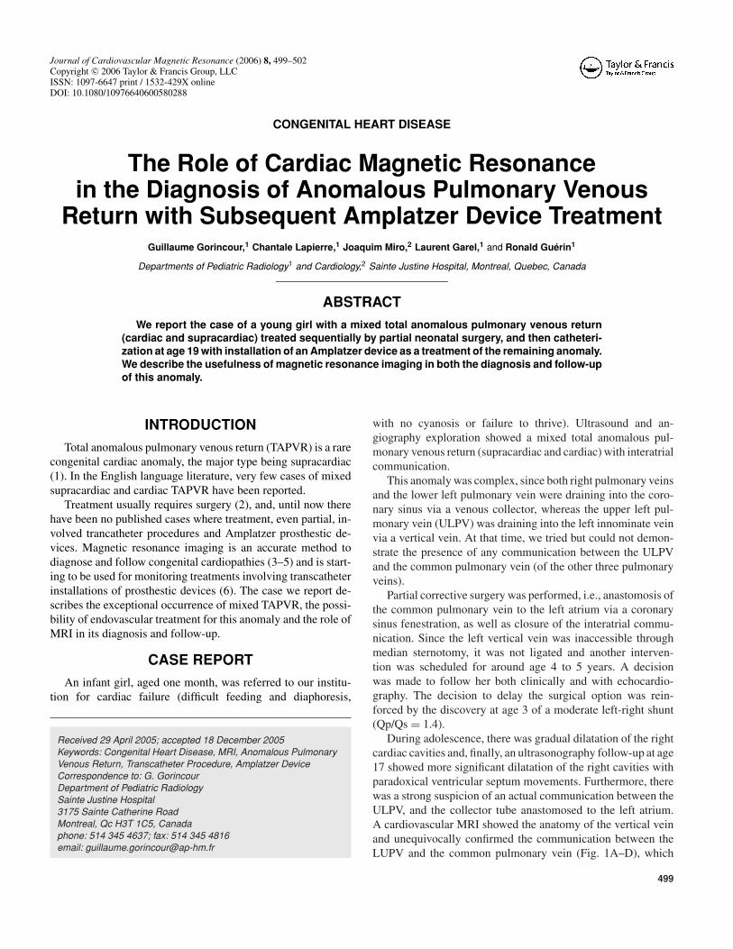
During adolescence, there was gradual dilatation of the rightcardiac cavities and, finally, an ultrasonography follow-up at age17 showed more significant dilatation of the right cavities withparadoxical ventricular septum movements. Furthermore, therewas a strong suspicion of an actual communication between theULPV, and the collector tube anastomosed to the left atrium.A cardiovascular MRI showed the anatomy of the vertical veinand unequivocally confirmed the communication between theLUPV and the common pulmonary vein (Fig. 1A–D), which
499

Figure 1. Successive, three-dimensional, native coronal slices showing the communication between the left pulmonary vein and the collectortube (A and B, white arrow), and between the left pulmonary vein and the vertical vein (C and D, white arrow).
presented with localized narrowing but no obstruction. It wastherefore decided to treat this residual anomaly.
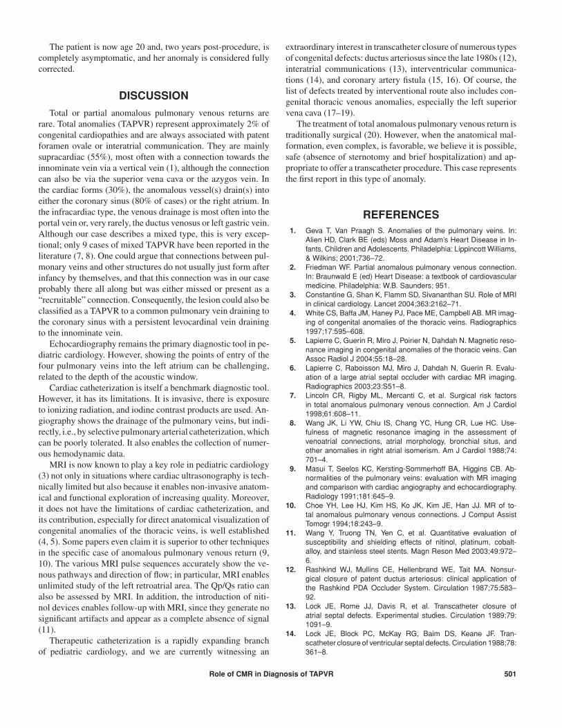
After multidisciplinary discussion, and given the institution’sexpertise in interventional procedures, a non-surgical treatmentwith the installation of an Amplatzer device was proposed toocclude the vertical vein. After angiographic confirmation ofthe anatomical MRI findings and the non-obstructive nature,the procedure was performed without complication (Fig. 2).Hospitalization lasted for 24 hours.
Follow-up showed quick regression of the dilatation of theright cavities, normalization of ventricular septum movementsand absence of residual shunt. The Qp/Qs ratio measured by
Figure 2. Antero-posterior chest x-ray after the procedure showingthe positioning of the Amplatzer device (white arrow).
isotope scan 10 days after the procedure was normal, with noperfusion deficit.
A follow-up MRI 9 months after the procedure demonstratedthat the device was in a satisfactory position to occlude the vein(Fig. 3A–B). There was no obstructive stenosis of the flow notedin the innominated vein or in the ULPV.
Figure 3. Oblique coronal dynamic sequence in the plane of thevertical vein showing its permeable appearance before the proce-dure (A, white arrow) and the absence of signal of the in-placedevice nine months after (B, white arrow).
500 G. Gorincour et al.
The patient is now age 20 and, two years post-procedure, iscompletely asymptomatic, and her anomaly is considered fullycorrected.
DISCUSSION
Total or partial anomalous pulmonary venous returns arerare. Total anomalies (TAPVR) represent approximately 2% ofcongenital cardiopathies and are always associated with patentforamen ovale or interatrial communication. They are mainlysupracardiac (55%), most often with a connection towards theinnominate vein via a vertical vein (1), although the connectioncan also be via the superior vena cava or the azygos vein. Inthe cardiac forms (30%), the anomalous vessel(s) drain(s) intoeither the coronary sinus (80% of cases) or the right atrium. Inthe infracardiac type, the venous drainage is most often into theportal vein or, very rarely, the ductus venosus or left gastric vein.Although our case describes a mixed type, this is very excep-tional; only 9 cases of mixed TAPVR have been reported in theliterature (7, 8). One could argue that connections between pul-monary veins and other structures do not usually just form afterinfancy by themselves, and that this connection was in our caseprobably there all along but was either missed or present as a“recruitable” connection. Consequently, the lesion could also beclassified as a TAPVR to a common pulmonary vein draining tothe coronary sinus with a persistent levocardinal vein drainingto the innominate vein.
Echocardiography remains the primary diagnostic tool in pe-diatric cardiology. However, showing the points of entry of thefour pulmonary veins into the left atrium can be challenging,related to the depth of the acoustic window.
Cardiac catheterization is itself a benchmark diagnostic tool.However, it has its limitations. It is invasive, there is exposureto ionizing radiation, and iodine contrast products are used. An-giography shows the drainage of the pulmonary veins, but indi-rectly, i.e., by selective pulmonary arterial catheterization, whichcan be poorly tolerated. It also enables the collection of numer-ous hemodynamic data.
MRI is now known to play a key role in pediatric cardiology(3) not only in situations where cardiac ultrasonography is tech-nically limited but also because it enables non-invasive anatom-ical and functional exploration of increasing quality. Moreover,it does not have the limitations of cardiac catheterization, andits contribution, especially for direct anatomical visualization ofcongenital anomalies of the thoracic veins, is well established(4, 5). Some papers even claim it is superior to other techniquesin the specific case of anomalous pulmonary venous return (9,10). The various MRI pulse sequences accurately show the ve-nous pathways and direction of flow; in particular, MRI enablesunlimited study of the left retroatrial area. The Qp/Qs ratio canalso be assessed by MRI. In addition, the introduction of niti-nol devices enables follow-up with MRI, since they generate nosignificant artifacts and appear as a complete absence of signal(11).
Therapeutic catheterization is a rapidly expanding branchof pediatric cardiology, and we are currently witnessing an
extraordinary interest in transcatheter closure of numerous typesof congenital defects: ductus arteriosus since the late 1980s (12),interatrial communications (13), interventricular communica-tions (14), and coronary artery fistula (15, 16). Of course, thelist of defects treated by interventional route also includes con-genital thoracic venous anomalies, especially the left superiorvena cava (17–19).
The treatment of total anomalous pulmonary venous return istraditionally surgical (20). However, when the anatomical mal-formation, even complex, is favorable, we believe it is possible,safe (absence of sternotomy and brief hospitalization) and ap-propriate to offer a transcatheter procedure. This case representsthe first report in this type of anomaly.
REFERENCES1. Geva T, Van Praagh S. Anomalies of the pulmonary veins. In:
Alien HD, Clark BE (eds) Moss and Adam’s Heart Disease in In-fants, Children and Adolescents. Philadelphia: Lippincott Williams,& Wilkins; 2001;736–72.
2. Friedman WF. Partial anomalous pulmonary venous connection.In: Braunwald E (ed) Heart Disease: a textbook of cardiovascularmedicine. Philadelphia: W.B. Saunders; 951.
3. Constantine G, Shan K, Flamm SD, Sivananthan SU. Role of MRIin clinical cardiology. Lancet 2004;363:2162–71.
4. White CS, Baffa JM, Haney PJ, Pace ME, Campbell AB. MR imag-ing of congenital anomalies of the thoracic veins. Radiographics1997;17:595–608.
5. Lapierre C, Guerin R, Miro J, Poirier N, Dahdah N. Magnetic reso-nance imaging in congenital anomalies of the thoracic veins. CanAssoc Radiol J 2004;55:18–28.
6. Lapierre C, Raboisson MJ, Miro J, Dahdah N, Guerin R. Evalu-ation of a large atrial septal occluder with cardiac MR imaging.Radiographics 2003;23:S51–8.
7. Lincoln CR, Rigby ML, Mercanti C, et al. Surgical risk factorsin total anomalous pulmonary venous connection. Am J Cardiol1998;61:608–11.
8. Wang JK, Li YW, Chiu IS, Chang YC, Hung CR, Lue HC. Use-fulness of magnetic resonance imaging in the assessment ofvenoatrial connections, atrial morphology, bronchial situs, andother anomalies in right atrial isomerism. Am J Cardiol 1988;74:701–4.
9. Masui T, Seelos KC, Kersting-Sommerhoff BA, Higgins CB. Ab-normalities of the pulmonary veins: evaluation with MR imagingand comparison with cardiac angiography and echocardiography.Radiology 1991;181:645–9.
10. Choe YH, Lee HJ, Kim HS, Ko JK, Kim JE, Han JJ. MR of to-tal anomalous pulmonary venous connections. J Comput AssistTomogr 1994;18:243–9.
11. Wang Y, Truong TN, Yen C, et al. Quantitative evaluation ofsusceptibility and shielding effects of nitinol, platinum, cobalt-alloy, and stainless steel stents. Magn Reson Med 2003;49:972–6.
12. Rashkind WJ, Mullins CE, Hellenbrand WE, Tait MA. Nonsur-gical closure of patent ductus arteriosus: clinical application ofthe Rashkind PDA Occluder System. Circulation 1987;75:583–92.
13. Lock JE, Rome JJ, Davis R, et al. Transcatheter closure ofatrial septal defects. Experimental studies. Circulation 1989;79:1091–9.
14. Lock JE, Block PC, McKay RG, Baim DS, Keane JF. Tran-scatheter closure of ventricular septal defects. Circulation 1988;78:361–8.
Role of CMR in Diagnosis of TAPVR 501

15. Al-Ata J, Amin M, Galal MO, Kouatli A, Hussain A. Transcatheterclosure of a large coronary artery to right superior vena cavafistula using the Amplatzer duct occluder device. Pediatr Cardiol2004;25:70–2.
16. Thomson L, Webster M, Wilson N. Transcatheter closure of a largecoronary artery fistula with the Amplatzer duct occluder. CatheterCardiovasc Interv 1999;48:188–90.
17. Seghaye MC, Wainwright U, Von Bernuth G. Use of an Amplatzerductal occluder to close a persistent left superior caval vein whichreopened after a total cavopulmonary anastomosis. Cardiol Young2002;12:81–3.
18. Geggel RL, Perry SB, Blume ED, Baker CM. Left su-perior vena cava connection to unroofed coronary sinusassociated with positional cyanosis: successful tran-scatheter treatment using Gianturco-Grifka vascular oc-clusion device. Catheter Cardiovasc Interv 1999;48:369–73.
19. Maheshwari S, Pollak J, Hellenbrabd WE. Transcatheter closureof an anomalous venous connection by a novel method. CatheterCardiovasc Interv 1998;45:269–71.
20. LeBlanc JG, Russell JL. Pediatric cardiac surgery in the 1990s.Surg Clin North Am 1998;78:729–47.
502 G. Gorincour et al.