the role of divalent metal ions in enzymatic dna ligation
TRANSCRIPT

The Role of Divalent Metal Ions in Enzymatic DNA Ligation
by
Mark Robert Taylor
A dissertation submitted in partial fulfillment
of the requirements for the degree of
Doctor of Philosophy
(Biological Chemistry)
in the University of Michigan
2014
Doctoral Committee:
Associate Professor Patrick J. O'Brien, Chair
Professor David R. Engelke
Professor Carol A. Fierke
Associate Professor Bruce A. Palfey
Associate Professor Thomas E. Wilson

ii
Dedication
“If I have seen further, it is because I stand on the shoulders of giants.”
-Sir Isaac Newton
This thesis is a testament to all those that have helped me along my path,
who have picked me up when I have stumbled,
and have pushed me forward when I have stubbornly sat still.
iii
Acknowledgements
This work could not have been accomplished without the help of countless people in my life.
None of this would have been possible without the support of my family, my friends, my
coworkers, and my peers in graduate school. Each of them has my eternal gratitude.
While I can’t hope to acknowledge everyone’s individual role in helping get this work done,
certain people deserve special mention for their contributions to the work presented herein. Work
by Daniel Wahl and John Conrad during rotations in the O’Brien lab was instrumental in getting
the DNA ligation assay working and in providing preliminary data that helped guide my work.
Neha Bokil performed the majority of the E720Q and D570N experiments as an undergraduate
researcher under my guidance andTom Jurkiw performed most of the E621Q experiments while
a rotation student in our lab. Both of these students truly astonished me with their quick grasp of
the concepts of kinetics and their dedication to science. This work could not have been done so
well or so quickly without them.

iv
Table of Contents
Dedication.......................................................................................................................................ii
Acknowledgements.......................................................................................................................iii
List of Figures.................................................................................................................................v
List of Tables................................................................................................................................vii
Chapter 1: Background on Enzymatic DNA Ligation...............................................................1
Chapter 2: Kinetic Mechanism of Human DNA Ligase I Reveals
Magnesium-dependent Changes in the Rate-limiting
Step that Compromise Ligation Efficiency....................................................................14
Appendix A: Figures, Tables and Experimental Methods to Accompany Chapter 2...........39
Chapter 3: Comparison of Manganese and Magnesium as
Cofactors for Human DNA Ligase I.....................................................................................50
Appendix B: Figures and Tables to Accompany Chapter 3....................................................73
Chapter 4: Role of Conserved Active Site Carboxylates in Enzymatic DNA Ligation.........78
Chapter 5: Conclusions and Future Directions......................................................................102
Appendix: Berkeley-Madonna Code for Reaction Fitting.....................................................110

v
List of Figures
Figure 1-1: ATP dependent DNA ligase domain structure..............................................................2
Figure 1-2: Steps of enzymatic DNA ligation.................................................................................4
Figure 1-3: Crystal structure of hLIG1 reveals 2 potential metal binding site................................6
Figure 2-1: Reaction pathway for ligation by eukaryotic DNA ligase..........................................16
Figure 2-2: Ligation assay using fluorescently labeled DNA........................................................23
Figure 2-3: Pre-steady state ligation reaction under burst conditions............................................25
Figure 2-4: Magnesium dependence of single-turnover ligation...................................................25
Figure 2-5: ATP dependence of LIG1...........................................................................................27
Figure 2-6: Magnesium dependence of multiple-turnover ligation...............................................29
Figure 2-7: Evidence for the release of adenylylated intermediate during multiple-
turnover ligation.................................................................................................................31
Figure 2-8: Minimal kinetic mechanism of LIG1..........................................................................32
Figure 2-9: Pathways for repair of 5'-adenylylated DNA resulting from abortive ligation...........36
Figure A-1: Evaluation of different quenching solutions..............................................................44
Figure A-2: Representative single-turnover ligation kinetics indicate that
the concentration of LIG1 employed is far above the Kd for substrate binding................44
Figure A-3: Active-site titration to determine the concentration of active
(adenylylated) LIG1...........................................................................................................45
Figure A-4: Stability of LIG1 in reaction buffer...........................................................................45
Figure A-5: DNA concentration dependence of the multiple-turnover ligation reaction..............46
Figure A-6: Multiple-turnover ligation is linearly dependent upon the
concentration of LIG1........................................................................................................46
Figure A-7: Representative initial rates for steady state ligation by LIG1....................................47
Figure A-8: Binding of Mg2+
to ATP decreases the concentration of
free Mg2+
available to LIG1...............................................................................................47

vi
Figure A-9: Kinetic and thermodynamic constants obtained from fits
of the ATP concentration dependencies with the two magnesium model for LIG1..........48
Figure A-10: Measurement of kcat/KM for utilization of ATP (kcat/KM)ATP
by LIG1.....................48
Figure 3-1: Multiple-turnover ligation with Mn2+ and saturating ATP and DNA.......................61
Figure 3-2: Multiple-turnover ligation with Mn2+
and sub-saturating ATP..................................62
Figure 3-3: Mn2+
is a noncompetitive inhibitor of Mg2+
-dependent ligation................................63
Figure 3-4: Mn2+
dependence of single-turnover ligation.............................................................65
Figure 3-5: Mn2+
is a noncompetitive inhibitor of adenylyl transfer, but not nick-sealing...........66
Figure B-1: LIG1 is stable under standard manganese reaction conditions..................................73
Figure B-2: Determination of proper quench solution for rapid-mixing experiments...................74
Figure B-3: Evidence of DNA saturation during manganese stimulated
multiple-turnover ligation..................................................................................................75
Figure B-4: ATP saturation of manganese stimulated multiple-turnover ligation........................75
Figure B-5: LIG1 saturation of manganese stimulated single-turnover ligation...........................77
Figure 4-1: Metal binding sites revealed by hLIG1 structure........................................................81
Figure 4-2: Active-site titration of D570N and E720Q LIG1 mutants..........................................89
Figure 4-3: E621Q exhibits extremely slow single-turnover ligation...........................................90
Figure 4-4: E621Q shows no significant decrease in substrate binding activity...........................91
Figure 4-5: Single-turnover ligation by D570N and E720Q.........................................................92
Figure 4-6: Representative steady-state time-course.....................................................................94
Figure 4-7: D570N and E720Q display wild-type substrate binding............................................95
Figure 4-8: D570N and E720Q show greatly weakened metal binding during
multiple-turnover reactions................................................................................................97
vii
List of Tables
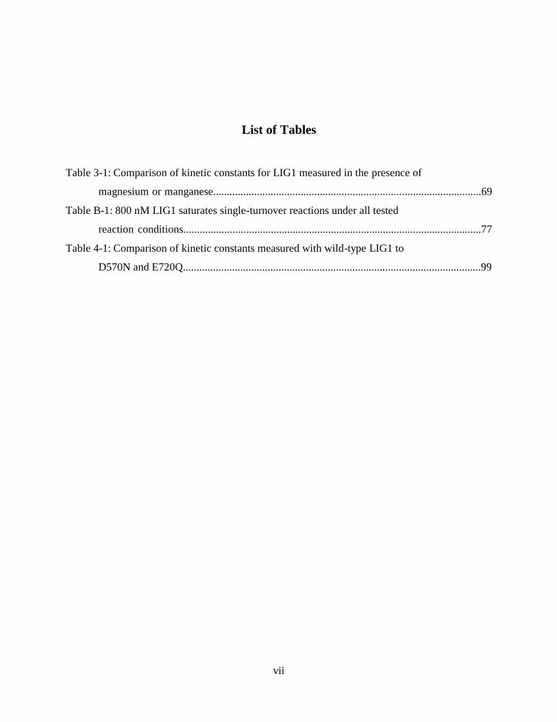
Table 3-1: Comparison of kinetic constants for LIG1 measured in the presence of
magnesium or manganese..................................................................................................69
Table B-1: 800 nM LIG1 saturates single-turnover reactions under all tested
reaction conditions.............................................................................................................77
Table 4-1: Comparison of kinetic constants measured with wild-type LIG1 to
D570N and E720Q.............................................................................................................99

1
Chapter 1: Background on Enzymatic DNA Ligation
The simultaneous discovery of DNA ligases by the Gellert, Hurwitz, Lehman and Richardson
groups in 1967 was a milestone in biology [1-4]. Identification of an enzyme capable of joining
two DNA molecules filled a fundamental gap in the process of discontinuous DNA replication
posited by Okazaki, et al. a year prior [5]. The ability to easily mend breaks formed in a DNA
molecule also encouraged the idea of a dynamic genome, where DNA molecules continually
experience transient breaks which are quickly mended. We now know this genomic dynamism to
be essential to genomic integrity, as DNA breaks are continually formed and resealed during
repair of DNA damage [6]. In addition to their importance in genomic maintenance, DNA ligases
have proven an invaluable scientific tool. The ability to stitch together different DNA molecules
enabled the technique of molecular cloning, which has proven essential to the advancement of
the biomedical sciences. DNA ligases remain widely used today for many biotechnology and
molecular biology applications.
Domain architecture of ATP-dependent DNA ligases
DNA ligases are a classic example of divergent evolution, with ATP dependent DNA ligases
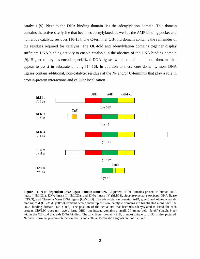
from viruses to bacteria to humans all sharing a conserved catalytic core [7] (Figure 1-1). This
core consists of three independently folding domains; the DNA binding domain, the
adenylylation domain, sometimes also referred to as the nucleotidyl transferase (NTase) domain,
and the oligonucleotide binding (OB)-fold domain. Closest to the N-terminus is the DNA
binding domain. This domain displays the largest size variability among DNA ligases, ranging
from the nearly 300 residue domain of human DNA ligase I to the miniscule 20 residue "latch"
of the Chlorella virus DNA ligase [8, 9]. True to its name, this domain displays the majority of
DNA binding activity for the mammalian DNA ligases, and carries no residues required for
2
catalysis [9]. Next to the DNA binding domain lies the adenylylation domain. This domain
contains the active-site lysine that becomes adenylylated, as well as the AMP binding pocket and
numerous catalytic residues [10-13]. The C-terminal OB-fold domain contains the remainder of
the residues required for catalysis. The OB-fold and adenylylation domains together display
sufficient DNA binding activity to enable catalysis in the absence of the DNA binding domain
[9]. Higher eukaryotes encode specialized DNA ligases which contain additional domains that
appear to assist in substrate binding [14-16]. In addition to these core domains, most DNA
ligases contain additional, non-catalytic residues at the N- and/or C-terminus that play a role in
protein-protein interactions and cellular localization.
Figure 1-1: ATP dependent DNA ligase domain structure. Alignment of the domains present in human DNA
ligase I (hLIG1), DNA ligase III (hLIG3), and DNA ligase IV (hLIG4), Saccharomyces cerevisiae DNA ligase
(CDC9), and Chlorella Virus DNA ligase (ChVLIG). The adenylylation domain (AdD, green) and oligonucleotide
binding-fold (OB-fold, yellow) domains which make up the core catalytic domains are highlighted along with the
DNA binding domain (DBD, red). The position of the active-site that becomes adenylylated is listed for each
protein. ChVLIG does not have a large DBD, but instead contains a small, 20 amino acid “latch” (Latch, blue)
within the OB-fold that aids DNA binding. The zinc finger domain (ZnF, orange) unique to LIG3 is also pictured.
N- and C-terminal protein-interaction motifs and cellular localization signals are not pictured.

3
Mechanism of Enzymatic DNA Ligation
All known DNA ligases follow a conserved reaction mechanism involving 3 independent
phosphoryl transfers [17] (Figure 1-2). The reaction mechanism begins with enzyme
adenylylation, where the enzyme catalyzes covalent attachment of an adenylyl group to an
active-site lysine, using either NAD+ or ATP as the AMP donor. During the next step, adenylyl
transfer, the protein catalyzes transfer of the AMP group from the adenylylated lysine to an
exposed 5' phosphate in DNA. The reaction completes with the nick-sealing step when the 3'
hydroxyl of an adjacent DNA strand attacks the adenylylated 5' phosphate, resulting in
phosphodiester bond formation. Each of these steps requires at least one divalent metal cofactor,
with magnesium presumably the physiologically relevant cofactor. Throughout the reaction, the
high energy of the original phosphoanhydride bond of ATP/NAD+ is stored within intermediates,
first as a phosphoramidate on the adenylylated lysine and then as a phosphoanhydride on the
adenylylated DNA. This pathway effectively links the original energy of ATP/NAD+ cleavage
with formation of a phosphodiester bond. Though each of these steps is inherently reversible,
hydrolysis of the pyrophosphate formed during enzyme adenylylation renders the overall
reaction essentially irreversible.
Mammalian DNA Ligases
There are three identified mammalian DNA ligases. DNA ligase I (LIG1) accomplishes most of
the most of the measured DNA ligation activity within a cell [18]. Found only in the nucleus and
with robust activity toward single-strand breaks, LIG1 is predicted to be the main DNA ligase for
resolving single-strand breaks formed during nuclear DNA replication and repair [19-21].
Immortalized cells originating from a patient with mutations in both LIG1 alleles suffer retarded
Okazaki fragment joining, as well as general growth and genome stability defects [22, 23]. The
patient herself suffered from developmental retardation as well as hypersensitivity to DNA
damage and other developmental abnormalities [24]. Similar problems occur in mouse models
containing LIG1 mutations [25]. Altogether these results suggest that LIG1 plays an essential
role in nuclear genomic maintenance.

4
Figure 1-2: Steps of enzymatic DNA ligation. The three steps of enzymatic DNA ligation are pictured. The side-
chain of the active-site lysine that becomes transiently adenylylated is pictured. Metal ions required for each reaction
step are not pictured.

5
DNA ligase III (LIG3) is the sole DNA ligase in mitochondria and as such is essential for all
mitochondrial DNA repair [26, 27]. Consistent with this role, LIG3 displays robust activity
toward both single- and double-strand break repair. Loss of mitochondrial LIG3 results in
abnormal mitochondrial structure and function, eventually leading to loss of the organelle and
cell death [28]. LIG3 is also expressed in the nucleus, however its function within the nucleus is
currently unknown. Numerous studies indicate LIG3 capable of participating in single- and
double-strand nuclear DNA repair pathways, however cells without nuclear LIG3 appear normal.
This suggests that LIG3 may assist in some nuclear DNA repair, but it is not essential and likely
serves as an emergency backup to the other DNA ligases.
DNA ligase IV (LIG4) is responsible for nuclear double-strand break repair. Much of the
activity of LIG4 is observed during non-homologous end joining (NHEJ) where two separate
DNA molecules are joined end-to-end using little to no sequence homology [29]. Accurate NHEJ
requires the activity of over half a dozen proteins including LIG4, and resolves otherwise
catastrophic damage to the genome [30]. In addition to combating double-strand breaks formed
as a result of DNA damage, NHEJ plays an essential role during immune system development by
ligating together the immunoglobulin genes during V(D)J recombination [31]. Inappropriate
NHEJ can cause chromosomal fusions and translocations, leading to severe cellular dysfunction
and cancer [32]. Overexpression of LIG3 leads to an increase in the rate of chromosomal
aberrations, suggesting that the double-strand break repair of LIG3 is more error prone than that
of LIG4.
DNA Ligase Structure
In 2004, the first crystal structure of a human DNA ligase was solved by Pascal et al. [9] (Figure
1-3A). The crystal captures a truncated form of human LIG1 in complex with an adenylylated
DNA nick. The truncation removes the N-terminal 231 residues of LIG1 which contain the
nuclear localization signal and protein-interaction motifs. Loss of these residues does not seem to
affect basic enzyme catalysis, as this form of the enzyme displays identical activity to the wild-
type protein under in vitro conditions. Capture of the enzyme in complex with adenylylated

6
Figure 1-3: Crystal structure of hLIG1 reveals 2 potential metal binding sites. A Crystal structure of hLIG1 in
complex with adenylylated DNA substrate [9]. DNA binding domain (red), adenylylation domain (green) and OB-
fold domain (yellow) of hLIG1 completely encircle DNA substrate. Motif VI residues of the OB-fold domain (blue)
which contain residues essential for enzyme adenylylation are oriented toward solvent. B The absence of a 3’
hydroxyl group on the 5’ side of the nick prevents nick-sealing. C Close-up view of hLIG1 active site. Magenta
lattices indicate location of potential metal binding sites. Free 5’ and 3’ groups at the nick and residues involved in
metal binding are labeled along with active-site lysine. D Protein sequence alignment of the three human DNA
ligases with ligase from Saccharomyces cerevisiea (CDC9), Schizosaccharomyces pombe (CDC17) and Chlorella
Virus DNA (ChVLIG) reveal strong conservation within the active-site. Alignment performed with default settings
in ClustalX using truncated versions of each protein that include only the catalytic core domains. * indicates a
residue involved in coordination of proposed active-site metal sites. † indicates Lys 568.

7
DNA was accomplished through the use of a DNA substrate containing a 3' dideoxy nucleotide
at the nick. Without the 3' hydroxyl required for catalysis during nick-sealing, the enzyme is
forced to stall between the adenylyl transfer and nick-sealing steps (Figure 1-3B). At this stage,
the enzyme has fully encircled the DNA at the nick, with contacts between the N-terminal DNA
binding domain and C-terminal OB-fold stabilizing the toroidal structure.
The difference in electron density between the crystal soaked in magnesium and manganese
reveals two potential metal binding sites within the active-site (Figure 1-3C). These sites are
coordinated by universally conserved residues that align very similarly in other DNA ligase
structures (Figure 1-3C,D) [8, 33, 34]. Both of these sites have excellent coordination geometry
for magnesium, and each is located in a position suitable for catalysis. The site on the 3' end of
the nick, coordinated by E720 and the phosphate groups of the 5' adenylylate, positions a metal
ion for activation of the 5' hydroxyl for nucleophilic attack on the adenylylated lysine during
adenylyl transfer, and would also stabilize AMP as a leaving group during nick-sealing. The site
on the 5' end of the nick, coordinated by E621 and D570, places a metal ion in position to
activate the 3' hydroxyl for attack during nick-sealing. Each of these amino acid residues is
universally conserved among LIG1 homologues, suggesting they serve an important role in LIG1
function. Mutations of the corresponding residues in Chlorella virus DNA ligase have large
effects on both metal affinity during catalysis and maximal catalytic rate.
Impact of DNA Ligase Research
The essential role of DNA ligases in everyday genomic maintenance make them fascinating
enzymes to study. The existence of multiple DNA ligases in humans with partially overlapping
function raises the possibility that different DNA repair pathways could be targeted by
specifically inhibiting individual DNA ligases. Used alone or combined with other DNA
damaging agents that target certain DNA repair processes, DNA ligase inhibitors could then be
potent chemotherapeutic agents. Recent research has identified some early small molecule
candidates for DNA ligase inhibitors specific to each of the three human DNA ligases [35, 36].
One LIG3 inhibitor has proven specifically interesting, as early studies have shown certain

8
cancer cell lines display an increased sensitivity to DNA damaging agents in the presence of sub-
toxic levels of the inhibitor. Though LIG1 specific inhibitors remain to be tested, these could be
an excellent tool against tumors such as glioblastomas which grow around senescent neurons.
Inhibition of LIG1 would preferentially harm cells going through replication, while normal DNA
repair within the cells could be accomplished by LIG3.
There are few reported clinical cases of acute hLIG1 dysfunction, presumably because such
dysfunction usually results in an extremely shortened lifespan. There are, however, multiple
avenues by which slow onset, chronic hLIG1 dysfunction may occur. As one example, previous
research has shown that different metals can inhibit the enzyme, and that this inhibition occurs
under concentrations similar to those observed in cells during heavy metal poisoning [37]. On a
cellular level, heavy metal poisoning leads to persistent DNA breaks, suggesting DNA ligases
may be unable to seal breaks under these conditions [38]. There is also evidence that dysfunction
of DNA ligases leads to neuronal dysfuntion in the disease Ataxia with Oculomotor Apraxia 1
(AOA1). Patients with AOA1 display late-onset, cumulative motor-neuron death, caused by loss
of the protein Aprataxin [39, 40]. Aprataxin catalyzes the removal of adenylyl groups from 5'
phosphates in DNA, whose only known mechanism of formation is abortive ligation by DNA
ligases [41]. Thus understanding the basics of DNA ligase function may give insight into these
and other diseases.
In addition to these clinically relevant reasons to study hLIG1, the importance of pursuing basic
research cannot be overstated. Only by understanding the basics of how enzymes work can
scientists begin to formulate the more important questions of how these enzymes relate to known
and unknown diseases. For example, it is only because we characterized the kinetic mechanism
of human LIG1 that we now know the adenylylated DNA problematic in AOA1 can be formed
by LIG1 under normal cellular conditions, and not just during acute cellular stress. Though there
is currently a popular push to pursue only the most relevant, immediately applicable research, we
cannot rationally claim to know biology well enough to suggest we know the direct path toward
any important discovery. In fact, the seemingly unimportant basic science research often leads to
some of the most important breakthroughs in history. In this manner, the focus of this thesis is
9
the basic science of human DNA ligase I. Here I report my investigations into the metal
dependence of enzymatic DNA ligation through kinetic characterization and active-site mutation.
The work in Chapter 2 characterizes the kinetic mechanism of hLIG1 in the presence of
magnesium, the presumptive physiological metal cofactor. The main purpose of this work is to
establish a kinetic framework for the function of hLIG1 against which future investigations can
be compared. LIG1 is found to perform flawlessly under most conditions, converting single-
strand nicks into ligated DNA. However, when magnesium concentrations are lowered, a
detectable amount of abortive ligation occurs. Abortive ligation by a DNA ligase essentially
exacerbates the damage at a nick, as the 5' adenylylated DNA that is formed is not easily rebound
by ligase, and the nick becomes a persistent DNA break until other repair pathways can remove
the adenylylated nucleotide. This type of damage is expected to play a role in the disease Ataxia
with Oculomotor Apraxia 1, where the protein that directly removes 5' adenylyl groups,
aprataxin, suffers loss of function.
Chapter 3 compares magnesium and manganese as cofactors for hLIG1. Full characterization of
the kinetic mechanism of hLIG1 in the presence of manganese reveals that manganese alone can
stimulate each step of the ligation reaction. The two metals display remarkable similarities with
similar metal affinities and maximal rate constants for each step of the reaction. One startling
difference between magnesium and manganese, however, is the presence of a lower affinity
inhibitory manganese binding site. This inhibition has the peculiar trait of inhibiting enzyme
adenylylation and adenylyl transfer, but not nick-sealing. The inhibitory metal site(s) also appear
to be specific for manganese over magnesium, as the inhibitory metal ion(s) cannot be competed
away with saturating levels of magnesium.
Chapter 4 investigates the effect of conservative mutations on the residues chelating the potential
metal sites observed in the hLIG1 crystal structure. The goal of this investigation was to identify
the role of each metal site during catalysis, and potentially whether one or two metals are
required for catalysis. The E621Q mutation results in greatly diminished enzyme activity,
creating an enzyme that is more than 10,000-fold slower than the wild-type. The enzyme appears
to still be adenylylated and is technically capable of DNA ligation, but does so at a greatly

10
decreased rate. The D570N and E720Q mutations result in very similar effects on the enzyme.
Both mutations weaken the magnesium affinity during adenylyl transfer and nick-sealing by ~10
fold. The mutations also decrease the maximal rate constants for adenylyl transfer and nick-
sealing, resulting in an ~40-fold decrease in adenylyl transfer and ~100-fold decrease in nick-
sealing for either mutation. These mutations also appear to affect the rate of enzyme
adenylylation, suggesting that the metal sites play a role in both the DNA and ATP dependent
reactions.
This body of work greatly deepens our understanding of the kinetic mechanism of enzymatic
DNA ligation, and provides new insight into the role of meal ions during catalysis. Future
research can build upon the information provided here to deepen our understanding of how metal
ions play a role in the individual reaction steps, and how the enzyme engages the DNA substrate.
The recent wide-spread availability of next-generation DNA sequencing technology allows the
possibility to ask new questions about DNA ligases that would have been technically impossible
a decade ago, like how does the sequence at a nick affect the rate, fidelity or efficiency of
enzymatic DNA ligation.

11
References
1. Gellert, M., Formation of covalent circles of lambda DNA by E. coli extracts. Proc Natl
Acad Sci U S A, 1967. 57(1): p. 148-55.
2. Gefter, M.L., A. Becker, and J. Hurwitz, The enzymatic repair of DNA. I. Formation of
circular lambda-DNA. Proc Natl Acad Sci U S A, 1967. 58(1): p. 240-7.
3. Olivera, B.M. and I.R. Lehman, Linkage of polynucleotides through phosphodiester
bonds by an enzyme from Escherichia coli. Proc Natl Acad Sci U S A, 1967. 57(5): p.
1426-33.
4. Weiss, B. and C.C. Richardson, Enzymatic breakage and joining of deoxyribonucleic
acid, I. Repair of single-strand breaks in DNA by an enzyme system from Escherichia coli
infected with T4 bacteriophage. Proc Natl Acad Sci U S A, 1967. 57(4): p. 1021-8.
5. Sakabe, K. and R. Okazaki, A unique property of the replicating region of chromosomal
DNA. Biochim Biophys Acta, 1966. 129(3): p. 651-4.
6. Sancar, A., et al., Molecular mechanisms of mammalian DNA repair and the DNA
damage checkpoints. Annu Rev Biochem, 2004. 73: p. 39-85.
7. Shuman, S. and B. Schwer, RNA capping enzyme and DNA ligase: a superfamily of
covalent nucleotidyl transferases. Mol Microbiol, 1995. 17(3): p. 405-10.
8. Nair, P.A., et al., Structural basis for nick recognition by a minimal pluripotent DNA
ligase. Nat Struct Mol Biol, 2007. 14(8): p. 770-8.
9. Pascal, J.M., et al., Human DNA ligase I completely encircles and partially unwinds
nicked DNA. Nature, 2004. 432(7016): p. 473-8.
10. Kodama, K., D.E. Barnes, and T. Lindahl, In vitro mutagenesis and functional expression
in Escherichia coli of a cDNA encoding the catalytic domain of human DNA ligase I.
Nucleic Acids Res, 1991. 19(22): p. 6093-9.
11. Sriskanda, V. and S. Shuman, Mutational analysis of Chlorella virus DNA ligase:
catalytic roles of domain I and motif VI. Nucleic Acids Res, 1998. 26(20): p. 4618-25.
12. Sriskanda, V., et al., Mutational analysis of Escherichia coli DNA ligase identifies amino
acids required for nick-ligation in vitro and for in vivo complementation of the growth of
yeast cells deleted for CDC9 and LIG4. Nucleic Acids Res, 1999. 27(20): p. 3953-63.
13. Sriskanda, V. and S. Shuman, Role of nucleotidyl transferase motif V in strand joining by
chlorella virus DNA ligase. J Biol Chem, 2002. 277(12): p. 9661-7.
14. Mackey, Z.B., et al., DNA ligase III is recruited to DNA strand breaks by a zinc finger
motif homologous to that of poly(ADP-ribose) polymerase. Identification of two
functionally distinct DNA binding regions within DNA ligase III. J Biol Chem, 1999.
274(31): p. 21679-87.
15. Taylor, R.M., et al., Role of the DNA ligase III zinc finger in polynucleotide binding and
ligation. Nucleic Acids Res, 1998. 26(21): p. 4804-10.
16. Taylor, R.M., C.J. Whitehouse, and K.W. Caldecott, The DNA ligase III zinc finger
stimulates binding to DNA secondary structure and promotes end joining. Nucleic Acids
Res, 2000. 28(18): p. 3558-63.

12
17. Lehman, I.R., DNA ligase: structure, mechanism, and function. Science, 1974.
186(4166): p. 790-7.
18. Soderhall, S. and T. Lindahl, Mammalian DNA ligases. Serological evidence for two
separate enzymes. J Biol Chem, 1975. 250(21): p. 8438-44.
19. Levin, D.S., et al., An interaction between DNA ligase I and proliferating cell nuclear
antigen: implications for Okazaki fragment synthesis and joining. Proc Natl Acad Sci U S
A, 1997. 94(24): p. 12863-8.
20. Montecucco, A., et al., DNA ligase I is recruited to sites of DNA replication by an
interaction with proliferating cell nuclear antigen: identification of a common targeting
mechanism for the assembly of replication factories. EMBO J, 1998. 17(13): p. 3786-95.
21. Levin, D.S., et al., Interaction between PCNA and DNA ligase I is critical for joining of
Okazaki fragments and long-patch base-excision repair. Curr Biol, 2000. 10(15): p. 919-
22.
22. Barnes, D.E., et al., Mutations in the DNA ligase I gene of an individual with
immunodeficiencies and cellular hypersensitivity to DNA-damaging agents. Cell, 1992.
69(3): p. 495-503.
23. Prigent, C., et al., Aberrant DNA repair and DNA replication due to an inherited
enzymatic defect in human DNA ligase I. Mol Cell Biol, 1994. 14(1): p. 310-7.
24. Webster, A.D., et al., Growth retardation and immunodeficiency in a patient with
mutations in the DNA ligase I gene. Lancet, 1992. 339(8808): p. 1508-9.
25. Bentley, D.J., et al., DNA ligase I null mouse cells show normal DNA repair activity but
altered DNA replication and reduced genome stability. J Cell Sci, 2002. 115(Pt 7): p.
1551-61.
26. Simsek, D., et al., Crucial role for DNA ligase III in mitochondria but not in Xrcc1-
dependent repair. Nature, 2011. 471(7337): p. 245-8.
27. Gao, Y., et al., DNA ligase III is critical for mtDNA integrity but not Xrcc1-mediated
nuclear DNA repair. Nature, 2011. 471(7337): p. 240-4.
28. Shokolenko, I.N., et al., Mitochondrial DNA ligase is dispensable for the viability of
cultured cells but essential for mtDNA maintenance. J Biol Chem, 2013. 288(37): p.
26594-605.
29. Wilson, T.E., U. Grawunder, and M.R. Lieber, Yeast DNA ligase IV mediates non-
homologous DNA end joining. Nature, 1997. 388(6641): p. 495-8.
30. van Gent, D.C. and M. van der Burg, Non-homologous end-joining, a sticky affair.
Oncogene, 2007. 26(56): p. 7731-40.
31. Grawunder, U., et al., DNA ligase IV is essential for V(D)J recombination and DNA
double-strand break repair in human precursor lymphocytes. Mol Cell, 1998. 2(4): p.
477-84.
32. Deriano, L. and D.B. Roth, Modernizing the nonhomologous end-joining repertoire:
alternative and classical NHEJ share the stage. Annu Rev Genet, 2013. 47: p. 433-55.
33. Ochi, T., X. Gu, and T.L. Blundell, Structure of the catalytic region of DNA ligase IV in
complex with an Artemis fragment sheds light on double-strand break repair. Structure,
2013. 21(4): p. 672-9.
34. Cotner-Gohara, E., et al., Human DNA ligase III recognizes DNA ends by dynamic
switching between two DNA-bound states. Biochemistry, 2010. 49(29): p. 6165-76.
35. Zhong, S., et al., Identification and validation of human DNA ligase inhibitors using
computer-aided drug design. J Med Chem, 2008. 51(15): p. 4553-62.

13
36. Chen, X., et al., Rational design of human DNA ligase inhibitors that target cellular DNA
replication and repair. Cancer Res, 2008. 68(9): p. 3169-77.
37. Yang, S.W., F.F. Becker, and J.Y. Chan, Inhibition of human DNA ligase I activity by
zinc and cadmium and the fidelity of ligation. Environ Mol Mutagen, 1996. 28(1): p. 19-
25.
38. Li, H., R. Swiercz, and E.W. Englander, Elevated metals compromise repair of oxidative
DNA damage via the base excision repair pathway: implications of pathologic iron
overload in the brain on integrity of neuronal DNA. J Neurochem, 2009. 110(6): p. 1774-
83.
39. Date, H., et al., Early-onset ataxia with ocular motor apraxia and hypoalbuminemia is
caused by mutations in a new HIT superfamily gene. Nat Genet, 2001. 29(2): p. 184-8.
40. Moreira, M.C., et al., The gene mutated in ataxia-ocular apraxia 1 encodes the new
HIT/Zn-finger protein aprataxin. Nat Genet, 2001. 29(2): p. 189-93.
41. Ahel, I., et al., The neurodegenerative disease protein aprataxin resolves abortive DNA
ligation intermediates. Nature, 2006. 443(7112): p. 713-6.
14
Chapter 2: Kinetic Mechanism of Human DNA Ligase I Reveals
Magnesium-dependent Changes in the Rate-limiting Step that
Compromise Ligation Efficiency
Abstract
DNA ligase I (Lig1) catalyzes the ligation of single-strand breaks to complete DNA replication
and repair. The energy of ATP is used to form a new phosphodiester bond in DNA via a reaction
mechanism that involves three distinct chemical steps; enzyme adenylylation, adenylyl transfer
to DNA and nick-sealing. We used steady state and pre-steady state kinetics to characterize the
minimal mechanism for the reactions catalyzed by human Lig1. The ATP dependence of the
reaction indicates that Lig1 requires multiple Mg2+
ions for catalysis and that an essential Mg2+
ion binds more tightly to ATP than to the enzyme. Further dissection of the magnesium ion
dependence of individual reaction steps revealed that the affinity for Mg2+
changes along the
reaction coordinate. At saturating concentrations of ATP and Mg2+
ions, the three chemical steps
occur at similar rates and the efficiency of ligation is high. However, under conditions of limiting
Mg2+
, the nick-sealing step becomes rate-limiting and the adenylylated DNA intermediate is
prematurely released into solution. Subsequent adenylylation of enzyme prevents rebinding to
the adenylylated DNA intermediate comprising an Achilles’ heel of Lig1. These ligase-generated
5’-adenylylated nicks constitute persistent breaks that are a threat to genomic stability if left
unrepaired. The kinetic and thermodynamic framework determined here for Lig1 provides a
starting point for understanding the mechanism and specificity of mammalian DNA ligases.
*This work was done with the help of Daniel Wahl and John A. Conrad. DW and JC performed early experiments to
determine proper reaction conditions for hLIG1, and collect early data on the kinetics of hLIG1 in the presence of
multiple metals. Mark Taylor collected and analyzed all data reported herein with help from Patrick O'Brien.
Citation for this published work: Taylor, M.R., et al., Journal of Biological Chemistry, 2011. 286(26): p. 23054-62.

15
Introduction
Breaks in DNA result from spontaneous hydrolysis of the phosphodiester backbone and are
formed as transient intermediates during DNA replication and repair pathways. Mammals have
three genes encoding DNA ligases that seal these breaks and restore the continuous nature of
chromosomes. DNA Ligase I (Lig1) is essential for ligation of single-strand breaks in the
nucleus, including the ligation of Okazaki fragments during discontinuous DNA replication [1].
DNA ligase III (Lig3) is required for mitochondrial DNA replication and repair. Although Lig3
has been assumed to play essential roles in nuclear DNA repair, it was recently shown to be
dispensable for nuclear genomic maintenance [2, 3]. DNA ligase IV (Lig4) is specialized for
repair of nuclear double-strand breaks, and is required for nonhomologous end joining and V(D)J
recombination [4, 5].
The overall reaction catalyzed by DNA ligases involves the formation of a phosphodiester bond
between an adjacent 3’ hydroxyl and a 5’ phosphate in DNA. The reaction proceeds via a
universally conserved pathway [6-8] (Figure 2-1). First, the apoenzyme catalyzes transfer of the
AMP group from a nucleotide cofactor to an active site lysine, forming an adenylylated enzyme
intermediate. Eukaryotic ligases use ATP as the adenylyl group donor, whereas bacterial ligases
utilize either ATP or NAD+. After binding a nicked DNA substrate, the adenylylated enzyme
catalyzes transfer of the adenylyl group to the 5’-phosphate present at the nick, forming an
adenylylated DNA intermediate. In the final step of the reaction, DNA ligase catalyzes the
nucleophilic attack of the 3’-hydroxyl on the adenylylated 5’-phosphate to form a new
phosphodiester bond and to release AMP. Although enzyme adenylylation and nick-sealing are
known to be reversible in the presence of a large excess of pyrophosphate or AMP, these steps
are effectively irreversible in their absence. All of the chemical steps catalyzed by ligase have
been shown to require magnesium, but the number of magnesium ions and their affinities during
each step have not been determined [9].
16
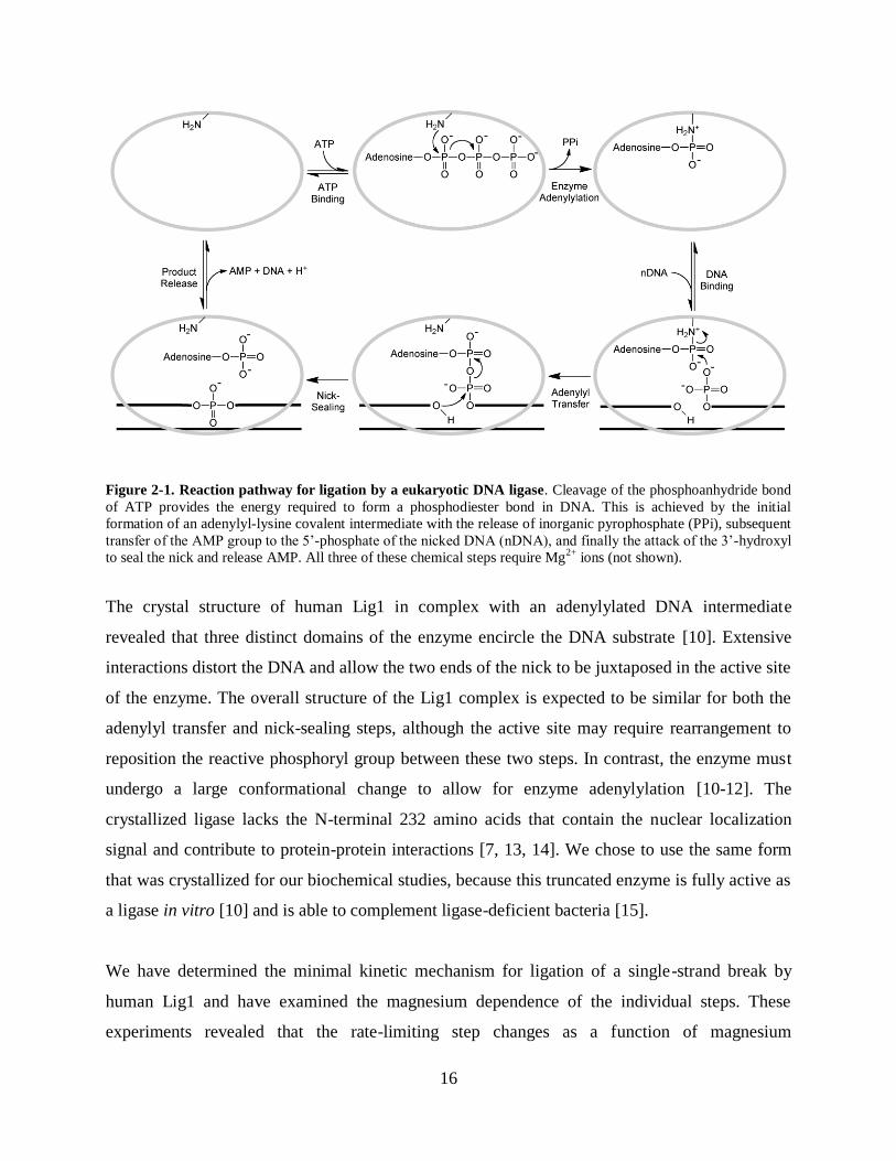
Figure 2-1. Reaction pathway for ligation by a eukaryotic DNA ligase. Cleavage of the phosphoanhydride bond
of ATP provides the energy required to form a phosphodiester bond in DNA. This is achieved by the initial
formation of an adenylyl-lysine covalent intermediate with the release of inorganic pyrophosphate (PPi), subsequent
transfer of the AMP group to the 5’-phosphate of the nicked DNA (nDNA), and finally the attack of the 3’-hydroxyl
to seal the nick and release AMP. All three of these chemical steps require Mg2+
ions (not shown).
The crystal structure of human Lig1 in complex with an adenylylated DNA intermediate
revealed that three distinct domains of the enzyme encircle the DNA substrate [10]. Extensive
interactions distort the DNA and allow the two ends of the nick to be juxtaposed in the active site
of the enzyme. The overall structure of the Lig1 complex is expected to be similar for both the
adenylyl transfer and nick-sealing steps, although the active site may require rearrangement to
reposition the reactive phosphoryl group between these two steps. In contrast, the enzyme must
undergo a large conformational change to allow for enzyme adenylylation [10-12]. The
crystallized ligase lacks the N-terminal 232 amino acids that contain the nuclear localization
signal and contribute to protein-protein interactions [7, 13, 14]. We chose to use the same form
that was crystallized for our biochemical studies, because this truncated enzyme is fully active as
a ligase in vitro [10] and is able to complement ligase-deficient bacteria [15].
We have determined the minimal kinetic mechanism for ligation of a single-strand break by
human Lig1 and have examined the magnesium dependence of the individual steps. These
experiments revealed that the rate-limiting step changes as a function of magnesium
17
concentration and exposed an Achilles’ heel of Lig1, whereby low magnesium concentrations
cause release of the adenylylated DNA intermediate to form a persistent DNA break. This
observation provides a rationale for the existence of the aprataxin pathway for repair of
adenylylated DNA intermediates and implies that magnesium deficiency could cause defects in
DNA repair and replication.

18
Experimental Procedures
Recombinant DNA Ligase I
The catalytic domain of human Lig1 (residues 232-919) was expressed in E. coli and purified as
previously described [10]. Cells were lysed in the presence of 1 mM EDTA to preserve the
adenylylated form of the enzyme, and subsequently purified over a phosphocellulose column and
a NTA-nickel column. The His tag was removed with Prescission protease, and Lig1 was further
purified with Q-sepharose. The final purified fractions were combined and dialyzed into storage
buffer (25 mM Tris•Cl, pH 7.6, 150 mM NaCl, 1 mM DTT, and 0.1 mM EDTA). Aliquots were
snap-frozen and stored at -80 oC. Purity was greater than 95% as judged by SDS-PAGE. Initial
concentrations were estimated from the absorbance at 280 nm, using the calculated extinction
coefficient. The concentration of active, adenylylated enzyme was determined by titration with
nicked DNA substrate, as described below, and this active concentration is reported throughout.
DNA Substrates
Oligonucleotides were synthesized by Integrated DNA Technologies or by the Keck Center at
Yale University and were purified on denaturing polyacrylamide gels. The portion of the gel
containing the full-length oligonucleotide was excised and crushed, and DNA was extracted by
soaking overnight in 500 mM NaCl and 1 mM EDTA. The extracted oligonucleotides were
desalted by binding to a C18 reverse phase column (Sep-pak, Waters) and eluted with 30% (v/v)
acetonitrile. Concentrations were obtained from the absorbance at 260 nm using the calculated
extinction coefficients. The three oligonucleotides used in this study had sequences 5’-
CCGAATCAGTCCGACGACGCATCAGCAC, 5’-GTGCTGATGCGTC, and 5’-P-
GTCGGACTGATTCGG-FAM (P indicates 5’ phosphorylation and FAM indicates the presence
19
of a 3’ fluorescein). The nicked, double-stranded DNA substrate (nDNA) was formed by mixing
equimolar amounts of the three oligonucleotides in 10 mM NaMES pH 6.5 and 50 mM NaCl and
cooling the mixture from 90 °C to 4 °C at a rate of 3 °C per minute.
Gel-based Ligation Assay
Ligation reactions were performed at 37 °C. Unless otherwise indicated, the standard buffer
contained 50 mM NaMOPS pH 7.5 (measured at 25 °C), 1 mM dithiothreitol, 0.05 mg/mL BSA,
and sufficient NaCl to maintain a constant ionic strength of 150 mM. The amounts of ATP,
MgCl2, nDNA, and Lig1 varied, as indicated below. Preincubation controls established that Lig1
retains 100% of its activity after 1 hr in this standard buffer, and all of the kinetic data was
collected within this window of time (Appendix Figure A-4). Reactions were quenched in
formamide/EDTA (30 mM EDTA in 94% formamide), heated to 95 °C for 5 min to denature the
DNA and resolved on 20% (w/v) denaturing polyacrylamide gels containing 8 M Urea.
Fluorescein-labeled oligonucleotides were detected with a Typhoon Trio+ imager (GE
Healthcare) with excitation at 488 nm and emission through a 520 nm band-pass filter. The
images were analyzed using ImageQuantTL (GE Healthcare). The intensity of the individual
DNA species was corrected for background fluorescence, and the fraction of the total
fluorescence was determined by dividing the fluorescence intensity for the desired species by the
signal for all other species in the sample. When necessary, this fraction was converted into its
concentration by multiplying by the total concentration of DNA in the reaction.
Rapid Quench Experiments
Rapid mixing experiments were performed in a Kintek RFQ-3 quench-flow apparatus. One
sample loop contained Lig1 and the other contained nDNA, each at double the reaction
concentration. The samples were in 1x reaction buffer with the desired magnesium concentration
and the drive syringes contained the same solution. Loaded reactants were allowed to equilibrate
for 90 seconds to reach the correct temperature. Reactions were initiated by mixing of 20 µL of

20
each sample and were quenched at the desired times by mixing with 20 µL of 50 mM EDTA in
90% formamide. Samples were analyzed with the standard gel-based ligation assay. Unless
otherwise indicated, the typical concentrations of nDNA and Lig1 were 80 nM and 600 nM,
respectively. The data were imported into the program Berkeley-Madonna
(www.berkeleymadonna.com) and the curve fitting function was used to globally fit the levels of
intermediate and product by the scheme represented in Figure 2-1 (see Appendix A for additional
details). Fitting of the data to a model including reversible chemical steps did not produce
significantly different results (data not shown). Fits of reactions from individual experiments
provided values for the rate constants for adenylyl transfer and nick-sealing and the reported
values reflect the average rates determined from at least three independent experiments at each
concentration of magnesium. These rate constants were plotted versus the concentration of
magnesium ions and fit by a hyperbolic binding curve (Equation 2-1), in which kobs is the
observed rate constant at a given concentration of Mg2+
, kmax is the maximal rate constant at
saturating Mg2+
, and KMg is the concentration of Mg2+
at which kobs is equal to one half kmax
(Kaleidagraph, Synergy Software).
kobs = kmax[Mg2+ ]/(KMg +[Mg2+ ]) Equation 2-1
Multiple-Turnover Ligation Assays
Steady state kinetic analysis was performed in the standard ligation buffer and the temperature
was maintained at 37 °C in a circulating water-bath. Reaction mixtures were preincubated for 5
min prior to addition of enzyme. Reactions were arrested by quenching a 4 µL aliquot in 15 µL
of a quench solution (50 mM EDTA in 95% formamide), and the extent of ligation was
determined as described above. The initial rates were determined from the linear rate of substrate
disappearance within the first 10% of the reaction. When both intermediate and product were
observed, the rate of formation of each species was determined. Values reported represent the
average of at least three experiments. The dependence on substrate concentration was fit by the
Michaelis-Menten equation (Equation 2-2) and the magnesium concentration dependence was fit
by Equation 2-3, in which KMg is the concentration of Mg2+
required to reach half of the maximal

21
rate of reaction. The full ATP dependence at 0.2 and 1 mM Mg2+
was fit by the equation for a
random, bi-reactant system that accounts for the depletion of one substrate by the other (see
Appendix A Additional Methods).
Vinit /[E] =Vmax[S]/(KM +[S]) Equation 2-2
Vinit /[E] =Vmax[Mg2+]/(KMg +[Mg2+]) Equation 2-3
The efficiency of ligation was defined as the partitioning between nick-sealing and release of
adenylylated DNA, and was determined by dividing the steady-state rate of product formation by
the sum of the rates of formation of product and intermediate Equation 2-4. See the Appendix A
materials for additional information including the equation used for fitting the Mg2+
dependence
for the efficiency of ligation.
Efficiency =Vprod / (Vprod +Vint ) Equation 2-4
22
Results
Gel-based Ligation Assay
We characterized the enzymatic activity of Lig1 on a synthetic 28mer oligonucleotide duplex
that contained a nick with a 3’-hydroxyl and a 5’-phosphate (Figure 2-2A). This substrate is
identical to the oligonucleotide that was crystallized in complex with Lig1 [10] except for the
addition of a fluorescein label at the 3’ end of the downstream 15mer. The fluorescein label
enables detection of the AMP-DNA intermediate and is not expected to alter reaction kinetics,
because the downstream end of this DNA does not contact Lig1 in the crystal structure. In order
to measure the pre-steady state kinetics for the Lig1-catalyzed reaction, we used a rapid-quench
apparatus. Under optimal conditions the enzymatic DNA ligation occurs on the second time
scale, requiring a rapid and efficient quench that traps all of the intermediates. Therefore, we
tested several chemical quenches, including EDTA/formamide, urea, and concentrated sodium
hydroxide, and found that EDTA/formamide and sodium hydroxide are equally effective in
stopping the ligation reaction (Appendix Figure A-1). In contrast, a urea quench solution takes
significantly longer to inactivate Lig1 and results in greater amounts of intermediate and product
than either the EDTA or hydroxide quench solutions. Samples quenched in EDTA/formamide
can be directly analyzed by gel electrophoresis, making this a convenient quench. A
representative time course for a single-turnover ligation reaction is shown in Figure 2-2. Greater
than 95% of the nicked DNA is ligated within two seconds and the build-up and break-down of
the adenylylated DNA intermediate can be readily quantified. The concentration of Lig1 was
varied in excess over the concentration of DNA to establish that the concentrations employed
were far above the dissociation constant for DNA binding and that the maximal single-turnover
rates were determined (Appendix Figure A-2). This gel-based assay allows both pre-steady state
and steady state kinetics of the Lig1-catalyzed reaction to be monitored.

23
Figure 2-2. Ligation assay using fluorescently-labeled DNA. A, Schematic of the 28mer nicked DNA substrate.
The 3’-hydroxyl (OH), 5’-phosphate (P), and 3’-fluorescein (FAM) label are shown. B, Representative denaturing
20% acrylamide gel of a single-turnover ligation reaction with 80 nM nDNA, 600 nM Lig1, and 1 mM MgCl2.
Shown to the right of the gel are the species represented in each band with the fluorescent molecule highlighted. C,
Results of quantification and fitting of substrate (○), intermediate (□) and product (◊) from three separate
experiments, including the example shown in B. The error bars indicate one standard deviation from the mean. The
lines indicate the global fit of all three species by Berkeley-Madonna using the reaction mechanism from Figure 2-1.
The rate constants for adenylyl transfer and nick-sealing are determined from these data.
Pre-steady State Burst and Active Site Titration of Ligase
Recombinant Lig1 was purified from E. coli in the fully adenylylated form [10, 16]. Therefore,
we titrated Lig1 against a fixed concentration of DNA in the absence of ATP and allowed the
ligation reaction to proceed to completion. The amount of active enzyme is indicated by the
amount of DNA that was ligated, because each enzyme molecule can turn over only once
(Appendix Figure A-3). This analysis assumes that 100% of the enzyme is in the adenylylated
form, which was confirmed by carrying out burst experiments in which enzyme was
preincubated with ATP and Mg2+
prior to the addition of nicked DNA substrate (Figure 2-3).
When 80-fold molar excess of ATP (4 M) is present, the burst phase is followed by a steady
state phase. The burst amplitude is identical within error to the amount of DNA that is ligated in
the absence of added ATP, indicating that all of the Lig1 molecules are adenylylated (Figure 2-

24
3). The observation of burst kinetics indicates that a step up to or including enzyme
adenylylation is rate-limiting at low concentrations of ATP. In contrast, when the concentration
of ATP is increased to 150 M, the burst phase is almost eliminated (Figure 2-3, ◊). This
indicates that the rate constant for enzyme adenylylation with saturating ATP is similar in
magnitude to the rate constant for adenylyl transfer from the enzyme to the DNA.
Mg2+
-dependence of Single-turnover Ligation
To investigate the steps of adenylyl transfer and nick-sealing, we performed single-turnover
ligation reactions at a range of Mg2+
concentrations. As Lig1 is fully adenylylated, it was not
necessary to add ATP to these reactions. The advantage of using single-turnover conditions with
excess enzyme over DNA is that neither the enzyme adenylylation nor product release steps are
monitored. The concentration of enzyme was far above the Kd for DNA binding and therefore
DNA binding is much faster than the subsequent steps (Appendix Figure A-2). Thus, the rate
constants for both adenylyl transfer and nick-sealing can be determined from this analysis (see
Figure 2-2C for representative data). These microscopic rate constants are plotted as a function
of magnesium concentration and the data are fit by a simple hyperbolic binding equation (Figure
2-4). This fit yields the maximal rate at saturating Mg2+
(kmax) and the concentration that is
necessary to reach half of the maximal rate (KMg). EDTA rapidly inactivates all Lig1-catalyzed
reactions, suggesting that Mg2+
binding is in rapid equilibrium. Therefore, KMg is expected to be
equal to the dissociation constant for the weakest essential Mg2+
ion. It is striking that the
adenylyl transfer reaction shows a much higher affinity for magnesium than the nick-sealing
reaction (KMg of 0.15 and 2.6 mM respectively; Figure 2-4). With saturating magnesium, the
nick-sealing step is significantly faster than the adenylyl transfer rate with kmax values of 12 s-1
and 2.6 s-1
, respectively. However, because of the lower affinity for magnesium during the nick-
sealing reaction, the nick-sealing step becomes rate-limiting at very low concentrations of Mg2+
.
This leads to an increase in the lifetime of the adenylylated DNA intermediate.

25
A B
Figure 2-3. Pre-steady state ligation reaction under burst conditions. Formation of DNA product (A) and
adenylylated intermediate (B) were monitored during single and multiple-turnover reactions containing 50 nM
ligase, 500 nM nDNA, and 10 mM MgCl2 in the absence of ATP (□), or in the presence of 4 M (○) or 150 M
ATP (◊). In reactions containing ATP, Lig1 was preincubated with ATP to allow for enzyme adenylylation. The
identical burst amplitude observed for multiple-turnover and single-turnover reactions indicates that all of the active
ligase is already adenylylated. At saturating ATP the steady state rate is only slightly slower than the pre-steady state
rate and the burst is poorly defined. This demonstrates that enzyme adenylylation and adenylyl transfer occur at
similar rates when the enzyme is saturated with Mg2+
ions and ATP and DNA substrates. The steady state level of
adenylylated DNA intermediate is at almost 20% the level of total enzyme when ATP is saturating (150 M, ◊),
providing additional evidence that the nick-sealing step is partially rate-limiting under these conditions.
Figure 2-4. Magnesium dependence of single-turnover ligation. Reactions containing 80 nM nDNA, 600 nM
Lig1, and Mg2+
concentrations that ranged from 0.02 to 19 mM were monitored by quenched-flow. The time-
dependent changes in the concentration of DNA product and intermediate were fit by the minimal kinetic scheme to
obtain the rate constants for adenylyl transfer (○) and nick-sealing (□). Both rates increase as a function of Mg2+
concentration and the curves shown are fits to a binding hyperbola. These hyperbolic fits yield maximal rate
constants of 2.6 ± 0.6 s-1
for adenylyl transfer and 12 ± 2 s-1
for nick-sealing. The KMg value for the adenylyl transfer
step (0.15 ± 0.06 mM) is much lower than the value observed for nick-sealing (2.6 ± 0.9 mM). Due to the difference
in affinity, adenylyl-transfer is mostly rate-limiting at high concentrations of MgCl2, but the two steps become more
closely matched at low concentrations of MgCl2.

26
ATP Dependence of Steady State Ligation
In order to characterize multiple-turnover ligation, we first measured the dependence of the Lig1-
catalyzed reaction on the concentration of ATP. Initial rates were measured with saturating
concentration of DNA (1 µM) and three different concentrations of Mg2+
. Under all conditions,
the initial rate portion of the reaction progress curves is linear (Appendix Figure A-7). At a high
concentration of Mg2+
(30 mM), the ATP dependence follows Michaelis-Menten behavior with a
KM value of 12 µM and a kcat value of 0.74 s-1
(Figure 2-5A). In contrast, at 1 mM Mg2+
the
velocity shows a biphasic dependence on the concentration of ATP (Figure 2-5B). The expected
Michaelis-Menten behavior is observed at low concentrations of ATP, but strong inhibition is
observed at higher concentrations of ATP. A similar biphasic ATP dependence is observed in
reactions with 0.2 mM Mg2+
, but the inhibitory phase is shifted to a lower concentration of ATP
than in the reactions performed at 1 mM Mg2+
(Figure 2-5C). The inhibition can be explained by
the model in which Lig1 requires two Mg2+
ions for enzyme adenylylation, with one metal
coming from ATP•Mg2+
and the other coming from solution (Figure 2-5D). The dissociation
constant for ATP binding to Mg2+
under similar conditions has been reported to be 10–30 µM
[17, 18], and thus the concentration of free Mg2+
is predicted to be dramatically decreased by the
presence of stoichiometric ATP (Appendix Figure A-8). Similar inhibition has been observed for
other ATP-dependent enzymes [19, 20]. Although the ATP concentration dependence could be
readily fit by the requirement for two Mg2+
ions at both 1 and 0.2 mM MgCl2 (Figure 2-5B, C),
these fits gave slightly different estimates for the Mg2+
affinity and the maximal rate of ligation
(Appendix Figure A-9). The discrepancy between different data sets could be due to
experimental error or to additional complexities such as inhibition by free ATP. Therefore, we
independently measured the Mg2+
dependence of kcat/KM and kcat under conditions in which
magnesium was always in excess over ATP.

27
Figure 2-5. ATP dependence of Lig1. Multiple-turnover ligation assays were performed with saturating nDNA (1
µM) and with varying concentration of ATP and Mg2+
. A, The initial rates determined with 30 mM MgCl2 are
plotted as a function of ATP concentration. These data were fit by the Michaelis-Menten equation which yields a kcat
value of 0.74 ± 0.09 s-1
and a KM for ATP of 11 ± 3 µM. B, The ATP dependence with 1 mM MgCl2. The low
concentrations of ATP can be fit by the Michaelis-Menten equation (not shown) to yield a kcat value of 0.4 ± 0.06 s-1
and a KM value of 13 ± 4 µM. C, The ATP dependence at 0.2 mM MgCl2. D, The kinetic model describing the
requirement of Lig1 for two Mg2+
ions in the enzyme adenylylation step. The biphasic concentration dependence
shown in panel B and C was fit by the model in D and takes into consideration the depletion of free Mg2+
due to the
presence of excess ATP (see Appendix A).
Mg2+
Dependence of Steady State Ligation
The magnesium dependence for kcat was determined with saturating DNA and ATP substrates. In
order to account for the binding of Mg2+
ions to ATP, which decreases the concentration of free
Mg2+
available for binding to the second site on Lig1, we calculated the free Mg2+
ion
concentration for each steady state reaction (Figure 2-6A). The steady state kcat value showed a

28
simple hyperbolic dependence on Mg2+
ions, with an apparent dissociation constant for Mg2+
(KMg) of 0.71 mM. The metal binding affinity cannot be ascribed to any single step, because the
rate-limiting step changes as a function of Mg2+
concentration. The maximal turnover number
(kcat) of 0.81 s-1
was used along with the independently determined microscopic rate constants
for adenylyl transfer and nick-sealing to calculate the microscopic rate constant for enzyme
adenylylation of 1.3 s-1
(see Appendix A Materials). This rate constant is very similar to the rate
constant for adenylyl transfer of 2.6 s-1
and explains why the burst phase is poorly defined in pre-
steady state reactions with saturating ATP (Figure 2-3).
In order to determine the affinity of Lig1 for Mg2+
during enzyme adenylylation, we monitored
the steady state reaction with saturating DNA, but sub-saturating ATP (kcat/KMATP
). This
apparent second-order rate constant for the utilization of ATP can be limited by steps up to and
including enzyme adenylylation. To avoid the problem of Mg2+
ion chelation that occurs at high
concentrations of ATP, Mg2+
was in excess of ATP. The dependence of the steady state rate on
the concentration of ATP is linear (Appendix Figure A-10). The resulting kcat/KM values show a
simple hyperbolic dependence on the concentration of Mg2+
and yield a KMg value of 1.4 mM for
an essential Mg2+
ion in the enzyme adenylylation step (Figure 2-6B). As discussed above, this
metal ion is in addition to the Mg2+
ion that is already associated with the ATP molecule that is
bound from solution (Figure 2-5D). At saturating Mg2+
the kcat/KM value for ATP is 6.2×10⁴ M
-
1s
-1, which is in reasonable agreement with the kcat/KM value of ~6.7×10
4 M
-1s
-1 determined by
varying ATP at saturating Mg2+
(Figure 2-5A). This number is significantly lower than the
kcat/KM value for the DNA substrate of ~3×10⁷ M
-1s
-1 (Appendix Figure A-5B).

29
A B
Figure 2-6. Magnesium dependence of multiple-turnover ligation. A, Reactions contained saturating nDNA (1
µM) and ATP (100 µM) and the concentration of MgCl2 was varied (0.2-30 mM). The concentration of free Mg2+
ion was calculated using the dissociation constant for the ATP•Mg2+
complex determined from the ATP dependence
fits (12 μM). Fitting of the data by a hyperbolic binding curve yields a maximal kcat value of 0.81 ± 0.1 s-1
and a
value of KMg of 0.71 ± 0.2 mM. B, The values of (kcat/KM)ATP
were determined with sub-saturating concentrations of
ATP at the indicated Mg2+
concentrations between 0.2 and 30 mM. The magnesium dependence of (kcat/KM)ATP
was
fit by a binding hyperbola yielding a maximal (kcat/KM)ATP
value of 6.2 ± 1.1×10⁴ M
-1s
-1 and a KMg value of 1.8 ± 0.5
mM.
Dead-end Ligation Intermediates
To investigate whether the long lifetime of the adenylylated DNA intermediate that is observed
in single-turnover reactions at very low Mg2+
concentrations results in off-pathway events, we
looked more closely at multiple-turnover reactions performed under similar conditions. The
dissociation of Lig1 from an adenylylated DNA intermediate is expected to be irreversible under
multiple-turnover conditions, because the free enzyme can adenylylate itself and is unable to
rebind the adenylylated DNA intermediate. Indeed, multiple-turnover reactions containing 0.2
mM MgCl2 show a significant build-up of adenylylated DNA intermediate (data not shown).
Given the complex interplay between ATP and free magnesium concentrations, we expected that
a similar situation would occur even at higher concentrations of total magnesium. To test this
possibility, we followed steady state ligation reactions with saturating DNA, 1 mM MgCl2, and
high levels of ATP. Under these conditions, the depletion of free Mg2+
results in significant off-
pathway release of the adenylylated DNA intermediate (Figure 2-7A, right panel). The level of
intermediate that is formed greatly exceeds the enzyme concentration, indicating multiple-
30
turnover release of the intermediate. In contrast, similar reactions with less than 200 μM ATP
that proceed to equivalent levels of product formation produce no detectable intermediate (Figure
2-7A, left panel). We measured the initial rates for steady state formation of adenylylated DNA
intermediate and ligated product in order to quantify the efficiency of ligation. As ligase can take
either the productive pathway of nick-sealing or the nonproductive pathway of release of
adenylylated DNA (Figure 2-7B), this efficiency is calculated from the ratio of the rate for
product formation divided by the sum of the rates for product and intermediate formation
(Equation 2-4). Over the range of ATP concentrations tested, the efficiency of ligation falls from
near 100% under optimal conditions to 60% with 1 mM Mg2+
and 2 mM ATP (Figure 2-7B).
Although the truncated enzyme that lacks the amino terminal 232 amino acids appears to be fully
active as a ligase, we were concerned that the removal of this portion of the protein might affect
the rate of dissociation from the adenylylated DNA intermediate. Therefore, we prepared full-
length Lig1 and measured the efficiency of ligation with 1 mM Mg2+
and 2 mM ATP. We
observed that the full-length enzyme had essentially the same efficiency of ligation as the
truncated enzyme under these conditions (Appendix Figure A-11).

31
Figure 2-7. Evidence for the release of adenylylated intermediate during multiple-turnover ligation. A,
Representative gels analyzing multiple-turnover ligation with 1 mM MgCl2 and 0.2 mM (left) or 2 mM (right) ATP.
Time-points follow the first 10% of product formation. At low ATP concentration no noticeable intermediate was
formed (left), whereas at high ATP concentration the intermediate accumulates in excess of enzyme concentration,
indicating substantial release of intermediate (right). B, Ligation efficiency as a function of free Mg2+
under
conditions of stoichiometric ATP and Mg2+
. Steady state rates of formation of intermediate and product were
determined for reactions containing 1 mM MgCl2 and 0.2–2 mM ATP. The concentration of ATP is indicated on the
upper x-axis and the calculated concentration of free Mg2+
is shown on the lower x-axis. The concentration of free
Mg2+
was determined using a Kd value of 12 M for the ATP•Mg complex (Figure 2-5 and Appendix Figure A-9).
Data in panel B were fit by the simple partitioning scheme shown as an inset, using the independently determined
values of kseal and the associated KMg for this step allowing a Mg2+
-independent value for koff of 0.05 s-1
to be
determined (see Appendix A and Appendix Equation A-12).
32
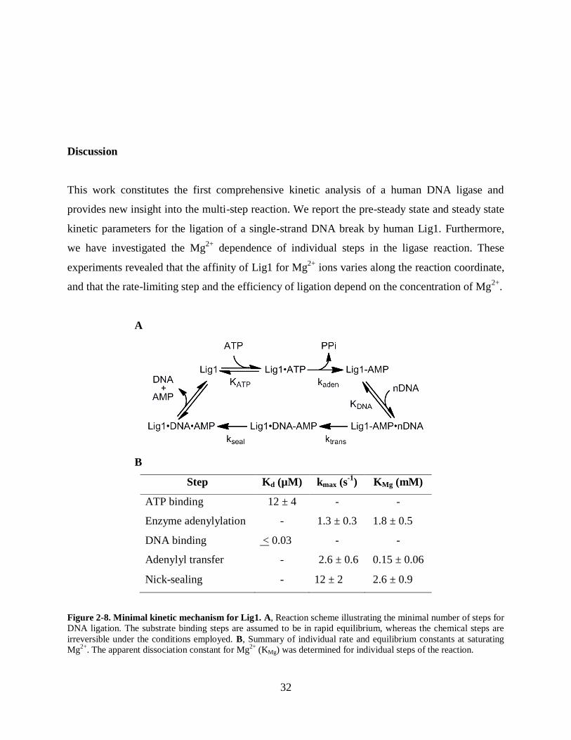
Discussion
This work constitutes the first comprehensive kinetic analysis of a human DNA ligase and
provides new insight into the multi-step reaction. We report the pre-steady state and steady state
kinetic parameters for the ligation of a single-strand DNA break by human Lig1. Furthermore,
we have investigated the Mg2+
dependence of individual steps in the ligase reaction. These
experiments revealed that the affinity of Lig1 for Mg2+
ions varies along the reaction coordinate,
and that the rate-limiting step and the efficiency of ligation depend on the concentration of Mg2+
.
A
B
Step Kd (µM) kmax (s-1
) KMg (mM)
ATP binding 12 ± 4 - -
Enzyme adenylylation - 1.3 ± 0.3 1.8 ± 0.5
DNA binding < 0.03 - -
Adenylyl transfer - 2.6 ± 0.6 0.15 ± 0.06
Nick-sealing - 12 ± 2 2.6 ± 0.9
Figure 2-8. Minimal kinetic mechanism for Lig1. A, Reaction scheme illustrating the minimal number of steps for
DNA ligation. The substrate binding steps are assumed to be in rapid equilibrium, whereas the chemical steps are
irreversible under the conditions employed. B, Summary of individual rate and equilibrium constants at saturating
Mg2+
. The apparent dissociation constant for Mg2+
(KMg) was determined for individual steps of the reaction.

33
Minimal kinetic framework for Lig1
The minimal kinetic mechanism for Lig1 is presented in Figure 2-8A. The three chemical steps
of ligase are essentially irreversible in the presence of excess ATP. The rates of enzyme
adenylylation and adenylyl transfer are evenly matched when the nicked DNA substrate is
saturating. The multiple-turnover rate constant (kcat) is 0.8 s-1
in this case. However, it is
important to note that DNA ligation occurring in excision repair pathways may not require
multiple-turnover ligation. We found that the recombinant Lig1 that we purified from E. coli is
fully adenylylated and extremely stable. The pool of adenylylated ligase may be sufficient for
ligation of low numbers of single-strand DNA breaks, and there may not be a strong driving
force to increase the rate of enzyme activation. When Mg2+
is saturating, the nick-sealing step is
significantly faster than the DNA adenylylation step, which would help to ensure efficient
coupling between nick recognition and nick-sealing. However, the lower affinity of Mg2+
in the
nick-sealing step leaves Lig1 vulnerable to uncoupling of these two chemical steps, which has
biological implications (see below).
Although the catalytic core is universally conserved in all ligases, there are additional domains
that vary between different isoforms in a given organism and between ligases from different
organisms. Therefore, it is interesting to compare the ligation activity of human Lig1, which is
one of the largest ligases, with the ligase from Chlorella virus, which is one of the smallest
ligases. Recent pre-steady state analysis of the Chlorella virus DNA ligase revealed almost
identical rate constants for adenylyl transfer and nick-sealing as we have observed for human
Lig1 [21]. Consistent with their similar kinetic parameters in vitro, both enzymes are able to
rescue the growth of yeast lacking cdc9, the replicative DNA ligase [22, 23].
DNA ligases require divalent metal ions for catalysis, therefore we have investigated the
magnesium dependence of the different steps in the Lig1-catalyzed reaction. The complex
behavior observed when ATP and Mg2+
concentrations are similar strongly suggests that Lig1
requires two Mg2+
ions for adenylylation (Figure 2-5D). One Mg2+
ion is expected to be bound
along with the ATP substrate, but a second is required that has much weaker affinity (KMg = 1.4

34
mM). A similar conclusion was reached in a study of the T4 bacteriophage DNA and RNA
ligases [24]. The use of more than one metal ion for phosphoryl and nucleotidyl transfer appears
to be quite common, with examples ranging from protein kinases [19] to DNA polymerases and
nucleases [25]. It is likely that subsequent steps also involve more than one metal ion, and there
is evidence of two metal ion binding sites in the structure of the adenylylated DNA intermediate
(10). The magnesium dependencies for adenylyl transfer and nick-sealing can be explained by a
single catalytic metal ion, but are also consistent with two metal ions that have different affinity.
In this case, the value of KMg that we have determined corresponds to the binding of the weakest
essential Mg2+
ion. Our experiments reveal that the affinity for this metal ion changes
significantly between different steps of the ligation reaction, increasing by an order of magnitude
between the enzyme adenylylation step and the adenylyl transfer step and then decreasing by a
similar degree in the nick-sealing step (Figure 2-8B). These changes in Mg2+
affinity could be
explained by conformational changes in the substrate or enzyme, or could indicate that unique
metal binding sites are involved in different steps of the reaction. Additional work is required to
determine the identity and catalytic roles of these Mg2+
ions. In many phosphoryl transfer
enzymes, Mg2+
ions play roles in activating the nucleophile and stabilizing the development of
negative charge.
Biological Implications of the Lig1 Magnesium Dependence
The decreased affinity of Lig1 for Mg2+
ions in the final nick-sealing step of the reaction renders
Lig1 susceptible to changes in the concentration of free Mg2+
ions. Lig1 faithfully completes
ligation at high concentrations of magnesium, however, low concentrations of magnesium cause
the enzyme to abort ligation. The released adenylylated DNA intermediate cannot be ligated,
because Lig1 reacts quickly with ATP and the occupancy of the AMP-binding pocket precludes
rebinding. Thus, repair pathways are needed to remove the 5’-blocking adenylyl group to allow
another opportunity for ligation. In the context of oxidative DNA damage, it has been suggested
that mammalian DNA ligases attempt to ligate nicks lacking 3’-hydroxyl groups [26]. Our
finding that low Mg2+
or high ATP concentration can lead to significant abortive ligation on a
normal DNA nick provides an additional possibility. If a DNA break is repaired under conditions
35
of imbalanced Mg2+
or Mg2+
-binding metabolites, then there is a significant risk that Lig1 will
fail to complete ligation. The abundance of Mg2+
(total concentration 10-40 mM) and similarity
to other metal ions has made it difficult to directly measure free Mg2+
concentration in the cell,
however the data that are available for several different cell types suggest that the majority of
Mg2+
is bound and the free Mg2+
is in the range of 0.2–3 mM [27]. The in vitro ligation
experiments show that this range of Mg2+
ion concentration is where Lig1 becomes susceptible
to abortive ligation and even slight decreases in the availability of free Mg2+
will further increase
the burden (Figure 2-7). Thus, abortive ligation intermediates could be frequently generated in a
wide variety of cellular contexts.
Although this abortive ligation was observed for a truncated form of Lig1 that lacks the first 232
amino acids, we observed that the amino terminus of Lig1 does not change the efficiency of
ligation under conditions of limiting Mg2+
in vitro (Appendix Figure A-11). However, this
portion of the protein is important for nuclear localization and for interactions with other
proteins, including the sliding clamp PCNA (7,13,14). It remains to be tested whether protein-
protein interactions alter the efficiency of Lig1 in the cell, but the existence of dedicated repair
pathways for adenylylated DNA suggest that abortive ligation does occur (26).
There are two pathways that would enable the repair of adenylylated DNA intermediates (Figure
2-9). The first pathway is the direct repair catalyzed by aprataxin. Aprataxin is a hydrolase that
catalyzes the removal of the AMP group from adenylylated DNA nicks to regenerate the 5’-
phosphate [26, 28]. Defects in aprataxin cause the neurodegenerative disease ataxia with
oculomotor apraxia 1, which is characterized by the loss of specific neuronal cells. It has been
suggested that the affected neuronal cells either experience higher levels of oxidative damage or
lack alternative repair pathways [26]. It is expected that long-patch base excision repair would
also be an effective way of repairing an abortive ligation intermediate (Figure 2-9). In this
pathway, strand displacement by a replicative polymerase would be followed by flap cleavage by
FEN1 to generate a new 5’-phosphate that would be a substrate of DNA ligase (Figure 2-9). In

36
support of this proposed pathway, strand displacement and FEN1 cleavage has been shown to
accept flap substrates with 5’ blocking lesions [29].
Figure 2-9. Pathways for repair of 5’-adenylylated DNA resulting from abortive ligation. Direct repair of a 5’-
adenylylated nick is catalyzed by aprataxin (APTX). Alternatively, strand displacement by a replicative DNA
polymerase generates a 5’-flap structure that can be processed by FEN1 to regenerate the adjacent 3’-hydroxyl and
5’-phosphate required by DNA ligase.
Our findings indicate that 3’-blocking lesions are not the only pathway for abortive ligation by
human ligases and suggest that there are multiple cellular perturbations that could impact the
frequency with which 5’-adenylylated DNA breaks are formed. As persistent DNA breaks are
expected to block transcription and to be converted into double-strand breaks upon DNA
replication, the formation and repair of 5’-adenylylated DNA is expected to be critical to cellular
viability and genomic stability. Additional work is needed to understand the physiological
conditions and molecular pathways important for the formation and repair of 5’-adenylylated
DNA breaks.

37
References
1. Petrini, J.H., Y. Xiao, and D.T. Weaver, DNA ligase I mediates essential functions in
mammalian cells. Mol Cell Biol, 1995. 15(8): p. 4303-8.
2. Simsek, D., et al., Crucial role for DNA ligase III in mitochondria but not in Xrcc1-
dependent repair. Nature, 2011. 471(7337): p. 245-8.
3. Gao, Y., et al., DNA ligase III is critical for mtDNA integrity but not Xrcc1-mediated
nuclear DNA repair. Nature, 2011. 471(7337): p. 240-4.
4. Sekiguchi, J., et al., Genetic interactions between ATM and the nonhomologous end-
joining factors in genomic stability and development. Proc Natl Acad Sci U S A, 2001.
98(6): p. 3243-8.
5. Barnes, D.E., et al., Targeted disruption of the gene encoding DNA ligase IV leads to
lethality in embryonic mice. Curr Biol, 1998. 8(25): p. 1395-8.
6. Lehman, I.R., DNA ligase: structure, mechanism, and function. Science, 1974.
186(4166): p. 790-7.
7. Tomkinson, A.E., et al., DNA ligases: structure, reaction mechanism, and function.
Chem Rev, 2006. 106(2): p. 687-99.
8. Shuman, S., DNA ligases: progress and prospects. J Biol Chem, 2009. 284(26): p.
17365-9.
9. Yang, S.W. and J.Y. Chan, Analysis of the formation of AMP-DNA intermediate and the
successive reaction by human DNA ligases I and II. J Biol Chem, 1992. 267(12): p. 8117-
22.
10. Pascal, J.M., et al., Human DNA ligase I completely encircles and partially unwinds
nicked DNA. Nature, 2004. 432(7016): p. 473-8.
11. Sriskanda, V. and S. Shuman, Specificity and fidelity of strand joining by Chlorella virus
DNA ligase. Nucleic Acids Res, 1998. 26(15): p. 3536-41.
12. Mackey, Z.B., et al., DNA ligase III is recruited to DNA strand breaks by a zinc finger
motif homologous to that of poly(ADP-ribose) polymerase. Identification of two
functionally distinct DNA binding regions within DNA ligase III. J Biol Chem, 1999.
274(31): p. 21679-87.
13. Ellenberger, T. and A.E. Tomkinson, Eukaryotic DNA ligases: structural and functional
insights. Annu Rev Biochem, 2008. 77: p. 313-38.
14. Song, W., et al., The DNA binding domain of human DNA ligase I interacts with both
nicked DNA and the DNA sliding clamps, PCNA and hRad9-hRad1-hHus1. DNA Repair
(Amst), 2009. 8(8): p. 912-9.
15. Kodama, K., D.E. Barnes, and T. Lindahl, In vitro mutagenesis and functional expression
in Escherichia coli of a cDNA encoding the catalytic domain of human DNA ligase I.
Nucleic Acids Res, 1991. 19(22): p. 6093-9.
16. Teraoka, H., et al., Expression of active human DNA ligase I in Escherichia coli cells that
harbor a full-length DNA ligase I cDNA construct. J Biol Chem, 1993. 268(32): p.
24156-62.

38
17. O'Sullivan, W.J. and D.D. Perrin, The Stability Constants of Metal-Adenine Nucleotide
Complexes. Biochemistry, 1964. 3: p. 18-26.
18. Phillips, R.C., P. George, and R.J. Rutman, Thermodynamic studies of the formation and
ionization of the magnesium(II) complexes of ADP and ATP over the pH range 5 to 9. J
Am Chem Soc, 1966. 88(12): p. 2631-40.
19. Waas, W.F. and K.N. Dalby, Physiological concentrations of divalent magnesium ion
activate the serine/threonine specific protein kinase ERK2. Biochemistry, 2003. 42(10):
p. 2960-70.
20. Cronin, C.N. and K.F. Tipton, The roles of magnesium ions in the reaction catalysed by
phosphofructokinase from Trypanosoma brucei. Biochem J, 1987. 247(1): p. 41-6.
21. Samai, P. and S. Shuman, Functional dissection of the DNA interface of the nucleotidyl
transferase domain of Chlorella virus DNA ligase. J Biol Chem, 2011.
22. Barnes, D.E., et al., Human DNA ligase I cDNA: cloning and functional expression in
Saccharomyces cerevisiae. Proc Natl Acad Sci U S A, 1990. 87(17): p. 6679-83.
23. Sriskanda, V., et al., Mutational analysis of Escherichia coli DNA ligase identifies amino
acids required for nick-ligation in vitro and for in vivo complementation of the growth of
yeast cells deleted for CDC9 and LIG4. Nucleic Acids Res, 1999. 27(20): p. 3953-63.
24. Cherepanov, A.V. and S. de Vries, Kinetic mechanism of the Mg2+-dependent
nucleotidyl transfer catalyzed by T4 DNA and RNA ligases. J Biol Chem, 2002. 277(3): p.
1695-704.
25. Steitz, T.A. and J.A. Steitz, A general two-metal-ion mechanism for catalytic RNA. Proc
Natl Acad Sci U S A, 1993. 90(14): p. 6498-502.
26. Ahel, I., et al., The neurodegenerative disease protein aprataxin resolves abortive DNA
ligation intermediates. Nature, 2006. 443(7112): p. 713-6.
27. Gunther, T., Concentration, compartmentation and metabolic function of intracellular
free Mg2+. Magnes Res, 2006. 19(4): p. 225-36.
28. Rass, U., I. Ahel, and S.C. West, Molecular mechanism of DNA deadenylation by the
neurological disease protein aprataxin. J Biol Chem, 2008. 283(49): p. 33994-4001.
29. Liu, Y., H.I. Kao, and R.A. Bambara, Flap endonuclease 1: a central component of DNA
metabolism. Annu Rev Biochem, 2004. 73: p. 589-615.

39
Appendix A: Figures, Tables and Experimental Methods to
Accompany Chapter 2
Appendix Methods
Fitting of ATP dependence
The ATP dependence of Lig1 suggests that adenylylation of Lig1 follows a random, bireactant
system in which there is a requirement for both the ATP•Mg2+
complex and a free magnesium
ion (Mg2+
F) for activity (Scheme A-1).
Scheme A-1: Kinetic model describing substrate binding in Lig1.
For simplicity, all of the steps following initial substrate binding have been grouped together into
kcat. The term is the cooperativity constant that represents the change in substrate affinity for
the second substrate after the first has already bound. The generic form of this system, where the
two substrates are represented by the variables A and B, has been described by Segel (1). The
equation describing the system is available as equation VI-2:

40
v
Vmax
[A][B]
(KA )(KB)(KA )[B](KB )[A][A][B] Appendix Equation A-1
Designating Vmax = kcat[E]t:
Appendix Equation A-2
For our model, the two substrates for the enzyme are ATP•Mg2+
and Mg2+
F. The concentrations
of these species present in our reactions must be determined from the total concentrations of
Mg2+
and ATP and the dissociation constants for Mg2+
and ATP. ATP is known to potentially
bind two magnesium atoms, resulting in the equilibrium pictured in Figure A-2.
Scheme A-2: Binding of magnesium by ATP.
Previous studies have shown that the second magnesium ion is bound with much lower affinity
than the first (K2 ~ 20 mM and K1 ~ 20 M). This results in a low population of the
ATP•(Mg2+
)2 complex under the conditions that we have employed and we have therefore
simplified the analysis by ignoring the negligible binding of the second magnesium ion to
ATP•Mg2+
from our calculations. Using the definition of the dissociation constant, K1, and the
law of conservation of mass, we can determine relations between the concentrations of
ATP•Mg2+
and Mg2+
F and our known constants. The relation for the dissociation constant K1:
Appendix Equation A-3

41
From conservation of mass:
Appendix Equation A-4
Appendix Equation A-5
Rearrangement of Appendix Equation A-4 and Appendix Equation A-5 yields Appendix
Equation A-6 and Appendix Equation A-7, respectively:
Appendix Equation A-6
Appendix Equation A-7
Substituting Appendix Equation A-6 and Appendix Equation A-7 into Appendix Equation A-3
and rearranging gives the following:
Appendix Equation A-8
Which can be input into the quadratic equation to yield an equation for [ATP•Mg2+
]:
Appendix Equation A-9
Only the negative root is used in this equation because use of the positive root would result in an
ATP•Mg2+
concentration greater than the concentration of ATPT or Mg2+
T, which is impossible.
The Mg2+
F concentration can then be calculated from Appendix Equation A-7, using the value of
[ATP•Mg2+
] obtained from Appendix Equation A-9.

42
Calculation of rate constant for enzyme adenylylation
We calculated the rate constant for enzyme adenylylation from the measured steady state rate
and the separately determined microscopic rate constants for adenylyl transfer and nick-sealing.
This analysis assumes that the substrate binding and release steps occur very quickly and that the
steady state rate constant (kcat) is controlled by the rate constants for the individual phosphoryl
transfer steps. The reciprocal of the overall rate of reaction is given by the sum of the recipricols
of the individual steps (2) as shown in Appendix Equation A-10:
Appendix Equation A-10
Rearranging yields Appendix Equation A-11:
Appendix Equation A-11
Using the experimentally determined values of 2.6 s-1
, 12 s-1
and 0.81 s-1
for the adenylyl
transfer, nick-sealing and steady-state ligation rate constants, respectively, we determine the
enzyme adenylylation rate constant to be 1.3 s-1
under saturation conditions.
Analysis of the abortive ligation catalyzed by Lig1 at limiting Mg2+
The simplest model to explain the abortive ligation involves direct partitioning between nick-
sealing and release of the adenylylated DNA intermediate (see the inset of Figure 2-7). The Mg2+
dependence of the nick-sealing step was determined independently from the single-turnover
ligation at high concentration of Mg2+
(Figure 2-8, using the data from Figure 2-4). In the
absence of direct information regarding the binding affinity of the adenylylated intermediate, we
assumed that the rate constant for its release (koff) is independent of Mg2+
concentration.

43
Substitution into the partitioning equation (Equation 2-4) gives Appendix Equation A-12, in
which kseal is the rate constant for nick sealing, and KMg is the apparent dissociation constant for
Mg2+
in the nick-sealing step.
Appendix Equation A-12

44
A B
Appendix Figure A-1: Evaluation of different quenching solutions. Single-turnover ligation reactions contained
600 nM Lig1 and 80 nM DNA in standard reaction buffer with 10 mM MgCl2 and were quenched at 4 ms (A) or 400
ms (B) in the indicated quench solution and the amounts of intermediate and product formed were quantified via the
gel-based ligation assay. A, Quench solutions that contain either EDTA or concentrated NaOH result in less than 1%
formation of intermediate or product when the reaction is quenched at 4 ms. In contrast, urea quench solutions allow
the reaction to proceed to roughly 40% completion. B, The EDTA and NaOH quench solutions stop the reaction at a
similar extent of reaction at 400 ms. Urea quenches appear to act more slowly, allowing for greater product
formation and intermediate breakdown.
A B
0.015 mM MgCl2
[Lig1] ktransfer (s-1
) kseal (s-1
)
400 nM 0.045 ± 0.013 0.040 ± 0.021
600 nM 0.054 ± 0.008 0.044 ± 0.011
800 nM 0.049 ± 0.020 0.045 ± 0.016
Appendix Figure B-2: Representative single-turnover ligation kinetics indicate that the concentration of Lig1
employed is far above the Kd for substrate binding. The rate constants for adenylyl transfer and nick-sealing were
determined for single-turnover reactions containing 80 nM DNA and 400–800 nM Lig1 in standard reaction buffer
with 0.015 mM MgCl2 (A) or 10 mM MgCl2 (B). The rate constants determined for each chemical step are
equivalent, within error, across all enzyme concentrations tested, indicating that the single-turnover reaction is
saturated with 400 nM Lig1 (Kd >> 400 nM). Unless otherwise indicated, we used 600 nM Lig1 to ensure that the
maximal single-turnover rate was measured.
10 mM MgCl2
[Lig1] ktransfer (s-1
) kseal (s-1
)
400 nM 2.1 ± 0.8 9.1 ± 1.1
600 nM 2.3 ± 0.4 9.6 ± 0.6
800 nM 2.2 ± 0.7 9.8 ± 1.3

45
Appendix Figure A-3: Active-site titration to determine the concentration of active (adenylylated) LIG1. Single-turnover reactions were in the standard buffer containing 250 nM DNA and 10 mM MgCl2 and the indicated
concentration of Lig1. Reactions were quenched after 10 minutes and the ligation was limited to a single turnover,
because no ATP was present. Prior to this experiment, the concentrations of ligase and DNA were based on
measurement of the UV-absorbance. According to the titration, 270 nM ligase would be required to fully ligate 250
nM nicked DNA. We adjusted the concentration of the Lig1 stock by 7.4 % to ensure that the concentration of
substrate and enzyme were equivalent. A similar titration was performed on enzyme that was loaded into the
quench-flow apparatus and this revealed that there was a reproducible loss of ~10% of the enzyme during the rapid-
mixing experiments. After this correction we measured identical steady state rate constants from reactions
performed either in the quench-flow or with manual mixing on the bench-top.
A B
Appendix Figure A-4: Stability of LIG1 in reaction buffer. Reactions containing 20 nM Lig1, 40 nM DNA and
10 mM MgCl2 in standard reaction buffer were quenched after 10 minutes and the fraction of ligated DNA was
determined. Because ATP was omitted from the reaction, ligation was limited to a single turnover. 100 nM stock
solutions of Lig1 used in the reactions had been incubating at 4oC (A) or 37
oC (B) in 1x reaction buffer for various
times prior to the experiment. There is no appreciable change in ligation % across the time-span tested under either
condition, indicating that ligase is very stable for up to 5 months at 4 o
C, or an hour at 37 o
C. All kinetic data
reported in the accompanying paper was obtained from reactions lasting less than 40 minutes.

46
A B
Appendix Figure A-5: DNA concentration dependence of the multiple-turnover ligation reaction. The rate of
multiple-turnover ligation was determined across a range of DNA concentrations for reactions containing 1 nM
ligase in 1x reaction buffer with 200 M ATP at either 0.2 mM MgCl2 (A) or 10 mM MgCl2 (B). Both sets of data
fit well to the Michaelis-Menten equation and yield a KM for DNA of 24 ± 8 nM. Reactions were performed in
duplicate.
Appendix Figure A-6: Multiple-turnover ligation is linearly dependent upon the concentration of LIG1. The
rate of product formation was determined from multiple-turnover reactions containing a range of ligase
concentrations and saturating DNA (1 M) in 1x reaction buffer with 200 M ATP and 10 mM MgCl2. The linear
response of the reaction velocity to enzyme concentration shows that Lig1 does not undergo any concentration
dependent changes in activity across this range and that similar reactions performed across this range of ligase
concentrations are directly comparable.
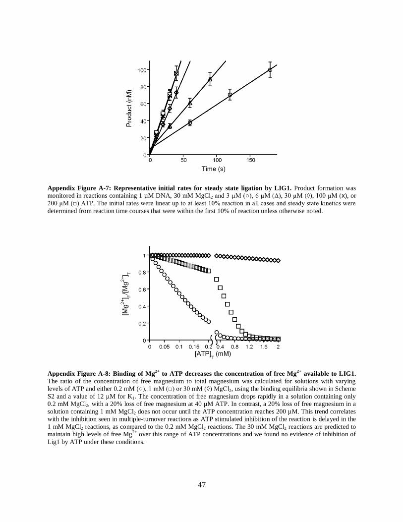
47
Appendix Figure A-7: Representative initial rates for steady state ligation by LIG1. Product formation was
monitored in reactions containing 1 µM DNA, 30 mM MgCl2 and 3 µM (○), 6 µM (Δ), 30 µM (◊), 100 µM (x), or
200 µM (□) ATP. The initial rates were linear up to at least 10% reaction in all cases and steady state kinetics were
determined from reaction time courses that were within the first 10% of reaction unless otherwise noted.
Appendix Figure A-8: Binding of Mg2+
to ATP decreases the concentration of free Mg2+
available to LIG1.
The ratio of the concentration of free magnesium to total magnesium was calculated for solutions with varying
levels of ATP and either 0.2 mM (○), 1 mM (□) or 30 mM (◊) MgCl2, using the binding equilibria shown in Scheme
S2 and a value of 12 µM for K1. The concentration of free magnesium drops rapidly in a solution containing only
0.2 mM MgCl2, with a 20% loss of free magnesium at 40 µM ATP. In contrast, a 20% loss of free magnesium in a
solution containing 1 mM MgCl2 does not occur until the ATP concentration reaches 200 µM. This trend correlates
with the inhibition seen in multiple-turnover reactions as ATP stimulated inhibition of the reaction is delayed in the
1 mM MgCl2 reactions, as compared to the 0.2 mM MgCl2 reactions. The 30 mM MgCl2 reactions are predicted to
maintain high levels of free Mg2+
over this range of ATP concentrations and we found no evidence of inhibition of
Lig1 by ATP under these conditions.

48
Parameter 0.2 mM MgCl2 1 mM MgCl2 30 mM MgCl2
K1 (M) 1.210-5
1.210-5
1.210-5
KMgF (M) 6.610-4
6.610-4
6.610-4
KATP•Mg (M) 5.010-5
1.710-5
1.110-5
kcat (s-1
) 0.59 0.8 0.8
α 5 5 5
Appendix Figure A-9: Kinetic and thermodynamic constants obtained from fits of the ATP concentration
dependencies with the two magnesium model for LIG1. These parameters are from the curve fits shown in the
text (Figure 2-5). The ATP concentration dependencies were fit to the equation describing the bisubstrate model of
Appendix Scheme A-1 (Appendix Equation A-2) using the program Kaleidagraph. In this equation the species A
and B represent the ATP•Mg2+
complex and free Mg2+
ion, respectively. The expressions describing the
concentrations of the ATP•Mg2+
complex and free Mg2+
ion were derived from the binding equilibria in Appendix
Scheme A-2 (Appendix Equation A-9 and A-7) and were substituted into Appendix Equation A-2 for fitting to the
known experimental conditions. We were unable to fit all three data sets to common parameters, but reasonable fits
were obtained with constant values for the affinity of ATP for Mg2+
, Lig1 affinity for free Mg2+
, and an α value for
cooperative binding of ATP•Mg2+
and Mg2+
. The resulting best fit values for kcat and the binding affinity of
ATP•Mg2+
to Lig1 are in reasonable agreement. The failure to obtain a perfect fit to all of the data could indicate
experimental error or additional complexities not considered in our model, such as inhibition by free ATP or
additional binding sites for Mg2+
on Lig1 or the DNA substrate.
Appendix Figure A-10: Measurement of kcat/KM for utilization of ATP (kcat/KM)ATP
by LIG1. Representative
data are shown for reactions containing 4 nM ligase and 1 M DNA in the standard reaction buffer with 0.5 M, 1
M, and 2 M ATP and 0.5 mM (○), 5 mM (□) or 15 mM (◊) MgCl2. The linear dependence of the initial rates upon
the concentration of ATP indicates that these reactions are sub-saturating for ATP and the value of (kcat/KM)ATP
is
given by the slope of the line. All reactions were performed in triplicate and the error bars indicate the standard
deviation.

49
References
1. Segel, I. H. (1975) Enzyme Kinetics: Behavior and Analysis of Rapid Equilibrium and
Steady-State Enzyme Systems, John Wiley & Sons, Inc., New York
2. Cleland, W. W. (1975) Biochemistry 14, 3220-3224

50
Chapter 3: Comparison of Manganese and Magnesium as Cofactors
for Human DNA Ligase I
Abstract
DNA ligases utilize a catalytic mechanism that involves three phosphoryl transfer reactions.
Each of these chemical steps requires the presence of one or more divalent metal cofactors.
While most phosphoryl transfer enzymes appear to use magnesium as the physiological cofactor,
many are also able to utilize manganese. Metal substitution often results in changes to enzyme
catalysis, including changes in substrate specificity and affinity, or the rate of catalysis during
metal-dependent steps. Characterization of single and multiple turnover reactions of human DNA
ligase I in the presence of manganese have shown that manganese can substitute for magnesium
in all steps of the reaction. Surprisingly, low affinity inhibition by manganese exists for some,
but not all of the reaction steps. Inhibition is not competitive with magnesium, suggesting that
inhibitory metal binding occurs outside of the active-site. This research shows the LIG1 active
site has enough plasticity to efficiently accommodate both magnesium and manganese cofactors
while also revealing a previously unknown inhibitory metal site.

51
Introduction
DNA ligases seal breaks in the DNA backbone by catalyzing formation of a phosphodiester bond
between adjacent DNA strands [1]. This function is absolutely essential to maintain the integrity
of the genome after nicks are formed during DNA repair and replication. Higher eukaryotes
encode three different DNA ligases, each specializing in a different area of DNA break repair.
DNA ligase I (LIG1) is the most abundant of the three and accomplishes the majority of nuclear
single-strand DNA break repair [2]. DNA ligase III exhibits both single- and double-strand break
repair activity and accomplishes all DNA repair within the mitochondria [3, 4]. DNA ligase III is
also expressed in the nucleus, however this form appears to dispensable for normal cellular
function. DNA ligase IV is responsible for nuclear double-strand break repair such as that during
non-homologous end-joining [5, 6]. All three ligases share a common catalytic core consisting of
three-independently folding domains. It is expected that a comprehensive understanding of LIG1
will provide a useful framework for understanding all human ligases.
Enzymatic DNA ligation follows a reaction pathway that is universally conserved from bacteria
to higher eukaryotes (Scheme 3-1) [1]. The pathway consists of 3 independent phosphoryl
transfer reactions. The first step of the pathway, enzyme adenylylation, involves attachment of an
AMP group to an active site lysine on the enzyme using either ATP or NAD+ as the nucleotide
donor. ATP serves as the nucleotide donor for all mammalian DNA ligases. Enzyme
adenylylation requires the presence of two divalent metals, one that comes in with the ATP
molecule and another captured from solution [7]. In the second step of the pathway, adenylyl
transfer, the enzyme catalyzes attack of the 5' phosphate at the nick on the adenylylated lysine,
resulting in transfer of the AMP to the DNA and formation of an adenylylated DNA
intermediate. The final step of the reaction involves catalyzing attack of the 3' hydroxyl at the
nick on the adenylylated phosphate, forming a phosphodiester bond and sealing the nick. Both
DNA dependent steps require the presence of at least one divalent metal ion [8]. A crystal
structure of LIG1 in complex with adenlylyated DNA pre-nick-sealing reveals two potential

52
metal binding sites well-coordinated by universally conserved active-site residues [9]. Metal ions
in these sites would interact closely with important substrate chemical groups, however the
function of a metal in either site is not known.
Scheme 3-1: Kinetic mechanism of enzymatic DNA ligation.
We recently characterized the kinetic mechanism of DNA ligation in the presence of magnesium,
the presumptive physiological cofactor [7]. Many magnesium-dependent phosphoryl transfer
enzymes are able to utilize manganese in place of magnesium as the catalytically required metal
cofactor [10-12]. In many cases, study of the differences between reactions catalyzed in
magnesium or manganese catalyzed reactions gives functional insight into the role of metal ions
during catalysis. We report here a systematic comparison of manganese and magnesium as metal

53
cofactors for enzymatic ligation by human DNA ligase I. We find manganese is able to substitute
for magnesium in every step of the reaction, displaying similar binding affinities and resulting in
similar maximal rate constants. We also discover the presence of an inhibitory metal site specific
for manganese over magnesium. Surprisingly, inhibition occurs during enzyme adenylylation
and adenylyl transfer, but not nick-sealing. More potent inhibition occurs in mixtures of
magnesium and manganese, suggesting that ligase activity could be compromised under
conditions of manganese metal toxicity.

54
Methods
Recombinant DNA Ligase I
The catalytic domain of human LIG1 (residues 232-919) was expressed in E. coli and purified as
previously described [7]. Cells were lysed in the presence of 1 mM EDTA to preserve the
adenylylated form of the enzyme, and subsequently purified over a phosphocellulose column and
a NTA-nickel column. The His tag was removed with Prescission protease, and LIG1 was further
purified with Q-sepharose. The final purified fractions were combined and dialyzed into storage
buffer (25 mM Tris•Cl, pH 7.6, 150 mM NaCl, 1 mM DTT, and 0.1 mM EDTA). Aliquots were
snap-frozen and stored at -80 oC. Purity was greater than 95% as judged by SDS-PAGE. Initial
concentrations were estimated from the absorbance at 280 nm, using the calculated extinction
coefficient. The concentration of active, adenylylated enzyme was determined by titration with
nicked DNA substrate, as described previously [7], and this active concentration is reported
throughout.
DNA Substrates
Oligonucleotides were synthesized by Integrated DNA Technologies or by the Keck Center at
Yale University and were purified on denaturing polyacrylamide gels. The portion of the gel
containing the full-length oligonucleotide was excised, crushed, and extracted by soaking
overnight in 500 mM NaCl and 1 mM EDTA. The extracted oligonucleotides were desalted by
binding to a C18 reverse phase column (Sep-pak, Waters) and eluted with 30% (v/v) acetonitrile.
Concentrations were obtained from the absorbance at 260 nm using the calculated extinction
coefficients. The three oligonucleotides used in this study had sequences 5’-
CCGAATCAGTCCGACGACGCATCAGCAC, 5’-GTGCTGATGCGTC, and 5’-P-
GTCGGACTGATTCGG-FAM (P indicates 5’ phosphorylation and FAM indicates the presence

55
of a 3’ fluorescein). The nicked, double-stranded DNA substrate (nDNA) was formed by mixing
equimolar amounts of the three oligonucleotides in 10 mM NaMES pH 6.5 and 50 mM NaCl and
cooling the mixture from 90 °C to 4 °C at a rate of 3 °C per minute.
Gel-based Ligation Assay
Ligation reactions were performed at 37 °C. Unless otherwise indicated, the standard buffer
contained 50 mM NaMOPS pH 7.5 (measured at 25 °C), 1 mM dithiothreitol, 0.05 mg/mL BSA,
and sufficient NaCl to maintain a constant ionic strength of 150 mM. The amounts of ATP,
MgCl2, MnCl2, nDNA, and LIG1 varied, as indicated below. Preincubation controls established
that LIG1 retains 100% of its activity after 1 hr in this standard buffer, and all of the kinetic data
was collected within this window of time (Appendix Figure B-1) [7]. Reactions were quenched
as described below, heated to 95 °C for 5 min to denature the DNA and resolved on 20% (w/v)
denaturing polyacrylamide gels containing 8 M Urea. Fluorescein-labeled oligonucleotides were
detected with a Typhoon Trio+ imager (GE Healthcare) with excitation at 488 nm and emission
through a 520 nm band-pass filter. The images were analyzed using ImageQuantTL (GE
Healthcare). The intensity of the individual DNA species was corrected for background
fluorescence, and the fraction of the total fluorescence was determined by dividing the
fluorescence intensity for the desired species by the signal for all other species in the sample.
When necessary, this fraction was converted into its concentration by multiplying by the total
concentration of DNA in the reaction.
Multiple-Turnover Ligation Assays
Steady state kinetic analysis was performed in the standard ligation buffer and the temperature
was maintained at 37 °C in a circulating water-bath. Reaction mixtures were preincubated for 5
min prior to addition of LIG1. Reactions were arrested by quenching a 4 µL aliquot in 15 µL of
quench solution (50 mM EDTA in 90% formamide), and the extent of ligation was determined as
described above. Initial rates were determined from the linear rate of substrate disappearance

56
within the first 10% of the reaction. When both intermediate and product were formed during the
reaction, the rate of formation for each species was determined. Values reported represent the
average of at least three experiments. Equations used to fit data are described in the Models of
Kinetic Parameters section and in the text. All fitting of multiple-turnover data was done using
Kaleidagraph, Synergy Software.
Rapid Quench Experiment
Rapid mixing experiments were performed in a Kintek RFQ-3 quench-flow apparatus. One
sample loop contained LIG1 and the other contained DNA, each at double the reaction
concentration. The samples were in 1x reaction buffer with the desired metal ion concentration
and the drive syringes contained the same solution. Loaded reactants were allowed to equilibrate
for 90 seconds to reach the correct temperature. Reactions were initiated by mixing of 20 µL of
each sample and were quenched at the desired times by mixing with 20 µL of quench solution,
held in the quenching syringe. Data obtained at concentrations above 1 mM manganese with the
50 mM EDTA in 90% formamide quench used previously [7] did not fit well to the 2-step
irreversible model (Appendix Figure B-2). Data from identical reactions instead quenched with
concentrated HCl do fit well to a 2-step irreversible model. Comparison of the data from
EDTA/formamide or HCl quenched reactions reveals that a larger amount of adenylylated DNA
intermediate is formed when reactions are quenched with EDTA/formamide. This suggests that
the EDTA/formamide quench does not stop the adenylyl transfer step on the sub-millisecond
time-scale required for quench-flow experiments. For this reason, 3M HCl was used as the
quenching solution for all single-turnover experiments. 5 seconds after quenching, samples were
brought to neutral pH by addition of 20 L of 3 M NaOH. 160 mL of 50 mM EDTA in 90%
formamide was added following neutralization and samples were analyzed with the standard gel-
based ligation assay. Unless otherwise indicated, the typical concentrations of nDNA and LIG1
were 80 nM and 600 nM, respectively. The data were imported into the program Berkeley-
Madonna (www.berkeleymadonna.com) and the curve fitting function was used to globally fit
the levels of intermediate and product by the scheme represented in Scheme 3-1. Fitting of the
data to a model including reversible chemical steps did not produce significantly different results

57
(data not shown). Fits of reactions from individual experiments provided values for the rate
constants for adenylyl transfer and nick-sealing and the reported values reflect the average rates
determined from at least three independent experiments at each metal concentration. These rate
constants were plotted versus the concentration of metal ions. Fitting of the data is discussed in
the results section.

58
Models of Kinetic Parameters
)][
1(*])[(
][*
I
A
Aobs
K
IAK
Akk
Equation 3-1
Equation 3-1 describes the non-competitive inhibition model where a reaction is activated by
binding of species A to a single stimulatory site and is inhibited by binding of species I to a
single inhibitory site. kobs is the observed rate constant for the step being measured, kA is the
maximal rate constant, KA is the observed affinity for species A during the observed step, and KI
is the observed affinity for species I. Equation 3-1 is used for fitting the manganese data in
Figures 3-1B, 3-2B, and 3-3B. It is also used for fitting the data in Figures 3-3A, 3-3B, and 3-5A
where manganese is inhibiting the magnesium stimulated reaction. In cases where manganese
acts to activate as well as inhibit catalysis, A = I.
][
][*
AK
Akk
A
Aobs
Equation 3-2
Equation 3-2 describes the model where a reaction is activated by binding of species A to a
single stimulatory site. kobs is the observed rate constant for the step being measured, kA is the
maximal rate constant, and KA is the observed affinity for species A during the observed step.
Equation 3-2 is used for fitting the magnesium data in Figures 3-1B, 3-2B, 3-4A, and 3-4B. It is
also used for fitting of the manganese dependence of nick-sealing in Figure 3-4B and takes the
form of the Michaelis-Menten equation for the ATP dependence of multiple-turnover in
Appendix Figure B-4.

59
BA
B
B
A
A
obs
K
B
K
A
K
Bk
K
Ak
k][][
1
][*
][*
Equation 3-3
Equation 3-3 describes the model where a reaction is activated by competitive binding of species
A or species B to a single stimulatory site. kobs is the observed rate constant for the step being
measured, kA and kB are the maximal rate constants when either species A or B is bound,
respectively, and KA and KB are the observed affinities for species A and B, respectively, during
the observed step. Equation 3-3 is used for fitting the data in Figure 3-5B.

60
Results
Manganese Stimulated Steady-State Ligation
To evaluate the ability of manganese to stimulate DNA ligation, we performed multiple-turnover
reactions at varying concentrations of manganese. These reactions spanned the range of
manganese concentrations from 100 M to 20 mM. The rate of all multiple-turnover reactions
were determined from data obtained at less than 10% product formation, ensuring measurement
of the steady-state reaction (Figure 3-1A). Observation of identical V/E values for reactions with
both 250 nM and 1 M DNA at both 100 M and 20 mM manganese indicate that the 1 M
DNA concentration used for the multiple-turnover experiments is saturating (Appendix Figure
B-3A, B). Product formation remains linear over the time-course of the reaction requiring the
most time (Appendix Figure B-3, B), and pre-incubation of the enzyme under typical reaction
conditions without DNA does not affect the rate of reaction (Appendix Figure B-1). These
results indicate that LIG1 is stable under these reaction conditions in the presence of manganese,
similar to what we have previously seen with magnesium [7]. The dependence of the manganese
stimulated steady-state rate on ATP was found to be similar to that observed with magnesium,
with a KM,ATP of 12 M (Appendix Figure B-4).
The maximal multiple-turnover rate constant at saturating DNA and ATP, kcat, displays a
biphasic dependence on manganese concentration, increasing from 0 mM to 1 mM manganese
and then decreasing above 1 mM manganese (Figure 3-1B). The biphasic dependence of kcat fits
well to a noncompetitive inhibition model where one manganese ion activates the enzyme and a
second manganese ion inhibits (Equation 3-1). This fit yields a kcat of 1.3 ± 0.1 s-1
, with a
stimulatory K1/2 for manganese, KMn, of 0.5 ± 0.08 mM and an inhibitory K1/2 for manganese, KI,
of 2.1 ± 0.6 mM. These data show that manganese can fulfill all the catalytically required roles
of magnesium during ligation. Data previously obtained in the presence of magnesium is plotted
alongside the manganese data for comparison (Figure 3-1B, Equation 3-2). Comparison of the

61
kinetic constants measured with manganese to those measured with magnesium reveals that the
two metals bind the stimulatory site with similar affinity and result in a similar reaction rate (0.5
mM and 1.3 s-1
vs. 0.71 mM and 0.81 s-1
for manganese and magnesium, respectively) (Table 3-
1).
A B
Figure 3-1: Multiple-turnover ligation with Mn2+
and saturating ATP and DNA. A, Representative initial rates
of DNA ligation with Mn2+
as the cofactor. Reactions contained saturating DNA (1 M) and 4 nM LIG1 and the
mean ± SD is shown for 3 independent reactions. B, The kcat value for LIG1-catalyzed ligation shows a biphasic
dependence on the concentration of Mn2+
(○). For comparison, the kcat dependence on magnesium is plotted from
[7]. The Mg2+
dependence is represented by a single-site binding model (Equation 3-2, dashed black line), the Mn2+
dependence is fit by a noncompetitive inhibition model (Equation 3-1, solid red line).
Manganese Stimulation of ATP-dependent Reactions
The use of subsaturating levels of ATP allows monitoring of the ATP dependent steps of the
reaction, ATP binding and enzyme adenylylation (Scheme 3-1). Analysis of the second order
rate constant for the utilization of ATP ((kcat/KM)ATP
) under these conditions allows us to
investigate how manganese affects the ATP-dependent reactions for DNA ligation. The linear
dependence of the steady-state rate on the concentration of ATP at less than 2 mM ATP confirms
that (kcat/KM)ATP
is determined (Figure 3-2A). The value of (kcat/KM)ATP
displays a biphasic
dependence on manganese (Figure 3-2B), similar to that observed when ATP is saturating in
multiple-turnover. A fit of this data to the equation describing a noncompetitive inhibition model

62
(Equation 3-1) where manganese acts as both the activating and inhibitory cofactor yields a
maximal (kcat/KM)ATP
value of (1.2 ± 0.2) x 105 M
-1s
-1, and identical values for KA and KI of 2.1 ±
0.6 mM (Table 3-1). The metal affinities have a large error associated with them in the fitting
program, Kaleidagraph. This suggests that multiple solutions exist that fit the model to the data.
Thus, while this set of numbers is the best fit for the data, they should be considered as rough
estimates of the real value.
A B
Figure 3-2: Multiple-turnover with Mn2+
and sub-saturating ATP. A, Representative initial rates of ligation for
sub-saturating amounts of ATP that are used to calculate the value of (kcat/KM)ATP
at different concentrations of
Mn2+
. B, The value of (kcat/KM)ATP
displays a biphasic dependence on the concentration of Mn2+
(○). For
comparison, the (kcat/KM)ATP
dependence on magnesium is plotted from [7].The Mg2+
dependence is represented by
a single-site binding model (Equation 3-2, dashed black line), and the Mn2+
dependence is fit by a noncompetitive
inhibition model (Equation 3-1, solid red line).
Inhibitory Manganese is Not Competitive with Magnesium in Multiple-Turnover
To investigate the effect of manganese on magnesium stimulated multiple-turnover ligation, we
followed reactions containing saturating concentrations of magnesium and varying
concentrations of manganese. The steady-state rate of product formation decreases in response to
increasing levels of manganese under these conditions (Figure 3-3A). The manganese
dependence of the observed multiple-turnover rate fits well to a noncompetitive inhibition model
(Equation 3-1), where magnesium is the activating cofactor, A, and manganese is the inhibitory

63
cofactor, I. This model yields superimposable fits to the data at 5 mM and 20 mM magnesium,
yielding a KI for the multiple-turnover reaction of 0.22 ± 0.06 mM and 0.19 ± 0.04 mM,
respectively. Absence of a change in the KI for manganese over a 4-fold change in magnesium
ion concentration indicates that the inhibitory manganese ion(s) do not compete with
magnesium. The significantly tighter affinity for the inhibitory manganese measured in a
background of magnesium in this experiment as opposed to that reported above with manganese
alone (0.2 mM vs. 2.1 mM, respectively) suggests the measurement of a different inhibitory
species between the two experiments. This will be evaluated further in the discussion section.
Figure 3-3: Mn2+
is a noncompetitive inhibitor of Mg2+
-dependent ligation. A, The rate constant for multiple
turnover ligation (kcat) with saturating DNA (1 M) and ATP (200 M) was measured at both 5 mM (○) and 20 mM
(⎕) Mg2+
and varying amounts of Mn2+
. The data were fit by a noncompetitive inhibition model (Equation 3-1). Fits
of the data for the two different concentrations of Mg2+
are superimposable and yield KI values of 0.22 mM and 0.19
mM Mn2+
at 5 mM and 20 mM Mg2+
, respectively. B, The second order rate constant for ligation at sub-saturating
ATP, (kcat/KM)ATP
, was determined at both 5 mM (○) and 20 mM (⎕) Mg2+
and varying concentrations of Mn2+
. The
data were fit by a noncompetitive inhibition model (Equation 3-1) and yield KI values of 4.1 mM and 8.4 mM Mn2+
at 5 mM and 20 mM Mg2+
, respectively.
The manganese dependence of the magnesium stimulated multiple-turnover rate under
(kcat/KM)ATP
conditions fits well to a noncompetitive inhibition model in which magnesium acts
as the stimulatory metal and manganese is inhibitory (Figure 3-3B, Equation 3-1). Though the
curves have different (kcat/KM)ATP
max values due to 5 mM magnesium being subsaturating under
(kcat/KM)ATP
conditions, the KI values are quite similar (4.1 ± 0.7 mM and 8.4 ± 0.9 mM at 5

64
mM and 20 mM magnesium, respectively). This suggests that inhibition by manganese is not
caused by displacement of active-site magnesium ions, but must instead be occurring via binding
to some other site(s).
Stimulation of Single-Turnover Ligation by Manganese
To determine the effect of manganese on adenylyl transfer and nick-sealing, we performed
single-turnover ligation reactions across a range of manganese ion concentrations. Under-single
turnover conditions, adenylylated enzyme is added to substrate in the absence of ATP, ensuring
that each enzyme can turnover only once. Use of saturating levels of enzyme in vast excess over
substrate ensures that the observed rate constants are not affected by substrate binding. The rates
for these steps were determined as described in the methods. Briefly, the level of intermediate
and product present in the reaction over time is fit by a 2-step irreversible mechanism using the
program Berkeley-Madonna. These fits give values for the rates of adenylyl transfer and nick-
sealing, which are reported here. 3 M HCl was used as the quench in the single-turnover
experiments reported here, as the EDTA/formamide quench used previously did not stop the
reaction on the required time-scale (see Methods, Appendix Figure B-2). This may reflect slower
dissociation of manganese than magnesium. Single-turnover reactions spanned the range of
manganese concentrations from 10 M to 10 mM. Identical rate constants were obtained from
reactions performed with either 800 nM or 3200 nM LIG1 at various manganese concentrations,
indicating saturating levels of enzyme (Appendix Figure B-5, Appendix Table B-1).
Similar to what was observed for kcat, the rate constant for adenylyl transfer, ktransfer, displays a
biphasic dependence on manganese concentration, resulting in an increase from 0 mM to 0.1 mM
Mn2+
, and then a decrease above 0.1 mM Mn2+
(Figure 3-4A). Fitting the data to the equation
describing a noncompetitive inhibition model (Equation 3-1), in which manganese acts as both
the activating and inhibiting cofactor, yields a maximal rate constant for adenylyl transfer,
ktransfer, of 8.7 ± 1.2 s-1
, with a KA of 0.12 ± 0.06 mM and a KI of 0.13 ± 0.08 mM. Similar to the
values obtained from the (kcat/KM)ATP
measurements, the numbers given here have large reported
errors from Kaleidagraph, suggesting they are not a unique solution to the data. In contrast to the

65
adenylyl transfer step, the nick-sealing reaction displays a simple hyperbolic binding dependence
on manganese concentration, similar to what is observed for magnesium (Figure 3-4B). Fitting of
the nick-sealing data to a hyperbolic binding curve (Equation 3-3) yields a maximal nick-sealing
rate constant, kseal, of 6.4 ± 0.6 s-1
and a KA for manganese of 0.44 ± 0.08 mM.
A B
Figure 3-4: Mn2+
dependence of single-turnover ligation. The adenylyl transfer and nick-sealing rate constants
were determined using rapid quench with 800 nM LIG1 and 80 nM nicked DNA at the indicated concentrations of
Mn2+
(○). For comparison the magnesium dependence is plotted from [7]. A, The manganese dependence for
adenylyl transfer is fit by a noncompetitive inhibition model (Equation 3-1, solid red line), whereas the magnesium
dependence is represented by a single-site binding curve (Equation 3-2, dashed black line). B, The metal dependence
for nick-sealing is fit by a single-site binding curve (Equation 3-2) for both manganese (solid red line) and
magnesium (dashed black line).
Manganese Inhibition of Magnesium Stimulated Single-Turnover Ligation
To gain insight into the inhibitory manganese sites observed during enzyme adenylylation, we
performed single-turnover reactions at a fixed concentration of magnesium and varying
concentrations of manganese. As expected, the addition of manganese decreases the observed
rate for adenylyl transfer (Figure 3-5A). Data obtained at either 5 mM or 20 mM magnesium fits
reasonably well to a noncompetitive inhibition model for manganese (Equation 3-1), and yields a
KI of 0.25 mM at both magnesium concentrations. An identical value for the KI at both 5 mM

66
and 20 mM magnesium suggests that the inhibitory manganese that affects adenylyl transfer is
not competitive with magnesium.
A B
Figure 3-5: Mn2+
is a noncompetitive inhibitor of adenylyl transfer, but not nick sealing. Single-turnover
ligation reactions were performed at both 5 mM and 20 mM Mg2+
and various concentrations of Mn2+
to evaluate
the inhibitory effects of Mn2+
on these two steps. A, The data for adenylyl transfer were fit to a noncompetitive
inhibition model (Equation 3-1). Fits of the data at both 5 mM and 20 mM Mg2+
yield nearly identical KI values of
0.54 mM and 0.51 mM, respectively. B, The data for nick-sealing were fit to a competitive, single-stimulatory site
model (Equation 3-3). The fit to the data at 5 mM Mg2+
yields a kseal of 6.5 s-1
and a K1/2 of 0.52 mM, and the fit to
the data at 20 mM Mg2+
yields a kseal of 6.2 s-1
and a K1/2 of 0.49 mM. The value of kseal at infinite [Mn2+
] in a
background of two different concentrations of magnesium approaches the value observed with manganese alone,
indicating manganese competes with magnesium for the active-site and that the inhibitory manganese sites do not
affect the nick-sealing step.
The manganese dependence of the magnesium stimulated nick-sealing reaction reveals an
apparent partial inhibition by manganese (Figure 3-5B). This apparent inhibition occurs because
stimulatory manganese ions displace magnesium ions in the active site due to their higher
affinity (KA,Mn = 0.44 mM and KA,Mg = 2.6 mM), while manganese ions have a lower maximal
rate for nick-sealing (kseal,Mn = 6.4 s-1
and kseal,Mg = 12 s-1
). When fit to the equation describing a
single-site competitive model (Equation 3-3), the fit gives values of 6.5 s-1
and 6.2 s-1
for kseal,Mn
at 5 mM and 20 mM magnesium, respectively, and 0.52 mM and 0.49 mM for KA,Mn at 5 mM
and 20 mM magnesium, respectively. This experiment directly shows that manganese ions

67
compete with magnesium ions for binding to the active-site, and that they do not inhibit the nick-
sealing reaction, unlike enzyme adenylylation and adenylyl transfer.

68
Discussion
Stimulatory manganese ions behave very similarly to magnesium ions
The observation of product formation during multiple-turnover ligation with only manganese
metal ions indicates that manganese can substitute for magnesium in all catalytically important
roles. Many of the kinetic constants obtained with manganese are quite similar to those obtained
with magnesium (Table 3-1). Together, this data suggests a substantial plasticity in the LIG1
active-site with regards to metal cofactors, considering the difference in size and preferred
binding partners of each metal. Manganese ions may then serve as an excellent tool to further
probe the reaction mechanism and substrate interactions of LIG1. For example, comparative
analysis of active-site mutations in the presence of magnesium or manganese may reveal which
residues specifically interact with active-site metal ions, and/or how many active-site metal ions
are required for catalysis. Similarly, characterization of DNA ligation of substrates with varying
chemical moieties at the nick in the presence of either metal may elucidate how the metal ions
specifically interact with the substrate. These questions are essential to understanding how LIG1
functions, and will be easier to answer now that changes in enzyme behavior in the presence of
manganese can also be evaluated.
Inhibitory manganese sites reveal new metal binding sites
The inhibitory behavior of manganese ions with LIG1 is unusual. The lack of a change in the
affinity of the inhibitory manganese across a 4-fold range of saturating magnesium
concentrations suggests that there is a site in LIG1 specific for manganese over magnesium.
Inhibition by manganese then cannot result from non-productive binding of manganese to a
catalytically required metal site, but instead to some other location on the enzyme (Scheme 3-2).
Perhaps even more interesting is the observation that manganese can inhibit the enzyme

69
adenylylation and adenylyl transfer steps, but not nick-sealing. This suggests significant
rearrangement occurs between the two DNA dependent steps of adenylyl transfer and nick-
sealing that either renders the enzyme insensitive to inhibition during the nick-sealing step or
simply eliminates the inhibitory manganese site. While it is expected that large scale domain
rearrangements occur between the enzyme adenylylation and adenylyl transfer steps [9], we
currently have little information as to how much conformational rearragnement occurs between
adenylyl transfer and nick-sealing.
Multiple-turnover Mg2+
a Mn
2+ relative
b
Saturating Conditions
kcat (s-1
) 0.81 ± 0.1 1.3 ± 0.1 0.62
KM2+ (mM) 0.71 ± 0.2 0.50 ± 0.08 1.4
KI (mM) N/A 2.1 ± 0.6 N/A
Subsaturating ATP
(kcat/KM)ATP
(M-1
s-1
) (6.2 ± 1.1) x 104 (1.2 ± 0.2) x 10
5 0.52
KM2+ (mM) 1.8 ± 0.5 2.1 ± 0.5 0.86
KI (mM) N/A 2.1 ± 0.6 N/A
Single-turnover
Adenylyl transfer
ktransfer (s-1
) 2.6 ± 0.3 8.7 ± 1.2 0.30
KM2+ (mM) 0.15 ± 0.06 0.12 ± 0.06 1.25
KI (mM) N/A 0.13 ± 0.08 N/A
Nick-sealing
kseal (s-1
) 12 ± 2 6.4 ± 0.6 1.9
KM2+ (mM) 2.6 ± 0.9 0.44 ± 0.08 5.9
Table 3-1: Comparison of kinetic constants for hLIG1 measured in the presence of magnesium or manganese.
Not applicable (N/A) is reported for inhibition of the magnesium reaction as magnesium was never observed to
inhibit ligation. a Values reported for the LIG1 reaction in magnesium come from Taylor et al. [7].
b The relative value is determined by dividing the value obtained in the presence of magnesium by the value
obtained in the presence of manganese. Values greater than 1 for rate constants indicate a faster maximal rate in
manganese, while values less than 1 indicate a slower maximal rate. Values greater than 1 for metal affinity indicate
a tighter affinity for manganese during the corresponding step, while values less than 1 indicate a weaker affinity.

70
Scheme 3-2: Kinetic model for metal ion binding to hLIG1.
Significance of manganese inhibition of magnesium stimulated reaction
One very surprising result is stronger inhibition by manganese of the magnesium stimulated
multiple-turnover reaction than the inhibition observed when manganese is the only metal ion
present (KI of 0.22 mM vs 2.1 mM). Under these conditions, manganese and magnesium
compete for binding to the enzyme active-site. Though we know from other reactions that the
enzyme is very active in magnesium or manganese alone, this experiment suggests that a hybrid
active site containing both metals is either much less active, or more susceptible to inhibition by
manganese. This could result from the need for two active-site metals during catalysis, and
though two of the same metal correctly structures the active site for catalysis, having two
different metal ions disrupts the active-site structure.
The low cellular concentration of manganese in the cell, combined with the relatively weak
affinity of the inhibitory site would normally rule out any biological significance of the
inhibitory behavior. However, because inhibition by manganese does not appear to be
competitive with magnesium, it is possible that inhibition by manganese occurs despite the
relative metal ion levels. Indeed, our results suggest manganese inhibition is more potent in the
71
presence of a high magnesium concentration. Previous reports have listed cadmium and zinc as
inhibitors of LIG1, though the affinity reported for inhibition is also above normal physiological
levels [13]. It will be interesting to see if inhibition by these metals occurs via similar means as
manganese, and if these metals may affect LIG1 activity in vivo despite their low relative
concentrations.

72
References
1. Lehman, I.R., DNA ligase: structure, mechanism, and function. Science, 1974.
186(4166): p. 790-7.
2. Soderhall, S. and T. Lindahl, Mammalian DNA ligases. Serological evidence for two
separate enzymes. J Biol Chem, 1975. 250(21): p. 8438-44.
3. Simsek, D., et al., Crucial role for DNA ligase III in mitochondria but not in Xrcc1-
dependent repair. Nature, 2011. 471(7337): p. 245-8.
4. Gao, Y., et al., DNA ligase III is critical for mtDNA integrity but not Xrcc1-mediated
nuclear DNA repair. Nature, 2011. 471(7337): p. 240-4.
5. Wilson, T.E., U. Grawunder, and M.R. Lieber, Yeast DNA ligase IV mediates non-
homologous DNA end joining. Nature, 1997. 388(6641): p. 495-8.
6. Grawunder, U., et al., DNA ligase IV is essential for V(D)J recombination and DNA
double-strand break repair in human precursor lymphocytes. Mol Cell, 1998. 2(4): p.
477-84.
7. Taylor, M.R., et al., Kinetic mechanism of human DNA ligase I reveals magnesium-
dependent changes in the rate-limiting step that compromise ligation efficiency. J Biol
Chem, 2011. 286(26): p. 23054-62.
8. Yang, S.W. and J.Y. Chan, Analysis of the formation of AMP-DNA intermediate and the
successive reaction by human DNA ligases I and II. J Biol Chem, 1992. 267(12): p. 8117-
22.
9. Pascal, J.M., et al., Human DNA ligase I completely encircles and partially unwinds
nicked DNA. Nature, 2004. 432(7016): p. 473-8.
10. Frank, E.G. and R. Woodgate, Increased catalytic activity and altered fidelity of human
DNA polymerase iota in the presence of manganese. J Biol Chem, 2007. 282(34): p.
24689-96.
11. El-Deiry, W.S., K.M. Downey, and A.G. So, Molecular mechanisms of manganese
mutagenesis. Proc Natl Acad Sci U S A, 1984. 81(23): p. 7378-82.
12. Tabor, S. and C.C. Richardson, Effect of manganese ions on the incorporation of
dideoxynucleotides by bacteriophage T7 DNA polymerase and Escherichia coli DNA
polymerase I. Proc Natl Acad Sci U S A, 1989. 86(11): p. 4076-80.
13. Yang, S.W., F.F. Becker, and J.Y. Chan, Inhibition of human DNA ligase I activity by
zinc and cadmium and the fidelity of ligation. Environ Mol Mutagen, 1996. 28(1): p. 19-
25.

73
Appendix B: Figures and Tables to Accompany Chapter 3
Figure B-1: LIG1 is stable under standard manganese reaction conditions. 4 nM LIG1 was incubated at 37 °C
in standard reaction buffer, 200 M ATP and 20 mM MnCl2 for 0 minutes to 60 minutes prior to the start of a
multiple-turnover ligation reaction. Reactions were initiated by addition of 1 M nDNA. The initial velocity was
divided by the concentration of enzyme to obtain kobs. The observed multiple-turnover rate constant was unaffected
by the length of incubation, indicating that LIG1 is stable under these reaction conditions.

74
Figure B-2: Determination of proper quench solution for rapid-mixing experiments. Data from single-turnover
reactions containing 80 nM substrate, 800 nM LIG1 and 10 mM Mn2+
quenched with either 50 mM EDTA in 90%
formamide (red, squares) or 3 M HCl (blue, circles). At early time-points, the EDTA/formamide quench results in
significantly more intermediate than the HCl quench. The EDTA/formamide data cannot be well-fit by a 2-step
irreversible model, while the HCl data is fit well by the model. This indicates that the EDTA/formamide quench is
insufficient to quench reactions on the time-scale required for rapid-mixing experiments, while 3 M HCl is
sufficient.

75
A B
Figure B-3: DNA Evidence of DNA saturation during manganese stimulated multiple-turnover ligation.
Multiple-turnover reactions performed at the lowest (200 M, A) and highest (20 mM, B) concentrations of
manganese used during multiple-turnover reactions. The rate of DNA ligation remains constant when either 250 nM
(red, circles) or 1 M DNA (blue, squares) is present in the reaction, indicating that the 1 M concentration of DNA
used for multiple-turnover reactions is saturating.
Figure B-4: ATP saturation of manganese stimulated multiple-turnover ligation. Multiple-turnover reactions
were performed under standard conditions with 20 mM manganese and a range of ATP concentrations. The ATP
dependence of the observed multiple-turnover rate constant fits well to the Michaelis-Menten equation, yielding a
kmax of 0.01 s-1
and a KATP of 12 M.

76
A
B
C

77
Figure B-5: LIG1 saturation of manganese stimulated single-turnover ligation. Reactions were performed with
80 nM substrate and either 800 nM or 3200 nM LIG1 at the lowest (0.01mM, A) and highest (10 mM, B)
concentrations of manganese studied, as well as the concentration of manganese that yields the fastest adenylyl
transfer rate (0.05 mM, C). The two LIG1 concentrations yield identical rate constants under all manganese
concentrations, indicating that 800 nM LIG1 is saturating for single-turnover reactions in manganese (Table B-1).
[Mn] [Lig1] ktransfer kseal
0.01 mM 800 nM 0.43 ± 0.053 s
-1 0.14 ± 0.036 s
-1
3200 nM 0.47 ± 0.061 s-1
0.15 ± 0.050 s-1
0.05 mM 800 nM 2.71 ± 0.27 s
-1 1.09 ± 0.15 s
-1
3200 nM 2.74 ± 0.43 s-1
1.10 ± 0.26 s-1
10 mM 800 nM 0.20 ± 0.018 s
-1 5.8 ± 0.42 s
-1
3200 nM 0.22 ± 0.034 s-1
6.0 ± 0.67 s-1
Table B-1: 800 nM LIG1 saturates single-turnover reactions under all tested reaction conditions. Single-
turnover reactions were performed in triplicate as described in Figure B-5. The values reported are the averages of
the value for ktransfer and kseal from 3 independent experiments, and the standard deviation of that measurement.

78
Chapter 4: Role of Conserved Active Site Carboxylates in
Enzymatic DNA Ligation
Abstract
Human DNA ligase I catalyzes the formation of a phosphodiester bond via a reaction mechanism
involving three independent phosphoryl transfer steps. Each step of the reaction requires divalent
metal ions, though the function of these metals during catalysis is unknown. Two potential
active-site metal binding site were observed in a crystal structure of the protein bound to a DNA
substrate. These sites are coordinated by the universally conserved residues D570, E621 and
E720 (LIG1 numbers). Kinetic analyses of mutant enzymes with conservative carboxyl to amide
substitutions reveal that mutations at any of these residues results in changes to metal affinity
and maximal catalytic rate for each step of the DNA ligation reaction. The E621Q mutant
retains wild-type substrate binding and is able to perform each step of the ligation reaction,
however the overall rate of ligation is 105-fold slower than wild-type. The D570N and E720Q
affect metal binding sites on opposite sides of the nick, yet the mutations result in similar
changes to metal binding affinity and maximal reaction rate for the DNA dependent steps. This
suggests the two residues are part of a cooperative network of interactions within the active-site.
The E720Q mutation disturbs the ATP dependent reaction much more severely than the D570N
mutation. The kinetic analysis done with these mutants extends previous mutational studies done
with a minimal DNA ligase to the more complex human DNA ligase I and provides functional
evidence that these residues coordinate metals in multiple steps of the reaction.
*The work reported here would not have been possible without the help of Neha V. Bokil and Thomas J. Jurkiw.
Neha collected most of the kinetic data for D570N and E720Q data while working with me as a UROP sponsored
undergraduate research assistant during the 2012-2013 school year, and as a UROP summer fellow during the
summer of 2013. Tom performed a majority of the experiments with E621Q under my guidance during a fall
research rotation in 2013. Patrick O'Brien made the mutant plasmids and Mark Taylor purified the mutant proteins.

79
Introduction
DNA ligases complete the processes of DNA replication and repair by catalyzing formation of a
phosphodiester bond between adjacent DNA strands [1]. DNA based life from viruses to humans
require the function of these enzymes to maintain the integrity of the genome [2-5]. All DNA
ligases share a universally conserved reaction pathway which consists of three independent
phosphoryl transfer reactions [1] (Scheme 4-1). During the first step of the reaction, enzyme
adenylylation, the enzyme catalyzes transfer of an adenylyl group from either ATP or NAD+ to
an active site lysine. All higher eukaryotes use ATP as the nucleotide donor, while bacteria and
some viruses use NAD+ [6]. The next step of the reaction is adenylyl transfer, in which the AMP
group is transferred from the enzyme to the 5’ phosphate of a DNA nick. The phosphodiester
bond is formed during the third step of the reaction, nick-sealing. Each of these reactions
requires the presence of one or more divalent metal ions, though the role of these ions during
each of the steps has not been well established [7].
A crystal structure of human DNA ligase I (LIG1) in complex with an adenylylated, nicked DNA
reveals two potential metal binding sites within the active-site of the enzyme (Figure 4-1A) [8].
Both of these sites are coordinated by catalytically essential substrate groups and active-site
residues that are universally conserved from viruses to humans [9] (Figure 4-1B). D570 and
E621 coordinate the site on the 5' side of the nick, which places a metal ion in close proximity to
the 3' hydroxyl that executes nucleophilic attack during nick-sealing. E720 and DNA substrate
phosphates coordinate the site on the 3' side of the nick, which places a metal near the active site
lysine and 5' phosphate. Conservative mutations of the corresponding residues in Chlorella Virus
DNA ligase result in decreased enzymatic activity, suggesting the importance of these residues in
catalysis [9].
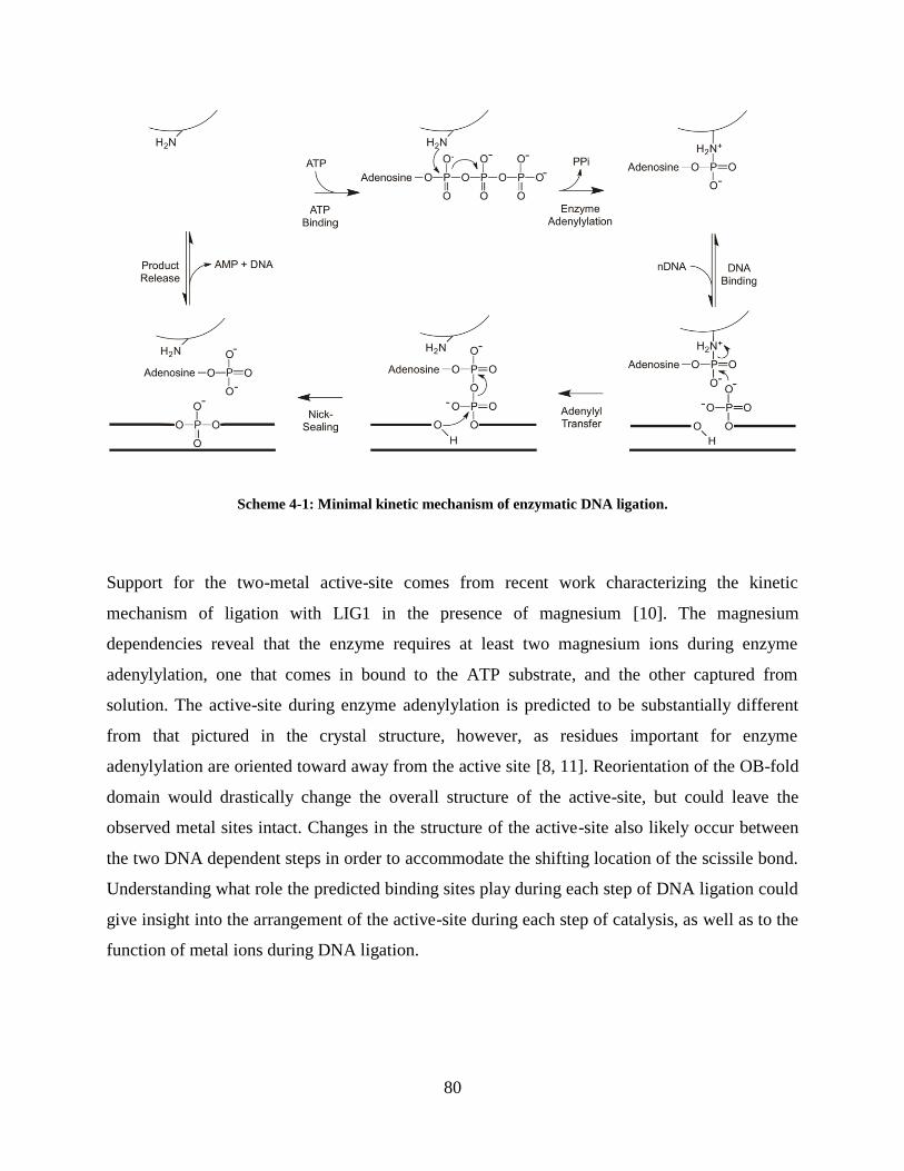
80
Scheme 4-1: Minimal kinetic mechanism of enzymatic DNA ligation.
Support for the two-metal active-site comes from recent work characterizing the kinetic
mechanism of ligation with LIG1 in the presence of magnesium [10]. The magnesium
dependencies reveal that the enzyme requires at least two magnesium ions during enzyme
adenylylation, one that comes in bound to the ATP substrate, and the other captured from
solution. The active-site during enzyme adenylylation is predicted to be substantially different
from that pictured in the crystal structure, however, as residues important for enzyme
adenylylation are oriented toward away from the active site [8, 11]. Reorientation of the OB-fold
domain would drastically change the overall structure of the active-site, but could leave the
observed metal sites intact. Changes in the structure of the active-site also likely occur between
the two DNA dependent steps in order to accommodate the shifting location of the scissile bond.
Understanding what role the predicted binding sites play during each step of DNA ligation could
give insight into the arrangement of the active-site during each step of catalysis, as well as to the
function of metal ions during DNA ligation.

81
A B
Figure 4-1: Metal binding sites revealed by hLIG1 crystal structure. A, Close-up of active-site of hLIG1 as
crystallized in complex with adenylylated DNA [8]. The protein backbone is represented in green cartoon form, with
important active-site residues and substrate DNA depicted in stick form. The lysine that goes through transient
adenylylation and active site residues involved in coordinating metal sites are labeled, as are the free 3’ and 5’ DNA
ends at the nick. Predicted metal sites are depicted with magenta lattices. B, Protein sequence alignment of human
DNA ligase I (LIG1), DNA ligase III (LIG3) and DNA ligase IV (LIGIV), Saccharomyces cerevisiea (CDC9),
Schizosaccharomyces pombe (CDC17) and Chlorella Virus DNA (ChVLIG) show conservation of residues within
the active-site. Alignment performed with default settings in ClustalX using truncated versions of each protein that
include only the catalytic core domains. * indicates a residue involved in coordination of proposed active-site metal
sites. † indicates Lys 568, that is adenylylated in the first step of the reaction.
To investigate the role of metal ions in predicted metal binding sites of LIG1, we have made
conservative carboxyl to amide mutations in the residues coordinating these sites. Comparison of
the magnesium dependence of the mutants during each step of the ligation reaction to wild-type
indicates during which step each residue serves as an important metal ligand. The E621Q
mutation results in a functional enzyme with greatly diminished ability to perform adenylyl
transfer and nick-sealing. The D570N and E720Q mutations result in a nearly identical defect in
adenylyl transfer and a similar defect in nick-sealing, despite being on opposite sides of the nick.
We propose that D570 and E720 form part of a network of cooperative interactions that structure
the enzyme active site during the DNA dependent reactions. In contrast, D570N has little effect
on enzyme adenylylation, while E720Q shows a significant decrease in the second order rate
constant for ATP utilization, (kcat/KM)ATP
. This suggests that E720Q may play a role in metal
binding during enzyme adenylylation, as well as the DNA dependent reactions. This work builds

82
on what is known about DNA ligases from work with the Chlorella Virus DNA ligase to help us
understand the more complicated human DNA ligase I.

83
Experimental Procedures
DNA Ligase I Mutants
The D570N, E621Q or E720Q mutants were each expressed as the catalytic domain of human
LIG1 (residues 232-919) in E. coli and purified as previously described [10]. Cells were lysed in
the presence of 1 mM EDTA to preserve the adenylylated form of the enzyme, and subsequently
purified over a phosphocellulose column and a NTA-nickel column. The His tag was removed
with Prescission protease, and LIG1 was further purified with Q-sepharose. The final purified
fractions were combined and dialyzed into storage buffer (25 mM Tris•Cl, pH 7.6, 150 mM
NaCl, 1 mM DTT, and 0.1 mM EDTA). Aliquots were snap-frozen and stored at -80 oC. Purity
was greater than 90% as judged by SDS-PAGE. Initial concentrations were estimated from the
absorbance at 280 nm, using the calculated extinction coefficient. The concentration of active,
adenylylated enzyme was determined by titration with nicked DNA substrate, as described in the
paper, and this active concentration is reported throughout. Due to the low activity of E621Q, we
used the concentration determined from UV-absorbance.
DNA Substrates
Oligonucleotides were synthesized by Integrated DNA Technologies or by the Keck Center at
Yale University and were purified on denaturing polyacrylamide gels. The portion of the gel
containing the full-length oligonucleotide was excised, crushed, and extracted by soaking
overnight in 500 mM NaCl and 1 mM EDTA. The extracted oligonucleotides were desalted by
binding to a C18 reverse phase column (Sep-pak, Waters) and eluted with 30% (v/v) acetonitrile.
Concentrations were obtained from the absorbance at 260 nm using the calculated extinction
coefficients. The three oligonucleotides used in this study had sequences 5’-
CCGAATCAGTCCGACGACGCATCAGCAC, 5’-GTGCTGATGCGTC, and 5’-P-

84
GTCGGACTGATTCGG-FAM (P indicates 5’ phosphorylation and FAM indicates the presence
of a 3’ fluorescein). The nicked, double-stranded DNA substrate (nDNA) was formed by mixing
equimolar amounts of the three oligonucleotides in 10 mM NaMES pH 6.5 and 50 mM NaCl and
cooling the mixture from 90 °C to 4 °C at a rate of 3 °C per minute.
Gel-based Ligation Assay
Ligation reactions were performed at 37 °C. Unless otherwise indicated, the standard buffer
contained 50 mM NaMOPS pH 7.5 (measured at 25 °C), 1 mM dithiothreitol, 0.05 mg/mL BSA,
and sufficient NaCl to maintain a constant ionic strength of 150 mM. The amounts of ATP,
MgCl2, nDNA, and LIG1 mutant enzyme varied, as indicated below. Preincubation controls
established that each LIG1 mutant retains 100% of its activity after 1 hr in this standard buffer,
and all of the kinetic data was collected within this window of time. Reactions were quenched as
described below, heated to 95 °C for 5 min to denature the DNA and resolved on 20% (w/v)
denaturing polyacrylamide gels containing 8 M Urea. Fluorescein-labeled oligonucleotides were
detected with a Typhoon Trio+ imager (GE Healthcare) with excitation at 488 nm and emission
through a 520 nm band-pass filter. The images were analyzed using ImageQuantTL (GE
Healthcare). The intensity of the individual DNA species was corrected for background
fluorescence, and the fraction of the total fluorescence was determined by dividing the
fluorescence intensity for the desired species by the signal for all other species in the sample.
When necessary, this fraction was converted into its concentration by multiplying by the total
concentration of DNA in the reaction.
Multiple-Turnover Ligation Assays
Steady state kinetic analysis was performed in the standard ligation buffer and the temperature
was maintained at 37 °C in a circulating water-bath. Reaction mixtures were preincubated for 5
min prior to addition of LIG1. Reactions were arrested by quenching a 4 µL aliquot in 15 µL of
quench solution (50 mM EDTA in 90% formamide), and the extent of ligation was determined as
85
described above. Initial rates were determined from the linear rate of substrate disappearance
within the first 10% of the reaction. When both intermediate and product were formed during the
reaction, the rate of formation for each species was determined. Values reported represent the
average of at least two experiments, unless otherwise noted in the text. Equations used to fit data
are described in the Models of Kinetic Parameters section and in the text. All fitting of multiple-
turnover data was done using Kaleidagraph, Synergy Software.
Single-turnover Ligation Assays
Unless otherwise indicated, the typical concentrations used for single-turnover reactions were 20
mM MgCl2, 800 nM nDNA and 80 nM LIG1 mutant enzyme. Reactions were quenched as
described above for multiple-turnover reactions and submitted to the standard gel-based ligation
assay. Levels of intermediate and product were imported into the program Berkeley-Madonna
(www.berkeleymadonna.com) and the curve fitting function was used to globally fit the levels of
intermediate and product by the scheme represented in Scheme 4-1. Fitting of the data to a model
including reversible chemical steps did not produce significantly different results (data not
shown). Fits of reactions from individual experiments provided values for the rate constants for
adenylyl transfer and nick-sealing and the reported values reflect the average rates determined
from at least two independent experiments at each metal concentration, unless otherwise noted in
the text. These rate constants were plotted versus the concentration of metal ions. Fitting of the
data is discussed in the results section.

86
Models of Kinetic Parameters
][
][*
AK
Akk
A
Aobs
Equation 4-1
Equation 4-1 describes the model where a reaction is activated by binding of species A to a
single stimulatory site. kobs is the observed rate constant for the step being measured, kA is the
maximal rate constant, and KA is the observed affinity for species A during the observed step.
Equation 4-1 is used for fitting the magnesium dependence in Figures 4-5B, 4-5C, and 4-8. This
equation takes on the form of the Michaelis-Menten equation when applied to Figure 4-7A and
B, where A becomes DNA and ATP, respectively.
I
AA
Aobs
K
IKKA
Akk
][][
][*
Equation 4-2
Equation 4-2 describes a competitive inhibition model where the substrate can either be bound
by a productive enzyme, A or a nonproductive enzyme, I. kobs is the observed rate constant for
the step being measured, kA is the maximal rate constant when bound by enzyme A, KA is the
affinity between enzyme A and the substrate, and KI is the affinity between the inactive enzyme,
I, and the substrate. Equation 4-2 is used for fitting the data in Figure 4-4.

87
n
cat
kkk
k1
...11
1
21
Equation 4-3
Equation 4-3 describes the relationship between the macroscopic rate constant for the steady-
state reaction (kcat) and the individual microscopic rate constants that define each step of the
reaction (k1, k2, etc.). Equation 4-3 is used to determine the value of kadenylylation from the
measured rate-constants for kcat, ktransfer and kseal. This value is reported in Table 4-1.

88
Results and Discussion
Determining the adenylylation state of LIG1 mutants
Wild-type LIG1 can be purified as the adenylylated enzyme and 100% of the protein is active
[10], however mutations of the residues corresponding to D570, E621 and E720 in Chlorella
Virus DNA ligase (ChVLIG) are reported to affect the enzyme’s adenylylation state [9]. In order
to determine the adenylylation state and active concentration of each mutant, active-site titrations
were performed in which varying amounts of enzyme was added to a fixed concentration of
nicked DNA (nDNA) substrate in standard reaction buffer without ATP (Figure 4-2). The
reaction is allowed to sit for 5 minutes and the amount of product formed then directly relates to
the amount of active, adenylylated LIG1 present in the reaction. D570N and E720Q each form
product in a 1.2:1 ratio of input enzyme to input substrate (Figure 4-2A, B), indicating that 80%
of the purified protein measured by UV-absorption is active and adenylylated. This small
discrepancy from 100% active enzyme can be explained by the contribution of the adenylyl
group to the absorbance at 280 nm. Reaction times of 15 minutes do not change the amount of
product formed at any DNA concentration, indicating the reaction is complete at 5 minutes (data
not shown).
E621Q forms product at a 500:1 ratio of input enzyme to input substrate under the same
conditions (data not shown). The amount of product formed with E621Q increases with time up
to 200 minutes (data not shown) indicating that the enzyme is active, but much slower than wild-
type LIG1. Pre-incubation of each mutant with ATP and magnesium prior to reaction does not
alter the amount of product formed, indicating each mutant is fully adenylylated following
purification (data not shown). This contrasts with what was seen with the ChVLIG mutants, in
which analogous mutations were reported to decrease, or even abolish enzyme adenylylation.

89
A B
Figure 4-2: Active-site titration of D570N and E720Q LIG1 mutants. Enzyme was titrated against a fixed
concentration of 250 nM DNA substrate in 20 mM MgCl2 and allowed to react for 15 minutes. Titration of D570N
(A) and E720Q (B) indicate ~80% of the protein measured by UV-absorption is adenylylated and active. Addition of
an adenylyl group increases the extinction coefficient of the protein by 20%, indicating all enzyme is actually active.
E621Q is severely impaired for the DNA dependent reactions
We investigated the slow reaction rate observed during active-site titration experiments by
performing single-turnover reactions with E621Q. Reactions containing 20 mM MgCl2, 600 nM
E621Q, 250 nM nDNA and no ATP show that E621Q does not complete ligation over a 48-hour
time-course (Figure 4-3). The rate of product formation rapidly decreases after 6 hours,
indicating the enzyme begins to lose activity at this point. Though we are unable to directly
determine any rate constants from this data, comparison of the linear rate of product formation to
wild-type can give a rough estimate of the relative difference between the two proteins. During
the linear phase of product formation, E621Q converts 1.3% of the input DNA to product per
hour, compared to 62% per second for wild-type LIG1. This rough comparison suggests E621Q
is ~1.8x10⁵-fold slower than wild-type under similar single-turnover conditions.
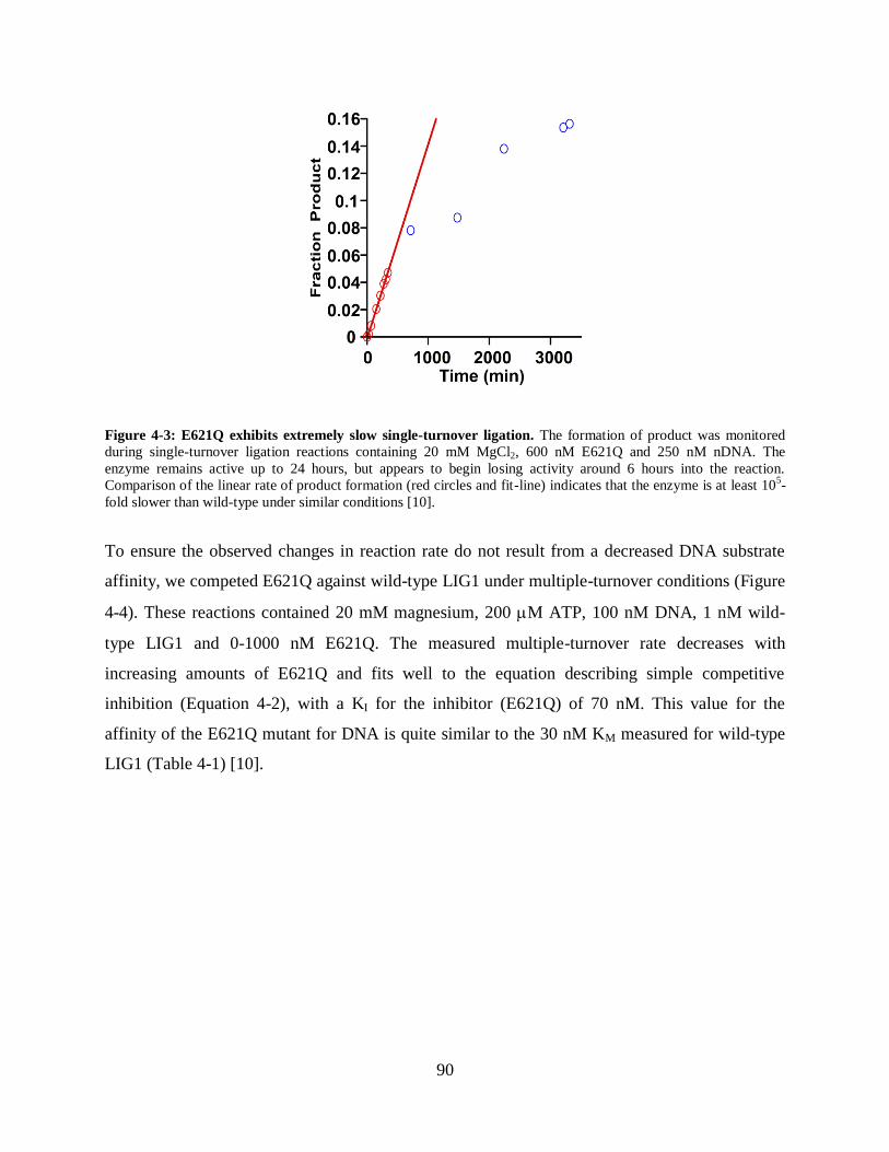
90
Figure 4-3: E621Q exhibits extremely slow single-turnover ligation. The formation of product was monitored
during single-turnover ligation reactions containing 20 mM MgCl2, 600 nM E621Q and 250 nM nDNA. The
enzyme remains active up to 24 hours, but appears to begin losing activity around 6 hours into the reaction.
Comparison of the linear rate of product formation (red circles and fit-line) indicates that the enzyme is at least 105-
fold slower than wild-type under similar conditions [10].
To ensure the observed changes in reaction rate do not result from a decreased DNA substrate
affinity, we competed E621Q against wild-type LIG1 under multiple-turnover conditions (Figure
4-4). These reactions contained 20 mM magnesium, 200 M ATP, 100 nM DNA, 1 nM wild-
type LIG1 and 0-1000 nM E621Q. The measured multiple-turnover rate decreases with
increasing amounts of E621Q and fits well to the equation describing simple competitive
inhibition (Equation 4-2), with a KI for the inhibitor (E621Q) of 70 nM. This value for the
affinity of the E621Q mutant for DNA is quite similar to the 30 nM KM measured for wild-type
LIG1 (Table 4-1) [10].
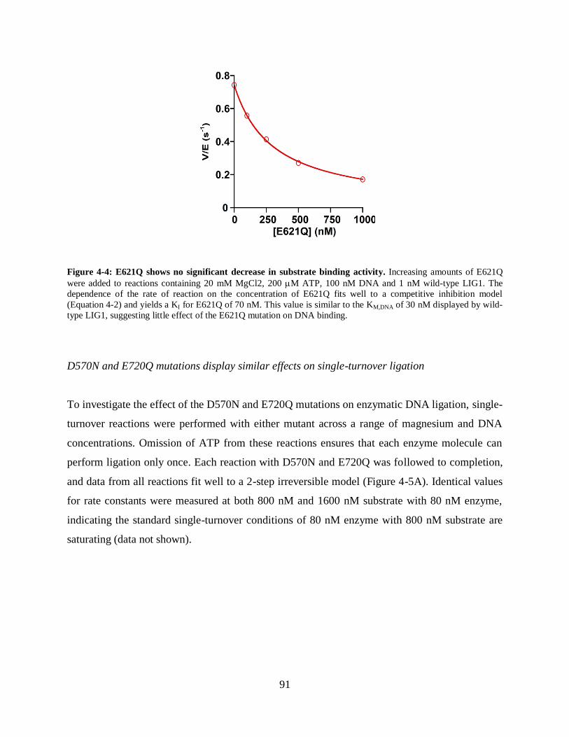
91
Figure 4-4: E621Q shows no significant decrease in substrate binding activity. Increasing amounts of E621Q
were added to reactions containing 20 mM MgCl2, 200 M ATP, 100 nM DNA and 1 nM wild-type LIG1. The
dependence of the rate of reaction on the concentration of E621Q fits well to a competitive inhibition model
(Equation 4-2) and yields a KI for E621Q of 70 nM. This value is similar to the KM,DNA of 30 nM displayed by wild-
type LIG1, suggesting little effect of the E621Q mutation on DNA binding.
D570N and E720Q mutations display similar effects on single-turnover ligation
To investigate the effect of the D570N and E720Q mutations on enzymatic DNA ligation, single-
turnover reactions were performed with either mutant across a range of magnesium and DNA
concentrations. Omission of ATP from these reactions ensures that each enzyme molecule can
perform ligation only once. Each reaction with D570N and E720Q was followed to completion,
and data from all reactions fit well to a 2-step irreversible model (Figure 4-5A). Identical values
for rate constants were measured at both 800 nM and 1600 nM substrate with 80 nM enzyme,
indicating the standard single-turnover conditions of 80 nM enzyme with 800 nM substrate are
saturating (data not shown).

92
A B
C
Figure 4-5: Single-turnover ligation by D570N and E720Q. Single-turnover reactions were performed with 800
nM nDNA, 80 nM mutant LIG1 and a range of MgCl2. A, Representative data from a single-turnover reaction with
E720Q at 20 mM MgCl2. The time dependence of the levels of intermediate (red, circles) and product (blue,
squares) fits well to the 2-step irreversible model used for single-turnover ligation (see Experimental Procedures). B,
The magnesium dependence of ktransfer fits well to a single-site binding model (Equation 4-1), and is almost identical
between D570N (red, circles) and E720Q (blue, squares). C, The magnesium dependence of kseal fits well to a
single-site binding model (Equation 4-1) for both D570N (red, circles) and E720Q (blue, squares). The E720Q
mutation results in a stronger decrease in the value of kseal at magnesium saturation, as well as a more weakened
affinity for magnesium. Values for kinetic constants measured in these fits reported in the text and Table 4-1.
Time (s) [MgCl2] (mM)
[MgCl2] (mM)
kseal (
s-1
)
60
50
40
30
20
10
Co
nce
ntr
ati
on
of
sp
ec
ies
(n
M)

93
The magnesium dependence of the single-turnover reaction was measured across a range of 0.5
mM to 20 mM magnesium. The magnesium dependence of ktransfer, the rate constant for adenylyl
transfer, appears very similar between the D570N and E720Q mutants (Figure 4-5B). The data
for ktransfer fit well to a single-site binding model (Equation 4-1) and yield similar values for
ktransfer of 0.066 s-1
and 0.055 s-1
for D570N and E720Q, respectively. Both mutants also display
similar values of 2.8 mM and 1.9 mM for KMg for D570N and E720Q, respectively, during
adenylyl transfer. These values correspond to an ~40 fold decrease in ktransfer and an ~10-fold
decrease in metal affinity from the 2.6 s-1
ktransfer and 0.15 mM KMg measured with wild-type
LIG1 (Table 4-1) [10]. The nick-sealing data also fit well to a single-site binding model
(Equation 4-1), yielding values of 0.18 s-1
and 0.11 s-1
for kseal, and 13 mM and 28 mM for KMg
with D570N and E720Q, respectively (Figure 4-5C). Compared to the values of 12 s-1
for kseal
and 2.6 mM KMg measured with wild-type LIG1, each mutant displays ~100-fold decrease in kseal
and ~10-fold decrease in KMg during nick-sealing (Table 4-1) [10]. It should be noted that these
reactions poorly define the values for kseal, as the highest value of magnesium tested for these
reactions, 20 mM, is close the measured KMg for both mutants. The buffering system used for
these reactions is unable to control for the changes in ionic strength that would occur at higher
concentrations of magnesium. Thus the values measured for kseal and KMg during nick-sealing
should be considered estimates.
D570N and E720Q have differing effects on enzyme adenylylation
To evaluate the effect of the D570N and E720Q mutations on enzyme adenylylation and the
overall steady-state reaction, we performed multiple-turnover reactions in which ATP is included
in the reaction. The standard conditions used for these reactions include 20 mM MgCl2, 200 M
ATP, 4 nM enzyme and 1 M nDNA. The rate of all multiple-turnover reactions were
determined from data at less than 10% substrate disappearance, ensuring measurement of the
steady-state reaction (Figure 4-6). Product formation remains linear for all steady-state reactions
up to the final time-point recorded (longest reaction time of 20 minutes) indicating that the
mutants are stable during the course of the reaction. To ensure saturation of the enzyme with
nDNA substrate during multiple turnover, we measured the V/E values for reactions spanning

94
the range of 50 nM to 2 M nDNA. The DNA dependences of the steady-state reaction rate for
both the D570N and E720Q mutants fit well to the Michaelis-Menten equation describing a
single-site binding interaction (Equation 4-1) (Figure 4-7A). Both mutants display a KM for
nDNA of less than 50 nM in steady-state reactions, similar to the affinity of 30 nM measured for
the wild-type enzyme (Table 4-1) [10]. The lack of a difference in DNA affinity between the
D570N and E720Q mutants and wild-type during either steady-state or single-turnover reactions
suggests that these two mutations do not significantly affect protein-DNA binding interactions.
The ATP dependence of the rate of steady-state ligation reveals a low KM value of these mutants
for ATP (Figure 4-7B). Fitting of the data to a single-site binding model yields a KM,ATP of 1.0
M and 4.3 M for D570N and E720Q, respectively. These are both considerably lower than the
12 M affinity for ATP measured with wild-type LIG1 (Table 4-1) [10].
Figure 4-6: Representative steady-state time-course. Multiple-turnover reactions were performed with 20 mM
MgCl2, 1 M nDNA, 4 nM D570N and either 1 M (red, circles), 5M (blue, squares), 20M (green, diamonds) or
200M (black, triangles) ATP. All data was collected within the linear portion of the reaction. The reaction remains
linear up to 16% product formation, indicating time-points taken within the first 10% reaction are not affected by
product inhibition.
Time (s)

95
A B
Figure 4-7: D570N and E720Q display wild-type substrate binding. A, Multiple-turnover reactions were
performed with 20 mM MgCl2, 200 M ATP, 4 nM D570N (red, circles) or E720Q (blue, squares) and a range of
DNA concentrations from 50 nM to 2 M. The DNA dependences of the reaction rate for both enzymes fit well to
the equation describing a single-site binding model (Equation 4-1). Both mutants display a lower than 50 nM
KM,DNA, similar to that measured for wild-type LIG1 [10]. B, Multiple-turnover reactions were performed with 20
mM MgCl2, 1 M nDNA, 4 nM D570N or E720Q and a range of ATP concentrations from 0.5 M to 200 M. The
ATP dependences of the multiple turnover reaction rate for D570N (red, circles) and E720Q (blue, squares) fit well
to the Michaelis-Menten equation (Equation 4-1). The KM value for ATP measured for D570N and E720Q are
considerably lower than that measured with wild-type LIG1 (1.0 M and 4.3 M vs. 12 M, respectively) [10].
The apparent increase in ATP affinity can be explained either by a true change in ATP binding,
or by the multiple-turnover rate being rendered less sensitive to ATP concentration due to fast
DNA dependent reactions. The second order rate constant (kcat/KM)ATP
describes the ATP
dependent steps of the reaction, up to and including enzyme adenylylation. The ATP dependence
at 20 mM mganesium gives (kcat/KM)ATP
values of 3.1x10⁴
M-1
s-1
and 6.3x10³
M-1
s-1
for D570N
and E720Q, respectively. Whereas the value measured for D570N is quite similar to the 5.6x10⁴
M-1
s-1
(kcat/KM)ATP
measured for wild-type LIG1 under similar conditions, E720Q shows a
significant decrease in (kcat/KM)ATP
compared to wild-type (Table 4-1) [10].
[DNA] (M) [ATP] (M)

96
D570N and E720Q display significant defect in magnesium utilization during steady-state
reactions
Multiple turnover reactions were performed at saturating DNA and ATP concentrations across a
range of magnesium concentrations in order to investigate the effect of the D570N and E720Q
mutations on magnesium binding during the overall multiple-turnover reaction. The magnesium
dependence of the steady-state rate fits well to the equation describing a single-site binding
model (Equation 4-1) and yields a maximal steady-state rate, kcat, of 0.042 s-1
and 0.063 s-1
and a
K1/2 for magnesium of 7.3 mM and 35 mM for D570N and E720Q, respectively (Figure 4-8).
These high values for KMg, that are similar to the values for the nick-sealing step, suggests that
multiple-turnover for these mutants is limited by the rate of nick-sealing at most concentrations
of magnesium. The second order rate constant for utilization of magnesium during multiple-
turnover, kcat/KMg2+, gives insight into how efficiently the magnesium cofactor binds to and
stabilizes the transition state. As determined from the magnesium dependence of multiple-
turnover, kcat/KMg2+ is 5.8 M
-1s
-1 and 1.8 M
-1s
-1 for D570N and E720Q, respectively (Table 4-1).
These values are much lower than the 1.1x10³
M-1
s-1
measured for the wild-type enzyme,
indicting a drastic decrease in the ability of magnesium to stabilize the reaction transition state
(Table 4-1) [10]. This observation supports the idea that both the D570 and E720 residues play
an important role in coordinating a metal ion during catalysis.

97
Figure 4-8: D570N and E720Q show greatly weakened metal binding during multiple-turnover reactions.
Multiple-turnover reactions were performed with 200 M ATP, 1 M nDNA, 4 nM D570N or E720Q and a range
of MgCl2 from 1 mM to 20 mM. The magnesium dependence of the rate of steady-state ligation if fit by the equation
describing a single-site binding model (Equation 4-1). D570N and E720Q both show a significant decrease in metal
binding affinity and maximal reaction rate vs. wild-type LIG1 (KMg = 7.3 mM and 35 mM vs. 0.71 mM and kcat =
0.042 s-1
and 0.063 s -1
vs. 0.81 s-1
, resepectively).
[MgCl2] (mM)

98
Conclusion
E621 plays an essential role during DNA dependent reactions
The amide to carboxyl substitution in E621Q is expected to be a relatively conservative mutation
with respect to metal ion binding. The expected change would be a weakening of metal binding
at the site with little effect on catalysis. The E621Q mutation in LIG1, however, has a
surprisingly deleterious effect on catalysis during the DNA dependent reactions of enzymatic
ligation, decreasing the observed rate constant for single-turnover reactions by a factor of 105. A
similar decrease in the rate of DNA ligation was observed for the corresponding mutation in
Chlorella Virus DNA ligase (ChVLIG) [9]. Such strong effects on the rate of reaction could
indicate that the hydroxyl of E621 may play a role either in overall stabilization in the active site,
or through direct or indirect activation of important catalytic groups. The carbonyl group of E621
could instead be responsible for metal ion coordination. Unfortunately, the extremely slow rate
of DNA ligation with E621Q prevented a full characterization. Future work characterizing other
mutations at E621 could help elucidate the obvious importance of this residue to catalysis.
D570 and E720 form a network of cooperative interactions during DNA dependent reactions
The D570N and E720Q mutations both result in a roughly 650-fold decrease in utilization for
magnesium during adenylyl transfer as compared to wild-type, due to nearly identical effects on
ktransfer and KMg (Table 4-1). An identical effect at two residues that appear to coordinate two
different metal sites suggests that they form a cooperative network of interactions that stabilizes
the enzyme active-site. No amino acid or substrate group appears to bridge these two residues in
the LIG1 crystal structure, however the oxygen atoms of the two carboxylates are spaced 0.44
nm apart, which could accomodate bridging water molecules [8]. In contrast, E720Q shows a
much greater decrease in magnesium utilization during nick-sealing than D570N (12,000-fold

99
and 3300-fold vs. wild-type, respectively) (Table 4-1). This suggests that while the two residues
appear to be equally important for adenylyl transfer, E720 plays a greater role during nick-
sealing than D570.
Multiple-turnover WT D570N relative E720Q relative
Saturating Conditions
kcat (s-1) 0.81 0.042 ± 0.007 (19) 0.063 ± 0.01 (13)
KMg (mM) 0.71 7.3 ± 3 (0.097) 35 ± 10 (0.02)
Subsaturating Mg2+
kcat/KMg (M-1s-1) 1.1 5.8 (190) 1.8 (610)
Subsaturating ATP
(kcat/KM)ATP (M-1s-1) 6.8x10⁴ 3.1x10⁴ (2.1) 6.3x10⁴ (10.8)
Enzyme adenylylation*
kadenylylation s-1 1.3 0.32 (4) fast (ND)
Single-turnover
Adenylyl transfer
ktransfer (s-1) 2.6 0.066 ± 0.005 (39) 0.055 ± 0.004 (47)
KMg (mM) 0.15 2.8 ± 0.4 (0.054) 1.9 ± 0.2 (0.079)
ktransfer/KMg (M-1s-1) 1.7x10⁴ 24 (710) 29 (590)
Nick-sealing
kseal (s-1) 12 0.18 ± 0.03 (67) 0.11 ± 0.02 (110)
KMg (mM) 2.6 13 ± 4 (0.2) 28 ± 7 (0.093)
kseal/KMg (M-1s-1) 4.6x10⁴ 14 (3300) 4 (12,000)
Table 4-1: Comparison of kinetic constants measured with wild-type ligase to D570N and E720Q. Relative
values between the constants measured for wild-type LIG1 and the mutants are calculated by dividing the value of
the wild-type constant by the value of the corresponding constant measured with the mutant enzyme. Thus, values
greater than 1 for rate constants and the efficiency of magnesium utilization during single-turnover steps indicates a
decrease in activity, while values less than 1 for metal affinity indicate a weakening in metal binding for the
indicated step.
*The value for kadenylylation is determined by using the equation that relates macroscopic and microscopic rate
constants (Equation 4-3), it is not directly determined by the experiments reported herein. Use of Equation 4-3 with
the rate constants measured for E720Q results in a negative number for kadenylylation, indicating the error in
measurements of the other rate constants is larger than the value of kadenylylation. This suggests enzyme adenylylation
is the fastest step of the ligation reaction catalyzed by E720Q under standard conditions.

100
E720 appears to play a significant role during enzyme adenylylation
Compared to the large effects the D570N mutation has on the utilization of magnesium during
the DNA dependent steps, changes to the same parameter for enzyme adenylylation appear quite
small. D570N shows only a 2-fold decrease compared to wild-type in the second order rate-
constant for utilization of ATP, (kcat/KM)ATP
(Table 4-1). The E720Q mutation, however, shows a
significant, 10-fold decrease in (kcat/KM)ATP
compared to wild-type. The difference in importance
of these two residues during adenylyl transfer coincides with their position within the active-site
as pictured in the LIG1 crystal structure [8]. E720 and the metal site it is proposed to coordinate
are nearby K568, the active-site lysine that becomes adenylylated during step 1 of catalysis. This
positioning allows E720 to play an important role during enzyme adenylylation. D570, however,
rests on the opposite side of the nick, well outside direct interaction range of the lysine residue.
Outstanding Questions
This work has extended our understanding of the role of conserved carboxylates in DNA ligase.
Kinetic characterization of these mutant enzymes has revealed further information about these
sites, supoorting the model of two metals being required for the DNA dependent steps. We also
found differences between LIG1 mutants and those of ChVLIG, such as observing complete
enzyme adenylylation following purification, unlike the ChVLIG mutants which purify with a
large portion deadenylylated. This work greatly enhances our knowledge of the metal binding
sites important for enzyme catalysis. Future research looking into additional mutations at these,
as well as other, important active-site residues will enable us to better understand the exact
function of metals in these sites.

101
References
1. Lehman, I.R., DNA ligase: structure, mechanism, and function. Science, 1974.
186(4166): p. 790-7.
2. Sancar, A., et al., Molecular mechanisms of mammalian DNA repair and the DNA
damage checkpoints. Annu Rev Biochem, 2004. 73: p. 39-85.
3. Levin, D.S., et al., Interaction between PCNA and DNA ligase I is critical for joining of
Okazaki fragments and long-patch base-excision repair. Curr Biol, 2000. 10(15): p. 919-
22.
4. Shokolenko, I.N., et al., Mitochondrial DNA ligase is dispensable for the viability of
cultured cells but essential for mtDNA maintenance. J Biol Chem, 2013. 288(37): p.
26594-605.
5. Grawunder, U., et al., DNA ligase IV is essential for V(D)J recombination and DNA
double-strand break repair in human precursor lymphocytes. Mol Cell, 1998. 2(4): p.
477-84.
6. Shuman, S., DNA ligases: progress and prospects. J Biol Chem, 2009. 284(26): p.
17365-9.
7. Yang, S.W. and J.Y. Chan, Analysis of the formation of AMP-DNA intermediate and the
successive reaction by human DNA ligases I and II. J Biol Chem, 1992. 267(12): p. 8117-
22.
8. Pascal, J.M., et al., Human DNA ligase I completely encircles and partially unwinds
nicked DNA. Nature, 2004. 432(7016): p. 473-8.
9. Sriskanda, V. and S. Shuman, Role of nucleotidyl transferase motif V in strand joining by
chlorella virus DNA ligase. J Biol Chem, 2002. 277(12): p. 9661-7.
10. Taylor, M.R., et al., Kinetic mechanism of human DNA ligase I reveals magnesium-
dependent changes in the rate-limiting step that compromise ligation efficiency. J Biol
Chem, 2011. 286(26): p. 23054-62.
11. Sriskanda, V. and S. Shuman, Mutational analysis of Chlorella virus DNA ligase:
catalytic roles of domain I and motif VI. Nucleic Acids Res, 1998. 26(20): p. 4618-25.

102
Chapter 5: Conclusions and Future Directions
The work presented in this thesis explores the relationship between metal ions and human DNA
ligase I. Our characterization of the kinetic mechanism of enzymatic DNA ligation by LIG1 in
the presence of magnesium provides the first kinetic evidence for two functionally important
metal ions during catalysis. This agrees with the many two-metal reaction mechanisms described
for other phosphoryl transfer enzymes [1-3], and with the observance of two potential metal-
binding sites within the LIG1 crystal active site [4]. We found that mutation of the residues
coordinating these two potential metal sites causes a decrease in the metal binding affinity during
each catalytic step, suggesting the involvement of metals at these two sites during catalysis.
Characterization of enzymatic DNA ligation in the presence of manganese revealed that LIG1 is
able to substitute manganese for magnesium during catalysis, similar to what has been seen with
many other phosphoryl transfer enzymes [5-7]. We also found evidence for the presence of at
least one inhibitory metal binding site that appears to be specific for manganese over
magnesium. Like all scientific research, this work has brought forth a large number of questions
even as it answered many basic questions about how metal ions interact with LIG1.
Abortive ligation
The adenylylated DNA intermediate formed during the adenylyl transfer step of DNA ligation
represents a significant danger to the genome if released from the enzyme prior to completing
nick-sealing. Rapid adenylylation of LIG1 prevents it from rebinding to the adenylylated
intermediate, resulting in a persistent DNA break. Multiple pathways can remove the adenylyl
group from DNA, allowing another chance at successful DNA ligation, including direct removal
of the adenylyl group by aprataxin [8], and strand displacement of the lesion with DNA
polymerase followed by single-strand cleavage with FEN-1[9]. Loss of aprataxin results in the
progressive neurodegenerative disease Ataxia with Oculomotor Apraxia 1 (AOA1) [10, 11],
suggesting that cells missing aprataxin are unable to efficiently repair adenylylated DNA.
Neurons are likely rendered more sensitive to loss of aprataxin due to their increased metabolism

103
and subsequent DNA repair activity compounded with the loss of replication-dependent DNA
repair pathways.
Previous work indicated LIG1 can form the adenylylated DNA intermediate at single-strand
nicks created through Fenton chemistry, presumably by acting on nicks with additional damage
[12]. Our work has shown that LIG1 can also form persistent adenylylated DNA at normal DNA
nicks by releasing the DNA intermediate prior to completing nick-sealing. Release of the
intermediate is most pronounced at sub-physiological concentrations of magnesium and
decreases with increasing magnesium, however a measurable amount of abortive ligation occurs
even at physiological concentrations of magnesium (.01% of ligation events resulting in abortive
ligation at 1 mM free magnesium) [13]. With the million or more ligation events occurring in the
genome of the average human cell per day due to DNA repair and DNA replication, even this
low rate of abortive ligation results in a substantial amount of total abortive ligation events. A
recent report suggests the main function of aprataxin may be to repair 5' adenylylated DNA
within the mitochondria, as loss of the protein appears to more adversely affect the mitochondrial
than the nuclear genome [14]. Studies of abortive ligation by LIG3 will need to be done in order
to understand how significant a problem this might be within the unique environment of the
mitochondria.
Substrate Specificity
The nature of substrate recognition by LIG1, and DNA ligases in general, remains one of the
least understood aspects of DNA ligase function. The ability of DNA ligases to discriminate
between proper ligation substrate and substrates with additional forms of damage seems almost
essential to prevent the accumulation of genomic aberrations. There are, however, numerous
reports of LIG1 catalyzing reactions on non-ideal DNA nicks, including abortive ligation of
Fenton-chemistry induced DNA nicks [12] or nicks lacking a 3’ hydroxyl [4], and full ligation of
hybrid duplexes containing mismatches or ribonucleotides [4]. In each of these cases, LIG1
appears to discriminate against the non-ideal substrate by showing a diminished rate of catalysis.
Other DNA ligases appear to show similar behavior with these non-ideal substrates [15, 16].

104
Many of these observations have been reported as qualitative observations from different sources
and different types of experiments, complicating synthesis of the data into a single model.
Perhaps the most straight-forward way to understand the importance of different chemical groups
at the nick and the effect of different forms of damage on ligation would be to compete
substrates with chemical substitutions against one another during a DNA ligation assay. Such a
systematic evaluation of how different forms of DNA damage at or near a nick affect ligation
would greatly increase our knowledge of how LIG1 interacts with the nick, and how these forms
of DNA damage may get exacerbated during ligation.
The crystal structure of LIG1 suggests that the nucleotide sequence of the substrate may also
affect catalysis in addition to chemical groups at the nick. The DNA within the LIG1 crystal
structure is highly distorted, with a 5 Å shift in the axis of the DNA backbone across the nick and
a partial unwinding of the DNA helix [4]. These movements require the energetically
unfavorable unstacking of adjacent base-pairs and stretching of hydrogen bonds in base-pairing
interactions, which must be overcome by the energy of binding to the protein. Differences in the
strength of base-stacking and base-pairing interactions due to DNA sequence can then cause
changes in the rate of catalysis, as seen in studies of other DNA modifying enzymes [17]. In the
case of LIG1, sequences prone to slow or abortive ligation would increase the likelihood of
chromosomal breaks. Sequence specificity of LIG1 could have profound impacts on the genome
by selecting over time against poorly ligatable sequences.
Preliminary work with three randomly different DNA sequences has shown that LIG1 shows a
10-fold range of kcat/KM values at physiological concentrations of magnesium, suggesting that
the sequence context of the nick can have a significant effect on catalysis. The number of
potential DNA sequences at a nick prohibits in-depth, manual characterization of sequence
space. Characterizing only the 2 nucleotides on either side of the nick, for example, would
require 256 different substrates. This type of question can only now be correctly asked by
utilizing the new technique of Next-Generation DNA Sequencing. Next-Gen Sequencing can
measure the rates of ligation of each of the millions of sequences formed during ligation of a
substrate containing degenerate sequence on either side of the nick. The relative rates correlate

105
with relative ligation efficiencies of each sequence with LIG1 and to how well each sequence
will be ligated within the cell. This data should reveal trends in what makes a “good” or “poor”
substrate for LIG1, potentially characteristics such as the effect of individual nucleotides at the
nick or overall AT or GC rich-ness for an area of the substrate. Knowing this sequence bias for
LIG1 could help explain why certain sequences are more or less prevalent within the genome.
One particularly interesting application would be to compare the sequences of “poor” LIG1
substrates to sequences within the genome that display an enigmatically high level of mutations.
Do fragile-sites within genomes arise due to persistent DNA breaks within sequences that are
poor ligation substrates? Do LIG1 and LIG3 show similar or different sequence specificity?
Perhaps both ligases exist in the nucleus in order to compensate for the sequence specificity of
the other. Understanding the substrate and sequence specificities of DNA ligases will provide a
great deal of information into how the enzymes interact with substrates, and under what
conditions DNA ligation may actually worsen DNA damage.
Alternative metals
Our characterization of the kinetic mechanism of DNA ligation by LIG1 in the presence of
magnesium supports the hypothesis that magnesium is the physiologically relevant metal
cofactor. LIG1 performs very efficient ligation at the estimated 1 mM concentration of free
magnesium within cells, at a rate adequate to accommodate all of the single-strand breaks
formed within the nucleus during DNA repair and DNA replication. The complex milieu of the
cell, however, raises the possibility that other divalent metals may affect catalysis in vivo.
Alternative metals such as manganese, cadmium and zinc have been shown to interfere with
protein-substrate interactions and catalysis through displacement of active site metal ions or
altering protein or substrate structures [18]. Knowledge of how these metals affect catalysis aids
in understanding both the basic function of metal ions during catalysis and the complexities of
how metal poisoning occurs at the cellular level.
Our characterization of the kinetic mechanism of DNA ligation in the presence of manganese
revealed a new method of metal interaction with LIG1. In addition to stimulating each step of the
106
reaction similar to magnesium, manganese also inhibits the enzyme adenylylation and adenylyl
transfer steps. The relatively low concentration of manganese within the cell, combined with the
similar affinities measured for manganese and magnesium, ensures that magnesium will out-
compete manganese for the active site. The observed noncompetitive inhibition by manganese,
however, suggests that it may alter LIG1 function even at low concentrations.
In addition to manganese, preliminary work with cadmium, zinc and calcium indicates that these
metals also stimulate and/or inhibit the enzyme in interesting ways. Cadmium and zinc are
unable to stimulate any of the ligation reaction steps and only inhibit the enzyme adenylylation
reaction. This observation contrasts with a previous report on inhibition of LIG1 by cadmium
and zinc where the two metals were reported to inhibit all steps of the reaction with similar metal
affinities during each step [19]. The discrepancy between our observations and the previous
report likely results from differences in protein purification and sample preparation which lead to
the previous group observing the multiple-turnover inhibition during their single-turnover
experiments. Both our work and the previous report establish an inhibitory metal affinity for zinc
of 350 M and an inhibitory metal affinity for cadmium of 12 M during the multiple-turnover
reaction [19]. The inhibitory zinc affinity is relatively weak compared to the < 1 M cellular
concentration of the metal within cells, however the inhibitory cadmium displays an affinity
similar or stronger than the concentration at which cells experience cadmium related toxicity
(10-60 M) [20, 21]. Cadmium poisoning leads to deficiencies in DNA replication and repair,
signified by persistent DNA breaks, suggesting that inhibition of LIG1 may be one of the
causative events for cadmium related cell death.
In a new pattern of metal ion interaction with LIG1, calcium stimulates adenylyl transfer and
nick-sealing, but is unable to promote enzyme adenylylation. Calcium also inhibits enzyme
adenylylation and adenylyl transfer, similar to what was observed with manganese. Preliminary
data suggests the calcium ion affinities lie within the low millimolar range for both the
stimulatory and inhibitory sites. Similar to manganese, this ensures that under normal conditions
magnesium will out-compete calcium for binding to the active site due to the much higher
concentration of magnesium in the cell compared to calcium (1 mM free magnesium vs. ~10 M
free calcium). However, under conditions of severe cell stress, such as during stroke, the level of
107
calcium within the cell is estimated to increase 10- to 100-fold [22]. Under these conditions, a
significant proportion of LIG1 molecules will contain active site calcium ions, allowing calcium
to promote DNA ligation. It is interesting to note that DNA fragmentation is one of the hallmarks
of neuronal cell death following stroke, perhaps suggesting that calcium stimulated DNA ligation
by LIG1 is more apt to lead to abortive ligation.
This small survey of divalent metals with LIG1 reveals 4 different patterns of metal interactions
with LIG1 based on which steps each metal stimulates and/or inhibits. The presence of an
inhibitory metal site that is not competitive with magnesium allows each metal to potentially
influence LIG1 catalysis, despite relatively low inhibitory metal affinities. As metal ions likely
play a significant role in the interaction between protein and substrate, it will be interesting to
investigate how the presence of alternative metals might affect substrate binding and recognition.
Do these inhibitory metals render LIG1 more or less sensitive to mismatches, damage or
ribonucleotides at the nick? Do they affect the rate of abortive ligation? These questions are
especially relevant in the context of calcium, as calcium will be able to out-compete magnesium
ions for active site metal binding during calcium spikes.

108
References
1. Yang, L., et al., Critical role of magnesium ions in DNA polymerase beta's closing and
active site assembly. J Am Chem Soc, 2004. 126(27): p. 8441-53.
2. Smith, D. and N.R. Pace, Multiple magnesium ions in the ribonuclease P reaction
mechanism. Biochemistry, 1993. 32(20): p. 5273-81.
3. Klumpp, K., et al., Two-metal ion mechanism of RNA cleavage by HIV RNase H and
mechanism-based design of selective HIV RNase H inhibitors. Nucleic Acids Res, 2003.
31(23): p. 6852-9.
4. Pascal, J.M., et al., Human DNA ligase I completely encircles and partially unwinds
nicked DNA. Nature, 2004. 432(7016): p. 473-8.
5. Tabor, S. and C.C. Richardson, Effect of manganese ions on the incorporation of
dideoxynucleotides by bacteriophage T7 DNA polymerase and Escherichia coli DNA
polymerase I. Proc Natl Acad Sci U S A, 1989. 86(11): p. 4076-80.
6. El-Deiry, W.S., K.M. Downey, and A.G. So, Molecular mechanisms of manganese
mutagenesis. Proc Natl Acad Sci U S A, 1984. 81(23): p. 7378-82.
7. Frank, E.G. and R. Woodgate, Increased catalytic activity and altered fidelity of human
DNA polymerase iota in the presence of manganese. J Biol Chem, 2007. 282(34): p.
24689-96.
8. Rass, U., I. Ahel, and S.C. West, Molecular mechanism of DNA deadenylation by the
neurological disease protein aprataxin. J Biol Chem, 2008. 283(49): p. 33994-4001.
9. Liu, Y., et al., DNA polymerase beta and flap endonuclease 1 enzymatic specificities
sustain DNA synthesis for long patch base excision repair. J Biol Chem, 2005. 280(5): p.
3665-74.
10. Moreira, M.C., et al., The gene mutated in ataxia-ocular apraxia 1 encodes the new
HIT/Zn-finger protein aprataxin. Nat Genet, 2001. 29(2): p. 189-93.
11. Date, H., et al., Early-onset ataxia with ocular motor apraxia and hypoalbuminemia is
caused by mutations in a new HIT superfamily gene. Nat Genet, 2001. 29(2): p. 184-8.
12. Ahel, I., et al., The neurodegenerative disease protein aprataxin resolves abortive DNA
ligation intermediates. Nature, 2006. 443(7112): p. 713-6.
13. Gunther, T., Concentration, compartmentation and metabolic function of intracellular
free Mg2+. Magnes Res, 2006. 19(4): p. 225-36.
14. Sykora, P., et al., Aprataxin localizes to mitochondria and preserves mitochondrial
function. Proc Natl Acad Sci U S A, 2011. 108(18): p. 7437-42.
15. Sriskanda, V. and S. Shuman, Specificity and fidelity of strand joining by Chlorella virus
DNA ligase. Nucleic Acids Res, 1998. 26(15): p. 3536-41.
16. Ho, C.K., J.L. Van Etten, and S. Shuman, Characterization of an ATP-dependent DNA
ligase encoded by Chlorella virus PBCV-1. J Virol, 1997. 71(3): p. 1931-7.
17. Stivers, J.T., 2-Aminopurine fluorescence studies of base stacking interactions at abasic
sites in DNA: metal-ion and base sequence effects. Nucleic Acids Res, 1998. 26(16): p.
3837-44.

109
18. Takiguchi, M., et al., Effects of cadmium on DNA-(Cytosine-5) methyltransferase activity
and DNA methylation status during cadmium-induced cellular transformation. Exp Cell
Res, 2003. 286(2): p. 355-65.
19. Yang, S.W., F.F. Becker, and J.Y. Chan, Inhibition of human DNA ligase I activity by
zinc and cadmium and the fidelity of ligation. Environ Mol Mutagen, 1996. 28(1): p. 19-
25.
20. Watjen, W., et al., Induction of apoptosis in mammalian cells by cadmium and zinc.
Environ Health Perspect, 2002. 110 Suppl 5: p. 865-7.
21. Shimoda, R., et al., Induction of apoptosis in cells by cadmium: quantitative negative
correlation between basal or induced metallothionein concentration and apoptotic rate.
Toxicol Sci, 2001. 64(2): p. 208-15.
22. Kristian, T. and B.K. Siesjo, Calcium in ischemic cell death. Stroke, 1998. 29(3): p. 705-
18.

110
Appendix: Berkeley-Madonna Code for Reaction Fitting
METHOD RK4 STARTTIME = 0 STOPTIME = 14 DT = 2e-5 {A single reaction scheme applies to all the traces. Madonna needs to associate a set of equations with each trace. Rather than repeat the equations for each trace, it can be done with one set using vectors indexed 1 through 3 for each trace.} {Shorthand reaction scheme: Lig + ATP <-k1-> Lig•ATP <-k2-> aLig + nDNA <-k3> aLig•nDNA <-k4> Lig•aDNA <-k5-> Lig•DNA <-k6-> Lig + DNA} {Explanation of variable names: Lig = deadenylylated Ligase, Lig•ATP = Ligase w/ ATP bound, aLig = adenylylated Ligase, nDNA = nicked DNA substrate, aLig•nDNA = adenylylated Ligase w/ DNA substrate bound, Lig•aDNA = Ligase w/ adenylylated (intermediate) DNA bound, DNA = product DNA, Lig•DNA = Ligase with product DNA bound} d/dt (Lig[1..3]) = k6f*LigDNA[i] - k6r*Lig[i]*DNA[i] - k1f*Lig[i]*ATP[i] + k1r*LigATP[i] d/dt (ATP[1..3]) = -k1f*Lig[i]*ATP[i] + k1r*LigATP[i] d/dt (LigATP[1..3]) = k1f*Lig[i]*ATP[i] - k1r*LigATP[i] - k2f*LigATP[i] + k2r*aLig[i] d/dt (aLig[1..3]) = k2f*LigATP[i] - k2r*aLig[i] - k3f*aLig[i]*nDNA[i] + k3r*aLignDNA[i] d/dt (nDNA[1..3]) = -k3f*aLig[i]*nDNA[i] + k3r*aLignDNA[i] d/dt (aLignDNA[1..3]) = k3f*aLig[i]*nDNA[i] - k3r*aLignDNA[i] - k4f*aLignDNA[i] + k4r*LigaDNA[i] d/dt (LigaDNA[1..3]) = k4f*aLignDNA[i] - k4r*LigaDNA[i] - k5f*LigaDNA[i] + k5r*LigDNA[i] d/dt (LigDNA[1..3]) = k5f*LigaDNA[i] - k5r*LigDNA[i] - k6f*LigDNA[i] + k6r*Lig[i]*DNA[i] d/dt (DNA[1..3]) = k6f*LigDNA[i] - k6r*Lig[i]*DNA[i] {"init X[i] = ##" sets value of reagent "X" in reaction "i" at t=0 to "##". By putting in a variable name for "##", the value can be changed easily in the parameters window.} {Explanation of variable names: "o" used as a "not" to symbolize amount of species present at time zero, deadLig = deadenylylated Ligase, LigATP = Ligase w/ ATP bound, aLig = adenylylated Ligase, nDNA = nicked DNA substrate, aLignDNA = adenylylated Ligase w/ DNA substrate bound, LigaDNA = Ligase w/ adenylylated (intermediate) DNA bound, DNA = product DNA, LigDNA = Ligase with product DNA bound}

111
init Lig[1..3] = deadLigo init ATP[1] = ATP1o init ATP[2] = ATP2o init ATP[3] = ATP3o init LigATP[1..3] = 0 init aLig[1] = aLigo1 init aLig[2] = aLigo2 init aLig[3] = aLigo3 init nDNA[1..3] = nDNAo init aLignDNA[1..3] = 0 init LigaDNA[1] = LigaDNA1o init LigaDNA[2] = LigaDNA2o init LigaDNA[3] = LigaDNA3o init LigDNA[1..3] = 0 init DNA[1..3] = 0 {"X = ##" sets value of variable "X" to "##" for the initial run. The value of "##" in this file will not change when the value of "X" is altered in the parameter window.} deadLigo = 0 ATP1o = 0 ATP2o = 4e-6 ATP3o = 150e-6 ATP4o = 0 aLigo1 = 50e-9 aLigo2 = 50e-9 aLigo3 = 50e-9 aLigo4 = 50e-9 nDNAo = 500e-9 LigaDNA1o = 0 LigaDNA2o = 0 LigaDNA3o = 0 LigaDNA4o = 0 {"X = Y[i]" allows the variable "X" to be used in fitting the reagent "Y" from reaction "i" in curve fitting. Also allows the variable "X" to be plotted independently on the graph.} DNA1 = DNA[1] DNA2 = DNA[2] DNA3 = DNA[3] ATP1 = ATP[1] ATP2 = ATP[2] ATP3 = ATP[3] LigaDNA1 = LigaDNA[1] LigaDNA2 = LigaDNA[2] LigaDNA3 = LigaDNA[3] {Rate constants must be set to a value using the following form. When fitting data, these values are simply initial guesses. Rate constants automatically appear in the parameters window and in the curve fitting menu.} k1f = 1e5 k1r = 2e-3

112
k2f = 0.5 k2r = 0 k3f = 1e8 k3r = 1 k4f = 2.5 k4r = 0 k5f = 12 k5r = 0 k6f = 1e8 k6r = 1