the role of morphology in mathematical models of · pdf fileintrauterine fetal death after the...
TRANSCRIPT

The role of morphology in mathematical models of placental gas exchange
A. S. Serov,1 C. Salafia,2 D. S. Grebenkov,1 and M. Filoche1
1Physique de la Matière Condensée, Centre National de la Recherche Scientifique, Ecole Polytechnique, Palaiseau, France;and 2Placental Analytics, LLC, Larchmont, New York
Submitted 29 June 2015; accepted in final form 8 October 2015
Serov AS, Salafia C, Grebenkov DS, Filoche M. The role of morphology inmathematical models of placental gas exchange. J Appl Physiol 120: 17–28, 2016.First published October 22, 2015; doi:10.1152/japplphysiol.00543.2015.—The per-formance of the placenta as a gas exchanger has a direct impact on the future healthof the newborn. To provide accurate estimates of respiratory gas exchange rates,placenta models need to account for both the physiology of exchange and the organmorphology. While the former has been extensively studied, accounting for thelatter is still a challenge. The geometrical complexity of placental structure requiresuse of carefully crafted approximations. We present here the state of the art ofrespiratory gas exchange placenta modeling and demonstrate the influence of themorphology description on model predictions. Advantages and shortcomings ofvarious classes of models are discussed, and experimental techniques that may beused for model validation are summarized. Several directions for future develop-ment are suggested.
flow patterns; diffusing capacity; porous medium; histomorphometry; experimentaltechniques
THE PLACENTA IS CRUCIAL FOR fetal development: this multifunc-tional organ combines respiratory, nutrition, excretion, im-mune, and endocrine functions. Previous works have convinc-ingly demonstrated that placental morphological and physio-logical characteristics are related to health of the newborn andthe future adult. Both placenta weight and placenta-fetusweight ratio have been associated with newborn health (60,159), while placenta size has been linked to risks of heartdiseases in adults (8). Correlations between placental morphol-ogy and intelligence quotient at age 7 yr (109) and risk ofhypertension in adults (33) have also been reported. Placentalpathologies were shown to be the main cause (64.9%) ofintrauterine fetal death after the 20th wk of pregnancy (72). Inthis situation, analyzing the placenta is of great interest as itmay help to determine the origin of fatal outcome, e.g. 1) anintrinsic placental abnormality (such as maternal floor infarc-tion, umbilical cord knot, or massive chorangioma), 2) adisease not originated in the placenta but ending in abnormalplacental function (such as maternal underperfusion or fetalthrombotic vasulopathy), or 3) abnormalities of the intrauterineenvironment (such as hypoxia, diabetes, or preeclampsia) thatare reflected in the placenta (for instance, in chorangiosis orincreased number of nucleated red blood cells) (7).
While the placenta can be considered as a “witness” (7) or a“diary of gestation life” (5) and may help to assess newbornhealth risks, this valuable and unique source of informationabout fetal development is usually discarded after birth asmedical waste. At the same time, it is the only organ that is notneeded after birth and, thus, can be extensively analyzedpostpartum.
Mathematical models of exchange across the placenta havebeen proposed for more than six decades. Most models focusedon respiratory gas exchange, since compromised exchange ofrespiratory gases has the most immediate fetal impact (withina minute) and as a worst case may lead to permanent braindamage or fetal death (16, 47, 128, 132). It was also argued thatalterations of placental morphology due to pathological condi-tions (such as diabetes and preeclampsia) are correlated withplacental gas exchange efficiency (31, 63, 79, 97, 101, 103).The primary goal of respiratory gas placenta models is toprovide a quantitative description of the placental exchangeand to estimate its efficiency based on the available experi-mental data. Once these estimations achieve a certain level ofprecision, they could be used in medical diagnosis of pathol-ogies of fetal development and assessment of newborn healthrisks. Finally, some predictions of respiratory gas exchangemodels (especially those related to organ morphology) can beextended to the exchange of other substances once substance-specific transport mechanisms are taken into account.
Models normally include two main aspects: physiology ofrespiratory gas transport and placental morphology as an ex-changer. The physiological part includes gas diffusion in bloodplasma in placental tissues and across placental membranes, aswell as kinetics of placental tissue respiratory metabolism andHill’s law of hemoglobin dissociation, all of which are wellstudied. In contrast, accounting for organ geometry in respira-tory gas exchange models is still challenging.
The internal structure of the placenta is complicated. Eachplacenta consists of a maternal and a fetal part, but theirorganization varies with species. In many species, maternalblood and fetal blood pass through the placenta in bloodvessels that come into close contact to allow exchange. Suchorganization is found in epitheliochorial (horse, pig), synepi-theliochorial (ruminants), and endotheliochorial (dog, cat) pla-
Address for reprint requests and other correspondence: M. Filoche, Labo-ratoire de Physique de la Matière Condensée, Ecole Polytechnique, CNRS,91128 Palaiseau Cedex, France (e-mail: [email protected]).
J Appl Physiol 120: 17–28, 2016.First published October 22, 2015; doi:10.1152/japplphysiol.00543.2015. Synthesis Review
8750-7587/16 Copyright © 2016 the American Physiological Societyhttp://www.jappl.org 17
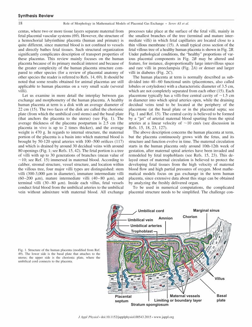
centas, where two or more tissue layers separate maternal fromfetal placental vascular systems (69). However, the structure ofa hemochorial labyrinthine placenta (human and primate) isquite different, since maternal blood is not confined to vesselsand directly bathes fetal tissues. Such structural organizationsignificantly complicates description of transport properties ofthese placentas. This review mainly focuses on the humanplacenta because of its primary medical interest and because ofthe greater complexity of the human placenta structure com-pared to other species (for a review of placental anatomy ofother species the reader is referred to Refs. 14, 69). It should benoted that some results obtained for animal placentas are stillapplicable to human placentas on a very small scale (severalvilli).
Let us examine in more detail the interplay between gasexchange and morphometry of the human placenta. A healthyhuman placenta at term is a disk with an average diameter of22 cm (15). The two faces of the disk are called the chorionicplate (from which the umbilical cord stems) and the basal plate(that anchors the placenta to the uterus) (see Fig. 1). Theaverage thickness of the placenta postpartum is 2.5 cm (theplacenta in vivo is up to 2 times thicker), and the averageweight is 470 g. In regards to internal structure, the maternalportion of the placenta is a basin into which maternal blood isbrought by 50-120 spiral arteries with 100–500 orifices (117)and which is drained by around 30 decidual veins with around80 openings (Fig. 1; see Refs. 15, 42). The fetal portion is a treeof villi with up to 30 generations of branches (mean value of�10; see Ref. 15) immersed in maternal blood. According tocaliber, stromal structure, vessel structure, and location withinthe villous tree, four major villi types are distinguished: stemvilli (300-3,000 �m in diameter), immature intermediate villi(60–200 �m), mature intermediate villi (40–80 �m), andterminal villi (30–80 �m). Inside each villus, fetal vesselsconduct fetal blood from the umbilical arteries to the umbilicalvein without admixture with maternal blood. All exchange
processes take place at the surface of the fetal villi, mainly inthe smallest branches of the tree (terminal and mature inter-mediate villi) in which fetal capillaries are located close to athin villous membrane (15). A small typical cross section of thefetal villous tree of a healthy human placenta is shown in Fig. 2B.Under pathological conditions, the “healthy” proportions of var-ious placental components in Fig. 2B may be altered andfeature, for instance, disproportionally large intervillous spaceand rare villi in preeclampsia (Fig. 2A) or denser and largervilli in diabetes (Fig. 2C).
The human placenta at term is normally described as sub-divided into 40–60 functional units (placentones, also calledlobules or cotyledons) with a characteristic diameter of 3.5 cm,which are not completely separated from each other (15). Eachplacentone typically has a villi-free central cavity of �1.5 cmin diameter into which spiral arteries open, while the drainingdecidual veins tend to be located at the periphery of theplacentone (at the basal plate or in the placental septa; seeFig. 1 and Ref. 15). The central cavity is believed to be formedby a “jet” of arterial maternal blood spurting from the spiralarteries at a linear velocity of �10 cm/s (see discussion inRefs. 15, 18, 23, 127).
The above description concerns the human placenta at term,but the placenta continuously grows with the fetus, and itsstructure and function evolve in time. The maternal circulationstarts in the human placenta only around 10th-12th week ofgestation, after maternal spiral arteries have been invaded andremodeled by fetal trophoblasts (see Refs. 15, 23). This de-layed onset of maternal circulation is believed to protect thedeveloping fetal tissues from the high velocity of maternalblood flow and high partial pressures of oxygen. Most mathe-matical models focus on gas exchange in the term humanplacenta, since extensive data about this stage can be obtainedby analyzing the freshly delivered organ.
To be used in numerical computations, the complicatedplacental structure needs to be simplified. The challenge con-
Fig. 1. Structure of the human placenta (modified from Ref.49). The lower side is the basal plate that attaches to theuterus; the upper side is the chorionic plate, where theumbilical cord connects to the placenta.
Synthesis Review
18 Role of Morphology in Mathematical Models of Placental Gas Exchange • Serov AS et al.
J Appl Physiol • doi:10.1152/japplphysiol.00543.2015 • www.jappl.org

sists in finding a proper balance between oversimplification(too far from the physiological reality) and an overdetailedmodel (computationally unworkable). With time, gas exchangeplacenta models have evolved through the following stages,each corresponding to an increased complexity of the morphol-ogy description: 1) capillary-scale (compartmental) modelsand studies of flow patterns, 2) morphometric diffusing capac-ity models, 3) distributed-parameters models (including po-rous-medium models), and 4) models based on histologicalplacental cross sections.
In the following sections, we discuss these stages of evolu-tion, focusing on advantages of each approach and emphasiz-ing drawbacks that required further progress. A separate sec-tion of the review is devoted to experimental techniques anddata available for validation of the models. In the last section,we discuss geometrical features of the placenta that should beaccounted for in future models.
Models of Placental Respiratory Gas Exchange
Capillary-scale models; flow patterns. Capillary-scale pla-centa models were the first models of respiratory gas exchange.They capitalized on anatomical observations that placentas ofdifferent species feature different co-orientations of maternaland fetal blood flows, and explored how this fact may influenceexchange properties of the organ. These compartmental modelsdistinguished five principal flow patterns (see Fig. 3 andRef. 34): 1) pool flow (a uniform solute concentration along thelength of maternal or fetal capillaries), 2) double pool flow(uniform solute concentrations in both maternal and fetalcapillaries), 3) countercurrent flow, 4) concurrent flow, and 5)cross-current flow (or multivillous flow: maternal capillariesperpendicular to fetal capillaries). Anatomical studies suggestthat the guinea pig and rabbit placentas are well represented bythe countercurrent pattern, the goat and sheep placentas arevenous equilibrators sharing features of the concurrent andpool flow patterns (35, 36, 168), and the human and primateplacentas can be approximated by the multivillous flow pattern
(although maternal blood is not confined to capillaries in theseplacentas). The exchange efficiency of the different flow pat-terns was compared, and it was demonstrated that the counter-current flow pattern provides the highest exchange rate (12–14,52, 55, 75, 107, 115, 145, 152, 168, 169). This conclusion, interalia suggesting higher exchange rate in the guinea pig placentathan in the human placenta, questions definitions of efficiencyand robustness of placental gas exchange and how they can becompared across species (see discussion in Refs. 36, 39).
The development of flow pattern studies continued withintroduction of dimensionless exchange parameters (transportfraction, flow ratio, placental permeability, and placentalclearance) used to compare experimental data on solute ex-change in different species (12, 14, 34–36, 38, 104, 108, 123,129, 130, 145) and to discuss the two specific cases of diffu-sion-limited (or permeability-limited) and flow-limited placen-
200 μm 200 μm200 μm
A B C
Fig. 2. Small fragments of typical 2D histological cross sections of the human placenta: preeclamptic placenta (sparse villi, reduced exchange surface; A), healthyplacenta (B), and diabetic placenta (densely packed villi, reduced surface accessibility; C). Note, however, that due to a very heterogeneous structure, a healthyplacenta may also contain regions similar to those shown in plates A and C, but their fraction is typically much lower. In all three images, the white spacecorresponds to the intervillous space, normally filled with maternal blood, which has been washed away during the preparation of the slides (some residual redblood cells are still present). The large red shapes are the cross sections of the fetal villi. Redder regions inside correspond to fetal capillaries. Histological crosssections are normally taken in the direction from the basal plate to the chorionic plate (see Fig. 1) and are stained with hematoxylin and eosin.
F
M
F
M
F
M
F
M
F
M
Poolflow
Counter-current
Con-current
Cross-current
Doublepoolflow
Fig. 3. Five principal patterns of blood flow based on the original scheme byFaber (34). The letters F and M mark fetal and maternal parts of the exchangersrespectively. Arrows show the direction of the blood flows. Their colorindicates high (red), intermediate (violet), and low (blue) blood oxygenation(for more details see Ref. 34 and references therein).
Synthesis Review
19Role of Morphology in Mathematical Models of Placental Gas Exchange • Serov AS et al.
J Appl Physiol • doi:10.1152/japplphysiol.00543.2015 • www.jappl.org
tal exchange (14, 104). More details on flow patterns can befound in Ref. 14, where experimental data are also summa-rized.
Flow-pattern models have been further used to study influ-ences of different physiological and geometrical parameters onplacental respiratory gas and other substance exchange (11, 13,24, 28, 34–37, 50–53, 55, 57, 58, 70, 75, 78, 85, 86, 88–90,107, 108, 110, 121–124, 129, 130, 134, 143, 145, 147, 152,156, 167). However, they oversimplify placental geometry,focusing on dynamics of gas concentration in fetal and mater-nal blood considered as two compartments separated by asingle effective membrane. This approach could not accountfor the complexity of the diffusion path of respiratory gases,and new, morphometric diffusing capacity models, had to beintroduced.
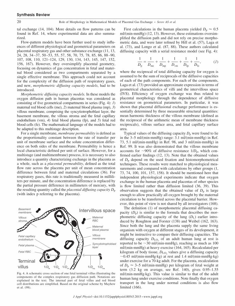
Morphometric diffusing capacity models. In these models theoxygen diffusion path in human placentas was considered asconsisting of five geometrical compartments in series (Fig. 4): 1)maternal red blood cells (me), 2) maternal blood plasma (mp), 3)villous membrane, comprising the syncytiotrophoblast layer, thebasement membrane, the villous stroma and the fetal capillaryendothelium (vm), 4) fetal blood plasma (fp), and 5) fetal redblood cells (fe). The mathematical language of the models had tobe adapted to this multistage description.
For a single membrane, membrane permeability is defined asthe proportionality constant between the rate of transfer perunit of membrane surface and the solute concentration differ-ence on both sides of the membrane. Permeability is hence alocal characteristic defined per unit of surface. However, for amultistage (and multimembrane) process, it is necessary to alsointroduce a quantity characterizing exchange in the placenta asa whole, such as a placental permeability, defined as the totalflow rate across the placenta per unit of mean concentrationdifference between fetal and maternal circulations (36). Forrespiratory gases, this rate is traditionally measured in millili-ters per minute, and the concentration difference is replaced bythe partial pressure difference in millimeters of mercury, withthe resulting quantity called the placental diffusing capacity Dp
(with index p referring to the placenta).
First calculations in the human placenta yielded Dp � 0.5ml/(min·mmHg) (12, 13). However, these estimations oversim-plified the diffusion path and did not rely on precise morpho-metric data, and were later refined by Hill et al. (57), Laga etal. (73), and Longo et al. (87, 88). These authors calculateddiffusing capacity with a serial resistance model (see Fig. 4):
1
Dp�
1
Dme�
1
Dmp�
1
Dvm�
1
Dfp�
1
Dfe, (1)
where the reciprocal of total diffusing capacity for oxygen isassumed to be the sum of reciprocals of the diffusive capacitiesof each of the path components. For each of the components,Laga et al. (73) provided an approximate expression in terms ofgeometrical characteristics of villi and the intervillous space(IVS). Efficiency of oxygen exchange was thus related toplacental morphology through the dependence of diffusiveresistance on geometrical parameters. In particular, it wasshown that placental diffusional exchange performance is es-sentially determined by three measurable structural quantities:mean harmonic thickness of the villous membrane (defined asthe reciprocal of the arithmetic mean of membrane thicknessreciprocals), villous surface area, and fetal capillary surfacearea.
Typical values of the diffusing capacity Dp were found to liein the 3–5 ml/(min·mmHg) range: 3.1 ml/(min·mmHg) in Ref.73, 5.1 ml/(min·mmHg) in Ref. 98, and 3 ml/(min·mmHg) inRef. 99. It was also demonstrated that the villous membraneaccounts for �90% of diffusive resistance 1/Dp, which con-firmed earlier findings (12, 13). Note that the obtained valuesof Dp depend on the used fixation and histomorphometricaltechniques. These results were matched to physiological mea-surements and compared with calculations by other groups (1,73, 74, 100, 101, 157, 158). It should be mentioned here thatindependent physiological experiments indicate that oxygenexchange in the human placenta and placentas of other speciesis flow limited rather than diffusion limited (36, 39). Thisobservation suggests that the obtained value of Dp is largeenough to allow practically all oxygen brought by the maternalcirculation to be transferred across the placental barrier. How-ever, this point of view is not shared by all investigators (168).
The definition (1) of morphometric placental diffusing ca-pacity (Dp) is similar to the formula that describes the mor-phometric diffusing capacity of the lung (DL) earlier intro-duced by Roughton and Forster (138) and Weibel (162, 163).Since both the lung and the placenta supply the same livingorganism with oxygen at different stages of its development, itmight be instructive to compare their diffusing capacities. Thediffusing capacity DLO2
of an adult human lung at rest isreported to be �30 ml/(min·mmHg), reaching as much as 100ml/(min·mmHg) at heavy exercise (164, 165). Recalculated perkilogram of body tissue, DLO2
values give a diffusing capacity�0.45 ml/(min·mmHg·kg) at rest and 1.4 ml/(min·mmHg·kg)under exercise for a 70-kg adult. For the placenta, recalculationof Dp � 3–5 ml/(min·mmHg) per kilogram of fetal weight atterm (3.2 kg on average, see Ref. 140), gives 0.95–1.55ml/(min·mmHg·kg). This value is similar to that of the adulthuman lung under exercise conditions. Note finally that oxygentransport in the lung under normal conditions is also flowlimited (166).
1/Dme1/Dmp1/Dvm1/Dfp1/Dfe
Intervillous spaceMaternalRBC
Maternalblood plasma
Villousmembrane
Syncytiotrophoblastlayer
Basementmembrane
Villousstroma
Fetal capillaryendothelium
Fetal plasma
Fetal RBC
Terminalvillus
Fig. 4. A schematic cross section of one fetal terminal villus illustrating thecomponents of the complex respiratory gas diffusion path. Notations areexplained in the text. The internal part of fetal villus and red bloodcell distributions are simplified. Based on the original scheme by Mayhewet al. (98).
Synthesis Review
20 Role of Morphology in Mathematical Models of Placental Gas Exchange • Serov AS et al.
J Appl Physiol • doi:10.1152/japplphysiol.00543.2015 • www.jappl.org
Morphometric diffusing capacity models present a signifi-cant improvement over capillary-scale models, since they areable to provide case-specific estimates of overall human pla-cental oxygen exchange rate based on histological placentaldata. However, the 1D geometry of these models does notallow proper consideration of convective transport of oxygen,which may create significant variations of oxygen concentra-tion in different parts of the IVS. To explore maternal bloodflow distribution in the human placenta and its impact onoxygen uptake, distributed-parameters models were proposed.
Distributed-parameters models. One of the first models ofthis class was introduced by Aifantis (2), who treated masstransport in the human placenta as a mixture problem. Theplacental tissue was considered as a solid constituent, whilefetal blood and maternal blood were modeled as two fluidconstituents. These three components were allowed to interactin a general way chemically, mechanically, and thermallyaccording to the laws of mass, momentum, and energy conser-vation. This model is, however, only a general framework;case-specific calculations are required to relate the model to theexperiment.
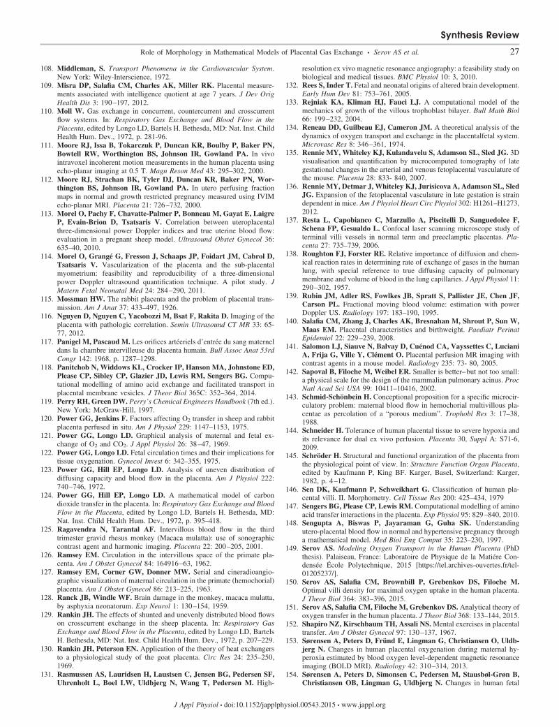
One possible way of introducing properties of solid and fluidconstituents is to consider the intervillous space as a porousmedium with maternal blood percolating through it. Mathe-
matically, such description can take the form of Darcy’s law,which assumes a linear relation between flow velocity, hydro-static blood pressure, and porosity of the solid component(119). A theoretical discussion of the applicability of porous-medium models to the human placenta from a hydrodynamicpoint of view and a method of calculating porous-mediumparameters from histological placental cross sections can befound in Refs. 25–27, 143.
The first calculation of distribution of the maternal bloodflow in a porous-medium human placenta model was per-formed by Erian et al. (32). This model describes a squareporous-medium placentone through which maternal blood per-colates following Darcy’s law (Fig. 5A). Several permeabilityconfigurations were solved, which all yielded significantlynonuniform (and hence inefficient) perfusion of the placentoneby maternal blood. In particular, it was observed that a portionof maternal blood directly flowed from the spiral artery todecidual veins without penetrating deep into the placentone.This short-circuiting was attributed to the fact that inertia offlow was disregarded (Darcy’s law) and that pulsatility ofmaternal blood flow was ignored. However, other explanationsare also possible: 1) the chosen ad hoc form of the velocitydependence of the permeability may not rightly represent villi
central cavity
spiral artery
decidual vein
decidual vein
chorionic plate
basal plate
central cavity
spiral artery
stream tubes
decidual veins
central cavity
decidualvein
decidualvein
spiral artery
A B
C
Fig. 5. A: scheme of a square 2D placentone filled with a porous medium as proposed by Erian et al. (32). One spiral artery and 2 decidual veins wereplaced at the bottom of the placentone. The boundary of the central cavity is marked by 2 dashed lines. An extension to a cylindrical 2D � 1D modelby rotation around the central “vertical” axis (dotted line) was proposed. B: scheme of the hemispherical 3D porous-medium placentone model ofChernyavsky et al. (26). A dotted line delimits the central cavity. The same number and location of maternal vessels as in A was used. Rotational symmetrywas assumed around the “horizontal” axis passing through the three vessels. C: scheme of stream tubes location in the placentone model of Serov et al.(150). Each stream tube (at left) corresponds to a small part of the whole maternal blood flow. At right, a gradual transformation of the maternal bloodfrom arterial into venous is schematically demonstrated.
Synthesis Review
21Role of Morphology in Mathematical Models of Placental Gas Exchange • Serov AS et al.
J Appl Physiol • doi:10.1152/japplphysiol.00543.2015 • www.jappl.org

properties or 2) the description of the placenta as a porousmedium may be inadequate.
To understand which of these explanations is correct andwhy the model of Erian et al. (32) predicted inefficient placen-tone perfusion, a modified 3D placentone model was con-structed, and the influence of location and size of the maternalveins on the blood flow distribution in the placentone wasstudied (Fig. 5B; see Ref. 26). The porous medium wasconsidered nondeformable, which allowed for an analyticalcalculation of maternal blood flow distribution. To modelplacental exchange, a uniform volumetric absorption coeffi-cient was assumed everywhere outside the central cavity.However, no account for specific transport kinetics of thesolute (e.g., oxygen-hemoglobin dissociation) was given, sothat model application was limited to transport of inert solutes(the authors’ choice of oxygen dissolved in the blood plasma asan illustration of inert solute transport being confusing).
Three main conclusions were drawn: 1) If decidual veins arelocated near the periphery of the placentone, maternal bloodflow penetrates deeper into the IVS and rather efficientlyperfuses the placentone. In other words, even with no accountfor flow inertia, more efficient perfusion of the placentone canbe obtained by the correct positioning of venous outlets withrespect to the arteries. 2) There is an optimal size of the centralcavity that is a compromise between hydraulic resistance tomaternal flow and the amount of villous tissue participating insolute uptake. 3) There is also an optimal volume fraction of“villous material” in the IVS as a result of trade-off betweenflow resistance and the uptake capacity of a placentone. How-ever, the obtained value of optimal villi density corresponds tohigh-altitude or preeclamptic placentas rather than to healthyones (26, 150).
In summary, the porous-medium approach was proved valu-able for estimating maternal blood flow distribution in theplacentone and analysis of the effects of shear stress and flowresistance. However, the porous medium concept imposesseveral serious limitations. 1) It is difficult to model respiratorygas uptake due to the absence of a clearly defined uptakesurface. 2) Darcy’s law that neglects diffusive transport ofsolutes in the IVS may be not valid for low maternal bloodvelocities in small IVS pores. 3) Human placenta morphologyis represented as random, which ignores the branching tree-likestructure of the villi. Addressing these issues required concep-tion of a new type of model, models based on histologicalplacental cross sections.
Models based on histological placental cross sections. Geo-metrical models reviewed below are based on 2D histologicalplacental cross sections (Fig. 2) and hence feature a clearlydefined uptake surface. A model of intravillous oxygen trans-port was first proposed by Gill et al. (46), who used 2D spatialdistributions of capillaries in several dozens of villi manuallytraced in histological cross sections of the human placenta.Uniform oxygen concentration was assumed on the perimeterof all villi, while perfect sink conditions were set on capillaryboundaries. Purely diffusive oxygen transport was then simu-lated. Calculations showed that fetal capillaries located close tothe center of a villus have smaller contribution to uptake of thevillus than those located near the villous boundary, and thiseffect was named screening of capillaries analogous to thescreening concept in the lungs (40, 142). A villus efficiency wasthen introduced, which may be potentially correlated to inde-
pendent indicators of the placental exchange efficiency, such asthe placenta-fetus birth weight ratio.
However, exchange efficiency calculated in this way may bestrongly affected by variations of oxygen concentration atvillous boundaries in different regions of the IVS, which arenot accounted for in this model. To study oxygen transport inthe IVS, diffusion and convection have to be consideredsimultaneously, and for this purpose it was found convenient tosubdivide the whole pattern of the maternal blood flow intosmall stream tubes (see Refs. 149–151 and Fig. 5C). Eachstream tube describes a path that a small volume of bloodfollows while moving from a spiral artery to a decidual veinand that is crossed by villi of arbitrary cross sections. Calcu-lations made with this model demonstrated that trade-off be-tween the amount of oxygen coming into the stream tube andthe absorbing villous surface inside it yields an optimal villidensity maximizing oxygen uptake. This optimal villi densityhas a different origin from that proposed in the porous-mediumapproach and was shown to correspond to villi density ob-served on average in a healthy human placenta (150).
While giving realistic predictions of villi density, the stream-tube placenta model has the following limitations: 1) its resultswere obtained with slip boundary conditions (nonzero velocityat the blood-tissue interface); 2) internal villous structure wasdisregarded (fetal blood circulation was ignored and perfect-sink conditions were assumed at the villi boundaries), and 3)geometry of stream-tube cross sections was assumed to beinvariant along the maternal blood flow. Further studies arenecessary to assess the influence of these assumptions on theobtained results.
Comparison with Experiment
After this overview of the evolution of morphologic descrip-tion in respiratory gas exchange placenta models, one maywonder how well predictions of these models correspond tomedical observations and experimental measurements. Oneshould take into account that a limited number of measure-ments are ethical in the human placenta in vivo, with mostmeasurements being available only after birth. In what follows,we summarize experimental techniques and results that arecurrently available either on a regular basis, or in clinicalresearch, and review the ways of experimental validation of themodels. Note that there exist many discrepancies within resultsof experimental studies, which are mainly due to inconsistenttechniques of investigation (such as different ways of placentalfixation, calculation of placental parameters, or umbilical cordclamping) or biological variations.
1) Macroscopic measurements such as placental weight,placenta-to-fetus weight ratio, placental diameter and thicknesscan be obtained after birth (see, for example, Ref. 109).
2) Histomorphometric measurements can be manually per-formed on histological placental cross sections obtained afterbirth (see Refs. 73, 146 and references therein). These studiesreport differences in placental morphometry between healthyand pathological placentas (similar to those shown in Fig. 2).Note, however, that placental morphometry obtained after birthmay not exactly represent the in vivo situation, because ofdetachment of the placenta from the uterine wall, cessation ofmaternal blood flow, mechanical stress of vaginal delivery, andfixation procedures (21, 22, 61, 67, 91, 96, 102).
Synthesis Review
22 Role of Morphology in Mathematical Models of Placental Gas Exchange • Serov AS et al.
J Appl Physiol • doi:10.1152/japplphysiol.00543.2015 • www.jappl.org

3) Placental corrosion casts can be prepared (80–82) pro-viding several generations of fetal villous tree branches. Theycan then be analyzed, in particular, by means of scanningelectron microscopy (20, 54, 68). Such corrosion casts can beused for visualizing fetal vasculature, but do not provideinformation about villi surfaces or small villous tree branches.
4) Confocal laser scanning microscopy with 3D reconstruc-tion can be used to qualitatively and quantitatively compareterminal villi structure in healthy and pathological placentas(see Refs. 64, 137 and references therein).
5) Artificial placenta perfusion can be performed after birthto imitate in vivo placental function (65, 144). However, thismethod requires that placental membranes are not damaged(which is often an issue with vaginal deliveries) and does notsupport long perfusion times.
6) Partial pressures of oxygen, carbon dioxide, and concen-trations of other solutes, as well as volumetric blood flows inuterine and umbilical arteries and veins can be measured inmultiple species (see Refs. 35, 36 and references therein).
7) X-ray photographs of in vivo placenta perfusion by thematernal blood have been obtained with a radioactive dye innormal and pathological human pregnancies and in primates(19, 43–45, 126, 127). The resulting data provide evidence forthe existence of a central cavity and allow estimation ofcharacteristic size of a placentone, transit time of bloodthrough the IVS, and an average velocity of maternal bloodflow (150). However, X-ray involving techniques are nowunethical and are used in humans only in serious pathologicalconditions.
8) Standard ultrasound techniques provide placental size,shape, and volume in utero. Typical values of linear bloodvelocity and pulsatility indexes in umbilical and uterine arteriesand veins, as well as in spiral arteries, can also be obtained (seeRef. 62 and note that mathematical models often require linearblood velocities rather than the integral blood flow). However,the sensitivity threshold of these techniques to linear flowvelocities is �0.1 cm/s, which is insufficient to measure theblood velocities expected in the IVS (�0.05 cm/s on theaverage, see Ref. 150). The use of potentially safe contrastagents (e.g., microbubbles of inert gas) may enhance theirresolution (125).
9) Ultrafast ultrasound techniques are likely to allow high-sensitivity mapping of blood flows simultaneously in the wholeplacenta analogous to brain studies (29), with the possibility ofdistinction between fetal and maternal blood by analyzingpulsatility indices.
10) 3D color/power Doppler angiography is able to providea low-resolution villous tree structure and patterns of bloodflow in the vicinity of central cavities (71). It can also be usedfor reproducible quantification of placental and myometrialvascularization (113, 114). Color Doppler sonograms measurelinear blood velocity and its direction (to or from the sensor),while power Doppler signals are proportional to the sum ofvelocities of all scatterers in a region (with a resolution of upto 0.1 cm/s) and are independent of the angle of insonation(30). Based on the power Doppler method, 2D and 3D frac-tional moving blood volume techniques were proposed toassess the fraction of moving blood in the placenta (139, 155).
11) T1- and T2-weighted contrast MRI techniques can beused to determine placental location in vivo and the main bloodcirculation regions (84, 94, 116, 170). Higher precision is
achieved in animal models by using contrast agents, whichallow for visualization of placental perfusion (141). Alterna-tively, diffusion of water molecules in the placenta can beobserved with diffusion-weighted MRI (17, 93). The resultingdata can be further analyzed by means of intravoxel incoherentmotion (IVIM) models that provide estimates of placentalblood volume (4, 111, 112). Similarly, the arterial spin labeling(ASL) method is used to study placental microflow patterns(6), while oxygen-enhanced MRI and blood-oxygen-level-de-pendent MRI (BOLD MRI) techniques allow one to estimateplacental blood oxygenation in vivo (59, 153, 154). Addition-ally, high-resolution ex vivo MRI angiography is able toprovide 3D structure of the human placenta up to the sixthgeneration of villous branching (131). Analysis of the sensi-tivity of MRI techniques shows that spatial resolution of MRImethods of �1 mm is enough to locate main fetal placentalvascular structures but would not resolve villi structure indense exchange regions (with a characteristic villous compo-nent size of �50 �m).
12) Microcomputed tomography techniques can be used toobtain 3D spatial organization of placental arteries and veins,as demonstrated on placentas of small rodents (135, 136) andin humans (76, 77).
Based on available experimental techniques, the followingstrategies for validating respiratory gas exchange models havebeen proposed (in addition to those mentioned in previoussections). Battaglia and Meschia (14) suggested measuringmaternal and fetal placental blood flows and transplacentaldiffusion rates of various test molecules and their concentra-tions in arteries and veins of both mother and fetus to integrallycharacterize the exchange in placentas of various species. Thisexperimental program was later realized in the sheep model(104, 105). Longo et al. (88, 90) compared predictions of theiroxygen and carbon dioxide exchange model to results of in situperfusion of an isolated placentone (89, 120). Chernyavsky etal. (26) demonstrated that radioactive dye diffusion patterns ofFreese (43) observed in the human placenta can be explainedby a porous-medium placenta model. Comparison with histo-logical cross sections showed that optimal density of “villousmaterial” of �30% calculated by Chernyavsky et al. (26)corresponds to pathological rather than healthy placentas,while the villi density of �47% obtained by Serov et al. (150)correlates well with histological measurements.
Perspectives
Starting with a pair of “capillaries” conducting all fetal andmaternal blood, placenta models have evolved to describingthe organ as a porous medium or a set of stream tubes whosegeometry is based on histological placental cross sections.Mathematical modeling has allowed to predict blood flowdistribution and respiratory gas exchange rates in the placentaacross different species.
However, our knowledge of gas exchange processes in thecomplex placental morphology still remains very incomplete.Further development in the field may move in two principaldirections. On one hand, porous-medium models should 1)account for inhomogeneity of villi density observed in histo-logical placental cross sections, 2) analyze shear stress exertedon villi by blood flow, 3) account for deformability of villoustissue by introducing a velocity-dependent permeability, 4)
Synthesis Review
23Role of Morphology in Mathematical Models of Placental Gas Exchange • Serov AS et al.
J Appl Physiol • doi:10.1152/japplphysiol.00543.2015 • www.jappl.org

study the influence on oxygen uptake of the lateral venousoutlets in placental septa at placentone borders, or 5) gobeyond a single placentone to describe blood flow patterns inthe whole placenta.
On the other hand, nonporous-medium models should 1)improve representation of placental geometrical structure (e.g.,by introducing branching of villi and variations in their shapes,sizes and orientations), 2) simultaneously account for maternaland fetal circulations by combining developed maternal trans-port models with fetal transport models, 3) develop placentalstructure acquisition and analysis techniques capable of pro-viding case-specific input for models, 4) evaluate influence onoxygen uptake of slip and nonslip boundary conditions (withrespectively nonzero and zero blood velocity) imposed on thevillous tree surface, 5) investigate villi screening in the IVS,and 6) explore influence of variations of maternal blow veloc-ity in the IVS.
Both porous-medium and nonporous-medium approachescould provide more accurate predictions by incorporating mod-els of structure of villous tree, fetal placental circulation, andumbilical cord circulation (3, 41, 48, 66, 133, 156, 161), ofmaternal blood flow in the spiral arteries (23, 148), or of thesignaling pathways and placental biochemistry (92, 160). It isalso important to construct placental models of gas exchangenot only at term, but earlier in the course of pregnancy, sincedevelopmental pathologies at these stages may cause thoseseen at later stages. Recent advances in in vivo placentalimaging techniques may provide ground for formulation ofnew early pregnancy gas exchange models.
A promising direction of development of all model typesconsists in analysis of efficiency and robustness of placentalgas exchange. Although the definitions of these notions aredebatable and depend on the model, calculation of efficiencyand robustness is required for a quantitative comparison ofrespiratory gas transport in different patients and for identifi-cation of cases of pathological placental function. This re-search axis may lead to important discoveries similar to thosemade in the human lung (95, 142). Some efficiency criteria,such as the coefficient of oxygen utilization (9, 10, 56, 106), theintegrated mean diffusion pressure gradient (75), the maternaland fetal transport fractions (34–36), the capillary screeningcoefficient (46), the villi density (26, 150), or efficiency dia-grams (149, 151) have already been proposed and may serve asa base for future theoretical and experimental studies.
The development of mathematical placenta models is alsohindered by missing or insufficient experimental data crucialfor validation of the models: 1) the high-resolution 3D geo-metrical structure of the placenta is yet unavailable for largeplacental regions (with at least a thousand villi), especially indense exchange regions. Meanwhile, models can be based onhigh-resolution 2D histological cross sections, on low-resolu-tion 3D structures obtained by ex vivo MRI angiography ormicro-CT, or on the local high-resolution 3D villi geometry(about dozens of villi) provided by the confocal laser micros-copy. 2) High resolution blood flow patterns in the humanplacenta have not yet been experimentally determined either invivo or in vitro. 3) Oxygen, carbon dioxide, and other sub-stances levels are not available on a regular basis either in theintervillous space of the human placenta or in umbilical anduterine arteries and veins. 4) There currently exists no auto-matic image-processing technique for analysis of histological
placental cross sections that can provide large histomorpho-metrical statistics and feed models based on histological crosssections and porous medium models with input data. 5) Thedegree to which formaldehyde-fixed histological cross sectionsfaithfully represent in vivo placental structure and the meth-odologies to correct for possible discrepancies are yet to beestablished. Advances in these directions would facilitate val-idation of existing models and stimulate development of newand more accurate ones.
It should be finally noted that respiratory gas exchangemodels (focusing on geometrical structure of the placenta) canalso be applied to studies of transport of other substances ifcombined with models of substance-specific transport mecha-nisms, such as amino acids (83, 118, 147), glucose (11), orwater (37, 167).
Conclusion
More than six decades of placenta modeling have signifi-cantly contributed to a better understanding of the organfunction. Modern mathematical models aim not only to de-scribe placental exchange based on available experimental databut to propose quantitative criteria of placental exchange effi-ciency that would allow diagnosing pregnancy pathologies. Inthe future, they may become a novel, powerful, and efficienttool in routine analysis of human placentas helping to identifyplacental pathologies and to assess pregnancy and newbornhealth risks.
GRANTS
This study was funded by the International Relations Department of EcolePolytechnique, by Placental Analytics, SAMOVAR Project of the AgenceNationale de Recherche No. 2010-BLAN-1119-05, and Agence Nationale deRecherche Project No. ANR-13JSV5-0006-01. The funding sources have notdirectly influenced the research process.
DISCLOSURES
C. Salafia is founder and head of Placental Analytics, LLC. There is noconflict of interest involved in Dr. Salafia’s contribution to this manuscript.
AUTHOR CONTRIBUTIONS
Author contributions: A.S.S., C.S., D.S.G., and M.F. conception and designof research; A.S.S., C.S., D.S.G., and M.F. analyzed data; A.S.S., C.S., D.S.G.,and M.F. interpreted results of experiments; A.S.S., C.S., D.S.G., and M.F.prepared figures; A.S.S., C.S., D.S.G., and M.F. drafted manuscript; A.S.S.,C.S., D.S.G., and M.F. edited and revised manuscript; A.S.S., C.S., D.S.G.,and M.F. approved final version of manuscript.
REFERENCES
1. Aherne W, Dunnill MS. Quantitative aspects of placental structure. JPathol Bacteriol 91: 123–139, 1966.
2. Aifantis EC. Towards a rational modeling for the human placenta. MathBiosci 40: 281–301, 1978.
3. Alastruey J, Sherwin SJ, Parker KH, Rubens DD. Placental transfu-sion insult in the predisposition for SIDS: a mathematical study. EarlyHum Dev 85: 455–459, 2009.
4. Alison M, Chalouhi GE, Autret G, Balvay D, Thiam R, Salomon LJ,Cuenod CA, Clement O, Siauve N. Use of intravoxel incoherent motionMR imaging to assess placental perfusion in a murine model of placentalinsufficiency. Invest Radiol 48: 17–23, 2013.
5. Altschuler G. Some placental considerations in alleged obstetrical andneonatology malpractice. In: Legal Medicine, edited by Wecht CH.Salem, MA: Butterworth Legal, 1994, p. 27–47.
6. Avni R, Raz T, Biton IE, Kalchenko V, Garbow JR, Neeman M.Unique in utero identification of fetuses in multifetal mouse pregnancies
Synthesis Review
24 Role of Morphology in Mathematical Models of Placental Gas Exchange • Serov AS et al.
J Appl Physiol • doi:10.1152/japplphysiol.00543.2015 • www.jappl.org

by placental bidirectional arterial spin labeling MRI. Magn Reson Med68: 560–70, 2012.
7. Baergen RN. The placenta as witness. Clin Perinatol 34: 393–407, 2007.8. Barker DJ. Fetal origins of coronary heart disease. Br Med J 311:
171–174, 1995.9. Barron DH. Some aspects of the transfer of oxygen across the syndes-
mochorial placenta of the sheep. Yale J Biol Med 24: 169–190, 195110. Barron DH, Meschia G. A comparative study of the exchange of the
respiratory gases across the placenta. Cold Spring Harb Symp Quant Biol19: 93–101, 1954.
11. Barta E, Drugan A. Glucose transport from mother to fetus a theoreticalstudy. J Theor Biol 263: 295–302, 2010.
12. Bartels H, Moll W. Passage of inert substances and oxygen in the humanplacenta. Pflüg Arch Gesamte Physiol Mensch Tiere 280: 165–177, 1964.
13. Bartels H, Moll W, Metcalfe J. Physiology of gas exchange in thehuman placenta. Am J Obstet Gynecol 84: 1714–1730, 1962.
14. Battaglia FC, Meschia G. An Introduction to Fetal Physiology. London:Academic, 1986.
15. Benirschke K, Kaufmann P, Baergen RN. Pathology of the HumanPlacenta (5th ed.). New York: Springer, 2006.
16. Berger R, Garnier Y. Pathophysiology of perinatal brain damage. BrainRes Rev 30: 107–134, 1999.
17. Bonel HM, Stolz B, Diedrichsen L, Frei K, Saar B, Tutschek B, RaioL, Surbek D, Srivastav S, Nelle M, Slotboom J, Wiest R. Diffusion-weighted MR imaging of the placenta in fetuses with placental insuffi-ciency. Radiology 257: 810–819, 2010.
18. Borell U, Fernstrom I, Westman A. Eine arteriographische studie desplazentarkreislaufs [Arteriographic study of placental circulation].Geburtshilfe Frauenheilkd 18: 1–9, 1958.
19. Burchell RC. Arterial blood flow into the human intervillous space. AmJ Obstet Gynecol 98: 303–11, 1967.
20. Burton GJ. The fine structure of the human placental villus as revealedby scanning electron microscopy. Scanning Microsc 1: 1811–1828, 1987.
21. Burton GJ, Palmer ME. Eradicating fetomaternal uid shift duringperfusion fixation of the human placenta. Placenta 9: 327–332, 1988.
22. Burton GJ, Ingram S, Palmer M. The influence of mode of fixation onmorphometrical data derived from terminal villi in the human placenta atterm: a comparison of immersion and perfusion fixation. Placenta 8:37–51, 1987.
23. Burton GJ, Woods AW, Jauniaux E, Kingdom JC. Rheological andphysiological consequences of conversion of the maternal spiral arteriesfor uteroplacental blood flow during human pregnancy. Placenta 30:473–482, 2009.
24. Butler LA, Longo LD, Power GG. Placental blood flows and oxygentransfer during uterine contractions: a mathematical model. J Theor Biol61: 81–95, 1976.
25. Chernyavsky IL. A Multiscale Analysis of Flow and Transport in theHuman Placenta (PhD. thesis). Nottingham, UK: Univ. Nottingham,2011.
26. Chernyavsky IL, Jensen OE, Leach L. A mathematical model ofintervillous blood flow in the human placentone. Placenta 31: 44–52,2010.
27. Chernyavsky IL, Leach L, Dryden IL, Jensen OE. Transport in theplacenta: homogenizing haemodynamics in a disordered medium. PhilosTrans A Math Phys Eng Sci 369: 4162–4182, 2011.
28. Costa A, Costantino ML, Fumero R. Oxygen exchange mechanisms inthe human placenta: mathematical modelling and simulation. J BiomedEng 14: 385–389, 1992.
29. Demené C, Pernot M, Biran V, Alison M, Fink M, Baud O, TanterM. Ultrafast Doppler reveals the mapping of cerebral vascular resistivityin neonates. J Cereb Blood Flow Metab 34: 1009–1017, 2014.
30. Dymling SO, Persson HW, Hertz CH. Measurement of blood perfusionin tissue using doppler ultrasound. Ultrasound Med Biol 17: 433–444,1991.
31. Egbor M, Ansari T, Morris N, Green CJ, Sibbons PD. Pre-eclampsiaand fetal growth restriction: how morphometrically different is theplacenta? Placenta 27: 727–734, 2006
32. Erian FF, Corrsin S, Davis SH. Maternal, placental blood flow: a modelwith velocity-dependent permeability. J Biomech 10: 807–814, 1977.
33. Eriksson JG, Kajantie E, Osmond C, Thornburg K, Barker DJ. Boyslive dangerously in the womb. Am J Hum Biol 22: 330–335, 2010.
34. Faber JJ. Application of the theory of heat exchangers to the transfer ofinert materials in placentas. Circ Res 24: 221–34, 1969.
35. Faber JJ. Steady-state methods for the study of placental exchange. FedProc 36: 2640–2646, 1977.
36. Faber JJ. Review of flow limited transfer in the placenta. Int J ObstetAnesth 4: 230–237, 1995.
37. Faber JJ, Anderson DF. Model study of placental water transfer andcauses of fetal water disease in sheep. Am J Physiol Regul Integr CompPhysiol 258: R1257–R1270, 1990.
38. Faber JJ, Hart FM. The rabbit placenta as an organ of diffusionalexchange. Comparison with other species by dimensional analysis. CircRes 19: 816–833, 1966.
39. Faber JJ, Thornburg KL, Binder ND. Physiology of placental transferin mammals. Integr Comp Biol 32: 343–354, 1992.
40. Felici M, Filoche M, Straus C, Similowski T, Sapoval B. Diffusionalscreening in real 3D human acini a theoretical study. Respir PhysiolNeurobiol 145: 279–293, 2005.
41. Franke VE, Parker KH, Wee LY, Fisk NM, Sherwin SJ. Time domaincomputational modelling of 1D arterial networks in monochorionicplacentas. ESAIM Math Model Numer Anal 37: 557–580, 2003.
42. Franken H. Beitrag zur veranschaulichung von struktur und funktion derplazenta. Zentralbl Gynakol 76: 729–745, 1954.
43. Freese UE. The uteroplacental vascular relationship in the human. Am JObstet Gynecol 101: 8–11, 1968.
44. Freese UE. Vascular relations of placental exchange areas in primatesand man. In: Respiratory Gas Exchange and Blood Flow in the Placenta,edited by Longo LD, Bartels H. Bethesda, MD: Nat. Inst. Child HealthHum. Dev. . . . . . ., 1972, p. 31–63.
45. Freese UE, Ranniger K, Kaplan H. The fetal-maternal circulation ofthe placenta. II. An X-ray cinematographic study of pregnant rhesusmonkeys. Am J Obstet Gynecol 94: 361–366, 1966.
46. Gill JS, Salafia CM, Grebenkov DS, Vvedensky DD. Modeling oxygentransport in human placental terminal villi. J Theor Biol 291: 33–41,2011.
47. Ginsberg, MD, Myers RE. Fetal brain damage following maternalcarbon monoxide intoxication: an experimental study. Acta Obstet Gy-necol Scand 53: 309–317, 1974.
48. Gordon Z, Eytan O, Jaffa AJ, Elad D. Fetal blood flow in branchingmodels of the chorionic arterial vasculature. Ann NY Acad Sci 1101:250–265, 2007.
49. Gray H. Anatomy of the Human Body (20th ed.). Philadelphia, PA: Lea& Febiger, 1918.
50. Groome LJ. A theoretical analysis of the effect of placental metabolismon fetal oxygenation under conditions of limited oxygen availability.Biosystems 26: 45–56, 1991.
51. Guilbeau EJ, Renea DD. Mathematical analysis of combined placental-fetal oxygen transport. Adv Exp Med Biol 37: 1007–1016, 1973.
52. Guilbeau EJ, Reneau DD, Knisely MH. The effects of placentaloxygen consumption and the contractions of labor on fetal oxygensupply. A steady and unsteady state mathematical simulation. In: Respi-ratory Gas Exchange and Blood Flow in the Placenta, edited by LongoLD, Bartels H. Bethesda, MD: Nat. Inst. Child Health Hum. Dev., 1972,p. 297–344.
53. Guilbeau EJ, Reneau DD, Knisely MH. A detailed quantitative anal-ysis of O2 transport in the human placenta during steady and unsteady-state conditions. In: Chemical Engineering Medicine. Advances in Chem-istry, edited by Reneau DD. Washington, DC: Am. Chem. Soc., vol. 118,chapt. 7, 1973, p. 130–171.
54. Hafez SA, Borowicz P, Reynolds LP, Redmer DA. Maternal and fetalmicrovasculature in sheep placenta at several stages of gestation. J Anat216: 292–300, 2010.
55. Heilmann L, Grebner H, Mattheck C, Ludwig H. Mathematical,clinical, and laboratory study of hemodynamic changes in the placentalcirculation. Arch Gynecol 227: 303–313, 1979.
56. Hellegers AE, Heller CJ, Behrman RE, Battaglia FC. Oxygen andcarbon dioxide transfer across the rhesus monkey placenta (macacamulatta). Am J Obstet Gynecol 88: 22–31, 1964.
57. Hill EP, Power GG, Longo LD. A mathematical model of placental O2
transfer with consideration of hemoglobin reaction rates. Am J Physiol222: 721–729, 1972.
58. Hill EP, Power GG, Longo LD. A mathematical model of carbondioxide transfer in the placenta and its interaction with oxygen. Am JPhysiol 224: 283–299, 1973.
59. Huen I, Morris DM, Wright C, Parker GJ, Sibley CP, Johnstone ED,Naish JH. R 1 and R 2* changes in the human placenta in response tomaternal oxygen challenge. Magn Reson Med 70: 1427–1433, 2013.
Synthesis Review
25Role of Morphology in Mathematical Models of Placental Gas Exchange • Serov AS et al.
J Appl Physiol • doi:10.1152/japplphysiol.00543.2015 • www.jappl.org

60. Hutcheon JA, McNamara H, Platt RW, Benjamin A, Kramer MS.Placental weight for gestational age and adverse perinatal outcomes.Obstet Gynecol 119: 1251–1258, 2012.
61. Illsley NP, Fox H, Van der Veen F, Chawner L, Penfold P. Humanplacental ultrastructure after in vitro dual perfusion. Placenta 6: 23–32,1985.
62. Jauniaux E, Jurkovic D, Campbell S. Current topic: in vivo investi-gation of the placental circulations by Doppler echography. Placenta 16:323–31, 1995.
63. Jirkovská M, Smídová J, Frýda T. Morphometric analysis of thevascularization of the terminal villi in normal and diabetic placenta. ActaStereol 13: 43–47, 1994.
64. Jirkovská M, Kubínová L, Janácek J, Moravcová M, Krejcí, V,Karen P. Topological properties and spatial organization of villouscapillaries in normal and diabetic placentas. J Vasc Res 39: 268–278,2002.
65. Jones NW, Hutchinson ES, Brownbill P, Crocker IP, Eccles D, BuggGJ, Raine-Fenning NJ. In vitro dual perfusion of human placentallobules as a flow phantom to investigate the relationship between feto-placental flow and quantitative 3D power doppler angiography. Placenta30: 130–135, 2009.
66. Kaplan AD, Jaffa AJ, Timor IE, Elad D. Hemodynamic analysis ofarterial blood flow in the coiled umbilical cord. Reprod Sci 17: 258–268,2010.
67. Kaufmann P. Influence of ischemia and artificial perfusion on placentalultrastructure and morphometry. Contrib Gynecol Obstet 13: 18–26,1985.
68. Kaufmann P, Sen DK, Schweikhart G. Classification of human pla-cental villi. I. Histology. Cell Tissue Res 200: 409–423, 1979.
69. King BF. Comparative studies of structure and function in mammalianplacentas with special reference to maternal-fetal transfer of iron. IntegrComp Biol 32: 331–342, 1992.
70. Kirschbaum TH, Shapiro NZ. A mathematical model of placentaloxygen transfer. J Theor Biol 25: 380–402, 1969.
71. Konje JC, Huppertz B, Bell SC, Taylor DJ, Kaufmann P. 3-Dimen-sional colour power angiography for staging human placental develop-ment. Lancet 362: 1199–1201, 2003.
72. Korteweg FJ, Erwich JJ, Holm JP, Ravisé JM, van der Meer J,Veeger NJ, Timmer A. Diverse placental pathologies as the main causesof fetal death. Obstet Gynecol 114: 809–817, 2009.
73. Laga EM, Driscoll SG, Munro HN. Quantitative studies of humanplacenta. I. Morphometry. Biol Neonate 23: 231–259, 1973.
74. Laga EM, Driscoll SG, Munro HN. Human placental structure: rela-tionship to fetal nutrition. In: Problems Human Reproduction. LactogenHormones Fetal Nutrition Lactation, edited by Josimovich JB, ReynoldsM, Cobo E. London: John Wiley, 1974, vol. 2, p. 143–181.
75. Lamport H. The transport of oxygen in the sheep’s placenta: thediffusion constant of the placenta. Yale J Biol Med 27: 26–34, 1954.
76. Langheinrich AC, Wienhard J, Vormann S, Hau B, Bohle RM,Zygmunt M. Analysis of the fetal placental vascular tree by X-raymicro-computed tomography. Placenta 25: 95–100, 2004.
77. Langheinrich AC, Vorman S, Seidenstücker J, Kampschulte M,Bohle RM, Wienhard J, Zygmunt M. Quantitative 3D micro-CTimaging of the human feto-placental vasculature in intrauterine growthrestriction. Placenta 29: 937–941, 2008.
78. Lardner TJ. A model for placental oxygen exchange. J Biomech 8:131–4, 1975.
79. Lee R, Mayhew TM. Star volumes of villi and intervillous pores inplacentae from low and high altitude pregnancies. J Anat 186: 349–355,1995.
80. Leiser R, Kohler T. The blood vessels of the cat girdle placenta.Observations on corrosion casts scanning electron microscopical andhistological studies. I. Maternal vasculature. Anat Embryol (Berl) 167:85–93, 1983.
81. Leiser R, Kohler T. The blood vessels of the cat girdle placenta.Observations on corrosion casts scanning electron microscopical andhistological studies. II. Fetal vasculature. Anat Embryol (Berl) 170:209–216, 1984.
82. Leiser R, Krebs C, Ebert B, Dantzer V. Placental vascular corrosioncast studies: a comparison between ruminants and humans. Microsc ResTech 38: 76–87, 1997.
83. Lewis RM, Brooks S, Crocker IP, Glazier J, Hanson MA, JohnstoneED, Panitchob N, Please CP, Sibley CP, Widdows KL, Sengers BG.
Review: modelling placental amino acid transfer from transporters toplacental function. Placenta 34, Suppl: S46–51, 2013
84. Linduska N, Dekan S, Messerschmidt A, Kasprian G, Brugger PC,Chalubinski K, Weber M, Prayer D. Placental pathologies in fetal MRIwith pathohistological correlation. Placenta 30: 555- 559, 2009.
85. Longo LD. Respiratory gas exchange. In: Handbook of Physiolgoy.Respiratory System. Gas Exchange, edited by Fishman AP, Farhi LE,Tenney SM, Geiger SR. Bethesda, MD: Am. Physol. Soc., sect. 3, vol.IV, 1987, p. 351- 401.
86. Longo LD, Power GG. Analyses of PO2 and PCO2 differences betweenmaternal and fetal blood in the placenta. J Appl Physiol 26: 48–55, 1969.
87. Longo LD, Power GG, Forster RE 2nd. Placental diffusing capacityfor carbon monoxide at varying partial pressures of oxygen. J ApplPhysiol 26: 360–370, 1969.
88. Longo LD, Hill EP, Power GG. Theoretical analysis of factors affectingplacental O2 transfer. Am J Physiol 222: 730–739, 1972.
89. Longo LD, Hill EP, Power GG. Factors affecting placental oxygentransfer. In: Respiratory Gas Exchange and Blood Flow in the Placenta,edited by Longo LD, Bartels H. Bethesda, MD: Nat. Inst. Child HealthHum. Dev., 1972, p. 345–393.
90. Longo LD, Hill EP, Power GG. Factors affecting placental oxygentransfer. In: Chemical Engineering Medicine, edited by Reneau DD.Washington, DC: Am. Chem. Soc., 1973, chapt. 6, p. 88–129.
91. Luckhardt M, Leiser R, Kingdom J, Malek A, Sager R, Kaisig C,Schneider H. Effect of physiologic perfusion-fixation on the morpho-metrically evaluated dimensions of the term placental cotyledon. J SocGynecol Investig 3: 166–171, 1996.
92. Mac Gabhann F, Popel AS. Model of competitive binding of vascularendothelial growth factor and placental growth factor to VEGF receptorson endothelial cells. Am J Physiol Heart Circ Physiol 286: H153–H1564,2004.
93. Manganaro L, Fierro F, Tomei A, La Barbera L, Savelli S, SollazzoP, Sergi ME, Vinci V, Ballesio L, Marini M. MRI and DWI: feasibilityof DWI and ADC maps in the evaluation of placental changes duringgestation. Prenat Diagn 30: 1178–1184, 2010.
94. Masselli G, Gualdi G. MR imaging of the placenta: what a radiologistshould know. Abdom Imaging 38: 573–587, 2013.
95. Mauroy B, Filoche M, Weibel ER, Sapoval B. An optimal bronchialtree may be dangerous. Nature 427: 633–636, 2004.
96. Mayhew TM, Burton GJ. Methodological problems in placental mor-phometry: apologia for the use of stereology based on sound samplingpractice. Placenta 9: 565–581, 1988.
97. Mayhew TM, Burton GJ. Stereology and its impact on our understand-ing of human placental functional morphology. Microsc Res Tech 38:195–205, 1997.
98. Mayhew TM, Joy CF, Haas JD. Structure-function correlation in thehuman placenta: the morphometric diffusing capacity for oxygen at fullterm. J Anat 139: 691–708, 1984.
99. Mayhew TM, Jackson MR, Haas JD. Microscopical morphology of thehuman placenta and its effects on oxygen diffusion: a morphometricmodel. Placenta 7: 121–131, 1986.
100. Mayhew TM, Jackson MR, Haas JD. Oxygen diffusive conductancesof human placentae from term pregnancies at low and high altitudes.Placenta 11: 493- 503, 1990.
101. Mayhew TM, Sørensen FB, Klebe JG, Jackson MR. Oxygen diffusiveconductance in placentae from control and diabetic women. Diabetologia36: 955–960, 1993.
102. Mayhew TM, Sørensen FB, Klebe JG, Jackson MR. The effects ofmode of delivery and sex of newborn on placental morphology in controland diabetic pregnancies. J Anat 183: 545–552, 1993
103. Mayhew TM, Wijesekara J, Baker PN, Ong SS. Morphometric evi-dence that villous development and fetoplacental angiogenesis are com-promised by intrauterine growth restriction but not by pre-eclampsia.Placenta 25: 829–833, 2004.
104. Meschia G, Battaglia FC, Bruns PD. Theoretical and experimentalstudy of transplacental diffusion. J Appl Physiol 22: 1171–1178, 1967.
105. Meschia G, Cotter JR, Makowski EL, Barron DH. Simultaneousmeasurement of uterine and umbilical blood flows and oxygen uptakes.Q J Exp Physiol Cogn Med Sci 52: 1–18, 1967.
106. Metcalfe J, Romney SL, Ramsey LH, Reid DE, Burwell CS. Estima-tion of uterine blood flow in normal human pregnancy at term. J ClinInvest 34: 1632–1638, 1955.
107. Metcalfe J, Moll W, Bartels H. Gas exchange across the placenta. FedProc 23: 774–780, 1964.
Synthesis Review
26 Role of Morphology in Mathematical Models of Placental Gas Exchange • Serov AS et al.
J Appl Physiol • doi:10.1152/japplphysiol.00543.2015 • www.jappl.org
108. Middleman, S. Transport Phenomena in the Cardiovascular System.New York: Wiley-Interscience, 1972.
109. Misra DP, Salafia CM, Charles AK, Miller RK. Placental measure-ments associated with intelligence quotient at age 7 years. J Dev OrigHealth Dis 3: 190–197, 2012.
110. Moll W. Gas exchange in concurrent, countercurrent and crosscurrentflow systems. In: Respiratory Gas Exchange and Blood Flow in thePlacenta, edited by Longo LD, Bartels H. Bethesda, MD: Nat. Inst. ChildHealth Hum. Dev., 1972, p. 281-96.
111. Moore RJ, Issa B, Tokarczuk P, Duncan KR, Boulby P, Baker PN,Bowtell RW, Worthington BS, Johnson IR, Gowland PA. In vivointravoxel incoherent motion measurements in the human placenta usingecho-planar imaging at 0.5 T. Magn Reson Med 43: 295–302, 2000.
112. Moore RJ, Strachan BK, Tyler DJ, Duncan KR, Baker PN, Wor-thington BS, Johnson IR, Gowland PA. In utero perfusing fractionmaps in normal and growth restricted pregnancy measured using IVIMecho-planar MRI. Placenta 21: 726–732, 2000.
113. Morel O, Pachy F, Chavatte-Palmer P, Bonneau M, Gayat E, LaigreP, Evain-Brion D, Tsatsaris V. Correlation between uteroplacentalthree-dimensional power Doppler indices and true uterine blood flow:evaluation in a pregnant sheep model. Ultrasound Obstet Gynecol 36:635–40, 2010.
114. Morel O, Grangé G, Fresson J, Schaaps JP, Foidart JM, Cabrol D,Tsatsaris V. Vascularization of the placenta and the sub-placentalmyometrium: feasibility and reproducibility of a three-dimensionalpower Doppler ultrasound quantification technique. A pilot study. JMatern Fetal Neonatal Med 24: 284–290, 2011.
115. Mossman HW. The rabbit placenta and the problem of placental trans-mission. Am J Anat 37: 433–497, 1926.
116. Nguyen D, Nguyen C, Yacobozzi M, Bsat F, Rakita D. Imaging of theplacenta with pathologic correlation. Semin Ultrasound CT MR 33: 65-77, 2012.
117. Panigel M, Pascaud M. Les orifices artéeriels d’entrée du sang materneldans la chambre intervilleuse du placenta humain. Bull Assoc Anat 53rdCongr 142: 1968, p. 1287–1298.
118. Panitchob N, Widdows KL, Crocker IP, Hanson MA, Johnstone ED,Please CP, Sibley CP, Glazier JD, Lewis RM, Sengers BG. Compu-tational modelling of amino acid exchange and facilitated transport inplacental membrane vesicles. J Theor Biol 365C: 352–364, 2014.
119. Perry RH, Green DW. Perry’s Chemical Engineers Handbook (7th ed.).New York: McGraw-Hill, 1997.
120. Power GG, Jenkins F. Factors affecting O2 transfer in sheep and rabbitplacenta perfused in situ. Am J Physiol 229: 1147–1153, 1975.
121. Power GG, Longo LD. Graphical analysis of maternal and fetal ex-change of O2 and CO2. J Appl Physiol 26: 38–47, 1969.
122. Power GG, Longo LD. Fetal circulation times and their implications fortissue oxygenation. Gynecol Invest 6: 342–355, 1975.
123. Power GG, Hill EP, Longo LD. Analysis of uneven distribution ofdiffusing capacity and blood flow in the placenta. Am J Physiol 222:740–746, 1972.
124. Power GG, Hill EP, Longo LD. A mathematical model of carbondioxide transfer in the placenta. In: Respiratory Gas Exchange and BloodFlow in the Placenta, edited by Longo LD, Bartels H. Bethesda, MD:Nat. Inst. Child Health Hum. Dev., 1972, p. 395–418.
125. Ragavendra N, Tarantal AF. Intervillous blood flow in the thirdtrimester gravid rhesus monkey (Macaca mulatta): use of sonographiccontrast agent and harmonic imaging. Placenta 22: 200–205, 2001.
126. Ramsey EM. Circulation in the intervillous space of the primate pla-centa. Am J Obstet Gynecol 84: 164916–63, 1962.
127. Ramsey EM, Corner GW, Donner MW. Serial and cineradioangio-graphic visualization of maternal circulation in the primate (hemochorial)placenta. Am J Obstet Gynecol 86: 213–225, 1963.
128. Ranck JB, Windle WF. Brain damage in the monkey, macaca mulatta,by asphyxia neonatorum. Exp Neurol 1: 130–154, 1959.
129. Rankin JH. The effects of shunted and unevenly distributed blood flowson crosscurrent exchange in the sheep placenta. In: Respiratory GasExchange and Blood Flow in the Placenta, edited by Longo LD, BartelsH. Bethesda, MD: Nat. Inst. Child Health Hum. Dev., 1972, p. 207–229.
130. Rankin JH, Peterson EN. Application of the theory of heat exchangersto a physiological study of the goat placenta. Circ Res 24: 235–250,1969.
131. Rasmussen AS, Lauridsen H, Laustsen C, Jensen BG, Pedersen SF,Uhrenholt L, Boel LW, Uldbjerg N, Wang T, Pedersen M. High-
resolution ex vivo magnetic resonance angiography: a feasibility study onbiological and medical tissues. BMC Physiol 10: 3, 2010.
132. Rees S, Inder T. Fetal and neonatal origins of altered brain development.Early Hum Dev 81: 753–761, 2005.
133. Rejniak KA, Kliman HJ, Fauci LJ. A computational model of themechanics of growth of the villous trophoblast bilayer. Bull Math Biol66: 199–232, 2004.
134. Reneau DD, Guilbeau EJ, Cameron JM. A theoretical analysis of thedynamics of oxygen transport and exchange in the placentalfetal system.Microvasc Res 8: 346–361, 1974.
135. Rennie MY, Whiteley KJ, Kulandavelu S, Adamson SL, Sled JG. 3Dvisualisation and quantification by microcomputed tomography of lategestational changes in the arterial and venous fetoplacental vasculature ofthe mouse. Placenta 28: 833- 840, 2007.
136. Rennie MY, Detmar J, Whiteley KJ, Jurisicova A, Adamson SL, SledJG. Expansion of the fetoplacental vasculature in late gestation is straindependent in mice. Am J Physiol Heart Circ Physiol 302: H1261–H1273,2012.
137. Resta L, Capobianco C, Marzullo A, Piscitelli D, Sanguedolce F,Schena FP, Gesualdo L. Confocal laser scanning microscope study ofterminal villi vessels in normal term and preeclamptic placentas. Pla-centa 27: 735–739, 2006.
138. Roughton FJ, Forster RE. Relative importance of diffusion and chem-ical reaction rates in determining rate of exchange of gases in the humanlung, with special reference to true diffusing capacity of pulmonarymembrane and volume of blood in the lung capillaries. J Appl Physiol 11:290–302, 1957.
139. Rubin JM, Adler RS, Fowlkes JB, Spratt S, Pallister JE, Chen JF,Carson PL. Fractional moving blood volume: estimation with powerDoppler US. Radiology 197: 183–190, 1995.
140. Salafia CM, Zhang J, Charles AK, Bresnahan M, Shrout P, Sun W,Maas EM. Placental characteristics and birthweight. Paediatr PerinatEpidemiol 22: 229–239, 2008.
141. Salomon LJ, Siauve N, Balvay D, Cuénod CA, Vayssettes C, LucianiA, Frija G, Ville Y, Clément O. Placental perfusion MR imaging withcontrast agents in a mouse model. Radiology 235: 73- 80, 2005.
142. Sapoval B, Filoche M, Weibel ER. Smaller is better–but not too small:a physical scale for the design of the mammalian pulmonary acinus. ProcNatl Acad Sci USA 99: 10411–10416, 2002.
143. Schmid-Schönbein H. Conceptional proposition for a specific microcir-culatory problem: maternal blood flow in hemochorial multivillous pla-centae as percolation of a “porous medium”. Trophobl Res 3: 17–38,1988.
144. Schneider H. Tolerance of human placental tissue to severe hypoxia andits relevance for dual ex vivo perfusion. Placenta 30, Suppl A: S71-6,2009.
145. Schröder H. Structural and functional organization of the placenta fromthe physiological point of view. In: Structure Function Organ Placenta,edited by Kaufmann P, King BF. Karger, Basel, Switzerland: Karger,1982, p. 4–12.
146. Sen DK, Kaufmann P, Schweikhart G. Classification of human pla-cental villi. II. Morphometry. Cell Tissue Res 200: 425–434, 1979
147. Sengers BG, Please CP, Lewis RM. Computational modelling of aminoacid transfer interactions in the placenta. Exp Physiol 95: 829–840, 2010.
148. Sengupta A, Biswas P, Jayaraman G, Guha SK. Understandingutero-placental blood flow in normal and hypertensive pregnancy througha mathematical model. Med Biol Eng Comput 35: 223–230, 1997.
149. Serov AS. Modeling Oxygen Transport in the Human Placenta (PhDthesis). Palaiseau, France: Laboratoire de Physique de la Matiére Con-densée École Polytechnique, 2015 [https://tel.archives-ouvertes.fr/tel-01205237/].
150. Serov AS, Salafia CM, Brownbill P, Grebenkov DS, Filoche M.Optimal villi density for maximal oxygen uptake in the human placenta.J Theor Biol 364: 383–396, 2015.
151. Serov AS, Salafia CM, Filoche M, Grebenkov DS. Analytical theory ofoxygen transfer in the human placenta. J Theor Biol 368: 133–144, 2015.
152. Shapiro NZ, Kirschbaum TH, Assali NS. Mental exercises in placentaltransfer. Am J Obstet Gynecol 97: 130–137, 1967.
153. Sørensen A, Peters D, Fründ E, Lingman G, Christiansen O, Uldb-jerg N. Changes in human placental oxygenation during maternal hy-peroxia estimated by blood oxygen level-dependent magnetic resonanceimaging (BOLD MRI). Radiology 42: 310–314, 2013.
154. Sørensen A, Peters D, Simonsen C, Pedersen M, Stausbøl-Grøn B,Christiansen OB, Lingman G, Uldbjerg N. Changes in human fetal
Synthesis Review
27Role of Morphology in Mathematical Models of Placental Gas Exchange • Serov AS et al.
J Appl Physiol • doi:10.1152/japplphysiol.00543.2015 • www.jappl.org
oxygenation during maternal hyperoxia as estimated by BOLD MRI.Prenat Diagn 33: 141–145, 2013.
155. Stevenson GN, Collins SL, Welsh AW, Impey LW, Noble JA. Atechnique for the estimation of fractional moving blood volume byusing three-dimensional power doppler US. Radiology 274: 132363,2015.
156. Talbert DG, Sebire NJ. The dynamic placenta: I. Hypothetical model ofa placental mechanism matching local fetal blood flow to local intervillusoxygen delivery. Med Hypotheses 62: 511–519, 2004.
157. Teasdale F. Morphometric evaluation. In: Contributions to Gynecol-ogy Obstetrics. Morphology Function Aspiration Placental Dysfunc-tion, edited by Soma H. Basel, Switzerland: Karger, 1982, vol. 9, p.17–28.
158. Teasdale F. Histomorphometry of the human placenta in Class Bdiabetes mellitus. Placenta 4: 1–12, 1983.
159. Teng J, Chang T, Reyes C, Nelson KB. Placental weight and neurologicoutcome in the infant: a review. J Matern Fetal Neonatal Med 25:2082–2087, 2012.
160. Thoumsin HJ, Albert A, Duvivier J. Preliminary clinical application ofa mathematical model for interpreting dehydroepiandrosterone-sulfateloading test in late pregnancy. J Perinat Med 6: 32–38, 1978.
161. van den Wijngaard JP, Westerhof BE, Ross MG, van Gemert MJ. Amathematical model of twin-twin transfusion syndrome with pulsatile
arterial circulations. Am J Physiol Regul Integr Comp Physiol 292:R1519–R1531, 2007.
162. Weibel ER. Morphometry and lung models. In: Quantitative MethodsMorphology/Quantitative Methoden der Morphology, edited by WeibelER, Elias H. Berlin, Germany: Springer, 1967, p. 253–267.
163. Weibel ER. Morphometric estimation of pulmonary diffusion capacity.I. Model and method. Respir Physiol 11: 54–75, 1970.
164. Weibel ER, 1984. The Pathway for Oxygen. Cambridge, MA: HarvardUniv. Press, 1984.
165. Weibel ER. What makes a good lung? Swiss Med Wkly 139: 375–386,2009.
166. West JB. Respiratory Physiology: The Essentials (8th ed.). Baltimore,MD: Lippincott Williams & Wilkins, 2008.
167. Wilbur WJ, Power GG, Longo LD. Water exchange in the placenta: amathematical model. Am J Physiol Regul Integr Comp Physiol 235:R181–R199, 1978.
168. Wilkening RB, Meschia G. Current topic: comparative physiology ofplacental oxygen transport. Placenta 13: 1–15, 1992.
169. Wilkin P. Les theories explicatives du mecanisme desechanges trans-placentaires. In: Le Placenta Human, edited by Snoeck J. Paris: Massonet Cie Paris, 1958, p. 248–279.
170. Yeh BM. Has the time arrived to image placental perfusion? Radiology241: 633–634, 2006.
Synthesis Review
28 Role of Morphology in Mathematical Models of Placental Gas Exchange • Serov AS et al.
J Appl Physiol • doi:10.1152/japplphysiol.00543.2015 • www.jappl.org